10 XII December 2022 https://doi.org/10.22214/ijraset.2022.47992
ISSN: 2321-9653; IC Value: 45.98; SJ Impact Factor: 7.538 Volume 10 Issue XII Dec 2022- Available at www.ijraset.com

ISSN: 2321-9653; IC Value: 45.98; SJ Impact Factor: 7.538 Volume 10 Issue XII Dec 2022- Available at www.ijraset.com
Khushi Bora1 , Ms.A.Y.Kerle2 , Vaidehi Phadke3 , Madiha Mujawar4 , Amruta Anuse5 1, 3, 4, 5 Third Year, 2
Department of Diploma in Computer Engineering, Sharad Institute of Technology, Polytechnic Yadrav, Ichalkaranji, Kolhapur, Maharashtra, India
Abstract: Brain tumor is the most commonly occurring malignancy among human beings, so study of brain tumor is important. The segmentation, detection, and extraction of infected tumor area from magnetic resonance (MR) images are a primary concern but a tedious and time taking task performed by radiologists or clinical experts, and their accuracy depends on their experience only. So, the use of computer aided technology becomes very necessary to overcome these limitations. In this paper, we propose a CNN model to identify or detect tumor from the brain magnetic resonance imaging (MRI). The mobile application is designed with a custom designed neural network that uses a combination of deep learning and image processing techniques to identify the presence of a brain tumor in medical images. The application is capable of providing accurate results within a short amount of time and mainly uses a combination of radiomics and morphometric features to evaluate the medical images. The application is also capable of providing support to physicians in diagnosing brain tumors.
Keywords: Brain, Brain tumor, CNN model, computer aided technology, MRI, radiomics, neural network, medical images, deep learning, app development.


In recent times, the introduction of information technology and e-health care system in the medical field helps clinical experts to provide better health care to the patient. Brain is the most complex organ in the body and is responsible for coordinating and controlling body functions. Nature has tightly safeguarded the brain inside a skull that hinders the study of its function as well as makes the diagnosis of its diseases more intricate. Brain tumor is a growth of abnormal cells in the brain that can cause serious neurological disorders, including seizures, headaches, vision loss, and personality changes. In this study, different magnetic resonance imaging (MRI) sequence images are employed for diagnosis. MRI segmentation is one of the essential tasks in medical area but is boring and time consuming. Visual study of MRI is generally more interesting and faster. Various researches and studies have been carried out on both type of tumors till date. These studies show that rate of tumors in human body has been increasing and there is no clue of their such a fast rate of increasing. Medical imaging techniques and analysis tools which play a vital role in the field of clinical study also enable the doctors and radiologists to arrive at a specific diagnosis. The detection of a brain tumor at an early stage is a key issue for providing improved treatment. Once a brain tumor is clinically suspected, radiological evaluation is required to determine its location, its size, and impact on the surrounding areas. On the basis of this information the best therapy, surgery, radiation, or chemotherapy, is decided. It is evident that the chances of survival of a tumor-infected patient can be increased significantly if the tumor is detected accurately in its early stage.
1) Brain Tumor: A brain tumor is a mass of cells that have grown and multiplied uncontrollable i.e. a brain tumor is an uncontrolled growth of solid mass formed by undesired cells either normally found in the different part of the brain such as glial cells, neurons, lymphatic tissue, blood vessels, pituitary and pineal gland, skull, or spread from cancers mainly located in other organs. Brain tumors are classified based on the type of tissue involved in the brain, the positioning of the tumor in the brain, whether it is benign tumor or malignant tumor and other different considerations. Brains tumors are the solid portion permeate the surrounding tissues or distort the surrounding structures. There are different types of brain tumor they are i) Gliomas, ii) Medulloblastoma, iii) Lymphoma, iv) Meningioma, v) Craniopharyngioma, vi) Pituitary adenoma.
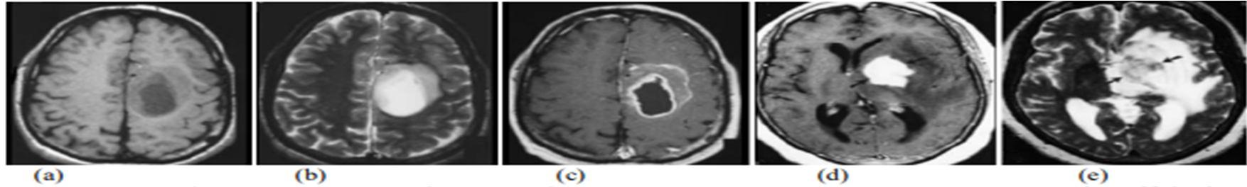
Figure 1: A set of brain tumor images from MRI of brain output cited
et al.(2003)[7]. a) Axial T1-weighted with tumor, b) T2-weighted with central positioning tumor, c) Contrast enhanced T1-weighted image showing ring formed tumor, d) Contrast enhanced T1- weighted image with high grade oligodendro glioma e) T2-weighted image with high grade oligodendro glioma from the same patient.
ISSN: 2321-9653; IC Value: 45.98; SJ Impact Factor: 7.538 Volume 10 Issue XII Dec 2022- Available at www.ijraset.com
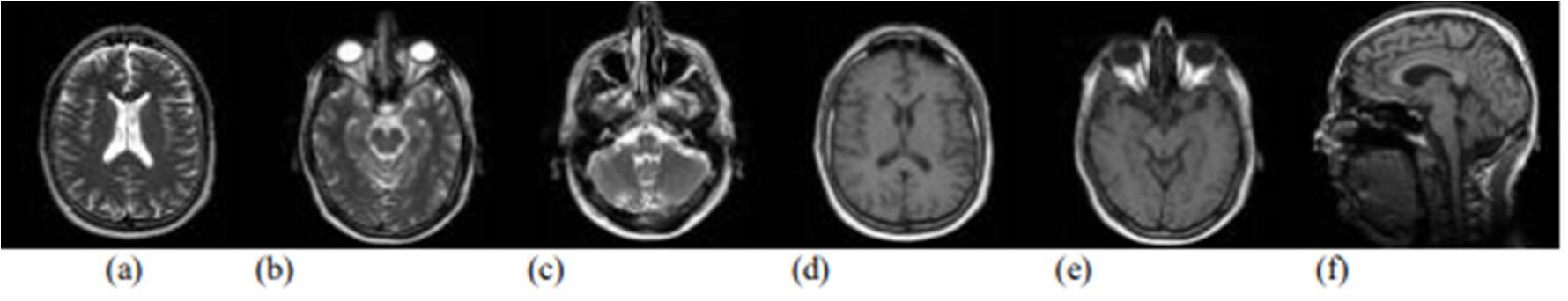
2) Magnetic Resonance Imaging (MRI): A magnetic resonance imaging instrument or MRI Scanner [3] uses powerful magnets to polarize and excite hydrogen nuclei i.e. proton in water molecules in human tissue, producing a detectable signal which is spatially encoded, resulting in images of the body [4]. MRI mainly uses three electromagnetic fields they are: i) A very strong static magnetic field to polarize the hydrogen nuclei, named as the static field, ii) A weaker time varying field(s) for spatial encoding, named as the gradient field, iii) A weak radio frequency field for manipulation of hydrogen nuclei to produce measurable signals collected through RF antenna. The variable behaviour of protons within different tissues leads to differences in tissue appearance. The different positioning of MRI of brain with T1 and T2 weight is shown below.
Figure 2 : MRI of brain cited by http://www.mr-tip.com/serv1.php?type=isimg. T2 weighted MR image (a) brain shows cortex, lateral ventricle, and falx cerebri, (b) brain shows eyeballs with optic nerve, medulla, vermis, and temporal lobes with hippocampal regions, (c) head shows maxillary sinus, nasal septum, clivus, inner ear, medulla, and cerebellum. T1 weighted MR image (d) brain shows cortex, white and grey matter, third and lateral venticles, putamen, frontal sinus and superior sagittal sinus, (e) brain shows eyeballs with optic nerve, medulla, vermis, and temporal lobes with hippocampal regions,(f) brain shows cortex with white and grey matter, corpus callosum, lateral ventricle, thalamus, pons and cerebellum from the same patients Most Research in developed countries show that the number of people who develop brain tumors and die from them has increased perhaps as much as 300 over past three decades. The National Brain Tumor Foundation (NBTF) for research in United States estimates that 29,000 people in the U.S are diagnosed with primary brain tumors each year, and nearly 13,000 people die. In children, brain tumors are the cause of one quarter of all cancer deaths. In India, totally 80,271 people are affected by various types of tumor (2007 estimates).
In this paper, we conduct and provide a comprehensive and systematic review of the developed apps for brain tumor detection available for iOS and Android phones. Specifically, we investigated:
a) What are the primary ways of Brain tumor detection?
b) The most efficient way of detecting Brain tumor
c) The accuracy of the different ways brain tumor is detected
The segmentation is the most important stage for analysing image properly since it affects the accuracy of the subsequent steps. Firstly, the original brain MRI image is found. Then the original Brain MRI image is pre-processed. Processing is necessary as it enhanced the image and prepare for segmentation. Pre-processing includes resizing the image, noise removal from the image, image enhancement, image normalization and background segmentation. Pre-processing and enhancement techniques are used to improve the detection of the suspicious region from Magnetic Resonance Image (MRI). Secondly, the removal of high frequency components using filtering technique that includes median filter, Adaptive filter and Spatial filter. Other artifacts like text removed by some morphological operations. RGB to grey conversion and reshaping also takes place here.
ISSN: 2321-9653; IC Value: 45.98; SJ Impact Factor: 7.538 Volume 10 Issue XII Dec 2022- Available at www.ijraset.com
There are various classification techniques used for classifying brain as normal or abnormal . These classification techniques are described below: -
In this technique, the image is mapped into a Neural Network. The neural network works in two phases- the training phase and the testing phase.The neural network was trained with training examples in the training phase. After training, the neural network is tested on the unknown instances. Neural network technique includes important step that is feature extraction. Feature extraction is very important as the features that are extracted forms the input part of the neural network.
Artificial Neural Network is divided into 2 categories: -
a) Feed-Forward Neural Network.
b) Recurrent Network or Feed-Backward Network. In feed-forward neural network, the neurons are arranged in layers and they have unidirectional connections between them. They produce only one set of output values. The output values do not depend on previous input values. They are called as memory less network. In feedback network, the neurons have bidirectional connections between them. Feedback or Recurrent networks produce a set of values which depends on the previous input values. Feedback network is also known as dynamic network because the output values always depend on the previous input values.
Back Propagation algorithm is used in feed-forward neural network. In this network, the neurons are arranged in layers and send the output in the forward direction. The errors generated are back propagated in the backward direction to the input layer. The network receives the input by neurons in the input layer of the neural network and the output of the network is given by the neurons on an output layer of the neural network. The neural network consists of one or more intermediate hidden layers. In back propagation algorithm, the supervised learning is used. The error between the input and the computed output is calculated and back propagated. The network is trained with random weights and then later the weights are adjusted by back propagation to get the minimal error. The network is perfect if the error is minimal. In back propagation, the weights are changed each time such that the error reduces gradually. This is repeated until there is no change in the error.
Advantages of artificial neural network: The neural networks have high parallel ability and fast computing. Expert intervention is reduced during the whole process.
Disadvantages of artificial neural network: Some of the information should be known beforehand. They should be first trained using learning process beforehand. Period of training neural networks may be very long.
It is a method of clustering. In this method, one pixel may belong to two or more clusters which represents group. In this algorithm, the finite collection of pixels are partitioned into a group of "c" fuzzy clusters according to some given criterion. The objective function of this algorithm is defined as the sum of distances between cluster centers and patterns. Different types of similarity measures are used to identify classes depending on the data and the application in which it is to be used. Some examples which can be used as similarity measures are intensity distance and connectivity.
The algorithm contain following steps:a) Initialize the matrix M. b) Centers vectors are calculated. c) Perform K steps until the termination value is reached.
Advantages of fuzzy c-means : It is very simple and fast algorithm.
This algorithm is more robust to noise and provides better segmentation quality.
Disadvantages of fuzzy c-means: It considers only image intensity values.
ISSN: 2321-9653; IC Value: 45.98; SJ Impact Factor: 7.538 Volume 10 Issue XII Dec 2022- Available at www.ijraset.com
SVM is a supervised classifier with associated learning algorithm. The SVM based on the training samples. It attempts to minimize the bound on the generalization error. The generalisation error is the error made by the learning machine on the test data not used during training phase. Thus, the SVM always performs well when applied to data which is outside the training set. SVM uses this advantage and focus on the training examples which are difficult to classify. These “borderline” training examples which are difficult to classify are called as support vectors. SVM formulation is somewhat modified by adding least squares term in its cost function. It helps to circumvent the need to solve a more difficult quadratic programming problem and only requires the solution of a set of linear equations. This approach significantly reduces the complexity and computation in solving the problem of classification. It is based on the hyperplane and the hyperplane maximizes the separating margin among the two classes. Support Vector Machines (SVM) works in the two stages- the training stage and the testing stage. SVM trains itself by learning features which are given as input to its learning algorithm. During the training phase, SVM selects the suitable margins between its two classes. Artificial neural network has a number of issues like having local minima and selection of number of neurons for each problem. Thus, the SVM classifier has no local minima. SVM is a systematic and effective method for two class problems. The MRI brain images are classified into two separate classes such as normal class and abnormal class using SVM classifier. The SVM classifier method is better than rule-based systems.
a) Advantages of support vector machines:
This algorithm has high generalization performance. It works well in case of high dimensional feature space. This algorithm works independent of the dimensionality of the feature space. The results given by support vector machines are very accurate.
b) Disadvantages of support vector machines:
The training time is very long. This algorithm is highly dependent on the size of data.
The KNN algorithm is based on a distance function (Euclidean Distance) and a voting function is used for the k-Nearest Neighbours. The distance metric used is the Euclidean distance, It shows the higher accuracy and stability for MRI images than other classifiers. The KNN algorithm has a slow running time.
The segmentation steps of KNN algorithm are as following :-
a) Determine k value where k gives the number of nearest neighbors.
b) Distance between query instance and all the training samples is calculated.
c) On the basis of kth minimum distance ,the distance is sorted.
d) The majority class is assigned.
e) The class is determined.
f) The brain abnormalities are segmented.
Advantages of KNN algorithm : KNN algorithm is fairly simple to implement. Real time image segmentation is done using KNN algorithm as it runs more quickly.
Disadvantages of KNN algorithm : There is some possibility of yielding an erroneous decision if the obtained single neighbour is an outlier of some other class.
In [1], brain segmentation is automated using Dual Localization method. In the first step of their process scull mask is generated for the MRI images. White matter and tumor region is used to improvise K-means algorithm. In the last step of their method, they assessed the breath and length.
Roy and Bandyopadhyay [2] introduce the symmetric analysis to detect the brain tumor by making calculations on the area of tumor. Magnetic resonance imaging is used to perform quantitative analysis.
ISSN: 2321-9653; IC Value: 45.98; SJ Impact Factor: 7.538 Volume 10 Issue XII Dec 2022- Available at www.ijraset.com
MR images give better results as compare to other techniques used in the field of medical sciences like CT images and X-rays and ultrasound. Automatic segmentation of images helps facilitate medical specialists to make manual labeling since a healthy brain has a strong symmetry which does not remain stronger in case of a tumor. Padole and Chaudhari [3] proposed an efficient method for brain tumor detection. One of the most important steps in tumor detection is segmentation. Combination of two standard algorithm, mean shift and normalized cut is performed to detect the brain tumor surface area in MRI. Pre-processing step is first performed by using the mean shift algorithm in order to form segmented regions. In the next step region nodes clustering are processed by Ncut method. In the last step, the brain tumor is detected through component analysis. Amin and Mageed [4] proposed neural network and segmentation base system to detect the brain tumor through MRI images. The PCA is used for feature extraction and then MLP is used classify the extracted features of MRI brain image. The average recognition rate is 88.2% and peak recognition rate is 96.7%. George and Karnan [5] proposed MRI image enhancement technique based on Histogram Equalization and Center Weighted Median (CWM) filter as they are used to enhance the MRI image more effectively. Joshi et al. [6] proposed brain tumor detection and classification system in MR images by first extracting the tumor portion from brain image, then extracting the texture features of the detected tumor using Gray Level Co-occurrence Matrix (GLCM) and then classified using neuro-fuzzy classifier.
MRI based detection is extensively used in numerous biomedical-imaging applications, e.g., the quantification of tissue volumes, study of anatomical structure, diagnosis, localization of pathology, treatment planning and computer-integrated surgery. As diagnosis tumor is a complicated and sensitive task; therefore, accuracy and reliability are always assigned much importance. Hence, an elaborated methodology that highlights new vistas for developing more robust brain tumor detection technique is much sought The paper reviews and summarise some existing method of segmentation for brain tumor detection from MRI image. Though this paper is not so much rich in describing many algorithm there are many other methods and algorithm present. Although there is no perfect method for image segmentation because the result of image depends on many factors i.e pixel, color, texture, intensity etc. Therefore, it is not possible to consider a single method for all type of image. Hence it is good to use hybrid solution.
[1] T.U Paul and S.K. Bandyopadhyay, ―Segmentation of Brain Tumor from Brain MRI Images Reintroducing K – Means with advanced Dual Localization MethodTuhin. International Journal of Engineering Research and Applications, June 2012.
[2] S. Roy and S. K. Bandyopadhyay, "Detection and Quantification of Brain Tumor from MRI of Brain and it’s Symmetric Analysis", International Journal of Information and Communication Technology Research, Volume 2 No. 6, June 2012.
[3] V.B Padole and D.S. Chaudhari, ―Detection of Brain Tumor in MRI Images Using Mean Shift Algorithm and Normalized Cut Method,‖ International Journal of Engineering and Advanced Technology, June 2012.
[4] Safaa E.Amin, M.A. Mageed," Brain Tumor Diagnosis Systems Based on Artificial Neural Networks and Segmentation Using MRI" , IEEE International Conference on Informatics and Systems, INFOS 2012.
[5] E. Ben George, M.Karnan, "MRI Brain Image Enhancement Using Filtering Techniques", International Journal of Computer Science & Engineering Technology ,IJCSET, 2012.
[6] Dipali M. Joshi, N. K. Rana, V. M. Misra, " Classification of Brain Cancer Using Artificial Neural Network" , IEEE International Conference on Electronic Computer Technology ,ICECT ,2010.