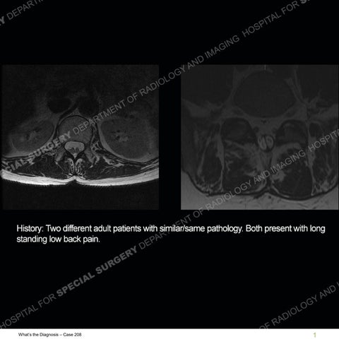


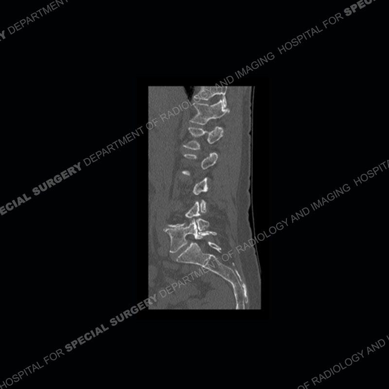
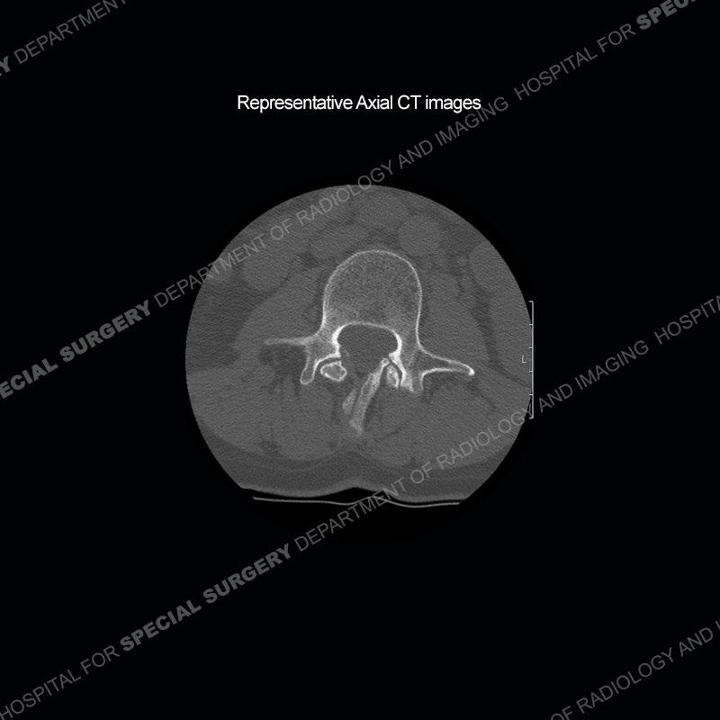
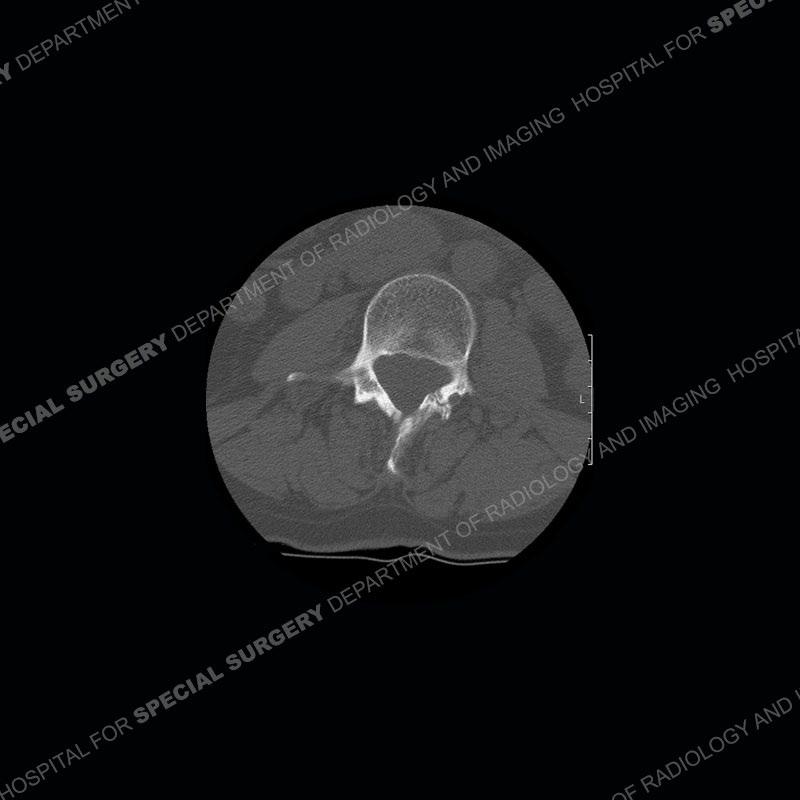


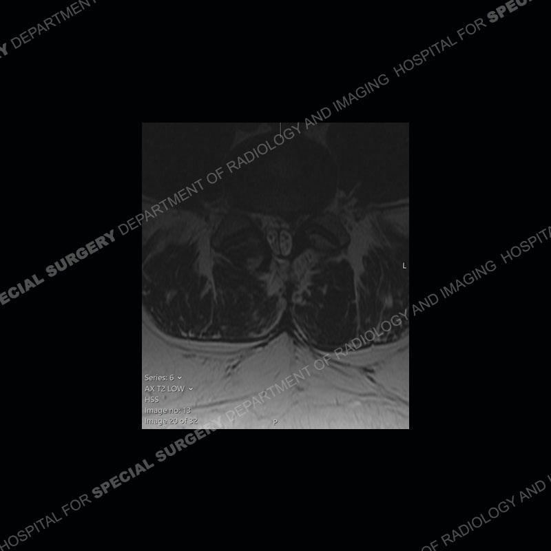

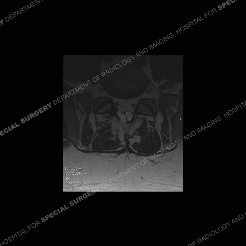

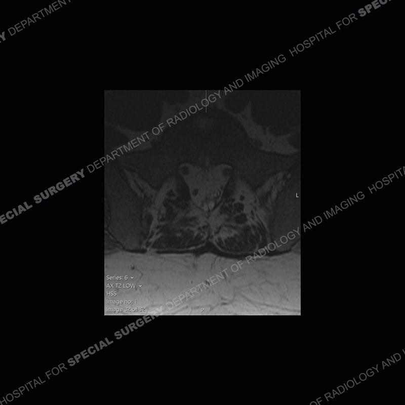
Findings
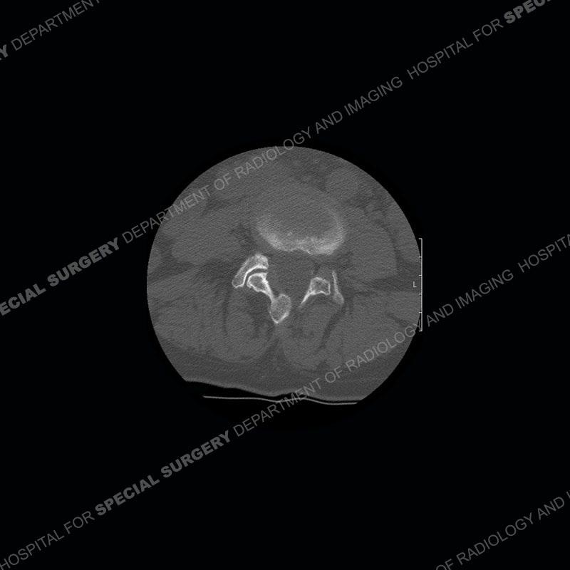
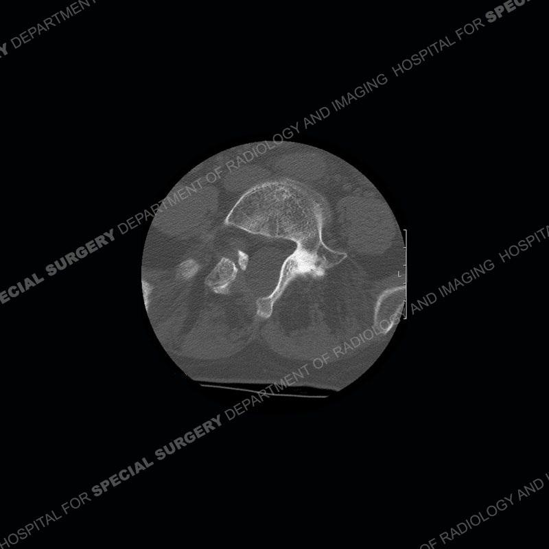
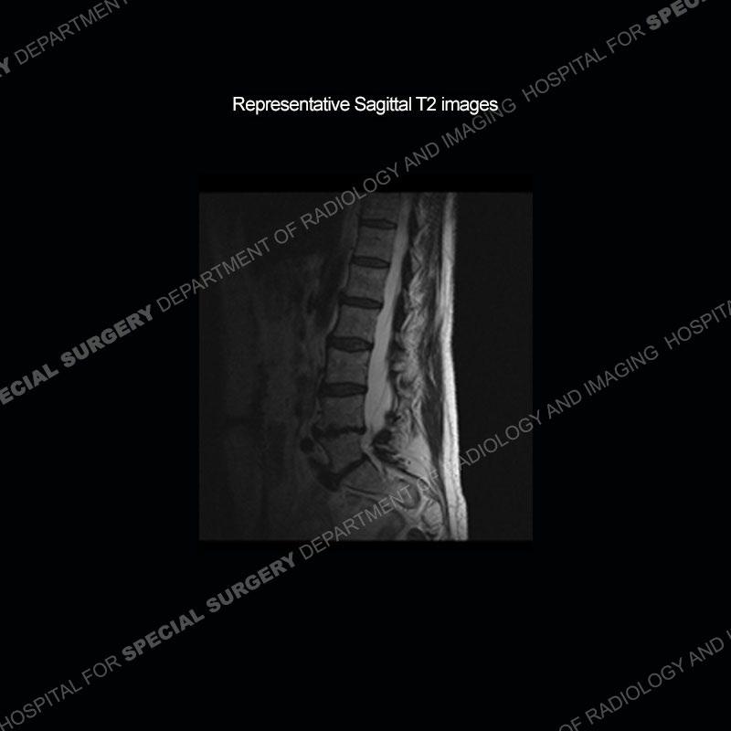
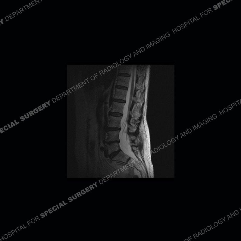
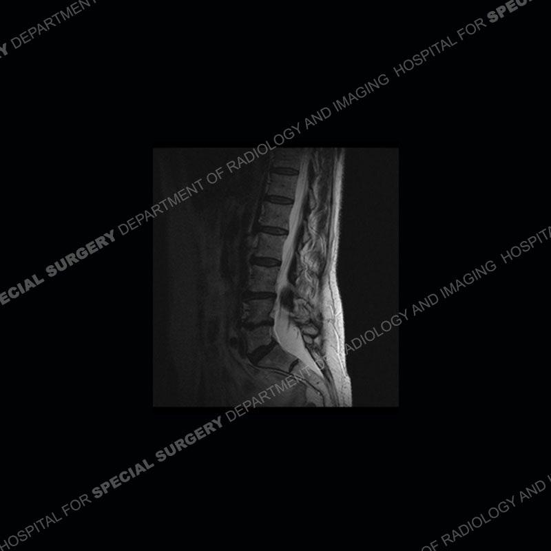
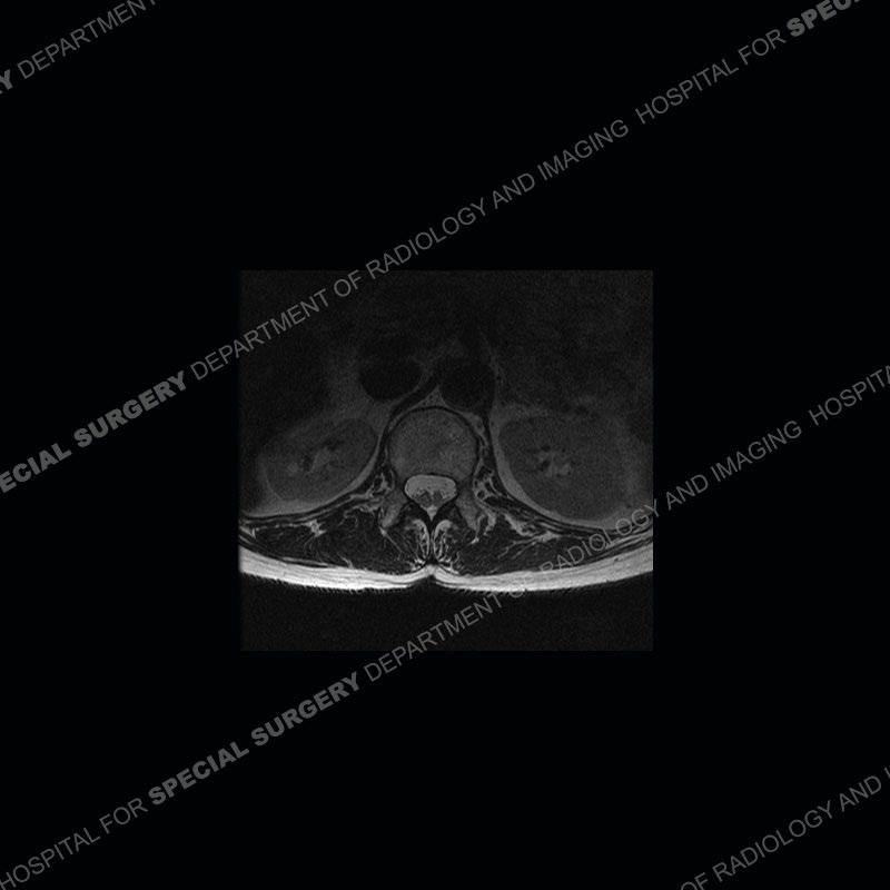
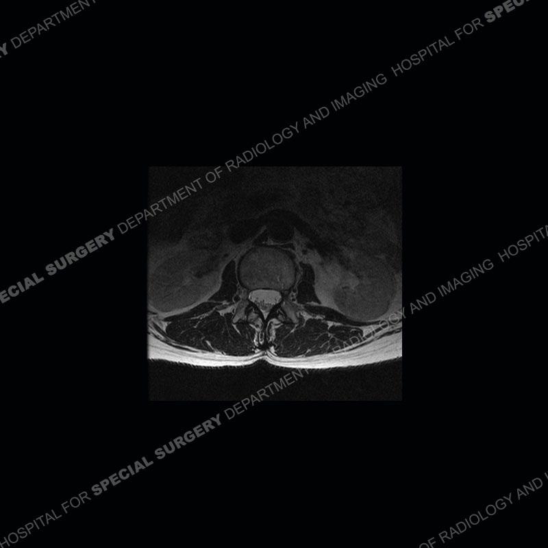
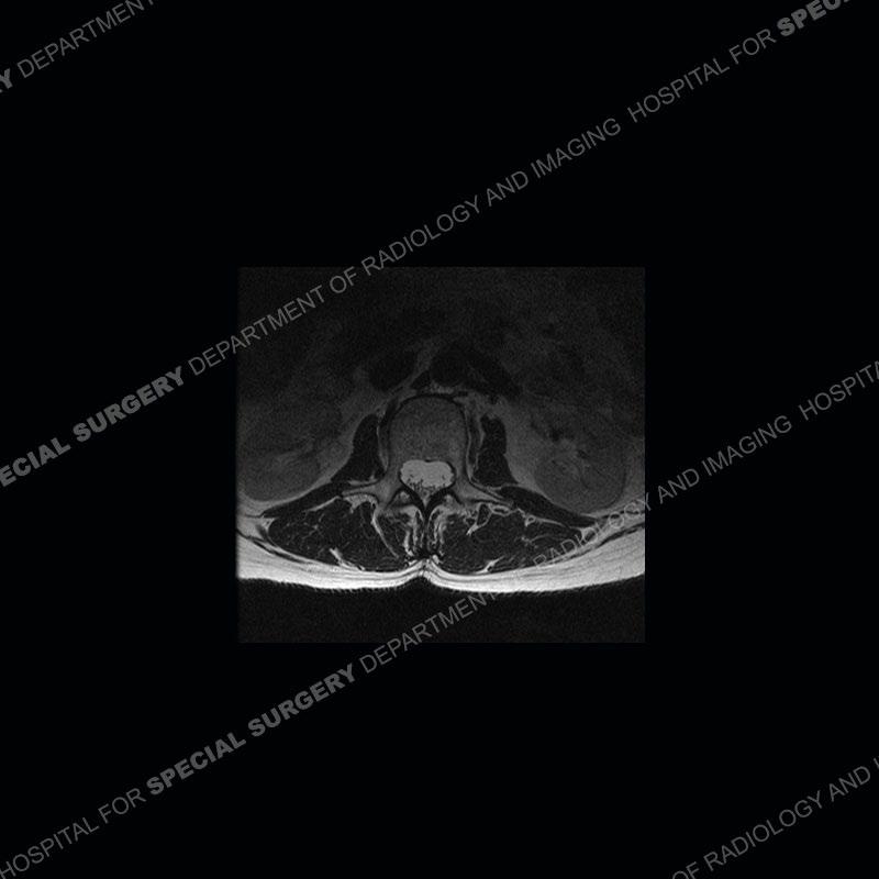
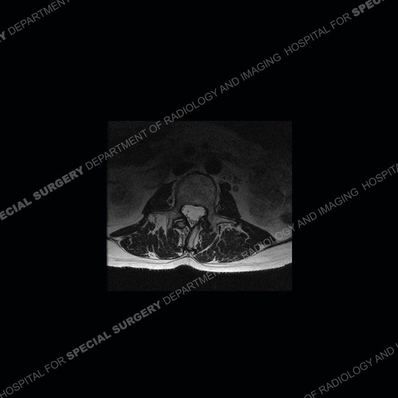
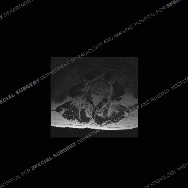
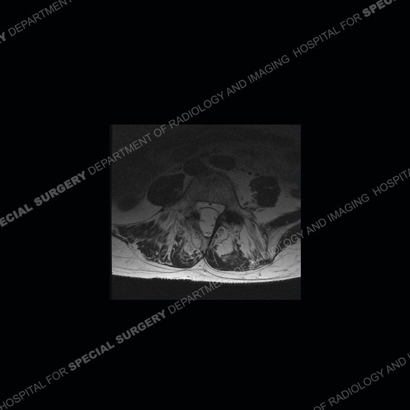
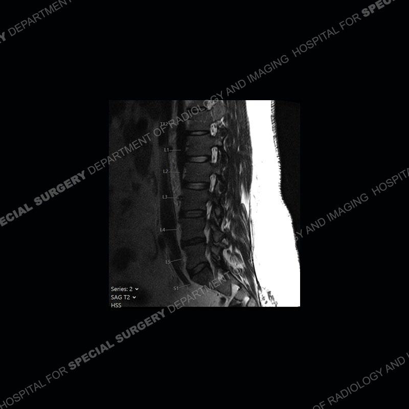
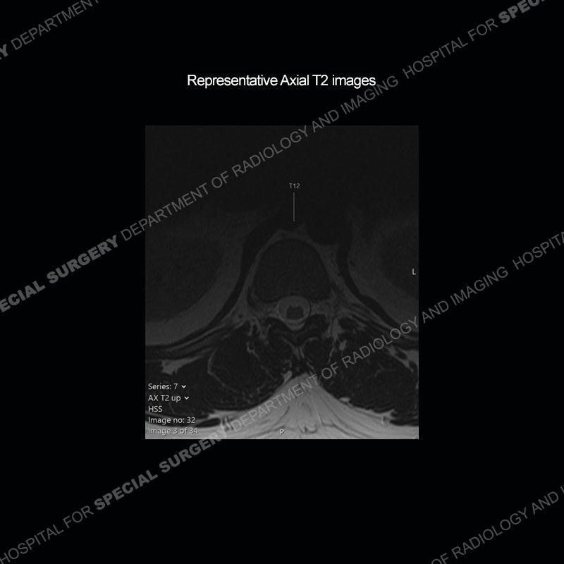
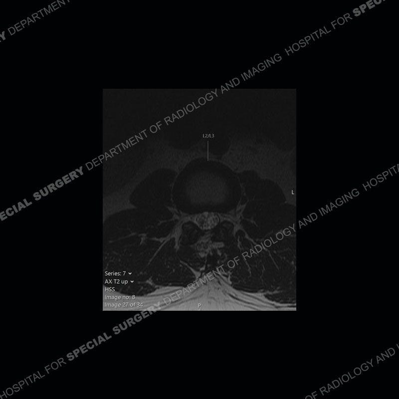
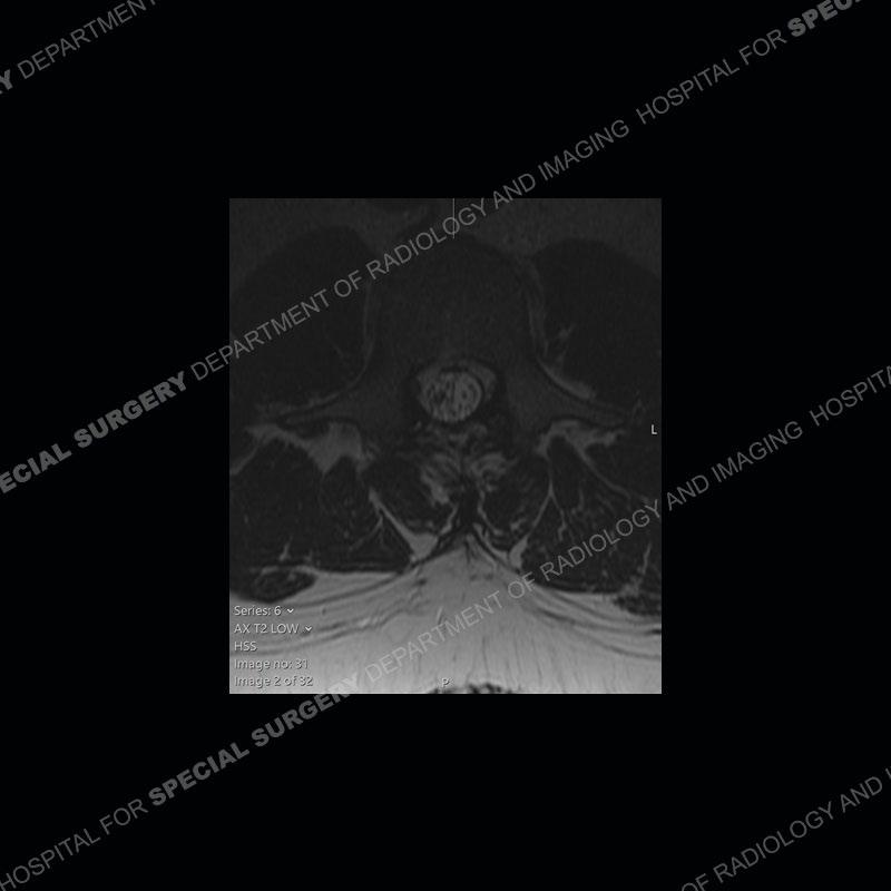
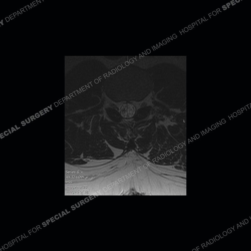
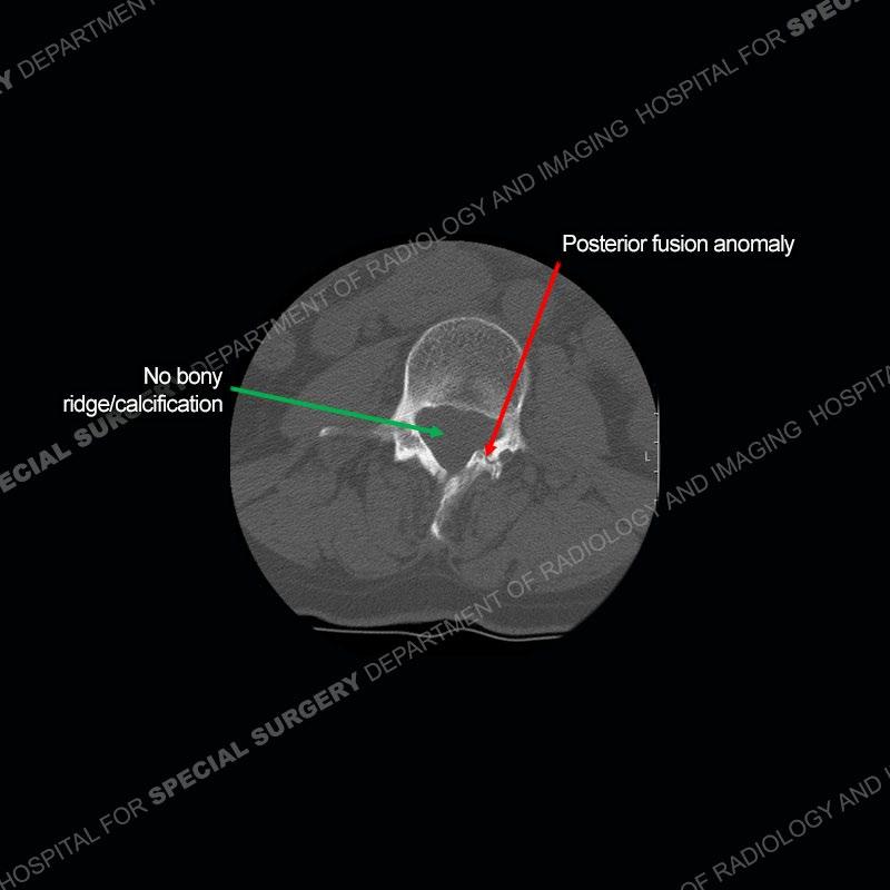
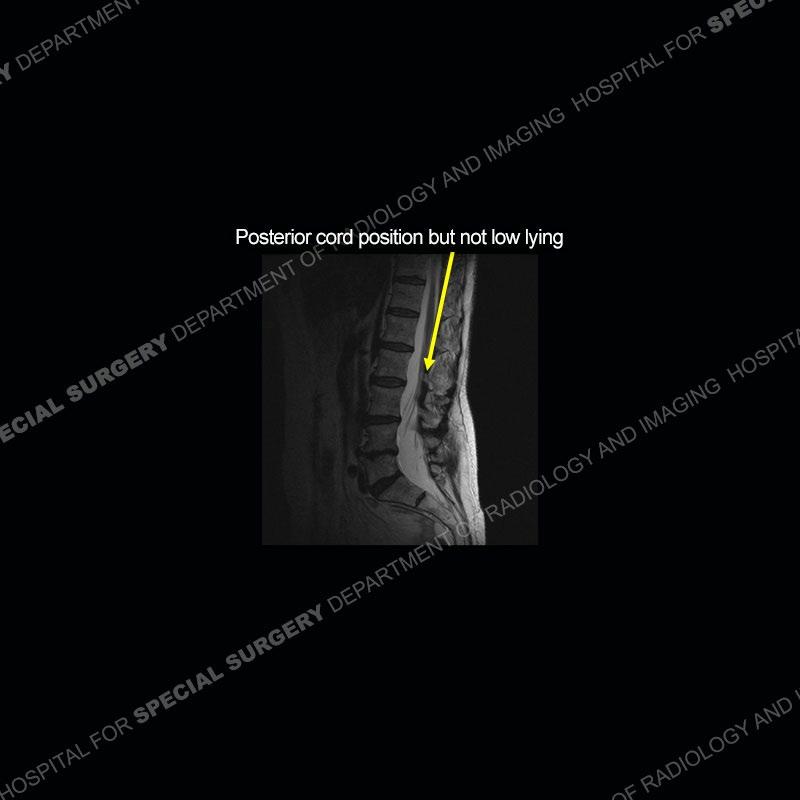
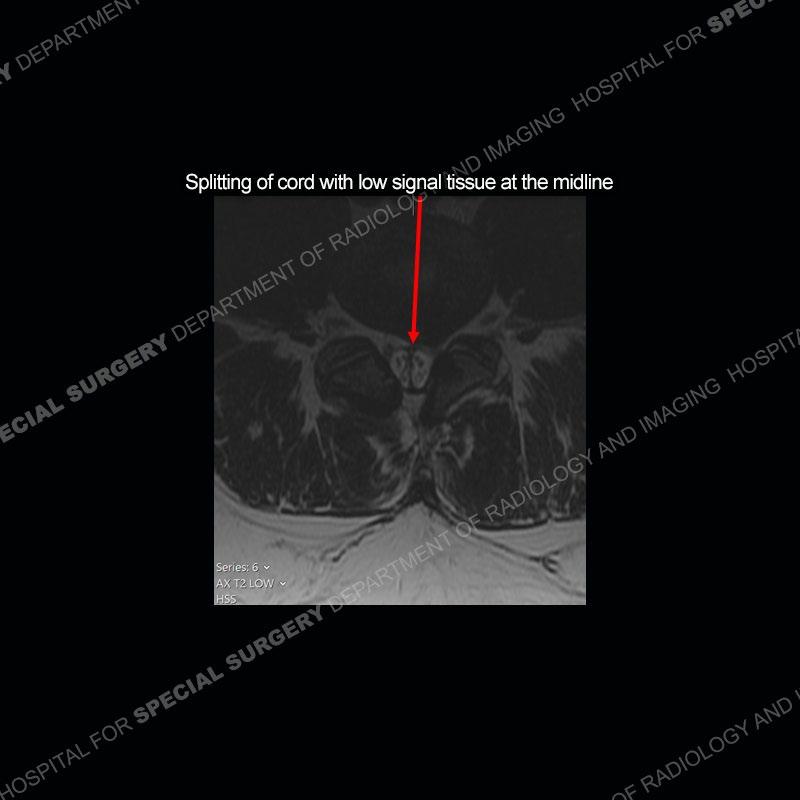
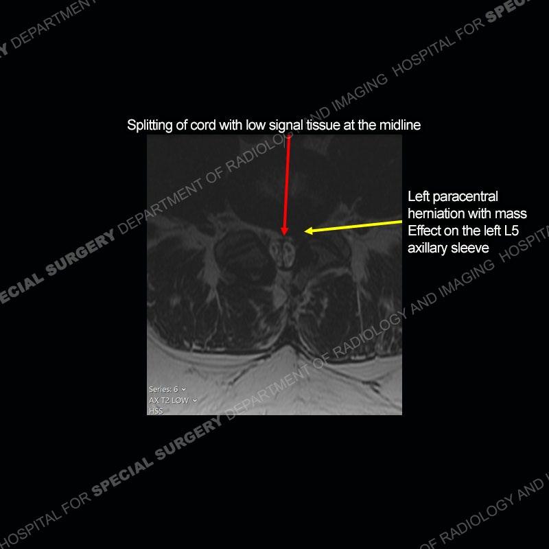
In both cases, seen are multiple bony anomalies of the posterior elements falling within the realm of spina bifida or a minor degree of spinal dysraphism. No vertebral body anomalies are present. In the first case, there is a very subtle splitting of the distal cord with one thecal sac identified. In the second case, there is a more prominent splitting of the cord with two thecal sacs identified. In both cases, the spinal cord is somewhat posteriorly positioned or tethered although the conus medullaris is not low lying in either situation. Noted on both CT studies is the absence of any ossification or mineralization at the areas of the splitting of the cord.


Diagnosis: Split Cord Malformation
The more global diagnosis of split cord malformation is now being used for the prior terms of distematomyelia and duplicated cord or diplomyelia. Although there is a spectrum, the split cord malformations are now being separated primarily as a type I with a duplicated dural sac with a rigid, midline cartilage or bony spur versus a type II with a thin if not imperceptible fibrous separation into two cords but only one thecal sac. Both entities are associated with posterior element anomalies with the type I and perhaps more prominent type II abnormalities additionally having vertebral body anomalies. These malformations relate to neuroenteric canal closure anomalies. They are very frequently (especially type I) associated with a tethered cord but not necessarily a low-lying cord.
Certain cases may be associated with more extreme radiological and neurological findings also being present. Although many patient do present with neurological deficits as children, a fair number of individuals as in these cases may present in adulthood with long standing pain and low-grade neurological deficits. As seen in the second case, more common age-related pathology such as disc herniation can also occur so multiple things can be happening at once.
References
Kleinrok J, Kleinrok K, Popiela TJ. Split cord malformation – a simple, current classification based on CT and MRI neuroimaging studies. Polish Journal of Radiology. 2025;90:46-54. doi:10.5114/pjr/199683.
A. Karim Ahmed, Elizabeth P. Howell, Stephen Harward, Eric W. Sankey, Jeffrey Ehresman, Andrew Schilling, Timothy Wang, Zachary Pennington, Linda Gray, Daniel M. Sciubba and C. Rory Goodwin. Split cord malformation in adults: Literature review and classification. Clinical Neurology and Neurosurgery, 2020-06-01, Volume 193, Article 105733, Copyright © 2020