


Findings
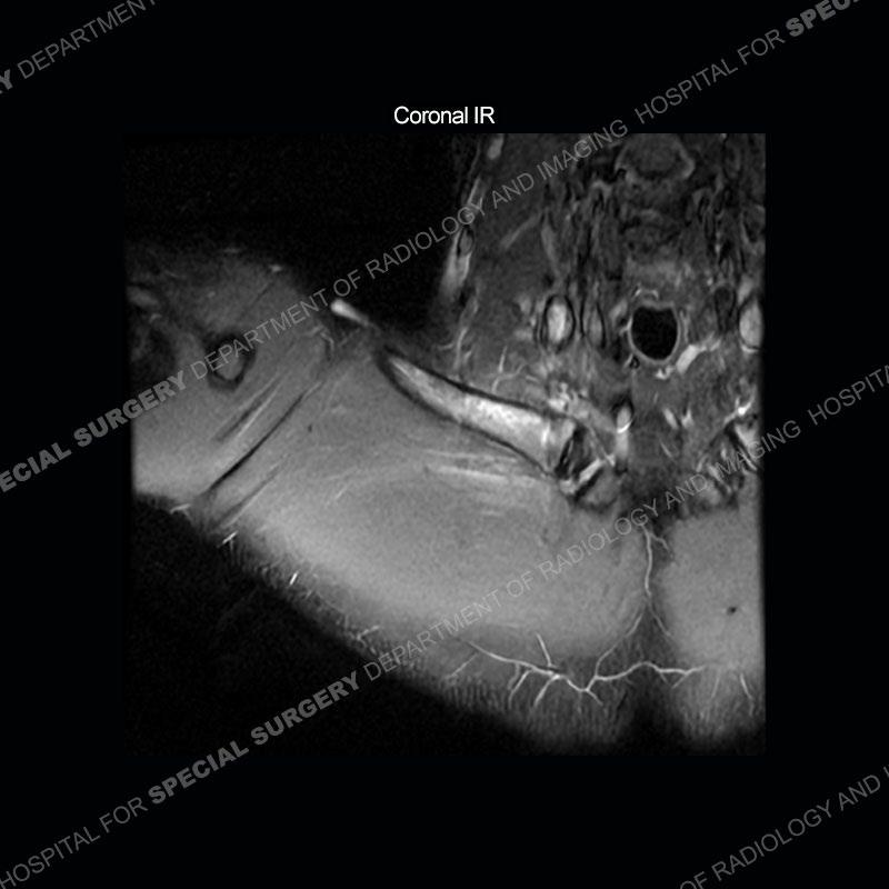
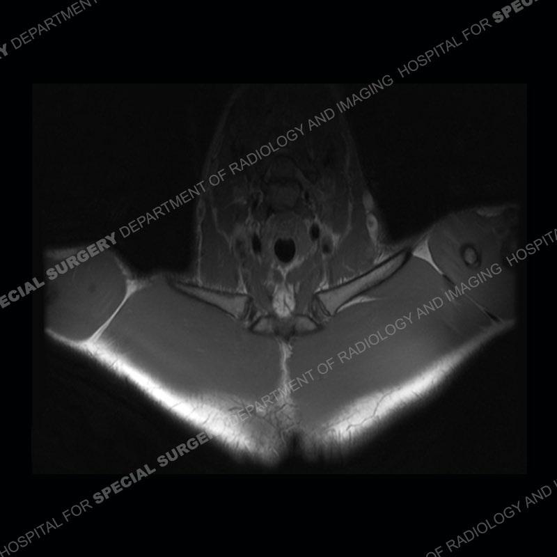
The initial MRI of the shoulder shows marked marrow edema pattern of the clavicle and marked edema/hematoma of the subclavius muscle. Given the pathology extending outside of the normal field of view, the exam was extended for further coverage than typical on the axial IR and axial PD images.
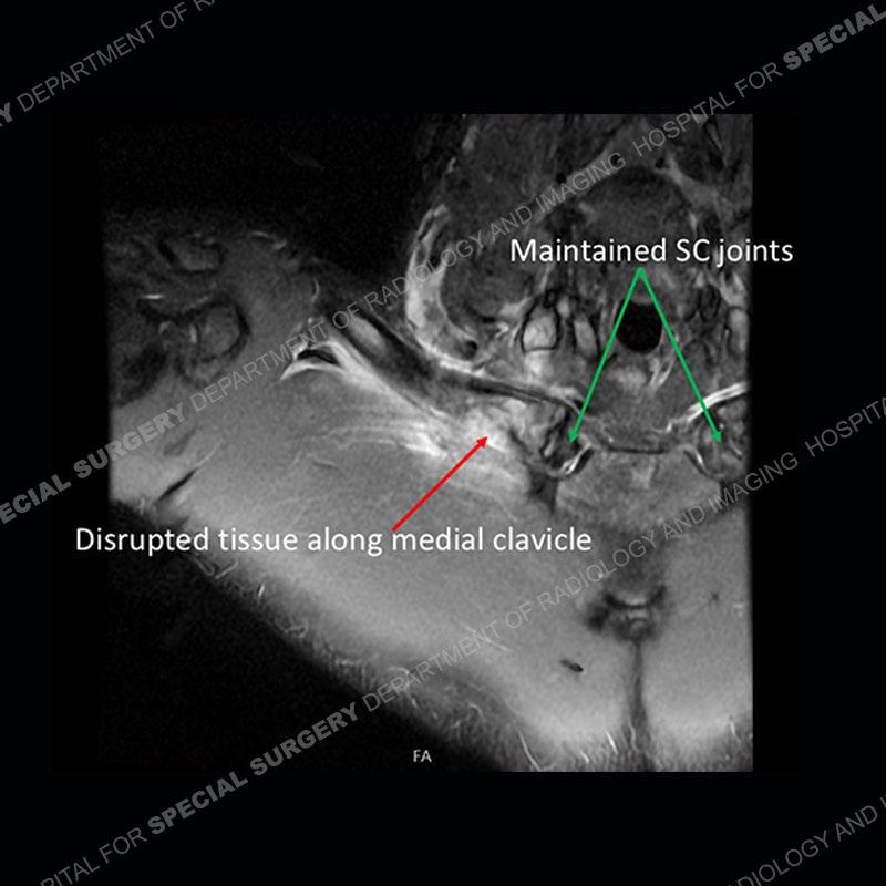
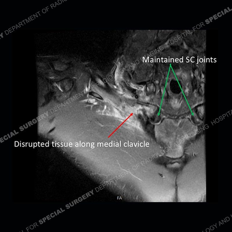
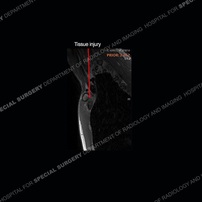
Subsequent imaging of the chest shows disruption of tissue along the medial aspect of the right clavicle and a deep insinuation of the bone along the medial aspect of the right clavicle. This is made more conspicuous with comparison to the left side. The edema pattern of the clavicle and the edema/hematoma of the subclavius persist.


Findings II
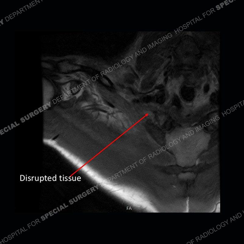
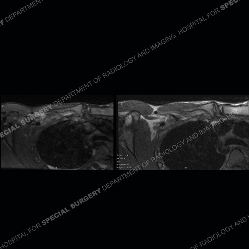
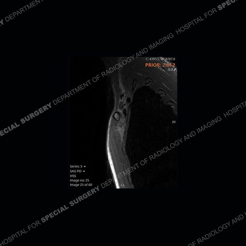
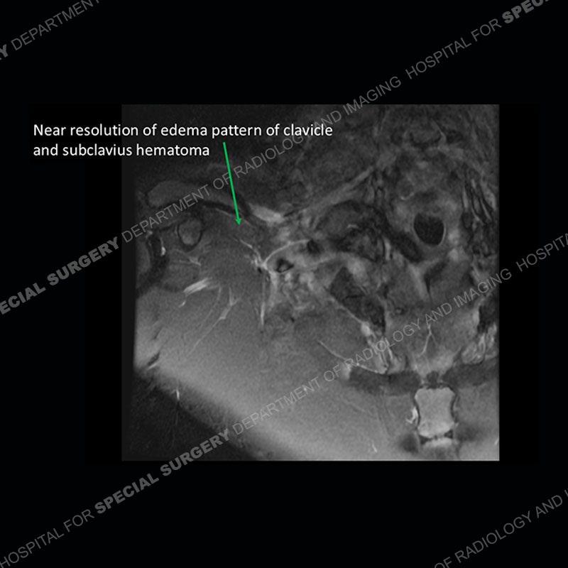
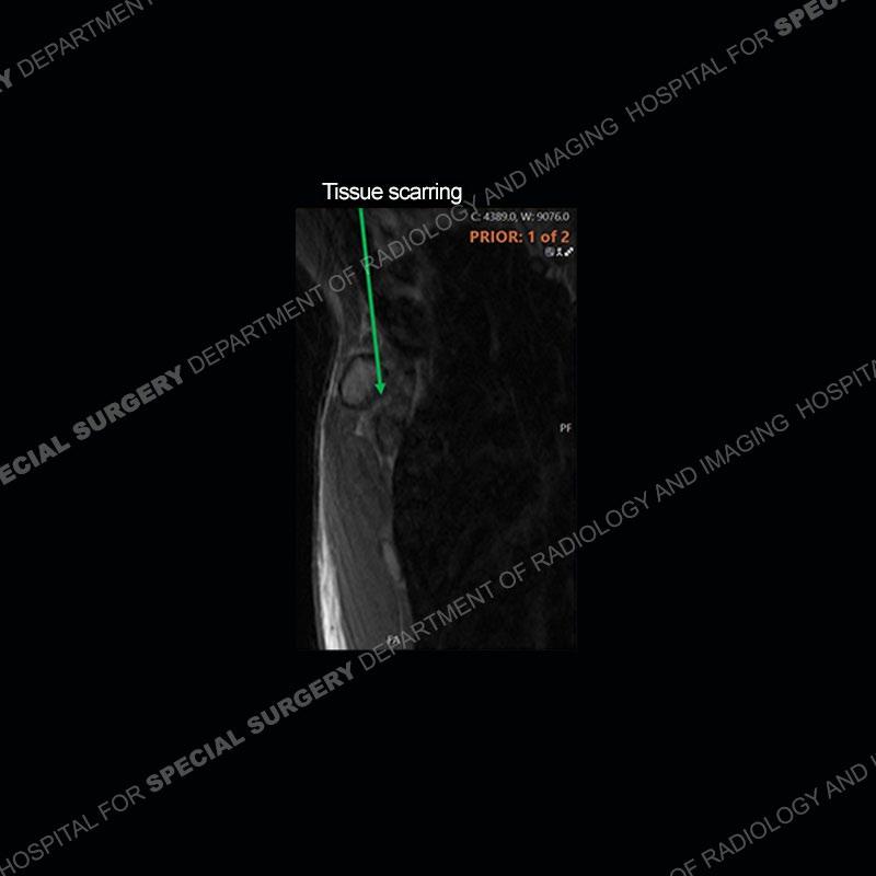
The subsequent imaging shows almost complete resolution of the edema pattern of the right clavicle and edema/hematoma of the right subclavius. The tissue along the medial aspect of the clavicle shows interval scarring, particularly when comparing the sagittal PD images between the two studies.


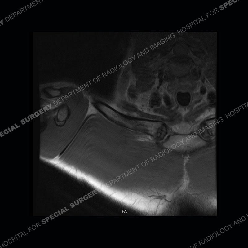
Diagnosis: Isolated Costoclavicular Ligament Disruption in the setting of a Rhomboid Fossa
The costoclavicular ligament (rhomboid ligament) extends from the undersurface of the clavicle to the first rib. The ligament attaches to the clavicle approximately 1cm lateral to the SC (sternoclavicular) joint. Injuries to the ligament are typically in the setting of an injury to the SC joint. Isolated injuries without SC joint injury are extremely uncommon with limited case reports in the literature.
The attachment of the ligament to the clavicle may have a normal variant with a deep fossa termed a rhomboid fossa. If this variant and its location are not known, it could mimic erosion or destruction of the bone. Adding more confusion is that the variant does not need to be bilateral or symmetric (as in this case where it is unilateral).
This case also highlights that at times, time can often be well utilized to help delineate injury vs. other pathological processes.
References
Dheer S, Zoga AC, Morrison WB. Clavicular avulsion of the costoclavicular (rhomboid) ligament: MRI findings. Radiol Case Rep. 2015 Nov 6;6(4):579. doi: 10.2484/rcr.v6i4.579. PMID: 27307942; PMCID: PMC4899931.