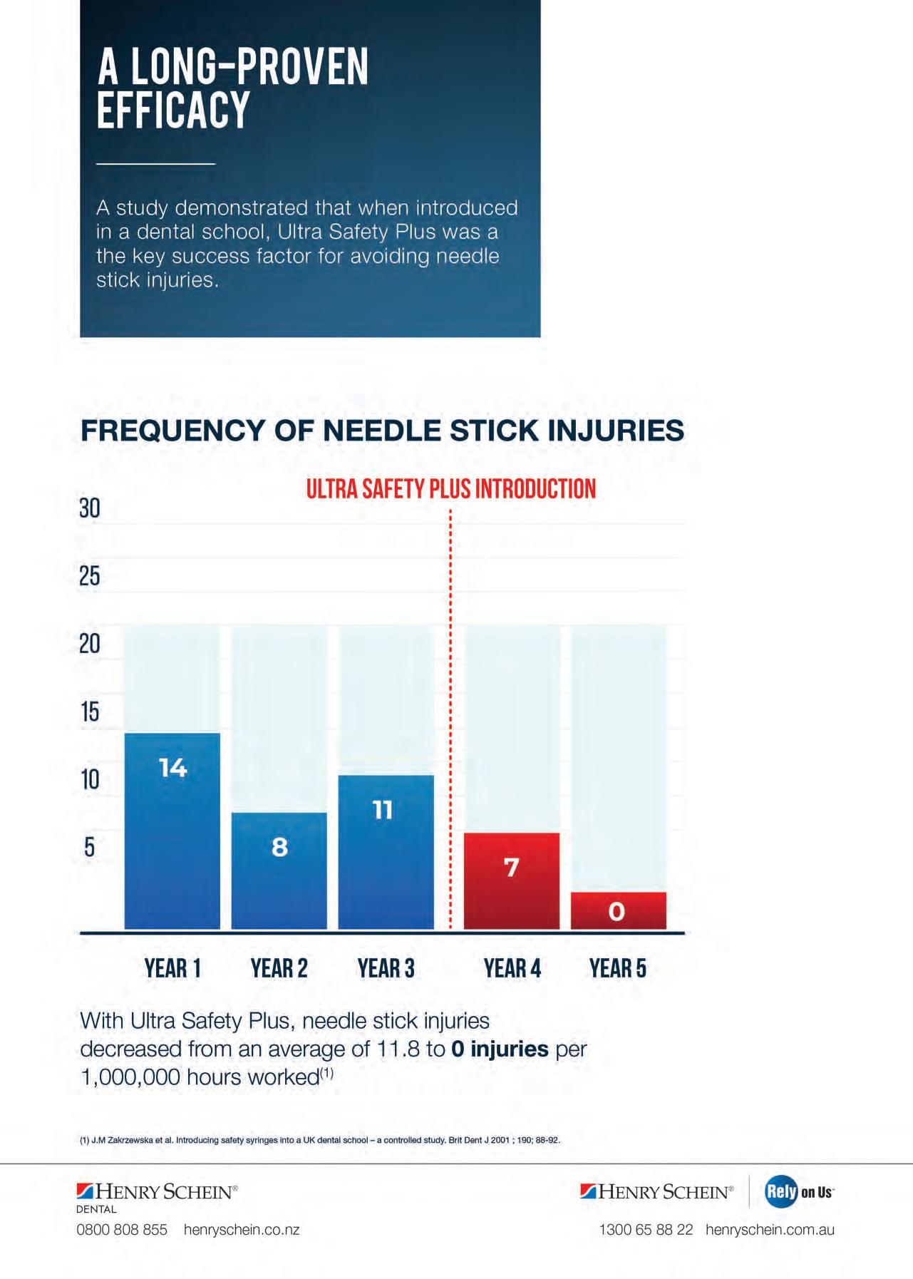
15 minute read
Clinical


Immediate placement and immediate restoration in the aesthetic zone: A review of the literature and case report
Peter Doherty1
1 Peter Doherty, BA, BDENTSC, MFDS (RCSI), PG CERT IMP DENT Private practice limited to implantology, Dublin, Ireland www.3dental.ie. Instagram: @drpeterdoherty Introduction The immediately placed and immediately restored dental implant has many advantages to offer us as clinicians and our patients: reduced total treatment times, less surgical procedures, preservation of the gingival architecture, less appointments, high patient satisfaction, and it also avoids the psychological impact of wearing a removable temporary prosthesis. Immediate placement is also nothing new, the concept of placing implants into fresh extraction sockets was first reported by Schulte and Heimke (1976). We have learned a lot in the intervening period about the procedure, however it remains clinically challenging and has the potential for significant aesthetic problems if inappropriately selected, and therefore requires some clear guidelines to operate within.
Literature review The timing between tooth extraction and implant placement has been categorised into four different protocols by the International Team for Implantology (ITI) (Chen and Buser, 2008). Type 1 placement is the immediate implant, where the implant is placed into the socket at the time of tooth extraction. In Type 2 placement (early placement with soft tissue healing) the implant is placed 4-8 weeks after extraction. The soft tissues have healed, but there is little bony healing. Type 3 placement (early placement with partial bone healing) occurs at 12-16 weeks. The soft tissues have healed and there is significant bony healing.
The main reasons for selection of Type 2 and 3 placements are to ensure an absence of pathology at the time of placement, and also increase the availability of soft tissues when a bone augmentation procedure is planned.
It has been shown that Type 1 placement has a greater variability of outcomes and is
associated with a greater risk of midfacial recession >1mm than Type 2 and Type 3 placement (Chen and Buser, 2014). Type 1 placement has also been shown to have survival rates equal to type 4 (fully healed) placement, even in teeth with associated pathology (Hammerle et al, 2004).
Planning immediate placement and immediate restoration cases require consideration of six vital areas: 1. The bone apical to the tooth 2. The buccal plate 3. The position of the gingival margin 4. The gingival biotype 5. The design of the implant 6. The jump gap.
The bone apical to the tooth 3-5mm of bone is required apical to the tooth to ensure sufficient implant stability at the time of placement. Immediate implants gain their primary stability from this apical bone, and from the palatal and lateral walls of the socket. The buccal plate must not be engaged to provide primary stability as this will often resorb leaving the implant facially positioned and at risk of an aesthetically compromised result. In addition to sufficient bone volume, the apical bone must be of sufficient density which is determined pre operatively with a CBCT (figures 2-4).
The buccal plate
The thickness of the buccal plate must be assessed with CBCT prior to immediate implant placement. If the buccal plate is thicker than 1mm there will be minimal bone loss following extraction. However, it has been shown that if the plate is less than 1mm thick there will be a median bone loss of 7mm in height (Chappuis et al, 2015).
Furthermore, in 90% of cases we see in practice, the buccal plate of the maxillary anterior teeth is 1mm or less (Huynh-Ba et al, 2010; Braut et al, 2011). This fragile piece of bone is critical to implant aesthetics. It derives its blood supply from the periodontal ligament and the periosteum mostly, with some thicker buccal plates having an endosseous supply from the bone marrow – though this is usually absent. If the buccal plate is 1mm thick or less then placement of an implant will not preserve the bone and the buccal plate will resorb (Arujo et al, 2005). It is critical to manage this resorption, as simply extracting a maxillary anterior tooth and placing an implant will lead to a buccolingual dimensional change of 1.1mm (Grunder, 2011) and the subsequent risk of aesthetic compromise. The same study by Grunder (2011) showed that placing a subepithelial connective tissue graft at the same time as immediate placement led to a gain of 0.34mm in the buccolingual dimension.
Strategies used in immediate implantation to avoid the problems posed by the resorption of the facial plate include:
A: Smaller implant diameter
B: Palatal implant position
C: Hard tissue grafting
D: Soft tissue grafting
E: Immediate provisional restoration.
The position of the gingival margin
Gingival margin position is critical to the success of maxillary anterior implants, particularly in high smile line cases. Where there is recession present on a tooth planned for implant replacement it would be advisable to select a type 2 or type 3 placement and avoid the immediate approach.
The gingival biotype
Described as either thick or thin, thick biotypes are said to have gingival tissues which are 2mm or more in thickness, and thin biotypes to be less than 1.5mm in thickness (Claffey and Shanley, 1986). In a population study by Olsson and Lindhe (1991) thick gingival biotypes were found to be more prevalent (85%), than thin gingival biotypes (15%). Thick biotypes are characterised by square teeth with short, broad papillae, broad band of keratinised tissue, and thick gingival tissues with thicker underlying bone. Thin biotypes exhibit triangular teeth, long and thin papillae, thin bands of keratinised tissue, and thin gingival tissues with thin underlying bone.
A useful method to assess gingival thickness is to insert a periodontal probe into the midfacial gingival sulcus. If the probe is visible through the tissues then you are dealing with a thin biotype, whereas a thick biotype will conceal the colour of the probe within the sulcus. The significance of gingival biotype in relation to immediate implants relates to the greater potential for facial recession and loss of papillae in thin biotype cases.
Gingival biotype may also be altered by the utilisation of a connective tissue graft, and the use of a connective tissue graft with immediate implant placement has been shown to contribute to mid facial soft tissue stability (Lorenz Seysens et al, 2021). This is a particularly useful technique when there is an increased risk of facial recession (for example a thin gingival biotype, or buccal plate less than 1mm in thickness).
Figure 1 Figure 2 Figure 3
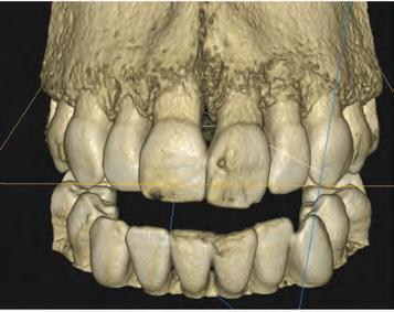
Figure 4 Figure 5 Figure 6
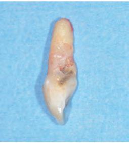

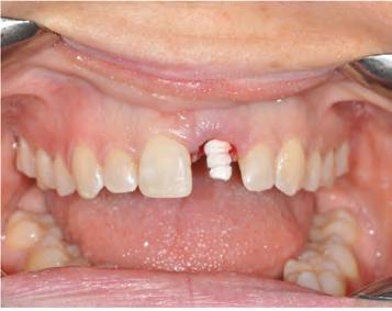
Figure 7
The design of the implant
For immediate placement primary stability is critical, and this is most reliably achieved with the use of tapered implants and an aggressive thread design. The tapered body shape facilitates bony compression as the implant is inserted and it has been shown that tapered implants can achieve higher stability in low density bone due to this compression (Atieh et al, 2018). The narrower tip also reduces the risk of apical fenestration in areas where there is a concavity present in the apical region of the alveolus.
Desirable features at the head of the implant are a platform shift to distance the implant-abutment junction from the rough surface, and a conical connection to create an effective seal at the implant-abutment junction preventing the ingress of bacteria. Both of these features ensure long term crestal bone stability.
The implant chosen is this particular case was a Neodent GM Drive implant. It was selected surgically for its tapered body and dual threaded design with cutting apical threads ensuring high primary stability in immediate placement. From a prosthetic viewpoint, it is ideally suited to use in the aesthetic zone due to its platform shift, horizontal polished portion and deep 16 degree morse taper.
The jump gap
As discussed previously, in 90% of cases in the anterior maxilla we can expect resorption of the buccal plate. To counteract this, immediate implants must be positioned palatally away from the buccal plate, creating a space between the buccal

Figure 8
Figure 10 Figure 9 Figure 9
Figure 11
Figure 12 Figure 13
surface of the implant and the buccal plate called the jump gap. Filling this gap with bone grafting material has no effect on osseointegration or survival rates, but it does have a significant effect on maintaining dimensions of the ridge and preventing collapse and reducing the chances of recession. To improve the dimensional stability of the ridge even further it has been shown that grafting the jump gap in combination with immediate provisionalisation results in almost no change in ridge contour (Tarnow et al, 2014). Case report
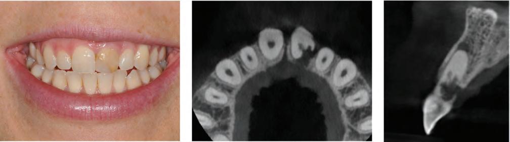
This 25 year old patient attended for assessment of UL1 which had been traumatised at age 7 and subsequently developed resorption and was deemed unrestorable. She expressed embarrassment at the colour and shape of the tooth, and felt self-conscious when speaking or smiling. She was also aware of its poor prognosis, and experienced intermittent pain from the tooth. Clinical examination revealed a high smile line with a discoloured, unaesthetic

Figure 14 Figure 15 Figure 16
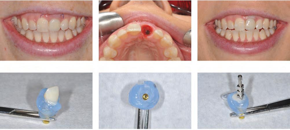
Figure 17 Figure 18 Figure 19
appearance to UL1 detracting from what was otherwise a nice smile (Figure 1). Probing pocket depths were normal, and there was no mobility, bleeding on probing or evidence of infection. The incisors were relatively square shaped, and the patient displayed a thick gingival biotype.
Radiographically, a CBCT revealed an extensive area of root resorption and associated apical radiolucency. There was also some distopalatal bone loss in the coronal one third of the root. The buccal plate was intact, approximately 1mm thick in the midportion of the root tapering out to less than that in the coronal one third, and comprised entirely of cortical bone (Figures 2-4).
The patient expressed a preference for a fixed temporary crown to avoid the use of a denture or other removable option as the implant integrated. Conditions were favourable for an immediate implant and immediate restoration given the thickness of the buccal plate, correct gingival margin level and the presence of good density bone apical to the UL1. There was also no evidence of parafunction and the patient had full posterior occlusion to the second molars. Complicating factors taken into account were the high smile line, a thinner buccal plate coronally (less than 1mm thick), and presence of bone loss both apically and at the distopalatal aspect towards the crown. Implant planning was carried out with CS 3D Imaging Software (Carestream Dental), and the implant virtually placed prior to the surgery.
On the day of the procedure the tooth was removed uneventfully (Figure 5) and the socket thoroughly curetted out, removing any granulation tissue.
Implant osteotomy was performed freehand towards the palatal aspect of the extraction socket, and advanced beyond the apex of the socket. A 3.5 x 16mm Neodent GM Drive implant was placed 1mm subcrestal on the facial aspect (Figure 6), and >45Ncm insertion torque achieved. The GM Drive implant offers very high torque values and exceptional primary stability, even in suboptimal bone density or in areas of limited bone availability – such as extraction sockets, which is one of the reasons I choose it in these immediate load cases. The second reason I select them is for their platform shift and tight morse taper connection which ensure the crestal bone level is maintained – vital in all areas of the mouth, but especially so in the aesthetic zone.
With the implant in position, attention was turned to management of the soft tissues and emergence profile. A PEEK abutment was modified intraorally (Figure 7) and a screw retained temporary crown constructed from Protemp and flowable composite using a pre op impression to recreate the emergence profile of the extracted tooth, and support the interdental papillae.
Further soft tissue management involved filling the space between the buccal aspect of the implant and the inner aspect of the buccal plate with Xenograft (Creos Xenogain, Nobel Biocare). Following this a deepithelialised connective tissue graft was harvested from the left hard palate and secured with two monofilament sutures into a tunnel created under the mucosa buccal to the implant to thicken the gingiva and

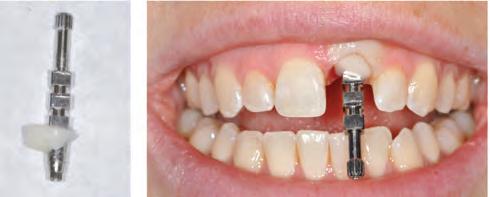
Figure 20 Figure 21
decrease the possibility of future gingival recession (Figures 8, 9 and 10). This was done in view of the thin buccal plate present at the margin and the presence of a high smile line.
Following insertion of the temporary crown, the static and dynamic occlusion was carefully checked and the crown adjusted out of occlusion in all excursions (Figures 11 and 12). The patient was instructed to treat the tooth as decorative and avoid eating on it during the osseointegration process. The final radiograph at placement can be seen in Figure 13.
On review two weeks later, the patient reported no discomfort, the surgical site was healing well, and the sutures removed (Figure 14).
Three months later, the buccal soft tissue contours had been maintained (Figure 15), and the patient actually had a lower gingival margin than was present pre operatively. The temporary crown was added to with composite in the region of the gingival margin to alter the emergence profile and move the gingival zenith slightly apically to match the adjacent central incisor (Figure 16).
Two weeks after this, final impressions were taken using a customised impression technique.
The temporary crown was removed and attached to an analogue, and then a transparent silicone (Memosil 2, Kulzer) was placed around the analogue and the submarginal area of the temporary crown to accurately copy the emergence profile. The buccal aspect of the tooth was marked on the silicone to aid orientation before the temporary crown removed and an impression coping placed into the analogue (Figures 17 and 18). Flowable composite was injected into the well around the base of the impression coping and light cured through the transparent silicone to create a customised impression coping which was a direct replica of the temporary crown (Figures 19 and 20). This was then seated intra-orally and an open tray impression taken with polyether (Impregum, 3M) (Figure 21).
The final crown was made from a zirconia core on a titanium base, cutback and layered in porcelain to mimic the natural anatomy of the adjacent teeth.
It was torqued to 20Ncm, the access sealed with PTFE tape and composite, and the occlusion checked and adjusted as appropriate (Figures 22a and 22b). The final radiograph is shown in Figure 23, displaying the platform shift and slender profile of the titanium base which aid bone and soft tissue preservation.
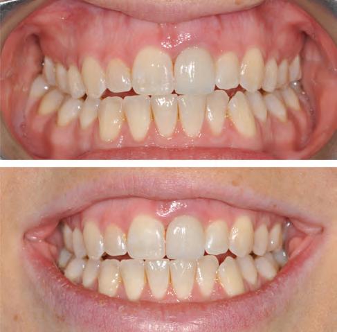
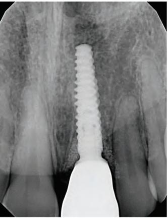
Figure 23
References
Araujo MG, Sukekava F, Wennstrom JL, Lindhe J (2005) Ridge alterations following implant placement in fresh extraction sockets: An experimental study in the dog. J Clin Periodontol 32:645-652
Atieh MA, Alsabeeha N, Duncan WJ (2018) Stability of tapered and parallel-walled dental implants: a systematic review and meta-analysis. Clinical implant dentistry and related research 20(4): 634-645
Braut V, Borenstein MM, Belser U, Buser D (2011) Thickness of the anterior maxillary facial bone wall – A retrospective radiographic study using cone beam computed tomography. Int J Periodontics Restorative Dent 31:125-131
Chappuis V, Engel O, Shahim K, Reyes M, Katsaros C, Buser D (2015) Soft Tissue Alterations in Esthetic Postextraction Sites: A 3-Dimensional Analysis. J Dent Res Sep 94 supplement:187S-93S
Chen S and Buser D (2008) ITI Treatment Guide Vol 3 Implant placement in post extraction sites p9-15
Chen S and Buser D (2014) Esthetic outcomes following immediate and early implant placement in the anterior maxilla - a systematic review. Int J Oral Maxillofac Implants 29 Supplement:186-215
Claffey N and Shanley D (1986) Relationship of gingival thickness and bleeding to loss of probing attachment in shallow sites following non-surgical periodontal therapy. J Clin Periodontol 13:654–57
Grunder U (2011) Crestal width changes when placing implants at the time of tooth extraction with and without soft tissue augmentation after a healing period of 6 months: Report of 24 consecutive cases. Int J Periodontics Restorative Dent 31:9-17
Hämmerle CHF, Chen ST, Wilson TG Jr (2004) Consensus statements and recommended clinical procedures regarding the placement of implants in extraction sockets. The International Journal of Oral & Maxillofacial Implants 19 supplement:26–28
Huynh-Ba G, Pjetursson BE, Sanz M et al (2010) Analysis of the socket bone wall dimensions in the upper maxilla in relation to immediate implant placement. Clin Oral Implants Res 21:37-42
Olsson M, Lindhe J (1991) Periodontal characteristics in individuals with varying forms of the upper central incisors. J Clin Periodontol 18:78-82
Schulte W, Heimke G (1976) The Tübinger immediate implant. Quintessenz 27(6):17-23
Seyssens L, De Lat L, Cosyn J (2021) Immediate implant placement with or without connective tissue graft: A systematic review and meta-analysis. J Clin Periodontol 48(2):284-301
Tarnow DP, Chu SJ, Salama MA, Stappert CFJ, Salama H, Garber DA, Sarnachiaro GO, Sarnachiaro E, Gotta SL, Saito H (2014) Flapless post-extraction socket implant placement in the esthetic zone: part 1. The effect of bone grafting and/or provisional restoration on facial-palatal ridge dimensional changea retrospective cohort study. Int J Periodontics Restorative Dent 34(3):323-31
Reprinted with permission by Private Dentistry Doherty P. Immediate placement and immediate restoration in the aesthetic zone: A review of the literature and case report. Private Dentistry April 2021: 64-68