
21 minute read
Clinical


Current state of CAD/CAM technology in implant dentistry
Dean Morton1, Waldemar Polido2, Wei-Shao Lin3
Abstract Complete or partial digital workflow utilizing computer-aided design and computer-aided manufacturing (CAD/ CAM) technology provides dental clinicians with ample options to treat their patients in a more effective way in daily practice. Dental clinicians and technicians need to recognize the limitations and proper indications of currently available technologies to accomplish optimal clinical care outcomes for patients. This article aims to narratively review various current CAD/CAM technologies (such as digital data collection, the development in CAD software programs and manufacturing platforms) in implant dentistry and discuss their advantages and limitations.
Keywords: CAD/CAM, digital, prosthesis, dental implant
1 Dr Dean Morton, BDS, MS, FACP, FDSRCS (Edin) Indiana Dental Association Professor and Chair of the Dept of Prosthodontics, Indiana University School of Dentistry, USA
2 Dr Waldemar Polido, DDS, MS, PhD, Clinical Professor and Chairman of the Oral & Maxillofacial Surgery Program, Indiana University School of Dentistry, Indianapolis, USA; CoDirector of the Center for Implant, Esthetic and Innovative Dentistry at IUSD.
3 Dr Wei-Shao Lin, DDS, FACP, Associate Professor & Program Director in the Advanced Education Program in Prosthodontics, Dept of Prosthodontics; Co-Director of the Center for Implant, Esthetic & Innovative Dentistry, Indiana University School of Dentistry, Indianapolis, USA. Dr Lin maintains an intramural dental implant & prosthodontics practice at the Indiana University School of Dentistry. Introduction A comprehensive digital workflow process for treatment including dental implants begins with data collection. Digital data collection may include 3-dimensional (3D) object scanning and obtaining a digital volume (CBCT). Together, the components of data collection allow for comprehensive treatment planning in which implant therapy is thought of as a singular entity and part of a total plan for the patient (Gallucci et al. 2019, Morton et al. 2019). Although the concept of comprehensive planning and treatment is embraced, this article will focus on those segments of the workflow traditionally considered to be of a restorative nature. Subsequent to data collection, the primary focus will therefore be computer-aided design and computer-aided manufacturing (CAD/CAM) of prostheses and associated processes.
CAD/CAM technology has found increasing popularity in dentistry. These processes can be seen as gradually replacing conventional fabrication methods, inclusive of implant-supported/retained prostheses. Available evidence supports incorporation of CAD/CAM protocols for single crown implant-supported crowns (Pjetursson et al. 2019), although the evidence for use of these procedures for fixed partial procedures is less compelling (Sailer et al. 2019). Research consistently demonstrates the precision and accuracy of CAD/CAM implant prostheses to be at least comparable and often improved when compared to the traditional lost-wax/casting technique. The advantages of CAD/CAM fabrication technology can be amplified for multi-unit implant prostheses and complete arch implant prostheses (Katsoulis et al. 2014). 3D object scanning can be undertaken with either a laboratory-based or intraoral scanner (Joda et al. 2017). Intraoral scanning offers clinical advantages including real-time evaluation of the impression and associated procedures (Figs 1–2). Intraoral
Fig. 1: An intraoral scan can be used for the preoperative diagnosis and treatment planning of static computer-aided implant surgery (sCAIS)

Fig. 3: An intraoral scan is more hygienic when an impression is desired during surgery. It may be of clinical convenience and ease if a patient is undergoing orthodontic treatment and is fitted with orthodontic brackets Fig. 2: When combined with a scan body, an intraoral scan can record the position and the timing of dental implants for the subsequent prosthesis fabrication
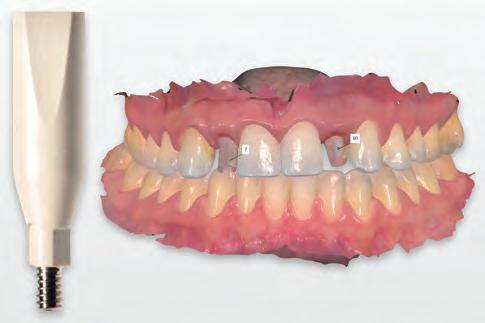
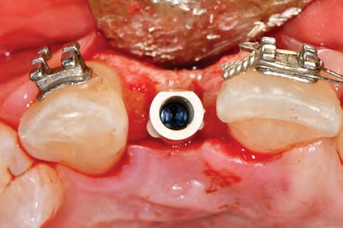
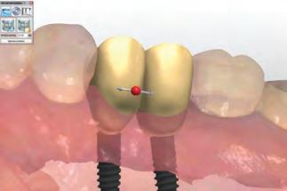
Fig. 4: The bevel near occlusal third on a scan body is a critical design for the CAD/CAM software to register the implant position and timing. The scan body should be positioned in a way that the bevel on the scan body facing area is easily accessible for intraoral scanning (generally speaking, towards the facial surface)
optical scanning offers flexibility and is compatible with in-office CAD/CAM systems and well as local and centralized laboratory options. Intraoral optical scanning may prove to be more hygienic in the transfer of information to the laboratory partners as there are no cast or analog components (Figs 3–4). There remains, however, a lack of high-quality evidence to support selection guidelines when considering conventional versus digital implant impression techniques. Available articles (mostly in-vitro laboratory studies) demonstrate however that digital impression options (intraoral and laboratory-based) offer a valid alternative for single and multi-unit implant prostheses. In particular, existing information supports improved patient perception and higher satisfaction with intraoral optical scanning (Wismeijer et al. 2014). Clinicians should be aware, however, that the distance between the implants (edentulous span), implant placement depth, implant angulation, operator experience, different intra-oral scanner systems and scan strategy may all affect the overall accuracy of intraoral scanning (Rutkūnas et al. 2017) (Figs 5–6).
Optical impressions or scans (intraoral or laboratorybased) can be used by both dental laboratory technicians and clinicians for the design and fabrication of prostheses using CAD/CAM options (Figs 7–9). For clinicians who
MORTON / POLIDO / LIN
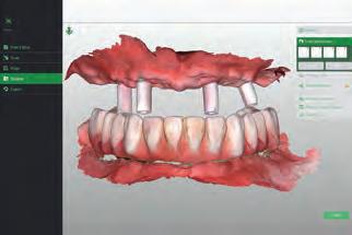
Fig. 5: The distance between implants (edentulous space) often affects the overall accuracy of intraoral scans greatly Fig. 6: Scanning the edentulous patient can pose a clinical challenge to capturing an accurate soft tissue extension, the maxillomandibular relationship, and interimplant relationships Fig. 7: The intraoral scan was sent to the dental laboratory for the design and fabrication of a CAD/CAM interim PMMA hybrid prosthesis

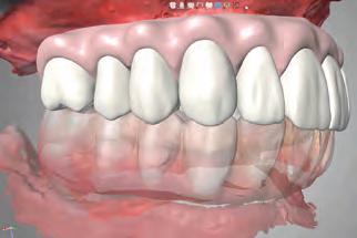
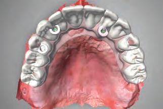
Fig. 8: Occlusal view of CAD/CAM interim PMMA hybrid prosthesis Fig. 9: The maxillary prosthesis was milled with a tooth-colored PMMA CAD/CAM block and layered with soft-tissue-colored laboratory composite resin Fig. 10: When the registration of soft tissue’s dynamic/functional movement is indicated for an implant-retained prosthesis, a traditional analog impression can be made to obtain proper soft tissue extension
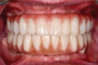

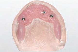
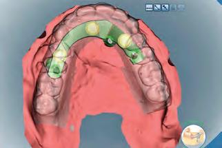
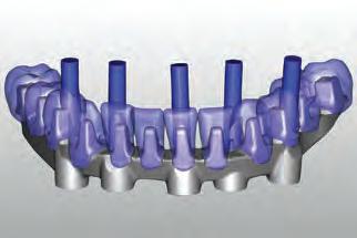
Fig. 11: A dental stone cast can be digitalized with a laboratory scanner for the subsequent CAD/CAM prosthesis fabrication Fig. 12: Representation of scanned dental cast, and the design of CAD/CAM bar on 3 dental implants Fig. 13: Computer validation of the design of CAD/CAM bar
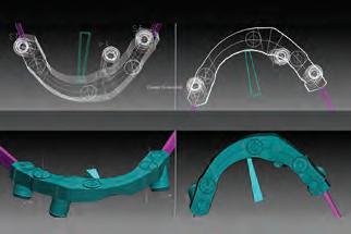
choose conventional impression methods or have limited access to an intra-oral optical scanner, or under certain clinical conditions, a conventional impression can be made (Fig. 10). The resulting cast can be digitalized using a laboratory scanner (Figs 11–12). This is a representation of a partial digital restorative workflow whereby CAD/ CAM technology can be applied for prosthesis design and fabrication, possibly improving precision and reducing human production errors when compared to a complete conventional analog technique (Fig. 13) (Kapos et al. 2014). Extraoral facial soft tissue scanning is a more recent option being marketed as part of a complete digital workflow

MORTON / POLIDO / LIN
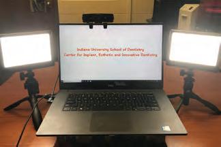
Fig. 14: Different economic extraoral facial soft tissue surface scanners are now commercially available Fig. 15: Ample lighting is desired while obtaining facial scans, and additional photography lighting equipment can be purchased to improve the overall quality of facial scans
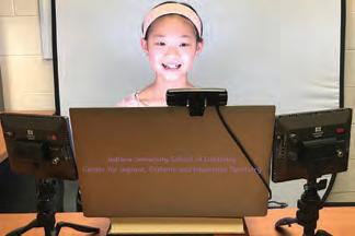
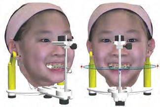
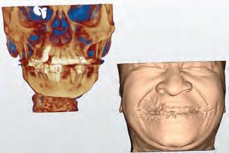
Fig. 17: Using the facial anatomic landmarks, a virtual articulator may be used to simulate/estimate the patient’s dynamic occlusal contacts Fig. 18: Although CBCT allows the 3D imaging of the craniofacial hard tissue, it only has a limited field of view and contrast resolution for facial soft tissue Fig. 16: A 3D virtual patient can be obtained with the superimposition of extraoral facial and intraoral dental scans
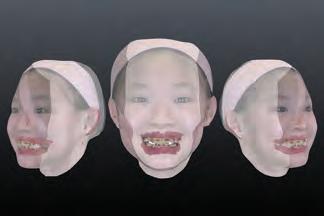
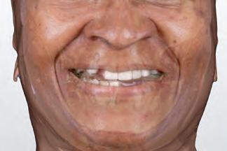
Fig. 19: The craniofacial hard tissue reconstructed from CBCT volumetric data can be superimposed with digital photographs to create a 3D virtual patient with photorealistic appearance
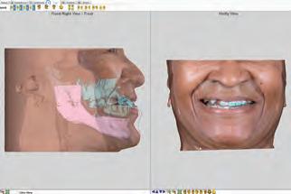
Fig. 20: A digital diagnostic tooth arrangement can be superimposed to the virtual patient to simulate soft tissue profile changes and obtain the patient’s approval Fig. 21: The approved diagnostic tooth arrangement can be used for prosthetically-driven sCAIS planning Fig. 22: CAD/CAM surgical templates for planned sCAIS
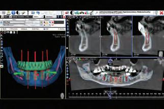

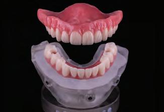
Fig. 23: CAD/ CAM interim prostheses for planned immediate provisionalization process (Figs 14–15). The overall esthetic treatment outcome is a decisive factor for the patient’s emotional acceptance of both tooth loss and treatment. In order to optimize clinical outcomes, clinicians seek harmony between dental and facial esthetics. Emerging technologies allow clinicians to capture the 3D surface texture of a patient’s extraoral facial soft tissue, including laser and optical-based surface scanners
MORTON / POLIDO / LIN

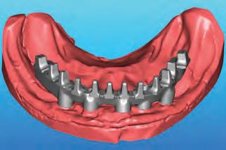
Fig. 24: Example STL file representing a mandibular intraoral scan of 4 dental implant scan bodies
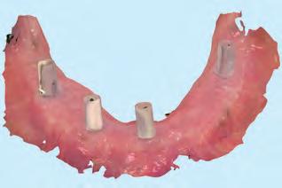
Fig. 27: Example proprietary file representing a mandibular intraoral scan of 4 dental implant scan bodies. The STL file in Fig. 24 was converted from this proprietary file format, and the surface texture and color cannot be preserved after conversion
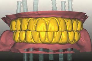
Fig. 30: Design of definitive zirconia prostheses Fig. 25: Example STL file representing a surgical template and complete dental prosthesis for sCAIS
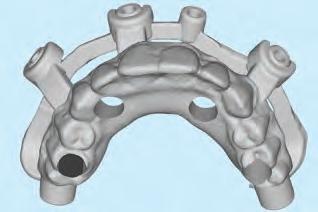
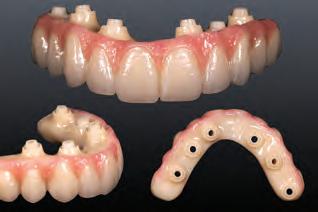
Fig. 28: Milled PMMA can be used as a prototype prothesis testing the functional and esthetic outcomes prior to the fabrication of definitive zirconia prosthesis

Fig. 31: Milled definitive zirconia prostheses prior to coloring and sintering Fig. 26: Example PLY file representing a postoperative intraoral scan
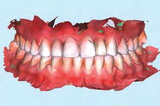

Fig. 29: After 1 to 2 months of intraoral function, the prototype prostheses may demonstrate occlusal changes resulting from patient’s dynamic occlusion. The prototype prostheses can be sent back to the dental technician to be used as a reference when designing the definitive prostheses

Fig. 32: Milled definitive zirconia prostheses ready for delivery
(Kau 2011) (Figs 16–17). In addition to surface scanners, 3D volumetric data obtained from a cone beam computed tomography (CBCT) can be superimposed (merged or registered) with digital photographs to create a 3D virtual patient with a photorealistic appearance. When combined with a laboratory or intraoral optical scan, a complete 3D virtual patient with photorealistic appearance can facilitate patient education and communication, diagnosis and treatment planning process for the static computeraided implant surgery (sCAIS), as well as the design and manufacturing of CAD/CAM prostheses (Figs 18–23). Currently, the accuracy of digital data superimposition is a limiting factor when composing an accurate 3D virtual patient. Consistent anatomic landmarks need to be present in all digital files (inclusive of the intraoral optical scanner, facial scanner, CBCT imaging scanner, and dental laboratory scanner) to facilitate accurate superimposition (Kuric et al. 2018). A goal in the development of future technology is the
MORTON / POLIDO / LIN
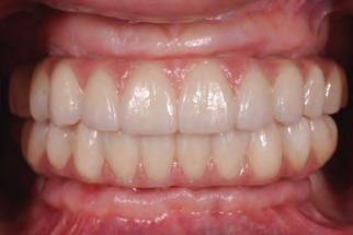
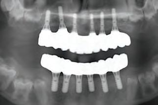
Fig. 33: Intraoral view of seated milled maxillary and mandibular definitive zirconia prostheses Fig. 34: Post-operative panoramic radiograph Fig. 35: 3D printed interim implant crown with a desktop DLP printer
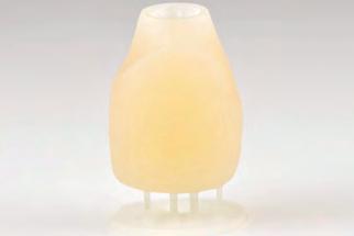
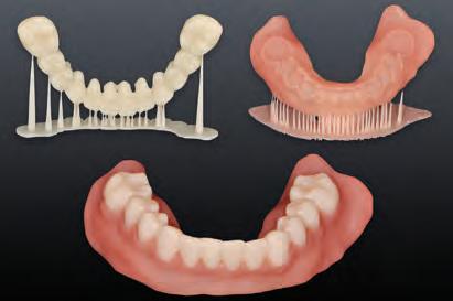
Fig. 36: 3D printed surgical template with a desktop SLA printer Fig. 37: 3D printed interim denture with a desktop DLP printer. Although desktop SLA or DLP printers are convenient for in-office production, one major limitation is that only one material (in color or stiffness) can be used in a single print job. When different materials are desired (such as pink and white resin in this instance), two separate prints and post-printing assembly are needed
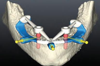
Fig. 38: Industrial 3D printer can operate different materials in the same print and produce objects with different colors, textures, gradients, transparencies and durometers in a single print operation. These dental casts were printed with a PolyJet printer (J750; Stratasys Ltd) Fig. 39: Design of CAD/CAM bone reduction and surgical template for sCAIS

creation of a 4-dimensional (4D) virtual patient inclusive of the patient’s dynamic motions. At present no commercially available digital system is able to compose such a 4D dental patient.
Most contemporary CAD/CAM systems have moved toward what is termed open architecture. Open architecture facilitates freedom of exchange of the digital data obtained from 3D scanners or computer software design programs.

MORTON / POLIDO / LIN
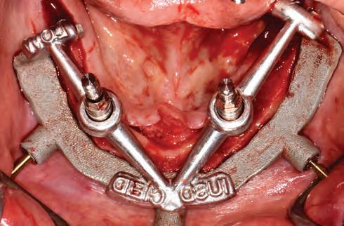
Fig. 40: The bone reduction and surgical template can be milled or printed from the cobalt-chromium (Co-Cr) or titanium alloy. Although the production cost with these materials could be higher, they offer superior mechanical strength and accuracy, which may in turn reduce the intra-operative complications, such as fracture of templates, insufficient mouth opening leading to insufficient operation access, and aggressive flap design needed to accommodate the bulky resin templates Fig. 41: A complete digital restorative workflow starts with an intraoral scan capturing implants’ position and timing with scan bodies
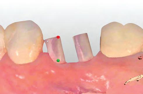
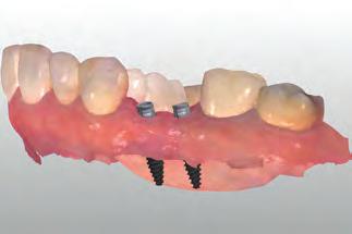
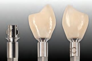
Fig. 42: Virtual titanium base abutments were placed onto the implants Fig. 43: Virtual design of CAD/CAM monolithic zirconia implant crowns Fig. 44: Milled zirconia crowns can be completed with surface finishing, polishing and characterization. Resin luting agent is used to lute the zirconia crowns and the titanium base abutments together prior to insertion
The data can then be effectively used for the subsequent prosthesis fabrication with a range of manufacturing devices. In contrast, a CAD/CAM system with closed architecture limits the digital data acquisition process and prosthesis design and manufacturing to one integrated system with no interchangeability. Open architecture increases flexibility and allows integration of information by appropriate CAD software programs, so enabling the composition of a virtual dental patient for the treatment planning or prosthesis design. Another important advantage of an open architecture is the increased opportunity for clinicians and dental technicians to access a wide variety of manufacturing technology and restorative materials without constraint (van Noort 2012).
Digital information can be transferred between CAD software programs and manufacturing platforms more effectively when digital file formats are standardized and consistent. There are more than 140 different recognized file formats, with Standard Tessellation Language (STL) being the most commonly used. The STL format, although widely used, is limited to the surface geometry of a 3D object, without any representation of color or texture (Grant et al. 2016) (Figs 24–25). In cases where surface color or texture are relevant,
MORTON / POLIDO / LIN
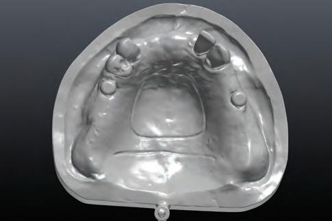
Fig. 45: When dynamic soft tissue movement is critical to a removable implant-retained prosthesis, a traditional impression should be made for optimal clinical outcomes. An analog cast can be digitalized in the dental laboratory with a laboratory scanner
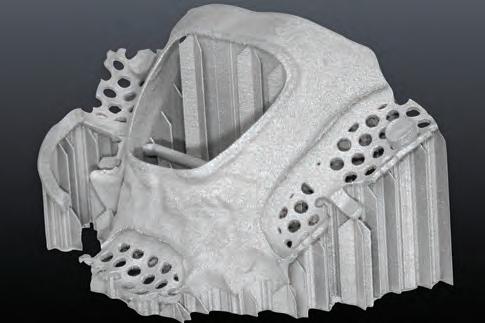
Fig. 47: Printed cobalt-chromium (Co-Cr) framework for removable dental prothesis Fig. 46: Continuation of digital prosthesis design after analog cast digitalization
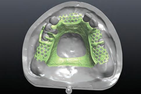
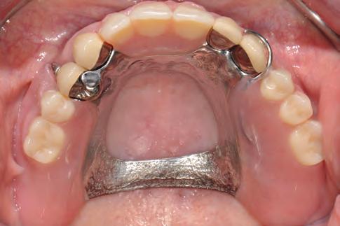
Fig. 48: Intraoral view of CAD/CAM implant-retained removable partial dental prosthesis
the OBJ (an open file format developed by Wavefront Technologies) or PLY (Polygon File Format or Stanford Triangle Format) file formats can be utilized to retain this information (Davies et al. 2017) (Fig. 26). OBJ or PLY files are therefore utilized to store extraoral facial information, preserving the 3D texture and color information of a patient’s facial expression (Morton et al. 2019) (Fig. 16). A closed CAD/CAM system may utilize a proprietary digital file format that is not recognized by all software options, and although conversion of the file formats is possible, it may lead to data loss (Fig. 27).
Noteworthy developments in the field of CAD/CAM include additive manufacturing technologies. Previously, CAD/CAM technology was dominated by subtractive manufacturing methods, such as computer numerically controlled machining, electrical discharge machining, electrochemical machining, electron beam machining, photochemical machining, and ultrasonic machining. Dental ceramic protheses and metal bar/frameworks are often milled from lithium disilicate, zirconia, titanium alloy or cobalt-chromium alloy (Figs 28–34). Although subtractive manufacturing has achieved a high degree of sophistication, it is not without limitations. It can be a wasteful process and does not easily allow for mass production as only one
MORTON / POLIDO / LIN
49 50
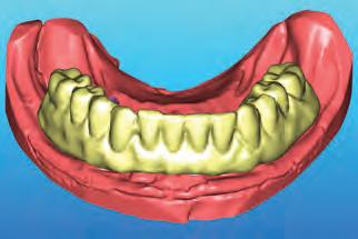
Fig. 49: When the clinician requires a clinical trial insertion, and/or the dental technician needs the articu-lated analog casts to re-position the manufactured framework or prosthesis for the subsequent finishing process, a partial digital workflow should be used. This figure illustrates the digitalization of a milled cast, and diagnostic tooth arrangement after obtaining the patient’s approval from clinical trial insertion. Fig. 50: Design for a CAD/CAM titanium framework on 5 dental implants. Fig. 51: Design of the CAD/CAM crowns fitting on the framework
object can be machined at a time. Additive manufacturing can create objects with fine surface details (such as undercuts and voids) and complex internal structures (van Noort 2012). Additive manufacturing is defined by the American Society for Testing and Materials (ASTM) as “the process of joining materials to make objects from 3D model data, usually layer upon layer, as opposed to subtractive manufacturing methodologies”. This process is increasingly utilized in dentistry to manufacture 3D objects from polymers and metal alloys. Additive manufacturing is categorized by the ASTM as vat polymerization, material extrusion, material jetting, powder bed fusion and direct energy deposition. Although vat polymerization requires more extensive postprocessing to remove build supports, unused material, and additional polymerization, it has become a popular CAD/ CAM option in dentistry with the rise of low-cost desktop stereolithography (SLA) and direct light projection (DLP) 3D printers. Dental casts, surgical templates, custom trays, fixed dental prostheses, removable dental prostheses, or occlusal splints can now be produced in the dental office with simplicity (Figs 35–37). Although the use of the low-cost desktop 3D printers has increased in-office production of dental protheses, these options have not been scientifically validated. Industrial 3D printers may utilize different manufacturing methods such as material jetting, which allow for the use of different materials (in color or stiffness) within the same print (Fig. 38), and powder bed fusion for the printing of cobalt-chrome and titanium alloy frameworks (Katkar et al. 2018) (Figs 39–40).
Regardless of current popularity, a complete digital restorative workflow is not routinely used in dental practice. The challenges of a complete digital restorative workflow include virtually defining the functional occlusal morphology of the prosthesis, and further processing and customization of a CAD/CAM prosthesis without a physical analog cast. An analog cast is still required in most instances for inspection, adjustment and individualization of a CAD/CAM prosthesis, limiting digital workflow options to fully anatomic contour, monolithic restorations (Martinez-Rus et al. 2013). Although there is only limited evidence available, fully anatomic, monolithic implant-supported crowns fabricated with a complete digital restorative work-flow seem promising and are increasingly feasible (Morton et al. 2019). Monolithic implant prostheses are characterized by a reduced risk of surface cracking or chipping, with the possible tradeoff of lower optical (and esthetic) properties (Joda et al. 2015) (Figs 41–44).
For these reasons complex, fixed, implant-supported prostheses and removable implant-supported/retained prostheses are often designed and fabricated using a mixed analog-digital (partial digital) restorative workflow (Kachalia et al. 2010). Issues remain with the effective capture of dynamic soft tissue movement, for example, leading clinicians to use conventional impression-making along with digitalization of the analog impression or cast in the laboratory (Figs 45–46). Clinicians and dental technicians see benefits associated with a CAD/CAM fabrication process in obtaining a well-designed prosthesis contour in the digital environment, and an accurately fabricated framework or prosthesis from the subtractively or additively manufacturing technology (Figs 47–48).
In summary, clinicians and technicians may still require
MORTON / POLIDO / LIN
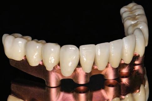
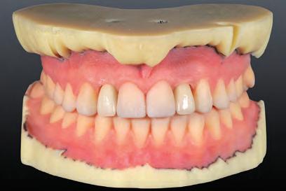
Fig. 52: CAD/CAM (milled) anodized titanium framework and zirconia crowns Fig. 53: Implant-supported complete fixed dental prosthesis (titanium framework and individual monolithic zirconia crowns)
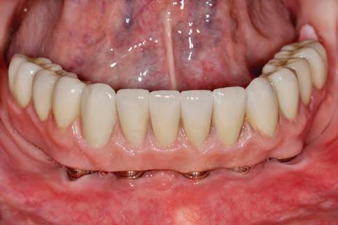
articulated analog casts fabricated from stone or printed or milled to facilitate or refine the finishing process inclusive of contouring, denture tooth arrangement/processing, adding veneering material, and surface characterization. A partial digital workflow may increase overall worktime when completing the prosthesis but remains a necessary phase in most cases in order to achieve satisfactory functional and esthetic outcomes for implant-supported/retained prostheses (Joda et al. 2017) (Figs 49–53).
References
Davies, T. G., Rahman, I. A., Lautenschlager, S., Cunningham, J. A., Asher, R. J., Barrett, P. M., ... & Braga, J. (2017). Open data and digital morphology. Proceedings of the Royal Society B: Biological Sciences 284: 20170194.
Grant, G.T, Campbell, S.D, Masri, R.M and Andersen, M.R. (2016) Glossary of Digital Dental Terms: American College of Prosthodontists. Journal of Prosthodontics 25 Suppl 2: S2–9.
Gallucci, G. O., Hamilton, A., Zhou, W., Buser, D., & Chen, S. (2018). Implant placement and loading protocols in partially edentulous patients: A systematic review. Clinical oral implants research 29: 106–134.
Joda, T. & Bragger, U. (2015) Time-efficiency analysis comparing digital and conventional work-flows for implant crowns: a prospective clinical crossover trial. The International Journal of Oral & Maxillofacial Implants 30: 1047–1053.
Joda, T., Ferrari, M., Gallucci, G.O., Wittneben, J.G. & Brägger, U. (2017) Digital technology in fixed implant prosthodontics. Periodontology 2000 73: 178–192.
Kachalia, P.R. & Geissberger, M.J. (2010) Dentistry a la carte: in-office CAD/CAM technology. Journal of the California Dental Association 38: 323–330.
Kapos, T. & Evans, C.A. (2014) CAD/CAM technology for implant abutments, crowns and superstructures. The International Journal of Oral & Maxillofacial Implants 29 Suppl: 117–136.
Katkar, R. A., Taft, R. M., & Grant, G. T. (2018) 3D Volume Rendering and 3D Printing (Additive Manufacturing). Dental Clinics of North America 62: 393–402.
Katsoulis, J., Mericske-Stern, R., Rotkina, L., Zbaren, C., Enkling, N. & Blatz, M.B. (2014) Precision of fit of implantsupported screw-retained 10-unit computer-aideddesigned and computer- aided-manufactured frameworks made from zirconium dioxide and titanium: an in vitro study. Clinical Oral Implants Research 25: 165–174.
Kau, C. H. (2011). Creation of the virtual patient for the study of facial morphology. Facial Plastic Surgery Clinics 19: 615–622.
Kuric, K. M., Harris, B. T., Morton, D., Azevedo, B., & Lin, W. S. (2018). Integrating hinge axis approximation and the virtual facial simulation of prosthetic outcomes for treatment with CAD/CAM immediate dentures: a clinical report of a patient with microstomia. The Journal of Prosthetic Dentistry 119: 879–886.
Martinez-Rus, F., Ferreiroa, A., Ozcan, M. & Pradies, G. (2013) Marginal discrepancy of mono-lithic and
MORTON / POLIDO / LIN
veneered all-ceramic crowns on titanium and zirconia implant abutments before and after adhesive cementation: a scanning electron microscopy analysis. The International Journal of Oral & Maxillofacial Implants 28: 480–487.
Morton, D., Phasuk, K., Polido, W.D. & Lin, W.S. (2019) Consideration for Contemporary Implant Surgery. Dental Clinics of North America 63: 309–329.
Morton, D., Gallucci, G., Lin, W. S., Pjetursson, B., Polido, W., Roehling, S., ... & Braut, V. (2018) Group 2 ITI Consensus Report: Prosthodontics and implant dentistry. Clinical Oral Implants Research 29: 215–223.
Pjetursson, B. E., Valente, N. A., Strasding, M., Zwahlen, M., Liu, S., & Sailer, I. (2018) A systematic review of the survival and complication rates of zirconia-ceramic and metal-ceramic single crowns. Clinical Oral Implants Research 29: 199–214.
Rutkūnas, V., Gečiauskaitė, A., Jegelevičius, D. & Vaitiekūnas, M. (2017) Accuracy of digital implant impressions with intraoral scanners. A systematic review. European Journal of Oral Implantology 10 Suppl 1: 101–120.
Sailer, I., Strasding, M., Valente, N. A., Zwahlen, M., Liu, S., & Pjetursson, B. E. (2018). A systematic review of the survival and complication rates of zirconia-ceramic and metal-ceramic multiple-unit fixed dental prostheses. Clinical Oral Implants Research 29: 184–198.
Van Noort, R. (2012) The future of dental devices is digital. Dental Materials 28: 3–12.
Wismeijer, D., Mans, R., van Genuchten, M. & Reijers, H.A. (2014) Patients’ preferences when comparing analogue implant impressions using a polyether impression material versus digital impressions (Intraoral Scan) of dental implants. Clinical Oral Implants Research 25: 1113–1118
Reprinted with permission by Forum Implantologicum. Morton D, Polido W, Lin W-S. Current state of CAD/CAM technology in implant dentistry. Forum Implantologicum Vol 15 Issue 2/2019:119-125


