CONTENTS
1 Defining Aging

CONTENTS
1 Defining Aging
Aging can be defined and categorized in several ways, including chronological age and biological aging. Biological aging refers to the accumulation of molecular and cellular damage, such as mitochondrial dysfunction over time (Table 1).1 These changes are sometimes referred to as age-associated cellular decline (AACD). Interventions that slow the accumulation or facilitate the repair of factors
Table 1. Biological Features of Aging
Features Description
Genomic instability
Telomere attrition
Epigenetic alterations
2 Mitochondria
3 Age-Associated Cellular Decline
5 Lifestyle Interventions and Healthy Aging
6 Nutritional Modulation of Cellular Function, Aging, and Longevity
7 Nutritional Factors and Compounds That Address Mitochondrial Health
associated with biological aging have the potential to extend human healthspan— the period of life in which people are in good health and free from the chronic diseases and common disabilities of aging.
9 Summary
The increased tendency for DNA mutations and other genetic changes to occur during cell division
The gradual loss of the protective caps of chromosomes, limiting the number of times that cells can divide
A heritable change that does not affect the DNA sequence but results in a change in gene expression; examples include promoter methylation and histone modifications
Loss of proteostasis The failure of the protein building machinery of the cell, and the accumulation of misfolded proteins and damaged proteins due to impaired autophagy
Deregulated nutrient Alterations in the capacity of the body to sense and respond to the ebb and flow of sensing nutrient levels
Mitochondrial dysfunction Loss of function in mitochondria, the organelle responsible for cellular energy production
Cellular senescence A condition in which a cell no longer can proliferate
Stem cell exhaustion
Age-related deficiency of stem cells
Altered intercellular Change in signals between cells communication
Source: Reference 1.
Sai Krupa Das, PhD Scientist I
Energy Metabolism Team
Jean Mayer USDA Human Nutrition Research Center on Aging
Associate Professor Friedman School of Nutrition Science and Policy Tufts University
Roger A. Fielding, PhD
Associate Director
Jean Mayer USDA Human Nutrition Research Center on Aging
Lead Scientist and Senior Scientist
Nutrition, Exercise Physiology, and Sarcopenia Team
Professor of Nutrition
Friedman School of Nutrition Science and Policy Tufts University
Professor of Medicine
Tufts University School of Medicine
Associate Director Boston Claude D. Pepper Older Americans Independence Center
Nathan K. LeBrasseur, PT, PhD
Professor and Co-Chair of Research Department of Physical Medicine and Rehabilitation
Scientific Director Office of Translation to Practice
Co-Director
Paul F. Glenn Center for Biology of Aging Research Mayo Clinic
Anthony J. A. Molina, PhD Vice Chief of Research Division of Geriatrics, Gerontology, and Palliative Care
Associate Professor Department of Medicine
University of California San Diego School of Medicine


Aging is the greatest risk factor for the majority of chronic diseases, including cancer, diabetes, cardiovascular disease, and neurodegenerative diseases; however, chronological age does not necessarily correlate with health, and some individuals remain healthy until late in life. Importantly, many chronic diseases are associated with molecular and cellular processes that are implicated in biological aging, including those involving mitochondria.1,2 Interventions intended to promote healthy aging, including both exercise and nutritional approaches, impact these cellular processes and may potentially improve healthspan.
Mitochondria are organelles that produce cellular energy (Figure 1).3 Oxygen is transported from the lungs to cells throughout the body by red blood cells. Mitochondria use this oxygen, combined with nutrients from food, to generate chemical energy in the form of adenosine triphosphate (ATP). Carbon dioxide is produced by this reaction and returned by the circulation to the lungs and exhaled. The energy produced by mitochondria provides more than 90% of the energy used to sustain life and support organ function.
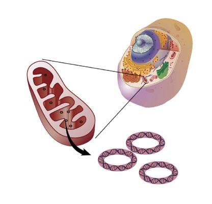
Source: Reference 3.
The energy produced by mitochondria provides more than 90% of the energy used to sustain life and support organ function.
The process of energy production by living systems is also sometimes referred to as bioenergetics. In this case, the term mitochondrial bioenergetics refers to how these organelles transform nutrients into chemical energy. The process that mitochondria use to convert energy from macronutrients to ATP is called oxidative phosphorylation, and forms the basis for mitochondrial respiration. Mitochondrial respirometry is often used to directly measure mitochondrial function and is related to multiple agerelated conditions.
Mitochondria are dynamic and can change size, shape, and position throughout their life cycle. They regularly undergo fission (division of a single mitochondrion into multiple mitochondria) or fusion (the combination of two or more mitochondria into a single mitochondrion).4 Mitochondrial homeostasis is maintained through a balance of fusion, fission, mitochondrial biogenesis (creation of new mitochondria), and mitophagy (the selective degradation of mitochondria).5 Together, these processes are referred to as mitochondrial dynamics, and serve to maintain the overall quality of a cell’s mitochondrial network.
Although the primary function of mitochondria is to produce energy for cells, they also have several other important functions, including regulating cellular metabolism, regulating apoptosis (programmed cell death), and signaling by producing reactive oxygen species (ROS) as a product of respiration. ROS are highly reactive molecules derived from oxygen that are key to many biochemical reactions; however, when present in excess, they can result in molecular damage. Mitochondria also have their own DNA (mtDNA) that encode for 13 proteins that are components of the respiratory chain and can develop mutations/deletions as a result of oxidative stress.
Abnormalities in the function of mitochondria are associated with many diseases, including cancer, cardiovascular diseases, and neurodegenerative diseases. Therefore, drivers of mitochondrial dysfunction are promising targets for addressing multiple age-related conditions with a single intervention.

Declines in mitochondrial function and metabolism are among the key components of AACD. AACD is systemic across all tissues but may be more evident in tissues with high metabolic demand (e.g., skeletal and cardiac muscle, central nervous system tissue). AACD is often associated with fatigue, reduced strength and daily energy, and low physical activity.6,7
Mitochondrial decline also can contribute to other forms of AACD, including cellular senescence, chronic inflammation, and the age-dependent decline in stem cell activity (Figure 2).7 Changes include a decrease in mitochondrial biogenesis, an increase in mitochondria-mediated apoptosis, and a decline in mtDNA, accompanied by increased mtDNA mutations.8 Specific molecular changes associated with mitochondrial decline include increased ROS and decreased glutathione and nicotinamide adenine dinucleotide (NAD+).
A number of physical and environmental risk factors can have a marked impact on trajectories of aging and rates of cellular damage accumulation, and have been associated with accelerated AACD (Table 2).9 Among these, the most important factors include smoking, obesity, sedentary lifestyles, physical or psychological stress, chronic disease, and an unfavorable genetic background.9 Clinical indicators for identifying AACD include fatigue, low quality of sleep, low mood, lack of motivation, subjective memory complaints, and poor exercise tolerance.9

Source: Adapted from Reference 7.
mtDNA mutations/Bioenergetics
In ammation
Stem cell function
Cellular senescence
Mitophagy and proteolysis
According to the mitochondrial theory of aging, damage to mitochondria and mtDNA contributes to aging by reducing the amount of energy available to the cell. Excessive ROS also can contribute to mitochondrial damage. ROS are vital to regulating a variety of cellular processes; excessive ROS can damage other cellular molecules, including DNA, proteins, and lipids.10 Homeostasis to balance the formation of free radicals and their elimination by antioxidants is normally tightly controlled and an imbalance can lead to excess ROS and oxidative stress.10 Excessive ROS and oxidative stress have been linked to several diseases, including cancer, cardiovascular disease, metabolic disease, Alzheimer’s disease, Parkinson’s disease, renal disease, and blood disorders.10
Table 2. Risk Factors Associated With Accelerated Age-Associated Cellular Decline
Clinical Risk Factors
• Clinical conditions
(e.g., cancer, cardiovascular, renal, or metabolic disease)
• Obesity
• Unfavorable genetic background
• Insulin resistance
• Low physical capacity
(e.g., slow gait speed, muscle weakness)
Source: Reference 9.
Behavioral/Environmental Risk Factors
• Smoking
• Sedentary lifestyle
• Low physical activity
• Persistent physical or psychological stress
• Low socioeconomic status
• Alcohol abuse
• Inadequate nutrition
• Air pollution
Endogenous antioxidants play a key role in protecting mitochondria against oxidative stress. For example, aging is associated with decreasing levels of the antioxidant glutathione. Acute glutathione deficiency in mitochondria results in mitochondrial damage or cell death.11 Chronic glutathione deficiency can contribute to the development of clinical conditions associated with aging, such as insulin resistance. Correcting glutathione deficiency by providing dietary supplementation with the amino acid precursors glycine and cysteine (which are converted by the body to glutathione after ingestion) has been suggested as a nutritional strategy to combat insulin resistance, obesity, and hepatic fat accumulation, and to replenish glutathione and improve mitochondrial function.11,12
NAD+ is a coenzyme that plays many beneficial roles in muscle development and homeostasis.13 Importantly, NAD+ is involved in reactions that produce ROS and is necessary for ATP production.14 It also acts as a signaling molecule that directs cells to increase energy production and utilization.15,16 NAD+ boosts cellular repair, coordinates circadian rhythms,15,16 and regulates intracellular processes such as immune function, DNA repair, telomere maintenance, and epigenetic regulation of gene expression.14 It is additionally involved in mitochondrial biogenesis13 and counters the age-associated decline in mitophagy that contributes to worsening of age-related disorders.17
Depletion of NAD+ results in reduced ATP production and apoptosis.14 NAD+ levels steadily decline as adults age; levels found in middle-aged adults are approximately half of levels in younger individuals.15 This decline is believed to be due to imbalances in NAD+ biosynthetic pathways and degradation pathways, which may be attenuated with nutritional and exercise interventions as well as dietary supplementation with NAD+ precursors.
Although life expectancy has increased dramatically over the past century, healthspan has not improved to the same extent.2 Instead, aging remains a primary risk factor for many chronic diseases as well as progressive loss of function and increased vulnerability to negative healthrelated outcomes across multiple disease states.6 Thus, interventions designed to address aging have explored the impact on both lifespan and healthspan.
Evidence suggests that changes associated with AACD act as triggers for age-associated diseases and conditions.18 For example, the age-related decline in NAD+ has been associated with the development of diabetes, nonalcoholic fatty liver disease, atherosclerosis, Alzheimer’s disease, retinal degeneration, chronic fatigue syndrome, and depression.18,19 The development of these diseases may be partially mediated through sirtuins, which are signaling proteins that act as downstream mediators of NAD+. Conversely, research has shown that NAD+ intermediates, such as nicotinamide mononucleotide and nicotinamide riboside, may potentially be effective for preventing and treating age-associated pathophysiology.18
The changes associated with AACD are also associated with several measures of declining function. Reduced NAD+ levels are associated with feeling tired faster, increased fatigue, slowed metabolism, and declining mental function.19,20 Glutathione levels are lower and oxidative stress is higher in conditions associated with mitochondrial dysfunction, including aging, HIV infection, diabetes, neurodegenerative disorders, cardiovascular disorders, neurometabolic diseases, cancer, and obesity.
Declining strength and increased fatigue are often physical effects of aging that precede cognitive decline and physical disability. Gait speed, which is correlated with systemic bioenergetic capacity, is a measure of physical function that is highly predictive of morbidity, disability, and mortality.21,22 Gait speed is associated with altered muscle mitochondrial bioenergetics in older adults, demonstrating the role of mitochondrial metabolism in physical functioning.21,22 In addition, pathological agerelated muscle wasting and weakness, known as sarcopenia, is associated with reduced muscular metabolism, including mitochondrial dysfunction, reduced oxidative capacity, and reduced NAD+ biosynthesis.23
Research has shown that behavioral factors and lifestyle interventions including exercise and healthy nutrition are important for healthy aging. Exercise can prevent or delay cardiovascular disease, diabetes, osteoporosis, sarcopenia, and depression and extends healthspan by reducing mortality from chronic diseases associated with aging.2 Even without weight loss, exercise has positive metabolic effects in adults with obesity.2 Resistance training in particular is key to preventing and reversing sarcopenia. In addition, exercise has been shown to extend the length of time older adults are able to live independently and support healthy aging by reducing frailty and mobility disability.2,24,25
Common molecular factors that appear to be associated with the beneficial effects of exercise on healthspan involve improved mitochondrial function, including stimulation of mitochondrial biogenesis and improved
oxidative capacity.2,8 Exercise also activates signaling to increase capillary density and, in turn, delivery of oxygen and nutrients to muscle.
Dietary patterns composed of energy-dense, nutrient-poor foods combined with a sedentary lifestyle have a dramatic impact on the development of age-related diseases.2 Conversely, some dietary patterns and nutrients have been found to have important roles in supporting cellular function and promoting active and healthy aging.
Evidence shows that weight management, healthy dietary patterns (e.g., Mediterranean, plant-based, DASH [Dietary Approaches to Stop Hypertension]), and diets high in specific nutrients (e.g., protein; calcium; vitamins C, D, and E; lutein; zeaxanthin; zinc; copper; folic acid; wheat bran fiber) are associated with reduced risk for a wide range of age-related conditions and disease states (e.g., sarcopenia, cognitive decline and dementia, osteoporosis, age-related macular degeneration, diabetic retinopathy, hearing loss, obstructive sleep apnea, urinary incontinence,


constipation).26,27 Nutrient-dense, plant-based diets that have relatively low glycemic loads have been recommended to meet the nutritional needs of older adults.26 Further, disease-specific nutritional interventions can reverse or slow the progression of many of these conditions.26
Nutritional interventions that modulate cellular function may impact AACD and longevity.
Calorie restriction (CR) and protein restriction are dietary interventions that have been shown to increase longevity across a wide range of species.28,29 CR involves reducing caloric intake while maintaining adequate nutrition. In multiple animal models, reduction of caloric intake by 20% to 50% extends both average and maximal lifespan.29 Moreover, CR reduces the incidence of cancer and slows the progression of neurodegenerative disorders, cardiovascular diseases, and metabolic syndrome.29
Longevity-promoting effects of CR affect multiple cellular pathways that regulate metabolism, oxidative stress, inflammation, and autophagy. In particular, CR improves mitochondrial metabolism, supports ATP production, and reduces ROS production and mtDNA mutations.30 In addition, CR may activate sirtuins and AMP-activated protein kinase, inhibit insulin-like growth factor 1, and inhibit the mechanistic target of rapamycin (mTOR).29 Overall, these changes reduce the rate of cellular aging by increasing NAD+ formation, leading to enhanced NAD+ levels in multiple tissues.20 CR also reduces cellular senescence by both preventing intracellular damage and promoting repair of existing damage and has been shown to improve muscle mitochondrial function, specifically the rate of ATP synthesis, in some but not all individuals.31
Additional research in animals indicates that restricting protein intake may extend lifespan by modulating the mTOR pathway. Low-carbohydrate ketogenic diets also were shown to extend lifespan and slow age-related decline in physiological function in mice.26,32
Due to practical and ethical limitations, there are limited data on the long-term benefits of CR or other restrictive dietary approaches in humans. Further, the extent of CR in animal models may not be realistic for most people.
In humans, a 2-year randomized controlled trial known as CALERIE (Comprehensive Assessment of Long-term Effects of Reducing Intake of Energy) found that an average of 12% CR is feasible in generally healthy humans without obesity, and associated with reduced oxidative stress, attenuated biological aging, and improvements in markers of healthspan.33-35 Promising alternatives to CR in humans include various forms of intermittent fasting such as time-restricted eating, alternate day fasting, and the 5:2 fast with 5 days of the week of normal intake and 2 fast days, all of which have been shown to improve markers of healthspan.36 Additionally, research is underway to develop CR mimetics—natural or synthetic molecules that mimic the effects of CR on cellular pathways without the need to alter food intake.29
Of note, CR combined with exercise does have some beneficial synergistic effects, such as improving insulin sensitivity, lowering systemic inflammation, and improving body composition.2,37 However, exercise may not provide an additive benefit for longevity when added to CR.2
Emerging research indicates that some nutritional compounds can support healthy aging by influencing mitochondrial repair and preservation, quality control, and signaling.7 In addition, research is being conducted on other small molecules (e.g., Szeto-Schiller [SS] peptides) and biologics that can be used to target mitochondria in different therapeutic indications.
Therapeutics for Mitochondrial Repair and Preservation
Examples of emerging compounds that have been shown to address mitochondrial damage and clinical disease states include SS peptides, coenzyme Q10 (CoQ10), MitoQ, and glycine and N-acetylcysteine (GlyNAC).
SS peptides (e.g., SS31, elamipretide) target the delivery of antioxidants to the inner mitochondrial membrane and help maintain mitochondrial structure and bioenergetics.38 These peptides reduce mitochondrial ROS and prevent oxidant-induced cell death.38 Preclinical studies have found benefits for the use of SS peptides for ischemiareperfusion injury and neurodegenerative disorders.39
CoQ10 is an essential electron and proton carrier in the mitochondrial respiratory chain that is widely available as a dietary supplement. In animal models, CoQ10 has been shown to have beneficial effects in several neurodegenerative diseases, including amyotrophic lateral sclerosis, Huntington’s disease, and Parkinson’s disease.40
MitoQ is a mitochondria-targeted antioxidant that has shown positive effects in animal models. In combination with SS31, MitoQ has been shown to reduce free radicals and increase ATP production in defective neurons from patients with Huntington’s disease. Researchers have postulated that both SS31 and MitoQ could be potential therapeutic targets in treating Huntington’s disease.41
GlyNAC is a precursor of glutathione.12 Because glutathione protects cells from damage due to oxidative stress, it is critical for maintaining mitochondrial health and healthy immune function, especially after middle age. Chronic glutathione deficiency results in the additional development of insulin resistance, which is reversed if glutathione levels are restored. GlyNAC supplementation appears to improve several markers of aging in humans, including improvements in mitochondrial dysfunction, inflammation, insulin resistance, and genomic damage. In older adults, GlyNAC supplementation has been shown to improve cellular protection, mitochondrial energy metabolism, physical strength, physical function, and cognitive health.12
Because glutathione protects cells from damage due to oxidative stress, it is critical for maintaining mitochondrial health and healthy immune function, especially after middle age.
In one study, GlyNAC supplementation for 24 weeks in older adults corrected glutathione deficiency in red blood cells, oxidative stress, and mitochondrial dysfunction; improved inflammation, endothelial dysfunction, insulin resistance, genomic damage, cognition, strength, gait speed, and exercise capacity; and lowered body fat and waist circumference. However, benefits declined after GlyNAC supplementation was discontinued.12
Compounds that may address mitochondrial quality control include sirtuins, mitochondrial division inhibitor (mdivi), urolithin A, and epicatechin.
Mitochondrial quality control involves a variety of mechanisms, including regulation by sirtuins. Sirtuins improve mitochondrial function, biogenesis, and fission/fusion balances involved in multiple cellular processes, including the regulation of mitochondrial function, oxidative stress, inflammation, and autophagy. Sirtuins have been identified as potential therapeutic targets for type 2 diabetes and diabetic renal disease.42
Mdivi inhibits mitochondrial fission, improves mitochondrial dysfunction and can reverse mitochondria-induced apoptosis. Mdivi has been postulated to have therapeutic potential for ischemic brain injury and Alzheimer’s disease.43
Mitochondrial quality control mechanisms also have been identified as targets for patients with heart failure.44 Urolithin A is a natural dietary metabolite produced from bacterial transformation of ellagitannins (polyphenols in foods such as berries and nuts). It stimulates mitophagy, which can facilitate cell regeneration. Urolithin A improves muscle health in animal and preclinical models of aging and stimulates mitochondrial biogenesis in the skeletal muscle of humans.45 Thus, it may improve skeletal muscle function, leading to improved mobility and extended independence in older adults.45 A clinical trial found that a 4-week regimen of urolithin A at 500 mg or 1,000 mg improved mitochondrial and cellular health in older adults who were healthy and sedentary.45
Epicatechin is a dietary flavonoid that has been associated with improvements in mitochondria in muscles and neurons. Chronic supplementation with epicatechin has been shown to increase mitochondrial biogenesis in human muscle tissue. Thus, it has been hypothesized that epicatechin supplementation may be beneficial for the treatment of diseases associated with mitochondrial dysfunction. The greatest benefits may be seen in sedentary patients.46
Nutritional compounds that have been shown to address mitochondrial signaling include nicotinamide riboside and nicotinamide mononucleotide.
Mdivi inhibits mitochondrial fission, improves mitochondrial dysfunction and can reverse mitochondria-induced apoptosis.
Nicotinamide riboside is an NAD+ precursor, and oral supplementation with nicotinamide riboside increases blood concentrations of NAD+ . 47 Preclinical research has found that raising NAD+ levels may help reverse signs of aging, improve cognition, and lower the risk of many chronic diseases.48,49
Nicotinamide mononucleotide is an intermediate of NAD+ biosynthesis. Preclinical studies of nicotinamide mononucleotide have found that it may have cardioprotective effects against ischemia for ischemia reperfusion injuries, diabetes, Alzheimer’s disease, and complications of obesity.50 In addition, nicotinamide mononucleotide supplementation is promising as a therapeutic strategy to support NAD+ function to address pathophysiologic processes associated with aging, including abnormal activity of sirtuins, mitochondrial ROS production, and oxidative stress responses.51
Researchers have identified several molecular pathways and cellular processes that appear to underlie both aging and age-related chronic disease. Cellular changes associated with aging are cumulatively referred to as AACD and include defects in mitochondrial function. Emerging research indicates that certain nutritional factors may influence AACD processes.
Adoption of healthful eating patterns and exercise has been shown to improve markers of age-associated diseases and attenuate biological aging. CR appears to improve markers of disease risk in humans, but its acceptability and feasibility particularly over the long term remains a challenge. Further, the duration, extent, feasibility, and acceptability of maintaining altered dietary patterns over time must be considered when making recommendations for dietary modifications. Emerging research indicates that nutritional components that target specific mechanisms associated with AACD hold promise for improving the health and well-being of adults. Dietary supplementation with these components may be an alternative approach to lifestyle interventions targeting AACD. Further, identifying AACD risk factors and intervening with cellular nutrients earlier in the aging process, before major mobility disabilities and disease-driven limitations emerge, could help improve overall healthy aging.
1. López-Otín C, Blasco MA, Partridge L, et al. The hallmarks of aging. Cell. 2013;153(6):1194-1217. doi: 10.1016/j.cell.2013.05.039
2. de Cabo R, Carmona-Gutierrez D, Bernier M, et al. The search for antiaging interventions: from elixirs to fasting regimens. Cell. 2014;157(7):1515-1526. doi: 10.1016/j.cell.2014.05.031
3. National Human Genome Research Institute. Mitochondrial DNA. Accessed October 15, 2021. https://www.genome.gov/ genetics-glossary/Mitochondrial-DNA
4. Scott I, Youle RJ. Mitochondrial fission and fusion. Essays Biochem. 2010;47:85-98. doi: 10.1042/bse0470085
5. Wu NN, Zhang Y, Ren J. Mitophagy, mitochondrial dynamics, and homeostasis in cardiovascular aging. Oxid Med Cell Longev. 2019;2019:9825061. doi: 10.1155/2019/9825061
6. Guralnik JM, Feige JN, Singh A, Fielding RA. Nutritional mediators of cellular decline and mitochondrial dysfunction in older adults. Geriatrics. 2021;6(2):37. doi: 10.3390/geriatrics6020037
7. Sun N, Youle RJ, Finkel T. The mitochondrial basis of aging. Mol Cell. 2016;61(5):654-666. doi: 10.1016/j.molcel.2016.01.028
8. Peterson CM, Johannsen DL, Ravussin E. Skeletal muscle mitochondria and aging: a review. J Aging Res. 2012;2012:194821. doi: 10.1155/2012/194821
9. Cesari M, Cherubini A, Guralnik JM, et al. Early detection of accelerated aging and cellular decline (AACD): a consensus statement. Exp Gerontol. 2021;146:111242. doi: 10.1016/j.exger.2021.111242
10. Valko M, Jomova K, Rhodes CJ, et al. Redox- and nonredox-metal-induced formation of free radicals and their role in human disease. Arch Toxicol. 2016;90(1):1-37. doi: 10.1007/s00204-015-1579-5
11. Nguyen D, Samson SL, Reddy VT, et al. Impaired mitochondrial fatty acid oxidation and insulin resistance in aging: novel protective role of glutathione. Aging Cell. 2013;12(3):415-425. doi: 10.1111/acel.12073
12. Kumar P, Liu C, Hsu JW, et al. Glycine and N -acetylcysteine (GlyNAC) supplementation in older adults improves glutathione deficiency, oxidative stress, mitochondrial dysfunction, inflammation, insulin resistance, endothelial dysfunction, genotoxicity, muscle strength, and cognition: results of a pilot clinical trial. Clin Transl Med. 2021;11(3):e372. doi: 10.1002/ctm2.372
13. Goody MF, Henry CA. A need for NAD + in muscle development, homeostasis, and aging. Skelet Muscle. 2018;8(1):9. doi: 10.1186/s13395-018-0154-1
14. Clement J, Wong M, Poljak A, et al. The plasma NAD + metabolome is dysregulated in “normal” aging. Rejuvenation Res. 2019;22(2):121-130. doi: 10.1089/rej.2018.2077
15. Schultz MB, Sinclair DA. Why NAD + declines during aging: it’s destroyed. Cell Metab. 2016;23(6):965-966. doi: 10.1016/j.cmet.2016.05.022
16. Imai S, Guarente L. NAD + and sirtuins in aging and disease. Trends Cell Biol. 2014;24(8):464-471. doi: 10.1016/j.tcb.2014.04.002
17. Babbar M, Basu S, Yang B, et al. Mitophagy and DNA damage signaling in human aging. Mech Ageing Dev. 2020;186:111207. doi: 10.1016/j.mad.2020.111207
18. Johnson S, Imai SI. NAD + biosynthesis, aging, and disease. F1000Res. 2018;7:132. doi: 10.12688/f1000research.12120.1
19. Castro-Marrero J, Cordero MD, Segundo MJ, et al. Does oral coenzyme Q10 plus NADH supplementation improve fatigue and biochemical parameters in chronic fatigue syndrome? Antioxid Redox Signal. 2015;22(8):679-685. doi: 10.1089/ars.2014.6181
20. Stein LR, Imai S. The dynamic regulation of NAD metabolism in mitochondria. Trends Endocrinol Metab. 2012;23(9):420-428. doi: 10.1016/j.tem.2012.06.005
21. Tyrrell DJ, Bharadwaj MS, Van Horn CG, et al. Respirometric profiling of muscle mitochondria and blood cells are associated with differences in gait speed among community-dwelling older adults. J Gerontol A Biol Sci Med Sci. 2015;70(11):1394-1399. doi: 10.1093/gerona/glu096
22. Braganza A, Corey CG, Santanasto AJ, et al. Platelet bioenergetics correlate with muscle energetics and are altered in older adults. JCI Insight. 2019;5(13):e128248. doi: 10.1172/jci.insight.128248
23. Migliavacca E, Tay SKH, Patel HP, et al. Mitochondrial oxidative capacity and NAD + biosynthesis are reduced in human sarcopenia across ethnicities. Nat Commun. 2019;10(1):5808. doi: 10.1038/s41467-019-13694-1
24. Pahor M, Guralnik JM, Ambrosius WT, et al.; LIFE study investigators. Effect of structured physical activity on prevention of major mobility disability in older adults: the LIFE study randomized clinical trial. JAMA. 2014;311(23):2387-2396. doi: 10.1001/jama.2014.5616
25. Trombetti A, Hars M, Hsu FC, et al.; LIFE study investigators. Effect of physical activity on frailty: secondary analysis of a randomized controlled trial. Ann Intern Med. 2018;168(5):309-316. doi: 10.7326/M16-2011
26. Roberts MN, Wallace MA, Tomilov AA, et al. A ketogenic diet extends longevity and healthspan in adult mice. Cell Metab. 2017;26(3):539-546.e5. doi: 10.1016/j.cmet.2017.08.005
27. Gonzalez-Armenta JL, Gao Z, Appt SE, et al. Skeletal muscle mitochondrial respiration is elevated in female cynomolgus macaques fed a western compared with a Mediterranean diet. J Nutr. 2019;149(9):1493-1502. doi: 10.1093/jn/nxz092
28. Ros M, Carrascosa JM. Current nutritional and pharmacological anti-aging interventions. Biochim Biophys Acta Mol Basis Dis. 2020;1866(3):165612. doi: 10.1016/j.bbadis.2019.165612
29. Maduro AT, Luís C, Soares R. Ageing, cellular senescence and the impact of diet: an overview. Porto Biomed J. 2021;6(1):e120. doi: 10.1097/j.pbj.0000000000000120
30. Ramsey JJ, Harper ME, Weindruch R. Restriction of energy intake, energy expenditure, and aging. Free Radic Biol Med. 2000;29(10):946-968. doi: 10.1016/s0891-5849(00)00417-2
31. Sparks LM, Redman LM, Conley KE, et al. Effects of 12 months of caloric restriction on muscle mitochondrial function in healthy individuals. J Clin Endocrinol Metab. 2017;102(1):111-121. doi: 10.1210/jc.2016-3211
32. Hill CM, Kaeberlein M. An anti-ageing mechanism for protein restriction. Nature. 2021;589(7842):357-358. doi: 10.1038/d41586-020-03662-x
33. Ravussin E, Redman LM, Rochon J, et al.; CALERIE study group. A 2-year randomized controlled trial of human caloric restriction: feasibility and effects on predictors of health span and longevity. J Gerontol A Biol Sci Med Sci. 2015;70(9):1097-1104. doi: 10.1093/gerona/glv057
34. Il’yasova D, Fontana L, Bhapkar M, et al.; CALERIE study investigators. Effects of 2 years of caloric restriction on oxidative status assessed by urinary F2-isoprostanes: the CALERIE 2 randomized clinical trial. Aging Cell. 2018;17(2):e12719. doi: 10.1111/acel.12719
35. Das SK, Balasubramanian P, Weerasekara YK. Nutrition modulation of human aging: the calorie restriction paradigm. Mol Cell Endocrinol. 2017;455:148-157. doi: 10.1016/j.mce.2017.04.011
36. Fontana L, Partridge L. Promoting health and longevity through diet: from model organisms to humans. Cell. 2015;161(1):106-118. doi: 10.1016/j.cell.2015.02.020
37. Nicklas BJ, Chmelo E, Delbono O, et al. Effects of resistance training with and without caloric restriction on physical function and mobility in overweight and obese older adults: a randomized controlled trial. Am J Clin Nutr. 2015;101(5):991-999. doi: 10.3945/ajcn.114.105270
38. Szeto HH. First-in-class cardiolipin-protective compound as a therapeutic agent to restore mitochondrial bioenergetics. Br J Pharmacol. 2014;171(8):2029-2050. doi: 10.1111/bph.12461
39. Rocha M, Hernandez-Mijares A, Garcia-Malpartida K, et al. Mitochondria-targeted antioxidant peptides. Curr Pharm Des. 2010;16(28):3124-3131. doi: 10.2174/138161210793292519
40. Dombi E, Mortiboys H, Poulton J. Modulating mitophagy in mitochondrial disease. Curr Med Chem. 2018;25(40):5597-5612. doi: 10.2174/0929867324666170616101741
41. Yin X, Manczak M, Reddy PH. Mitochondria-targeted molecules MitoQ and SS31 reduce mutant huntingtin-induced mitochondrial toxicity and synaptic damage in Huntington’s disease. Hum Mol Genet. 2016;25(9):1739-1753. doi: 10.1093/hmg/ddw045
42. Xu J, Kitada M, Koya D. The impact of mitochondrial quality control by sirtuins on the treatment of type 2 diabetes and diabetic kidney disease. Biochim Biophys Acta Mol Basis Dis. 2020;1866(6):165756. doi: 10.1016/j.bbadis.2020.165756
43. Ruiz A, Alberdi E, Matute C. Mitochondrial division inhibitor 1 (mdivi-1) protects neurons against excitotoxicity through the modulation of mitochondrial function and intracellular Ca2+ signaling. F ront Mol Neurosci. 2018;11:3. doi: 10.3389/fnmol.2018.00003
44. Ong SB, Kwek XY, Katwadi K, et al. Targeting mitochondrial fission using mdivi-1 in a clinically relevant large animal model of acute myocardial infarction: a pilot study. Int J Mol Sci. 2 019;20(16):3972. doi: 10.3390/ijms20163972
45. Andreux PA, Blanco-Bose W, Ryu D, et al. The mitophagy activator urolithin A is safe and induces a molecular signature of improved mitochondrial and cellular health in humans. Nat Metab. 2019;1(6):595-603. doi: 10.1038/s42255-019-0073-4
46. Daussin FN, Heyman E, Burelle Y. Effects of (−)-epicatechin on mitochondria. Nutr Rev. 2021;79(1):25-41. doi: 10.1093/nutrit/nuaa094
47. Trammell SA, Schmidt MS, Weidemann BJ, et al. Nicotinamide riboside is uniquely and orally bioavailable in mice and humans. Nat Commun. 2016;7:12948. doi: 10.1038/ncomms12948
48. Gong B, Pan Y, Vempati P, et al. Nicotinamide riboside restores cognition through an upregulation of proliferator-activated receptor-γ coactivator 1α regulated β-secretase 1 degradation and mitochondrial gene expression in Alzheimer’s mouse models. Neurobiol Aging. 2013;34(6):1581-1588. doi: 10.1016/j.neurobiolaging.2012.12.005
49. Frederick DW, Loro E, Liu L, et al. Loss of NAD homeostasis leads to progressive and reversible degeneration of skeletal muscle. Cell Metab. 2016;24(2):269-282. doi: 10.1016/j.cmet.2016.07.005
50. Poddar SK, Sifat AE, Haque S, et al. Nicotinamide mononucleotide: exploration of diverse therapeutic applications of a potential molecule. Biomolecules. 2019;9(1):34. doi: 10.3390/biom9010034
51. Hong W, Mo F, Zhang Z, et al. Nicotinamide mononucleotide: a promising molecule for therapy of diverse diseases by targeting NAD + metabolism. Front Cell Dev Biol. 2020;8:246. doi: 10.3389/fcell.2020.00246

geron.org