ULTRASOUND ZOOM
Point-of-Care Ultrasound in Pulmonary Embolism By Andrea Alvarado, MD, PGY-2 Jackson Memorial Hospital
Are you concerned about your patient who is short of breath? Is your patient hypotensive, tachycardic and too unstable for the “donut of truth” (also known as the CT scanner)? I know—I have been there. Identifying the etiology of shock can be nerve-racking in unstable patients, and when it is pulmonary embolism (PE), it can be scary as a large percentage of patients end in sudden death. We as emergency physicians need to be empowered to diagnose and manage such patients at bedside in the emergency department, and one of the best ways to do that is with point-of-care ultrasound. Our strategy needs to be directed towards examining the heart, lungs and lower extremity veins. Read on for my technique and tips!
TECHNIQUE A. Cardiac Probe: Phased array probe
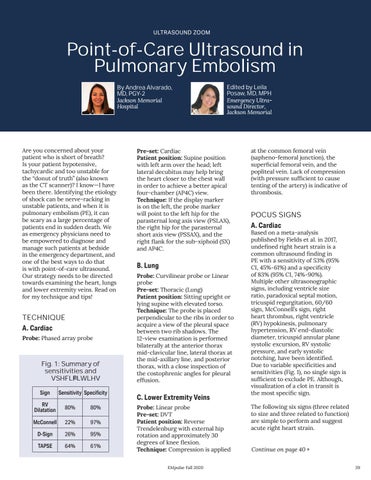
Fig. 1: Summary of sensitivities and specificities Sign
Sensitivity Specificity
RV Dilatation
80%
80%
McConnell
22%
97%
D-Sign
26%
95%
TAPSE
64%
61%
Edited by Leila Posaw, MD, MPH
Emergency Ultrasound Director, Jackson Memorial
Pre-set: Cardiac Patient position: Supine position with left arm over the head; left lateral decubitus may help bring the heart closer to the chest wall in order to achieve a better apical four-chamber (AP4C) view. Technique: If the display marker is on the left, the probe marker will point to the left hip for the parasternal long axis view (PSLAX), the right hip for the parasternal short axis view (PSSAX), and the right flank for the sub-xiphoid (SX) and AP4C.
B. Lung Probe: Curvilinear probe or Linear probe Pre-set: Thoracic (Lung) Patient position: Sitting upright or lying supine with elevated torso. Technique: The probe is placed perpendicular to the ribs in order to acquire a view of the pleural space between two rib shadows. The 12-view examination is performed bilaterally at the anterior thorax mid-clavicular line, lateral thorax at the mid-axillary line, and posterior thorax, with a close inspection of the costophrenic angles for pleural effusion.
C. Lower Extremity Veins Probe: Linear probe Pre-set: DVT Patient position: Reverse Trendelenburg with external hip rotation and approximately 30 degrees of knee flexion. Technique: Compression is applied EMpulse Fall 2020
at the common femoral vein (sapheno-femoral junction), the superficial femoral vein, and the popliteal vein. Lack of compression (with pressure sufficient to cause tenting of the artery) is indicative of thrombosis.
POCUS SIGNS A. Cardiac
Based on a meta-analysis published by Fields et al. in 2017, undefined right heart strain is a common ultrasound finding in PE with a sensitivity of 53% (95% CI, 45%-61%) and a specificity of 83% (95% CI, 74%-90%). Multiple other ultrasonographic signs, including ventricle size ratio, paradoxical septal motion, tricuspid regurgitation, 60/60 sign, McConnell’s sign, right heart thrombus, right ventricle (RV) hypokinesis, pulmonary hypertension, RV end-diastolic diameter, tricuspid annular plane systolic excursion, RV systolic pressure, and early systolic notching, have been identified. Due to variable specificities and sensitivities (Fig. 1), no single sign is sufficient to exclude PE. Although, visualization of a clot in transit is the most specific sign. The following six signs (three related to size and three related to function) are simple to perform and suggest acute right heart strain. Continue on page 40 » 39


