
10 minute read
Interview with Lynette Johns – Drug delivery in scleral lens wear


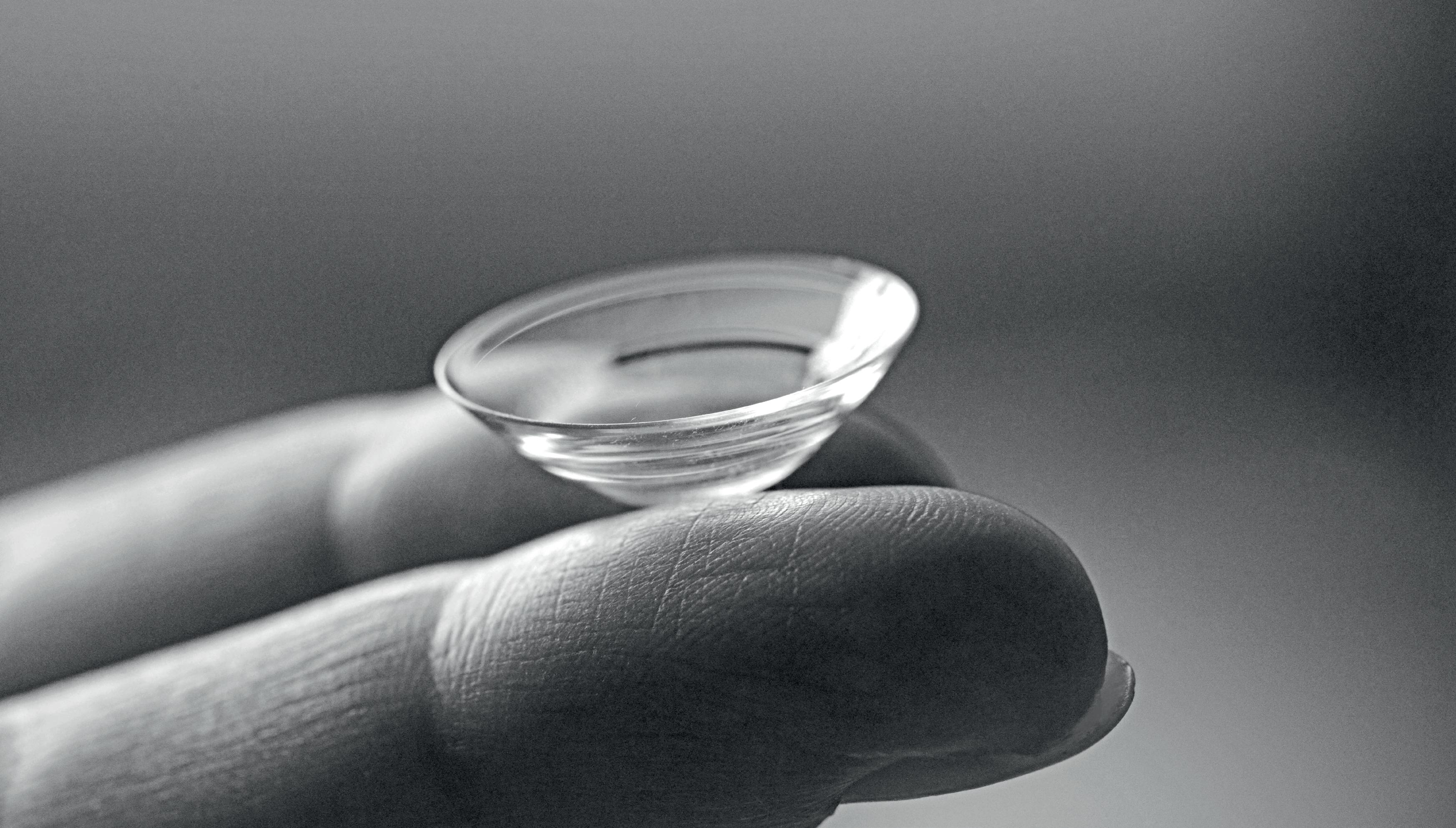
Drug delivery in scleral lens wear
Interview with Dr Lynette Johns, Harvard Medical School Department of Ophthalmology
Advertisement
Drug delivery in scleral lens wear is still a bit uncharted territory, although talk about this and pioneering work in the field date back at least a few decades. The areas of potential application could be prevention against corneal infection in case of corneal defects, and particularly in case of persistent epithelial defect healing. These defects can be very difficult to re-epithelialize, and it is important to use prophylactic antibiotics, to prevent corneal infection in these vulnerable eyes. Another area of potential application could be the use of medication that prevents neovasculasation in the cornea. These drugs can prevent new blood vessel growth in the cornea. Other areas such as glaucoma medication application are still under investigation. Here, like in general scleral lens fitting, it is the balance between the benefits and the costs (including risks etc.) that need to be considered. Here is an overview of what we know so far. By Silke Sage
GlobalCONTACT: Which drugs are generally suitable for the administration through scleral lenses? Lynette Johns: If there is an epithelial defect on the cornea, it may be wise to have an antibiotic present to prophylactically prevent against a corneal infection. One of the antibiotics we use is Moxifloxacin – that provides dual protection against gram-negative and gram-positive microorganisms. Moxifloxacin is a preservative-free ocular therapeutic, which makes it ideal for applying in the reservoir of a scleral lens. Preservatives of topical medications are toxic to the cornea with extended exposure times or dosing in drop form. When preservatives are used in a scleral lens, they can cause dense superficial corneal staining.
GlobalCONTACT: When are they applied? Lynette Johns: If there is a persistent epithelial defect, then this may be particularly useful. A persistent epithelial defect is a non-infectious focal loss of corneal epithelial cells. Often, they are caused by mechanical trauma, neurotrophic corneal disease, or post-surgically. Depending on the underlying condition, they can be very difficult to re-epithelialize. Using aggressive lubrication and bandage soft contact lenses is typically needed. It is important to use prophylactic antibiotics as described above, to
prevent corneal infection in these vulnerable eyes. In extreme longstanding cases the eyelids are sewn partially closed (tarsorrhaphy) in order to heal and resurface the cornea. To prevent this, scleral lenses have been used to resurface corneal epithelial defects. Drug delivery using scleral lenses was first suggested in a publication in 2000 by Dr. Perry Rosenthal and colleagues.[1] In this retrospective study, 14 eyes with persistent epithelial defects were treated with extended wear of scleral lenses and in some cases, there were prophylactic antibiotics placed in the reservoir of the lens with the saline. Unfortunately, there were four cases of microbial keratitis, but eight eyes completely resurfaced. Moxifloxacin was not used in this study. Later, this same group retrospectively reviewed 20 different cases with epithelial defects where Moxifloxacin was used in all cases, and 17 eyes resurfaced. There were no cases of microbial keratitis in theses 20 cases using moxifloxacin in the reservoir.2 I worked with Dr. Perry Rosenthal using extended wear of scleral lenses with prophylactic Moxifloxacin in the reservoir of the lens in many of these cases. It was remarkable to see how quickly and effectively this worked. The protocol for this treatment method was published by Ciralsky et al. in 2015.3

GlobalCONTACT: What is the requirement for these types of scleral lens fitting and how is the drug administered? Lynette Johns: Basically, a scleral lens is fit as optimally as possible, with the plan to have no scleral lens compression or impingement on the conjunctiva. The lens is filled with preservative-free saline and one drop of moxifloxacin and applied on the eye using the typical method for scleral lens application (head parallel to the floor). The lens is worn on an extended-wear basis for 24 hours and the eye is examined carefully the next day. The lens is removed, photos are taken for comparison, the epithelial defect is measured, and all signs of microbial keratitis are ruled out. The lens is disinfected, and the same process is repeated daily until the defect has resurfaced. In some cases, the epithelium is very fragile and can break down the first night the lens is not worn. We would have patients wear the lens an extra night with the antibiotic to allow the epithelium to “tack down”. In my experience, the majority of corneas would resurface within two weeks. If
the healing was slow, we would manufacture two lenses for the patient and train him/her on careful application and removal of the lens. When proficient we dispensed both lenses. They would wear them on a “12/12” schedule. Basically, they would wear a fresh lens with the antibiotic in the morning while the nighttime lens is disinfecting during the day, and then remove that lens at bedtime and put a fresh lens in with the antibiotic and then disinfect the daytime lens overnight. When epithelial defects healed, the patients would wear the lenses daily and lubricate aggressively at night. If their lids open at night while sleeping, we would have them tape the eye closed while they slept.
GlobalCONTACT: Are there other potential fields of application? Lynette Johns: Another medication used in the lens was topical 1% bevacizumab which is an anti-vascular endothelial growth factor (VEGF) medication. VEGF is a prominent factor that promotes neovascularization – new blood vessel growth in the cornea, and this drug can slow down the progression of that process. It has been injected into the eye to prevent retinal neovascularization and also injected into the conjunctiva against corneal blood vessel formation. It also had been used topically to prevent corneal neovascularization. I worked with Dr. Deborah Jacobs who was the first to use this medication in the lens for patients who demonstrated new blood vessel growth that was potentially sight-threatening. This use of 1% bevacizumab is off-label and carries potential risks of poor wound healing, corneal thinning, and ulceration of the cornea. To avoid systemic absorption, the patients had punctal occlusion. The drug needed to be compounded made-to-order and preservative-free – which is expensive. The lens was worn daily only, and patients removed the lens and reapplied the drop midday for a total of two days dosing in the lens. This treatment continued for three months and was continued based on the discretion of the corneal specialist.4,5 Long-term results ranging from six months to eleven years of 13 patients who used a course of bevacizumab in the lens were reviewed and approximately 90% of eyes had a reduction in corneal neovascularization and approximately 75% had an improvement in best corrected visual acuity.6
GlobalCONTACT: How do you make sure, the dosage is correct? Lynette Johns: In the previously described studies, one drop was placed in the reservoir with the saline solution. The problem with drug delivery in scleral lenses is dosing. The reservoir is different for each patient and the dilution would also be difficult to determine. It is unknown what percentage of the drug is ideal. Drug delivery with a scleral lens is a novel use of scleral lenses and is truly in its infancy. Practitioners should be cautious if delivering topical medications using this technique. In topical medications where drops are administered directly to the eye, most of the ocular medication is lost within 15-30 seconds after instillation.7 The tear turnover rate is approximately 16% per minute whereby all the drug disappears within ten minutes after dosing. Furthermore, the instilled drop is diluted by 1/3 of the original drop.8
GlobalCONTACT: When should scleral lenses vs eye drops be used? Lynette Johns: I would be very cautious considering scleral lenses for drug delivery at this time unless further research is conducted. Risk and benefits should be clearly delineated before attempting drug delivery versus topical therapies such as eye drops. In the presence of an epithelial defect that does not respond to conventional therapy, I would use a scleral lens to resurface the defect with moxifloxacin since it has been shown to be effective. Close monitoring of the patient is required and may involve daily monitoring which can be difficult depending on practice scheduling. The expense of compounding drops for use in scleral lenses may be cost-prohibitive for some patients. But of course this needs to be performed in a highly controlled, hospital setting – and its use may be restricted depending on what part of the world and what profession you are in.
GlobalCONTACT: Does this cause any problems? Lynette Johns: I think many people would like to consider using a scleral lens to deliver glaucoma medications; however, I think that’s premature at this time. I am concerned about the actual concentration of the drug in the reservoir of the lens. Would one drop of a conventional glaucoma medication be too strong for long-term exposure? Would it be ineffective and too diluted? We just don’t know those answers. Also, the volume of the actual reservoir of a vaulted scleral lens is unknown and changes with settling. I hope that this is researched to determine and titrate the appropriate concentration in the lens of all topical medications. Once it is carefully researched, this could be an effective pressure lowering treatment especially for noncompliant patients with glaucoma. To be clear, I am not advocating its use in this capacity at this time.
GlobalCONTACT: Is the application (over the surface of the cornea and limbus) evenly distributed by the scleral lenses? Or does it not matter? Lynette Johns: This is a great question, and something that would need further study. But the same question applies to eye drops potentially too.
GlobalCONTACT: Is the medication of the eye drops generally different from the administration via scleral lenses – is only the quantity lower? Or is a different concentration necessary? Lynette Johns: Most probably it will be different, but we have no clue at this point on how much and why.
GlobalCONTACT: Is the application for non-professionals doable? Or does the optometrist or ophthalmologist have to apply it? Lynette Johns: Patients have been able to apply the medications independently in the lens if they were proficient at application and removal. When using topical drugs in the lens, patients must be carefully watched by optometrists or ophthalmologists. Again, it depends on the licensing and regulations of each country. Even in the United States, therapeutic licensing for optometrists varies from state to state.
GlobalCONTACT: Could atropine also be administered in this way? And if so, what would be the advantages? Lynette Johns: Atropine is a very potent medication with significant side effects even with topical application. This most certainly requires further study before using in a lens on a patient.
GlobalCONTACT: Are there specifications for the lens material? Lynette Johns: There are no specifications regarding lens materials in my experience with either of these medications. In the cases of anti-VEGF treatment, the highest Dk materials were used to enhance the oxygen permeability because of the presence of corneal neovascularization.
GlobalCONTACT: Thank you very much for this Interview!
References: [1] Rosenthal P, Cotter JM, Baum J. Treatment of persistent corneal epithelial defect with extended wear of a fluid-ventilated gas-permeable scleral contact lens. Am J Ophthalmol. 2000 Jul;130(1):33-41. doi: 10.1016/s00029394(00)00379-2. PMID: 11004257. [2] Lim P, Ridges R, Jacobs DS, Rosenthal P. Treatment of persistent corneal epithelial defect with overnight wear of a prosthetic device for the ocular surface. Am J Ophthalmol. 2013 Dec;156(6):1095-101. doi: 10.1016/j. ajo.2013.06.006. Epub 2013 Sep 25. PMID: 24075432. [3] Ciralsky JB, Chapman KO, Rosenblatt MI, Sood P, Fernandez AG, Lee MN, Sippel KC. Treatment of Refractory Persistent Corneal Epithelial Defects: A Standardized Approach Using Continuous Wear PROSE Therapy. Ocul Immunol Inflamm. 2015 Jun;23(3):219-24. doi: 10.3109/09273948.2014.894084. Epub 2014 Mar 21. PMID: 24654929. [4] Lim M, Jacobs DS, Rosenthal P, Carrasquillo KG. The Boston Ocular Surface Prosthesis as a novel drug delivery system for bevacizumab. Semin Ophthalmol. 2009 May-Jun;24(3):149-55. doi: 10.1080/08820530902802013. PMID: 19437350. [5] Keating AM, Jacobs DS. Anti-VEGF Treatment of Corneal Neovascularization. Ocul Surf. 2011 Oct;9(4):227-37. doi: 10.1016/s1542-0124(11)70035-0. PMID: 22023817. [6] Yin J, Jacobs DS. Long-term outcome of using Prosthetic Replacement of Ocular Surface Ecosystem (PROSE) as a drug delivery system for bevacizumab in the treatment of corneal neovascularization. Ocul Surf. 2019 Jan;17(1):134141. doi: 10.1016/j.jtos.2018.11.008. Epub 2018 Nov 20. PMID: 30468876; PMCID: PMC6340761. [7] Shell JW. Pharmacokinetics of topically applied ophthalmic drugs. Surv Ophthalmol. 1982 Jan-Feb;26(4):207-18. doi: 10.1016/0039-6257(82)90081[8] Maurice D, Mishima S: Ocular pharmacokinetics. in SearsML (ed): Pharmacology of the Eye. New York, Springer-Verlag,1984, pp 19–116 [9] PMID: 7041308.
Many thanks to Eef van der Worp for his comments.
Dr. Lynette Johns is a research associate at Harvard medical school department of ophthalmology. She exclusively fits scleral lenses at Massachusetts Eye and Ear Infirmary. She is an author and co-editor with Melissa Barnett of the textbook: Contemporary Scleral Lenses: Theory and Application. She has published and speaks internationally on scleral and specialty contact lenses.


