MAZE Navigating the
Determining the right combination in cataract and glaucoma surgery

CATARACT & REFRACTIVE | CORNEA | RETINA | GLAUCOMA PAEDIATRIC OPHTHALMOLOGY June 2022 | Vol 27 Issue 5

Determining the right combination in cataract and glaucoma surgery
ESCRS’s vision is to educate and help our peers excel in our field. Together, we are driving the field of ophthalmology forward.

Publishers Therese Dolan
Publishers Therese Dolan Operations Director ESCRS
Operations Director ESCRS
Barbara Calderwood
Barbara Calderwood
Mark Wheeler
Mark Wheeler
Executive Editor
Executive Editor
Stuart Hales stuart.hales@eurotimes.org
Stuart Hales stuart.hales@eurotimes.org
Editor-in-Chief
Editor-in-Chief
Sean Henahan
Sean Henahan
Senior Content Editor
Senior Content Editor Kelsey Ingram
Kelsey Ingram
Design Director
Design Director
Kelsy McCarthy
Kelsy McCarthy
Designer Jen Basel
Designer Jen Basel
Circulation Manager
Circulation Manager
Vanessa McCourt
Vanessa McCourt
Contributing Editors
Contributing Editors
Cheryl Guttman Krader
Cheryl Guttman Krader
Howard Larkin
Howard Larkin
Dermot McGrath
Dermot McGrath Roibeárd O’hÉineacháin
Roibeárd O’hÉineacháin
Contributors
Contributors
Soosan Jacob
Leigh Spielberg
Clare Quigley
Gearóid Tuohy
Priscilla Lynch
Colour and Print
Soosan Jacob
W&G Baird Printers
Colour and Print
Advertising Sales
W&G Baird Printers
Roo Khan MCI UK
Advertising Sales
NAVIGATING THE MAZE

Tel: +44 203 530 0100 roo.khan@wearemci.com
Roo Khan MCI UK Tel: +44 203 530 0100 roo.khan@wearemci.com
Published by the European Society of Cataract and Refractive Surgeons, Temple House, Temple Road, Blackrock, Co Dublin, Ireland. No part of this publication may be reproduced without the permission of the executive editor. Letters to the editor and other unsolicited contributions are assumed intended for this publication and are subject to editorial review and acceptance.
Published by the European Society of Cataract and Refractive Surgeons, Temple House, Temple Road, Blackrock, Co Dublin, Ireland. No part of this publication may be reproduced without the permission of the executive editor.
Letters to the editor and other unsolicited contributions are assumed intended for this publication and are subject to editorial review and acceptance.
ESCRS EuroTimes is not responsible for statements made by any contributor. These contributions are presented for review and comment and not as a statement on the standard of care. Although all advertising material is expected to conform to ethical medical standards, acceptance does not imply endorsement by ESCRS EuroTimes ISSN 1393-8983
ESCRS EuroTimes is not responsible for statements made by any contributor. These contributions are presented for review and comment and not as a statement on the standard of care. Although all advertising material is expected to conform to ethical medical standards, acceptance does not imply endorsement by ESCRS EuroTimes ISSN 1393-8983


Correction: An editing error was made in the “Taming ‘Misbehaving’ Irises” article from the May 2022 issue. Instead of referring to “tamsulosin and other beta blockers,” the article should read “tamsulosin and other alpha blockers.”
As certified by ABC, the EuroTimes average net circulation for the 10
Included with this issue... Keys to Maximizing Outcomes for Today’s Presbyopia
Correction Cataract Patients


Noel Alpins (Australia), Bekir Aslan (Turkey), Roberto Bellucci (Italy), Hiroko Bissen-Miyajima (Japan), John Chang (China), Béatrice Cochener-Lamard (France), Oliver Findl (Austria), Nino Hirnschall (Austria), Soosan Jacob (India), Vikentia Katsanevaki (Greece), Daniel Kook (Germany), Boris Malyugin (Russia), Marguerite McDonald (USA), Cyres Mehta (India), Sorcha Ní Dhubhghaill (Ireland), Rudy Nuijts (The Netherlands), Leigh Spielberg (The Netherlands), Sathish Srinivasan (UK), Robert Stegmann (South Africa), Ulf Stenevi (Sweden), Marie-José Tassignon (Belgium), Manfred Tetz (Germany), Carlo Enrico Traverso (Italy)
Even in the midst of COVID and the Ukraine war, we do need to address the issue of climate change and sustainability. Four percent of the world’s total trash is medical waste, and of that, cataract surgery is a key contributing factor.
Operating rooms are the single largest contributor to this disproportionately high environmental footprint and the supply chain accounts for the largest percentage of carbon emissions from ophthalmic surgery. Globally, rigid OR regulations that are not evidence-based may substantially and unnecessarily increase costs to the healthcare system and eye patients. Ophthalmologists have become increasingly aware of, and concerned about, the environmental impact of this waste as well.
We are only at the beginning stage of determining how to tackle this complex issue as it applies to ophthalmology. At the recent annual meeting of the American Society of Cataract and Refractive Surgeons in Washington, DC, a new programme was introduced called EyeSustain. Co-sponsored by the ASCRS and the ESCRS, the programme goal is to engage, network, and educate the global ophthalmic community about more sustainable practices.
The developers of the new programme, led by David Chang MD, have established a global coalition of ophthalmologists and ophthalmic societies seeking to collaborate on making ophthalmic care and surgery more economically and environmentally sustainable. For the first time, the most current information and resources for reducing surgical waste and ophthalmology’s carbon footprint will be curated and freely available in one central location, www.eyesustain.org, for the global ophthalmic community to access.
Sustainability has become a priority of the ESCRS. This will be a theme at the upcoming ESCRS annual Congress in Milan, Italy (16–20 September). At the opening ceremony, we will have a young ophthalmologist from the Netherlands who is very involved in sustainability describing the problems and possible solutions we could implement.
Conferences have a major carbon footprint. The ESCRS is now working towards making our meetings carbon neutral. For the Milan meeting, we are working with a company that focuses on sustainability and congresses. They are helping us understand what we are doing and how we can reduce our carbon footprint.

We will be using more recyclables and placing an emphasis on sustainable food options in Milan. My ideal would be for people to take a step towards less red meat, maybe refrain from eating meat altogether during the four days of the meeting. Delegates registering for the meeting will have the option of paying a fee to offset the effect of flying—I very much hope all will participate.
Working together on the global scale, we can begin to make a difference in facing this massive challenge.
Oliver Findl President, ESCRS
Everybody talks about the weather, but nobody does anything about it.
—Mark Twain

Powerful technology for sustained intraocular pressure and medication reduction. Predictable outcomes from a truly tissue-sparing procedure. All on a proven platform with the most clinical evidence of any MIGS device.

iStent inject® W IMPORTANT SAFETY INFORMATION

INDICATION FOR USE: The iStent inject W, is intended to reduce intraocular pressure safely and effectively in patients diagnosed with primary open-angle glaucoma, pseudo-exfoliative glaucoma or pigmentary glaucoma. The iStent inject W, can deliver two (2) stents on a single pass, through a single incision. The implant is designed to stent open a passage through the trabecular meshwork to allow for an increase in the facility of outflow and a subsequent reduction in intraocular pressure. The device is safe and effective when implanted in combination with cataract surgery in those subjects who require intraocular pressure reduction and/or would benefit from glaucoma medication reduction. The device may also be implanted in patients who continue to have elevated intraocular pressure despite prior treatment with glaucoma medications and conventional glaucoma surgery. CONTRAINDICATIONS: The iStent inject W System is contraindicated under the following circumstances or conditions: • In eyes with primary angle closure glaucoma, or secondary angle-closure glaucoma, including neovascular glaucoma, because the device would not be expected to work in such situations. • In patients with retrobulbar tumor, thyroid eye disease, Sturge-Weber Syndrome or any other type of condition that may cause elevated episcleral venous pressure. WARNINGS/PRECAUTIONS: • For prescription use only. • This device has not been studied in patients with uveitic glaucoma. • Do not use the device if the Tyvek® lid has been opened or the packaging appears damaged. In such cases, the sterility of the device may be compromised. • Due to the sharpness of certain injector components (i.e. the insertion sleeve and trocar), care should be exercised to grasp the injector body. Dispose of device in a sharps container. • iStent inject W is MR-Conditional; see MRI Information below. • Physician training is required prior to use of the iStent inject W System. • Do not re-use the stent(s) or injector, as this may result in infection and/or intraocular inflammation, as well as occurrence of potential postoperative adverse events as shown below under “Potential Complications.”
• There are no known compatibility issues with the iStent inject W and other intraoperative devices. (e.g., viscoelastics) or glaucoma medications. • Unused product & packaging may be disposed of in accordance with facility procedures. Implanted medical devices and contaminated products must be disposed of as medical waste. • The surgeon should monitor the patient postoperatively for proper maintenance of intraocular pressure. If intraocular pressure is not adequately maintained after surgery, the surgeon should consider an appropriate treatment regimen to reduce intraocular pressure.
• Patients should be informed that placement of the stents, without concomitant cataract surgery in phakic patients, can enhance the formation or progression of cataract. ADVERSE EVENTS: Please refer to Directions For Use for additional adverse event information. CAUTION: Please reference the Directions For Use labelling for a complete list of contraindications, warnings and adverse events.
Glaukos®, iStent®, and iStent inject ® W are registered trademarks of Glaukos Corporation. All rights reserved. ©2022 PM-EU-0187 *Based on units sold.As we have all seen, the fighting in the Donbas region has intensified whilst attacks continue elsewhere in Ukraine, including five railway stations and even two missile attacks on Kyiv during the visit by the UN Secretary General. It would seem the war is not going to end anytime soon and the support from ESCRS will be very much needed for months to come.
Our cool storage was nearly at full capacity in Krakow, but many of these stores are now heading into Ukraine, where they will be distributed to 20 main sites across the country. Also, eyedrops paid for by ESCRS (20,000 euro) will ship at ESCRS’s expense to Ukraine from Giessen in Germany—where we were able to purchase them with the help of Dr Lyubomyr Lytvynchuk, whose assistance along with that of Anelia Nehanova in Krakow, has been invaluable throughout. We have had to find the most practical means of purchasing, importing, exporting, storing, and transporting the varied supplies and ensure the most needed items reach the surgeons who can put them to best use; this is not straightforward and will not fully satisfy everyone, but we now have a process that is tried and tested as well as expedient and transparent. We also are very happy to include EURETINA and ESOPRS in these efforts.

In particular, we are very grateful for the donations of equipment from Alcon, Bausch + Lomb, BVI Medical, Johnson & Johnson Vision, the Lions Institute, Oertli, and Zeiss as well as the monetary donations from sister societies and members.
James Hampton from the ESCRS Head Office has been working with the ESCRS IT team and David Verity (ESOPRS), Stuart Seiff (ASOPRS), and Ben Williams (a medical website specialist who is helping pro bono) to establish a platform that will act as a database of organisations supporting the ESCRS initiative and for support with clinical functionality. Regarding having a network of consultants willing to discuss cases with our Ukrainian colleagues, the feed-
back is that this could be very helpful, even allowing for the fact many Ukrainian surgeons now have considerable experience, gained since 2014, with battlefield trauma.
Once again, I would like to thank all those who are supporting ESCRS in its efforts to help our colleagues and their patients in Ukraine.

The Society has established a fund to accept financial donations, which will be directed exclusively to support ophthalmology-related relief efforts arising from the conflict in Ukraine. We can accept donations to the fund from ESCRS members as well as industry partners and fellow societies.
We are able to accept these donations via bank transfer, and if you are an ESCRS member and wish to contribute, please simply log in at this link https://appescrs-donations.azurewebsites.net/ using your membership details to access information on how to donate.
For industry partners or fellow societies, please email escrs@mci-group.com for information on how to make your donation.
Support for the medical and surgical needs of Ukrainian ophthalmologists is coming in from professional organisations, individuals, and industry groups from around the world.


A group of Canadian ophthalmologists has established a project called Eyes on Ukraine to provide additional financial and medical support. The project is directed by Larissa DerzkoDzulynsky MD.

EuroTimes spoke with George Beiko BM, BCh, one of the nine team members. Dr Beiko was born in Ukraine and came to Canada in 1964, fleeing communism.
“There are nine of us in the group, all with Ukrainian backgrounds. We got together because of this invasion. We decided we would try and do something worthwhile,” explained Dr Beiko, a Lecturer at University of Toronto.
The Eyes on Ukraine project has already delivered several hundreds of thousands of dollars’ worth of medical/surgical eye
equipment to ophthalmologists in Ukraine via partner clinics in Poland. This included 500 vials of silicone oil, surgical instruments, and emergency packs. He credited the generous donations of money and material that the programme received from the Canadian divisions of companies, including Labtician, Bausch + Lomb, Alcon, Zeiss, Aurolab, and Epsilon.
The Canadian effort is part of a growing coalition of ophthalmology groups sending aid to Ukraine.
“We’ve been in touch with the ESCRS from the beginning. We have been letting them know what we’re sending, and we have been coordinating our efforts,” Dr Beiko emphasised.
georgebeiko@hotmail.com


Determining the right combination in cataract and glaucoma surgery
Dermot McGrath reports

Surgical strategies for treating concomitant cataract and glaucoma have evolved considerably over the past decade. Substantial evidence has accumulated in support of the use of cataract surgery as a primary treatment modality for both acute angle-closure glaucoma and chronic angle-closure glaucoma. The widespread adoption of minimally invasive glaucoma surgery (MIGS) has transformed the management of open-angle glaucoma, with surgeons now intervening earlier and more frequently compared to traditional filtering surgery.
Yet while techniques and technology have advanced, a consensus on the optimal approach to tackle the increasing number of patients with coexisting cataract and glaucoma remains elusive. Surgeons face a daunting variety of factors before deciding which approach—standalone, combined, or sequential surgeries—is best suited to a particular patient, depending on age, severity of glaucoma, cataract grade, lifestyle, profession, and other criteria.
“Combined surgery has many potential advantages, and cataract surgery is a window of opportunity, but from the perspective of the payers and health service, we must prove the value for the patient of the different techniques and justify the indications,” said Julián GarcíaFeijoo MD, Professor and Chairman of Ophthalmology at Hospital Clínico San Carlos, Madrid, Spain.
From Dr García-Feijoo’s perspective, combined surgery is an excellent and justified option in patients with ocular surface disease, adherence problems, and ocular or systemic side effects from glaucoma medications. “The same is true for early glaucoma and ocular hypertension (OHT) patients with high IOP on maximum medical therapy. But more evidence is needed to indicate the surgery to improve quality of life related to glaucoma medication use, at least in the public health system,” he told EuroTimes
Choosing the best strategy is not always easy, but certain basic principles can help point surgeons in the right direction, believes Roberto Bellucci MD, Head of the Ophthalmic Unit at the University Hospital Verona, Italy.
“I have a very clear opinion on patients with cataract and open-angle glaucoma. If there is some optic nerve damage, both diseases should be treated at the time of cataract surgery. In these cases, I prefer filtration surgery, either by adding a trabeculectomy to the cataract procedure or implanting a filtering device like the XEN® Gel Stent (Allergan Inc.) or the Preserflo™ MicroShunt (Santen). On the other hand, when there is no optic nerve damage, and the IOP is elevated but controlled by topical betablockers, I do the cataract first and leave the glaucoma balance for later stages,” he said.
Not all cases are so clear-cut, and many eyes fall in between these two conditions, Dr Bellucci added. “They may have ocular hypertension well or poorly controlled by two or more topical drugs but no optic nerve damage. These eyes are the ideal candidates for non-filtering MIGS, and implants like the Hydrus® Microstent (Ivantis) or the iStent inject® (Glaukos) are my preferred choice for these patients.”
Although there is now robust evidence from the EAGLE triali and other studies of the pressure-lowering benefit of cataract extraction (and even clear lens exchange in angleclosure glaucoma), the situation is more nuanced and complex when it comes to primary open-angle glaucoma, said Professor Gus Gazzard, Director of the Glaucoma Service at Moorfields Eye Hospital and UCL Professor of Ophthalmology, London, United Kingdom.
“We know cataract surgery alone will reduce IOP quite well. In the HORIZON study of patients with mild to moderate glaucoma, the control arm was 48% drop free at the end of two years after cataract extraction without a MIGS procedure. Other randomised studiesii have also shown the control arm of cataract extraction alone gives significant pressure reduction, so clearly lens extraction can help,” he said.
Prof Gazzard said the weight of evidence reduced the threshold at which he now considers performing cataract surgery.
“I have now got an extra reason for doing cataract surgery, supported by good evidence that it lowers pressure out to five years in the HORIZON trial. And depending on the specific situation, the addition of a MIGS procedure may reduce the threshold for cataract surgery still further,” he said.
Dr García-Feijoo echoed the view that the threshold for cataract surgery, with or without MIGS, has been lowered in glaucoma patients in recent years.
“Ocular hypertensive and early glaucoma patients can certainly benefit from combined phaco-MIGS surgery. We are essentially offering medication-free time and probably a better quality of life. And in general, postoperative complications and recovery time are very similar to phacoemulsification alone,” he said.
Although standalone phacoemulsification is probably a better first surgical
option for the majority of primary angleclosure glaucoma cases, combined phacoMIGS surgery may be viable in mild openangle glaucoma, Dr García-Feijoo said.
In cases with uncontrolled moderate to advanced glaucoma or primary open-angle glaucoma with very high IOP, he usually opts for sequential surgery.
“The key question is what surgery comes first, and there is no easy answer—it depends on glaucoma stage and progression rate, IOP, age, visual acuity, and related quality of life issues,” he said.
Dr Bellucci said it was important to distinguish between filtering and non-filtering MIGS as the demands of each surgery are significantly different from a patient perspective.
“I think non-filtering MIGS can be implemented in every patient, since the postoperative protocol does not require anything special as compared with cataract surgery. Filtering MIGS is different. Patients should fully understand the need to apply the proper therapy at the right time by coming for control visits as scheduled by the surgeon and ultimately by understanding we need them to play an active role in their postoperative glaucoma care,” he said.
The significance of chair time and patient compliance should not be underestimated, Dr García-Feijoo agreed.
“It is important to talk to the patient and address aspects such as barriers to medication use, side effects of medications, fears or bad medical or surgical experiences, specific surgical complications, and consequences of the different treatment options. If the patient is informed and takes responsibility for their care planning and treatment, then the decisions we take will be better—and probably the long-term outcome too,” he said.
For Prof Gazzard, any patient scheduled for cataract surgery taking medications for glaucoma should automatically be considered for a MIGS procedure—preferably one with some clinical trial evidence behind it.
“I would strongly advocate choosing a MIGS device based on the available evidence and assessing the risk according to the individual patient and the surgeon’s technique. There is now a very good review of MIGS procedures summarising the evidence from randomised controlled
“I would strongly advocate choosing a MIGS device based on the available evidence and assessing the risk according to the individual patient and the surgeon’s technique.”
— Prof Gus Gazzard
trials (RCTs).iii We see that apart from Hydrus Microstent and iStent, there is currently very little evidence for any of the other MIGS devices. So we definitely need more RCTs of these devices going forward,” he said.
In planning the surgery, the risk-benefit profile of each MIGS technique will inevitably influence the risk profile for that particular patient, Prof Gazzard added.
“For example, haemorrhage is not usually of concern with Hydrus or iStent but is a worry for a Gonioscopy-Assisted Transluminal Trabeculotomy (GATT) procedure. Likewise, prolonged inflammation is not usually a worry for the implantable devices but is a concern for endoscopic cyclophotocoagulation. So we need to bear in mind that one technique may be riskier for one individual than another,” he said.
As surgeons gain experience with MIGS and pharmacologic and surgical therapies continue to evolve, glaucoma surgeons will increasingly be able to offer individualised treatment strategies. However, despite the advantages, MIGs will probably not be effective for the entire lifespan of younger patients, Dr Bellucci cautioned.
“Glaucoma care is changing from a single, more dramatic surgery to minimal and safer procedures that may be repeated over time and titrated according to actual patient needs.
This prospect should also be explained to our glaucoma patients,” he said.
Dr García-Feijoo agreed the new glaucoma surgeries, often combined with phacoemulsification, are durably changing the treatment algorithm, with shorter recovery time and less impact on quality of life compared to conventional filtering surgeries.
“All this could help us indicate glaucoma surgery earlier, but we should not forget the increased costs and the fact more evidence for many of these new surgeries is still needed. Newer could be cooler, but it is not always better. Filtering procedures such as trabeculectomy or deep sclerectomy are excellent glaucoma surgeries routinely used for combined procedures with excellent results,” he said.
Prof Gazzard also insisted on the importance of glaucoma care as ultimately guided by the evidence of benefits that actually matter to the patient.
“In the Laser in Glaucoma and Ocular Hypertension (LiGHT) trial, we were able to demonstrate that laser trabeculoplasty gave better visual field protection than eyedrops, even when treated to the same pressure. The HORIZON trial also demonstrated that the Hydrus was able to reduce medication burden and protect against the need for additional glaucoma surgery over five years. These are the type of outcomes that really matter to patients, and our management strategies
need to be guided by the available evidence,” he concluded.
i Azuara-Blanco, Augusto, et al. “Effectiveness of Early Lens Extraction for the Treatment of Primary Angle-Closure Glaucoma (Eagle): A Randomised Controlled Trial.” The Lancet, 2016 Oct; 388(10052): 1389–1397, https://doi. org/10.1016/s0140-6736(16)30956-4.
ii Craven ER, et al, Journal of Cataract & Refractive Surgery. 2012 Aug; 38(8): 1339–45; Samuelson TW, et al, Ophthalmology. 2011 Mar; 118(3): 459–67.
iii Bicket, A. K., et al, JAMA Ophthalmology. https:// doi.org/10.1001/jamaophthalmol.2021.2351
Professor Gus Gazzard MA(Cantab), MD, MBBChir, FRCOphth: g.gazzard@nhs.net
Professor Roberto Bellucci MD: roberto.bellucci52@gmail.com
Professor Julián García-Feijoo MD: jgarciafeijoo@hotmail.com

ESCRS
Research
True innovation comes from good research.
The 2022
Clinical
Awards is an initiative sponsored by the ESCRS to support and encourage independent clinical research in the eld of cataract and refractive surgery.

Anatural language artificial intelligence (AI) assistant “Dora” (Ufonia, Oxford, UK)— which performs automated followup safety checks over the telephone for cataract patients—met with high levels of satisfaction among participants in a mixed-method cohort study, said Sarah Khavandi MBBS, BSc(Hons).


“Dora is seen as a highly acceptable method of routine follow-up post-cataract surgery by patients. Whilst many enjoy human interaction, patients appreciate that automation saves time and money for the National Health Service and find automated telephone followup simple and easy to use,” Dr Khavandi said.
She noted that Dora is the first CE-marked (now UKCA) AI technology-driven clinical assistant capable of delivering cataract surgery follow-up calls. Clinicians simply provide Dora with a patient list, and it then automatically contacts patients by telephone, without any additional need for training of patients or clinicians. Dora uses speech transcription, natural language understanding, speech generation, and a machine-learning conversation model to enable contextual conversations.
As part of the conversation, patients gave a Net Promoter Score (NPS) in answer to the question, “On a scale of 1 to 10, how likely would you be to recommend this automated service to a friend or colleague?” The median NPS response was 9 out of 10.
A randomly selected cohort of 21 patients also underwent a remote semi-structured interview to assess their opinions about Dora’s usability, acceptability, appropriateness, and level of satisfaction. Emerging themes from interview data include convenience, ease of use, and the preference of some patients to speak to a clinician for human interaction.
The patients gave responses to the validated Telephone Usability Questionnaire (TUQ). On a scale of one to five, the patients gave overall satisfaction a mean score of four. Simplicity, timesaving, and ease of use scored the highest with a median of five, while “speaking to Dora feels the same as speaking to a clinician” scored a median of three.
In their study, Dr Khavandi and her associates used Dora to call 184 patients with planned telephone follow-up calls after uncomplicated cataract surgery from June to September 2021. The patients were between 41 and 98 years old and had a mean age of 76 years. They received calls three to four weeks postoperatively, and a human ophthalmologist supervised all calls in real time.
When patients received the automated call from Dora, they first confirmed they were the patient, providing their name and date of birth. They then answered a series of questions regarding symptoms, such as pain, eye redness, change in vision, flashing lights, and floaters. Dora then inquired if they wished to go ahead with the second eye surgery and responded to patients’ queries and frequently asked questions.
“Patient views are an integral part of improving the design and development of such innovation. With high rates of patient acceptability, the use of automated AI assistants such as Dora has the potential for a transformative, system-wide increase in efficiency of high volume, low complexity care,” Dr Khavandi said.
AI system performs automated follow-up calls for cataract surgery patients.
Diseases of the corneal endothelium and the retina may be contraindications to the implantation of hydrophilic intraocular lenses (IOLs) because the procedures to treat the conditions put the lenses at an increased risk of calcification, said Professor Andrzej Grzybowski MD, PhD, MBA and his co-authors in an article recently in published in the American Journal of Ophthalmology.
“Our aim was to start the discussion and increase awareness of the problem. First, doctors should not use hydrophilic IOLs in these clinical scenarios leading to possible future keratoplasty or pars plana vitrectomy surgeries. Secondly, patients should be informed about possible risks related to the IOL material. Finally, reimbursement agencies should acknowledge the lifelong economic impact of hydrophilic versus hydrophobic IOLs,” Prof Grzybowski told EuroTimes.
“Historically—that is, in the last two decades—the problem of calcification with hydrophilic IOLs was of limited importance. But new technologies, including lamellar endothelial keratoplasty and mini-invasive vitrectomies, are more and more common, and there is growing evidence for significant association of IOL calcification with these new surgical techniques. This is probably related to intracameral air/gas injection during the surgery,” Prof Grzybowski said.
He noted that in a retrospective analysis by Silvia Schrotenboer and associates there was a 2.5% incidence of IOL calcification after Descemet’s membrane endothelial keratoplasty triple-procedures, and 79% of the opacified IOLs were hydrophilic. ii In another study, Peter Belin and colleagues reported opacification was observed in 2% of scleral-fixated hydrophilic Akreos® AO60 IOLs. In the same study, IOL calcification occurred in 25% of all patients who underwent DSAEK. iii
“According to the existing data—which is quite limited—between 2–20% endothelial keratoplasty procedures may be related with hydrophilic IOLs opacifications. Some, up to 50%, lead to IOL explantations. No registries exist for this purpose, and it is difficult to obtain the real data,” Grzybowski said.

He noted reports of calcification-related opacification of IOLs have appeared since the early years of this century. The opacifications in hydrophilic IOLs differed from the glistenings of hydrophobic IOLs, and their impact on vision can reach the point where the IOLs must be explanted and replaced. By comparison, glistenings in hydrophobic IOLs rarely lead to explantation. He cited a recent study by Mackart (et al)i that showed of a series of 75 opacified and explanted IOLS, 92% were hydrophilic. He added there is no association proven between calcification and IOL design or manufacturer.
Studies have also pointed to additional risk factors associated with hydrophilic IOL calcification. They include ocular comorbidities and possible changes in the IOL’s microenvironment, such as the breakdown of the blood-aqueous barrier, which can occur in diabetes mellitus—the main systemic disease associated with opacification formation. Complex or prolonged surgery where surgical trauma leads to increased postoperative inflammation also increases the risk. On the other hand, there is no proven association between calcification and IOL design or manufacturer.
“I personally do not see any argument [in using] hydrophilic IOLs when hydrophobic IOLs’ are available. However, we must note that our present understanding of the pathophysiology of hydrophilic IOLs opacification is rather limited, and we still cannot truly evaluate the range of the problem.”
The 2017 Global IOL Market Shares for Optic Materials reported a 56% share for hydrophobic acrylic, 29% for hydrophilic acrylic, 12% for PMMA, and 3% for silicone. In Europe, hydrophilic IOL use ranges from less than 5% (Finland) to 45% (Poland).
“There are many reasons for that: differences among EU countries in healthcare organisation—we cannot compare the system in Finland with Poland or Germany—and differences in the reimbursement regulations between EU countries. Obviously, hydrophilic IOLs are much cheaper than hydrophobic ones,” he noted.
One of the proposed advantages of hydrophilic IOLs compared to hydrophobic IOLs is they are injectable through incision sizes lower than 2.0 mm, compared to a minimum incision size of 2.2 mm for injecting hydrophobic IOLs, Prof Grzybowski said. But he argues that in real life, using an incision smaller than 2.2 mm is of little, if any, additional benefit. Therefore, most surgeons use an incision size of at least 2.2 mm.
“I personally do not see any argument [in using] hydrophilic IOLs when hydrophobic IOLs are available. However, we must note that our present understanding of the pathophysiology of hydrophilic IOLs’ opacification is rather limited, and we still cannot truly evaluate the range of the problem. But finally, even the real risk of opacification is closer to 2% than 20%, better to avoid it, especially when we have an option related with much smaller risk,” Prof Grzybowski added.
The article, “Should we abandon hydrophilic intraocular lenses?” was published in the American Journal of Ophthalmology (2021), doi: https://doi.org/10.1016/j.ajo.2021.11.021. The paper’s co-authors were Reda Zemaitiene, Agne Markeviciute, and Raimo Tuuminen.
BMJ Open Ophthalmology 2021.
ii Schrittenlocher et al, American Journal of Ophthalmology. 2018; 190: 171–178.
iii Belin et al, Journal of VitreoRetinal Diseases. 2021; 5(2): 157–162.
Andrzej Grzybowski MD, PhD, MBA, is a Professor of Ophthalmology and Chair of Department of Ophthalmology, University of Warmia and Mazury, Olsztyn, Poland, and Head of Institute for Research in Ophthalmology, Foundation for Ophthalmology Development, Poznan, Poland. ae.grzybowski@gmail.com
The new OS 4 marks the beginning of the next generation of retina, glaucoma and cataract surgery. The all-in-one platform has received numerous exciting features that provide even more comfort, precision and safety.
Laser integration: More safety, fully automated user protection filter
Light: 45% more power*, maximum visibility

Pedal: Multifunctional with over 100 setting options
Phaco: Speedier readiness, greater controllability
User comfort: Even more userfriendly and communicative
Make the difference –with the new OS 4: www.oertli-instruments.com

There is growing evidence for significant association of IOL calcification with these new surgical techniques.”
Combining cataract surgery with limbal relaxing incisions (LRIs) delivers comparable visual outcomes and quality of life improvements as implantation with toric IOLs for patients with mild to moderate corneal astigmatism, according to the results of a recent randomised, controlled study.
“Our study showed that cataract surgery, combined with astigmatic correction, offers an improvement in quality of life which is comparable for both LRIs and toric lenses, and also offers similar one-year and five-year visual outcomes for mild corneal astigmatism between 0.75 D to 2.5 D,” Ritika Mukhija MD said.
Correcting pre-existing corneal astigmatism can result in significant improvement in visual quality for patients, with LRIs and toric lenses the two most common techniques employed combined with cataract surgery, Dr Mukhija noted. Both methods, however, have their advantages and disadvantages.
“On one hand, the relaxing incisions are simple and low cost. However, they can be less predictable than toric lenses and can only be used for mild astigmatism. Although toric intraocular lenses can be used for a wide range of astigmatism, high cost is often a limiting factor,” she said.
Dr Mukhija’s study included 70 patients with symptomatic cataract with good visual potential and corneal astigmatism between 0.75 D and 2.5 D. Those younger than 18 years or with visually significant comorbidity were excluded. Randomisation was performed using an online random number generator and in two sets to ensure equal spacing throughout the range of astigmatism.
“Both groups were comparable in terms of the baseline parameters. Uncorrected logMAR distance visual acuity, which was our primary outcome, significantly improved from baseline in both groups and was comparable at one month, one-year, and five-year follow-up in both groups. The logMAR best-corrected distance visual acuity was significantly better in the LRI group. However, this was only at one month and was not maintained at one-year and five-year follow-ups,” she said.
The residual mean arithmetic refractive spherical equivalent and the residual mean arithmetic cylinder were also comparable in both groups at one year and five years. The mean overall QIRC scores improved in both groups at one month, one year, and five years as compared to the baseline, and there was no statistically significant difference between the two groups at any point, Dr Mukhija said.
Putting the outcomes in context, Dr Mukhija said the main strength of the study lay in the design and the length of follow-up.
The research team learned of the intervention only before surgery. Manual preoperative marking using a Tomark toric IOL marker (Geuder) and standard zero- and 180-degree marks were performed for all cases. Follow up was at baseline, one month, one year, and five years after surgery.
Anterior keratometric data from a “4-map refractive” display (Pentacam HR) was used for calculation. A single experienced surgeon performed all surgeries. For LRIs, the calculation was done on the standard website using Donenfeld’s nomogram (LRIcalculator.com). A single or double LRI was placed on the limbus after draping and before the start of cataract surgery. The surgeon used a standard 600-micron disposable blade, the Rayner T-flex for toric intraocular lenses, and the company website for calculating IOL power.
The primary outcome was uncorrected distance visual acuity. Secondary outcomes were best-corrected distance visual acuity, residual spherical equivalent, residual refractive astigmatism, and quality of life impact of refractive correction (QIRC) score.
Dr Mukhija said 34 patients had toric lens implantation and 36 LRIs.
“However, there are a few limitations. Other visual properties such as contrast sensitivity, glare, and halos, were not compared. Manual marking technique was used, though it was the same for all cases. Toric intraocular lens power calculation was done using the company’s recommended website, which did not incorporate the posterior corneal astigmatism at the time of study,” she said.
Ultimately, when it comes down to deciding between LRIs or toric IOLs, cost and health economics are definitely major deciding factors, Dr Mukhija told EuroTimes

“Here, in the UK’s National Health Service, toric IOLs are not routinely available in many trusts, meaning patients may have to go privately and spend much more should they want that option. Not all cataract surgeons perform toric IOL implantation or LRIs, and both have a short learning curve. But LRI being a simple, low-cost technique, may be extremely useful over toric IOL in some situations,” she concluded.
Fellow, ritika.mukhija@nhs.net
LRIs are a viable alternative for mild astigmatism. Dermot McGrath reports
“Our study showed that cataract surgery, combined with astigmatic correction, offers an improvement in quality of life which is comparable for both LRIs and toric lenses.”
The key to ensuring satisfaction in refractive lens exchange patients is achieving a visual postoperative vision that suits their visual and lifestyle requirements better than what they had with their natural crystalline lens, emphasises Francesco Carones MD.

“Visual acuity improvement is not the only goal anymore. Europeans aged 55 years or older spend at least six hours a day on leisure and sports activities as well as other daily activities and occupational needs that involve several working distances,” he said.


Dr Carones noted several major differences between patients who seek clear lens exchange for high ametropia and those who wish to undergo refractive lens exchange for presbyopia. In high ametropes, clear lens exchange aims to reduce the degree of correction needed with spectacles or contact lenses. Such patients seldom receive presbyopia-correcting IOLs, and emmetropia without correction is not always the goal.
Patients seeking presbyopia correction from lens exchange include those with cataracts and those seeking treatment for presbyopia. Both groups have a strong desire for spectacle independence. However, as with high ametropes, the lens exchange aspect of the procedure in cataract patients is therapeutic, and an uneventful surgery with some degree of spectacle independence
When counselling presbyopia patients considering refractive lens exchange, Dr Carones noted the importance of explaining dysfunctional lens syndrome (DLS), differentiating the inflexible presbyopic lens from the early stages of cataract. Using advanced diagnostics and a proper DLS grading scale can educate patients on their condition and help determine the best procedure in their case.
In addition, he advised listening to the patient as they express their needs and concerns regarding their vision. At the same time, the clinician must take the role of the expert, making confident recommendations, using straightforward, consistent language, and explaining the rationale behind their recommendations.

It is also important to consider factors that may affect postoperative visual outcomes and patient satisfaction. When considering which IOL to use, the physician needs to determine what kinds of compromise the patient is willing to accept—such as the need for spectacle correction for reading or the presence of halos in scotopic conditions—emphasising to the patient there is no perfect lens design.
“Messaging is crucial. Therefore, educate patients on important diagnostic information and show them why they are or are not good candidates. Refractive lens exchange is an elective procedure, so you need to ensure postoperative vision quality and refractive outcomes are comparable, if not better, than what they have with the crystalline lens,” Dr Carones concluded.
This presentation was made at the ESCRS Virtual Winter Meeting 2022.
may satisfy their principal motivation, Dr Carones said.
In contrast, spectacle independence is the principal motivation for refractive lens exchange in presbyopes without cataracts, and such patients may be more disappointed with their outcomes if they need spectacle correction for daily activities. As it is an elec tive procedure, presbyopia patients may be more sensitive to any loss in uncorrected distance visual acuity.
Moreover, presbyopes need to understand refractive lens exchange procedures carry the same risks as cataract surgery, such as posterior capsule opacification, cystoid macular oedema (CME), and endophthalmitis. Furthermore, myopes undergoing such procedures are at an increased risk of retinal detachment.
Francesco Carones MD is the Medical Director and Physician CEO at Carones Vision Advalia, Milan, Italy. fcarones@carones.comCommunication crucial to maximising outcomes in presbyopes. Roibeárd O’hÉineacháin reports
“Visual acuity improvement is not the only goal anymore. Europeans aged 55 years or older spend at least six hours a day on leisure and sports activities as well as other daily activities and occupational needs that involve several working distances.”
Aclinical audit can provide many benefits in a busy practice, from improving outcomes to optimising workflow. An audit is typically conducted to address a single question or problem.
Dr Imran Yusuf, a Clinical Research Fellow from the Oxford Eye Hospital, UK, gave an interesting presentation on optimising workflow and audit of postcataract astigmatism during the ESCRS Virtual Winter Meeting.
He noted a clinical audit is a systematic process involving an evaluation against predefined criteria, followed by proposals for change and re-audit to assess their impact.
Various European studies have shown a significant prevalence of pre-existing corneal astigmatism in cataract patients, with approximately 20% having more than 1.5 D of corneal astigmatism. This is a sizable number considering the large number of cataract surgeries performed.
The significance of 1.5 D as a threshold for considering toric IOLs is explained well in a study by Schallhorn (et al, 2021) that examined more than 15,000 individuals undergoing cataract and refractive lens exchange. This study concluded that with 1.5 D of residual refractive astigmatism, the odds of achieving 20/20 vision drops from around 84% to less than 10%. The chance of achieving 20/16 drops from about 50% to essentially zero. The study also concluded that postoperative astigmatism results in significantly decreased patient satisfaction. Dr Yusuf noted this particular result might be even more significant than the reported study, using validated visual function questionnaires and other tools.
any clear benchmarks for these aims and how often these are expected to be achieved.
Definition of standards must be the starting point for any audit, with different surgical techniques that tackle astigmatism assessed separately. Differences such as digital versus manual marking, femtosecond laser versus manual surgery, IOL technology type, presence or absence of ocular comorbidities—all may need separate auditing. Cohorts may differ in the magnitude of pre-existing corneal astigmatism, visual expectations, and the proportion of resident-performed operations.
Surgeons, too, demand excellent outcomes, and auditing these outcomes is essential in driving improvement. “You can’t improve what you don’t measure!” Dr Yusuf said. As he explained, the aims of astigmatism correcting surgery are to reduce/eliminate refractive astigmatism and spectacle dependence for the target distance, ideally in one procedure, with very few or no complications.
Large data sets such as EUREQUO, UK National Ophthalmology Database, and Swiss registries have not set
“Published data can help establish benchmarking,” Dr Yusuf said. However, differences in data reporting are common between studies. Without standardised methods of reporting, aggregating data and benchmarking from small clinical studies is challenging. Access to “Big Data” is essential to assess certain surgical outcomes. For example, a study of 6,000 eyes from the American Academy of Ophthalmology Iris Registry found the surgical repositioning rate was 1.3%, with IOL design and patient age identified as significant risk factors for increased rates of surgical repositioning. Smaller studies report a repositioning rate up to 9%.
Some obvious benchmarks to include in any minimum data set are parameters for efficacy (unaided distance visual acuity and residual refractive stigmatism), safety (corrected distance visual acuity; complication rate, misalignment, and repositioning rates), spectacle independence, and patient satisfaction. However,
“A minimum meaningful data set should be defined with a compromise between what is important in terms of an outcome measure and what is practical to gather.”
any minimum data set for astigmatic correction in cataract surgery needs to be agreed through a consensus between key stakeholders, including surgeons and patient advocates.


Data flows are also relevant factors to consider in an audit. Electronic medical record systems help greatly to automate data entry and analysis. These systems can provide meaningful data sets since they integrate outcomes and audits into routine clinical practice by allowing digital entries of all meaningful outcome measures. Data can then be continuously audited within the service—either against one’s own outcomes, against the whole service, or against external sources—which further help refine standards because of the greater number of patients included.
Dr Yusuf stressed again in his conclusion the need for large data sets to support clinical audit in managing astigmatism in cataract surgery, clarity on the minimum data set required, standardisation of outcome measures to allow comparison between studies, and continuous digital data capture to facilitate real-time analysis.
“A minimum meaningful data set should be defined with a compromise between what is important in terms of an outcome measure and what is practical to gather, especially in high volume practices where it may be difficult to gather large volumes of information on all patients,” he advised.
Dr Imran H Yusuf MBChB(Hons), MRes, MRCP(UK), PG Dip Ed, DPhil, FRCOphth is a Clinical Research Fellow at Oxford Eye Hospital and the University of Oxford, UK. imran.yusuf@eye.ox.ac.uk
Dr Soosan Jacob is Director and Chief of Dr Agarwal’s Refractive and Cornea Foundation at Dr Agarwal’s Eye Hospital, Chennai, India, and can be reached at dr_soosanj@hotmail.com.


An online visual acuity test enables the measurement of postoperative visual acuity in cataract patients with an accuracy akin to those achieved with Snellen and ETDRS charts performed in a clinical setting, according to the results of a study presented by Joukje Wanten MD. Called Easee, the web-based tool uses smartphone and computer screens to take measurements.
The single-centre prospective hospital-based validation study, conducted in the University Eye Clinic Maastricht, the Netherlands, involved 75 eyes of 46 patients who had undergone cataract surgery. The participants included 22 women and 24 men with a mean age of 62.8 years. All had undergone uncorrected (UDVA) and corrected distance visual acuity (CDVA) assessments at their fourweek postoperative check-up using Snellen charts.
After providing informed consent, the participants performed UDVA and CDVA testing using the web-based tool and underwent conventional ETDRS visual acuity chart tests supervised at the outpatient clinic under the most ideal circumstances. When performing the test with the web-based tool, patients used a smartphone as a remote control, submitting input from a distance of three metres to a computer screen displaying a sequence of optotypes the user must correctly identify.
The mean difference between the measurements of the webbased tool and the ETDRS chart for the UDVA and CDVA was a value of -0.05±0.10 logMAR and -0.04±0.08 logMAR, respectively. The Pearson correlation coefficients between these tests were 0.94 and 0.89 for the uncorrected and corrected visual acuity measurements, respectively. In total, 82.9% to 88.2% of the visual acuity measurement differences were within the clinically acceptable range of 0.15 logMAR.
The web-based tool was developed by Easee BV in collaboration with the University Medical Centre Utrecht, the Netherlands, and is CE-marked. It is designed for use at home and provides patients with instructions—which are currently available in Dutch, English, and German—to guide them through the test. Dr Wanten noted the web-based tool has shown similar
success compared to conventional visual acuity testing in previous studies involving healthy volunteers, keratoconus patients, and uveitis patients.
“The Easee web-based tool has been validated for the assessment of visual acuities in patients who undergo cataract surgery when considering a difference of 0.15 logMAR as clinically acceptable. Future studies need to be conducted to assess the applicability of this tool in regular cataract care,” Dr Wanten concluded.
Dr Wanten presented at the ESCRS Virtual Winter Meeting 2022.
Joukje Wanten MD is a full-time PhD student at the Department of Ophthalmology, University Eye Clinic Maastricht, the Netherlands. joukje.wanten@mumc.nl

While not a replacement for Descemet membrane endothelial keratoplasty (DMEK), Descemet stripping only (DSO) could be a useful surgical option for patients with Fuchs’ dystrophy.
Nino Hirnschall MD, PhD discussed patient selection, surgical factors affecting the corneal clearance rate, Rho-kinase inhibition as adjuvant medical therapy, and genetic screening at a recent conference.
“Size of the descemetorhexis matters a lot, and with use of a Rho-kinase inhibitor, corneal clearance can be achieved in about 80% of cases. Then, in the future, it may be possible to increase this rate by excluding unsuccessful cases through TCF4 screening,” he said.
Dr Hirnschall observed that DMEK is still the gold standard procedure when surgery is indicated for patients with Fuchs’ dystrophy. However, DSO might be something to consider in patients with severe glaucoma for whom there is a concern about any IOP spike or for patients who cannot lie flat on their back for any reason. In addition, DSO might be something to offer patients who decline DMEK because they reject the idea of having any transplant.
“And, if there is a shortage of corneal grafts, which could be the case in the future, DSO may be the way to go,” Dr Hirnschall said.
Early reports of outcomes after DSO showed low success rates. But in those series, the descemetorhexis size measured 6 to 8 mm. A subsequent series achieved a higher rate of corneal clearance with a 3- to 4-mm descemetorhexis for DSO.
Avoiding stromal contact during Descemet removal is also paramount to success because surgical trauma to the stromal tissue is believed to stimulate fibrosis that will serve as a barrier to the migration of endothelial cells from the periphery to the centre. In a video, Dr Hirnschall presented the preferred peeling technique for descemetorhexis instead of a scoring technique to avoid stromal trauma.
Supplementary treatment with ripasudil 0.4% (Glanatec®, Kowa Pharma) or another topical Rho-kinase inhibitor has also been identified as important for increasing the corneal clearance rate after DSO and accelerating the time to clearance. Average time to corneal clearance is eight to 10 weeks without the medical treatment but reduces to about three weeks with use.
The Rho-kinase inhibitor drops must be continued for a period post-clearance to prevent relapse of corneal oedema and should not stop abruptly. Dr Hirnschall suggested using the medication for a minimum of six weeks or for four weeks after achieving corneal clearance. Then, the dosing regimen should be tapered slowly in the same fashion as would be done for a steroid after DMEK but should restart in case of relapse.
In following patients at the slit-lamp after DSO, surgeons should see an expanding clear zone between the descemetorhexis margin and a contracting area of epithelial oedema. Unique find-
ings that emerge with topical Rho-kinase inhibitor use include a honeycomb appearance of the oedema and pseudoguttata.
“It is important to be aware that honeycomb oedema is a normal reaction to the Rho-kinase inhibitor that will disappear within one to two weeks. The pseudoguttata are also seen only during the first one or two weeks after DSO. They are part of the migration process and not a sign of recurrent Fuchs’,” Dr Hirnschall explained.
Even when topical Rho-kinase inhibitor treatment supplements DSO, 20% of patients will not achieve a clear cornea and will need to undergo DMEK, which is done by increasing the size of the descemetorhexis and then implanting the graft. Encouragingly, previous DSO does not seem to compromise the outcome after DMEK, assuming there is not a prolonged delay in performing the graft procedure.
“Waiting three months after DSO is not a problem. You should still have a good result with DMEK. However, if you wait longer to perform DMEK, perhaps one or two years, there will be a fibrotic process and poorer results,” Dr Hirnschall said.
In a recently published paper, Dr Hirnschall and colleagues reported on a pilot study investigating the possibility of using genetic screening as a tool to predict patients who are likely to fail DSO. The study found that having a very high allele repeat of TCF4 (≥80) alleles was associated with an 18-fold increased risk for incomplete corneal clearance after DSO.
“If we could identify patients who are most likely to benefit from DSO, it could be easier to introduce the technique,” he said.
This presentation was made at the ESCRS Virtual Winter Meeting 2022.
Nino Hirnschall MD, PhD is a clinician and researcher at the Kepler University Clinic, Linz, Austria. nino.hirnschall@kepleruniklinikum.at

“Size of the descemetorhexis matters a lot, and with use of a Rho-kinase inhibitor, corneal clearance can be achieved in about 80% of cases.”
Eyes with yeast fungal keratitis were shown to have better clinical outcomes than eyes with filamentous keratitis in a large-scale retrospective study carried out over 15 years at a Portuguese centre.
“Our study also showed commencement of antifungal therapy within 72 hours of clinical onset was associated with greater visual acuity improvement,” said Rosa Pinheiro MD.
Fungal keratitis is considered one of the major causes of ocular morbidity, particularly in developing countries, she explained.
“Recent reports in the scientific literature indicate the incidence is increasing in temperate regions such as Portugal. While the risk factors are well known, there are fewer largescale studies of fungal keratitis compared to bacterial keratitis.”
Although the recent Mycotic Ulcer Treatment Trial (MUTT) did study the outcomes of various treatment regimens for filamentous mycotic keratitis, the topical natamycin used in that study is not readily available in Portugal.
Dr Pinheiro’s retrospective study compared risk factors, clinical features, and management outcomes of culture-proven filamentous and yeast fungal keratitis. The team identified all cases of fungal keratitis from the microbiologic records between 2005 and 2020 at Coimbra University Hospital in Portugal. They noted demographic data, risk factors, logMAR visual acuity (VA), therapeutic management, and functional outcomes.
The overall enucleation rate of 16% compared favourably to a previous Portuguese study that reported a rate of 28%.
No correlations were found between filamentous and yeast fungi and contact lens use, history of trauma or surgery, corneal perforation, and previous penetrating keratoplasty. Interestingly, initiation of antifungal therapy within 72 hours of clinical onset was associated with a greater visual acuity improvement, Dr Pinheiro said.
“Antifungal therapy took an average of eight days to prescribe, but in 31% of patients, it was administered empirically within the first 72 hours of onset. This turned out to be the only predictive factor of greater visual acuity improvement.”
Of the 49 eyes of 49 patients identified with fungal keratitis, 33 had filamentous fungus (group 1), and 16 had yeast fungus (group 2). The most prevalent fungi were fusarium and aspergillus (group 1), while candida was the most prevalent for those with yeast fungus (group 2). Patients with filamentous fungi had significantly better visual acuity at presentation and a greater visual acuity improvement following treatment.
The most prevalent risk factors overall were systemic diseases (62%), previous ocular surgery (41%), and penetrating keratoplasty (34%).
“These are all well-known predisposing factors to fungal keratitis. Penetrating keratoplasty was performed in 43% of our patients and is considered an effective treatment for fungal keratitis that does not respond to antifungal medication,” Dr Pinheiro said.
Sorcha Ní Dhubhghaill MD, PhD underscored the problem many treating physicians in European countries had in sourcing topical natamycin at an economically viable price. “It is produced in such small quantities that the manufacturers charge something in the range of 400 to 500 euros per 15 mL bottle, yet it is so cheap and readily available in other countries,” she said.
Bruce Allan MD confirmed there were no problems of availability in the United Kingdom and said his hospital uses natamycin as a first-line treatment for fungal infections.
Rosa Pinheiro MD is on the faculty of the University of Coimbra, Portugal. info@uoc.pt
“Our study also showed commencement of antifungal therapy within 72 hours of clinical onset was associated with greater visual acuity improvement.”

Dermot McGrath reports
Anovel high-oxygen epithelium-on (epi-on) treatment protocol may be a promising alternative to epi-off protocol in room air for customised cross-linking in keratoconus patients, according to preliminary data from an ongoing study by Anders Behndig MD, PhD and colleagues.
“So far, our results indicate the novel high-oxygen epi-on treatment protocol may offer certain advantages compared to epi-off customised cross-linking. We hope to be able to confirm the data when the final results are available in the near future,” Dr Behndig said.
Dr Behndig’s study compared the efficacy, safety, and healing phases of two different customised remodelled vision (CuRV) cross-linking protocols in 45 patients treated bilaterally for keratoconus: one eye with epi-on CuRV with high oxygen and one eye with epi-off CuRV in room air.
The treatment protocol used HPMC-based riboflavin application for 10 minutes followed by individualised treatment patterns with a maximum effect of 7.2 to 15 joules/cm2, with energy distribution determined by the maximum keratometry reading (Kmax). The posterior corneal surface was the basis for treatment localisation, with the maximum effect at the steepest point of the corneal posterior surface and a tapering of 2 joules per 2 D of reduced effect towards the periphery. There was a 6 mm vertex centred zone for all eyes treated with 5.4 joules/cm2
For the epi-on eye, humidified oxygen at 2.5 litres per minute was flushed over the eye during the whole treatment. The treatment time was always 16 minutes and 40 seconds, which Dr Behndig explained is important because the chemical reaction in cross-linking is oxygen dependent.
Dr Behndig first noticed this difference when previously performing treatments on low-grade myopia. When he treated patients in normal room air, there was almost no effect, but when Dr Behndig enhanced the oxygen concentration around the cornea by applying goggles and flushing oxygen into it, he obtained a much higher cross-linking effect.
This explains why accelerated cross-linking protocols will have less effect, noted Dr Behndig, as the oxygen is consumed too quickly. “High energy and short time are not the same as low energy and a longer time,” he said.
In terms of results, Dr Behndig said there was significantly less pain from eight hours after treatment in the epi-on eyes. Corrected and uncorrected distance visual acuity improved at key follow-up intervals out to 12 months for both groups, with no differences between the treated eyes.
However, low contrast visual acuity improved faster with the epi-on protocol, with improvement at one month for epi-on and at six months for epi-off eyes. There were no significant differences between the groups at six and 12 months. The safety data showed an improvement in corrected distance visual acuity, with no adverse events and no change in endothelial cell counts, he concluded.
Dr Behndig presented these results at the ESCRS Virtual Winter Meeting 2022.
Anders Behndig MD, PhD is Professor of Ophthalmology at Umeå University Hospital, Umeå, Sweden. anders.behndig@umu.se

Photoactivated Chromophore for Keratitis-Corneal Collagen CrossLinking (PACK-CXL) can achieve rapid resolution of antibiotic-resistant bacterial corneal infections and should be considered the first line of treatment in some cases, said Chiara Bonzano MD, PhD, FEBO.
“PACK-CXL stiffens the cornea and prevents digestion by bacterial enzymes. At the same time, reactive oxygen produced by photo-activated riboflavin destroys bacterial cellular membranes and, by disrupting DNA, also inhibits pathogen replication,” she said.
Dr Bonzano described the case of an 87-year-old Caucasian male who presented to a corneal service complaining about blurry vision and corneal hypoesthesia in his left eye for about two days. The initial evaluation revealed iatrogenic lagophthalmos, cicatricial ectropion, a corneal ulcer 5.0 mm in diameter on fluorescein stain-

ing, and an inferior hypopyon of 2.0 mm. The right eye was normal.
She noted the patient’s medical history included cornea perforation in the same eye managed by a cyanoacrylate glue corneal patch and a bandage contact lens three months earlier. He also reported he had undergone surgical excision of basal cell carcinoma on the forehead, left temple, left cheek, and left medial canthus one year earlier. He had no history of diabetes or herpes infection.

“After appropriate oncological surgery on the eyelids, the reconstruction must be extremely precise to preserve eyelid function and avoid consecutive exposure keratopathy and infectious keratitis,” Dr Bonzano added.
On the day of admission, Dr Bonzano and her team examined the patient’s eye with in vivo confocal microscopy and found no signs of fungal or Acanthamoeba infection. As the clinical picture suggested bacterial keratitis, they performed PACK-CXL. After removing the epithelium around the infection site to allow better penetration of the riboflavin, they irradiated the area of the ulcer with UV-A using an accelerated high fluence mode of 30 mW/cm2 for four minutes. Afterwards, they administered topical fortified antibiotics hourly and applied a bandage contact lens.
Within 24 hours of initiating treatment, the hypopyon disappeared, and after a few days, the corneal ulcer improved, she said. In addition, anterior segment OCT showed the cornea had markedly ameliorated by two weeks. Furthermore, there was an improvement in visual acuity. After treatment, only minimal residual corneal scarring remained.

“Performed as a firstline treatment, PACKCXL has been extremely useful in treating Serratia marcescens keratitis.”Severe infectious keratitis caused by the gram-negative rod Serratia marcescens. Top: UV treatment application in a patient undergoing PACK-CXL. Bottom: Pre- and post-treatment anterior segment photography and anterior segment OCT of a patient with the infectious keratitis. Cicatricial iatrogenic lower eyelid malposition after skin cancer surgery.
Dr Bonzano noted corneal scraping performed at initial examination revealed the presence of a multidrug-resistant Serratia marcescens. The organism is highly virulent in part from its production of proteases and bacterial endotoxins which cause liquefactive necrosis by destruction of proteoglycans. PACK-CXL has shown effective in treating both fungal and bacterial infections, including methicillin-resistant Staphylococcus aureus. It therefore overcomes some of the diagnostic dilemma whenever surgeons face a challenging clinical scenario.

“Performed as a first-line treatment, PACK-CXL has been extremely useful in treating Serratia marcescens keratitis. It has shown a triple benefit: avoiding re-perforation by immediately stopping corneal melting and quickly stopping infection, which can become increasingly important in the context of increasing antimicrobial resistance. It also prevented emergency keratoplasty, which is associated with higher rates of re-infection and rejection,” Dr Bonzano said.

The study was presented at the ESCRS Virtual Winter Meeting 2022.


Thanks to the newly developed measurement workflow and the automatic quality check, you are always on the safe side. Optimized workflows, satisfied patients, and the best possible clinical results: always achieved quickly, reliably, and without long training.
No risk, just fun – the new Pentacam® AXL Wave
Corneal collagen cross-linking (CXL) appears to halt the progression of keratoconus in paediatric patients and results in visual acuity and topographic parameter stabilisation over the long term, a recent study concludes.
“Our results are in line with the published international series for paediatric keratoconus, showing overall good results but with a higher risk of progression than adult patients. Alternative protocols also seem to be equally effective as standard CXL in stopping progression in these younger patients,” said Raquel Félix MD.

Explaining the study background, Dr Félix said there is a need for more studies of CXL in younger patients.
“We know the management of paediatric keratoconus is particularly challenging since younger age has been associated with faster progression and more severe forms of the disease,” she said.
Although the safety and efficacy of CXL in adult keratoconus patients have been established, there are very few studies comparing different CXL protocols in paediatric patients, she added.
Dr Félix’s retrospective cross-sectional study reviewed the data of 44 eyes of 33 patients with progressive keratoconus under the age of 18 who underwent CXL between 2010 and 2021 at two centres in Coimbra, Portugal. The mean follow-up period was 21.9 months, and the mean patient age at the time of treatment was 15 years.
Four different CXL modalities were used, including standard CXL in 9 eyes, accelerated CXL in 14, combined CXL and partial topography-guided photorefractive keratectomy (PRK) in 7, and customised topography-guided CXL in 14. Evaluation included best spectacle-corrected visual acuity (BSCVA), manifest refraction, and Scheimpflug tomography evaluation.
The BSCVA and spherical equivalent for all patients remained stable between preoperative and final follow-up visits—with a significant reduction in the manifest cylinder.
“When analysing the four groups separately, there was a significant improvement in BSCVA in the customised topography-guided CXL group and a significant reduction in the manifest cylinder in the combined CXL and PRK group,” she said.
Mean keratometric values were stable for all patients between preoperative and last follow-up visits. A significant thinnest corneal thickness (TCT) reduction appeared between
preoperative and postoperative values. Evaluating the groups separately, all four showed a significant TCT reduction.
The overall success rate was 90.9%, with four eyes showing progression—two from the accelerated CXL group and two from the customised topography-guided CXL group.
Analysing the overall results, Dr Félix noted keratometric and visual acuity values remained stable after CXL and reported the progression rate of 9.1% was slightly better than other paediatric CXL studies, which tend to be in the range of 20% to 24%.

“We found that efficacy in the stabilisation of paediatric keratoconus seems to be similar between different protocols, although combined PRK and CXL and customised CXL resulted in a better improvement in best-spectacle corrected visual acuity. Accelerated CXL with a shorter treatment time may also be considered for paediatric patients, as it was shown to be at least as effective as standard CXL in our study,” she concluded, adding that larger prospective studies are needed to draw firm conclusions regarding the optimal CXL protocol to adopt in paediatric patients.
She presented the study at the ESCRS Virtual Winter Meeting 2022.
Alarge-scale IRIS Registry study found intraocular pressure declined a mean 8% in glaucoma patients 90 days after standalone phacoemulsification surgery.

Overall, 21% of more than 668,000 eyes of patients with glaucoma or ocular hypertension diagnoses saw an IOP reduction of greater than 20% based on two measurements, one within 90 days after surgery and one later. This classified them as clinically significant IOP reduction responders.
However, to compensate for follow-up challenges, Adam Rothman MD conducted a Kaplan-Meier survival analysis that estimated 41% may have confirmed reductions of 20% or more if followed for 90 days. The study data were drawn from the American Academy of Ophthalmology’s IRIS Registry and extended over six years.
To confirm operated eyes had an IOP reduction, Dr Rothman also compared pressures after surgery in the surgical eyes with the non-surgical fellow eyes as a control. From 11 through 90 days after surgery, surgical eyes showed a significant mean reduction in IOP, whereas control eyes hovered around zero change in IOP.
Parameters associated with being an IOP responder included higher baseline IOP, older age at baseline, male sex, no prostaglandin use at baseline, Rho-kinase inhibitor use at baseline, and non-aphakia after surgery, Dr Rothman said.
“They were either pseudophakic or status unspecified after surgery.”
Regarding glaucoma diagnosis, pre-glaucomatous eyes with ocular hypertension had a greater IOP response than glaucomatous eyes, as did those with various forms of angle closure and pseudoexfoliation.
Race was not associated with IOP response in glaucomatous eyes, Dr Rothman said. However, a separate analysis of healthy eyes found race and hypermature cataracts were associated with greater IOP reductions.
Weaknesses of the IRIS registry data include no information on visual fields other than whether one was conducted and limited information about medication use after surgery. Post-surgical medication adherence tends to increase, and Dr Rothman noted future studies should investigate this factor.
In addition to not capturing a complete medication administration record, another limitation of the IRIS Registry is it does not specify for which eye a recorded medication was prescribed.
“We just assumed it was for the eye of interest,” he said.
When IOP medications were added after surgery those eyes were excluded going forward, he noted.
Another big question is how long the IOP reduction effect lasts.
“Our next step will be looking at survival of the response,” he concluded.
Dr Rothman presented at the American Glaucoma Society 2022 annual meeting in Nashville, Tennessee, USA.
Adam Rothman MD is assistant professor of clinical ophthalmology at Bascom Palmer Eye Institute, Miami, Florida, USA. alr235@med.miami.edu
“Our next step will be looking at survival of the response.”
Femtosecond laser-assisted cataract surgery (FLACS) is usually safe to use for glaucoma patients even though it briefly raises intraocular pressure. But patient needs and conditions should dictate its use as well as the use of multifocal or extended depth of focus (EDOF) lenses, emphasises Michael Greenwood MD.
While docking the femtosecond laser raises IOP about 10–16 mmHg above baseline for 30–90 seconds, this is less than standard cataract surgery and should be safe, Dr Greenwood said. However, the presence of a bleb may complicate docking, which can cause subconjunctival haemorrhages. The haem can obscure visualisation if inserting a conjunctival MIGS device.
On the plus side, the laser allows precise customisation of the capsulorhexis and softens hard lenses for easier removal, Dr Greenwood said. However, cortex removal may be more challenging, which can be problematic in patients with weak zonules.
“Overall, it’s a good option, but there are a couple of things to look out for.”
Referencing the writings of Reay Brown MD, Dr Greenwood noted glaucoma patients want refractive surgery with a good outcome, just like any other patient. With courage and skill, multifocal lenses can work in many glaucoma patients, but a careful workup, excellent surgery, and enhancements (as needed) are essential.
Preoperatively, in addition to standard biometry, topography, aberrometry, and retina checks, glaucoma patients should have a complete glaucoma workup, including visual fields and OCT. Patients with more advanced disease may have different options than those with early disease.

Dr Greenwood said patient counselling is essential to manage expectations, adding comparisons with patients who do not have glaucoma are not valid and stressing glaucoma patients need to understand this. Depending on the IOL and the severity of their glaucoma, these glaucoma patients may need spectacles more after surgery than their friends with multifocal lenses. They also should be prepared for bleeding and slower healing if the surgeon needs to use a MIGS device.
And surgeons need to nail the refractive outcome, Dr Greenwood emphasised.
“If you don’t nail it, they are going to have some dysphotopsias, and they are not going to be happy with you.”
A recent study in the Journal of Ophthalmology suggests multifocals can be successfully implanted in glaucoma patients. It found that while patients without glaucoma or with preperimetric glaucoma had better visual outcomes and greater spectacle independence than those with visual field loss, satisfaction with multifocal IOL outcomes was similar across all groups.i
In addition, the range of IOLs available today—from monofocal, monofocal toric, EDOF, and multifocal lenses—creates options that did not exist even a few years ago, Dr Greenwood said. The proliferation of MIGS technologies also helps improve refractive outcomes because they are astigmatically neutral, so they don’t affect cataract surgery.
This wide range of options allows Dr Greenwood to tailor the lens to the patient. For a patient with no visual field loss, minimal OCT changes, and IOP controlled on one medication, almost any lens on the spectrum would be appropriate. These patients often opt for a multifocal and can be very successful with the lenses, he said.
For patients with moderate visual field loss, OCT changes, and requiring two or more medications, an EDOF, monofocal, or monofocal toric may be more suitable, with MIGS customised for the patient. For patients with severe glaucoma and significant visual field loss, a monofocal, monofocal toric, or light-adjustable lens may be best to ensure good quality of vision, Dr Greenwood said.
He suggested going the extra step after surgery to ensure a very good refractive outcome.
“If they need a touch-up, whether it is a laser fine-tuning with LASIK, an IOL exchange, an IOL rotation, arcuate incisions— whatever it is, your patient will be happy if you do it,” he said.
“Glaucoma patients don’t deserve the same attention to the refractive outcome as ‘normal’ patients—they need more attention. You’ve got to have that little extra time. You’ve got to look at a little extra testing and have a little bit more conversation with them. So be brave, manage expectations, and make sure you deliver on these patient expectations,” he advised.
Dr Greenwood offered advice on patient counselling before surgery. “On the bench, these lenses work the same for everyone. In glaucoma patients, [these lenses] react differently once they are in the eye due to the disease process.”
He advised surgeons to remind patients they will do everything they can, there may be reactions, and there still might be dependence on glasses after surgery.
Dr Greenwood spoke at the American Glaucoma Society 2022 annual meeting.

Patient condition should govern use of FLACS and multifocal lenses. Howard Larkin reports
Adeep-learning programme developed at the Wilmer Eye Institute was more accurate in detecting worsening of visual fields in glaucoma patients than ophthalmologists evaluating the same cases in day-to-day clinical practice, a new study concludes.

In evaluating a mean of about 12 visual fields taken over about the same number of years in 871 eyes of 836 test patients, the programme correctly detected progression with a receiver operating characteristic (ROC) area under the curve (AUC) of 0.94, with a sensitivity of 0.93 and specificity of 0.99. By contrast, clinicians correctly noted progression in electronic medical records with a ROC AUC of 0.64, a sensitivity of 0.63, and specificity of 0.65.
“Overall, the deep learning model performs quite well and actually outperforms the clinicians in a non-controlled setting,” noted Jasdeep S Sabharwal MD, PhD.
The deep learning programme was trained with visual field tests performed in 6,964 eyes of 4,655 glaucoma patients seen at the Wilmer Eye Institute in Baltimore, Maryland, USA, with a mean of 11.95 fields per eye. It was validated on 870 eyes in 833 patients.
Because of low agreement among individual measures of VF progression, the study used three event-based mechanisms— AGIS, CIGTS, and GPA—and three trend-based methods—VFI slope, MD slope, and PLR slope—for training the deep learning programme. If four of the six indicators agreed, the case scored as progressing. Using four measures ensured the inclusion of at least one event-based and one trend-based measure, though trend-based measures generally show greater agreement.
“There is a lot of variation, and that is why there is difficulty finding a reference standard for these deep learning algorithms. So, as others have proposed, we used a consensus,” Dr Sabharwal explained.
The study also compared clinician assessment of progression at the last appointment, though its agreement with other measures was generally low.

The machine learning model synthesized 52 points measuring total deviation and eight global metric vectors for all visual
fields of each eye to make its prediction. This data was processed using a long, short-term memory training architecture—which processes not only single data points but multiple time points in a sequence to generate a prediction.
“The output was a probability of worsening,” Dr Sabharwal said. He foresees the programme as having direct clinical use. “Once you train the algorithm with a large set of data, it can give you a prediction for a series of visual fields it has not been trained on.”
The programme could serve as an additional piece of data with current VF printouts, Dr Sabharwal said. He currently prints out MD and VFI slopes, and GPA data, and finds them helpful in assessing cases, especially where progression is not obvious. “Each different point helps add to it.”
The study was presented at the American Glaucoma Society 2022 annual meeting in Nashville, Tennessee, USA.
Jasdeep S Sabharwal MD, PhD is an ophthalmology resident at Wilmer Eye Institute, Johns Hopkins Medicine, Baltimore, Maryland, USA. jsab@jhmi.edu
II am delighted to announce that after a two-year absence, the ESCRS will be holding an in-person Practice & Development programme in Milan in September.
Our last in-person meeting was held at the 37th Congress of the ESCRS in Paris in 2019.
The COVID-19 pandemic disrupted some of our activities, but thanks to the splendid work of my fellow committee members Arthur Cummings, Daniel Kook, Guy Sallet, Keith Barton, Pavel Stodulka, and Sheraz Daya, we continued to deliver the programme through a series of webinars and articles in EuroTimes , the official news magazine of the society.
The principal focus of the ESCRS is clinical education and research, but there are other aspects to our professional lives which we ignore at our peril.
To advance and introduce innovative technologies, we have to maximise the efficiency of our practices, public or private, and work in a more businesslike way.
Ophthalmologists, as a rule, are not natural business people, but they need to know about the business of ophthalmology in its broadest sense in both the public and private systems.
In our two-day programme in Milan, we will hold a series of workshops looking at some of the components that will help ophthalmologists to improve their skills in marketing, economic management, staff development, retention, and effective leadership.
If you are in public or private practice, you must have the tools that help you develop a vision when you are leading and collaborating with your teams.
The ESCRS Practice Management & Development programme is aimed at ophthalmologists working in pri vate practice, business managers (non-ophthalmologists) working in private practice, senior clinicians who run their own ophthalmology departments in public hospitals and clinics, and young ophthalmologists approaching the end of their training.
The programme has been run successfully at the annual congress for the last 14 years since its first meeting in Berlin in 2008.
The COVID-19 pandemic has revolutionised the way ophthalmologists think about their businesses and deliver healthcare. Now is the time to reinvent the ESCRS Practice Management & Development programme.
With that in mind, our programme in Milan will focus on a number of essential topics, including Leadership and Innovation, Negotiation, Building and Developing Your Private Practice, Managing and Developing a Public Ophthalmology Department, and Sustainable surgery.
One of the key questions we will be asking is “Who Owns Ophthalmology?”
In recent years, there has been growing pressure on ophthalmologists—in both public and private practice—to cede some independence to private investors who wish to include ophthalmology in their healthcare portfolios.

The ESCRS has always valued the support of industry in assisting us in delivering premium services to our patients. It is our belief this should be done in the spirit of collaboration. We also believe ophthalmology should be physician-led, and that we must retain control of our practices both in the public and private environments.
At our meeting in Milan, we will be engaging with key opinion leaders in industry and discussing how we can collaborate in the future.
As I have noted in the past, when you work with hospital managers, government regulators, insurance negotiators, bankers, investors, and existing and prospective patients, you need a different skillset to the clinical skillset which makes you a successful surgeon.
Understanding the fundamentals of finance, marketing, people management, strategic planning, process mapping, organisational behaviour, and leadership, you are required to support patients and colleagues alike.
I look forward to seeing you in Milan where you will enjoy an engaging and informative ESCRS Practice Management & Development programme.
Dr Paul Rosen, Chairman of the ESCRS Practice Management & Development Committee, previews the two-day programme for ophthalmologists and practice managers in public and private practice.Paul Rosen is Chairman of the ESCRS Trustees, Chairman of the ESCRS Practice Management & Development Committee, and Past President of the ESCRS.


Anew global fellowship programme represents a timely and highly relevant means of promoting refractive surgery through education, research, management, and patient care, according to the first European ophthalmologist selected to the programme.
“Participation in the fellowship programme represents a unique, high-quality opportunity. Uncorrected refractive error is the second leading cause of blindness in the world after cataract, concerning 3.7 million people worldwide, so refractive surgery will stay on top of best international practices,” said Sigrid Freissinger MD, FEBO.
Dr Freissinger shared her experience as the only European fellow in the Refractive Surgery Alliance Society (RSA) fellowship programme based in the United States. Founded by Guy Kezirian MD, the RSA strives to increase the access and availability of quality refractive surgery to all patients and counter some of the myths and misinformation about refractive surgery.
The RSA Fellowship Network provides training and mentorship for recent residency graduates. The two-year programme provides a standardised curriculum experience with a strong focus on skills transfer across several dimensions of expertise—
clinical, surgeon, business, practice development, innovation, teaching, research, and collaboration.
“There are nine fellows selected from all over the globe. We hold regular web meetings with didactic content including lectures, case reviews, surgical videos, and journal clubs. The programme includes teaching with renowned experts and surgeons, as well as preparation based on selected literature and books to generate case reports for discussion,” Dr Freissinger reported. Each fellow is accompanied and supervised by a preceptor, an experienced refractive surgeon.
Dr Freissinger described the programme in a poster study presented at the ESCRS Virtual Winter Meeting 2022.
Dr Freissinger is currently completing a fellowship with Bernhard Febrer Bowen MD, CEO of Augerlin MVZ in Berlin, Germany, while also participating in the RSA Fellowship Network under the academic guidance of Helen Wu MD and Julie Schallhorn MD. sigrid.freissinger@augen-artz-berlin.de Sigrid.Freissinger@hotmail.com
2022 – 2023
The 2022/2023 ESCRS Systematic Review Award (“SRA”) is a new initiative sponsored by the ESCRS to produce a high quality body of research aimed to prepare, collate, analyse, synthesise and report medical research.
In episode 23 of the interactive video programme EyeJC, Artemis Matsou (Greece) and Imran Yusuf (UK) discussed “Phakic intraocular lens explantation: a series of 175 surgeries” with Professors José Güell and Miguel Rechichi. Phakic intraocular lenses (PIOLs) are a popular lensbased refractive surgical option for the correction of moderate to high (and even low) refractive errors, offering the advantages of preserving corneal integrity and crystalline lens accommodation. The indications for PIOL implantation have expanded in recent years with the evolution of PIOL design and longer follow-up data supporting their safety. Unsurprisingly, PIOLs come with an “expiry date”, and removal becomes necessary as patients age and cataract extraction is indicated.

In their retrospective study “Phakic intraocular lens explantation: a series of 175 surgeries”, Heitor Marques and colleagues investigated the main indications for PIOL explantation over a 10-year period. The study identified 175 eyes that required PIOL removal, with three types of lenses included: two anterior chamber (AC) (iris-fixated [IF], 65.5% and angle-supported [AS], 25.5%) and a posterior chamber (PC, 9.1%). Mean global survival time from implantation to removal was approximately 13 years in this cohort comprised exclusively of explanted PIOLs. Surprisingly, the AS PIOLs had the longest survival at nearly 15 years, an interesting observation considering these lenses were withdrawn from the market due to significant endothelial cell loss (ECL). The main indications for explantation were cataract formation (44%, mainly with PC type) and ECL (50%, mainly with AC types), with 4.6% of eyes requiring a corneal transplant due to corneal decompensation.
The outcomes of this study were discussed with Professors José Güell and Miguel Rechichi during the ESCRS Eye Journal

The study identified 175 eyes that required PIOL removal, with three types of lenses included: two anterior chamber (AC) (iris-fixated [IF], 65.5% and angle-supported [AS], 25.5%) and a posterior chamber (PC, 9.1%).

Club, who offered further insights. AC PIOLs dominated the PIOL market until the introduction of PC PIOLs, explaining why they form the majority of explanted PIOLs in this study, with ECL as primary cause for explantation. With surgeons’ preference favouring newer generation PC PIOLs, future studies will have these data for comparison. Regardless of the common perception that AC PIOLs can lead to ECL and PC PIOLs to
cataract, both complications can be encountered with both types of lenses, making long-term (decades!) follow-up crucial—in contrast to laser refractive surgery, where patients can usually be safely discharged to their optician. Of note, cases with corneal decompensation in this study were all lost to follow-up.
The role of chronic microinflammation also merits further investigation as a contributing factor to ECL and early cataract development in these eyes. Although not examined in this study, microinflammation may represent a mechanism that might explain chronic changes to structures not in direct contact with the PIOL. Moreover, ECL seems to arrest after PIOL removal with even reversal of corneal oedema in some cases. The observed increase in EC density is more likely attributable to repopulation and morphological alterations of the endothelial cells. Professor Güell suggested ECL greater than two standard deviations from the physiological loss on two consecutive visits should prompt removal of the lens, particularly when the rate of loss is increasing.
As a take-home message, both speakers highlighted their positive experience with PIOL outcomes and the importance of patient counselling regarding PIOL survival time, possible complications, and requirement for long-term follow-up for early identification of complications, irrespective of PIOL selection.
For more information on the study, see: Marques JH, Baptista PM, Abreu AC, Monteiro S, Pinto MDC. “Phakic intraocular lens explantation: a series of 175 surgeries”, Journal of Cataract & Refractive Surgery. 2022, Apr 4. doi: 10.1097/j.jcrs.0000000000000952.
Tarsus Pharmaceuticals announced that TP-03 (lotilaner ophthalmic solution, 0.25%) met the primary endpoint and all secondary endpoints in the Saturn-2 pivotal Phase 3 trial with a favourable safety profile, reinforcing its ability to resolve Demodex blepharitis. With these positive results, Tarsus plans to submit a New Drug Application (NDA) to the US Food and Drug Administration (FDA) in the second half of 2022.
Tarsusrx.com
Haag-Streit announced the launch of the Eyestar 900 Anterior Chamber Suite (AC-Suite) to complement its Cataract Suite. The Anterior Chamber Suite provides Class A-topography to the 12 mm diameter of the anterior and posterior surface of the cornea. It also provides high quality 18 mm diameter OCT images of the anterior chamber, including the crystalline lens, for visual inspection of key structures, such as lens position, ICL vault, and chamber angle. The Suite’s corneal topography feature includes difference and trend views for maps and indices and sophisticated screening aids for corneal ectasia. The integrated Belin ABCD grading system provides intuitive data for efficient keratoconus screening and progression assessment. Other tools include Zernike wavefront analysis of the cornea and vision simulation for patient education. Haag-streit.com
SCHWIND eye-tech-solutions is celebrating a significant milestone in its history, as the company marks 30 years of driving solutions for refractive corneal surgery. Founded in 1958 and initially a successful one-stop supplier for ophthalmologists, at the start of the 1990s, SCHWIND launched eye-tech-solutions for the development, production, and sales of eye lasers for correcting visual deficiencies. A SCHWIND laser was first used in myopia treatment in South Korea in 1992. In 1999, SCHWIND moved over entirely to laser eye surgery. eye-tech-solutions.com
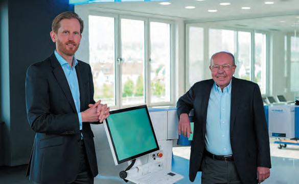
Alcon introduced its new Alcon Fidelis™ Virtual Reality (VR) Ophthalmic Surgical Simulator at the recent American Society of Cataract and Refractive Surgery annual meeting. The portable VR tool should expand training opportunities around the world, the company says.


The portable Fidelis VR Ophthalmic Surgical Simulator features a VR headset, two haptic engines, and an integrated Centurion® footswitch. It provides a realistic virtual OR environment, complete with Alcon equipment. It offers the ability to connect with other remote users and instructors within the same virtual OR. It also provides real-time feedback and performance tracking over time to enhance surgeon technique.
alcon.com


New research shows that subconjunctival triamcinolone is clinically effective and cost-effective in preventing cystoid macular oedema (CME) in diabetic cataract patients. Using data from the ESCRS PREMED study, the prospective analysis compared quality-adjusted life years (QALYs)-based cost-effectiveness in diabetic cataract patients randomised to receive perioperative subconjunctival triamcinolone acetonide, perioperative intravitreal bevacizumab, both treatments combined, or topical steroids. It showed bevacizumab and control treatment were the most costly and least effective. The main complication occurring more frequently in eyes receiving subconjunctival triamcinolone was increased IOP. These results support earlier overall findings from the ESCRS PREMED study, which demonstrated that subconjunctival triamcinolone is the most effective strategy in preventing CME after cataract surgery, the authors said. RWP Simons et al, “Economic evaluation of prevention of cystoid macular oedema after cataract surgery in diabetic patients: ESCRS PREMED study report 6”, 48 (5): 555–563, May 2022.


Exchanging a bag-in-the-lens (BIL) intraocular lens (IOL) in the event of a refractive surprise is a safe technique with a low rate of complications that can significantly improve UDCVA, regardless of the indication, suggest the findings of an observational retrospective study. It looked at 59 eyes of 59 patients who underwent a BIL exchange between 2007 and 2020. Their mean decimal uncorrected visual acuity (UDVA) and corrected (CDVA) distance visual acuity improved from 0.36 and 0.79, respectively, before BIL exchange to 0.66 and 0.89, respectively, one month postoperatively. The study authors note that because of its easy exchangeability, the BIL is their IOL of choice in complex cases at risk of refractive surprise. D Dragnea et al, “Intraocular bag-inthe-lens exchange: indications, outcomes, and complication”, 48 (5): 568–575, May 2022.

The authors of a review/update article have raised a call to action in preventing postoperative dry eye disease (DED) in patients undergoing cataract and refractive procedures. They stress all ocular surgery patients should be assessed for signs of ocular surface disease (OSD). Pre-existing DED can exacerbate postoperative dry eye symptoms and reduce patient satisfaction. Symptom screening should be followed by non-invasive multimodal objective evaluation of OSD signs. Treatment goals include reducing inflammation and restoring tear film homeostasis to optimise preoperative measurements, maximise postoperative outcomes, and improve patient satisfaction. K Donaldson et al, “Call to action: treating dry eye disease and setting the foundation for successful surgery”, 48 (5): 623–629, May 2022.


JUNE

15th European Glaucoma Society Congress


4 – 7 June
Athens, Greece
ESONT Abstract Submission

Deadline: 15 June 2022, 23:59 CET
EUCornea
25 – 26 June
Athens, Greece
40th Congress of the ESCRS
Early Bird Registration
Deadline: 29 June 2022, 23:59 CET
EURETINA
1 – 4 September
Hamburg, Germany
ESCRS iNovation Day
16 September
Milan, Italy
WSPOS Subspecialty Day
16 September
Milan, Italy
40th Congress of the ESCRS
16 – 20 September
Milan, Italy

MILAN
16–20 September 2022
MiCo, Milano Convention Centre, Milan Italy
Friday 16th September 2022
A safe environment for in-person interaction
Networking opportunities with 500 clinical and business experts
Learn about the latest developments in the European commercial and capital markets, projected 2023 innovations, regulatory changes, and reimbursement trends


