How to Identify MASLD Forms Secondary to Endocrine Derangements in Clinical Practice
Interviews
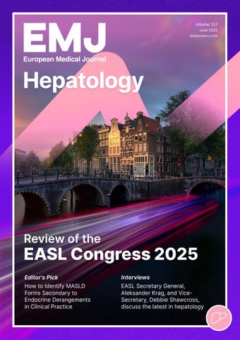
EASL Secretary General, Aleksander Krag, and ViceSecretary, Debbie Shawcross, discuss the latest in hepatology
10 Review of the European Association for the Study of the Liver (EASL) Congress 2025 Congress Features
24 Phosphatidylethanol in Steatotic Liver Disease: Unveiling Alcohol Use and Enhancing Diagnostic Precision
Nikolaj Torp
27 MASH Redefined: Insights from the EASL Congress 2025
Thirlwell and Campana
30 Evolving Paradigms in Albumin Therapy For Decompensated Cirrhosis: Highlights from the EASL Congress 2025
Nikolaj Torp
32 Pregnancy and Liver Disease: A Multidisciplinary Challenge
Bertie Pearcey Abstract Reviews
38 Unveiling the Link Between Neddylation and Hepatic Zonation: Key Insights Into Liver Metabolism and Diseases
Abruzzese et al.
40 Niche-Specific Reprogramming of Macrophages Reveals Myeloid Cell-Centric Targets During Pro-Senescence Therapy in Liver Cancer
Tsouri et al.
42 Mitigation of Immune Dysfunction in Patients with Sustained Suppression of Hepatitis B Surface Antigen After siRNA Treatment
Mak et al.
44 Hepatocyte–Neutrophil Interaction in the Liver via SAA-FPR2 Is Key in the Pathogenesis of Alcohol-Associated Hepatitis
Martínez-Álvarez et al.
47 Abstract Highlights
Congress Interviews
55 Aleksander Krag
62 Debbie Shawcross Interviews
66 Redefining Diagnosis and Management in Hepatology with Multiparametric Ultrasound
76 Scott Friedman
82 Massimo Colombo Articles
86 Editor's Pick: How to Identify MASLD Forms Secondary to Endocrine Derangements in Clinical Practice
Lonardo et al.
94 Safe Anticoagulation Timing After Variceal Bleeding and Acute Portal Vein Thrombus: A Narrative Review
Doshi et al.
101 Secondary Bacterial Peritonitis Due to an Inadequately Treated Renal Abscess
Patel et al.
106 Evaluating the Effectiveness of Three Different Anchoring Methods on Percutaneous Drainage Catheter: A Pilot Study
Rajasegeran et al.
"This year's congress also highlighted EASL’s commitment to its four strategic pillars: Education, Advocacy, Science, and Leadership"
Editorial Board
Editor-in-Chief
Markus Peck-Radosavljevic
Klinikum Klagenfurt am Wörthersee, Austria
Markus Peck-Radosavljevic is a renowned expert in gastroenterology and hepatology, currently serving as Professor of Medicine and Chairman of the Department of Internal Medicine & Gastroenterology at Klinikum Klagenfurt, Austria. With a distinguished career spanning clinical practice, research, and academia, he has contributed extensively to the study of liver diseases, including end-stage cirrhosis, liver transplantation, viral hepatitis, and liver cancer.
Ahmed Elsharkawy
University Hospitals Birmingham, UK
Kieron B.L Lim
Mount Elizabeth Hospital Liver Transplant Program, Singapore
Fabio Marra University of Florence, Italy
Ken Simpson University of Edinburgh, UK
Ashwani Singal
University of South Dakota Sanford School of Medicine, USA
Dhiraj Tripathi
Queen Elizabeth Hospital Birmingham,UK
Amr Amin
United Arab Emirates University, UAE
Aims and Scope
EMJ Hepatology is an open-access, peer-reviewed eJournal committed to helping elevate the quality of liver healthcare globally by informing healthcare professionals on all aspects of liver health and disease.
The journal is published annually, six weeks after the European Association for the Study of the Liver (EASL) Congress, and features highlights from this congress, alongside interviews with experts in the field, reviews of abstracts presented at the congress, as well as in-depth features on congress sessions. The journal also covers advances within the clinical and pharmaceutical arenas by publishing sponsored content from congress symposia, which is of high educational value for healthcare professionals. This undergoes rigorous quality control checks by independent experts and the in-house editorial team.
EMJ Hepatology also publishes peer-reviewed research papers, review articles, and case reports in the field. In addition, the journal publishes features and opinion pieces intended to create a discussion around key topics in the field and broaden readers’ professional interests. The journal is managed by a dedicated editorial team that adheres to a rigorous doubleblind peer-review process, maintains high standards of copy editing, and ensures timely publication.
EMJ Hepatology focuses on topics that are relevant to healthcare professionals in hepatology. We do not publish veterinary science papers or laboratory studies that are not linked to patient outcomes. We have a particular interest in topical studies that advance knowledge and inform of coming trends affecting clinical practice in hepatology.
Further details on coverage can be found here: www.emjreviews.com
Editorial Expertise
EMJ is supported by various levels of expertise:
• Guidance from an Editorial Board consisting of leading authorities from a wide variety of disciplines.
• Invited contributors who are recognised authorities in their respective fields.
• Peer review, which is conducted by expert reviewers who are invited by the Editorial team and appointed based on their knowledge of a specific topic.
• An experienced team of editors and technical editors.
Peer Review
On submission, all articles are assessed by the editorial team to determine their suitability for the journal and appropriateness for peer review.
Editorial staff, following consultation with either a member of the Editorial Board or the author(s) if necessary, identify three appropriate reviewers, who are selected based on their specialist knowledge in the relevant area.
All peer review is double blind. Following review, papers are either accepted without modification, returned to the author(s) to incorporate required changes, or rejected.
Editorial staff have final discretion over any proposed amendments.
Submissions
We welcome contributions from professionals, consultants, academics, and industry leaders on relevant and topical subjects. We seek papers with the most current, interesting, and relevant information in each therapeutic area and accept original research, review articles, case reports, and features.
We are always keen to hear from healthcare professionals wishing to discuss potential submissions, please email: editorial.assistant@emjreviews.com
To submit a paper, use our online submission site: www.editorialmanager.com/e-m-j
Submission details can be found through our website: www.emjreviews.com/contributors/authors
Reprints
All articles included in EMJ are available as reprints (minimum order 1,000). Please contact hello@emjreviews.com if you would like to order reprints.
Distribution and Readership
EMJ is distributed through controlled circulation to healthcare professionals in the relevant fields across Europe.
Indexing and Availability
EMJ is indexed on DOAJ, the Royal Society of Medicine, and Google Scholar®; selected articles are indexed in PubMed Central®
EMJ is available through the websites of our leading partners and collaborating societies. EMJ journals are all available via our website: www.emjreviews.com
Open Access
This is an open-access journal in accordance with the Creative Commons Attribution-Non Commercial 4.0 (CC BY-NC 4.0) license.
Congress Notice
Staff members attend medical congresses as reporters when required.
All information obtained by EMJ and each of the contributions from various sources is as current and accurate as possible. However, due to human or mechanical errors, EMJ and the contributors cannot guarantee the accuracy, adequacy, or completeness of any information, and cannot be held responsible for any errors or omissions. EMJ is completely independent of the review event ( EASL 2025) and the use of the organisations does not constitute endorsement or media partnership in any form whatsoever. The cover photo is of Amsterdam, the Netherlands, the location of EASL 2025.
Katrina Thornber, Katie Wright, Aleksandra Zurowska
Creative Director
Tim Uden
Design Manager
Stacey White
Senior Designer
Owen Silcox
Creative Artworker
Dillon Benn Grove
Designers
Shanjok Gurung, Fabio van Paris
Senior Performance & Insight Lead
Darren Brace
Marketing Director
Kristina Mestsaninova
Chief Executive Officer
Justin Levett
Chief Commercial Officer
Dan Healy
Founder and Chairman
Spencer Gore
Welcome
Dear Readers,
I am thrilled to welcome you to the 2025 issue of EMJ Hepatology, which features coverage of cutting-edge insights from this year’s European Association for the Study of the Liver (EASL) Congress, held in Amsterdam, the Netherlands. With a focus on bridging the gap between the bench and the bedside, and a spotlight on reducing the harm associated with alcohol use, this year’s event saw an increase in the number of ‘Basic Science’ sessions and included several ‘Alcohol-Related Harm’ sessions that addressed the need for public health advocacy and multidisciplinary collaboration to effectively achieve this goal.
Alongside our congress review, you will find features that explore updates on the role of human albumin therapy in managing decompensated cirrhosis, the new nomenclature for steatotic liver disease and its implications for clinical trials, and the potential of using phosphatidylethanol as a serum biomarker to objectively quantify 4-week alcohol intake. Also, be sure not to miss our exclusive interviews with key opinion leaders from EASL, as well as experts in hepatocellular carcinoma and liver fibrosis.
Our peer-reviewed content includes a topical review article that provides valuable clinical insights into the underlying pathogenetic mechanisms and how to diagnose metabolic dysfunction-associated steatotic liver disease secondary to endocrine disorders.
I would like to take this opportunity to thank our Editorial Board, the authors, peer reviewers, and interviewees for their support and key contributions to this issue. I hope you enjoy reading!
Editorial enquiries: editor@emjreviews.com
Sales opportunities: salesadmin@emjreviews.com
Permissions and copyright: accountsreceivable@emjreviews.com
Evgenia Koutsouki Editor
Reprints: info@emjreviews.com Media enquiries: marketing@emjreviews.com
Foreword
It is with great pleasure that I welcome you to EMJ Hepatology, a new edition that reflects the dynamic evolution of our field and the vibrant global hepatology community. In these pages, you will find a rich selection of peer-reviewed articles, expert interviews, and in-depth insights from the European Association for the Study of the Liver (EASL) Congress 2025, which took place in Amsterdam, the Netherlands, from 7th–10th May.
This year’s Congress brought together leading minds in hepatology from across the globe, fostering interdisciplinary dialogue and presenting the latest advances in clinical and basic science. A broad programme featured not only traditional symposia and abstract sessions, but also an increased focus on basic science, popular ‘Do’s and Don’ts’ sessions, and a variety of postgraduate courses. Among the many highlights was the symposium ‘Future of Hepatology: Novel Therapeutics and Evolving Challenges in Managing MASLD’, which offered valuable perspectives on emerging treatments and the complexities of managing this increasingly prevalent condition.
Complementing the Congress coverage, EMJ Hepatology features conversations with several of today’s most influential hepatologists. We are delighted to include interviews with EASL Secretary General Aleksander Krag and ViceSecretary Debbie Shawcross, each sharing insights into the society’s vision and future direction. In addition, Scott Friedman discusses
the evolving role of the gut–liver axis in liver disease therapeutics, while Massimo Colombo reflects on innovative strategies for managing hepatocellular carcinoma, including CAR-T cell therapies.
We are delighted to include interviews with EASL Secretary General Aleksander Krag and ViceSecretary Debbie Shawcross
We also present four original articles tackling a diverse range of topics, from anticoagulation timing after variceal bleeding, to new clinical approaches for identifying MASLD caused by endocrine disorders. These contributions exemplify the breadth of thought and inquiry that characterise our field today.
Finally, I would like to extend my sincere thanks to everyone who has made this issue possible: our authors, peer reviewers, interviewees, and our EMJ Hepatology Editorial Board.
I hope you find this edition informative and inspiring.
Markus Peck-Radosavljevic
Professor of Medicine and Chairman, Department of Gastroenterology and Hepatology, Endocrinology and Nephrology, Klinikum Klagenfurt am Wörthersee, Austria
EASL 2025
This year's Congress also highlighted EASL’s commitment to its four strategic pillars: Education, Advocacy, Science, and Leadership
Review of the European Association for the Study of the Liver (EASL) Congress 2025 Congress Review
THIS YEAR, the vibrant city of Amsterdam, the Netherlands played host to the European Association for the Study of the Liver (EASL) Congress 2025, welcoming 7,742 participants from 119 countries. The event served as a powerful hub of scientific exchange, advocacy, and professional development for clinicians, researchers, policymakers, and allied health professionals committed to tackling liver disease worldwide.
Over four dynamic days, attendees were invited to explore 190 scientific sessions spanning a broad spectrum of hepatology, from basic science to translational research, and clinical best practices to public health initiatives. The congress featured a rich mix of symposia, abstract presentations, handson training, and the popular ‘Do’s and Don’ts’ sessions, offering something of value for everyone in the hepatology community.
The Opening Ceremony set a powerful tone, with a panel discussion addressing the urgent health crisis of alcohol-related liver disease. This discussion featured Aleksander Krag, EASL Secretary General; Riina Sikkut, Member of the Estonian Parliament and former Minister of Health of Estonia; and Carina Ferreira-Borges, Regional Advisor for Alcohol and Illicit Drugs, WHO, Geneva, Switzerland. The panel, 'From Evidence to Action', was moderated by David Barrett, WHO, Geneva, Switzerland, and explored the challenge of translating science into policy. Krag highlighted the scale of the issue, citing over 800,000 alcohol-related deaths annually in Europe, and called for action on three evidence-based policy levers: pricing, availability restrictions, and marketing
regulation. “The challenge is not evidence, it’s political will,” he stated.
Ferreira-Borges emphasised the need for cross-sector collaboration and support for policymakers facing pressure from powerful industry lobbies. “There is no safe level of alcohol consumption. That’s not opinion, it’s evidence,” she said, while detailing WHO’s effort to combat misinformation and raise awareness of under-recognised risks such as the link between alcohol and breast cancer. The panel's message was clear: while science provides the solutions, meaningful progress depends on coordinated advocacy and political courage. “We must be louder than the lobbyists,” Krag concluded.
This year's Congress also highlighted EASL’s commitment to its four strategic pillars: Education, Advocacy, Science, and Leadership, the letters that form the organisation’s acronym. With its clear vision of ‘Beating liver disease’ and its mission to be ‘The Home of Hepatology for everyone engaged in beating liver disease’, the Opening Ceremony reaffirmed EASL’s central role in shaping the future of liver health.
The awards provided a fitting conclusion to the session, celebrating excellence and emerging leadership within the field. Silvia Affo, Instituto de Investigaciones Biomédicas August Pi i Sunyer (IDIBAPS), Barcelona, Spain, and Mattias Mandorfer, Medical University of Vienna, Austria, were recognised as recipients of the EASL Emerging Leader Award. Meanwhile, the EASL Rising Star Award was presented to Sally Tilden, University Hospitals Bristol NHS Foundation Trust, UK, and Thomas Maurel, Pitié Salpêtrière University Hospital, Paris, France. Meanwhile the EASL Daniel Alagille Award, honouring outstanding contributions to paediatric hepatology, was awarded to Barbora Smolkova, Oslo University Hospital, Norway.
From educational initiatives, such as the updated clinical guideline app and postgraduate courses, to advocacy efforts like the ‘Love Your Liver’ project and the newly launched European Health Alliance on Alcohol, the EASL Congress 2025 showcased a united global effort to confront the burden of liver disease head-on.
As EASL continues to foster inclusive leadership, scientific innovation, and public health advocacy, the Congress in Amsterdam will be remembered not just for its depth of knowledge, but for its heart, driven by a shared mission to improve lives.
Stay tuned for EASL 2026, and in the meantime, explore our full coverage and reflections on an unforgettable week in hepatology.
The EASL Congress 2025 showcased a united global effort to confront the burden of liver disease head-on
to diagnose metabolic dysfunction-associated steatotic liver disease (MASLD).
The study aimed to assess the diagnostic accuracy of a wide range of imaging and blood-based biomarkers against liver biopsy, the current gold standard, in patients with MASLD. Participants underwent clinical evaluation, blood sampling, liver stiffness measurement, and MRI imaging within 6 months of biopsy, with all data centrally processed and histology scored by expert pathologists using standardised methods.
The study aimed to assess the diagnostic accuracy of a wide range of imaging and blood-based biomarkers against liver biopsy
Of the 357 participants included, 50% had Type 2 diabetes, with a mean BMI of 33.8 kg/m². Fibrosis staging revealed a significant burden of liver disease, with over 70% of participants showing Stage F2 or greater. When assessing biomarkers for diagnosing ‘at-risk metabolic dysfunction-associated steatohepatitis (MASH)’, defined as active steatohepatitis with moderate fibrosis, NIS2+® (Genfit, France) showed the highest accuracy (area under the curve [AUC]: 0.82) but did not surpass the statistical threshold required to be considered a valid replacement for biopsy. Other biomarkers, such as controlled attenuation parameter (CAP), magnetic resonance imaging derived proton density fat fraction (MRI-PDFF), and iron-corrected T1 mapping (cT1), yielded lower diagnostic accuracies.
For identifying advanced fibrosis (≥F3), magnetic resonance elastography and the composite scoring system AGILE3+™ (Echosens, Paris, France) performed exceptionally, both exceeding the minimum acceptable criterion, with AUCs of 0.91 and 0.84, respectively. Magnetic resonance elastography also proved the most reliable marker for cirrhosis (F4), followed by vibration-controlled transient elastography, AGILE3+, and AGILE4™ (Echosens), all meeting or surpassing the minimum acceptable criterion. In contrast, several blood biomarkers, such as PROC3 and CK18 variants, fell short of the performance threshold.
The study reinforces the utility of elastography-based imaging for fibrosis assessment in MASLD, while simultaneously exposing the ongoing limitations in reliably diagnosing active steatohepatitis noninvasively. Although promising, the current biomarkers, including NIS2+, require further validation before they can replace biopsy. Ongoing studies aim to assess the prognostic value of these biomarkers, with the ultimate goal of improving patient stratification and guiding treatment decisions without the need for invasive procedures.
%
Of the 357 participants included, 50% had Type 2 diabetes
Primary Prevention of Variceal Bleeding: Carvedilol or Band Ligation?
THE MAIN findings from the CALIBRE trial, recently presented at the EASL Congress 2025, indicate no significant difference between carvedilol and variceal band ligation (VBL) in preventing first variceal bleeding in patients with cirrhosis and medium to large oesophageal varices, with both treatments demonstrating similar safety profiles.2
The CALIBRE trial was designed to address the ongoing uncertainty regarding the optimal primary prevention strategy for variceal bleeding in liver cirrhosis, specifically comparing the efficacy and safety of carvedilol, a non-selective β-blocker, with VBL.
This multicentre, randomised controlled, open-label study was conducted across 60 UK hospitals, enrolling adult patients with cirrhosis and medium to large oesophageal varices who had not previously bled or received either intervention. The trial’s primary aim was to determine if carvedilol 12.5 mg daily was superior or equivalent to VBL in reducing the risk of first variceal bleed within one year of randomisation.
A total of 265 participants were randomised between January 2019–August 2022, with 133 assigned to carvedilol and 132 to VBL. The incidence of variceal bleeding within 1 year was 3.8% in the carvedilol group and 7.6% in the VBL group, with a risk ratio of 0.50 (95% CI: 0.17–1.41; p=0.189) and a risk
difference of –0.038 (95% CI: –0.094–0.017; p=0.178). No statistically significant differences were observed in secondary outcomes, including mortality, transplantfree survival, or other complications of cirrhosis. Both interventions were well tolerated, with only one serious treatmentrelated adverse event in each arm, and no treatment-related deaths. Economic analysis favoured carvedilol, which was less costly and associated with a slight increase in quality-adjusted life years. Qualitative findings suggested no strong patient preference for either intervention, though clinicians tended to favour carvedilol as a first-line therapy.
In conclusion, the CALIBRE trial, despite being underpowered due to early recruitment closure, found no evidence of a difference in efficacy or safety between carvedilol and VBL for primary prevention of variceal bleeding in cirrhosis. Future research should focus on larger, adequately powered studies to confirm these findings and further explore patient-centred outcomes and cost-effectiveness.
New Therapy Significantly Slows Liver Disease in Patients With Cystic Fibrosis
A LANDMARK French study presented at the EASL Congress 2025 has revealed that the introduction of Elexacaftor-Tezacaftor-Ivacaftor (ETI) therapy has dramatically reduced the progression of liver disease and mortality in people living with cystic fibrosis.3
The 10-year national study analysed data from nearly 10,000 patients aged 12 years and older, comparing cystic fibrosis liver disease outcomes before and after ETI became available in France in December 2019. Researchers used data from the French National Discharge Database to examine liver disease progression, transplant rates, and mortality.
The incidence of liver disease progression dropped sharply in the ETI era, from 20.7 to just 1.14 cases per 1,000 personyears. Five-year risk of cystic fibrosis liver disease progression fell from 11.4% to 0.27%, while transplant-free mortality decreased from 18.5% to 0.57%. All major liver-related complications, including gastro-oesophageal varices and hepatocellular carcinoma, declined significantly during the same period.
The study also showed that these improvements were not simply due to reduced life expectancy. Even after accounting for mortality as a competing risk, the adjusted hazard ratio for liver
The findings strongly support incorporating ETI into clinical care for patients with cystic fibrosis Who have liver involvement
disease progression was 0.083, with transplant-free mortality at just 0.011.
The findings strongly support incorporating ETI into clinical care for patients with cystic fibrosis who have liver involvement, shifting the outlook from high-risk to manageable with modern therapy.
Five-year risk of cystic fibrosis liver disease progression fell from 11.4% to 0.27%, while transplant-free mortality decreased from 18.5% to 0.57%
Liver Stiffness Identified as Key Predictor in Primary Biliary Cholangitis
AT THE EASL Congress 2025, new data from the GLOBAL-PBC study revealed that liver stiffness measurement (LSM) is a more robust predictor of liver-related complications in primary biliary cholangitis (PBC) than biochemical response alone.4
Liver stiffness measurement (LSM) is a more robust predictor of liver-related complications in primary biliary cholangitis (PBC) than biochemical response alone
In the multinational cohort of 1,793 patients, selected from 4,096 with available LSMs, participants had at least two reliable LSMs performed via vibration-controlled transient elastography at least 6 months apart. Patients with prior hepatic decompensation (HD), liver transplantation, or hepatocellular carcinoma were excluded.
Over a median follow-up of 22 months, 3.3% of patients developed HD. Biochemical response, defined using the Paris-2 criteria, was achieved in 51% of patients, while 52% had stable or reduced LSMs. ALP normalisation and deep biochemical response were seen in 39% and 25% of patients, respectively, all associated with a significantly reduced risk of HD (p<0.05 for all).
However, LSM response alone did not predict HD unless the reduction exceeded certain thresholds. Reductions in LSM of 10%, 20%, and 30% were each independently associated with lower HD risk (p<0.05), but the clearest predictor was the most recent LSM value (LSMc). An LSMc ≥10 kPa was strongly predictive of HD (hazard ratio [HR]: 14.5; 95% CI: 6.9–30.6), with LSMc alone having a predictive accuracy (C-statistic) of 0.87.
Among patients with discordant biochemical and LSM responses (seen in 52%), LSMc >10 kPa remained predictive of HD (HR: 37.4; 95% CI: 4.8–289.7) and of the composite outcome of liver transplantation or liverrelated death (HR: 2.6; 95% CI: 1.3–5.4).
Researchers concluded that in routine clinical practice, LSMc alone offers a simple yet highly effective means of prognostication, particularly when biochemical markers are inconclusive.
'3-in-1' Metabolomic Test Could Transform Cholangiocarcinoma Detection
A NEW metabolomic blood test could transform the way clinicians diagnose and monitor primary sclerosing cholangitis (PSC) and detect associated cholangiocarcinoma (CCA) at an early stage.5
In a large international study, presented at the EASL Congress 2025, researchers evaluated serum samples from 434 individuals across 13 centres in eight countries. Participants included patients with PSC, those who developed CCA during follow-up (PSC-to-CCA), individuals with concurrent PSC-CCA, patients with ulcerative colitis, and healthy controls. The team employed ultra-high-performance liquid chromatography–mass spectrometry to assess metabolite profiles, followed by machine learning to identify the most predictive biomarkers. The study aimed to uncover serum-based markers capable of diagnosing PSC, detecting CCA in patients with PSC, and predicting CCA development prior to clinical manifestation.
Results identified 50 metabolites strongly associated with PSC, unaffected by age, sex, cirrhosis, or ulcerative colitis status. A 13-metabolite model differentiated patients with PSC from healthy controls with 98% accuracy in both discovery and validation cohorts. Among those with PSC-CCA, 57 metabolites showed significant alterations. Another 13-metabolite model accurately distinguished PSC-CCA from PSC cases with area under the curve (AUC) values of 0.91
and 0.90, excelling in early-stage cancer detection (AUC=0.930) and outperforming CA19-9 (AUC=0.646). Impressively, this model retained diagnostic power (AUC=0.92) in patients with low CA19-9 levels. Furthermore, a separate seven-metabolite model predicted future CCA development in patients with PSC, with positive predictive values of 83% and 73% in discovery and validation cohorts, respectively.
This ‘3-in-1’ blood test offers a promising, non-invasive solution for diagnosing PSC, detecting early PSC-CCA, and forecasting cancer risk before clinical signs emerge. Its clinical adoption could enable earlier intervention, tailored surveillance, and more precise management strategies in high-risk PSC populations.
A 13-metabolite model differentiated patients with PSC from healthy controls with 98% accuracy in both discovery and validation cohorts
New Treatment Offers Relief for PBC-Related Itch
A NEW Phase III clinical study, presented at the EASL
Congress
2025, has shown promising results for patients experiencing cholestatic pruritus associated with primary biliary cholangitis.6
This debilitating and often undertreated condition significantly impacts quality of life, particularly due to persistent itching and sleep disturbance. The trial evaluated the efficacy and safety of linerixibat, an ileal bile acid transporter inhibitor, in individuals with moderate-to-severe pruritus.
In the double-blind, randomised, placebocontrolled study, 238 patients received either linerixibat 40 mg or a placebo twice daily over 24 weeks. The majority of participants were female, with a mean itch severity score of 7.34 at baseline. The primary endpoint measured the change in the worst itch score, while secondary outcomes assessed sleep interference, early response at Week 2, and overall patient impressions of symptom changes.
In this trial, linerixibat demonstrated a significant and rapid reduction in pruritus compared to the placebo. By Week 24, patients treated with linerixibat reported a greater mean reduction in itch severity, with an adjusted mean difference of –0.72 (p=0.001). Improvements were noticeable as early as Week 2, with a significant difference of –0.71 (p<0.001). Sleep quality also improved more markedly in the linerixibat group, with a statistically significant reduction in pruritusrelated sleep interference (p=0.024).
Moreover, a higher proportion of patients on linerixibat achieved clinically meaningful improvements in itch relief, with 41% experiencing a ≥4-point reduction, and 21% reporting an absence of pruritus altogether. While gastrointestinal side effects such as diarrhoea and abdominal pain were more common with linerixibat, they were generally manageable, with only 4% of patients discontinuing treatment due to these issues.
238 patients received either linerixibat 40 mg or a placebo twice daily over 24 weeks
These results support the potential use of linerixibat for pruritus in patients with primary biliary cholangitis, offering meaningful symptom relief and improved sleep.
Phase II Study Reveals New Treatments for Hepatitis B Virus
THE
PHASE II MARCH study, presented at the EASL Congress 2025, investigated the efficacy and safety of tobevibart (VIR-3434) and elebsiran (VIR-2218), administered alone or combined with pegylated interferon (IFN) alfa-2a, in people living with chronic hepatitis B virus (HBV) infection.7
Tobevibart is an engineered human monoclonal antibody targeting the hepatitis B surface antigen (HBsAg), while elebsiran is a small interfering RNA designed to silence the HBx region of the HBV genome.
Participants receiving nucleos(t)ide-reverse transcriptase inhibitors were treated for 44–48 weeks with either tobevibart alone, tobevibart plus elebsiran, or the combination of tobevibart, elebsiran, and pegylated IFN alfa-2a. All medications were administered via subcutaneous injection at specified intervals. The study primarily evaluated treatment-emergent adverse events and rates of HBsAg seroclearance (defined as HBsAg levels below 0.05 IU/mL) at the end of treatment (EOT). The authors noted that functional cure rate at 24 weeks post-EOT, measured in participants who discontinued nucleos(t)ide-reverse transcriptase inhibitors after achieving seroclearance, will be reported later.
At EOT, no participants treated with tobevibart alone achieved HBsAg seroclearance, whereas 15.7% of those receiving tobevibart plus elebsiran and 22.2% of those receiving the triple
lower baseline HBsAg levels (<1,000 IU/mL) experienced higher seroclearance rates: 38.9% in the two-drug group, and 45.5% in the three-drug group.
Treatment-emergent adverse events were mostly mild or moderate (Grade 1–2). Severe adverse events (Grade ≥3) were rare, but more common in the triple therapy group, including one case of leukopenia and one hepatitis event linked to the study drugs. Both of these were resolved without lasting issues.
These encouraging findings highlight the potential of tobevibart combined with elebsiran, with or without pegylated IFN alfa-2a, to achieve HBsAg loss in chronic HBV infection, especially in patients with lower antigen levels, and support further clinical development.
Findings highlight the potential of tobevibart combined with elebsiran, with or without pegylated IFN alfa-2a, to achieve HBsAg loss in chronic HBV infection
Portal Hypertension Found in Half of Patients After Vein Thrombosis
RESEARCH presented at the EASL Congress 2025 has found that nearly half of patients with non-cirrhotic, non-tumoural recent portal vein thrombosis (RPVT) develop signs of portal hypertension within 5 years.8
The study, conducted across multiple centres and published after two decades of patient data collection, followed 485 individuals with RPVT for a median of 67 months. Portal hypertension was defined by the presence of portosystemic collaterals, gastrooesophageal varices, ascites, gastrointestinal bleeding, or hepatic encephalopathy. The cumulative incidence of portal hypertension reached 48% by the 5th year.
The results showed that patients who did not achieve full recanalisation of the portal vein system were at a significantly higher risk, with a 67% incidence at 5 years. In contrast, those who achieved complete recanalisation had a much lower risk of 17%.
Several clinical factors were independently associated with the development of portal hypertension. These included the presence of ascites at diagnosis, a diagnosis of myeloproliferative neoplasm, thrombosis affecting multiple veins, and incomplete or absent recanalisation during follow-up.
Interestingly, a small group of patients did not develop portal hypertension, although there was no evidence of recanalisation. A majority of these individuals had clots confined to intrahepatic portal vein
branches, suggesting that the location and extent of the thrombosis are critical to long-term outcomes.
Several clinical factors were independently associated with the development of portal hypertension
This research highlights the importance of long-term monitoring and early intervention in patients with RPVT, even in the absence of cirrhosis or cancer. The authors emphasise that early identification of highrisk patients may allow for more targeted follow-up, recanalisation strategies, and preventive care to avoid the development of complications linked to portal hypertension.
67 %
Patients who did not achieve full recanalisation of the portal vein system were at a significantly higher risk, with a 67% incidence at 5 years
Dual Immunotherapy Extends Survival in Unresectable Liver Cancer
AT THE EASL Congress 2025, an interim analysis of the Phase III CheckMate 9DW trial revealed that first-line immunotherapy with nivolumab plus ipilimumab significantly improves survival in patients with unresectable hepatocellular carcinoma, regardless of liver function status.9
In the study of 668 previously untreated patients, nivolumab plus ipilimumab demonstrated superior overall survival (OS) compared with lenvatinib or sorafenib, with a median OS of 23.7 months versus 20.6 months (hazard ratio [HR]: 0.79; p=0.018). Objective response rate was markedly higher with nivolumab plus ipilimumab (36% versus 13%; p<0.0001), with responses lasting over twice as long (median duration of response: 30.4 versus 12.9 months).
Outcomes were stratified by liver function using albumin-bilirubin (ALBI) scores. Among the 396 patients with ALBI Grade 1 (better liver function), median OS was 35.4 months with nivolumab plus ipilimumab versus 23.2 months with lenvatinib or sorafenib (HR: 0.75). In those with ALBI Grades 2 or 3 (272 patients), OS was 16.9 versus 14.0 months, respectively (HR: 0.75).
In both subgroups, nivolumab plus ipilimumab showed higher objective response rates (37% versus 14% in Grade 1; 35% versus 11% in Grades 2 or 3) and greater complete response rates. Safety profiles were consistent across ALBI grades.
These findings support nivolumab plus ipilimumab as a promising first-line treatment for unresectable hepatocellular carcinoma, offering durable responses and survival benefits irrespective of underlying liver function.
In both subgroups, nivolumab plus ipilimumab showed higher objective response rates
Objective response rate was markedly higher with nivolumab plus ipilimumab (36% versus 13%; p<0.0001)
Novel Therapy Reduces Flare Risk in IgG4-Related Disease
A NOVEL treatment targeting B cells significantly reduces disease flares in patients with IgG4-related disease affecting the pancreas, bile ducts, or liver, according to new results from the MITIGATE Phase III trial, presented at the EASL Congress 2025.10
IgG4-related disease is a chronic, immune-mediated fibroinflammatory condition that commonly affects the pancreas and hepatobiliary system, leading to progressive organ damage. The MITIGATE study, a global, randomised, placebocontrolled trial, evaluated the safety and efficacy of inebilizumab, a CD19-targeting monoclonal antibody that depletes B cells. A post hoc subgroup analysis focused on patients with active pancreatic, biliary, or liver involvement at baseline. Participants with a history of multi-organ disease and recent glucocorticoid-treated flare were randomised to receive inebilizumab or placebo at set intervals during a 1-year treatment period. Glucocorticoids were tapered off by Week 8, and no other immunosuppressants were allowed.
Of 135 trial participants, 52% had prior pancreatic disease, 32% had biliary involvement, and 7% had liver disease. At baseline, active disease was present in 38% (pancreas), 21% (bile ducts), and 4% (liver). Inebilizumab dramatically reduced flare risk in all groups. In the pancreas group, the hazard ratio for flare compared to placebo was 0.03 (nominal p=0.005). Notably, none of the inebilizumab-treated
patients with biliary or liver involvement experienced a flare, while 12/15 and 3/3 placebo-treated patients did, respectively.
Inebilizumab dramatically reduced flare risk in all groups
Annualised flare rates were significantly lower with inebilizumab in all subgroups, with a rate ratio of 0.04 for pancreatic disease (nominal p=0.0015). Complete remission without treatment was achieved more frequently with inebilizumab, with an odds ratios of 10.8 and 35.8 in the pancreas and bile duct groups, respectively. Steroid use was also significantly lower in these groups (nominal p<0.001). Adverse events were consistent with the overall trial population, with no new safety concerns identified.
These findings highlight the therapeutic potential of CD19-targeted B cell depletion in managing IgG4-related disease with pancreatic and hepatobiliary involvement. Inebilizumab may offer a more effective, steroid-sparing option for long-term disease control.
References
1. Pavlides M et al. Diagnostic performance of imaging and serum based MASLD biomarkers: robust validation in the prospective LITMUS imaging study. Abstract GS-001. EASL Congress, 7-10 May, 2025.
2. Tripathi D et al. A multicentre randomised controlled trial of carvedilol versus variceal band ligation in primary prevention of variceal bleeding in liver cirrhosis (CALIBRE trial). Abstract GS-006. EASL Congress, 7-10 May, 2025.
3. Mouliade C et al. ElexacaftorTezacaftor-Ivacaftor Era and cystic fibrosis liver disease progression: a 10year national study. Abstract GS-008YI. EASL Congress, 7-10 May, 2025.
4. Wong YJ et al. Latest liver stiffness measurements predict liverrelated outcomes in primary biliary
cholangitis patients with discordant biochemical and liver stiffness responses. Abstract GS-009. EASL Congress, 7-10 May, 2025.
5. Lapitz A et al. Novel “3-in-1” blood metabolomic test for the early diagnosis and risk stratification of primary sclerosing cholangitis and cholangiocarcinoma. Abstract GS-007YI. EASL Congress, 7-10 May, 2025.
6. Hirschfield GM et al. Linerixibat significantly improves cholestatic pruritus in primary biliary cholangitis: results of the pivotal phase 3 GLISTEN trial. Abstract GS-011. EASL Congress, 7-10 May, 2025.
7. Gane EJ et al. Outcomes of 48 weeks of therapy and subsequent 24-week post-treatment period with tobevibart (VIR-3434) and elebsiran (VIR-2218) with or without pegylated interferon alfa-2a in chronic hepatitis
B virus infection. Findings from the MARCH study. Abstract GS-010. EASL Congress, 7-10 May, 2025.
8. Alabau BB et al. Development of portal hypertension after noncirrhotic non-tumoral recent portal vein thrombosis (RPVT). A long-term follow-up study. Abstract GS-004. EASL Congress, 7-10 May, 2025.
9. Sangro B et al. Outcomes by liver function in patients with unresectable hepatocellular carcinoma treated with nivolumab plus ipilimumab vs lenvatinib or sorafenib in the CheckMate 9DW trial. Abstract GS005. EASL Congress, 7-10 May, 2025.
10. Culver E et al. Safety and efficacy of inebilizumab in IgG4 related disease in participants with pancreatic, biliary, and hepatic involvement: results from the phase 3 MITIGATE trial. Abstract GS002. EASL Congress, 7-10 May, 2025.
Phosphatidylethanol in Steatotic Liver Disease: Unveiling Alcohol Use and Enhancing Diagnostic Precision
ACCURATELY discerning the contribution of alcohol intake to the pathogenesis of steatotic liver disease (SLD) is fundamental to its classification, management, and prognosis. As the field transitions toward a refined taxonomy, including metabolic dysfunction-associated SLD (MASLD), alcohol-associated liver disease (ALD), and the hybrid phenotype metabolic dysfunction and alcohol-related liver disease (MetALD), the role of objective biomarkers becomes increasingly crucial. At the European Association for the Study of the Liver (EASL) Congress 2025, three pivotal studies illustrated the emerging clinical value of phosphatidylethanol (PEth), a blood-based biomarker that directly quantifies alcohol intake over a ~4-week window, proving an important supplement to the self-reported history.
DISCORDANCE BETWEEN SELF-REPORT AND PHOSPHATIDYLETHANOL QUANTIFICATION
The study by Bech et al.1 evaluated PEth concentrations in 2,925 individuals undergoing screening for SLD. The investigators highlighted significant discrepancies between self-reported alcohol consumption and PEth-based measurements.
Despite an Alcohol Use Disorders
Identification Test-C (AUDIT-C) score indicative of hazardous drinking in 71% of the cohort, the median PEth concentration (40.8 ng/mL) revealed underestimation.
Correlation coefficients between PEth and self-reported alcohol intake were modest at best (r=0.400 for past-week intake) and poor for average intake over 3 months (r=0.131). Notably, among individuals classified with MASLD who were ostensibly free of harmful alcohol use, 8% had PEth levels ≥200 ng/mL (indicative of heavy intake), and 31% had levels consistent with significant intake (≥20 ng/mL).
This substantial misclassification raises concerns regarding diagnostic precision in both clinical and research contexts. Here, PEth may prove to be an important aid in the subclassifications of patients across the spectrum of steatotic liver disease.
PROSPECTIVE VALIDATION IN A COMMUNITY-BASED COHORT
Building on this, Tavaglione et al.2 conducted a prospective, population-based study assessing under-reported alcohol use via PEth in 556 community-dwelling adults, 391 of whom had MRI-confirmed SLD. Using established PEth thresholds (≥25 ng/mL for MASLD and ≥200 ng/mL for MetALD), the authors found that 16% of patients were misclassified based on self-report alone.
These data have immediate clinical ramifications: reliance on tools such as AUDIT or Lifetime Drinking History (LDH) may inadvertently categorise individuals into inappropriate SLD subtypes, thereby affecting therapeutic strategy and prognosis. Multivariable analysis showed that underreporting was more common among male, Caucasian participants without diabetes.
Crucially, PEth enabled correction of this misclassification, supported a data-driven transition away from purely subjective assessments toward integrative, biomarker-based diagnosis in liver clinics.
RISK STRATIFICATION AND FIBROSIS IN METABOLIC DYSFUNCTION AND ALCOHOLRELATED LIVER DISEASE
In the third key presentation, Diaz et al.3 examined a cohort of 617 overweight or obese adults, identifying 97 individuals at risk of MetALD. The risk was defined based on either self-reported alcohol intake within MetALD thresholds or lower reported intake but with PEth ≥25 ng/mL.
Using a tiered screening approach, beginning with fibrosis index based on four factors (FIB-4) followed by VCTE, the researchers demonstrated that non-invasive testing effectively identified individuals with significant fibrosis (defined as magnetic resonance elastography ≥3.14 kPa or VCTE ≥7.6 kPa). The diagnostic performance was robust (FIB-4 area under the receiver operating characteristic curve=0.78; VCTE area under the receiver operating characteristic curve=0.85) and importantly, this strategy maintained a false negative rate as low as 2% when applied sequentially.
These findings reinforce the utility of PEth not only for diagnostic classification but also for augmenting fibrosis risk assessment when coupled with established non-invasive tests. Such strategies are indispensable for efficient triage and resource allocation in hepatology care pathways.
CLINICAL IMPLICATIONS
Together, these studies paint a compelling picture of PEth as an indispensable tool for modern hepatology practice. Key implications include:
• Improved diagnostic precision: PEth overcomes recall and social desirability bias, thereby enabling more accurate phenotypic categorisation among MASLD, MetALD, and ALD.
• Impact on clinical trials: misclassification due to reliance on self-report can distort eligibility criteria and confound outcomes in therapeutic studies.
References
1. Bech K et al. Discrepancies between self-reported alcohol intake and phosphatidylethanol in 2,925 individuals at risk of steatotic liver disease. Abstract OS-030-YI. EASL Congress, 7-10 May, 2025.
PEth incorporation into trial screening protocols could enhance external validity and reproducibility.
• Potential for risk stratification: beyond classification, PEth may have prognostic relevance, particularly when integrated with non-invasive fibrosis markers such as FIB-4 and VCTE.
CONCLUSION
As hepatology embraces a precision medicine paradigm, PEth emerges as a robust biomarker that bridges the gap between subjective history and objective phenotyping. Its value lies not only in detecting under-reported alcohol consumption but also in refining diagnostic classification and guiding individualised care. The EASL Congress 2025 marks a turning point in the endorsement of PEth as a standard adjunct in the diagnostic armamentarium for SLD.
2. Tavaglione F et al. Prospective study of phosphatidylethanol as a quantitative, objective biomarker to detect underreported alcohol use in steatotic liver disease. Abstract SAT-482-YI. EASL Congress, 7-10 May, 2025.
3. Diaz LA et al. Stratification of liver fibrosis in individuals at-risk of metabolic dysfunction and alcoholassociated liver disease (MetALD). Abstract SAT-495. EASL Congress, 7-10 May, 2025.
MASH Redefined: Insights from the EASL Congress 2025
Authors: Kayleigh Thirlwell,1 *Lara
Campana1,2
1. Resolution Therapeutics Ltd, Edinburgh, UK
2. University of Edinburgh, UK
*Correspondence to lara.campana@resolution-tx.com
Disclaimer:
Citation:
Both authors are Resolution Therapeutics employees. Lara Campana is the scientific co-founder. No sponsorship was received for this content, and the views expressed are the authors’ only and not a reflection of the company's position.
EMJ Hepatol. 2025;13[1]:27-29.
https://doi.org/10.33590/emjhepatol/SUIX7527
THE EUROPEAN Association for the Study of the Liver (EASL) Congress 2025, Amsterdam, the Netherlands, placed patients and the public at the heart of its agenda, emphasising the importance of liver health through early detection, prevention, and improved access to emerging treatments. The meeting served as a strategic inflection point to assess real-world data and address the implications of the newly adopted steatotic liver disease (SLD) nomenclature. This feature distils the key emerging themes from the congress, spanning public engagement, policy change, implications of the new SLD framework, and clinical data from repurposed drugs to target a wider patient population.
EARLY DETECTION, PUBLIC ENGAGEMENT, AND AFFECTING POLICY CHANGE
Undiagnosed liver fibrosis remains a major public health challenge. Results from the LIVERSCREEN cohort (30,541 participants) revealed a high prevalence of elevated liver stiffness (>8 kPa) primarily linked to SLD driven by metabolic risk factors and high-risk alcohol use. Hospital referrals were made for 8% of participants, and 32% of those were diagnosed with chronic liver disease with fibrosis, a total of 782 individuals.1 These findings underscore the urgent need for early detection to prevent advanced liver disease, where treatment options are limited.
Public interest in liver health is growing, as seen by the long lines of individuals eager to check their liver health at the conference. This highlights both rising awareness and limited access to testing.
The 2025 EASL Congress reaffirmed its call to recognise SLD as a non-communicable disease and to prioritise liver health within national and international health agendas by aiming to tackle Europe's alcohol burden. Despite clear evidence linking alcohol to liver morbidity and mortality, policy action remains inconsistent.
The message at EASL 2025 was clear: to bridge the gap between science and care, researchers, clinicians, and policymakers must act together. Evidence-based interventions like alcohol pricing, labelling reforms, and raising the legal drinking age were suggested tools to reduce harm.
The 2025 EASL Congress reaffirmed its call to recognise steatotic liver disease as a non-communicable disease and to prioritise liver health
UNTANGLING LIVER DISEASE: IMPLICATIONS FOR TRIALS AND CARE
The recent reclassification of liver diseases into metabolic dysfunction-associated steatotic liver disease (MASLD), metabolic dysfunction and alcohol-related liver disease (MetALD), and alcohol-related liver disease (ALD) has presented both clarity and complexity for clinical trial design. While MASLD is now defined by the presence of steatosis and metabolic dysfunction, MetALD and ALD introduce overlapping aetiologies, particularly where alcohol consumption coexists with metabolic risk. Despite alcohol's well-established role in liver pathology, only 2–5% of reported cases fall under ALD or MetALD, which is grossly unrepresentative of global alcohol exposure. Correcting for underreporting using tools such as AUDIT-C and longitudinal phosphatidylethanol (PEth) testing suggests that up to 50% of patients currently categorised as MASLD may meet criteria for MetALD, highlighting the urgent need for more accurate patient stratification. This diagnostic fluidity complicates trial recruitment and endpoint interpretation. In one study, 38% of patients with MASLD and over 60% of those with MetALD or ALD changed categories based on alcohol intake and steatosis status.2 Given this variability, a combined approach using non-invasive tests (NIT) and validated alcohol screening tools is essential to stratify patients reliably.
From a therapeutic perspective, shared pathogenesis between metabolic dysfunction-associated steatohepatitis (MASH) and alcohol-related liver diseases offers a rationale for drug repurposing. Drugs with a mechanism of action that have the potential to target both MASH and ALD, such as thyroid hormone receptor beta (THR-β) agonists, glucagon-like peptide-1 (GLP-1) receptor agonists, and fibroblast growth factor 21 (FGF-21) analogues, are now under investigation across these categories. For instance, MASH drugs are being trialled in ALD, and peroxisome proliferator-activated receptor (PPAR) agonists and hydroxysteroid 17-beta-dehydrogenase 13 (HSD17B13) inhibitors are being evaluated for efficacy in
MetALD and ALD, with early trends showing promise in fibrosis markers.
To this end, strategic trial design will ensure that medicines reach more patients in an efficient and cost-effective way. Some suggestions included basket trial designs encompassing MASLD, MetALD, and ALD, or hybrid designs that cluster MASLD with MetALD, or MetALD with ALD. Regardless, safety remains a concern; alcohol may increase gut permeability and affect oral drug solubility, impacting both efficacy and tolerability.
Considering the shift in trial design from a regulatory standpoint, the evolving terminology necessitates clarity in inclusion criteria, biomarkerbased endpoints, and justification of population definitions, especially for therapies aiming for broad SLD indications. To this end, it is encouraging to see that real world data is supporting the move from needing liver biopsies to non-invasive tests for diagnosis (LITMUS consortium)3 and endpoints (HARMONY Trial).4 This is a welcome step in reducing the biopsy bottleneck, and it highlights the need to develop a new reasonably likely surrogate endpoint. However, with fewer trials now relying on liver biopsies, there is a growing reliance on NITs; yet no single NIT reliably identifies both fibrosis and steatohepatitis across the SLD spectrum. Despite the challenges, the collective shift from biopsies to NITs reflects an interdisciplinary, patient-centered approach being adopted by both drug developers and clinicians.
CLINICAL EVIDENCE IN METABOLIC DYSFUNCTION-ASSOCIATED STEATOHEPATITIS CIRRHOSIS
At the EASL Congress 2025, several investigational agents demonstrated potential in improving fibrosis and reducing portal hypertension in patients with compensated MASH cirrhosis (F4c):
• Resmetirom (MAESTRO-NAFLD-1):5,6
Two-year open-label data showed sustained improvements in liver stiffness, fibrosis biomarkers, and portal hypertension risk, with good tolerability, supporting its clinical potential ahead of outcome data from MAESTRO-NASH OUTCOMES.
• Efruxifermin (SYMMETRY):7
First randomised controlled trial to show histologic reversal of cirrhosis due to MASH with a 96week treatment, along with NIT and metabolic improvements, reinforcing its antifibrotic and metabolic efficacy.
• Belapectin (NAVIGATE): While the primary endpoint was not met, 2 mg dosing significantly reduced new varices and improved liver stiffness in a perprotocol analysis, suggesting delayed portal hypertension progression; extended 36-month data are awaited (unpublished; NCT04365868).8
CONCLUSION
EASL 2025 has paved the way for future efforts by highlighting some key themes:
1. Undiagnosed fibrosis is a major health issue, with real-world data supporting the urgent need for early detection tools.
2. Despite public awareness of the importance of liver health, there is a strong need for evidence-based policy reform to tackle alcohol-related liver damage, an action that EASL and partners are actively pursuing.
3. In the context of MASLD, MetALD, and ALD, patient stratification remains a challenging and dynamic situation.
4. Alternative trial designs, such as basket trials and the promise of large omics and AI, could offer a viable path forward for accelerating drug development across the spectrum of SLD.
5. Drugs are reaching more patients, whether they are being repurposed from patients with MASH to patients with MetALD or ALD, or from patients with MASH to those with compensated MASH cirrhosis.
At the EASL Congress 2025, several investigational agents demonstrated potential in improving fibrosis and reducing portal hypertension
References
1. Graupera I et al. High prevalence of undiagnosed liver fibrosis in the adult European population driven by metabolic risk factors and alcohol consumption: results from the prospective LIVERSCREEN cohort in 30,541 participants. Abstract LB2553. EASL Congress, 7-10 May, 2025.
2. Krag A et al. Reporting discrepancy of alcohol intake affecting estimated prevalence of MetALD and ALD. Lancet Gastroenterol Hepatol. 2025;10(4):282-4.
3. Vali Y et al. Biomarkers for staging fibrosis and non-alcoholic
steatohepatitis in non-alcoholic fatty liver disease (the LITMUS project): a comparative diagnostic accuracy study. Lancet Gastroenterol Hepatol. 2023;8(8):714-25.
4. Harrison SA et al. Safety and efficacy of once-weekly efruxifermin versus placebo in non-alcoholic steatohepatitis (HARMONY): a multicentre, randomised, double-blind, placebo-controlled, phase 2b trial. Lancet Gastroenterol Hepatol. 2023;8(12):1080-93.
5. Harrison SA et al. A Phase 3, randomized, controlled trial of resmetirom in NASH with liver fibrosis.
N Engl J Med. 2024;390(6):497-509.
6. Harrison SA et al. Resmetirom for nonalcoholic fatty liver disease: a randomized, double-blind, placebocontrolled phase 3 trial. Nat Med. 2023;29(11):2919-28.
7. Noureddin M et al. Efruxifermin in compensated liver cirrhosis caused by MASH. N Engl J Med. 2025;DOI:10.1056/NEJMoa2502242.
8. Galectin Therapeutics Inc. Study Evaluating the Efficacy and Safety of Belapectin for the Prevention of Esophageal Varices in NASH Cirrhosis (NAVIGATE). NCT04365868. https:// clinicaltrials.gov/study/NCT04365868.
Evolving Paradigms in Albumin Therapy for Decompensated Cirrhosis: Highlights From the EASL Congress 2025
THE MANAGEMENT of decompensated cirrhosis remains a formidable challenge in hepatology, owing to its multifaceted complications and limited therapeutic options. At the European Association for the Study of the Liver (EASL) Congress 2025, several pivotal studies highlighted new insights into the clinical utility of human albumin therapy, expanding our understanding of its role in improving patient outcomes. This feature reviews the latest data presented on long-term albumin administration, mechanistic studies on endothelial dysfunction, and randomised controlled evidence for its role in correcting hyponatraemia.
LONG-TERM ALBUMIN ADMINISTRATION: THE PRECIOSA TRIAL
The PRECIOSA trial, a large-scale, multicentre, Phase III randomised controlled study, presented top-line results evaluating the efficacy of long-term albumin therapy in patients with cirrhosis, prior or current ascites, and acute decompensation.1
This trial enrolled 410 patients from 14 countries, randomising them to receive either standard medical treatment alone, or standard medical treatment plus Albutein® (Grifols, Barcelona, Spain) 20% (1.5 g/kg every 10 days for up to 12 months). While the primary endpoint of 1-year transplantfree survival was not met with statistical significance (hazard rate: 0.80; 95% CI: 0.58–1.10), the study nonetheless revealed promising trends. Notably, the incidence of disease-related complications was significantly lower in the treatment arm, with marked reductions in cirrhosis-related complications, such as spontaneous bacterial peritonitis (odds ratio 0.28; 95% CI: 0.09–0.86) and hepatorenal syndrome (odds ratio 0.24; 95% CI: 0.09–0.64). Importantly, the safety profile of albumin was favourable, with no new safety signals.
These findings suggest that, although the survival benefit was not statistically conclusive, long-term albumin therapy may exert clinically meaningful effects by stabilising the disease course and reducing major complications. However, more targeted approaches are needed to identify patients who are likely to respond to therapy.
Long-term albumin therapy may exert clinically meaningful effects by stabilising the disease course and reducing major complications
PATHOPHYSIOLOGICAL MECHANISM: ENDOTHELIAL DYSFUNCTION AS A THERAPEUTIC TARGET
Addressing the mechanistic underpinnings of albumin’s effects, a translational study from Leiden University Medical Center, the Netherlands, explored the impact of human albumin on endothelial cell (EC) dysfunction, which is a key contributor to acute decompensation and acute-on-chronic liver
failure.2 Using a high-content imaging model, cultured endothelial cells were exposed to plasma from patients with decompensated cirrhosis and hypoalbuminaemia, with and without albumin supplementation. The administration of albumin shifted EC morphology towards a healthier phenotype, particularly reversing detrimental mitochondrial changes. These effects were not replicated when ECs were stimulated by inflammatory mediators such as TNF-α or lipopolysaccharide, suggesting a unique corrective effect of albumin on circulating factors in cirrhotic plasma. This study bolsters the view of albumin as a biologically active therapeutic agent, rather than merely a plasma expander.
ALBUCAT: TARGETING HYPONATRAEMIA IN CIRRHOSIS
Hyponatraemia is a common and prognostically unfavourable complication in decompensated cirrhosis. The ALBUCAT trial addressed this issue through a randomised, multicentre design evaluating short-term intravenous albumin administration versus standard of care in patients with dilutional hyponatraemia (serum sodium ≤133 mEq/L).3 Among 52 patients, those receiving daily albumin exhibited significantly higher rates of hyponatraemia resolution (48% versus 15%; relative risk 3.39; p=0.0145) and greater increases in serum sodium (median 133 mEq/L versus 129 mEq/L). The data confirms that intravenous albumin can be a valuable adjunct in managing dilutional hyponatraemia where therapeutic options remain limited.
CLINICAL IMPLICATIONS
The collective data presented at EASL 2025 contribute to a paradigm shift in
References
1. O’Leary JG et al. Efficacy and safety of long-term human albumin therapy in cirrhotic patients with acute decompensation and ascites: topline results of the PRECIOSA trial. Abstract LBO-003. EASL Congress, 7-10 May, 2025.
how albumin is conceptualised in cirrhosis care. The PRECIOSA trial offers a possible affirmation of the disease-modifying potential of long-term albumin, despite a neutral primary endpoint. Meanwhile, mechanistic insights into endothelial protection and randomised evidence in hyponatraemia management underscore albumin’s multifactorial benefits.
These findings support a more personalised approach to albumin therapy, particularly in patients with advanced disease phenotypes, such as those prone to circulatory dysfunction, systemic inflammation, and impaired sodium homeostasis. Yet, critical questions remain regarding optimal dosing regimens, patient selection, and health-economic considerations.
CONCLUSION
The collective data presented at EASL 2025 contribute to a paradigm shift in how albumin is conceptualised in cirrhosis care
At EASL 2025, albumin therapy was redefined, not merely as a volume expander or rescue agent, but as a pleiotropic modulator of disease progression in cirrhosis. As evidence continues to accumulate, future guidelines may increasingly recommend individualised albumin protocols for targeted subpopulations. These developments mark an important evolution in our therapeutic arsenal against decompensated liver disease.
2. Fischer S et al. Albumin as a therapeutic target for endothelial dysfunction in patients with decompensated cirrhosis. Abstract THU-177. EASL Congress, 7-10 May, 2025.
3. Juanola A et al. Short-term intravenous albumin administration increases serum sodium levels in hospitalized patients with decompensated cirrhosis and dilutional hyponatremia. A randomized, multicenter, controlled trial (ALBUCAT). Abstract LBP-010-YI. EASL Congress, 7-10 May, 2025.
Pregnancy and Liver Disease: A Multidisciplinary Challenge
AT THIS year’s European Association for the Study of the Liver (EASL) Congress 2025, held in Amsterdam, the Netherlands, a thrilling, expert-lead session chaired by Isabelle Colle, ASZ Aalst, Belgium, and Catherine Williamson, Imperial College London, UK, entitled, 'Pregnancy and liver disease: a double-edged sword', explored the clinical challenges and management approaches to liver-related complications in pregnancy, providing both practical insights and recent research developments.
DIAGNOSTIC CHALLENGES IN PREGNANCY-RELATED LIVER CONDITIONS
The session began with Rebecca Painter, Erasmus Medical Centre, Rotterdam, the Netherlands, delivering a talk entitled, 'The diagnostic conundrum: acute fatty liver of pregnancy vs HELLP syndrome'. Painter began by stating that, as an obstetrician, she is aware that she “knows less about the liver than the audience”, setting a collaborative tone. She outlined the scenarios in which obstetricians typically seek input from hepatologists, specifically when liver disease is pre-existing or newly suspected, or in cases of liver failure. In contrast, she mentioned that they do not routinely consult hepatologists in cases of preeclampsia or Haemolysis, Elevated Liver enzymes, and Low Platelet count (HELLP) syndrome.
Painter defined preeclampsia and detailed the diagnostic criteria, including elevated blood pressure, proteinuria, and associated clinical or biochemical features such as low platelets, elevated creatinine, raised transaminases, pulmonary oedema, neurological symptoms, and fetal growth restriction.
She went on to explain that acute fatty liver of pregnancy (AFLP), though rare, presents
a significant diagnostic challenge due to overlapping symptoms with HELLP. AFLP is diagnosed using the Swansea Criteria, which requires the presence of six or more specific findings, including coagulopathy, elevated bilirubin and transaminases, leukocytosis, and abdominal pain, among others. Painter noted the nuanced interpretation of certain markers; for example, leukocytosis alone is common in pregnancy, but can be a red flag when liver function is severely impaired.
Data was presented summarising the incidence of obstetric syndromes: preeclampsia affects approximately 3–5% of pregnancies, or slightly more in high-risk clinics, while HELLP is seen in 1–2% of pregnancies. In comparison, AFLP remains extremely rare, affecting around 1 in 20,000 pregnancies.
Painter then discussed the evolving understanding of pathogenesis, pointing to endothelial dysfunction as a central feature in preeclampsia. This dysfunction is linked to an imbalance of angiogenic factors, such as soluble fms-like tyrosine kinase and placental growth factor, which affect the blood vessels of the liver. AFLP, by contrast, appears to be rooted in mitochondrial dysfunction, although this is likely not the only cause.
To illustrate diagnostic complexities, Painter presented two anonymised cases. The first involved a woman at 26 weeks gestation with early-onset HELLP. Although she presented with raised blood pressure, mild thrombocytopenia, transaminase elevation, and fetal growth restriction, her liver function remained intact. HELLP was diagnosed, and the patient was managed without hepatology input. Delivery proceeded, but the neonate did not survive due to severe prematurity and growth restriction.
The second case featured a twin pregnancy where one fetus showed growth restriction. The patient presented with malaise, polydipsia, polyuria, and mild laboratory abnormalities. The day after admission, jaundice and raised ammonia levels developed alongside altered mental status, and AFLP was diagnosed. As a result, the babies were delivered urgently. Painter then recalled how maternal encephalopathy was highlighted in this case by the patient’s inability to recall the names of her mother’s dogs, which was a clinical clue that prompted urgent hepatology input.
Painter concluded by stressing that AFLP is a rare but severe condition with a high rate of complications, including liver failure, renal dysfunction, and even death; thus, it must be considered in any pregnant patient with liver dysfunction. Importantly, she underlined that those with HELLP do not present with liver failure.
Therefore, recognising this distinction and acting promptly is imperative to providing appropriate care.
THE IMPACT OF PREGNANCY ON AUTOIMMUNE LIVER DISEASES
Ulrich Beuers, Tytgat Institute for Liver and Intestinal Research, AGEM, Amsterdam University Medical Center, the Netherlands, was the recipient of an EASL recognition award at this year’s congress. He followed Painter with a talk entitled, 'The effect of pregnancy on immunerelated liver diseases'. This covered autoimmune hepatitis (AIH), primary biliary cholangitis (PBC), and primary sclerosing cholangitis (PSC). To start things off, Beuers acknowledged the rarity of these conditions, which are typically diagnosed in fewer than 50 per 100,000 individuals.
Summarising the incidence of AIH, he noted that it affects around 15–25 per 100,000 people, occurring at any age. He explained that fertility is usually preserved and pregnancy is possible with careful planning, emphasising that pre-conception counselling focused on disease stability and medication safety is essential. He stressed that mycophenolate mofetil should be avoided, while azathioprine and prednisolone can be continued if already tolerated. However, medications should not be unnecessarily tapered prior to conception.
Diagnosis during pregnancy mirrors standard criteria, using autoantibody titres, IgG levels, liver histology, and viral markers. Beuers showed how scores from these categories can support a diagnosis of probable or definite AIH. He also noted that AIH often coexists with features of cholestatic liver diseases, such as PBC or PSC.
PBC often affects around 40 per 100,000 people. These are typically women beyond childbearing age, although approximately 25% of diagnoses occur in younger women. While discussing the necessary pre-conception counselling considerations, Beuers reported that up to 50% of patients experience worsening or de novo pruritus during pregnancy, likely due to hormonal changes. Preterm births occur more commonly, and live birth rates may be reduced, but he optimistically stated: “it will probably change when we are more consistent at treating our pregnant women.” Furthermore, he mentioned that postnatal liver serum test deterioration is common, but medication can be adapted post-pregnancy to combat this, if lactation is assessed. Ursodeoxycholic acid remains the treatment of choice during pregnancy and lactation, but other agents such as rifampicin and bile acid resins may be used to manage pruritus, while obeticholic acid is not recommended.
Reduced live birth rates will probably change when we are more consistent at treating our pregnant women
PSC, the rarest of the three conditions Beuers discussed, is usually diagnosed at childbearing age, and similarly shows increased pruritus in up to half of pregnant patients. Liver function generally remains stable with ursodeoxycholic acid therapy, though postnatal deterioration of serum liver tests is reported in around 30% of cases. As with PBC, monitoring bile acids is important, as higher serum bile acids are associated with reduced gestation length in pre-existing cholestatic liver diseases due to toxicity.
Overall, Beuers emphasised that pregnancy is stable in AIH, PBC, and PSC, but prepartum counselling and multidisciplinary surveillance are crucial; outcomes can be improved through continued therapy and close monitoring.
MANAGING PORTAL HYPERTENSION DURING PREGNANCY
The session continued with Melanie Nana, King’s College London, UK, who focused on 'Pre-conception counselling and management of portal hypertension in pregnancy'. She began by describing the surge in incidence of cirrhosis among women of childbearing age: a sevenfold increase from 2.0 per 100,000 in 2000, to 14.9 per 100,000 in 2016, with 71% of cases caused by non-alcoholic fatty liver disease.
The surge in incidence of cirrhosis among women of childbearing age: a sevenfold increase from 2.0 per 100,000 in 2000, to 14.9 per 100,000 in 2016
Nana then presented data from her own research published in March 2025, where the incidence was 2.5 per 100,000 women of childbearing age. After remarking that the aetiology reflects that of older studies, she continued: “Our concern is that we are 10 years behind the curve, and we will eventually be seeing more women with this underlying aetiology in the future.”
She emphasised that pre-conception counselling plays a pivotal role in ensuring maternal and fetal health. Disease control prior to conception improves outcomes, and medication review is critical to avoid drug toxicity. She referenced data showing that 76% of women felt pre-conception counselling helped them make informed decisions about pregnancy, and that the main worries for those considering pregnancy included deterioration of maternal health and loss of pregnancy. Additionally, for some women, particularly those with decompensated cirrhosis, pregnancy may be contraindicated, requiring a discussion about contraception options.
Maternal risks associated with cirrhosis in pregnancy include cholestasis, puerperal infections, and induction of labour. Neonatal risks include preterm delivery and respiratory complications. Nana noted that, while maternal mortality remains low (0.89%), variceal haemorrhage is the leading cause of death. While presenting evidence that mortality rates are decreasing, likely due to better clinical care and multidisciplinary approaches, she remarked that “it’s very reassuring that the incidence of maternal mortality is declining over time,” and that “we probably have better care and are better equipped to take these women through pregnancy.”
Following on from the data, Nana offered some important information about the risks of variceal bleeding, which is reported in up to 33% of pregnant women with cirrhosis and up to 50% in pregnant women with portal hypertension. She stressed that endoscopic surveillance for oesophageal varices is essential, and presented guideline recommendations on the timing of such interventions. In women with cirrhosis, endoscopy should be performed within a year before pregnancy, while in others, it may be done in the second trimester. The rare but deadly risk of splenic artery aneurysm rupture was also highlighted,
with Nana recommending pre-pregnancy management for any patient with a previous rupture or an aneurysm over 2 cm.
Delivery planning in women with varices is complex. While caesarean sections are not routinely recommended, assisted vaginal delivery may be considered to reduce the strain of prolonged labour. Nana noted that postpartum haemorrhage occurs in 5–45% of these patients, warranting careful haemostatic planning.
Our concern is that we are 10 years behind the curve, and we will eventually be seeing more women with this underlying aetiology in the future
In summary, Nana explained that both mothers and neonates face an increased risk of severe complications compared to the general population, underscoring the essential nature of pre-pregnancy counselling. Her final remark echoed the previous talks, as she emphasised the need for multidisciplinary team involvement and management to achieve the gold standard of care.
It’s very reassuring that the incidence of maternal mortality is declining over time
BENIGN LIVER LESIONS IN PREGNANT WOMEN: DIAGNOSIS AND MANAGEMENT
The final talk, delivered by Joris Erdmann, Amsterdam University Medical Centre, the Netherlands, focused on hepatocellular adenomas (HCA) in women of childbearing age. Erdmann described HCAs as rare, benign liver lesions often driven by oestrogen and associated with obesity and metabolic disorders. While benign, they carry risks of bleeding and, in rare cases, malignant transformation.
He reviewed the histological subtypes of HCA, noting that β-catenin-activated HCAs carry the highest risk of malignancy. These require surgical resection, whereas others may be managed conservatively. Erdmann advocated for biopsy and subtyping where possible to avoid unnecessary surgery.
He continued explaining that, when planning liver surgery, it is essential to examine the anatomy, as there are places where lesion resection is considered ‘easy surgery’. These surgeries can even be completed in pregnant patients.
However, Erdmann urged caution when performing surgeries involving an extra anatomical section during pregnancy, as these carry a high risk of bile leak and other complications. He specifically highlighted hemihepatectomy, a highrisk procedure involving the removal of two-thirds of the liver.
He stated that overall surgical outcomes are good, quoting data from a study showing that mortality was zero from 2014–2019, though he noted selection bias. Interestingly, during the same period, the mortality for right hemihepatectomy indicated for colorectal liver metastasis was around 4%, and for primary liver tumours, it was around 20%.
Erdmann specified that bleeding risk in pregnancy remains a concern, especially with lesions over 5 cm. He pointed to alternative therapies, such as transarterial embolisation and ablation, as particularly useful during the second trimester when surgery can be dangerous. Studies were cited showing that bleeding is more likely in larger lesions and is rare after pregnancy, underlining the potential for conservative management if lesions remain stable.
Importantly, he stressed that HCA does not preclude pregnancy in most women. With proper imaging, multidisciplinary planning, and monitoring, many patients can achieve favourable outcomes. The key, he concluded, is to balance risk while supporting reproductive goals.
We probably have better care and are better equipped to take these women through pregnancy
CONCLUSION
Across all four talks, a common theme emerged: successful management of liver disease in pregnancy hinges on multidisciplinary collaboration, early planning, and individualised care. While these conditions are rare, they carry substantial risk, making informed, proactive management essential. Whether dealing with autoimmune conditions, portal hypertension, or benign hepatic lesions, clinicians must engage with patients early, optimise disease control, and maintain vigilant monitoring throughout pregnancy. The talks highlighted that individualised care, grounded in current evidence and thoughtful communication, is essential to optimise outcomes for both mother and child.
EASL 2025
Abstract Reviews
Drawing on insights from the European Association for the Study of the Liver (EASL) 2025, these abstract reviews spotlight notable new advancements and key focuses in the field of hepatology.
Unveiling the Link Between Neddylation and Hepatic Zonation: Key
2. Centro de Investigación Biomédica en Red de Enfermedades Hepáticas y Digestivas (CIBERehd), Madrid, Spain
3. Centro de Investigación en Medicina Molecular y Enfermedades Crónicas, University of Santiago de Compostela, Spain
4. Instituto de Investigación Sanitaria, CIBER Fisiopatología de la Obesidad y Nutrición (CIBERobn), Spain
5. Department of Medicine, University of California San Francisco Medical Center, USA
6. Metabolism and Cell Signaling Laboratory, Spanish National Cancer Research Centre, Madrid, Spain
*Correspondence to mlmartinez@cicbiogune.es
Disclosure: Abruzzese is supported by an MSCA Postdoctoral Fellowship; and received a full bursary to attend EASL 2025 from the organisation. Efeyan has received support for the present manuscript from the LaCaixa Foundation and Agencia Estatal de Investigación. All other authors have declared no conflicts of interest.
The liver exhibits metabolic zonation, with gene expression and metabolic functions distributed across specific zones along the lobule radial axis.1,2 Disruption of zonation is linked to the initiation and progression of liver pathologies. Neddylation, a post-
translational modification essential for metabolism, influences zonal metabolic processes and is associated with liver pathogenesis.3 However, its relationship with liver zonation remains understudied. This study aims to investigate the connection between neddylation and liver zonation, evaluating its metabolic implications and role in liver diseases.
METHOD
Spatial transcriptomics was used to assess the localisation of neddylation machinery gene expression in mice. Biotinylated ubiquitin and biotinylated NEDD8 mice were employed to evaluate post-translational modifications in known zonated proteins. The authors also utilised different transgenic mouse models to study: 1) the impact of zonation loss on neddylation using LiTSC1KO RagAGTP mutants,4 which exhibit zonation disruption without hepatic disease at early stages; and 2) the role of neddylation modulation on zonation, using models that silence or overexpress NEDD8, silence NAE1 (a key NEDD8 activator), or overexpress SENP8 (a major deneddylation protease).
RESULTS
Neddylation genes were more highly expressed in pericentral hepatocytes than periportal ones. Proteomic data from biotinylated NEDD8 and biotinylated ubiquitin mice revealed neddylation and ubiquitination enrichment on periportal zonation markers (e.g., ALB, ASS, ASL, CYP2F2). In LiTSC1KO RagAGTP mutant mice, zonation loss altered the expression of neddylation mediators. Immunofluorescence showed a decentralised pattern of NEDD8 in the mutants compared to its periportal localisation in controls. Silencing NAE1 or overexpressing SENP8 (which encodes the deneddylase NEDP1) disrupted periportal
marker gene expression, while NEDD8 modulation also affected central zone markers’ mRNA levels. Mutant models showed altered expression of genes involved in zonated metabolic processes, with pronounced effects when NEDD8 or NAE1 were manipulated.
CONCLUSION
The results demonstrate a significant association between neddylation and liver
zonation, suggesting a zonal expression pattern of the neddylation machinery. Genetic manipulation of neddylation mediators influenced zonal markers and altered the expression of genes involved in zonated metabolic processes, reinforcing the link between neddylation and liver zonation (Figure 1). Future research will explore zonal neddylation modulation to elucidate its role in liver metabolism and pathology.
Neddylation Alterations
Zonation Loss
References
1. Abruzzese GA et al. Unveiling the link between neddylation and hepatic zonation: key insights into liver metabolism and diseases. Abstract number 986. EASL Congress, 7-10 May, 2025.
2. Paris J, Henderson NC. Liver zonation, revisited. Hepatology. 2022;76(4):1219-30.
3. Gonzalez-Rellan MJ et al. Neddylation of phosphoenolpyruvate carboxykinase 1 controls glucose metabolism. Cell Metab. 2023;35(9):1630-45.e5.
4. Plata-Gómez AB et al. Hepatic nutrient and hormone signaling to mTORC1 instructs the postnatal metabolic zonation of the liver. Nat Commun. 2024;15(1):1878.
Figure 1: Schematic representation of the relationship between neddylation and liver zonation.
Central Vein
Portal Vein
Niche-Specific Reprogramming of Macrophages Reveals Myeloid Cell-Centric Targets During ProSenescence Therapy in Liver Cancer
Authors: Efi Tsouri,1,2 Jing Xu,3 Masami
Ando-Kuri,1,2,4 Eduardo Martin-Quintana,1,2 Leila
Akkari,1,2 *Serena Vegna1,2
1. The Netherlands Cancer Institute, Amsterdam, the Netherlands
2. Oncode Institute, Utrecht, the Netherlands
3. Sun Yat-sen University Cancer Center, Guangzhou, P.R. China
4. Sanger Institute, Cambridge, UK
*Correspondence to s.vegna@nki.nl
Disclosure: Tsouri is supported by funding from Oncode. Vegna has been awarded a KWF Young Investigator grant. Ando-Kuri and Martin-Quintana are supported by the KWF Young Investigator grant awarded to Vegna. All other authors have declared no conflicts of interest.
Acknowledgements: Tsouri and Xu contributed equally.
Hepatocellular carcinoma (HCC), the main form of primary liver cancer, is an increasingly prevalent and highly heterogeneous disease.1,2 Owing to its complexity, effective therapeutic strategies remain limited, making HCC a leading cause of cancer-related deaths worldwide.3 Recent immunomodulatory strategies have achieved unprecedented results, revolutionising the HCC treatment landscape. However, only a small proportion of patients respond to treatments, with modest survival benefit, underscoring the need for novel combinatorial strategies targeting the tumour microenvironment (TME).
One such strategy involves the use of pro-senescence therapy, whereby cellular senescence is enforced in cancer cells to restrict their proliferation.4 Beyond their
cell-autonomous tumour-suppressive effects, senescent tumour cells can also influence the TME, offering promising therapeutic potential.
CDC7 inhibition (CDC7i) was previously identified as a promising pro-senescence treatment in Tp53-deficient tumours in earlier work from this group.5,6 This study investigates the dynamic interactions between senescent tumour cells and the TME, focusing on tumour-associated macrophages (TAM) and their niche-specific roles during pro-senescence therapy.
METHODS
In this study, a Tp53-deficient HCC mouse model was used to evaluate the effects of CDC7i-induced cancer cell senescence on the liver TME. Two time points were selected for in-depth TME analyses, based on the kinetics of senescence induction in cancer cells, allowing TME changes during distinct phases of therapy response to be captured. At these stages, histological and spatial analyses were performed to study vasculature remodelling, T cell infiltration, and TAM distribution. To unravel TAM reprogramming during treatment, singlecell RNA sequencing was combined with high-dimensional spectral flow cytometry. Finally, to assess the translational relevance of the findings, tissue microarrays of 488 human HCC samples were analysed,7 integrating TAM profiles and location with patient survival data.
RESULTS
Two distinct phases of therapy response were observed following CDC7i treatment. The initial response phase, characterised by progressive senescence induction in cancer cells, was accompanied by extensive extracellular matrix reorganisation, tumour vascular
remodelling, accumulation of exhausted T cells, and increased abundance of TAMs. Following this response phase, senescence induction was lost (therapy resistance, non-response phase), and the effects on tumour vasculature and T cell responses waned. In contrast, TAM accumulation persisted, suggesting a role for TAMs in limiting therapy response. Supporting this hypothesis, pan-macrophage blockade through anti-CSF-1R treatment enhanced pro-senescence therapy efficacy in vivo.
Analysis of TAM functions revealed that TAM subsets displayed enhanced T cell activation features during the treatment response phase, along with angiogenic capacities. This corroborated the enhanced T cell engagement and tumour vasculature remodelling observed during the initial response phase to CDC7i. As resistance emerged, TAMs were enriched in angiogenic CD36+ subsets localised in perivascular and hypoxic niches, with the latter displaying heightened immunosuppressive features. These findings highlight the influence of local microenvironmental cues in shaping TAM phenotype during pro-senescence therapy.
Spatial analysis of a large human HCC dataset7 confirmed the perivascular and hypoxic localisation of CD36+ TAMs, corroborating findings from the HCC mouse model. Importantly, niche-specific CD36+ TAMs significantly correlated with different patient prognoses, suggesting distinct functions for these cells depending on the tumour niche they reside in.
CONCLUSION
This study reveals that pro-senescence therapy reshapes the HCC TME in a nichespecific manner, mainly by modulating TAMs. The identification of angiogenic and immunosuppressive CD36+ TAMs within hypoxic niches underscores their role in tumour progression and therapy resistance. This highlights CD36 as a potential therapeutic target to improve the efficacy of pro-senescence strategies in liver cancer.
References
1. Tsouri E et al. Crosstalk between senescent cancer cells and the TME reveals myeloid cell centric therapeutic targets in HCC. Abstract OS005-YI. EASL Congress, 7-10 May, 2025.
2. Llovet JM et al. Hepatocellular carcinoma. Nat Rev Dis Primers. 2021;7(1):6.
3. Sung H et al. Global cancer statistics 2020: GLOBOCAN estimates of incidence and mortality worldwide for 36 cancers in 185 countries. CA Cancer J Clin. 2021;71(3):209-49.
4. Wang L et al. Exploiting senescence for the treatment of cancer. Nat Rev Cancer. 2022;22(6):340-55.
5. Wang C et al. Inducing and exploiting vulnerabilities for the treatment of liver cancer. Nature. 2019;574(7777):268-72.
6. Ramirez CF et al. Cancer cell genetics shaping of the tumor microenvironment reveals myeloid cellcentric exploitable vulnerabilities in hepatocellular carcinoma. Nat Commun. 2024;15(1):2581.
7. Wu C et al. Myeloid signature reveals immune contexture and predicts the prognosis of hepatocellular carcinoma. J Clin Invest. 2020;130(9):4679-93.
Mitigation of Immune Dysfunction in Patients with Sustained Suppression of Hepatitis B Surface Antigen After siRNA Treatment
Authors: Lung-Yi Mak,1,2 Upkar Gill,2
Liqiong Yang,1
Wai-Kay Seto,1 Patrick Kennedy,2
*Man-Fung Yuen1
1. The University of Hong Kong, Hong Kong SAR of China
2. Blizard Institute, Queen Mary University of London, UK
*Correspondence to mfyuen@hku.hk
Disclosure: Yuen has received research funding from Janssen and Arrowhead Pharmaceuticals. All other authors have declared no conflicts of interest.
Keywords: Finite treatment, hepatitis B surface antigen (HBsAg), immune dysfunction, partial cure, regulatory T cells (Treg), small interfering RNA (siRNA), transcriptional silencing.
A chronic hepatitis B (CHB) infection is characterised by host immune dysfunction related to a high viral antigen load.1 The restoration of immune function is critical for achieving and maintaining a cure for hepatitis B virus (HBV). Novel drugs aiming for finite treatment through achieving a functional or partial cure are in development.2 The authors explored the immunological profile of patients with CHB who received small interfering RNA (siRNA).
METHODS
Patients with CHB in the author’s centre, who received siRNA in clinical trials3,4 at a median duration of 5.8 years (range: 5.0–8.3) from their last dose, were recruited. A total of 10 patients with sustained hepatitis B surface antigen (HBsAg) <100 IU/mL (low HBsAg; median log101.77) during longterm follow-up (LTFU) were identified,
and matched (baseline age and hepatitis B e antigen) with 10 patients with HBsAg ≥100 IU/mL (high HBsAg; median log102.98) at LTFU. Peripheral blood mononuclear cells were evaluated by multicolour flow cytometry characterising CD4+, CD8+, and regulatory (Treg; CD25+, CD127lo, and FoxP3+) T cell subsets; B cells; and markers of exhaustion (PD-1, CTLA-4, TIM-3, and LAG-3). Synthetic HBV peptides were cocultured with peripheral blood mononuclear cells to assess HBV T cell effector functions, including activation (CD38, HLA-DR), proliferation (Ki-67), cytokine production (IFNγ, IL-2), and cytotoxicity (granzyme B).
RESULTS
In the low HBsAg group, the frequency of Treg among CD4+ T cells was significantly lower compared to the high HBsAg group (6.474% versus 8.646%; p=0.0006). The expression of LAG-3 and TIM-3 were significantly reduced in CD4+, CD8+, Treg, and B cells in the former group (all p<0.05), along with reduced PD-1 and a co-expression of PD-1 and CTLA4+ on CD8+ T cells (all p<0.05). There was a trend of enrichment of central memory (CD45RA-, CCR7+, CD27+, CD28+) among CD4+ T cells (36.6% versus 30.22%; p=0.056) in the low HBsAg group. Upon HBsAg stimulation, IFNγ production in CD4+ T cells and IL-2 production in CD8+ T cells markedly increased in the low HBsAg group. No significant differences were observed regarding the degree of activation, proliferation, and cytotoxicity. HBsAg at LTFU, but not at baseline, positively correlated with the frequency of Tregs (r=0.531; p=0.016), as well as TIM3 and LAG-3 expression on B and T cells, while it negatively correlated with Ki-67 (r= –0.501; p=0.025) and IFNγ (r= –0.516; p=0.020) expression on CD4+ T cells.
CONCLUSION
In patients who received siRNA and achieved sustained low HBsAg <100 IU/mL at >5 years from the last dose, the degree of immune dysfunction in peripheral blood lymphocytes was diminished. Effective siRNA treatment can calibrate a new equilibrium between the host immunity and the virus.
References
1. Mak LYL et al. Mitigation of immune dysfunction in patients with sustained suppression of hepatitis B surface antigen after siRNA treatment. Abstract OS-071. EASL Congress, 7-10 May, 2025.
2. Ghany MG et al.; 2022 AASLD-EASL HBV-HDV Treatment Endpoints Conference Faculty. Guidance on treatment endpoints and study design for clinical trials aiming to achieve cure in chronic hepatitis B and D: report from the 2022 AASLDEASL HBV-HDV Treatment Endpoints Conference. Hepatology. 2023;78(5):1654-73.
3. Yuen MF et al. Combination treatments including the small-interfering RNA JNJ-3989 induce rapid and sometimes prolonged viral responses in patients with CHB. J Hepatol. 2022;77(5):1287-98.
4. Yuen MF et al. RNA interference therapy with ARC-520 results in prolonged hepatitis B surface antigen response in patients with chronic hepatitis B infection. Hepatology. 2020;72(1):19-31.
Hepatocyte–Neutrophil Interaction in the Liver via SAA-FPR2 Is Key in the Pathogenesis of Alcohol-Associated Hepatitis
Authors: Alicia Martínez-Álvarez,1 Aina Rill,2
Jordi Gratacós-Ginès,3 Laia Aguilar-Corominas,4 Queralt Herms,3 Javier Vicente-Mora,1 Àlex Guillamón-Thiery,1 Raquel Ferrer-Lorente,1 Silvia Ariño,1 Celia Martínez-Sánchez,1 Xènia Almodòvar,1 Martina Perez-Guasch,3 Ana Belén Rubio,3 Pau Sancho-Bru,1 Ramón Bataller,3 Pere Ginès,3 Elisabetta Mereu,2 *Mar Coll,1,4 Elisa Pose3
1. Instituto de Investigaciones Biomédicas August Pi i Sunyer (IDIBAPS), Barcelona, Spain
2. Josep Carreras Leukaemia Research Institute, Badalona, Spain
3. Liver Unit, Hospital Clínic de Barcelona, IDIBAPS, Spain
4. University of Barcelona, Spain
*Correspondence to mdcoll@recerca.clinic.cat
Disclosure: Project ‘PI20/00579’ has been funded by the Instituto de Salud Carlos III (ISCIII) and co-funded by the European Union through payments made to the institution. Martínez-Álvarez has received support for attending meetings and/or travel from Asociación Española para el Estudio del Hígado. Pose has participated in the scientific advisory board for GSK. Gratacós-Ginès has received payments from Instituto de Salud Carlos III and the Asociación Española para el Estudio del Hígado; and support for attending meetings and/or travel from the Asociación Española para el Estudio del Hígado and the European Association for the Study of the Liver (EASL). Ginès has received research funding from Gilead & Grifols; has consulted or attended advisory boards for Gilead, RallyBio, SeaBeLife, Merck, Sharp and Dohme (MSD), Ocelot Bio, Behring, Roche Diagnostics International, Boehringer Ingelheim, and Astra-Zeneca; and has received speaking fees from Pfizer. Bataller has received consulting fees from Novo Nordisk, GSK, Boehringer Ingelheim, and Resolution Therapeutics; and has received payment or honoraria for lectures, presentations, speakers bureaus, manuscript writing or educational events from Gilead and Abbvie.
Acknowledgements: The authors would like to acknowledge the support of project PI20/00579, funded by the Instituto de Salud Carlos III (ISCIII) and co-funded by the European Union. They also wish to thank Silvia Affò’s group at IDIBAPS for their valuable assistance with imaging.
Alcohol-associated hepatitis (AH) is the most severe form of alcohol-related liver disease (ALD), which is characterised by an impairment of liver function associated with marked inflammation and poor prognosis.1 Therapeutic options remain limited, largely due to an incomplete understanding of the cellular and molecular mechanisms driving disease progression. Using single-cell RNA sequencing (scRNA-seq) of liver biopsies, the authors previously identified distinct hepatocyte and neutrophil subpopulations that were significantly enriched in AH.
AIM
In this study, the authors investigated the interplay between hepatocyte and neutrophil subpopulations enriched in AH, with particular emphasis on the serum amyloid A-formyl peptide receptor 2 (SAAFPR2) signalling axis, to better understand their role in disease pathogenesis.
METHODS
The authors performed scRNA-seq on liver biopsies from patients with ALD encompassing disease spectrum (healthy controls: n=3; early ALD: n=3; AH: n=6). Distinct cellular subpopulations were characterised and validated via immunofluorescence (IF) in liver sections of patients with ALD. Cell–cell communication networks were analysed using a CellChat
algorithm. Protein expression of hepatic SAA2 across ALD stages was evaluated by IF. Additionally, the authors developed a patient-derived liver organoid model treated with an ‘AH medium’, which contained pro-inflammatory stimuli (IL1β, TNF-α, LPS, ethanol) to mimic the AH microenvironment. Gene and protein expression of acute-phase SAAs (A-SAAs), including SAA1 and SAA2, was evaluated in liver organoids by RNA sequencing and IF, respectively, while A-SAAs secretion levels were quantified using an ELISA test. Finally, plasma A-SAAs levels were measured in a well-characterised ALD cohort (healthy controls: n=9; early ALD [Stages F0/F1, stage F2, compensated cirrhosis]: n=50; decompensated cirrhosis: n=21; AH: n=60), and associations with clinical data such as disease stage, survival (at 1, 3, and 12 months), and bacterial infections were examined.
RESULTS
A scRNA-seq analysis identified two hepatocyte subpopulations enriched in AH (AH- Hepatocyte [1] and AHHepatocyte [2]), characterised by a high gene expression of A-SAAs, as well as a neutrophil subpopulation (AH-Neutrophil) that overexpressed IL1R1, as well as FPR2, a known receptor for SAA. In silico analysis revealed a strong interaction between these hepatocyte and neutrophil subsets via the SAA-FPR2 axis. The authors confirmed elevated protein expression of A-SAAs in AH-Hepatocytes (Figure 1), and of IL1R1 and FPR2 in AH-Neutrophils in liver sections of patients with AH. In hepatic organoids, A-SAAs expression at mRNA levels, and secretion levels, was significantly increased upon AH-medium stimulation. Moreover, A-SAAs circulating levels differed significantly across ALD stages (healthy: 7,388 ng/mL; early ALD: 23,884 ng/mL; decompensated cirrhosis: 17,948 ng/ mL; AH: 54,232 ng/mL; p=0.0338, Kruskal–Wallis test). Post hoc analysis revealed that this difference was primarily driven by a significant increase in A-SAAs levels in patients with AH compared to healthy controls (p=0.0468). Among patients with AH, higher A-SAAs levels were significantly
1: Elevated protein expression of SAA2 in alcohol-associated hepatitis-hepatocytes.
associated with short term mortality at 1, 3, and 12 months (p=0.0103, 0.0266, and 0.0180, respectively, using Mann–Whitney tests). A-SAAs levels were also elevated in patients with active bacterial infections (78,799 versus 40,009 ng/mL; p=0.0665, Mann–Whitney test).
CONCLUSION
This study highlights the pivotal role of SAA-FPR2 in mediating interactions between hepatocytes and neutrophils, two of the most relevant cell populations in AH pathogenesis. The authors show how A-SAAs increase their expression in response to inflammation, resulting in an intense interaction with neutrophils in the liver and A-SAAs secretion into the general circulation. This shows their potential as a novel therapeutic target in AH.
Reference
1. Martínez-Álvarez A et al. Hepatocyte-neutrophil interaction in the liver via SAA-FPR2 is key in the pathogenesis of alcohol-associated hepatitis. Abstract 2350. EASL Congress, 7-10 May, 2025.
The following highlights showcase late-breaking research presented at the European Association for the Study of the Liver (EASL) Congress 2025. Topics ranged from alanine transaminase testing as a screening tool for paediatric metabolic dysfunction-associated steatotic liver disease to the risks of moderate alcohol use in liver disease, and novel siRNA therapies for hepatitis B.
A RECENT study, presented at the EASL Congress 2025, has demonstrated that alanine aminotransferase (ALT) testing is an effective method for the primary screening of advanced metabolic dysfunction-associated steatotic liver disease (MASLD) in children who are either obese or overweight with additional risk factors.1
MASLD is known to be highly prevalent in this population, yet screening remains debated due to uncertainty over the best approach and the potential risks or benefits. The study examined 322 children aged 8–18 years, who were attending obesity outpatient clinics, and divided them into four groups based on their ALT levels. Probable significant liver fibrosis was assessed using vibration-controlled transient elastography, with a threshold of ≥7.4 kPa.
Findings revealed a strong correlation between elevated ALT levels and the prevalence of significant liver fibrosis. Only 1.9% of children with normal ALT had a vibration-controlled transient elastography measurement of ≥7.4 kPa, compared to 16.4% for mild ALT elevation, 21.3% for moderate elevation, and 38.9% for those with strong ALT elevation (≥80 IU/L). The diagnostic value of ALT outperformed other non-invasive tests in this cohort. Furthermore, higher ALT (≥80 IU/L), older age, male gender, increased BMI z-score, and higher insulin resistance were all positively associated with significant fibrosis.
These results suggest that ALT, a simple and widely available blood test, can be an effective initial tool in identifying children at greater risk of advanced MASLD. As the presence of liver fibrosis in childhood can lead to serious long-term health consequences, early detection is crucial. This study supports the use of ALT-based screening in clinical practice for at-risk paediatric populations, offering a practical step forward in managing a growing public health concern.
These results suggest that ALT, can be an effective initial tool in identifying children at greater risk of advanced MASLD
Moderate Drinking Increases Liver Damage in Patients With MASLD
NEW research presented at the EASL Congress 2025 has revealed that low-to-moderate alcohol consumption is associated with a significantly higher risk of liver fibrosis progression in patients with metabolic dysfunction-associated steatotic liver disease (MASLD).2
The study, conducted at Hospital Universitario Vall d’Hebron, Barcelona, Spain, followed 482 outpatients with MASLD over a median period of 66 months. Patients were grouped based on alcohol consumption levels, ranging from ‘very low’ (0–40 g/week) to ‘moderate’ (91–210 g/week for men and 91–140 g/week for women), and also reclassified into Metabolic and Alcohol-Related Liver Disease (MetALD) and Alcohol-Related Liver Disease (ArLD) where applicable.
The results showed that patients in the moderate drinking group had the highest levels of liver stiffness at followup, with a mean value of 47.3 kPa, which was significantly higher than those in the very low group. Interestingly, low-tomoderate alcohol consumption was linked to ongoing fibrosis progression, even in patients without signs of advanced disease at baseline.
While the study found no significant differences in cardiovascular events or survival between groups, moderate drinkers had a higher, though not statistically significant, risk of new cardiovascular events. The study’s authors also highlighted the role of alcohol abstinence, noting stark differences in liver stiffness outcomes depending on whether patients remained abstinent by the study’s end, and noted that abstinence should be strongly encouraged, even in patients who appear stable.
Patients in the moderate drinking group had the highest levels of liver stiffness at follow-up, with a mean value of 47.3 kPa
siRNA Treatment Linked to Long-Term Immune Restoration in Hepatitis B
A STUDY presented at the EASL Congress 2025 has shown that small interfering RNA (siRNA) therapy can lead to durable immune recovery in patients with chronic hepatitis B, with significant changes in immune cell profiles observed more than 5 years after treatment cessation.3
The research team, led by Lung Yi Loey Mak from The University of Hong Kong, Hong Kong SAR of China, as well as other researchers from Queen Mary University of London, UK, analysed peripheral blood samples from 20 participants who had previously received siRNA therapy in clinical trials. Ten patients with sustained low hepatitis B surface antigen (HBsAg; <100 IU/mL; median log101.77) were compared with 10 matched patients whose HBsAg remained ≥100 IU/mL (median log102.98) at long-term follow-up.
Key immune markers differed significantly between groups. Regulatory T cell (Treg) frequency was lower in the low HBsAg group (6.47% versus 8.65%; p=0.0006).
Expression of immune exhaustion markers LAG-3 and TIM-3 was also reduced across CD4+, CD8+, Treg, and B cells (p<0.05) in the low HBsAg group. Moreover, CD8+ T cells in the low HBsAg group also showed lower co-expression of PD-1 and CTLA-4 (p<0.05).
Functionally, T cells in the low HBsAg group produced higher levels of interferon-γ
(CD4+ T cells) and IL-2 (CD8+ T cells) upon hepatitis B virus peptide stimulation. HBsAg levels at follow-up, but not at baseline, positively correlated with Treg frequency (r=0.531; p=0.016) and with TIM-3 and LAG3 expression on B and T cells, while they negatively correlated with Ki-67 (r=–0.501; p=0.025) and interferon-γ production (r=–0.516; p=0.020) on CD4+ cells, suggesting that persistent antigen suppression plays a critical role in rebalancing immune function.
The findings support siRNA’s potential to induce long-term immunological improvements
The findings support siRNA’s potential to induce long-term immunological improvements, paving the way for finite treatment approaches aimed at a functional cure.
Hepatitis D Screening Gaps Revealed Across Europe
A MULTINATIONAL study, presented at the EASL Congress 2025, has highlighted substantial disparities in hepatitis D virus (HDV) screening and diagnosis across Europe and parts of the Middle East, despite the virus’s known severity in liver disease progression.4
The study, conducted from 2021–2023 across 16 tertiary centres, involved 15,200 individuals with hepatitis B surface antigen positivity. Screening rates for HDV varied significantly by region, with Southern Europe showing the highest rates (79.1%), followed by the Middle East (71.5%), Northwestern Europe (59.3%), and Eastern Europe (23.9%). Of the 8,801 individuals screened, 5.9% tested positive for anti-HDV antibodies, with the highest prevalence in Romania (10.4%), Türkiye (8.7%), Germany (7.9%), and Italy (6.8%).
Among the 523 individuals with anti-HDV antibodies, 82.6% were tested for HDV RNA, revealing active infection in nearly half (46.9%) of those assessed. Alarmingly, active HDV infection was also detected in a small number (2%) of individuals who tested negative for antiHDV, mostly from Eastern and Southern Europe, as well as Africa, raising concerns about current diagnostic reliability.
People with HDV showed significantly more advanced liver disease, higher hepatitis B surface antigen levels, and lower hepatitis B virus DNA than those with hepatitis B virus alone. Additionally, co-infection with HIV was more prevalent among HDV-positive individuals (10.8% versus 4.1%).
There were notable regional differences in both HDV RNA detection and viral load. Eastern Europe reported the highest proportion of active infections (71.6%), while the Middle East had the highest viral loads.
The study’s findings highlight the need to standardise HDV screening practices and expand access to care across Europe. Improved diagnostics and equitable treatment access are essential to addressing this underrecognised but severe viral infection.
Among the 523 individuals with anti-HDV antibodies, were tested for HDV RNA, revealing active infection in of those assessed 82.6 % 46.9 %
Limited Prognostic Value of MELD 3.0 in Decompensated Cirrhosis
PRESENTED at the EASL Congress 2025, new research comparing the Model for End-Stage Liver Disease (MELD) and MELD 3.0 found only modest improvements in predicting 90-day mortality among patients hospitalised with acute decompensation of cirrhosis, highlighting limitations in their clinical utility outside transplant settings.5
Researchers analysed data from 777 patients enrolled in the ATTIRE trial, a multicentre study of individuals admitted with acute decompensation of cirrhosis. Patients receiving palliative care or with advanced hepatocellular carcinoma were excluded. Both MELD and MELD 3.0 scores were calculated at the time of trial randomisation. The prognostic performance of each model was assessed, using the Akaike Information Criterion (AIC) for model fit, C-statistic for discrimination, and the Hosmer-Lemeshow (HL) test for calibration.
The cohort had a mean age of 53.8 years (±10.6), with 29% female and alcoholrelated cirrhosis as the leading aetiology (90%). The vast majority (97%) were admitted to general medical wards, predominantly for new or worsening ascites (67%), with only 10% presenting with organ dysfunction. Median MELD at randomisation was 20 (interquartile range [IQR]: 15–23), and MELD 3.0 was 23 (IQR: 18–27). Overall 90-day mortality was 24%, while liver transplant was rare (n=4; <1%). The MELD 3.0 model demonstrated a slightly higher C-statistic (0.699) than MELD (0.675), indicating marginally better discrimination. MELD 3.0 also showed improved model fit (p=0.0003), though calibration was inferior (p=0.0295) compared to MELD, which demonstrated adequate alignment between predicted and observed mortality.
Researchers analysed data from 777 patients enrolled in the ATTIRE trial, a multicentre study of individuals admitted with acute decompensation of cirrhosis
Although MELD 3.0 slightly outperforms MELD in identifying patients at risk of shortterm mortality, both models have notable limitations in ward-based settings with low transplant activity. Clinicians should therefore interpret these scores cautiously when discussing prognosis or making decisions about escalation or palliation.
AI Model Identifies High-Risk Liver Transplant Patients
A RECENT study from the Ajmera Transplant Centre, Toronto, Canada, presented at the EASL Congress 2025, has spotlighted the growing need for accurate tools to predict liver allograft fibrosis in transplant recipients.6
As surgical and perioperative outcomes improve, more patients are living longer with their grafts; however, this has also led to an increasing number facing fibrosis, particularly among older recipients and those transplanted for steatohepatitis. With limited clinical tools available to guide decision making, the research aimed to develop a machine learning model capable of identifying patients at the highest risk of fibrosis progression.
The study examined 1,318 liver transplant recipients with paired fibrosis measurements obtained via biopsy or transient elastography. Only extreme fibrosis stages (F0 and F4) were included to ensure higher accuracy. Researchers extracted clinical, demographic, and laboratory data from the year preceding the fibrosis assessments. Three deep learning methods were evaluated: a recurrent neural network, a long short-term memory model, and a novel temporal attenuation Gated Recurrent Unit model developed by the team.
The temporal attenuation Gated Recurrent Unit model outperformed the others, with a mean squared error of 0.61 and a Pearson’s correlation coefficient of 0.84. Performance improved further to a mean squared error of 0.28 and correlation of 0.92 when a weighted loss function was applied to emphasise data points closest to the fibrosis measurement. The five most influential variables linked to fibrosis progression were current fibrosis stage, time from transplant, donor gender, pretransplant diabetes, and leukocyte count.
The findings highlight the potential of advanced machine learning models to support clinicians in assessing fibrosis risk non-invasively.
The study examined liver transplant recipients
1,318
The five most influential variables linked to fibrosis progression were; current fibrosis stage, time from transplant, donor gender, pre-transplant diabetes, and leukocyte count.
The findings highlight the potential of advanced machine learning models to support clinicians in assessing fibrosis risk non-invasively. These tools could reduce unnecessary biopsies and offer more precise monitoring, especially in settings where transient elastography is not readily available. By harnessing routinely collected clinical data, this model represents a significant step towards personalised posttransplant care.
References
1. Stroes AS et al. ALT is an effective screening tool for advanced MASLD in children with obe-sity and overweight. Abstract WED-371. EASL Congress, 7-10 May, 2025.
2. Villagrasa A et al. Maintaining a low to moderate alcohol consumption over time in MASLD patients increases the risk of fibrosis progression over moderate drinkers and MetALD pa-tients. Abstract SAT-469. EASL Congress, 7-10 May, 2025.
3. Mak YLL et al. Mitigation of immune dysfunction in patients with sustained suppression of hepatitis B surface antigen after siRNA treatment. Abstract OS-071. EASL Congress, 7-10 May, 2025.
4. Salpini R et al. The burden of HDV chronic infection and the gaps in HDV screening across Europe: an urgent need to strengthen diagnostic strategies at pan-European level. Abstract OS-015. EASL Congress, 7-10 May, 2025.
5. Torp N et al. Prognostic performance of MELD and MELD 3.0 in 777 patients hospitalized with an acute decompensation of cirrhosis. Abstract SAT-196-YI. EASL Congress, 7-10 May, 2025.
6. Tan XXE et al. AI-driven prediction of fibrosis progression in liver transplant recipients using a temporal deep learning framework. Abstract FRI-479. EASL Congress, 7-10 May, 2025.
Congress Interviews
EMJ had the pleasure of interviewing the two leading members of the 2025 EASL committee: Secretary General Aleksander Krag, and Vice Secretary Debbie Shawcross. Krag reflects on how his vision for the EASL has evolved in recent years, as well as the accelerating impact of AI and personalised medicine in hepatology. Shawcross discusses her agenda as the upcoming Secretary General of the EASL, the evolving understading of the gut–liver axis, and the classification of steatotic liver disease as a noncommunicable disease.
Featuring: Aleksander Krag and Debbie Shawcross.
Aleksander Krag
Secretary General, European Association for the Study of the Liver (EASL); Professor of Hepatology, University of Southern Denmark; Department Chair, Department of Gastroenterology and Hepatology, Odense University Hospital, Denmark
EASL exists because of the patients
Disclosure: Krag has served as a speaker for Novo Nordisk, Norgine; participated in advisory boards for Boehringer Ingelheim, GSK and Novo Nordisk, all outside the submitted work; received research support from AstraZeneca, Siemens, Nordic Bioscience, GSK, Echosense; and is a board member and co-founder of Evido.
Q1You’ve now spent 2 years as Secretary General, having first joined the European Association for the Study of the Liver (EASL) Governing Board as Vice-Secretary. How has your vision for EASL evolved over this time, and what specific initiatives have you focused on building or transforming this year?
What we set out to do, and what we’ve continued working on, is exploring all the ways we could create the best possible Congress, one that appeals to everyone. Ultimately, our goal is to promote liver health and beat liver disease. That aligns with our strategic pillars: E for Education, A for Advocacy, S for Science, and L for Leadership. These are the foundations of a successful congress, summit, journal, or policy intervention.
Behind all of this is our team. We have more than 30 employees in Geneva (Switzerland) and over 200 volunteers, which makes EASL a large and dynamic organisation. Take our journals, for example. Each may have around 100 people involved across various committees, each with different goals and responsibilities.
When we set out to improve our congresses, we started by asking ourselves: ‘What do we want EASL to look like?’ and then, ‘How do we make that happen?’ That meant revisiting why EASL exists, and importantly, it’s not because of me. EASL exists because of the patients.
Once you've identified the people who care about liver disease, the next step is helping them. You have to design a program that
draws them in. Not just through scientific sessions, but also through educational content, advocacy efforts, industry presence, and exhibitions, because that’s what leads to new drugs and interventions for patients who might otherwise have no options.
This begs the question: how do you make it appealing? We had nearly 8,000 participants this year, with a complete gender balance and a wide age distribution, from people in their 20s to others in their 80s. That tells us we’re reaching different generations, and each one has different needs. Someone who’s been a professor for 30 years has a different want to a clinician in training. It’s necessary to understand this matrix and offer something for everyone: highly specialised content, general overviews, and hands-on sessions.
We’ve included skills-based learning sessions, such as how to do endoscopy, ultrasonography, and elastography, involving hands-on training. We also have ‘Meet the Expert’ sessions, where you can sit down with someone, maybe even someone like me, and ask, ‘How does Aleksander Krag
approach this?’ It’s very close to clinical research. Then there are the ‘Do’s and Don’ts’ sessions, on how to apply what you've learned.
We have symposia that give state-of-the-art overviews, designed so you go home with something that changes your practice. At the forefront, we have almost 2,000 abstracts. This is where you see the future of liver research. In parallel, we run purely educational programs, like the postgraduate course.
Our focus has been on bringing all these elements together. You need a clear vision. How do we become the centre of the liver universe? How do we allocate resources, not just today or tomorrow, but to shape what we want to see in five years? That’s why, even before the official start of the meeting, we held a session on clinical endpoints. You can't run a trial or develop a drug if you don’t know what you're measuring. It was about reaching a consensus on endpoints in areas like primary biliary cholangitis and intermediary hepatocellular carcinoma. This wide portfolio reflects our broader ambitions.
Inclusivity is absolutely essential. Just look at the diversity of participants. There are not only physicians, but many allied health professionals, nurses, patient organisations, and representatives from several industries. Moreover, these industries are not just pharma, but also imaging, basic science, biomarker development, and more. We don’t have an exact count, but there were likely over 1,000 side meetings: small, focused discussions. I had one myself this afternoon, just a quick 30-minute conversation. That’s part of the value here.
You’ll also see journal editors from outside EASL attending because they recognise this as the place to be. I had brief discussions with several of them. They want to know what’s next. This is the kind of ecosystem we want to build: a marketplace.
This isn’t a traditional congress, and it shouldn’t be. It’s a marketplace where everyone, regardless of background or focus, can find their place, connect, share ideas, and contribute. EASL provides the platform, the space, and the
coordination to make it all happen. It has been a big undertaking, but it’s incredibly rewarding to see it all come together.
Q2At last year’s Congress, you stressed the urgency of tackling non-communicable diseases and the need for better policy engagement. What real-world policy changes or partnerships have EASL helped influence since then, and what’s next on the policy front?
What we launched at this Congress, during the opening ceremony, was the European Health Alliance on Alcohol. We’ve been working on that for almost a year. It’s a partnership between EASL and WHO Europe, and it represents a very strong connection.
We have many strong public health experts involved, and from our side, we bring the scientific perspective. More than 20 organisations are a part of the alliance. The goal is to bring evidence and turn it into action. How do we translate science into something a politician can understand and use? How can we better protect populations through policy? That’s a very concrete initiative.
Another initiative we began is a global public health and policy alliance. It’s not only about Europe; although we are based here, we care about the rest of the world too. EASL’s reach goes far beyond Europe, and you can see that here at the Congress, with participants coming from 119 countries. In fact, our guidelines are used in over 180 countries. With that kind of global influence comes great responsibility.
These are very important, concrete actions. Additionally, coming up next is the World
Health Assembly at the end of May. We will be holding a side meeting there, with a focus on getting non-communicable diseases onto the agenda. That’s especially important with the upcoming United Nations (UN) meeting in New York, USA, this September. The UN sits above the WHO and defines what counts as a non-communicable disease, and the problem for us is that liver disease is still not formally recognised as a non-communicable disease.
That’s an issue because if you want to influence policy, the disease needs a name. It needs recognition. We’re pushing for liver disease, including conditions like metabolic dysfunction–associated steatotic liver disease, metabolic dysfunctionassociated steatohepatitis, and alcohol-related liver disease, to be acknowledged as part of that spectrum. If liver disease remains excluded, it doesn’t appear on the radar for governments or funders. We’re working hard to get it on the agenda in time for the meeting in September. These meetings only happen every 4 years, so you don’t get many chances to influence that kind of definition.
This is the kind of long-term work we do that takes years to come to fruition. Some of what we’re starting now won’t fully unfold for another five years. That sits alongside very immediate, hands-on work too, like our skills learning labs that directly benefit patients and clinicians.
There’s a lot more happening as well. We have an entire policy track, including how we engage with the European Parliament and activities around World Liver Day. There are many ongoing efforts. The same goes for education: we have EASL Schools, masterclasses,
and we’re now working on implementing a core hepatology curriculum across Europe.
In addition to this Congress, on the scientific side, we host summits like the Liver Cancer Summit and the Steatotic Liver Disease (SLD) Summit. EASL doesn’t just go quiet for most of the year before suddenly holding a Congress. It’s a year-round effort, with continuous work across policy, education, science, and advocacy.
Q3
Climate change is a topic you’ve previously written about in relation to liver disease. What role do you believe EASL should play in promoting sustainable hepatology practices, and did this theme feature at all in EASL 2025?
EASL can, and does, play a role in promoting sustainable practices. I co-authored a commentary exploring how liver disease and climate change interact, which is published in Gastroenterology and Journal of Hepatology. It was a joint publication between EASL, the American Association for the Study of Liver Diseases (AASLD), the British Society of Gastroenterology (BSG), and the British Association for the Study of the Liver (BASL).
We looked at how these things link together, and even with this meeting, considering that we have almost 8,000 people, how do we ensure that we’re not wasting resources? Fortunately, we’ve collaborated closely with the venue, and they really care about sustainability.
As an organisation, EASL doesn’t produce a huge carbon footprint directly, but bringing people from 119 countries together does. Lots of changes have been made, and EASL is embracing these ideas.
For example, we used to serve food for everyone, but most of it ended up being thrown away. We didn’t want to be a part of that anymore, so now that has changed. We also encourage people to travel more sustainably. On social media this year, we said: "If you can avoid flying, please do. Come by train, or even by bike if you can."
There are also more direct links between liver disease and global warming. Some are very clear, while others are indirect. However, in the environment we live in, things like pollution and obesity are clearly connected. There is a growing understanding of how urban planning, lifestyle, and liver health are intertwined. We’ve written about this already, but it will also come up in the upcoming EASL–Lancet Commission. These are the kinds of topics we need to highlight. Politicians can’t make informed decisions without evidence, so we’re trying to provide that direction.
Take cities, for example. This city, like my own, is a relatively healthy city, though still slightly polluted. It works because people can bike everywhere, but in many places, that’s just not possible.
Then there’s the issue of food access. We have what are called food deserts: large, often deprived areas where people simply can’t buy healthy food. That’s very common in the UK, actually. Then there are food swamps, where all you can get is ultra-processed food. We know this food is directly linked to liver diseases. Moreover, when you add very liberal alcohol laws into the equation, it becomes clear how the environment people live in contributes significantly to liver disease.
We’re also doing work on migrant health. Climate change is pushing large groups of people out of their homes. Sooner or later, there may be parts of Africa that are no longer liveable. Where will those people go? How do we support them? We’ve already developed a major guideline and policy paper on how to manage and support these underserved groups. At the end of the day, as an influential organisation, we are accountable. Not just in what we say, but also in what we do and what we enable.
There’s no doubt that climate change is a driver of liver disease. As the environment shifts, we see more challenges from both infectious diseases like viral hepatitis and non-communicable diseases like alcohol-related liver disease, steatotic liver disease, and obesity. It all reinforces the need for a stronger response to these diseases.
Q4In your view, what recent developments in personalised hepatology, from ‘omics’ to AI, are closest to making a real difference in everyday clinical decision-making?
At EASL, we have a task force specifically dedicated to looking into AI and advising us. They're coming out with a position paper in the Journal of Hepatology very soon. I can’t disclose the details yet, but speaking now as a researcher, and acknowledging that I have some conflicts of interest, it's clear that we can’t do the kind of research we do today without AI. You can hardly publish anything anymore without using the lighter ends of AI, such as machine learning, which can analyse the massive amounts of data we have.
Then there are other layers beyond the analytical. For instance, AI is already being used in pathology.
You take slides, images of tissues, and analyse them. In my country, all pathology has been digitalised. The physical slides are almost obsolete; it’s all digitised. That’s the reality today, and it’s an area where AI has been strong for quite a while, just like in radiology. It allows for very rapid, precise analysis. If I have a liver biopsy slide, how do I see the amount of scarring that is present? That’s something AI can already do, and it’s even being commercialised.
I was also one of the co-founders of a small company called Evido, which uses advanced algorithms to help detect liver disease early. Some of the tools we use now, like the Fibrosis-4 (FIB-4) index, are not very useful; they were developed 20 years ago for different purposes. If you have very large datasets, like we did, and you want to assess the risk of liver disease at a simple, affordable level, you can integrate all that knowledge. That’s difficult for a human brain, but a computer can do it. This system was built from around 200 different algorithms. It uses whatever data is available, even if some is missing, and gives you a stronger estimate of your liver disease risk.
These are what I would call ‘first-generation’ AI tools. They represent a huge leap from the simple tests we used to scribble out on the back of an envelope. These diagnostic or prognostic tools, which can sort patients into different risk categories for the future, are already being used.
I also work a lot with omics, which produce huge volumes of data that are hard to manage. However, AI gives us a way to process and make sense of the data. The challenge is, even if the tool works well, there’s still a long
way to go before it reaches the market. The company I mentioned already got a Conformité Européenne (CE) mark, so that tool can now be deployed, but I do a lot of work with biomarkers too, and the problem there is that the CE mark process can take five years in hepatology.
Pro-C3 from Nordic Bioscience (Herlev, Denmark) recently received the CE mark, which they announced the other day. It took them 10 years. Otherwise, we have the Enhanced Liver Fibrosis (ELF) test from Siemens (Erlangen, Germany), and the FibroScan® from Echosens (Paris, France). These are the tools that have been fully approved. Meanwhile, there are thousands of papers on biomarkers out there. That’s the barrier we face when trying to translate brilliant science into something usable for patients.
Commercialisation is necessary. Someone has to take the next step. We want to carry out our research and publish papers, but someone has to take those findings and turn them into real tools. It’s a real challenge: how do we pick up and carry forward the most promising ideas?
I still believe in omics. For example, we’ve done a lot with proteomics, which is proving very powerful. The technology
around mass spectrometry is getting better, faster, and cheaper. Most hospitals today have a mass spectrometer. If we have the knowledge and can link it to meaningful treatment decisions, I don’t see why we shouldn’t implement it. If a clinical decision can have huge consequences or comes with major costs, then the decision-making tool can afford to be more expensive.
The regulatory pathway is definitely a barrier. For traditional biomarkers, the pathway is more or less clear, but for AIdriven tools, especially the more dynamic, self-learning models, it’s a different story. People need to know what, exactly, they are approving. If a model changes over time and keeps learning, that’s a problem. The current systems aren’t set up for that. Therefore, unfortunately, some of the most advanced forms of AI, the ones that could help us the most, can’t be approved yet.
Still, AI tools can and should be applied as close to the patient as possible, ideally outside of hospitals, where care is cheaper. The whole idea is to create a broad screening tool, which is where our current tools fall short. Take FIB-4 again. If you're trying to use it filter out people who don’t have liver disease, it fails about half the time. That’s a big problem.
Q5
As co-chair of this year’s ‘Navigating Clinical Practice and New Therapies in Steatotic Liver Disease’ session, what were some of the most promising solutions, or challenges, brought to light?
I think there’s a lot of enthusiasm, but it’s still early. We have two drugs that made it through Phase III: one is already approved, and the other has met its primary endpoint, so it will receive conditional approval. They’ll be on the market very soon, so we’ll finally have something to offer these patients. There’s a huge pipeline, and many other positive Phase II studies are now entering Phase III. This will be a real game changer.
The challenge is translating this into the real-world and actually getting these treatments to all the patients who need them. How do we make that happen? That’s the conversation we’ve been having, and it comes down to case finding. How do you identify people who don’t necessarily want to go to a doctor, especially for a disease they don’t know or can’t feel they have, but which could ultimately kill them? How do you build a healthcare system that can proactively find and support these patients? That brings us back to using tools like FIB-4. We know it’s not perfect, but how do we make it work in practice? That still isn’t entirely clear.
The most important thing that could accelerate all of this is an effective surrogate biomarker. Right now, all the studies rely on liver biopsies. It’s a very tedious and expensive process, which means you need a lot of resources just to give a new therapy a try.
Imagine if you could run a shorter trial, like with hypertension, where you can simply measure blood pressure, or in diabetes, where you check HbA1c, or in hepatitis C, where you measure sustained virologic response. In these areas, there are established endpoints, and that makes it much easier to run trials. We don’t have that for liver disease at the moment, but imagine if we had a biomarker that could be measured, indexed, and then checked again 6 months later. You wouldn’t be so hesitant to try new drugs. That could make a huge difference. The issue is in creating awareness and finding a healthcare system that’s ready.
At this event, I think we’ve screened close to 2,000 people in just the past few days. That tells you something. People don’t go to their GP or somewhere else to get checked. That’s why we do things like the Love Your Liver project,
so we can raise awareness in the community. It’s important to teach people what the liver is, how it works, and why it matters.
We’ve also been to 13 different schools in the area. Some of the kids didn’t know what the liver is, or whether you can live without one. It’s very basic stuff, but that’s what liver health awareness is about: starting from the ground up.
Q6
You’ve long been a champion of education and supporting young hepatologists. What new opportunities, programmes, or mentorship initiatives did EASL introduce this year to help shape the next generation of leaders in liver research and care?
We’re very proud of the EASL schools, which is where we invite around 30 people to a certain centre and try to give them 3 days of the best education and the best networking in the field. We also have a master class that we usually do with the American Association for the Study of Liver Diseases (AASLD) this year, bringing the community together once again.
We have launched many different initiatives. We have an initiative around guideline implementation, and we have an initiative focused on building a core curriculum, on defining what hepatology is and what the minimum standard should be to drive hepatology forward.
Another part of that portfolio is our guideline app. We just made a new one, and it is slowly moving towards a more AI-driven model. We want to have the best guidelines in the world, a closed environment, and tools that allow you to say, “I have patient, this is the problem they’re having, and here are their lab results. What should I do? What do EASL guidelines suggest?” Instead of just having PDFs, we want to make guidelines more interactive and easily accessible. These guidelines need to be in everyone’s pockets, so having them on mobile devices is key, but that’s a long-term process.
We also launched what we call EASL Deep Dives, which are fancy versions of webinars. In addition to that, we have the whole studio portfolio, which is very popular. On our educational webpage, we have more than 30,000 documents.
Last year, we had over 326,000 page views from 180 countries. One of the most visited pages is actually EASL studio. Many people like to listen to experts discussing topics while they’re biking, driving, or doing other things.
Q7Finally, what was your favourite moment or session from EASL 2025, either one you chaired or attended, and why did it resonate with you?
It was a bit emotional for me because it’s my last congress as Secretary General, and it’s been a very intense thing to
do alongside my research and clinical duties. I’m very proud of the Opening Ceremony. I think we managed to capture something important and had a great discussion about putting alcohol on the agenda. For a long time, there has been an imbalance between the burden alcohol causes and the attention it gets. Alcohol is, without comparison, the leading cause of liver disease in countries like the UK and Denmark, accounting for approximately 70% of cases. Nevertheless, we were finally able to shine a spotlight on alcohol, form the Alcohol Health
Alliance, hold this session, and have a studio with WHO. If I had to pick one thing, something that I was personally involved in, it would be that.
I also felt privileged in my position when giving awards to people who have made a real difference. Seeing people who have made those differences, and how happy they are to have been acknowledged, is really special. That is the moment when it becomes an international, inclusive family. Everybody’s welcome. EASL really is the home of hepatology, and everyone inside it is like a family.
Debbie Shawcross
Vice-Secretary, European Association for the Study of the Liver (EASL); Professor of Hepatology and Chronic Liver Failure, King’s College London, UK; Clinician–Scientist, King’s College Hospital NHS Foundation Trust, London, UK
As Vice-Secretary of the European Association for the Study of the Liver (EASL) 2025, how did you collaborate with the Secretary General and other leadership members in shaping the scientific direction of this year’s EASL Congress?
It always takes a village to organise an event the size of the EASL Congress. We had close to 8,000 registrants, so it was a huge undertaking. As part of the structure of the EASL board, the Vice Secretary General holds the position for 2 years, and then transitions to the role of Secretary General. As Vice Secretary General, you work closely with the Scientific Committee, the Education Committee, and the entire Governing Board to help in the development of the scientific programme.
The bottom line is, it’s a massive team effort to deliver the scientific and educational sessions, and to put on the variety of activities and symposia that we did. We can’t do these things without working together, and I am very proud to be a part of that team.
Q2
What were your primary goals going into the EASL Congress 2025?
The aims and objectives of the Congress were to bring that community together in a global marketplace in Amsterdam, to exchange ideas and knowledge, and to further the agenda of improving patient care, not just in Europe, but across the globe. I hope that we were able to achieve that.
Another point I should mention is that one of our growing aims is to work in public health and advocacy within hepatology. A large part of the opening ceremony was dedicated to discussing the harmful effects of alcohol. There was a very eminent panel on stage, including WHO representatives and the ex-Estonian Health Minister, among others.
Ultimately, we wanted to use the Congress as an opportunity to advance the work we’re doing to raise awareness, not only of liver disease worldwide, but also of the harmful effects of alcohol.
Q3 The microbiomefocused session you chaired drew attention to emerging therapeutic pathways. How did the research presented reflect the evolving understanding of the gut–liver axis, and what future directions were proposed?
I think the gut microbiome has been a very underappreciated area that drives the development of liver disease
I think of EASL as the home of hepatology; the beating heart of hepatology. When EASL was set up in 1966, it was very much a European organisation, with quite a small remit (70 attendees). However, we’re now a global organisation, with more than 7,000 members across 128 countries.
I think the gut microbiome has been a very underappreciated area that drives the development of liver disease and its outcomes. We’re becoming more familiar with, and aware of, how changes in the gut microbiome, which can be influenced by what you eat as much as anything else, are a huge driving force behind liver disease and its complications.
The aim of the symposium was to raise awareness of the gut microbiome’s involvement in the early development and progression of liver disease. Some of the topics we covered included changes in the gut microbiome in patients who are obese and have steatotic or fatty liver disease, which was particularly important.
We also addressed how gut bacteria themselves can produce alcohol as a byproduct. Thus, even if you completely abstain from drinking alcohol, your gut microbiome may produce alcohol that you then absorb. There was a fascinating talk by Abraham Stijn Meijnikman, Amsterdam University Medical Center, the Netherlands, who highlighted that some species of gut microbiota can produce enough alcohol to push you over the legal driving limit.
Bernd Schnabl, Department of Medicine, University of California San Diego, La Jolla, USA; Department of Medicine, VA San Diego Healthcare System, California, USA, spoke about some of the novel methods that researchers are working on to manipulate the gut microbiome, potentially offering
new treatments and therapies for patients with liver disease. I finished the session by discussing the role of faecal microbiota transplantation, often nicknamed ‘poop transplantation’, as a mechanism to improve outcomes in people with liver disease. These patients are particularly vulnerable to infections, and we’re starting to understand that this procedure may help improve immune function and reduce the risk of infection.
Interest in the gut microbiome really began around 15 years ago when researchers started to describe it as an entity. The gut microbiome was fully sequenced, and people began thinking of it as almost a second genome, since there are as many genes in the gut microbiome influencing the body as there are our own genes.
There has been so much work in this area that we now understand that the gut microbiome is involved in many chronic diseases. It’s not just liver disease, but also obesity, diabetes, Parkinson’s, Alzheimer’s, autoimmunity, and more. It’s a huge and rapidly developing field.
Q4
EASL Congress 2025 showcased significant work on cirrhosisrelated complications. Which presentations or posters did you find particularly interesting, or aligned with your own clinical and research interests?
Cirrhosis is always a popular topic, understandably so, as it represents the end-stage process of liver disease and is something we are trying to prevent our patients from developing. Unfortunately, when they present for the first time, around 70% of patients already have cirrhosis, particularly decompensated cirrhosis. Therefore, it’s a hugely important area, and I think it attracts more interest from attendees than any other session.
One of the most interesting areas that we are currently exploring, and one that I was involved in moderating a studio session on, is the concept of recompensation. By that, I mean exploring ways in which we could reverse the disease process. Cirrhosis is the final stage of liver disease, and once it progresses to endstage liver failure, the only option that is currently available is liver
transplantation. It isn’t ideal for patients, and it’s a limited resource that isn’t available in every country.
As a result, one key focus is on therapies that could potentially bring patients back to a ‘recompensated’ state, effectively reversing some of the damage and improving liver function. Some of the most exciting talks, posters, and discussions centred on this concept. If successful, it could make a huge difference in managing these patients and reduce the need for liver transplantation. While I’m not sure we’re there yet, it would certainly be a wonderful advancement.
Q5
Last year, during the opening ceremony, you advocated for the global recognition of steatotic liver disease (SLD). How did this year’s congress continue the conversation around SLD and global health policy?
At the moment, the topic on everyone’s radar is the United Nations’ review of the noncommunicable diseases (NCD) agenda this September, where they will consider whether or not additional conditions will be included. Frustratingly for all of
us, as I mentioned in last year’s opening ceremony, chronic liver disease is not currently recognised as an NCD. Neither is obesity, which is quite surprising.
What we have all been working towards is gaining recognition of chronic liver disease, particularly SLD, as an NCD. SLD is closely linked to the wider metabolic syndrome; many patients who develop complications such as heart disease, stroke, diabetes, or obesity also have SLD. Unfortunately, it has often been dismissed as a ‘bystander’ condition, and something that people ‘just get’; it isn’t considered important.
What we managed to achieve this year was to raise awareness of SLD’s significance. It is a driver of clinical outcomes and needs to be recognised globally. In particular, our focus on the harms of alcohol during the congress was an important step forward.
Whether liver disease will be officially recognised as an NCD in 2025 remains uncertain. This is likely to be a long-term effort that will require continuous work over the next 5 years or more, but we’re making small steps.
Q6 Looking back at EASL Congress 2025, what were some of the standout moments or highlights for you personally, in terms of scientific breakthroughs, impactful discussions, or the overall atmosphere of the congress?
It’s difficult to single out just one thing, but I think the obvious highlight was the queues outside the Congress centre, where the general public came to have their livers checked. To see people sleeping overnight in sleeping bags just to be at the front of the queue to get their livers scanned and tested for hepatitis C was, for me, a very sobering sight.
What this demonstrated is that liver disease is very much on everyone’s agenda, and that was a huge victory in terms of public awareness. I’m not sure exactly how many people were scanned in total, but it must have been in the thousands. By the end of the week, whether we identified 10, 50, or 100 cases of liver disease within that population, we would have made a significant impact.
That was, in my opinion, one of the most powerful achievements of the Congress. We had several exciting scientific and educational
sessions, but this outreach probably made the biggest immediate difference overall.
It was a very special moment. We have done similar activities at previous congresses; it started in London in 2022 on a much smaller scale, with just a small FibroScan® (HCA Healthcare UK) station where we scanned maybe
Being the first female Secretary General feels very special. It’s important to me that EASL becomes truly inclusive
200 people. Seeing it grow into what it became in Amsterdam, where we tested 845 people, was truly remarkable.
Q7Looking to EASL 2026 and your upcoming transition to Secretary General, what aspirations do you hold for further enhancing the congress?
We have a lot of plans. First and foremost, I’m very excited because I’m EASL’s first female Secretary General. When EASL was founded in 1966 in Germany, there were around 70 delegates at the first meeting: 68 men, and only two women. I’m not even certain those women were even hepatologists.
Fast forward to 2025, and over 50% of the participants were women, as were 50% of the faculty. Being the first female Secretary General feels very special. It’s important to me that EASL becomes truly inclusive. It should be a place where hepatology embraces everyone around the world and offers a level playing field for the advancement of liver disease research and care. That is my number one priority.
Next year, when the Congress is held in Barcelona, the focus will be on developing that inclusive environment and bringing everyone together to push the field forward. We’ll have many diverse sessions aimed, much like this year, at continuing this vital work.
Redefining Diagnosis and Management in Hepatology with Multiparametric Ultrasound
Interviewees:
Alina Popescu,1 Felix Bende,1 Roxana Sirli,1 Ioan Sporea1
1. Department of Gastroenterology and Hepatology, Victor Babeș University of Medicine and Pharmacy, Timișoara, Romania
Disclosure: Healthcare professionals featured in this article received honoraria from GE HealthCare for their participation.
Acknowledgements: Medical writing assistance was provided by Yolande Chalmers, EMJ, London, UK
Disclaimer: Products mentioned in the material may be subject to government regulations and may not be available in all countries. Please check with local GE HealthCare representatives for details.
Support: The publication of this article was funded by GE Healthcare.
Interview Summary
With the increasing global prevalence of steatotic liver disease (SLD), there is a growing demand for fast, accurate, and low-cost diagnostic methods. This interview summarises the insights of expert gastroenterologists who participated in a series of videos focusing on the evolving global prevalence of liver disease. Alina Popescu, Felix Bende, Roxana Sirli, and Ioan Sporea, Department of Gastroenterology and Hepatology, Victor Babeș University of Medicine and Pharmacy, Timișoara, Romania, shared their clinical insight and recommendations regarding best-practice diagnosis and assessment using multiparametric ultrasound technology. They highlighted gold-standard recommendations for the diagnosis of liver diseases, the advantages and limitations of different diagnostic approaches, and emphasised the importance of early diagnosis and intervention to prevent metabolic dysfunctionassociated steatotic liver disease (MASLD) from progressing to its most severe form of the disease, metabolic dysfunction-associated steatohepatitis (MASH). Each expert explored a patient case study from their clinical practice, navigating patient presentation, real-world assessment, and outcomes when using a high midrange AI-powered multiparametric ultrasound system (LOGIQ Totus™, GE HealthCare), a recent advancement in diagnostic ultrasound for liver disease.
THE ESCALATING PREVALENCE OF CHRONIC LIVER DISEASE
Liver disease is characterised by hepatocyte injury, inflammatory cell infiltration, and activation of hepatic stellate cells (HSC), causing liver dysfunction and disruption.1 Globally, cases of SLD are on the rise, and MASLD (previously known as non-alcoholic fatty liver disease) is now the most common cause of chronic liver disease globally.2,3 A recent meta-analysis revealed that 38% of adults worldwide have MASLD, (between 2016–2019), reflecting a 50% increase since 1990–2006.4
Disease epidemiology varies between world regions, and the increasing prevalence of MASLD is closely associated with rising numbers of obesity and Type 2 diabetes.2,3 The incidence of alcoholic liver disease is also on the rise, in line with an increase in global alcohol consumption.1
Popescu echoed this paradigm shift with recent observations from her clinical practice: “Among the most common causes of chronic liver disease in our centre’s experience are viral causes, namely hepatitis B and D, as well as MASLD. The increase in obesity, diabetes, and metabolic syndrome have led to the increased frequency of steatotic liver in all of its facets.”
The prevalence of MASLD is projected to increase significantly across several world regions,1-4 with forecasts predicting a global prevalence of MASLD to reach 55.4% by 2040.4 Early diagnosis and intervention are paramount to prevent the disease from progressing to MASH. MASH is the most severe form of MASLD, and can result in cirrhosis, liver failure, and liver cancer.5,6 Popescu added that: “Even in advanced stages, chronic liver disease can be asymptomatic and silent. The patient will usually consult the doctor due to an accidental discovery of elevated transaminase levels.”
From an initial accumulation of fat (steatosis), the liver becomes inflamed. If this lipid accumulation is prolonged, the liver enters a chronic inflammatory
response, and progresses to end-stage liver disease, including fibrosis, cirrhosis, and finally hepatocellular carcinoma.1,5 MASLD is detectable and preventable, yet prevalence is on the rise. As such, global organisations are calling for action to tackle the global health burden of MASLD and prevent widespread progression to MASH.6
Current guidelines regarding first-line diagnostic recommendations vary. The European Association for the Study of the Liver (EASL) recommends ultrasound as the first-line diagnostic procedure, discouraging MRI use due to high cost.7 Conversely, the American Association for the Study of Liver Diseases (AASLD) recommends MRI-proton density fat fraction and vibration-controlled transient elastography as the first-line diagnostic approach.8
THE DEMAND FOR NON-INVASIVE DIAGNOSTICS
Liver biopsy histological analysis is the current gold-standard for the diagnosis of liver disease.9,10 Biopsies can determine the degree of steatosis, necroinflammatory activity, and extent of fibrosis, allowing the clinician to distinguish disease staging, severity, and treatment indication.10 Despite its gold-standard status, the invasive procedure is not without risk, and may result in life-threatening complications.10,11 Moreover, limitations in both sampling and intra/interobserver variabilities in fibrosis staging mean the method is unfeasible for large-scale screening.10,11
As the global prevalence of liver disease rises, the diagnostic focus has shifted toward alternative non-invasive approaches. MRIproton density fat fraction is an increasingly used, non-invasive technique that accurately evaluates liver fat fraction. However, it is costly, access is limited, and it cannot be used for routine screening and monitoring.12 Popescu remarked that MRI “takes weeks for an appointment, during which you can imagine the worry of the patient”.
Traditional brightness mode (B-Mode) 2D ultrasound is another commonly used, quick, and cost-effective non-invasive
technique that can detect liver texture changes and hepatic steatosis, but also liver texture changes suggestive of advanced fibrosis.10 Recalling a patient case, Popescu explained: “If you take only one sign, the irregular surface of the liver is the most sensitive sign of liver cirrhosis.” Once severe liver damage is suspected through ultrasound imaging, Popescu assessed for splenomegaly and gallbladder thickness to add to the signs of advanced liver disease, explaining that: “Ultrasound is the most sensitive and easy tool to measure the spleen, due to its accessibility, noninvasiveness and lack of radiation.”
However, B-Mode ultrasound does not allow for quantitative assessment of fat infiltration, as liver texture and brightness may vary depending on imaging parameters or sonographer technique, and cannot quantify the degree of fibrosis. Moreover, sensitivity is decreased in patients with obesity or mild steatosis, and can be affected by intraobserver subjectivity.10-14
Alternative well established non-invasive methods, using elastography and attenuation to assess liver fibrosis and steatosis without imaging guidance, are known to be painless, reproducible and fast, but can be unreliable in patients with obesity, those with ascites, and cases of mild/moderate fibrosis.10 Popescu also noted that technological developments within ultrasound include additional functions and parameters, enhancing the versatility and effectiveness of clinical examinations.
BOTTLENECKS IN THE DIAGNOSTIC JOURNEY
Diagnosing and managing liver disease remains a challenge, due to a limited awareness of the disease, its asymptomatic nature, a lack of alignment in diagnostic pathways and expert usage of a specific technique, and a lack of approved pharmacological treatments.15 The experts discussed the use of multiparametric ultrasound imaging in their clinical practice for scanning, diagnosing, and assessing liver disease, as well as the use of
AI-powered ultrasound multiparametric technology in real-world patient cases.
Popescu highlighted the difficulties and time-consuming nature of confirming a diagnosis in all its pathological aspects: “When there is a focal liver lesion, fibrosis or steatosis, you have to refer the patient for another investigation. This can be expensive and time-consuming.”
This diagnostic complexity presents challenges for patients as well as clinicians. Sirli recalled a particular patient evaluation where the patient became agitated once she knew Sirli had identified a lesion. Bende said: “If we have a focal liver lesion in a standard ultrasound, we order another investigation, such as CT or MRI. These are expensive, and the waiting time can be over a month. During this time, the patient may be afraid of a severe problem.” Additionally, in CT imaging, the patient will undergo radiation exposure.16
Popescu highlighted that practitioners have previously struggled to interpret results of ultrasound-based elastography, as different ultrasound techniques and devices from ultrasound vendors required specific cut-offs. In 2020, a consensus statement update to the Society of Radiologists in Ultrasound on liver elastography proposed the ‘Rule of 4’, facilitating easier application in clinical settings, aiming to decrease variability across different ultrasound vendors and provide clearer guidance on the assessment of liver disease.17
Recommendations for interpreting liver stiffness values obtained with acoustic radiation force impulse-based techniques in patients with viral hepatitis and MASLD are as follows:17
• A stiffness value of ≤5 kPa (1.3 m/sec) suggests a high probability of the liver being normal.
• A value of <9 kPa (1.7 m/sec) in the absence of other known clinical signs, indicates that compensated advanced chronic liver disease (cACLD) is unlikely. If there are known clinical signs, further testing may be needed for confirmation.
• A value between 9–13 kPa (1.7–2.1 m/ sec) is indicative of possible cACLD but requires additional tests for confirmation.
• A value >13 kPa (2.1 m/sec) rules in cACLD.
• A value >17 kPa (2.4 m/sec) is suggestive of clinically significant portal hypertension.
MULTIPARAMETRIC ULTRASOUND IN CLINICAL PRACTICE
The experts recognised the major role of ultrasound in the diagnosis of chronic liver disease.16,18 It is the most widely used non-invasive diagnostic method, and different functional parameters enable diagnosis through measurements including diffuse hyperechoic echotexture, deep attenuation, liver echotexture homogeneity, and vascular blurring.16
Once first-line diagnostic ultrasound has detected abnormal texture in the liver, pathological features such as steatosis and fibrosis help to define the disease stage.19 When using multiparametric ultrasound, pre-existing features within the device can assess other pathological features within one clinical evaluation.19 A multiparametric ultrasound device can contain an embedded range of parameters to assess and diagnose liver disease, including B-Mode, contrast-enhanced ultrasound (CEUS), vascular assessment, and quantitative assessment techniques for liver stiffness and steatosis.11,19
Embedded within the multiparametric ultrasound, ultrasound-guided attenuation parameter (UGAP) is a real-time, imageguided, quantitative method that assesses liver attenuation, which is an indicator of steatosis, using an automated algorithmic approach. UGAP has proven intra/interobserver reliability regardless of patient status.20,21
2D shear wave elastography (2D-SWE) measures liver stiffness, which is correlated to the degree of fibrosis. This is displayed as a real-time colour map
of tissue elasticity, superimposable with B-Mode ultrasound.22 Alongside the UGAP parameter, Popescu commented that the technology can be used “to quantify attenuation and find out if the patient has steatosis and liver fibrosis, […] even if the patient has ascites”.
Popescu described a 46-year-old male patient presenting to the emergency room with abdominal discomfort. Further consultation found the patient’s alcohol intake exceeded 30 units a day. Carrying out B-Mode examination, Popescu noted the irregular surface of the liver, which was brighter than the kidney. While this was the most sensitive sign, it did not, on its own, determine cirrhosis, she explained. Imaging of the gallbladder displayed a hypoechoic, posterior shadow, indicating gallstones and a thickened wall, and an enlarged spleen. Popescu noted there were several signs of chronic liver disease in this patient and that quickly investigating all signs is imperative. By using the same machine for UGAP and elastography, she went on and was able to confirm the presence of advanced chronic liver disease.
Metabolic Dysfunction-Associated Steatotic Liver Disease, Obesity, and Ultrasound
The prevalence of MASLD, which is associated with metabolic syndrome, Type 2 diabetes, and obesity, has been estimated at 60–95% in patients with obesity. Yet ultrasound, the most widely used non-invasive diagnostic approach in liver disease, exhibits lower sensitivity in people who are severely obese.13
Sporea described a case involving a 42-year-old patient who is overweight. With a BMI of 29, investigation revealed mildly elevated aminotransferases and hepatitis B with a low viraemic load. Initial B-Mode ultrasound revealed a degree of steatosis and excluded splenomegaly. UGAP then allowed quantitative evaluation of steatosis as severe (Figure 1).
To investigate further, Sporea carried out 2D-SWE, and excluded significant fibrosis. With confirmed steatosis but no fibrosis, a
Values A1-12 indicate severe steatosis.
dB/m: decibels per metre.
follow-up period was ordered for viraemic evaluation and to encourage the patient to make some lifestyle changes. B-Mode ultrasound, UGAP, and 2D-SWE were all carried out in a single evaluation, he noted. Otherwise, he continued, “the patient would have been sent for an ultrasound, then separate fibrosis evaluation and liver fat quantification”.
Sporea emphasised the time saved when using a multiparametric ultrasound, explaining that “everything is in one room, and evaluation happens within a short time”. Additionally, the clinicians emphasised the importance of a user-friendly, intuitive system to improve clinical confidence and operator dependency.
Focal Liver Lesions and Multiparametric Ultrasound CEUS enables clinicians to detect
and characterise focal liver lesions in real-time23,24 (Figure 2 and 3).
In a patient presenting with abdominal discomfort, Bende explained, B-Mode ultrasound displayed hyperechoic lesions in the gallbladder with posterior shadowing, and a slight hyperechoic lesion on the left lobe of the liver. A slightly hyperechoic lesion is usually hard to see, explained Bende, adding: “When we discovered this lesion in the liver, we immediately went to perform a contrast-enhanced ultrasound.”
CEUS revealed ring-like arterial phase enhancement, characteristic of haemangioma. In the late phase, the lesion displayed no washout, and Bende confirmed no malignancy. The central area did not enhance, from which he concluded haemangioma with central thromboses. Bende commented: “With contrast enhance ultrasound [...],
Figure 1: Ultrasound-guided attenuation parameter quantification of liver steatosis in patient.
Figure 2: Characterisation of benign focal liver lesions using CEUS.22
liver lesions using CEUS.22
Figure 2 Characterisation of focal liver lesions using CEUS.22
Haemangioma
Haemangioma
Figure 2 Characterisation of focal liver lesions using CEUS.22
Focal Nodular Hyperplasia (FNH
Haemangioma B Focal Nodular Hyperplasia (FNH C Hepatic adenoma
Focal Nodular Hyperplasia (FNH
A1) B-mode image showing a subcapsular predominantly hyperechogenic lesion (arrow) in hepatic segment VI. The lesion is well-demarcated and smooth-bordered. A2) B-flow image showing blood flow signals positioned in a peripheral nodular pattern (arrows). A3) Early arterial phase (9 seconds) CEUS image showing arterial phase hyper-enhancement in a peripheral, nodular, and discontinuous pattern (arrows). Micro-bubbles uptake in the enhancing parts of the lesion equals that of arteries. The central part of the lesion is non-enhancing at this stage (asterisk). A4) Later arterial phase (16 seconds) CEUS image showing a gradual centripetal pattern of enhancement. The micro-bubbles move towards the central part of the lesion, while intense enhancement is sustained at the periphery. A5) Portal-venous phase (54 seconds) CEUS image showing homogeneous enhancement of the lesion. Note the enhancement of the lesion is higher compared to the adjacent liver parenchyma and similar to the juxtapositioned vessels (arrows). A6) Parametric CEUS image adjusted at 5 seconds of enhancement timing. The centripetal pattern of arterial phase enhancement is confirmed. The micro-bubbles initially appear at the periphery of the lesion, forming globules and then gradually and slowly move toward the centre of the lesion (purple central area).
B1) B-mode image showing a large focal liver lesion (asterisk). The lesion is ill-defined and has a similar echogenicity to the adjacent liver parenchyma, making it difficult to be discerned. B2) Colour Doppler hybrid image showing the hypervascularity of the lesion. Note the presence of a feeding artery (arrow) and the abundance of internal blood flow signals throughout the lesion. Arteries can be seen positioned in a ‘spoke-wheel’ appearance radiating from a central stellate vascular structure (arrow). B3) Early arterial phase (12 seconds) CEUS image showing quick and homogeneous enhancement of the lesion (arrows). The lesion quickly uptakes micro-bubbles earlier and at a higher degree than the adjacent parenchyma. The lesion enhances similarly to the juxtapositioned blood vessels (arrows), being a highly vascularised lesion. B4) Portal venous phase (47 seconds) CEUS image showing homogeneous enhancement of the lesion. The lesion is now iso-enhancing to the adjacent parenchyma and cannot be easily discerned. B5) Delayed phase (2:10) CEUS image showing sustained homogeneous enhancement of the lesion. Note the presence of a centrally located non enhancing linear structure, representing a central scar (arrowhead). B6) CEUS parametric image adjusted at 5 seconds, given the quick arterial enhancement of the lesion. The lesion demonstrates a centrifugal pattern of enhancement, meaning that micro-bubbles initially appear at the central part of the lesion and then quickly move toward the periphery. Note the presence of feeding arteries (arrow head).
C1) B-mode image showing a mixed echogenicity ill-defined lesion (outlined by arrows). Note the presence of anechoic areas in keeping with necrosis or previous episode of haemorrhage (asterisk). C2) Colour Doppler image showing peripheral blood flow signals (arrows). These signals correspond to peripherally located feeding vessels. C3) Arterial phase (11 seconds) CEUS image showing mild arterial phase hyper-enhancement of the lesion (arrows). Note is made of peripherally located feeding arteries. The haemorrhagic material does not show any enhancement (asterisk). C4) Portal-venous phase (54 seconds) CEUS image showing the lesion iso-enhancing to the adjacent liver parenchyma. The lesions’ margins cannot be discerned. The haemorrhagic material does not show any enhancement, excluding active haemorrhage (asterisk). C5) Delayed phase (4:29) CEUS image showing the lesion still iso-enhancing to the adjacent liver parenchyma. The absence of wash-out even at this delayed stage is suggestive of a benign lesion. The haemorrhagic material is still non-enhancing (asterisk). C6) CEUS parametric image adjusted at 4.7 seconds, based on the rapid arterial enhancement of the lesion. The lesion demonstrates a centripetal pattern of enhancement, meaning that microbubbles initially appear at the periphery of the lesion (arrows), originating from the feeding arteries and then gradually move toward the central part of the lesion (asterisk).
we can evaluate focal liver lesions and incidentalomas within the same examination.” They explained that there is “no need to schedule another appointment for contrast/CT/MRI if the lesion has a typical CEUS pattern for a benign lesion, for example”.
Sirli shared her clinical insights from a patient presenting with an abdominal complaint. B-Mode ultrasound revealed an uncharacteristic small hypoechoic lesion in the left liver lobe. To investigate further, “I could perform contrast-enhanced ultrasound immediately after B-Mode examination. In 10–15 minutes, I was able to tell them they have no worries […] otherwise, I would have had to send the patient for an MRI or CT, and it would have taken weeks for them to get an appointment,” said Sirli. After confirming the lesion as focal nodular hyperplasia through CEUS, Sirli was able to make a diagnosis in 10 minutes.
Popescu noted that focal liver lesions (FLL) are often detected in routine abdominal ultrasound, and although frequently benign, need further examination to determine an exact diagnosis. She explained how multiparametric ultrasound can evaluate the vascularisation and stiffness of the liver and FLL, and that the severity of liver fibrosis can be determined or ruled out using elastography. Then, by quantifying FLL perfusion using CEUS in portal and late phases assesses the presence of washout suggestive for malignancy; and finally, CEUS enhancement in the arterial phase allows for a positive diagnosis of the lesion.
If the diagnosis is still inconclusive, CT and MRI are recommended, or Popescu explained that liver biopsy for certain aetiologies or cases in which non-invasive methods cannot establish a final diagnosis. “Multiparametric ultrasound devices,” remarked Popescu, “have been successfully implemented for disease evaluation across multiple
Figure 3: Characterisation of malignant focal liver lesions using CEUS.22
MALIGNANT
D Hepatocellular carcinoma (HCC
Hepatocellular carcinoma (HCC)
D Hepatocellular carcinoma (HCC
D Hepatocellular carcinoma (HCC
MALIGNANT
E Hypovascular metastasis
E Hypovascular metastasis
Hypovascular metastasis
E Hypovascular metastasis
F Hypervascular metastases
Hypervascular metastases
F Hypervascular metastases
subcapsular predominantly hyperechogenic lesion (arrow) in hepatic segment VI. The lesion is wellsmooth-bordered. AD.2 B-flow image showing blood flow signals positioned in a peripheral nodular pattern (arrows). seconds) CEUS image showing arterial phase hyper-enhancement in a peripheral, nodular and Micro-bubbles uptake in the enhancing parts of the lesion equals that of arteries. The central part of this stage (asterisk). AD.4 Later arterial phase (16 seconds) CEUS image showing a gradual enhancement. The micro-bubbles move towards the central part of the lesion, while intense enhancement is Portal-venous phase (54 seconds) CEUS image showing homogeneous enhancement of the lesion. lesion is higher compared to the adjacent liver parenchyma and similar to the juxtapositioned vessels
A. AD.1 B-mode image showing a subcapsular predominantly hyperechogenic lesion (arrow) demarcated and smooth-bordered. AD.2 B-flow image showing blood flow signals positioned AD.3 Early arterial phase (9 seconds) CEUS image showing arterial phase hyper-enhancement discontinuous pattern (arrows). Micro-bubbles uptake in the enhancing parts of the the lesion is non-enhancing at this stage (asterisk). AD.4 Later arterial phase (16 seconds) centripetal pattern of enhancement. The micro-bubbles move towards the central part sustained at the periphery. AD.5 Portal-venous phase (54 seconds) CEUS image showing Note the enhancement of the lesion is higher compared to the adjacent liver parenchyma
subcapsular predominantly hyperechogenic lesion (arrow) in hepatic segment VI. The lesion is well-
AD.2 B-flow image showing blood flow signals positioned in a peripheral nodular pattern (arrows). seconds) CEUS image showing arterial phase hyper-enhancement in a peripheral, nodular and Micro-bubbles uptake in the enhancing parts of the lesion equals that of arteries. The central part of stage (asterisk). AD.4 Later arterial phase (16 seconds) CEUS image showing a gradual The micro-bubbles move towards the central part of the lesion, while intense enhancement is Portal-venous phase (54 seconds) CEUS image showing homogeneous enhancement of the lesion. is higher compared to the adjacent liver parenchyma and similar to the juxtapositioned vessels
A. AD.1 B-mode image showing a subcapsular predominantly hyperechogenic lesion (arrow) demarcated and smooth-bordered. AD.2 B-flow image showing blood flow signals positioned AD.3 Early arterial phase (9 seconds) CEUS image showing arterial phase hyper-enhancement discontinuous pattern (arrows). Micro-bubbles uptake in the enhancing parts of the lesion the lesion is non-enhancing at this stage (asterisk). AD.4 Later arterial phase (16 seconds) centripetal pattern of enhancement. The micro-bubbles move towards the central part of sustained at the periphery. AD.5 Portal-venous phase (54 seconds) CEUS image showing Note the enhancement of the lesion is higher compared to the adjacent liver parenchyma
AD.3 Early arterial phase (9 seconds) CEUS image showing arterial phase hyper-enhancement discontinuous pattern (arrows). Micro-bubbles uptake in the enhancing parts of the lesion is non-enhancing at this stage (asterisk). AD.4 Later arterial phase (16 centripetal pattern of enhancement. The micro-bubbles move towards the central sustained at the periphery. AD.5 Portal-venous phase (54 seconds) CEUS image Note the enhancement of the lesion is higher compared to the adjacent liver parenchyma Hyperplasia (FNH
AD.1 B-mode image showing a subcapsular predominantly hyperechogenic lesion demarcated and smooth-bordered. AD.2 B-flow image showing blood flow signals
a subcapsular predominantly hyperechogenic lesion (arrow) in hepatic segment VI. The lesion is wellsmooth-bordered. AD.2 B-flow image showing blood flow signals positioned in a peripheral nodular pattern (arrows). seconds) CEUS image showing arterial phase hyper-enhancement in a peripheral, nodular and (arrows). Micro-bubbles uptake in the enhancing parts of the lesion equals that of arteries. The central part of at this stage (asterisk). AD.4 Later arterial phase (16 seconds) CEUS image showing a gradual enhancement. The micro-bubbles move towards the central part of the lesion, while intense enhancement is AD.5 Portal-venous phase (54 seconds) CEUS image showing homogeneous enhancement of the lesion. lesion is higher compared to the adjacent liver parenchyma and similar to the juxtapositioned vessels
A1) B-mode image showing a rounded hypoechoic lesion in the right liver lobe (asterisk). The lesion is subcapsular in location and has lobulated margins. A2) Microvascular imaging (MVI) image illustrating the internal vascular architecture of the lesion. The lesion exhibits a rich network of internal irregular blood vessels. Note the origin of the vascular branches from a single feeding artery (arrow). The term ‘basket’ pattern of vascularity has been used to describe HCC vascularity. A3) Arterial phase (0:17) CEUS image showing clear and homogeneous arterial phase hyper-enhancement of the lesion (arrow). The lesion uptakes the micro-bubbles
prior to the adjacent parenchyma and at a higher grade. A4) Portal venous phase (0:52) CEUS image showing the lesion isoenhancing to the adjacent liver. The 1st minute is an important timepoint as wash-out prior to this point suggests a malignancy of non-hepatocellular origin in the setting of liver cirrhosis. A5) Delayed phase (5:42) CEUS image showing wash-out of the lesion (asterisk), which is now hypo-enhancing to the adjacent liver. Lesions in cirrhotic liver should be followed up for up to 6 minutes, as even late phase wash-out indicates a malignancy of hepatocellular origin. A6) CEUS parametric image adjusted at 8 seconds, based on the rapid arterial enhancement of the lesion. The lesion demonstrates a rather homogeneous and rapid pattern of enhancement. Note the presence of feeding arteries inside the lesion (asterisks).
B1) B-mode image showing a rounded hypoechoic lesion in hepatic segment I. (asterisk). The lesion is homogeneous in echogenicity and has smooth borders. B2) Early arterial phase (0:19) CEUS image showing the lesion (asterisk) as hypo-enhancing compared with the adjacent normal liver parenchyma. The lesion uptakes micro-bubbles at a lower rate than the liver parenchyma. B3) Late arterial phase (0:30) CEUS image showing the lesion (asterisk) iso-enhancing to the adjacent parenchyma. The lesion has uptaken micro-bubbles at the same degree with the parenchyma but with a slower rate. B4) Portal venous phase (0:44) CEUS image showing the lesion (asterisk) with early and marked wash-out. The lesion has retained only a minimal degree of enhancement, starting prior to the time cut-off of 1 minute, a criterion in keeping with malignancy and specifically metastatic disease. B5) Delayed phase (1:54) CEUS image showing the lesion (asterisk) completely wash-out. There are essentially no micro-bubbles circulating inside the lesion, which appears as a ‘black hole’. B6) Time-intensity-curve analysis showing that the lesion (green curve) uptakes the contrast with a slower rate and at a lower degree than the adjacent liver parenchyma (yellow curve). This pattern is in keeping with a hypovascular metastasis, particularly in the setting of a patient with a known primary colon cancer. Objective quantitative parameters can be derived from this type of analysis, useful for prospective monitoring of treatment response to anti-angiogenetic treatment.
C1) B-mode image showing multiple mildly hyperechoic lesions (asterisks) throughout the right liver lobe in a patient with known primary colon cancer. Some of the lesions have a hypoechoic halo, in keeping with ‘target’ appearance. C2) B-Flow image revealing peripheral blood flow signals in some of the lesions (asterisks). The peripheral vascularity pattern follows the hypovascular halo finding on B-mode. C3) Late arterial phase (0:25) CEUS image showing arterial phase hyper-enhancement of the lesions (asterisks) in a peripheral ‘rim’ like pattern. The lesions uptake the micro-bubbles quicker than the normal liver parenchyma and at a higher degree, especially in their periphery, and later at their central part. C4) Portal venous phase (0:59) CEUS image showing early and intense wash-out of the lesions (asterisks). Following their arterial phase hyper-enhancement, the lesions washedout quickly and intensely, prior to the cut-off of 1 minute. This pattern of wash-out is suggestive of malignancy and specifically hypervascular metastatic disease. C5) Delayed phase (3:44) CEUS image showing the lesions (asterisk) completely wash-out. There are essentially no micro-bubbles circulating inside the lesions, which appear as a ‘black holes’. C6) During the delayed phase and once the vascularity pattern of liver metastases is established, a ‘swipe through’ examination of the entire liver parenchyma should be performed in every patient assessed for liver metastatic disease, in order to assess the extent (number and size) of liver metastases. In this delayed phase (3:44) CEUS image, multiple metastases can be seen as washed-out lesions. The wash-out appearance enables the detection of metastases which could be missed on B-mode due to similar echogenicity with the liver parenchyma.
pathologies, for example parathyroid lesions, thyroid nodules, prostate cancer and breast lesions.”
The Future of Chronic Liver Disease
Popescu weighed up the possibilities of large-scale quantitative screening for those at risk of chronic liver disease, explaining that screening the asymptomatic population would enable early disease detection, initiation of lifestyle changes and therapeutic intervention, and therefore decrease the number of patients reaching cirrhosis and end-stage disease. Moreover, she commented, early diagnoses would improve patient quality of life, social integration, and alleviate the burden on healthcare systems managing the complex disease. Additionally, she added, with the development of novel therapeutics for MASH,25 the role of ultrasound will not only be prominent in establishing early diagnoses but also in the continual
monitoring of therapeutic effect and disease evolution in patients receiving treatment.
CONCLUSION
As the prevalence of chronic liver disease rises across the globe, experts from the Department of Gastroenterology and Hepatology, Victor Babeș University of Medicine and Pharmacy, emphasised the growing need for prompt diagnosis, disease staging, and intervention. To fulfil these unmet needs within the chronic liver disease diagnostic landscape, it is crucial that healthcare professionals streamline their approaches, initiate liver assessment promptly, and utilise screening methods that rapid, accessible, and comprehensive, such as multiparametric ultrasound. As Popescu highlighted, the early detection and precise staging of liver disease are crucial for the initiation of lifestyle changes or suitable therapeutic intervention. These can prevent or slow down disease
progression and alleviate the burden of chronic liver disease on patients, caregivers, and healthcare professionals.
References
1. Gan C et al. Liver diseases: epidemiology, causes, trends and predictions. Nature. 2025;10(1):33.
2. Loomba R, Sanyal AJ. The global NAFLD epidemic. Nat Rev Gastroenterol Hepatol. 2013;10(11):686-90.
3. Younossi ZM et al. The global epidemiology of nonalcoholic fatty liver disease (NAFLD) and nonalcoholic steatohepatitis: a systematic review. Hepatology. 2023;77(4):1335-47.
4. Younossi ZM et al. Epidemiology of metabolic dysfunction-associated steatotic liver disease. Clin Mol Hepatol. 2025;31(Suppl): S32-50.
5. Petta S et al. The first MASH drug therapy on the horizon: current perspectives of resmetirom. Liver Int. 2024;44(7):1526-36
6. Krag A et al. Uniting to defeat steatotic liver disease: a global mission to promote healthy livers and healthy lives. J Hepatol. 2023;79(5):1076-8.
7. European Association for the Study of the Liver (EASL); European Association for the Study of Diabetes (EASD); European Association for the Study of Obesity (EASO). EASL;EASD;EASO. EASL-EASD-EASO clinical practice guidelines for the management of non-alcoholic fatty liver disease. J Hepatol. 2016;64(6):1388-402.
8. Chalasani N et al. The diagnosis and management of nonalcoholic fatty liver disease: Practice guidance from the American Association for the Study of Liver Diseases. Hepatology. 2018;67(1):328-57.
9. Piazzolla VA, Mangia A. Noninvasive diagnosis of NAFLD and NASH. Cells. 2020;9(4):1005.
10. Tsai E and Lee TP. Diagnosis and evaluation of nonalcoholic fatty liver disease/nonalcoholic steatohepatitis, including noninvasive biomarkers and transient elastography. Clin Liver Dis. 2018;22(1):73-92.
Additional videos, information, and learnings on multiparametric ultrasound can be accessed online, intended for healthcare professionals only.26
11. Ryu H et al. Reproducibility of liver stiffness measurements made with two different 2-dimensional shear wave elastography systems using the comb-push technique. Ultrasonography. 2019;38(3):246-54.
12. De Robertis R et al. Ultrasoundderived fat fraction for detection of hepatic steatosis and quantification of liver fat content. Radiol Med. 2023;128(10):1174-80.
13. De Moura Almeida A et al. Fatty liver disease in severely obese patients: diagnostic value of abdominal ultrasound. World J Gastroenterol. 2008;14(9):1415-8.
14. Imajo K et al. Utility of ultrasoundguided attenuation parameter for grading steatosis with reference to MRI-PDFF in a large cohort. Clin Gastroenterol Hepatol. 2022;20(11):2533-41.
15. Eskridge W et al. Metabolic dysfunction-associated steatotic liver disease and metabolic dysfunctionassociated steatohepatitis: The patient and physician perspective. J Clin Med. 2023;12(19):6216.
16. Bozic D et al. Ultrasound methods for the assessment of liver steatosis: A critical appraisal. Diagnostics (Basel). 2022;12(10):2287.
17. Barr RG et al. Update to the society of radiologists in ultrasound liver elastography consensus statement. Radiology. 2020;296(2):263-74.
18. Gerstenmaier JF and Gibson RN. Ultrasound in chronic liver disease. Insights Imaging. 2014;5(4):441-55.
19. Sporea I et al. “Perspective chapter: multiparametric ultrasound in the evaluation of fatty liver disease,” Stoian D (ed). Ultrasound - The Next Step in Clinical Evaluation [Internet](2024). IntechOpen. Available at: https://www. intechopen.com/online-first/1183755. Last accessed: 5 June 2025
20. GE HealthCare. Ultrasound-guided attenuation parameter (UGAP).
Available at: https://www.gehealthcare.com/-/jssmedia/GEHC/US/Files/ Products/Ultrasound/whitepaperugap-giu-logiq-may-2024-jb29271xx. Last?srsltid=AfmBOor48pcKA50bdlSkjo9l3HZiNmr1DD775x2VDq-Z__ UUMcT_pN5-&srsltid=AfmBOoqTxy8WUlQuUghLom0ft-qDRddOI5LDM4Wypoy_k2usnHovKTu5. Last accessed 28 May 2025.
21. Zhao Y et al. Reproducibility of ultrasound-guided attenuation parameter (UGAP) to the noninvasive evaluation of hepatic steatosis. Sci Rep. 2022;12(1):2876.
22. Sporea I et al. Hepatic Elastography Using Ultrasound Waves Revised Edition of Volume 1. 2D-ShearWaves Elastography (2D-SWE). Bentham Science Publishers, 2016. Avaliable at: https://benthambooks.com/ book/9781681084015/ Last accessed: 5 June 2025.
23. Marschner CA et al. Benefits of contrast-enhanced ultrasonography for interventional procedures. Ultrasonography. 2021;40(2):207-16.
24. Vasileios R. See and characterize a liver lesion in a single exam. GE HealthCare. 2024. Available at: https:// gehealthcare-ultrasound.com/en/ logiq-family/liver-package-upgrade/ download-poster/. Last accessed: 5 June 2025.
25. Madrigal Pharma. Madrigal announces new clinical data demonstrating Rezdiffra™ (resmetirom) significantly improved multiple noninvasive tests and portal hypertension risk in patients with compensated MASH cirrhosis. Available at: https://ir.madrigalpharma.com/ news-releases/news-release-details/ madrigal-announces-new-clinicaldata-demonstrating-rezdiffratm. Last accessed: 28 May 2025.
26. GE Healthcare. Clinical educationliver. Availiable at: https://logiqportal. net/education/clinical-education/ liver/?utm_medium=article&utm_ id=INTL-INTL-ULT-GIM-EMJ-LiverArticle-AWR-AD-25-. Last accessed: 28 May 2025.
Interviews
EMJ is delighted to welcome Scott Friedman and Massimo Colombo in two insightful interviews that explore the latest advancements in liver disease research. Friedman discusses the evolution of liver disease treatment, focusing on early diagnosis, personalised approaches, and the promising roles of genomics, the microbiome, and weightloss medications. Meanwhile, Colombo shares his expertise on groundbreaking treatments, including the potential of CAR-T cell therapies for hepatocellular carcinoma and emerging breakthroughs in hepatitis B therapies.
Featuring: Scott Friedman and Massimo Colombo
Scott Friedman
Dean for Collaborative Research and Partnerships, Fishberg Professor of Medicine; Director, Institute for Liver Research; CoDirector, Cancer Mechanisms Program, Tisch Cancer Institute, Icahn School of Medicine at Mount Sinai, New York, USA.
What specifically sparked your initial interest in investigating the underlying causes of scarring or fibrosis associated with chronic liver disease?
My interest started when I was a medical student at Mount Sinai, New York, USA. There were lectures by leading experts at the time, the late Hans Popper and Fenton Schaffner, who made the liver seem extremely interesting, complex, and mysterious; that mystery attracted me. A med school rotation at the Royal Free in London, UK, where I worked for a few months with the team under Dame Sheila Sherlock, solidified my interest.
patients. I was mostly a clinical doctor, and that was a lab run by Monty Bissell, UCSF Liver Center, who was beginning to study scarring or fibrosis, and I thought this was something that, as a clinician, made sense to me.
It wasn't esoteric, yet in the end, it could impact the lives of patients. That turned out to be a good choice because, firstly, my mentor was outstanding and supportive, and secondly, because the question we were tackling needed answers. We have made some progress since then, and the ongoing challenge has kept me interested for the last 40 years.
Liver disease is widespread across the globe, not just in the USA or Western Europe
Then, when I got to residency and fellowship, particularly at the University of California San Francisco (UCSF), USA, I chose a lab that was attacking a problem that seemed very relevant to my
It just so happens, though it didn't influence my decision, that liver disease is widespread across the globe, not just in the USA or Western Europe. As a result, I’ve had the privilege of travelling to incredible places where people speak the universal language of
liver disease, giving us much to share and learn from one another.
Q2
Given the significant impact of metabolic dysfunction-associated steatotic liver disease (MASLD) and its progression to metabolic dysfunction-associated steatohepatitis (MASH) and cirrhosis, what are the most critical unmet needs in this field?
The biggest unmet need is that the problem is vast, with somewhere on the order of 30–40% of the world's population having fat in the liver. The number one unmet need is that we don't have a good handle on who's at risk, because patients generally don't have too many symptoms. Therefore, systematic screening efforts are very nascent and need a lot of attention to uncover silent disease that could end up being very serious.
The biggest unmet need is that the problem is vast, 30–40% of the world's population has fat in the liver
Another related problem is that we are still required to do biopsies as we test drugs for FDA approval. Fortunately, with the availability of a new therapy in the USA, resmetirom, the FDA has not required that patients undergo a liver biopsy to justify the use of the drug. Instead, they have to demonstrate non-invasive evidence of moderate-to-advanced scarring. However, for Phase III clinical trials, we're still obligated to do liver biopsies, which are invasive; we are moving away from them as quickly as possible.
I would say the next unmet need, even for resmetirom and the drugs that are likely to be approved in the next couple of years, is they don't benefit every patient. In fact, on average they benefit less than half of patients. So, we need more potent and more personalised drugs that can ensure that every patient responds and benefits, and that we can find a way to monitor that response without having to do biopsies.
The final unmet need is for patients who have very, very advanced scarring or advanced cirrhosis, who, as we are discovering, have a much lower likelihood of responding to many
of the drugs that work in patients at earlier stages. We're going to need a new class of drugs that helps degrade the scar that's already there.
Q3
As a pioneer in understanding the role of hepatic stellate cells in liver fibrosis, how has our understanding of these cells evolved in recent years, and what are the most promising therapeutic targets within this pathway?
We were instrumental in getting the field accelerated many years ago when we developed a method to isolate, grow, and characterise the cells in culture and then in vivo. That led to a series of observations, both by us and now many labs in the world, that defined the cells in one of two and then three states. In a healthy liver, they remained quiescent; in an injured liver, they became activated; and as the disease regressed they were either deactivated or underwent senescence.
What we've learned now with the development of fantastic new technologies, particularly single cell and single nuclear sequencing, where we can obtain
the transcriptomic sequence of every individual cell, is that there are many ‘flavours’ of stellate cells. It's not just three types, but probably six or even more. We're still trying to figure out what those different subtypes of stellate cells do, how important they are to fibrosis, and how we can target them for therapeutic benefit, both in fibrosis and liver cancer, as fibrosis is an important contributor to both. Single-cell sequencing has been, in my experience, the most transformative advance in the fibrosis field for some time in terms of therapeutic targets.
Most of the therapies that are currently under development, or the one now approved for MASH, target the hepatocyte, which is the source of almost all the fat, injury, and damage signals. The resulting benefits of these agents to fibrosis are likely indirect, achieved by reducing the injury and inflammation that drive fibrosis. But there is a new class of drugs emerging that targets the fibrogenic stellate cells themselves by zeroing in on different receptors that drive fibrosis on the cell surface; in particular, integrins, a class of cell surface receptors that we've known about for more than 25 years.
Increasingly, we know that some integrins are important in activating fibrogenic signals, especially TGF-β. So, the principle is to turn off the integrin's ability to activate TGF-β as an anti-fibrotic strategy. There's been some progress there, so far primarily in animal models, except for one company that already has clinical trials going on for fibrosis to antagonise integrin αvβ6. There are a lot of targets for stellate cells, but none have reached the point of approval yet. However, I'm highly confident that their success is only a matter of time. In my lab we have some of our own new targets that we
think are very promising and can be blocked by antibodies or small molecule inhibitors.
Q4Can you discuss the interplay between the gut microbiome, the liver, and the immune system in the pathogenesis of liver disease, and how this knowledge can inform novel therapeutic strategies?
That's a complex question. The microbiome, more generally, is emerging as one of the unsung regulators of health and disease in many different diseases, including cancer. Of course, because the gut communicates directly with the liver through the portal blood, it's not surprising that the products and the composition of the microbiome can influence normal and diseased liver.
There are examples in animal models where one can collect the microbiome of an obese mouse and transplant it into a skinny mouse, making them obese. There's a school of thought that argues that the reason that we have a MASH epidemic now, and not 50 years ago, is because our microbiome as a population is evolving to promote fat in the liver as a result of exposure to antibiotics in our world, both in what we eat and antibiotics that we take therapeutically. Lessons from the microbiome have not translated into new treatments yet, but I think there are a lot of mysteries yet to unfold in terms of how the microbiome contributes not only to liver disease but to cancer, where it may influence responsiveness to chemotherapy. Gastrointestinal diseases and many other systemic conditions are likely also influenced by microbiome composition.
It's not so easy to harness the complex information from the
microbiome to turn it into a treatment, but I believe we'll get there as well, and the possibilities are fascinating and very expansive in terms of the immune system. That's an even more complicated question because we're still developing the tools to characterise, which is the different components of the immune system in healthy and injured liver. We do know that there are certainly more inflammatory cells and their composition evolves, especially macrophages and the T cells, during the course of disease progression and regression.
I
think there are a lot of mysteries yet to unfold in terms of how the microbiome contributes not only
to liver disease but to cancer
There is likely to be a whole class of therapeutic approaches targeting immune cells that we have not been fully able to exploit, with the exception of some very exciting work. Some of this is being done by Stuart Forbes, University of Edinburgh, UK, where he's using macrophages as a new kind of modality, infusing specialised types of macrophages to treat liver disease.
As we develop new technologies, there's yet another approach that involves either T cells, called CAR-T cells, which is an exciting method to programme a T cell into becoming a guided missile that attacks very specific target cells one would like to eliminate. The target can be a tumour cell, which is where CAR-T cells have largely been developed for liquid tumours (e.g., leukaemias), but increasingly non-cancerous cell types as well.
We're really excited about their prospects in liver.
Q5
Given the rising prevalence of MASLD worldwide, what are the most effective strategies to improve early diagnosis and intervention?
That's an important question, and as I mentioned earlier, most people with MASLD or MASH don't know they have it. We need to have a high index of suspicion in patients who are at risk. For example, anybody with Type 2 diabetes has around a 70% risk of having fat in their liver. So, one recommendation is to screen every Type 2 diabetic to determine if they have an underlying liver disease associated not only with diabetes, but also with hyperlipidaemia, hypertension, and obesity, because MASH and MASLD are part of a systemic condition called metabolic syndrome. Therefore, the first step is to suspect the disease in high-risk patients.
I don't think we're at the point yet where we should go around just screening every patient who is obese or overweight, but there are increasingly sophisticated blood tests that could eventually be
part of a normal blood panel, and certainly, the standard liver tests like alanine transaminase (ALT) and aspartate transaminase (AST) can be a subtle but important sign that the liver is not 100% healthy. Therefore, it’s crucial to raise awareness among highrisk patients and their immediate family members about this issue, as well as to recognise that any abnormal liver tests, such as the AST, ALT, bilirubin, international normalised ratio (INR), or platelet count, should be taken seriously. In particular, first-degree relatives of patients with MASLD and MASH are at higher risk of these conditions as well. Persistent abnormalities in these blood tests warrant further investigation and follow-up.
Overall, I think right now, the best strategy is to screen all high-risk patients, (e.g., those with Type 2 diabetes), and be suspicious of the disease in patients who have elevated liver tests and don't have any other explanation, since we want to exclude viral liver disease and alcohol-associated liver disease. Moreover, we increasingly recognise that many patients with metabolic liver disease or MASLD also drink alcohol. In fact, one of the rationales behind developing
a new nomenclature for steatotic liver disease has been to allow for patients who have both metabolic liver disease or MASLD as well as alcohol-induced liver disease. So, those patients are kind of a hybrid, with a condition called MetALD, as they may require different strategies for screening, diagnosis, and treatment. Things are moving very quickly, and awareness is the first step.
Most patients with MASLD and MASH are not being seen by liver specialists, they're being seen by primary care doctors, GPs, endocrinologists who manage their diabetes, and sometimes cardiologists as well. So now, particularly because we have an approved therapy, we are trying to heighten awareness among those physician groups to think about the possibility of steatotic liver disease when they see patients with Type 2 diabetes, hypertension, and/or hyperlipidaemia, especially if liver tests are abnormal.
Q6
Your research has emphasised the importance of precision medicine in liver disease. How do you think we can leverage genomics, metabolomics, and other ‘omics’ technologies to personalise treatment for patients with liver fibrosis and cirrhosis?
There's been a revolution in the ability to sequence our genome and identify genes that put patients at risk. The most obvious way that's played out in MASLD and MASH is a growing list of so-called gene polymorphisms, or sequence variants, that correlate with a higher or lower risk of disease.
One of the newer approaches is to screen patients who have identified MASLD or MASH, to see if they have those at-risk polymorphisms. If they do, there are emerging specific gene therapies that can correct the defect that the gene variant imposes, to reduce risk or treat disease. Two that are now being developed as potential treatment targets are gene variants are in PNPLA3, which was discovered several years ago in Dallas Heart Study by Helen Hobbs, University of Texas Southwestern Medical Center, USA, and more recently, HSD17B13, which was reported around 7 years ago by the scientists at Regeneron Pharmaceuticals (Tarrytown, New York, USA) who identified a variant that protects patients. Gene
therapy is trying to mimic the protective function of this gene variant. So that's one way that genomics is mainstream in liver disease and particularly MASH.
Also, as we learn more, the prospects for targeting different components of the microbiome become more likely. For example, there's fantastic work being done by Bernd Schnabl at University of California San Diego (UCSD), USA, in which he identified certain types of bacteria that may predispose to alcoholrelated liver disease in animals exposed to alcohol. If they use highly specialised viruses known as bacteriophages to clear just those disease-causing bacteria, this ameliorates the disease. It’s possible in humans that targeting specific bacteria could help reduce the risk or disrupt the liver-related signalling that drives fibrosis and injury.
These are two clear ways genomics is transforming medicine and increasing awareness. Genomics has significant implications for treatment personalisation; for example, patients with certain genetic profiles may be more likely to respond to one drug over another. This approach aligns with the core principle of precision medicine: identifying unique patient or disease characteristics to match individuals with the most effective therapies. That will also be
increasingly available to us as part of our armamentarium.
Particular ethnic backgrounds may also influence disease risk. For example, the PNPLA3 polymorphism is particularly prevalent in certain populations. Individuals descended from native Mesoamericans (e.g, Southern USA, and Central and South America) have a higher prevalence of the risk PNPLA3 polymorphism, and therefore have higher rates of MASLD and MASH. Interestingly, this polymorphism only exacerbates the disease in individuals who are also obese. Lean patients with the variant do not appear to have the same risk of MASLD and MASH. However, once they become obese, likely due to other factors, their risk significantly increases. Similarly, this gene variant also heightens the risk in individuals who consume alcohol, suggesting that the pathogenesis of metabolic-associated liver disease and alcohol-associated liver disease may share overlapping mechanisms.
Q7 What do you think are the most promising areas of research that are just over the horizon in the field of liver fibrosis and MASH, and how do you anticipate these advancements will impact patient care?
I believe the most immediate benefit won’t come solely from
liver-directed drugs, but rather from the wave of weight-loss medications that are transforming healthcare. GLP-1 agonists, as well as combination therapies involving GLP-1, glucagon, and GIP, are driving remarkable weight loss while also addressing many aspects of metabolic syndrome, including liver-related effects. As these so-called weight-loss drugs become more widely used and closely monitored, I expect we’ll see a meaningful reduction in liver disease across a substantial portion of patients.
Beyond that, the emergence of liver-specific therapies,
particularly FGF21 drugs, looks very promising. There are currently four different FGF21 drugs in development, and alongside the already beneficial GLP-1s and resmetirom, they represent the next major wave of novel treatments. With multiple FGF21 drugs expected to become available, I believe we will see significant improvements in outcomes for patients with liver disease. Many of these therapies are either already approved or are likely to receive approval within the next 2–3 years.
I think we will then turn to some of the precision medicine
ideas, including gene therapy to correct those with high-risk polymorphisms, as well as potential strategies to manipulate the microbiome. Certainly, the work that we're doing in my laboratory is directed towards developing direct antifibrotic therapies, but they're a stage or two behind since we are still performing those studies in animal models.
Overall, between the imminent successes, the intermediate prospects, and the long-term successes, the future for treating steatotic liver disease and fibrosis is going to be very bright.
Massimo Colombo
Past Professor of Gastroenterology at the University of Milan, Italy; and Director of the Department of Medical Specialties, Gastroenterology and Hepatology Policlinic Hospital Milan, Italy
What inspired you to transition from specialising in gastroenterology to focusing on hepatology, and how did your time as a researcher at Mount Sinai Hospital shape this shift in your career?
To break the ice with bench work, I started practising techniques of immunohistopathology, an approach that helped me gain confidence with autoimmune liver diseases
The incipit of my academic career was navigating liver diseases in the years subsequent to the discovery of the aetiologic agent of hepatitis B and a kind of fatal attraction for the elusive hepatitis non A, non B, later reclassified as hepatitis C. In the 90s, I focused on liver cancer as it was a relevant cause of liver morbidity and mortality in my country. In an era devoid of clinically impactful non-invasive techniques for the diagnosis and prognostication of liver diseases, I felt it necessary to refine my professional skills through high-profile training in liver histopathology due to the fact that liver biopsy had been emerging as the ultimate guidance for the management of liver patients. With this in mind, I joined the Department of Pathology at Mount Sinai Hospital in New York, USA, confident that the group led by Hans Popper, one of the founders of modern hepatology, could represent a unique opportunity for feeding both my clinical and scientific curiosity. To break the ice with bench work, I started practising techniques of immunohistopathology, employed in the routine detection of serum tissue antibodies, an approach that helped me gain confidence with autoimmune liver diseases, which at that time was an unrecognised risk factor of liver morbidity in my country. The next
steps were developing research programmes in immunopathology of the liver, that, once back home, led me and my coworkers to establish productive collaborations with colleagues in many parts of the world, including the National Institute of Health (NIH), Pasteur Institute in Paris, BCLC group in Barcelona, and with the Nobel Prize awardee Michael Houghton, Alberta, Canada, the discoverer of the aetiologic agent of non A, non B hepatitis.
Q2
Having previously been appointed as Editor-in-Chief of the Journal of Hepatology, the official journal of the European Association for the Study of the Liver (EASL), and lately as the Chairman of the EASL International Liver Foundation, what actions did you take in your role to shape the organisation’s approach to liver disease research, and how did the goals of EASL influence the advancement of liver disease research on a larger scale?
To gain readership among clinicians, the journal embraced the policy of prioritising the publication of robust, wellstructured multicentre clinical studies that, together with the periodic release of updated evidence-based recommendations (Clinical Practice Guidelines) for the management of liver diseases, significantly contributed to expanding the outreach of the journal beyond the European borders. No surprise, the impact factor of the journal almost doubled at the end of our term, thus paving the way for the next
editors to further increase it up to the current amazing score of 26.8.
The EASL Foundation was activated in 2017 with the aim of flanking EASL in promoting both educational and philanthropic activities while enhancing fundraising to promote science. One major engagement was promoting research and disseminating educational programmes in Eastern Europe and the Middle East and North Africa (MENA), where European hepatology, for many reasons, undisputedly holds a relevant credit. A European registry of the national interventions and programmes of management of MASLD was generated by the Vice President of the Foundation, Jeff Lazarus, in Barcelona, which highlighted the lack of national programmes and preparedness to fight the impact of MAFLD in terms of public health, therefore emphasising the need for funding campaigns of awareness among the healthcare institutional providers in Europe. One major achievement was the establishment of an EASL Centre of Excellence for Viral Hepatitis Elimination in Tbilisi, Georgia, that was meant to flank the local authorities partnering with the CDC in Atlanta in what later turned out to be a very successful programme of hepatitis C elimination, as recently outlined
by the WHO. In collaboration with the University of Tashkent, Uzbekistan, the Foundation sponsored a programme for the analysis of morbidity and mortality of the hepatitis delta virus (HDV) in an endemic area for hepatitis B infection that was carried out under the guidance of Mario Rizzetto, University of Torino, Italy, the discoverer of HDV. The Foundation was also active in other fields like funding the educational activity for physician assistants engaged in the rehabilitation programmes of people who inject drugs and in the release of bursaries to sustain educational and research programmes in Europe, with special attention to liver regeneration.
Q3How do you think immune checkpoint inhibitors (ICI) like atezolizumab and bevacizumab enhance the body’s immune response to target and fight cancer cells in advanced hepatocellular carcinoma (HCC), and what are your thoughts on the future potential of combining ICIs with other therapies, such as targeted therapies, to further improve treatment efficacy in hepatology?
For almost 15 years, systemic treatment of advanced HCC relied on monotherapy with oral tyrosine kinase inhibitors (TKI), whose
effectiveness and tolerability turned out to be unsatisfactory. The advent of ICIs-based combination regimens heralded a new dawn in the pharmacological treatment of HCC as it proved to increase the survival of patients with such a lethal condition as advanced HCC. This was the case for four ICIs-based regimens that have been validated as effective first-line therapies for patients with advanced HCC which develops in compensated liver (ChildPugh A status): the anti-PD-L1 atezolizumab plus the anti-VEGF bevacizumab and the anti-PD-L1 durvalumab plus the anti-CTL4 tremelimumab, both recommended by the FDA and EMA; the antiPD-1 camrelizumab plus the TKI anti-VEGFr2 rivoceranib; and the biosimilar anti-PD-1 sintilimab plus the anti-VEGF bevacizumab. More recently, in a preplanned interim analysis of a Phase III RCT, the combination of anti-PD1 nivolumab plus the anti-PD-L1 ipilimumab was shown to provide more survival benefits than monotherapy with the TKI lenvatinib or sorafenib. Emerging data suggest that ICIbased regimens are effective and well-tolerated even in certain groups of patients with HCC who have more impaired liver function, such as those with ChildPugh B status. Additionally, ICI combinations are being explored beyond first-line treatment for advanced HCC. These
investigations include their use in second-line therapy, as part of pretransplant downstaging for HCC exceeding the Milan criteria, and as adjuvant therapy following hepatic resection or tumour ablation.
Q4
In terms of emerging therapies, how do you see CAR-T cell therapies offering new potential angles for the treatment of hepatocellular carcinoma (HCC)?
No doubt, CAR-T cell therapy, together with therapeutic cancer vaccines, is among the most promising areas of cancer treatment. However, adoptive cell therapies to treat unresectable HCC are still in their infancy, as they are confronted with major barriers like the molecular and immunologic heterogeneity of HCC while exposing patients with impaired liver function to potentially life-threatening side effects like cytokine release syndrome, neurotoxicity, and infections. As a matter of fact, CAR-T cell therapy is currently approved by the FDA for the treatment of refractory blood cancers only.
Q5
A recent paper you co-authored entitled “Hepatocellular Carcinoma
Prevention in the Era of Hepatitis C Elimination”, discussed the persistent risk of HCC even after the successful treatment of HCV. Given the ongoing risk of HCC in cured patients, especially those with additional co-morbidities such as diabetes, obesity, and alcohol consumption, what do you think are the most important steps to take in terms of risk-stratified surveillance and early detection in this growing population?
Owing to the fact that the risk of neoplastic transformation of the liver stays lifelong even after
pharmacological eradication of HCV and that the number of patients with a cured hepatitis C is restlessly growing, the implementation of cost-effective, risk-stratified screening aimed at optimising both early diagnosis and cure of liver cancer, is imperative.
Working against this goal, however, is the histological and molecular heterogeneity of HCC, not to speak of the time dependence of the outcomes in patients with HCC, the lack of calibration studies of the currently available biomarkers, and their nonlinear trajectory over time, which conflicts with the linearity of the models utilised for assessing the predictive power of such biomarkers. That said, EASL recommends semi-annual examination with abdominal ultrasound of patients with cirrhosis and advanced liver fibrosis as they can be identified with vibration-controlled transient elastography (FibroScan®) or share wave elastography, whereas screening of patients with advanced fibrosis but lacking cirrhosis is not endorsed by the American Association for the Study of Liver Diseases (AASLD). As screening with ultrasound may yield poor visualisation of the liver in certain patients, like individuals who are obese and those with decompensated cirrhosis, in those individuals a user-friendly diagnostic approach like abbreviated MRI and low-dose CT scan might better serve the purpose. In one controlled study of an ordinary population with cirrhosis, a screening programme based on alternating MRI and ultrasound every 6 months led to an increased yield of early-stage HCC identification compared to the delivery of ultrasound alone.
The implementation of cost-effective, risk-stratified screening aimed at optimising both early diagnosis and cure of liver cancer, is imperative
Q6
What are the most promising advancements in hepatitis B treatment, such as long-acting injectable therapies or gene-editing approaches, and how do they compare in terms of efficacy and accessibility to current standard treatments like daily antivirals?
According to the WHO report, last year >900,000 people died from hepatitis B worldwide, >80% of the 250 million people living with chronic HBV infection were unaware of their status, and 3% had been put only on antiviral treatment. As the advent of nucleot(s)ide analogues (NUC) did not impact global mortality rates from HBV, more efforts are deemed necessary to curb the epidemics of hepatitis B worldwide, with an eye to support operational research into simplified care pathways needed to identify and treat as many as possible infected individuals. There are, however, barriers to the elimination of serum HBsAg with NUCs (the so-called functional cure) related to the persistence of occult HBV infection expressed by the accumulation of covalently closed circular DNA (cccDNA) of HBV in the liver cell nuclei and chromosomal integrations of viral DNA that are resistant to NUCs and interferon while being prone to transcribing potentially carcinogenetic viral genes. On the other hand, the achievement of a
functional cure for HBV is hindered by the impaired immune system of the infected host, whose integrity is essential for clearing infected hepatocytes. However, this is compromised due to defective responses from CD8 T cells, B cells, and innate immunity. With this as a background, research in HBV therapy has been directed at exploring the clinical efficacy of different antiviral agents administered alone and in combination with other agents, spanning from the inhibitors of virus entry (one of these, bulevirtide, has been marketed for the treatment of chronic hepatitis D) to inhibitors of translation, capsid assembly, and HBsAg secretion. The other pocket of the therapeutic toolkit includes immune modulators, i.e., activators of innate and adaptive immunity, as well as the recently developed, engineered therapeutic viral vaccines. While the latter lag behind in Phase I and II studies, more advanced studies are available to tell us that, depending on the number and the turnover of infected liver cells, the combination of two or three inhibitors and/or immunologic agents accelerates the decay of cccDNA and increases the rates of functional cure. Of note, interferon alfa, which in the 80s pioneered the treatment of HBV, still retains a pivotal role in some of the newly developed therapeutic regimens of HBV.
Q7 With the advancements in targeted medicine, bioengineering, and personalised treatments for HCC, how do you envision the future of HCC therapies evolving, particularly with the integration of biomarkers and genetic testing to improve patient outcomes?
The future of HCC therapy looks bright; however, let me pinpoint
that a pragmatic approach to reduce HCC-related mortality stands on the prevention grounded on the identification and neutralisation of the dominant risk factors for liver cancer, like viral hepatitis, metabolic syndrome complications, and alcohol abuse. For the populations already carrying HCC risk like those with MASLD, viral hepatitis, and cirrhosis of any aetiology, risk-stratified algorithms are recommended by the professional societies for HCC screening that currently are grounded on liver disease severity. Unfortunately, this stands as a hurdle given the difficulty of identifying liver patients within the general population and the limited accessibility of the general population to non-invasive tests required for assessing liver disease severity. The initial enthusiasm of utilising genetic markers in the risk stratification algorithms did not meet the expectations of improving costeffectiveness of screening programmes for liver cancer even when they were incorporated into algorithms built on the usual demographic and clinical predictors of HCC. All in all, data gathered in the last years suggests that genetic variants act more as a disease modifier rather than as a disease driver.
Another barrier to the implementation of cost-effective programmes of HCC screening is the lack of high-precision tests for HCC, given the fact that comprehensive tumour molecular data remain elusive due to the infrequency of tissue biopsy in patients with an early-stage tumour. Liquid biopsies analysing tumour components such as DNA, RNA, circulating tumour cells, and extracellular vesicles are promising, but they are associated with challenges related to
sensitivity, cost, and accessibility. Preliminary data highlighting the sensitivity of metabolomics in the prediction of HCC need to be validated by further studies aiming to solve the issue of the possible association between cancer risk and liver disease aetiology. As a matter of fact, the molecular profile of metabolic-related liver cancer differs substantially from that observed in patients with viral or autoimmune-related HCC.
Data gathered in the last years suggests that genetic variants act more as a disease modifier rather than as a disease driver
How to Identify MASLD Forms Secondary to Endocrine Derangements in Clinical Practice
Editor's Pick
This insightful review expertly explores secondary metabolic dysfunction-associated steatotic liver disease, which is linked to endocrine disorders such as hypothyroidism, polycystic ovary syndrome, growth hormone deficiency, and panhypopituitarism. The authors provide a thorough analysis of the mechanisms involved, advancing understanding of targeted treatments in at-risk populations and providing valuable insights for clinicians
Markus Peck-Radosavljevic Klinikum Klagenfurt am Wörthersee,
Secondary forms of metabolic dysfunction-associated steatotic liver disease (MASLD) have different pathogeneses, outcomes, and specific treatment approaches. The aim of this narrative review is to discuss the principal forms of MASLD secondary to endocrine disorders. MASLD is associated with hypothyroidism both in adults and in children. Impaired intrahepatic thyroid hormonce receptor β signalling contributes to the progression of metabolic dysfunction-associated steatohepatitis and explains why resmetirom, a liverspecific thyromimetic, improves lipid profile and liver histology in this condition. Thyroidstimulating hormone testing should be performed in all patients with MASLD. Women with the classic hyperandrogenic polycystic ovary syndrome (PCOS) phenotype are strongly at risk of MASLD, suggesting that hyperandrogenism determines visceral adiposity, metabolic dysfunction, and progressive MASLD in this patient population. All women with PCOS should therefore undergo MASLD screening. The risk of MASLD is significantly increased among growth hormone deficiency (GHD) subjects versus matched controls without GHD. This is
due to the roles of growth hormone (GH) and insulin-like growth factor 1, which act on various hepatic cell types to mitigate the progression of steatosis and liver fibrosis. In adults, GHD presents with central obesity, sarcopenia, and osteopenia. MASLD rapidly develops among subjects with hypothalamic-pituitary diseases and 'hypothalamic obesity' owing to impaired GH/ insulin-like growth factor axis, hypogonadotropic hypogonadism, and hypothyroidism. Medical history of any conditions predisposing to panhypopituitarism may offer clues to identify panhypopituitarism-related MASLD, which may also be suspected in subjects with 'cryptogenic' cirrhosis and hypernatremic hyperosmolality. These MASLD forms secondary to endocrine disorders carry important implications for further research and clinical practice. Endocrine aspects of MASLD may disclose novel therapeutic pathways. A high index of suspicion is requested in clinical practice to triage subjects with MASLD secondary to endocrine disorders.
Key Points
1. The evolving nomenclature of metabolic dysfunction-associated steatotic liver disease (MASLD) still requires conceptual and nosographic refinement of disease forms secondary to specific endocrine disorders. These endocrine MASLD forms have distinct manifestations, pathogenesis, and treatment approaches.
2. Here we describe MASLD forms secondary to hypothyroidism, polycystic ovary syndrome, growth hormone deficiency, and panhypopituitarism. For each of these conditions, theevidence, mechanisms, and diagnosis are illustrated.
3. Clinicians should be aware that hypothyroidism, polycystic ovary syndrome, growth hormone deficiency, and panhypopituitarism are risk factors for the development and progression of MASLD. Triaging these MASLD forms secondary to endocrine disorders is key for early diagnosis and specific treatment.
BACKGROUND
Defined as the accumulation of triglycerides within hepatocytes exceeding 5% of liver weight, non-alcoholic fatty liver disease (NAFLD) encompasses a gamut of disorders from simple steatosis to non-alcoholic steatohepatitis (NASH), with or without fibrosis, cirrhosis, and hepatocellular carcinoma. 'Primary' NAFLD is typically associated with either the full-blown metabolic syndrome (MetS) or its individual constituents that represent manifestations of insulin resistance and often arise in association with visceral obesity.
There is a strong rationale for distinguishing primary NAFLD from secondary forms of steatotic liver disease (SLD), which have distinctly different pathogenesis, outcomes, and specific treatment approaches.1-3 However, the spectrum of SLD forms secondary to endocrinopathies has evolved over time (Table 1).3-7
In 2002, Angulo listed four categories of what he called “secondary NAFLD”, referring to SLD forms not associated with the MetS: nutritional, drug-induced, owing to rare genetic and metabolic causes, and 'others', which includes inflammatory bowel disease, infection with HIV, and hepatotoxins.1 In 2012, Kneeman et al.2 also mentioned hepatitis C virus infection among the secondary forms of SLD. However, neither Angulo nor Kneeman described any NAFLD forms secondary to endocrine disorders.
In 2006 and 2009, the pathogenic role of endocrine pathways in NAFLD and NASH, respectively, was clearly described.8,9 In 2010, the Italian guidelines on NAFLD suggested suspecting polycystic ovary syndrome (PCOS) in women in whom NAFLD was associated with altered menses and hirsutism.10
The 2016 European Association for the Study of the Liver (EASL) guidelines suggested investigating thyroid diseases,
Table 1: The evolving nosography of steatotic liver disease forms secondary to selected endocrine disorders.
Endocrine disorders
Hypogonadism
Primary aldosteronism
EASL: European Association for the Study of the Liver; GH: growth hormone; HPA: hypothalamic-pituitary axis; PCOS: polycystic ovary syndrome.
and PCOS as a part of the 'extended diagnostic protocol' of suspected NAFLD, to be implemented based on a priori probability of disease.6 Studies published in 2021 further contributed to encoding secondary SLD forms.3,5
In 2023 'fatty liver' was renamed as SLD, and NAFLD was replaced with MASLD.11 While NAFLD identifies SLD for what it is not, the MASLD definition is based on the positive recognition of the prominent role played by metabolic dysfunction in the development of SLD. SLD may be classified as either cryptogenic (with no clear cause) or secondary to specific aetiologies comprising drugs, monogenic disease, or miscellaneous forms such as hepatitis C virus, malnutrition, celiac disease, and HIV.11 However, endocrine disorders are not mentioned in this classification.11
The 2024 EASL MASLD guidelines clearly state that SLD may be secondary to some specific endocrinopathies, such as hypothyroidism, PCOS, growth hormone (GH) deficiency (GHD), and panhypopituitarism.7 In addition to these conditions, Hutchison et al.6 have also added hypogonadism and primary hyperaldosteronism as potential contributors.
Here the authors discuss the principal endocrine disorders associated with
secondary SLD/MASLD forms (Figure 1). For each endocrinopathy, they reappraise the evidence supporting the association with MASLD, the pathomechanisms involved, and finally pinpoint the specific diagnostic strategy to adopt.
MASLD IN HYPOTHYROIDISM
The Evidence
Robust epidemiological evidence supports the association of hypothyroidism and MASLD both in adults and in children (Table 2).12-15
The Mechanisms
Euthyroid MASH individuals have a reduced ability to convert the prohormone thyroxine to the bioactive triiodothyronine form while also exhibiting increased conversion of prohormone thyroxine to the inactive reverse triiodothyronine metabolite.16 These changes connote local hypothyroidism owing to impaired intrahepatic TRβ signalling, which promotes MASH progression and explains why Resmetirom, a liver-specific thyromimetic, improved liver inflammation, fibrosis, and lipid profile in patients with MASH through ameliorated intrahepatic TRβ signalling.16
Overview of the best characterised among the so called MASLD forms secondary to endocrine disorders. The identification of these distinct MASLD forms requires a high index of suspicion and carries relevant diagnostic, prognostic, and therapeutic implications.
Table 2: Evidence for the association of hypothyroidism and metabolic dysfunction-associated steatotic
AUTHOR, YEAR [REF] METHOD
Mantovani A et al.12 2018 Meta-analysis of 12 cross-sectional and three longitudinal studies, totaling 44,140 individuals.
FINDINGS
Hypothyroidism was associated with increased odds of MASLD (OR: 1.42, 95% CI 1.15–1.77) irrespective of confounding factors and in parallel with the severity of liver MASLD histology (OR for prevalent NASH or advanced fibrosis was 2.73; CI: 1.90–3.93)
CONCLUSION
Hypothyroidism is strongly associated with the presence and severity of MASLD.
Untalan M et al.13 2024 Hypothyroidism was defined by self-reported history or by abnormal results of thyroid function.
TSH values were associated with increased steatosis (p=0.03). SH was associated with borderline or definite MASH at the baseline (p=0.03) and with variations of fibrosis over time (p=0.01).
This study suggests an independent role of TSH in MASLD.
MASLD/ MASH hypothyroidism
MASLD/ MASH
Table 2. Evidence for the association of hypothyroidism and metabolic dysfunction-associated steatotic liver disease. (Continued.)
Wang S et al.14 2024
Observational study of 218 cases of biopsy-proven pediatric MASH compared to a population-based cohort of 2,198 individuals aged ≤18 years.
Kouvari M et al.15 2024 2,901 participants were followed for a 15.6 monthmedian follow-up.
Cross-sectional analy-sis of 677 MASLD eu-thyroid individuals.
At LRA low thyroid function (HR: 1.53; 95% CI: 1.22–1.92) and SH (HR: 1.95; 95%; CI 1.47-2.60) were associated with MASLD.
SH was associated with fibrosis F ≥2 (OR=3.47; 95% CI 1.50–8.05), MASH (OR=3.44; 95% CI: 1.48–7.98), and at-risk MASH (OR=3.88; 95% CI 1.76–8.55), after controlling for VO and IR. but significance was lost including leptin, adiponectin, or GDF-15. At RCSA TSH was associated with liver outcomes.
MASLD is associated with low thyroid function.
TSH is a major determinant of liver-related outcomes and its effects on liver tissue are mediated by leptin, adiponectin, and GDF-15 rather than by VO and IR.
Primary stable hypothyroidism, resulting from thyroid failure, can occur because of chronic autoimmune thyroiditis or iatrogenic procedures; while transient hypothyroidism may result from silent thyroiditis.17 Therefore, careful medical history taking may offer important diagnostic clues in the individual patient. However, based on expert opinion, Thyroid-stimulating hormone (TSH) testing should be performed in all patients with MASLD to rule out MASLD forms secondary to hypothyroidism.18 To differentiate sub-clinical from 'clinical' (i.e. overt) hypothyroidism necessitating hormone replacement therapy, free thyroid hormone levels should be assessed whenever TSH is abnormal.
MASLD IN PCOS
The Evidence
A meta-analysis of 17 published studies totalling 2,734 patients with PCOS and 2,561 age- and BMI-matched controls19 found that patients with PCOS had a higher risk of prevalent MASLD (odds ratio [OR]: 2.54; 95% CI: 2.19–2.95). This study showed that, compared to PCOS women without hyperandrogenism, the classic
hyperandrogenic PCOS phenotype had a higher prevalence of MASLD irrespective of confounders.19
PCOS is independently associated with more severe forms of MASH, including advanced liver fibrosis. Retrospective analysis of a multi-ethnic cohort of 102 women aged 18–45 years with biopsyproven MASH20 found MASH in 76% of women with PCOS, compared to 66% of those without it. Age- and BMI- adjusted analysis showed that PCOS was associated with severe hepatocyte ballooning (OR: 3.4; 95% CI: 1.1– 0.6; p=0.03) and women with advanced fibrosis were, on average, 5 years younger than those without PCOS, indirectly suggesting that PCOS may accelerate the fibrotic progression of MASH.20
The Mechanisms
Various pathomechanisms, including the individual features of the MetS, account for the increased prevalence of MASLD and its accelerated histological progression among women with PCOS.18 Obesity-associated insulin resistance, by promoting intrahepatic steatogenesis, hyperandrogenism, and chronic anovulation, is a strong pathogenic nexus associating PCOS and MASLD.18
However, young women with PCOS exhibit a high risk of MASLD/MASH irrespective of obesity, suggesting that hyperandrogenism is the most likely causal factor determining the accumulation of visceral adiposity, metabolic dysfunction, and progressive MASLD in this patient population.21,22
The Diagnosis
Clinically, amenorrhea and hirsutism in a woman of fertile age should raise the suspicion of PCOS, even in the absence of visceral obesity,23 which justifies gynaecological referral for further assessment. PCOS is defined by the presence of at least two out of three factors: oligo-anovulation, clinical or biochemical hyperandrogenism, or ≥12 follicles in a single ovary or ovarian volume >10 cm3 24
All women with PCOS should undergo MASLD screening, including family history of liver disease, physical examination, clinical and endocrinological phenotyping, liver enzymes, MASLD biomarkers, and imaging techniques that quantify steatosis, steatohepatitis, and fibrosis.25 Liver biopsy should be reserved for selected cases who either prove non-responsive to first-line lifestyle changes or whenever competing causes of chronic liver disease are suspected other than PCOS.26
GROWTH HORMONE DEFICIENCY
The Evidence
Observational and interventional studies support the notion that GHD causes a distinct type of MASLD.
A pioneering study27 found that serum GH concentrations were significantly lower in 61 MASLD subjects compared to 104 controls (0.03 ng/mL versus 0.1 ng/mL, respectively, p<0.01) and that reduced GH levels independently predicted MASLD in male patients, suggesting the existence of a central neuroendocrine dysregulation.
Twenty-one years later,28 a meta-analysis of 10 published studies totalling 782
participants showed that the risk of MASLD was significantly higher in patients with GHD compared to matched controls without GHD (pooled OR: 4.27; 95% CI: 1.33–13.68%; p=0.015). The prevalence of MASH in subjects with GHD (18% versus 1.33%) was significantly higher than in the general population, supporting targeted strategies for the early diagnosis of MASLD/MASH in patients with GHD.28
A meta-analysis on the efficacy and safety of GH therapy in MASLD29 assessing data from three randomised controlled trials totalling 120 patients, found that 6 months of GH therapy in MASLD significantly reduced intrahepatic lipid content compared to placebo (p=0.04). Consistently, GH treatment was associated with significantly higher reduction of visceral adipose tissue area (p=0.03), significantly raised serum insulin-like growth factor-1 (IGF-1) (p<0.0001), significantly reduced highsensitivity C-reactive protein (p=0.0006) and gamma-glutamyl transpeptidase (p=0.0008) without any significant changes in fasting glucose, triglyceridemia, and newonset hypothyroidism.29 Additional studies are needed to assess the impact of GH therapy on liver fibrosis progression.
The Mechanisms
GH is a key player of growth in children and metabolic health in adults. GHD exhibits visceral obesity, metabolic dysfunction, sarcopenia, and osteopenia,30 which are potentially reversible with hormone replacement therapy,30 and are recognised risk factors for MASLD and fibrosing MASH.
Moreover, GH stimulates the liver to secrete circulating IGF-1, and GHD can progress to end-stage liver cirrhosis in some adults and children.31 Interestingly, GH and IGF-1 can act on hepatocytes, macrophages, and hepatic stellate cells to mitigate progression of steatosis and fibrosis.31
The Diagnosis
GHD in adults is commonly associated with a non-specific clinical presentation featuring central obesity, and decreased muscle and bone mass,31 which may raise
the suspicion of GHD in selected cases. However, the link between GHD and MASLD is often overlooked and underrecognised,32 and no established recommendations specify when GHD should be investigated among patients with MASLD in clinical practice. While MASLD and MASH subjects frequently exhibit decreased levels of GH and IGF-1, the true clinical significance of these findings remains unclear, and further studies are needed to determine the most appropriate and cost-effective medical conduct to follow in this field.
Whenever specialistic assessment is requested for those referred to the hepatologist after documented diagnosis of GHD, it will be important to recognise MAFLD/MASH. Indeed, GH replacement therapy, combined with management of concurrent metabolic dysfunction, should be considered in these patients.31
Increased awareness of the GHD and MASLD association would facilitate early diagnosis of comorbid MASLD/MASH. Therefore, a multidisciplinary approach involving hepatology and endocrinology should become a standard of care for these patients.32
PANHYPOPITUITARISM
The Evidence
The first clinical evidence of 'hypothalamic obesity' developing in patients with hypothalamic-pituitary diseases dates back to 1840.33 This syndrome, featuring intense overfeeding, locomotor inactivity, altered carbohydrate metabolism, and massive hepatic steatosis, was experimentally characterised in mammals in 1944.33 In 2004, a study reported 21 patients who developed MASLD 3 years after being diagnosed with hypothalamic-pituitary dysfunction.34 Similar to the experimental studies in mammals, these patients experienced a rapid development of metabolic dysfunction and MASLD, which could not be attributed to any explanations other than the neuroendocrine dysfunction itself.34 Recent progress in metabolomics has clarified subtle pathomechanisms driving fibrosing MASLD in individuals with hypothalamic obesity.35
The Mechanisms
MASLD developing in the context of panhypopituitarism is multifactorial and involves an impaired GH/IGF axis, hypogonadotropic hypogonadism, and hypothyroidism.33 Each of these endocrinopathies contributes to MASLD development and its fibrotic progression through mechanisms such as lipid peroxidation, exacerbated oxidative stress, reduced glutathione, modification in cytochrome P450s, mitochondrial dysfunction, PPARA activation, altered metabolism of fatty acids and NADP, enhanced peroxisome β-oxidation and ω-oxidation, downregulation of the Janus kinase 2–signal transducer and activator of transcription 5B, and upregulation of mTOR signalling pathway.35,36
The Diagnosis
Detailed medical history capturing any genetic, neoplastic, traumatic, or inflammatory predisposing conditions, may represent clues to panhypopituitarismrelated MASLD. This condition may also be suspected in subjects with 'cryptogenic' cirrhosis and hypernatremic hyperosmolality, which are positively associated with fasting hyperinsulinaemia,37 and should prompt endocrinological referral for early recognition and management of panhypopituitarism.
CONCLUSION
Ever-increasing awareness of the secondary MASLD forms calls for classifying 'endocrine MASLD' as a distinct disease pathotype for which replacement hormone therapy offers the opportunity of pathogenic treatment and, in principle, full MASLD reversal. Multidisciplinary approaches should routinely be offered to these patients. Additionally, endocrine MASLD forms may disclose novel therapeutic pathways, such as proven by resmetirom. Based on the considerations illustrated in the present study, a high index of suspicion is requested in clinical practice to triage subjects with MASLD/MASH secondary to endocrine disorders.
References
1. Angulo P. Nonalcoholic fatty liver disease. N Engl J Med. 2002;346(16):1221-31.
2. Kneeman JM et al. Secondary causes of nonalcoholic fatty liver disease. Therap Adv Gastroen-terol. 2012;5(3):199-207.
3. Lonardo A et al. NAFLD in some common endocrine diseases: prevalence, pathophysiology, and principles of diagnosis and management. Int J Mol Sci. 2019;20(11):2841.
4. European Association for the Study of the Liver (EASL) et al. EASL-EASDEASO clinical practice guidelines for the management of non-alcoholic fatty liver disease. J Hepatol. 2016;64(6):1388-402.
5. Liebe R et al. Diagnosis and management of secondary causes of steatohepatitis. J Hepatol. 2021;74(6):1455-71.
6. Hutchison AL et al. Endocrine aspects of metabolic dysfunction-associated steatotic liver dis-ease (MASLD): beyond insulin resistance. J Hepatol. 2023;79(6):1524-41.
7. European Association for the Study of the Liver (EASL) et al. EASL-EASDEASO clinical practice guidelines on the management of metabolic dysfunction-associated steatotic liver disease (MASLD). J Hepatol. 2024;81(3):492-542.
8. Lonardo A et al. 'Endocrine NAFLD' a hormonocentric perspective of nonalcoholic fatty liver disease pathogenesis. J Hepatol. 2006;44(6):1196-207.
9. Loria P et al. Endocrine and liver interaction: the role of endocrine pathways in NASH. Nat Rev Gastroenterol Hepatol. 2009;6:236-47.
10. Loria P et al. Practice guidelines for the diagnosis and management of nonalcoholic fatty liver disease. A decalogue from the Italian Association for the Study of the Liver (AISF) expert com-mittee. Dig Liver Dis. 2010;42:272-82.
11. Rinella ME et al. A multisociety Delphi consensus statement on new fatty liver disease nomenclature. J Hepatol. 2023;79(6):1542-56.
12. Mantovani A et al. Association between primary hypothyroidism and nonalcoholic fatty liver disease: a systematic review and meta-analysis. Thyroid. 2018;28(10):1270-84.
13. Untalan M et al. Prevalence of subclinical hypothyroidism and longitudinal thyroid-stimulating hormone changes in youth
with metabolic dysfunctionassociated steatotic liver disease: an observational study. Hepatology. 2024;DOI:10.1097/ HEP.0000000000001095.
14. Wang S et al. Low thyroid function is associated with metabolic dysfunction-associated steatotic liver disease. JGH Open. 2024;8:e13038.
15. Kouvari M et al. Thyroid function, adipokines and mitokines in metabolic dysfunction-associated steatohepatitis: A multi-centre biopsybased observational study. Liver Int. 2024;44:848-64.
16. Kuchay MS et al. Intrahepatic hypothyroidism in MASLD: role of liver-specific thyromimetics including resmetirom. Diabetes Metab Syndr. 2024;18(5):103034.
17. Nygaard B. Hypothyroidism (primary). BMJ Clin Evid. 2010;2010:0605.
18. Lonardo A, Ballestri S. Perspectives of nonalcoholic fatty liver disease research: a personal point of view. Explor Med. 2020;1:85-107
19. Rocha ALL et al. Non-alcoholic fatty liver disease in women with polycystic ovary syndrome: systematic review and meta-analysis. J Endocrinol Invest. 2017;40(12):1279-88.
20. Sarkar M et al. Polycystic ovary syndrome (PCOS) is associated with NASH severity and advanced fibrosis. Liver Int. 2020;40(2):355-9.
21. Maldonado SS et al. Polycystic ovary syndrome is associated with nonalcoholic steatohepatitis in women of reproductive age. Hepatol Commun. 2022;6(10):2634-39.
22. Wen X et al. Metabolic characteristics of different phenotypes in reproductive-aged women with polycystic ovary syndrome. Front Endocrinol (Lausanne). 2024;15:1370578.
23. Rosenfield RL, Ehrmann DA. The Pathogenesis of Polycystic Ovary Syndrome (PCOS): The Hypothesis of PCOS as Functional Ovarian Hyperandrogenism Revisited. Endocr Rev. 2016;37(5):467-520.
24. Kostroun KE et al. Impact of updated international diagnostic criteria for the diagnosis of polycystic ovary syndrome. F S Rep. 2022;4(2):173-8.
25. Jamalinia M, Lonardo A. MASLD: predictive value for liver-related events and extra-hepatic complications. Expert Rev Gastroenterol Hepatol. 2024;18(11):685-88.
26. Neuberger J et al. Guidelines on the use of liver biopsy in clinical practice from the British Society of Gastroenterology, the Royal
College of Radiologists and the Royal College of Pathology. Gut. 2020;69(8):1382-1403.
27. Lonardo A et al. Growth hormone plasma levels in nonalcoholic fatty liver disease. Am J Gastroenterol. 2002;97(4):1071-2.
28. Kong T et al. Association of nonalcoholic fatty liver disease and growth hormone deficiency: a systematic review and meta-analysis. Endocr J. 2023;70(10):959-67.
29. Dutta D et al. Role of growth hormone therapy in metabolic-dysfunctionassociated steatotic liver disease: a systematic review and metaanalysis. Indian J Endocrinol Metab. 2024;28(4):336-42.
30. Attallah H et al. Visceral obesity, impaired glucose tolerance, metabolic syndrome, and growth hormone therapy. Growth Horm IGF Res. 2006;16(Suppl A):S62-7.
31. Takahashi Y. Nonalcoholic fatty liver disease and adult growth hormone deficiency: an under-recognized association? Best Pract Res Clin Endocrinol Metab. 2023;37(6):101816.
32. Doycheva I et al. Growth hormone deficiency and NAFLD: An overlooked and underrecognized link. Hepatol Commun. 2022;6(9):2227-37.
33. Lonardo A, Weiskirchen R. From hypothalamic obesity to metabolic dysfunction-associated steatotic liver disease: physiology meets the clinics via metabolomics. Metabolites. 2024;14(8):408.
34. Adams LA et al. Nonalcoholic fatty liver disease among patients with hypothalamic and pituitary dysfunction. Hepatology. 2004;39(4):909-14.
35. Zhang Q et al. Serum metabolomic and lipidomic profiling reveals the signature for postopera-tive obesity among adult-onset craniopharyngioma. Metabolites. 2024;14(6):338.
36. Zhang Y et al. Proteomic and metabolomic analysis of GH deficiency-induced NAFLD in hypopituitarism: insights into oxidative stress. Front Endocrinol (Lausanne). 2024;15:1371444.
37. Yuan XX et al. Clinical characteristics of non-alcoholic fatty liver disease in Chinese adult hypopituitary patients. World J Gastroenterol. 2019;25(14):1741-52.
Safe Anticoagulation Timing After Variceal Bleeding and Acute Portal Vein Thrombus: A Narrative Review
Authors: *Krishna
Doshi,1
Laurence Vaitiekunas,1 Tariq Masood1 1. Gold Coast University Hospital, Southport, Australia *Correspondence to kdoshi08@gmail.com
Disclosure: The authors have declared no conflicts of interest.
Background: Acute portal vein thrombus (APVT) refers to the abrupt occlusion of the portal vein and often requires anticoagulation. Oesophageal varices may occur in patients with cirrhosis and APVT and may require endoscopic eradication. The timing of anticoagulation post-treatment remains a clinical challenge, with considerable variability in approach. The risk of rebleeding versus thrombus progression must be weighed, but no clear guidance on safe timing exists.
Purpose: This review aims to identify the safe timing for recommencing anticoagulation for APVT post-treatment of oesophageal varices in patients with cirrhosis.
Methods: Three databases, Cochrane, Embase, and PubMed, were searched with the terms “portal vein thrombus,” “oesophageal varices,” and “anticoagulation.” Out of 235 studies, five were eligible for review.
Results: In 372 patients with APVT (across five studies), 25 (6.72%) experienced rebleeding at a median follow-up of 21 days. The most common anticoagulant was low molecular weight heparin (LMWH). The median time to recommence anticoagulation was 59 hours post-treatment after endoscopic band ligation. One RCT (43 patients) showed zero rebleeds at 14 days with nadroparin starting within 48 hours, followed by warfarin for 5 months.
Conclusion: Early recommencement of anticoagulation for APVT, particularly LMWH, within 59 hours post-endoscopic band ligation of oesophageal varices may be safe, with a low rebleed rate. Further research is required to address this clinically significant topic, with no definite guidance to date.
Key Points
1. Portal vein thrombosis occurs in up to 16% of patients with cirrhosis, and the risk increases with severity. Oesophageal varices are common in cirrhosis. Optimal timing for restarting anticoagulation after variceal bleeding remains unclear, highlighting the need to balance thrombosis and rebleeding risks.
2. This literature review includes five studies, over 6 years, across 415 patients, aimed at identifying the safe timing of recommencing anticoagulation in patients with acute portal vein thrombus and treated oesophageal varices.
3. Commencement within 59 hours post-treatment of oesophageal varices, primarily with low molecular weight heparin, carries less risks of rebleeding at 21 days and may be safe, but the area requires ongoing research.
INTRODUCTION
Portal vein thrombus (PVT) is characterised by narrowing or blockage of the portal vein by a blood clot. Obstruction may be complete or partial. Acute PVT (APVT) occurs with abrupt portal vein occlusion and is defined as symptom duration under 60 days and absence of collateralisation features.1 Chronic PVT is often asymptomatic and has features of portal hypertension and collateralised circulation. The most common cause of PVT is liver cirrhosis, and in patients without cirrhosis, the main causes are inherited or acquired prothrombotic states. PVT is reported in up to 16% of patients with cirrhosis. Treatment options for PVT include anticoagulation with low molecular weight heparin (LMWH) or novel oral anticoagulants, thrombolysis, and transvenous intrahepatic portal shunt placement.2
Another common complication of liver cirrhosis is oesophageal varices. At the time of diagnosis, 30% of patients with cirrhosis have varices and this progresses to 90% at 10 years. Oesophageal varices occur due to portal hypertension, leading to portocaval anastomoses in an effort to decompress the portal system. Collateral vessels slowly enlarge and connect the systemic and portal venous systems.3
The modified Paquet’s classification of oesophageal varices is one method used to endoscopically classify the severity of varices: microcapillaries located in the distal oesophagus or oesophagogastric junction (Grade I), one or two small varices located in the distal oesophagus (Grade II), medium-sized varices of any number (Grade III), and large-sized varices in any part of the oesophagus (Grade IV).4
Endoscopic treatment of bleeding oesophageal varices includes endoscopic variceal ligation and sclerotherapy, typically
performed for Grade III and IV varices. In 10–20% of patients, rebleeding may occur.5
In patients with both APVT and oesophageal varices, the timing of recommencing anticoagulation for APVT, after haemostasis of oesophageal varices, remains a question with no definitive guidance. Given the critical need to balance the risk of rebleeding and progression of PVT, this topic remains pertinent for review.
AIM
This literature review aims to identify the safe timing for recommencing anticoagulation in patients with APVT and treated oesophageal varices, by reviewing recent literature and assessing outcomes including rebleed from anticoagulation and death.
METHODS
Three databases, Cochrane, Embase, and PubMed, were searched with MeSH terms ([‘portal vein thrombus’] AND [‘oesophageal varices’] AND [‘anticoagulation’]). A total of 235 studies were identified, and after applying inclusion and exclusion criteria, five studies were suitable for inclusion (Figure 1 and Table 1).6–10
Studies were included with the following criteria: if they were in English, randomised controlled trials, systematic reviews, observational studies, studies examining the timing of anticoagulation, studies with adults more than 18 years of age, liver cirrhosis, endoscopically treated oesophageal varices, portal vein thrombosis and given anticoagulation, studies examining oesophageal variceal rebleeding events, and death from rebleeding events.
Three databases, Cochrane, Embase and PUBMED, were searched with MeSH terms ('portal vein thrombus') AND ('oesophageal varices'] AND ['anticoagulation'].
Total of 235 students identified:
• 0 results were found from Cochrane database
• 119 results on Embase
• 116 results on PUBMED
Inclusive and exclusion criteria applied
Five studies suitable for inclusion
Studies were excluded with the following criteria: if they were not in English; participants with chronic PVT; studies that did not address the timing of anticoagulation post-varix treatment; studies with participants less than 18 years of age; participants with untreated oesophageal varices, non-anticoagulation treatment of APVT, or experienced anticoagulation for conditions other than APVT; and participants who were pregnant.
Study Characteristics
The studies included are all somewhat recent (within the last 10 years), relatively similar regarding the age of patients (median age: 59 years), and show a relatively even distribution of male and female participants (285 [68.6%] were male and 130 [31.3%] were female), as well as the use of endoscopic band ligation (EBL) and the type of anticoagulant used (mostly heparins). There is variance in methodology, with only one study being a RCT and therefore representing the most robust methodology. All the remaining studies
are retrospective. This may be related to the challenging nature of this topic and the difficulty of performing an RCT due to risks to participants, including haemorrhage and death. Overall, within these limitations, the five included studies are broadly homogeneous.
RESULTS
A total of 235 studies were identified (zero results were found from the Cochrane database, 119 results on Embase, and 116 results on PubMed). After title and abstract screening, zero studies were suitable from Cochrane, zero studies were suitable from Embase, and five studies were suitable from PubMed (Figure 1).
Five studies were included for review. The median age was 59 years. A total of 415 patients were recorded for outcomes, of which 187 were on anticoagulation, all with APVT and oesophageal varices treated with EBL. Of the total, 285 (68.6%) were male and 130 (31.3%) were female.
Figure 1: Search methodology.
Table 1: Characteristics of the five included studies.
LMWH (75 IU/kg/d)
sclerotherapy, and argon plasma coagulation
Nadroparin (1 mg/kg) for one month, then 5 months warfarin (aiming INR 2-3)
Most common treatment: EBL Most common anticoagulant: LMWH
Maruyama et al. 6
on
(80 on anticoagulation)
patients (187 on anticoagulation)
Turnés et al. 8
Ponthus et al. 9
Bianchini et al. 10
BD: twice a day; EBL: endoscopic band ligation; INR: international normalised ratio; LMWH: low molecular weight heparin; RCT: randomised controlled trial; Rx: treatment; UFH: unfractionated heparin; VKA: vitamin K antagonist.
Maruyama et al.6 performed a prospective study with five participants, administering LMWH. All five (100%) achieved recanalisation by day 11. At a median followup of 351 days, there were zero rebleeds or deaths.6 Gao et al.7 performed an RCT with a total of 86 patients, of which 43 were given nadroparin (for 1 month) and then warfarin (for 5 months). Of the 43 patients, 29 (67.4%) achieved recanalisation at 6 months. There was one rebleed at 4 weeks, two rebleeds at 6 weeks, and 8 rebleeds at 6 months. No patients died from rebleeding in their study.7 Turnés et al.8 performed a retrospective study with 27 participants, of which 16 received unfractionated heparin (UFH) and 11 received LMWH, both for a median of 15 days. Twelve participants (44.4%) achieved recanalisation at a median follow-up of 43 months. At a median followup of 4 months, four (14.8%) patients had variceal bleeding.
Ponthus et al.9 performed a retrospective study with 32 participants, of which 23 received LMWH and nine received a vitamin K antagonist (VKA), both dosed to maintain blood at an international normalized ratio of 2–3. Of the 32 participants, 12 participants (37.5%) had complete recanalisation at a median follow-up of 2 weeks. Seven (21.2%) had variceal bleed on anticoagulation.
Bianchini et al.10 performed a retrospective study with 265 participants, of whom 80 received LMWH, which was not withheld post-EBL. At a follow-up of 4 weeks, three (1.13%) on LMWH had a rebleed, two (0.75%) died on LMWH within 28 days (of whom one died within 48 hours), and zero (0%) deaths were due to rebleeds.
Overall, the most common form of variceal treatment was EBL. The most common anticoagulant used was LMWH. The median follow-up duration was 21 days. Of 107 patients recorded for recanalisation, 58 (54.21%) achieved recanalisation. The median time to recommence anticoagulation was within 59 hours post-EBL. A total of 25 patients (6.72%) experienced rebleed. No patients died due to rebleeding from anticoagulation (Table 1).
DISCUSSION
No standardised treatment guideline exists for managing APVT in the setting of treated oesophageal variceal bleeding. A survey of 136 physicians in the United States demonstrated that 56% felt comfortable starting anticoagulation on their own and 40% referred to haematology. The most preferred anticoagulant was apixaban (47%), and 23% restarted immediately after EBL, 59% within 1 week, and 17% beyond 1 week post-EBL.11 There is considerable variation in the approach to managing anticoagulation in APVT and in patients treated for variceal bleeds, particularly with regard to timing.
This literature review aimed to understand the optimal timing of commencing anticoagulation in patients with APVT and oesophageal variceal bleeding. Overall, LMWH was the most commonly used agent, with a rebleeding rate of 6.72%. Most patients received EBL. The median time to recommence anticoagulation was 59 hours post-treatment of oesophageal varices.
The most robust literature from the five included studies was the RCT by Gao et al.7 They demonstrated that with the use of the regimen nadroparin calcium for 1 month and then 5 months of warfarin, the rate of rebleeding was 0% at 14 days. There was one rebleed at 4 weeks, two rebleeds at 6 weeks, and eight rebleeds at 6 months. No patients died from rebleeding in their study. 7
Nadroparin is a type of LMWH. It is not currently available in Australia. When a proposal was submitted for consideration of its use in venous thromboembolism prophylaxis, it was rejected for use in all patients by Therapeutic Goods Administration (TGA) in September 2018, due to lack of adequate representation of patient subgroups, including myocardial infarcts, unstable angina, and stroke.12
Odriozola et al.13 published a review article in 2022 discussing anticoagulant management of APVT with LMWH, UFH, VKAs and direct oral anticoagulants. With their four included meta-analyses, they demonstrated recanalisation rates (of any amount) from 66.6–71.5%. Early
administration ranging from within 14 days to 6 months was found to be crucial for treatment success. There were major and minor bleeding rates (when compared to untreated patients) of 10.3–11.0%, with no increase in 5-day treatment failure rates or 6-week mortality rates. However, there were no data correlating differences in bleeding outcomes post-EBL based on subacute, acute, and delayed commencement of anticoagulation.
Artaza et al.14 performed a retrospective cohort study (in 2018) involving 32 patients with cirrhosis and PVT. All patients received non-selective beta-blocker treatment or EBL. LMWH was given to 29 patients and VKA to three patients, aiming for an INR of 2–3. The median time to commencement of anticoagulation was 14 days. Sixteen patients commented within week 1, nine patients between weeks 2 and 3, and seven patients commenced beyond 3 weeks. Recanalisation was achieved in 23 patients (complete in 17 patients (53.1%) and partial in six patients (18.1%)). Three patients (9%) had haemorrhage, of which two were variceal rebleeds (one at 7 months and the other at 9 months), and one was brain haemorrhage (at 2 months). Overall, it was found that there were minimal rebleed rates when commencing LMWH within a median commencement time of 14 days.
Chen et al.15 performed a retrospective cohort study on 66 patients with PVT from 2002–2014. Thirty patients were on warfarin (2.5 mg daily, aiming INR of 2–3) and 36 patients did not receive any anticoagulation. Thrombus improvement was found in 15 (68.2%) patients on warfarin, remained stable in four patients (18.2%), and progressed in three patients (13.6%). Thirteen (43.3%) patients had anticoagulation within the first month, 12 (40%) from 1–3 months, and five patients (16.7 %) commenced after 3 months. The median time on anticoagulation was 7.6 months (range: 0.2–52.0 months). No patients underwent EBL earlier. There were six deaths (all in the non-anticoagulated group); four due to gastrointestinal bleeding, one due to renal failure, and in one the cause was undetermined. Regarding variance in timing of commencement of anticoagulation,
there were no patient outcomes (such as haemorrhage or mortality) measured directly in relation to the change in timing, in this retrospective cohort study.
Amitrano et al.16 performed a retrospective cohort study of 39 patients with nonneoplastic PVT from 2005–2006. Twentyeight patients received 200 U/kg/day of LMWH (enoxaparin) for a minimum of 6 months. Of these, 14 patients (50%) were also admitted with gastrointestinal bleeding. In this subgroup, LMWH was commenced after EBL was completed and a beta-blocker prescribed. The median time for LMWH commencement was 4 months. Of the 28 patients, complete recanalisation occurred in 33.3%, partial in 50%, and no improvement in 16.7%. There were no adverse events such as haemorrhage or death at a median follow-up time of 11 months (range: 7–17 months). Overall, there is considerable variation in the literature but no definitive guidance surrounding the timing of anticoagulation. It appears that relatively early anticoagulation may be safe in APVT and EBL-treated oesophageal varices, when using LMWH, resulting in low rate of rebleed. There may be a role for nadroparin that would require further investigation pertaining to patients with AVPT.
CONCLUSION
In conclusion, there is a lack of consensus regarding the safe timing of recommencing anticoagulation in patients with APVT, following treatment of oesophageal varices. On review of the literature, it appears that commencement within 59 hours posttreatment of oesophageal varices, primarily with LMWH, is associated with lower risks of rebleeding at 21 days and may be safe; however, this is not definitive without further investigation. Nadroparin may have a role and has been investigated with a robust RCT; however, it is not currently approved by Therapeutic Goods Administration (TGA) for use in Australia. Ultimately, further investigation into this topic is necessary, as it remains a contentious topic of clinical importance without clear guidance.
References
1. Ma J et al. Rational classification of portal vein thrombosis and its clinical significance. PLoS One. 2014; 9(11):e112501.
2. Samant H, Garfield K. Portal vein thrombosis [Internet] (2019) Treasure Island: StatPearls. Available at: https://www.ncbi.nlm.nih.gov/books/ NBK534157/. Last accessed: 18 January 2022.
3. Meseeha M, Attia M. Esophageal varices [Internet] (2023) Treasure Island: StatPearls. Available at: https://www.ncbi.nlm.nih.gov/books/ NBK448078/#article-21350.s8. Last accessed: 18 January 2022.
4. Philips CA, Sahney A. Oesophageal and gastric varices: historical aspects, classification and grading: everything in one place. Gastroenterol Rep (Oxf). 2016;4(3):186-95.
5. Bajaj JS, Sanyal AJ. Methods to achieve hemostasis in patients with acute variceal hemorrhage. UpToDate. Available at: https:// www.uptodate.com/contents/ methods-to-achieve-hemostasisin-patients-with-acute-varicealhemorrhage?search=treatment%20 of%20varices&source=search_ result&selectedTitle=2%7E150&usage_ type=default&display_rank=2#H15. Last accessed: 3 June 2024.
6. Maruyama H et al. Emergency anticoagulation treatment for cirrhosis
patients with portal vein thrombosis and acute variceal bleeding. Scand J Gastroenterol. 2012;47(6):686-91.
7. Gao Z et al. Anticoagulation therapy early is safe in portal vein thrombosis patients with acute variceal bleeding: a multi-centric randomized controlled trial. Intern Emerg Med. 2023;18(2):513-21.
8. Turnés J et al. Portal hypertensionrelated complications after acute portal vein thrombosis: impact of early anticoagulation. Clin Gastroenterol Hepatol. 2008;6(12):1412-7.
9. Ponthus S et al. Safety of variceal band ligation in patients with cirrhosis and portal vein thrombosis treated with anticoagulant therapy: a retrospective study. Eur J Gastroenterol Hepatol. 2020;32(3):395-400.
10. Bianchini M et al. Low molecular weight heparin does not increase bleeding and mortality post‐endoscopic variceal band ligation in cirrhotic patients. Liver Int. 2018;38(7):1253-62.
11. Mui BG et al. Variations in practice among cirrhotic patients with portal vein thrombosis and esophageal varices: a North American survey study. Am J Gastroenterol. 2024;119(4):774-7.
12. Australian Government Department of Health, Disability and Ageing Therapeutic Goods Administration
(TGA). AusPAR: Nadroparin. Available at: https://www.tga.gov.au/resources/ auspar/auspar-nadroparin. Last accessed: 4 June 2024.
13. Odriozola A et al. Portal vein thrombosis in the setting of cirrhosis: a comprehensive review. J Clin Med. 2022;11(21):6435.
14. Artaza T et al. Efficacy and safety of anticoagulation in non-malignant portal vein thrombosis in patients with liver cirrhosis. Gastroenterol Hepatol. 2018;41(10):611-7.
15. Chen H et al. Efficacy and safety of anticoagulation in more advanced portal vein thrombosis in patients with liver cirrhosis. Eur J Gastroenterol Hepatol. 2016;28(1):82-9.
16. Amitrano L et al. Safety and efficacy of anticoagulation therapy with low molecular weight heparin for portal vein thrombosis in patients with liver cirrhosis. J Clin Gastroenterol. 2010;44(6):448-51.
Secondary Bacterial Peritonitis Due to an Inadequately Treated Renal Abscess
Authors: Vraj Patel,1 *Lakshya Seth,1 Daniel Faught2
1. Department of Medicine, Medical College of Georgia, Augusta University, USA.
2. Department of Interventional Radiology, Rochester Regional Health, New York, USA.
*Correspondence to lakshya10g@gmail.com
Disclosure: The authors have declared no conflicts of interest.
Secondary bacterial peritonitis is a life-threatening disease requiring early distinction from spontaneous bacterial peritonitis due to shared features such as fever, leukocytosis, and abdominal pain. Despite appropriate antibiotic therapy, persistent fever, leukocytosis, and culture-positive fluid should always prompt further investigation for a secondary source of infection.
The authors present a rare case of a patient with cirrhosis who had an untreated perinephric abscess and developed antibiotic-resistant peritonitis, raising concerns of secondary bacterial peritonitis arising from the abscess. The patient presented to the emergency department with malaise, nausea, and vomiting in the context of a recent admission 2 months prior for a urinary tract infection caused by extended-spectrum betalactamase-producing Klebsiella pneumoniae, which is sensitive to ertapenem. This was complicated by the development of multiple renal abscesses. At the time, the patient received a percutaneous drain in his largest renal abscess and was discharged with an outpatient ertapenem infusion scheduled 1 month later, but he was unable to follow up. Despite adequate antibiotic therapy, peritoneal drainage demonstrated persistent culturepositive fluid and elevated neutrophils, raising suspicion of a secondary source of peritonitis that ultimately required surgical intervention. This case emphasises the importance of early suspicion of a secondary source of peritonitis in patients who demonstrate persistent fever, leukocytosis, and culture-positive peritoneal fluid despite adequate antibiotic therapy, and a difference in management for spontaneous bacterial peritonitis versus secondary peritonitis.
Key Points
1. Secondary peritonitis is a surgical emergency with a mortality rate approaching 20%.
2. This case report outlines a rare case of a patient with cirrhosis who had an untreated perinephric abscess and developed antibiotic-resistant peritonitis, raising concerns of secondary bacterial peritonitis arising from the perinephric abscess.
3. Persistent fever, leukocytosis, and culture-positive fluid despite appropriate antibiotic therapy should initiate further investigations for a secondary source of infection, given the high morbidity caused by a lack of prompt treatment for this condition. Surgery is necessary for patients with secondary bacterial peritonitis.
INTRODUCTION
Bacterial peritonitis can either arise spontaneously or occur secondary to abscesses or perforations in the abdominal cavity. Both aetiologies present similarly with fever, leukocytosis, and abdominal pain, but a full response to antibiotics is expected in spontaneous peritonitis. Persistent fever, leukocytosis, and culturepositive fluid despite antibiotic therapy should prompt further investigation for a secondary source of infection, which requires surgical treatment.
Spontaneous bacterial peritonitis is characterised by an infection of ascitic fluid that does not have an evident intraabdominal source amenable to surgery.1 An absolute polymorphonuclear leukocyte count of at least 250 cells/cmm defines the presence of infection.2 The most common causative pathogens, in terms of isolated species from ascitic fluid cultures, are Escherichia coli (33%), Streptococcus (15%), Klebsiella (13%), and Enterococcus (9%).2
The most common predisposing condition to spontaneous bacterial peritonitis is cirrhosis of the liver. In cirrhosis, spontaneous bacterial peritonitis is thought to occur due to decreased liver function and increased pressure in the portal system. As a result of these factors, ascitic fluid builds up in the abdominal cavity, and the passage of intestinal bacteria into intestinal lymph vessels, systemic circulation, and ascitic fluid can occur, leading to infection.3
While secondary bacterial peritonitis has several similarities to spontaneous bacterial peritonitis in terms of clinical presentation (fever, leukocytosis, abdominal pain), the aetiology is different. As previously mentioned, spontaneous bacterial peritonitis most commonly presents in patients with cirrhosis and subsequent ascites. The aetiologies of secondary bacterial peritonitis, however, usually stem from a perforation in the gastrointestinal
system or from abdominal abscesses.4,5 Furthermore, early recognition of secondary bacterial peritonitis is imperative, as the treatment significantly differs from spontaneous bacterial peritonitis. Surgery is the primary treatment of secondary bacterial peritonitis, given the most common aetiologies, whereas in spontaneous bacterial peritonitis, surgery is not considered.6,7 Another important consideration is that, in patients with cirrhosis, there is an increased risk of mortality with laparoscopic procedures.8 An unnecessary laparotomy in a patient with spontaneous bacterial peritonitis has an approximately 80% mortality rate.9 A patient with secondary bacterial peritonitis has a near 100% mortality rate if they are treated with antibiotics alone and surgery is not performed to address the source of infection.4
Here, the authors present a patient with a past medical history of cirrhosis and an incompletely treated renal abscess, who later developed peritonitis that failed to improve despite adequate antibiotic therapy, leading to suspicion of bacterial peritonitis secondary to the renal abscess.
CASE PRESENTATION
A 40-year-old male presented to the emergency department with malaise, nausea, and vomiting. Past medical history revealed that he was admitted 2 months prior for a urinary tract infection caused by extended-spectrum β-lactamase (ESBL)-producing Klebsiella pneumoniae, which is sensitive to ertapenem. This was complicated by the development of multiple renal abscesses and alcoholic cirrhosis with minimal ascites on previous admission. The patient received a percutaneous drain in his largest renal abscess and was discharged with an outpatient ertapenem infusion scheduled 1 month later, with plans for repeat imaging to monitor the resolution
of his abscesses. However, the patient had not followed up in the outpatient setting or received his ertapenem infusion. He was admitted for concerns of sepsis and was found to have a leukocytosis of 21,000 cells/cmm (reference: 4,500–11,000 cells/cmm), an elevated lactic acid of 4.5 mmol/L (reference: 0.50–2.2 mmol/L), and tachycardia of 140 beats per minute. Urine culture demonstrated the growth of ESBL-producing Klebsiella pneumoniae Blood cultures did not show the growth of any organisms. The abdominal and pelvic CT demonstrated lobulated areas of fluid density along the right kidney, with the largest being 4.18 cm; a moderate amount of intra-abdominal fluid with a possible loculated component; and peritoneal hyperenhancement that was concerning for possible peritonitis (Figure 1). Diagnostic paracentesis demonstrated a white blood cell count of 2,875 cells/cmm (reference: 0–10 cells/cmm) that was 95% neutrophils, with the growth of ESBL-producing Klebsiella pneumoniae sensitive to ertapenem. The patient received a repeat placement of a renal percutaneous drain that demonstrated recurrent growth of
ESBL-producing Klebsiella pneumoniae sensitive to ertapenem. There was persistent leukocytosis despite ertapenem therapy and drainage of the abscess, pointing towards a diagnosis of secondary peritonitis rather than spontaneous bacterial peritonitis. Given concerns for possible intraperitoneal loculations, the patient underwent a repeat paracentesis along with the placement of a peritoneal drain. One litre of frank pus was removed, which demonstrated persistent growth of ESBL-producing Klebsiella pneumoniae sensitive to ertapenem, with a white blood cell count of 408,000 cells/cmm (reference: 0–10 cells/cmm) that was 100% neutrophils. General surgery was consulted to discuss surgical abdominal washout, but the patient was deemed a poor surgical candidate due to him having Child-Pugh Class C cirrhosis, which corresponded to an estimated perioperative mortality of 82%. A repeat abdominal and pelvic CT demonstrated persistent loculated fluid collections with repeat peritoneal drain placement, demonstrating purulent fluid and growth of ESBL-producing Klebsiella pneumoniae sensitive to ertapenem. The patient initially
An abdominal and pelvic CT demonstrating multiple lobulated areas of fluid density located along the right kidney. The largest discrete lesion measures 4.18 cm and is delineated with a blue line.
Figure 1: Abdominal and pelvic CT.
demonstrated transient improvement following drainage of the loculated collection, but his condition subsequently worsened. Given the patient’s high surgical risk, repeat percutaneous drainage attempts were made, but with inadequate drainage and continued deterioration, he developed septic shock requiring vasopressor support. He then underwent a laparotomy and an abdominal washout procedure with clearance of multiple purulent pockets. The postoperative course was complicated by bleeding, requiring a blood transfusion and tranexamic acid administration. Following the blood transfusion, the patient demonstrated marked clinical improvement and was weaned off vasopressors. The patient was deemed stable for transfer to a long-term acute care facility for continued ertapenem infusion along with outpatient follow-up with general surgery and infectious disease.
DISCUSSION
Bacterial peritonitis poses clinical challenges that must be addressed to decrease the chance of further complications. Two aetiologies include spontaneous bacterial peritonitis and secondary bacterial peritonitis. Both share some similarities, but it is crucial to highlight how they differ in aetiology, patient presentation, and treatment to prevent a poor prognosis. Understanding these differences can guide clinical management and prevent misdiagnoses in the future.
A low index of suspicion should be held for spontaneous bacterial peritonitis in patients with cirrhosis and ascites. The most common presenting symptoms are fever, chills, abdominal pain, and confusion, although some patients may be entirely asymptomatic.10 At times, the presence of ascites can make abdominal pain less obvious, and some patients might even present without abdominal pain.11 Early recognition is essential to increase the likelihood of a positive prognosis because, if not treated in time, spontaneous bacterial peritonitis can lead to septic shock with resultant organ failure. Kumar A et al.4
highlight the decreased survival rate in patients with sepsis for each hour without antibiotic treatment. Within the first hour of documented hypotension, if proper antibiotic treatment was administered, there was a 79.9% survival rate.4 Once a diagnosis of spontaneous bacterial peritonitis has been made via paracentesis, patients should be initiated on empiric antibiotic treatment, such as with a third-generation cephalosporin, until their cultures come back with a specific pathogen, at which time antibiotic treatment can be tailored accordingly.11
Discerning between the two conditions accurately can mean the difference between life and death for a patient. Thus, it is prudent to be aware of the signs of secondary bacterial peritonitis so that appropriate measures can be taken. In the authors’ case, sepsis was a major concern during the patient’s second admission, and a follow-up abdominal and pelvic CT revealed intra-abdominal fluid with findings that were concerning for peritonitis. The patient received ertapenem therapy as well as placement of a renal percutaneous drain due to lobulated densities along the right kidney. Even with the appropriate antibiotic treatment, the patient continued to be febrile and demonstrated persistent leukocytosis, something that would be unexpected in spontaneous bacterial peritonitis. His condition did not improve until surgery was performed, pointing towards a diagnosis of secondary peritonitis. While reviewing the literature, another case report about a patient with secondary bacterial peritonitis noted that consistent temperature spikes and increased leukocyte counts in the setting of appropriate antibiotic therapy should raise concern for secondary peritonitis,11 as was seen in this patient. In this case, Runyon11 notes that secondary peritonitis via perforations tends to be polymicrobial due to gut flora leakage into the abdominal cavity. The patient was found to have a recurrent monomicrobial infection with ESBL-producing Klebsiella pneumoniae, which would be a supportive clinical manifestation of a localised abscess instead of an intestinal perforation.
LITERATURE REVIEW
Secondary peritonitis is a rare but serious complication in patients with cirrhosis, accounting for 4.5% of all cases of peritonitis in this cohort of patients and having a mortality rate between 50–80%.12,13 Given its similar presentation and starkly different treatment to the more commonly occurring spontaneous bacterial peritonitis, early recognition is necessary to reduce mortality. While spontaneous bacterial peritonitis is treated with antibiotic therapy, secondary bacterial peritonitis requires surgical removal of the infectious source in addition to antibiotic therapy.6,7 Secondary bacterial peritonitis usually stems from a perforation in the gastrointestinal system or from an abdominal abscess, with one study finding that the most common sources of infection were the colon or rectum followed by the stomach or duodenum.4-6 This case reports a source of infection that, to the authors' knowledge, has not been previously reported in other literature: a perinephric abscess, secondary to an untreated ESBL urinary tract infection from a prior hospital admission. This case challenges current knowledge behind common sources of secondary peritonitis, and highlights the poorly understood link between retroperitoneal abscesses and secondary peritonitis.
References
1. Lu MLR et al. Infected ascites: distinguishing secondary peritonitis from spontaneous bacterial peritonitis in a cirrhotic patient with classic symptoms. IDCases. 2017;8:29-31.
2. Furey C et al. Impact of bacteria types on the clinical outcomes of spontaneous bacterial peritonitis. Dig Dis Sci. 2023;68(5):2140-48.
3. Arroyo V et al. Spontaneous bacterial peritonitis in liver cirrhosis: treatment and prophylaxis. Infection. 1994;22 Suppl 3:S167-75.
4. Kumar A et al. Duration of hypotension before initiation of effective antimicrobial therapy is the critical determinant of survival in human septic shock. Crit Care Med. 2006;34(6):1589-96.
CONCLUSION
The aetiology of secondary peritonitis is traditionally categorised into either gastrointestinal tract perforation or an abdominal abscess without evidence of perforation. This case revealed a previously unreported origin: a retroperitoneal abscess, as the source of the patient’s peritonitis was a right perinephric abscess. Despite ertapenem therapy, peritoneal fluid drainage demonstrated persistent growth of ESBL-producing Klebsiella pneumoniae. This would be a phenomenon rarely seen in the setting of spontaneous bacterial peritonitis, as most cases show culture-negative therapy ascitic fluid upon the initiation of appropriate antibiotic treatment. Persistent fever, leukocytosis, and culture-positive fluid, despite appropriate antibiotic therapy, should initiate further investigation for a secondary source of infection, particularly given the high morbidity that can occur from a lack of prompt treatment for this condition. Surgery is necessary for patients with secondary bacterial peritonitis.
INFORMED CONSENT
Written informed consent was obtained from the patient’s next of kin for this case report, as at the time of drafting the manuscript, the patient had unfortunately passed away.
5. Ross JT et al. Secondary peritonitis: principles of diagnosis and intervention. BMJ. 2018;361:k1407.
6. Rimola A et al. Diagnosis, treatment and prophylaxis of spontaneous bacterial peritonitis: a consensus document. J Hepatol. 2000;32(1):142-53.
7. Jang JY et al. Epidemiology and microbiology of secondary peritonitis caused by viscus perforation: a singlecenter retrospective study. Surg Infect (Larchmt). 2015;16(4):436-42.
8. Teh SH et al. Risk factors for mortality after surgery in patients with cirrhosis. Gastroenterology. 2007;132(4):1261-69.
9. Garrison RN et al. Clarification of risk factors for abdominal operations in patients with hepatic cirrhosis. Ann Surg. 1984;199(6):648-55.
11. Runyon BA. Bacterial peritonitis secondary to a perinephric abscess. Case report and differentiation from spontaneous bacterial peritonitis. Am J Med. 1986;80(5):997-98.
12. Würstle S et al. Differentiation of spontaneous bacterial peritonitis from secondary peritonitis in patients with liver cirrhosis: retrospective multicentre study. Diagnostics (Basel). 2023;13(5):994.
13. Soriano G et al. Secondary bacterial peritonitis in cirrhosis: a retrospective study of clinical and analytical characteristics, diagnosis and management. J Hepatol. 2010;52(1):39-44.
Evaluating the Effectiveness of Three Different Anchoring Methods on Percutaneous Drainage Catheter: A Pilot Study
Authors: *Darshini Devi Rajasegeran,1 Chua Siew Huang,1 Nanthakumahrie Gunasegaran,1 Wang Yuwei,1 Julian Lim Cong En,1 Mohammad Shafiq Bin Omar,1 Julianah Bee Abdul Latiff,1 Fazila Binte Abu Bakar Aloweni,1 Ang Shin Yuh2
1. Singapore General Hospital, Singapore
2. SingHealth, Singapore
*Correspondence to darshini.devi.rajasegeran@sgh.com.sg
Disclosure: The authors have declared no conflicts of interest.
Acknowledgements This research was supported by the SGH Patient Safety Grant 2018 Grant ID: FY18PSG009.
Received: 31.08.23
Accepted: 04.02.25
Keywords: Catheter dislodgement, drainage care, management, medical adhesive-related skin injury (MARSI), percutaneous drainage catheter (PDC), securement dressing.
Aim: To compare the effectiveness of three different anchoring methods for percutaneous drainage catheter (PDC) in preventing accidental dislodgement, occurrence of medical adhesive-related skin injury (MARSI), ease of application, and additional anchorage changes.
Methodology: Patients undergoing PDC were randomly allocated into intervention groups: medical silk tape (DP), micropore tape (MP), or securement dressing (GL). The anchoring dressing was changed once a week or when it was soiled. Weekly follow-up via a phone call was conducted for 4 weeks.
Results: Fifty-four participants were recruited. There was a significant difference in MARSI for DP compared to MP (z: -2.45, p=0.014) and GL (Z-score [z]: -2.51, p=0.012). DP (median: 1) was significantly easier to apply when compared to MP (median: 3, H-score [H]: 13.30, p=0.015). There was a significant difference in additional changes required for MP (n=5, z: -2.44, p=0.015) and DP (n=4, z: -2.21, p=0.027) as compared to GL. There were no PDC dislodgements.
Conclusion: This pilot study suggests that securement dressing has the lowest association with MARSI and does not require frequent changes. However, further research with a larger patient population is necessary to validate these findings, as definitive conclusions cannot be drawn from a small sample size.
Key Points
1. There are no gold standards on percutaneous drainage catheter anchorage methods despite seemingly high rates of dislodgement.
2. A quasi-experimental study was conducted to compare the effectiveness of three different line anchoring methods in preventing accidental dislodgement, occurrence of medical adhesive-related skin injury, ease of application, and additional anchorage changes.
3. Using a securement dressing may be an ideal method for percutaneous catheter anchorage while maintaining skin integrity with good yet comfortable adhesion.
INTRODUCTION
Percutaneous drainage catheter (PDC) insertion is an effective, safe, and welltolerated method of relieving obstructions and drainage, especially in the renal or biliary system. It plays a significant role especially when biliary and renal systems are not inaccessible due to obstructions.1 PDC insertion is a minimally invasive procedure requiring radiology imaging guidance during the insertion of the catheter through the skin to drain the infected fluid or relieve it.2,3 While PDC is effective for up to 6–12 months, some patients may require long-term placement.4
One of the most common complications related to PDC is accidental dislodgement.5-8 Dislodgement can be defined as the displacement of the PDC followed by possible leakage, insufficient drainage, or protruding catheter.1 An unpublished retrospective data review conducted among patients with PDC reported an average of two to three accidental dislodgements per month within the authors’ institution. Each re-insertion procedure alone can cost from 900 to 3,200 SGD. With dislodgment, the patient can wait at least 3–5 days for a reinsertion appointment, leading to frequent readmissions, longer hospitalisation stays, complications from reinsertion, increased use of resources, significant delay in treatment, and hospitalisation cost.5,9 This compromises patient satisfaction and quality of care.10 A rapid improvement project was carried out to investigate the root cause of the dislodgment. The tubing anchoring method and dressing material used were identified as the root causes of accidental dislodgement.
The standard of care for external catheter anchorage is to suture the catheter to the skin at the insertion site.11 Within our institution, external catheter anchorage is usually accomplished in two steps: to suture the catheter to the skin at the insertion site and to use an adhesive medical tape in a crisscross technique to anchor the tubing onto the patient’s skin. Common reasons for accidental dislodgement apart from suture techniques are patient movement on the bed, accidental line tugging, bed transfers, skin integrity, patient ambulation, catching onto things in their environment, and loosening of tapes.12
Peripherally inserted catheters, which include short peripheral intravenous catheters, peripherally inserted central catheters, and central venous catheters, are the most inserted catheters worldwide.13 As such, the anchoring of these catheters has been extensively studied.4,7,8,13 The gold standard of anchoring the central venous catheters is the usage of transparent occlusive dressing, preferably impregnated with chlorhexidine gluconate, to firstly lower catheter failure and lower central lineassociated bloodstream infection.13 In short, intravenous catheters and peripherally inserted central catheters; however, there is no established gold standard for anchoring.14 Literature strongly recommends using two options concurrently: a transparent dressing and securing the catheter to provide anchorage, as the usage of an additional form of securement was found to keep the catheter in place and reduce potential complications.14
In comparison, however, there is limited data on anchoring methods pertaining specifically
to PDC, although high rates of dislodgment are also reported in the literature.
A review of the clinical evidence and guidelines on PDC care showed a variety of dressing methods.2 It involved multiple tube fixation devices to anchor the catheter with different dressing methods. As PDC can remain on patients from a few days to months, applying multiple dressings can be challenging for both the patients and caregivers. PDC is often longer in length as compared to many other catheters. This, in addition to the forces encountered on a day-to-day basis over an extended duration, can render current anchorage guidelines as inadequate.11
For a potentially long-term PDC that is at a fixed position, there is a need for repeated removal and application of the anchoring dressing. Such repeated action compromises the skin barrier function and can lead to skin injury, also known as medical adhesive-related skin injury (MARSI). MARSI is commonly associated with drainage catheters.15 It is a preventable skin injury; it is defined as an occurrence of erythema, vesicle, erosion, or tears persisting for more than 30 minutes after adhesive removal.16 Even when there is no visible trauma, the adhesive can lead to detachment of superficial epidermal cell layers. MARSI can be classified into skin tears, skin stripping, tension injury or blister, maceration, folliculitis, allergic contact dermatitis, and irritant contact dermatitis.16,17 MARSI are painful and can cause deep tissue injuries that can take more than 6 weeks to re-epithelialise.18
The ideal PDC line anchoring method should provide excellent anchorage to prevent accidental dislodgment and be comfortable, non-irritating, cost-effective, and easy to use. Decreasing PDC dislodgment can significantly impact patient morbidity and treatment costs. To date, however, there is no consensus on the optimal anchoring method for PDC. There is now a large variety of securement methods available specifically designed to anchor catheter tubing. While it has been extensively
studied on central venous catheters to show an overall reduction in complications, there is minimal data on its use on PDC.19,20
A stabilisation and securement dressing for medical lines, catheters, and tubings recently became available at the authors’ institution. It offers a secure attachment without sutures and hypoallergenic adhesives.21 The meta-analysis by Xu et al.22 suggested that there is low evidence for the usage of sutureless securement devices in minimising movement and improving anchorage when compared to other dressings.22 There is currently limited research comparing securement dressings with established medical tapes often used for anchorage. Hence, it was of interest to not only evaluate the securement dressing, but also to compare the medical tapes against the securement dressing regarding important patient outcomes.
METHODS
Study Design
A quasi-experimental study was conducted in Singapore General Hospital, an acute tertiary hospital in Singapore, and National Cancer Centre, Singapore, a tertiary specialist cancer centre.
Study Aims
This study aimed to compare the effectiveness of three different line anchoring methods for PDCs in:
1. preventing accidental dislodgement; 2. occurrence of MARSI; 3. ease of application; and 4. additional anchorage changes and applications, if any.
Participants
Patients were recruited according to the following criteria from February 2019 –July 2021:
Inclusion criteria
1. All patients with scheduled PDCs to be inserted.
2. At least 21 years old for consent purposes.
Exclusion criteria
1. Critically ill patients.
2. Patients with complication(s) such as post-insertion bleeding, sepsis, and injury to an adjacent organ.
Ethical Consideration
This study conformed to the ethical guidelines of the Declaration of Helsinki. This study was approved by the SingHealth Institutional Review Board (CIRB reference number: CIRB 2018/2912), where informed consent with a witness was obtained from the participants. REDCap, a Singhealth secure web application complying with CIRB data security requirements for managing databases, was used to enter and store the collected data.
Study Procedure
Patients were recruited from four inpatient surgical units and outpatient clinics. Potential participants were identified from the listing provided by the Interventional Radiology department or by speciality nurses. Potential participants were then approached for recruitment. Post PDC creation, IV advance dressing was applied at the PDC exit site for all participants, which is the standard dressing applied for all PDC exit sites. Recruited participants were allocated using computer-generated randomisation to one of the three intervention groups with different anchoring methods: (1) IV advance with medical silk tape (DP) (n=17), (2) IV advance with micropore tape (MP) (n=18), or (3) IV advance with securement dressing (GL) (n=21). DP and MP were applied in a crisscross technique to anchor the tubing onto the patient’s skin. Patient education on the anchoring method was done prior to discharge. The anchoring dressing was changed by the patient or caregiver once a week, or when it was soiled. Weekly follow-up via a phone call was conducted for 4 weeks to check on any accidental dislodgement, ease of application of anchoring method, and presence of MARSI
(dichotomous questions with a yes or no option). Ease of application was assessed on a Likert scale rating, ranging from 1 (easy) to 5 (challenging).
Statistical Analysis
Statistical analysis was performed using the Statistical Package for Social Sciences (IBM, Version 26). Descriptive statistics were applied to analyse the frequency of MARSI, ease of change, and number of additional changes and applications. Nonparametric tests were used given the sample size and categorical data that was collected. The Mann-Whitney U test was conducted to analyse pairwise differences for ease of application and presence of MARSI. The Kruskal-Wallis test was conducted to analyse pairwise differences between ease of application.
RESULTS
A total of 60 patients were recruited (Figure 1). Six patients were lost to followup and were excluded. A total of 54 patients completed the study.
Most patients were between 61–80 years of age (Table 1). The most common site for PDC insertion was the biliary system (n=22; 40.7%), and the abdomen or peritoneum (n=20; 37.0%). Caregivers were available for 52 patients (96.3%). Up to 44 patients (81.5%) had their first PDC insertion in this study. There were no PDC dislodgments seen during this study.
DP was the only anchoring method involved in MARSI in 5 patients (Tables 1 and 2). As such, there was a significant difference in MARSI occurrence for DP as compared to MP (Z-score [z]: -2.45; p=0.014) and GL (z: -2.51; p=0.012). DP (median=1) was significantly easier to apply when compared to MP (median: 3, H-score [H]: 13.30, p=0.015) (Tables 1 and 2). MP was reported to be not sticking well (n=5) and requiring multiple strips for adherence (n=2) (Tables 1 and 2). GL was associated with no additional changes as compared to DP and MP. As such, there was a significant difference in additional
Assessed for eligibility N=174
Recruitment
Not recruited n=114 related to 1. refused participation 2. post procedure complications
Table 2: Comparison and patient feedback on the anchoring techniques on medical adhesive-related skin injury, additional changes and ease of application (n=54).
Mann-Whitney U test comparing MARSI between three different anchoring methods (n=54)
Mann-Whitney U test comparing number of additional changes between three different
Kruskal-Wallis
comparing
DP: silk tape; GL: Grip-Lok; MARSI: medical adhesive-related skin injury; MP: micropore. *Significant when p<0.05
n2 ≥0.14: large effect where outcome was affected by the anchorage method (Cohen 199223).
changes required for MP (n=5; z: -2.44; p=0.015) and DP (n=4; z: -2.21; p=0.027) as compared to GL.
DISCUSSION
Accidental dislodgement of PDC is crucial to prevent delayed treatment, complications from reinsertion, increased morbidity, and treatment costs. While there are expert and literature recommendations to secure catheters for anchorage,14 studies often use different primary dressings with different securement methods.12,22,24 Given that transparent occlusive dressing is the recommended dressing for catheters,14 this study evaluated the effectiveness of three different line anchoring methods in preventing PDC dislodgement.
There was no accidental dislodgement seen across the three different anchoring dressings during the study period. Given the institution’s retrospective data and literature,14 accidental dislodgments were expected when only tape was used for anchorage. Given that both tapes were changed more frequently as compared to DP, it is plausible that the frequent changes ensured anchorage and prevented dislodgement.
Apart from DP, neither MP nor GL was significantly associated with MARSI. Five patients experienced DP-associated MARSI, ranging from itch and skin rashes to skin tears. Silk tape, an acrylate adhesive tape with woven polyester backing, has been previously reported to possibly cause greater skin trauma as compared to silicone tape.25 The skin is the body’s natural defence system against pathogens from the external environment.26 Patients with impaired skin integrity are subjected to more pain and discomfort,27 and are at greater risk of localised and systemic infections.16 Impaired skin integrity also creates a burden of sickness, including anxiety, depression, reduced quality of life, and impaired social well-being of both patients and carers.28 While MARSI can appear to be a minor injury, it is a specific and challenging wound, especially when there are skin tears involved.29 PDC can remain on patients for
long periods, requiring repeated application and removal of anchoring dressing. As such, skin integrity related to the effects of anchoring dressing is an important outcome that must be considered.14 In addition, patients can experience PDC-related complications such as pain, infection, and leakage.5 It is crucial that the anchoring dressing does not contribute to or aggravate such possible developments.
DP was the only dressing associated with MARSI, despite being the easiest to apply. MP was reported as being the least easy anchoring method, which can be related to the tape not sticking well, requiring multiple strips. Two of the participants had reported GL application to be confusing. This can be attributed to its four-step application. Despite that, there were no additional changes required with GL, suggesting that the patient or caregiver was able to apply it correctly. The frequency of change may not be the most frequently assessed outcome in similar studies.24 As frequency of change reflects durability and thus contributes to potential displacement or dislodgment, it was one of the primary outcomes of this study. GL was the only method requiring no additional changes as well. With the duration of PDC ranging from days to months, caregivers and patients can be expected to care for the PDC for a longer time frame. Patients and caregivers may need to make lifestyle modifications when living with a catheter,30 and the need for frequent changing of anchoring dressing can have a negative impact on their daily lifestyle.31
LIMITATIONS
One limitation of this study is the use of convenient sampling, which may not have the same methodological rigour when evaluating the effectiveness of an intervention.32 There is the risk of sample selection bias as only selected units were included for data collection, outlying patients were not included.33 As this was a pilot study, the target sample size for each group was set at 30 participants.34 Given that the patient numbers in both inpatients and outpatients were affected due to the
pandemic, the target sample size could not be met within the study timeline. The ease of application was based on a Likert scale rating and it is not a validated tool. While there are validated tools such as the USE Questionnaire,35,36 certain questions may not be applicable to dressing application or pertaining to patients’ or caregivers’ perspectives, and thus were not utilised for this study.
IMPLICATION FOR PRACTICE
The nursing quality team conducted a further evaluation and decided to implement the securement device as an improvement to the inpatient work process. Instead of medical tapes, the use of the securement device for anchorage has been expanded for all catheters and drains, where appropriate. The securement device has currently been made available within all inpatient wards.
FUTURE RESEARCH
While this pilot study did show a significant difference between the three different anchoring methods, further high-quality evidence is required, especially since there was no incidence of slippage among the study participants. Hence, evaluation of the securement device is necessary, especially for inpatients, through follow-up care
References
1. Nennstiel S et al. Drainage-related complications in percutaneous transhepatic biliary drainage. J Clin Gastroenterol. 2015;49(9):764-70.
2. Dalgleish T et al. Nephrostomy and biliary tube management: a review of the clinical evidence and guidelines. J Exp Psychol Gen. 2014;136(1).
3. Robson PC et al. Prospective study of outcomes after percutaneous biliary drainage for malignant biliary obstruction. Ann Surg Oncol. 2010;17(9):2303-11.
4. Yoshitomi M et al. Assessing the incidence of complications and malignancies in the long-term management of benign biliary strictures with a percutaneous
from both the caregivers and patients for continuous improvement. As PDC can remain for extended periods, a more rigorous efficacy trial over a longer follow-up period, including cost-effectiveness analyses and time spent on anchorage dressing by nurses or caregivers should be considered.
CONCLUSION
This study compared three anchoring methods specifically with regards to PDC. There was no dislodgement seen across the three different anchoring methods. Although DP may be the easiest dressing to apply, it was the only dressing associated with MARSI. MP was associated with additional changes and applications. There were no additional changes or MARSI with GL. While there are four steps for GL application, only two patients had reported difficult application. As such, GL might be an ideal dressing for PDC anchorage while maintaining skin integrity with good yet comfortable adhesion.
AVAILABILITY OF DATA AND MATERIALS
Due to ethical concerns, study data cannot be made openly available. Data may be made available upon request from the corresponding author.
transhepatic drain. Medicine (Baltimore). 2022;101(10):e29048.
5. Radecka E, Magnusson A. Complications associated with percutaneous nephrostomies. A retrospective study. Acta radiol. 2004;45(2):184-8
6. Robson PC et al. Prospective study of outcomes after percutaneous biliary drainage for malignant biliary obstruction. Ann Surg Oncol. 2010;17(9):2303-11.
7. Luo X et al. Effectiveness, safety and comfort of StatLock securement for peripherally-inserted central catheters: a systematic review and meta-analysis. Nurs Health Sci. 2017;19(4):403-13.
8. Ullman AJ et al. Dressings and securement devices for central venous
9. Lorente L et al. Accidental catheter removal in critically ill patients: a prospective and observational study. Crit Care. 2004;8(4):R229-33.
10. Ganter Ritz V et al. Reducing complications and hospitalizations through an innovative catheter care clinic for percutaneous nephrostomy catheter patients. J Radiol Nurs. 2016;35(4).
11. Russo M et al. Preventing inadvertent drain removal using a novel catheter securement device. Sci Rep. 2023;13(1):16130.
12. Moureau N. Impact and safety associated with accidental dislodgement of vascular access devices: a survey of professions,
settings, and devices. JAVA. 2018;23(4):203-15.
13. Keogh S, Mathew S. Peripheral intravenous catheters: a review of guidelines and research. 2019. Available at: https://www. safetyandquality.gov.au/sites/default/ files/2019-06/literature-reviewperipheral-intravenous-catheters-areview-of-guidelines-and-research_ qut.pdf. Last accessed: 12 September 2024.
14. Hill S, Moureau NL, “Right securement, dressing, and management,” Moureau N (ed.), Vessel Health and Preservation: The Right Approach for Vascular Access (2019) Cham: Springer, pp.117-30.
15. Fumarola S et al. Overlooked and underestimated: medical adhesiverelated skin injuries. J Wound Care. 2020;29(Sup3c):S1-24.
16. McNichol L et al. Medical adhesives and patient safety: state of the science consensus statements for the assessment, prevention, and treatment of adhesive-related skin injuries. J Wound Ostomy Continence Nurs. 2013;40(4):365-E2.
17. Maene B. Hidden costs of medical tape-induced skin injuries. Wounds UK. 2013;9(1).
18. Yang B et al. Skin avulsion injuries caused by the application of adhesive drapes during total knee arthroplasty a case report. Medicine (Baltimore). 2018;97(24):e11049.
19. Frey AM, Schears GJ. Why are we stuck on tape and suture? A review of catheter securement devices. J Infus Nurs. 2006;29(1):34-8.
20. Moureau N. Impact and safety associated with accidental dislodgement of vascular access devices: a survey of professions, settings, and devices. J Assoc Vasc Access. 2018;23(4):203-15.
22. Xu H et al. The effectiveness of dressings and securement devices to prevent central venous catheterassociated complications: a systematic review and meta-analysis. Int J Nurs Stud. 2024;149:104620.
23. Cohen J. Statistical power analysis. current directions in psychological science. 1992;1(3):98101.
24. Corley A et al. Peripheral intravenous catheter dressing and securement practice is associated with site complications and suboptimal dressing integrity: a secondary analysis of 40,637 catheters. Int J Nurs Stud. 2019;100:103409.
25. Zeng LA et al. Comparison of medical adhesive tapes in patients at risk of facial skin trauma under anesthesia. Anesthesiol Res Pract. 2016;2016:4878246.
26. Proksch E et al. The skin: an indispensable barrier. Exp Dermatol. 2008;17(12):1063-72.
27. Gorecki C et al. Patient-reported pressure ulcer pain: a mixed-methods systematic review. J Pain Symptom Manage. 2011;42(3):443-59.
28. Galhardo VÂC et al. Health-related quality of life and depression in older
patients with pressure ulcers. Wounds. 2010;22(1):20-6.
30. Parás-Bravo P et al. Living with a peripherally inserted central catheter: the perspective of cancer outpatients - a qualitative study. Support Care Cancer. 2018;26(2):441-9.
31. Cooper AL et al. Exploring the patient experience of living with a peripherally inserted central catheter (PICC): a pilot study. Aust J Cancer Nurs. 2017;18(1):10-4.
32. Faraoni D, Schaefer ST. Randomized controlled trials vs. observational studies: why not just live together?. BMC Anesthesiol. 2016;16(1):102.
33. Handley MA et al. Selecting and improving quasi-experimental designs in effectiveness and implementation research. Annu Rev Public Health. 2018;39:5-25.
34. Hertzog MA. Considerations in determining sample size for pilot studies. Res Nurs Health. 2008;31(2):180-91.
35. Lund A. Measuring usability with the USE questionnaire. Usability User Exp Newsl STC Usability SIG. 2001;8.
36. Tet Kun C, Sahari N. Utilitarian or experiential? An analysis of usability questionnaires. Int J Comput Theory Eng. 2015;7:167-71.