Editor's Pick
Exclusive Interview
Kelly Hirko discusses disparities in breast cancer treatment and within the wider field of oncology
Article
Health-Economic Determinants of COVID-19 Pandemic and Countries' Efficiency

Creative Commons Attribution-Non Commercial 4.0 ● September 2023 ● EMJ 1 Volume 8.3 Autumn 2023
emjreviews.com
Assessing the Relationship Between Vitiligo and Cardiovascular Disease Risk Factors Flagship Journal
4 Editorial Board 6 Welcome 7 Foreword Symposium Reviews 9 The Use of Evidence-Based Dietary Interventions for the Management of Obesity 18 Improving the Effectiveness of Anticoagulant Therapy: The Promise of Factor XI Inhibition KOL Interview 30 The Unspeakable Disease: A Tale of Two Siblings Podcast Summary 38 Insights From an Expert Roundtable Discussion: Navigating Intermittent Catheterisation Associated Complications Interview 49 Kelly Hirko Infographic 56 Immune Benefits of HMO Supplementation in Infants with CMPA Contents 2 T Area ● Month 2023 ● Creative Commons Attribution-Non Commercial 4.0 EMJ ● September 2023 ● Creative Commons Attribution-Non Commercial 4.0
Articles 58 Editor's Pick: Assessing the Relationship Between Vitiligo and Cardiovascular Disease Risk Factors Rahman et al. 67 Collaborating to Overcome the Barriers to Implementing Planetary Health Education for Medical Students: The International Medical Education Collaboration on Climate and Sustainability (IMECCS) Bevan et al. 76 A Case Control Study of Mesoamerican Nephropathy in Farmers with Long-Term Exposure to Agrochemical Compounds in El Salvador Aguilar et al. 82 Diagnosis of Tuberculosis in Low-Resource Settings: Overcoming Challenges Within Laboratory Practice Shaozae et al. 92 Health-Economic Determinants of COVID-19 Pandemic and Countries' Efficiency Ahangar and Prybutok 106 Lupus Enteritis: A Case Report Zambiasi et al. 111 Sensitivity And Specificity of FEF25–75/Forced Vital Capacity for Diagnosing Restrictive Lung Disease Tarkhorani et al. 119 Assessment of Post-Traumatic Stress Disorder in Patients Who Recovered from COVID-19 Patidar et al. Creative Commons Attribution-Non Commercial 4.0 ● September 2023 ● EMJ 3 reative
Editorial Board
Editor-in-Chief
Prof Markus Peck-Radosavljevic Klinikum Klagenfurt am Wörthersee, Austria
Editorial Board
Dr Pierfrancesco Agostoni St. Antonius Hospital, the Netherlands
Dr Fernando Alfonso Hospital Universitario de La Princesa, Spain
Dr George Anifandis University of Thessaly, Greece
Dr Emanuele Angelucci IRCCS Ospedale Policlinico, San Martino, Italy
Dr Riccardo Autorino Virginia Commonwealth University, USA
Prof Ahmad Awada Jules Bordet Institute, Belgium
Prof Sorin T.Barbu “Iuliu Hațieganu” University of Medicine and Pharmacy, Romania
Prof Andrew Bush Imperial College London, UK
Dr Abdullah Erdem Canda Yildirim Beyazit University, Türkiye
Prof Ian Chikanza Harley Street Clinic, UK
Dr Lorenz Räber Bern University Hospital, Switzerland
Prof Lászlo Vécsei University of Szeged, Hungary
Dr Mátyás Benyó University of Debrecen, Hungary
Dr Hassan Galadari United Arab Emirates University, United Arab Emirates
Dr Amir Hamzah Abdul Latiff Pantai Hospital, Malaysia
4 EMJ ● September 2023 ● Creative Commons Attribution-Non Commercial 4.0
Aims and Scope
EMJ is an online only, peer-reviewed, open access general journal, targeted towards readers in the medical sciences. We aim to make all our articles accessible to readers from any medical discipline.
EMJ allows healthcare professionals to stay abreast of key advances and opinions across Europe.
EMJ aims to support healthcare professionals in continuously developing their knowledge, effectiveness, and productivity. The editorial policy is designed to encourage discussion among this peer group.
EMJ is published quarterly and comprises review articles, case reports, practice guides, theoretical discussions, and original research.
EMJ also publishes 18 therapeutic area journals, which provide concise coverage of salient developments at the leading European congresses. These are published annually, approximately 6 weeks after the relevant congress. Further details can be found on our website: www.emjreviews.com
Editorial Expertise
EMJ is supported by various levels of expertise:
• Guidance from an Editorial Board consisting of leading authorities from a wide variety of disciplines.
• Invited contributors are recognised authorities from their respective fields.
• Peer review, which is conducted by EMJ’s Peer Review Panel as well as other experts appointed due to their knowledge of a specific topic.
• An experienced team of editors and technical editors.
Peer Review
On submission, all articles are assessed by the editorial team to determine their suitability for the journal and appropriateness for peer review.
Editorial staff, following consultation with either a member of the Editorial Board or the author(s) if necessary, identify three appropriate reviewers, who are selected based on their specialist knowledge in the relevant area.
All peer review is double blind.
Following review, papers are either accepted without modification, returned to the author(s) to incorporate required changes, or rejected.
Editorial staff have final discretion over any proposed amendments.
Submissions
We welcome contributions from professionals, consultants, academics, and industry leaders on relevant and topical subjects.
We seek papers with the most current, interesting, and relevant information in each therapeutic area and accept original research, review articles, case reports, and features.
We are always keen to hear from healthcare professionals wishing to discuss potential submissions, please email: editorial.assistant@emjreviews.com
To submit a paper, use our online submission site: www.editorialmanager.com/e-m-j
Submission details can be found through our website: www.emjreviews.com/contributors/authors
Reprints
All articles included in EMJ are available as reprints (minimum order 1,000). Please contact hello@emjreviews.com if you would like to order reprints.
Distribution and Readership
EMJ is distributed through controlled circulation to healthcare professionals in the relevant fields across Europe.
Indexing and Availability
EMJ is indexed on DOAJ, the Royal Society of Medicine, and Google Scholar®; selected articles are indexed in PubMed Central®.
EMJ is available through the websites of our leading partners and collaborating societies.
EMJ journals are all available via our website: www.emjreviews.com
Open Access
This is an open-access journal in accordance with the Creative Commons Attribution-Non Commercial 4.0 (CC BY-NC 4.0) license.
Congress Notice
Staff members attend medical congresses as reporters when required.
This Publication
ISSN 2397-6764
EMJ is published four times a year. For subscription details please visit: www.emjreviews.com
All information obtained by EMJ and each of the contributions from various sources is as current and accurate as possible. However, due to human or mechanical errors, EMJ and the contributors cannot guarantee the accuracy, adequacy, or completeness of any information, and cannot be held responsible for any errors or omissions. The cover photo is of New York City, New York, USA, the location of work for the primary author of our Editor's Pick.
Front cover and contents photograph: New York City, New York, USA© dell / stock.adobe.com
Creative Commons Attribution-Non Commercial 4.0 ● September 2023 ● EMJ 5
Editor
Evgenia Koutsouki
Editorial Manager


Anaya Malik
Copy Editors
Noémie Fouarge
Kirsty Hewitt, Jaki Smith
Editorial Co-ordinators
Natasha Meunier-McVey, Robin Stannard
Editorial Assistants
Abigail Craig, Evan Kimber, Jivitesh Newoor, Darcy Richards
Head of Publishing
Operations
Tian Mullarkey
Design Manager
Stacey Rivers
Senior Designer
Roy Ikoroha
Designer
Steven Paul
Junior Designers
Dillon Benn Grove, Shanjok Gurung
Head of Sales
Robert Hancox
Business Unit Leader
Billy Nicholson
Director of Performance
Keith Moule
Chief Operating Officer
Dan Scott
Chief Commercial Officer
Dan Healy
Founder and Chief Executive Officer
Spencer Gore
Welcome letter
Evgenia Koutsouki Editor

Dear Readers,
Welcome to the third issue of our flagship journal for 2023, covering a plethora of topics across oncology, dermatology, and respiratory health, among other disciplines.
In this issue, we are proud to feature an interview with Kelly Hirko from Michigan State University (MSU), East Lansing, USA, where she discusses modifiable lifestyle risk factors for cancer and disparities in the treatment of breast cancer. Also focusing on disparities is an article on health-economic determinants of COVID-19 pandemic, which examines the relationship between vaccination and inflation in battling the COVID-19 pandemic across nations, with data from 85 countries.
Our Editor’s Pick is a review highlighting the emerging links between vitiligo and cardiovascular disease risks factors, also discussing the contradicting evidence on the association of vitiligo with hypertension and lipid profiles, two known cardiovascular disease risk factors.
For the microbiologists among you, our article discussing challenges in laboratory practice in the diagnosis of tuberculosis in low-resource settings will be of interest. Finally, a case control study highlighting the potential impact of long-term exposure to agrochemical compounds on the incidence of Mesoamerican nephropathy could be a trigger for further investigations around the risk factors contributing to this condition.
I would like to take this opportunity to thank everyone who contributed to bringing this issue together, including the EMJ Editorial Board, the peer reviewers, contributors, and of course the EMJ team, who worked tirelessly to compile this collection of high-quality content. Keep an eye our for our next issue, which will feature topics around primary care. Until then, I hope you enjoy reading this issue.
Contact us
Editorial enquiries: editor@emjreviews.com
Sales opportunities: salesadmin@emjreviews.com
Permissions and copyright: accountsreceivable@emjreviews.com
Reprints: info@emjreviews.com
Media enquiries: marketing@emjreviews.com
6 EMJ ● September 2023 ● Creative Commons Attribution-Non Commercial 4.0
Foreword
Dear Colleagues,
I am delighted to welcome you to the latest edition of our flagship journal, featuring fascinating articles spanning a range of therapy areas. The articles included in this issue encompass an array of subjects centred around our mini-focus of disparities in healthcare.

One article delves into the precision of diagnostic tools, investigating the sensitivity and specificity of forced mid-expiratory flow 25–75 and forced vital capacity in the context of diagnosing restrictive lung disease. Another delves into the intricate interplay of health-economic factors in the context of the ongoing COVID-19 pandemic, and its impact on different nations' efficiency in managing the crisis.
Our fascinating Editor’s Pick highlights key information surrounding the reported emerging link between vitiligo and cardiovascular disease. This journal also proudly features an insightful interview with Kelly Hirko, Assistant Professor of Epidemiology and Biostatistics at Michigan State

University, USA, which focuses on the crucial topic of disparities in cancer care.
A case-control study examines the prevalence of Mesoamerican nephropathy among farmers in El Salvador, specifically those with long-term exposure to agrochemical compounds. This study contributes to our understanding of the intricate relationship between occupational exposures and renal health. Also included is a feature article that addresses the challenges entailed in the diagnosis of tuberculosis within low-resource environments. This comprehensive exploration underscores the innovative approaches and strategies needed to combat this global health concern under limited conditions.
As always, I extend my heartfelt appreciation to all authors, reviewers, and members of our esteemed Editorial Board who have dedicated their expertise and effort to bring this edition of EMJ to fruition. I hope this journal proves to be an interesting and thought-provoking read for all healthcare professionals.
 Lászlo Vécsei Head of Neuroscience Research Group, Department of Neurology, University of Szeged, Hungary
Creative Commons Attribution-Non Commercial 4.0 ● September 2023 ● EMJ 7


Stay up to date with new advancements across European healthcare Visit EMJ for our comprehensive collection of peerreviewed research articles, latest interviews, and features across a range of therapeutic disciplines. Visit EMJ www.emjreviews.com
The Use of Evidence-Based Dietary Interventions for the Management of Obesity
Presentations took place on 19th May 2023, as part of a symposium at the 30th European Congress on Obesity (ECO 2023), held in Dublin, Republic of Ireland
Speakers: L. Busetto,1,2 F. Casanueva,3 J. Ard,4
B. Van der Schueren,5,6 B. Burguera7
1. Department of Medicine, University of Padova, Italy
2. Center for the Study and the Integrated Management of Obesity, Padova University Hospital, Italy
3. Division of Endocrinology, Department of Medicine, Santiago de Compostela University, Santiago, Spain
4. Department of Epidemiology and Prevention, Wake Forest University School of Medicine, Winston-Salem, North Carolina, USA
5. University Hospitals Leuven, Belgium
6. Laboratory of Clinical and Experimental Endocrinology, University of Leuven, Belgium
7. Cleveland Clinic Lerner College of Medicine, Case Western Reserve University, Ohio, USA
Disclosure:
Busetto disclosed relationships with Burno Farmaceutici, Novo Nordisk, Pronokal, Rythm, and Therascience. Casanueva is the Editor-in-Chief of Reviews in Endocrinology and Metabolism Disorders; and has received lecture fees, research grants, or advisory board compensation from Novo Nordisk and Pronokal. Ard reports relationships with Boehringer Ingelheim, Nestlé Health Science, Novo Nordisk, Eli Lilly and Company, and Weight Watchers. Van der Schueren reports receiving a senior clinical research fellowship from FWO, the Flemish Research Council. Burguera reports research and grant support from Novo Nordisk; and philanthropic funds from the Cosgrove Transformation Fund, Lennon Philanthropic Fund, Lozick Philanthropic Fund, and McWilliams Philanthropic Fund.
Acknowledgements: Eleanor Roberts, Beeline Science Communications Ltd., London, UK.
Disclaimer: The views and opinions expressed are exclusively those of the speakers.
Support: The writing and publication of this article were supported by independent funding from Nestlé Health Science, who had no input into the content.
Keywords: Comorbidities, meal replacement, obesity, very low calorie ketogenic diet (VLCKD), weight management.
Citation: EMJ. 2023; DOI/10.33590/emj/10302068. https://doi.org/10.33590/emj/10302068.
Creative Commons Attribution-Non Commercial 4.0 ● September 2023 ● EMJ 9 Symposium Review
Meeting Summary
Obesity has become a serious public health issue worldwide, with its prevalence steadily increasing. The potential consequences of this chronic disease, including cardiovascular disease, increased morbidity, and mortality, pose a significant burden on individuals and healthcare systems. Evidence has established that lifestyle and dietary modification are central to achieving effective weight loss. One approach shown to be efficacious in achieving weight loss is the use of a very low calorie ketogenic diet (VLCKD), which includes stages of induced ketosis, followed by a reintroduction to a low calorie diet and maintenance diet. Such regimens have been shown to result in sustained weight loss and, for some people, remission of Type 2 diabetes (T2D). A similar approach, which may also be a component of the VLCKD, is the use of total or partial replacement of meals using nutritionally complete shakes, bars, or soups. These may be combined with other weight loss measures, including bariatric surgery or medications. It is important that such programmes are delivered in a structured, medically-monitored, and supportive environment, such as laid out by Obesity Canada’s ‘5As’ programme. An ‘obesity shared medical appointment’ model is a multidisciplinary approach, whereby a patient with obesity is seen by a number of healthcare specialists, depending on their comorbidities. The patient also has the opportunity to meet with obesity specialists and engage in monthly patient support groups, all of which have been shown to be successful interventions in helping patients lead a healthier lifestyle, and gain more control over their weight. The following proceedings are based on talks given by leading obesity experts, presented at the 30th European Congress on Obesity (ECO 2023), which took place in Dublin, Republic of Ireland, in May 2023.
Introduction
Obesity is generally defined as having a BMI of ≥30 kg/m2, although, as will be discussed, this measure alone may be insufficient to determine severity of obesity.1 A 2015 survey found that North and Central America leads the world in obesity rates among people ≥15 years of age (25.8–38.2%), followed by Australasia (27.9–30.7%), and several European countries (9.8–30.0%).2 According to a 2016 World Health Organization (WHO) Global Burden of Disease report, more than 1.9 billion adults met the criteria of overweight (BMI ≥25 kg/m2), and roughly 650 million people had obesity.1
Obesity is strongly associated with an increased risk of developing complications, such as cardiovascular disease, cancer, chronic kidney disease, and T2D.3 As such, having a BMI of ≥30 kg/m2 has been associated with premature death. An analysis of the 2017 Global Burden of Disease data found that high BMI accounted for approximately 4.72 million deaths worldwide.4
Studies show that a 5–10% weight loss can lower mortality risk and improve obesityrelated comorbidities, including hypertension, dyslipidaemia, polycystic ovary syndrome, gastrointestinal reflux disease, and osteoarthritis.5 Moreover, there is evidence suggesting that weight loss can help delay progression to, or lead to remission of, T2D. In a symposium held at ECO 2023, five leading experts gave presentations focused primarily on the role of weight loss and dietary interventions for people with obesity. Below is a summary of their presentations.
Scientific Update of Overweight and Obesity Treatments and the New International Guidelines of Medical Nutrition
Luca Busetto
According to Luca Busetto, Department of Medicine, University of Padova, Italy, and Center for the Study and the Integrated Management
10 EMJ ● September 2023 ● Creative Commons Attribution-Non Commercial 4.0 Symposium Review
of Obesity, Padova University Hospital, Italy, there is increasing use of a VLCKD for weight management worldwide. This programme, they recounted, “is a powerful intervention that should be carried out under medical supervision.” While the whole programme is called a VLCKD, the ‘active’ stage, which includes a 600–800 kcal/ day low carbohydrate, higher fat, and protein diet to induce ketosis is only administered for a few weeks or handful of months. This is followed by a ‘re-introduction’ stage that consists of a 1,200–1,500 kcal/day low calorie diet, and the addition of other foods. At 6 months, the person transitions to the ‘maintenance’ stage, whereby a diet of approximately 1,500–2,200 kcal/day is followed.6
Busetto described how they helped to convene the Obesity Management Task Force (OMTF) of the European Association for the Study of Obesity (EASO) in 2021, and conducted a meta-analysis of randomised controlled trials to develop guidelines regarding the use of a VLCKD for obesity management.6 They found a significant and meaningful effect of a VLCKD on body weight (-7.06 kg difference; 95% confidence interval: -11.16, -2.97; p=0.0007) and BMI (-2.45 difference; 95% confidence interval: -3.88, -1.01; p=0.0008) at a number of time points over 2 years, compared with other foodbased dietary interventions. Loss of fat free mass (e.g., bone and muscles) was similar between people following a VLCKD and those following other diets. The VLCKD also significantly reduced HbA1c and the Homeostatic Model Assessment for Insulin Resistance (HOMA-IR) index, showing a potential to impact T2D and pre-diabetes.6
The conclusion of the OMTF was the recommendation for use of a VLCKD for individuals with obesity, especially people with obesity-related comorbidities, such as cardiovascular, joint, and/or metabolic diseases. The recommendation also included that such a diet should be personalised, and combined with long-term lifestyle changes that include physical activity and nutritional counselling.6
Busetto concluded their presentation with a discussion on the EASO 2023 position statement on medical nutrition therapy for overweight and obesity in adults, which was developed in collaboration with the European Federation of the Associations of Dietitians (EFAD).7 An update to the adult obesity guidelines was needed,
whereby, based on the latest evidence, partial meal replacements were now recommended to help with a reduction in body weight and blood pressure, and improved glycaemic control. Intermittent or continuous calorie restriction may be included in future updates, but the evidence is ‘currently inconclusive.’7
Main Studies on Very Low Calorie Ketogenic Diets in Obesity, and Future Directions
Felipe Casanueva
In this session, Felipe Casanueva, Division of Endocrinology, Department of Medicine, Santiago de Compostela University, Santiago, Spain, discussed how treatments, such as lifestyle changes or hypocaloric diets alone, may not have long-term success in regard to effective weight reduction. While there is some evidence for drug therapy, this may be expensive and, Casanueva emphasised, “we do not know what will happen in 4–5 years when we have not changed the lifestyle of the patient after taking these drugs.” Additionally, they discussed that even though bariatric surgery may be suitable for some, it is irreversible, depending on the type of surgery.5
The concept of protein-sparing to induce ketosis was first introduced in the 1970s by George L. Blackburn and colleagues.8 This has now8,9 evolved to the current VLCKDs, specifically designed to be low in carbohydrates and fats, while maintaining a normal protein content. In a study by Casanueva and colleagues, it was demonstrated that a specific type of VLCKD,10 which is both low in carbohydrates and low in fat content, was advantageous with regard to loss in body weight and visceral fat area over a simple low-calorie diet in the short term (2–6 months), during the ketosis and low-calorie phases, and long term (1–2 years), during the maintenance phase. This diet was also shown to be well-tolerated, without significantly impacting participants’ mood or energy.11
Casanueva also noted in the question and answer session, how another advantage of the specific VLCKD is that in the initial phase "you attain a huge amount of weight loss without hunger." This, he discussed, means that "patients are motivated by good results to continue.”
Creative Commons Attribution-Non Commercial 4.0 ● September 2023 ● EMJ 11 Symposium Review
Casanueva stressed that a broad approach is needed to treat obesity, which involves assessment of loss in both body fat and muscle mass. This is because sarcopenia, a reduction in muscle mass more typically seen with ageing,12 can be dangerous, as it has been associated with increased morbidity and mortality due to, for example, heart failure via a reduction in left ventricular ejection fraction.13 As such, muscle mass and strength, stated Casanueva, are important factors to monitor in patients undergoing a weight management programme.
With respect to the use of a VLCKD, studies have shown that reduction in free fat mass was predominantly due to total body water loss, most notably extracellular water and not muscle, with a much lower reduction in muscle mass. Studies also observed that changes in measures of muscle strength, despite a mean loss of approximately 20 kg in body weight, did not occur.14 In closing, Casanueva discussed how the VLCKD also led to an increase in antiinflammatory cytokines and a decrease in proinflammatory cytokines, thereby suggesting the potential to reduce the risk of cardiovascular disease and other metabolic diseases.15
Improving Obesity Treatment Engagement and Treatment Response: The Role of Meal Replacements
Jamy Ard
Jamy Ard, Department of Epidemiology and Prevention, Wake Forest University School of Medicine, Winston-Salem, North Carolina, USA, started their presentation with a discussion on the challenges of asking someone to adhere to a restricted dietary intake.16 Indeed, studies have shown that over time, people tend to decline in their ability or motivation to continue with dietary changes.17 This, Ard noted, is likely due to a number of variables, including degree of nutritional literacy and abilities in the realms of inhibition and restraint. “Meal replacement,” said Ard, “may support patients by dealing with this concept of self-efficacy.” With meal replacements, such as via a complete nutrition shake, soup, or bar, portion control is much simpler. Meal replacement may be partial, where some food-based meals are allowed, or where all
nutrition is provided as a total meal replacement (TMR).18 However, Ard pointed out, while the European Union (EU) has detailed guidance regarding energy and nutritional content of meal replacements,19 the U.S. Food and Drug Administration (FDA) does not.
TMR also works through the concept of sensoryspecific satiety, such that when a person is presented with a single food stimulus, also termed ‘stimuli-narrowing’, they can soon become satiated by it. However, if presented with a meal that has a high variety of foods, with a large number of food stimuli, such as texture, flavour, and macronutrients, it will likely lead to a higher caloric intake due to lack of sensoryspecific satiety.20 This is problematic in the USA, reported Ard, where food portions when eating out tend to be large,16 and/or there is availability of a wide variety of highly palatable food.21 Meal replacements, in contrast, provide few stimulating cues and enhance sensory-specific satiety,22 explained Ard; that is, they provide a meal which, “decreases the hedonic drive for food and increases satiety.” This has been shown via functional MRI studies that highlight how brain regions associated with hedonic drive, self-control, and appetite regulation, such as the dorsolateral prefrontal cortex, exhibit higher executive control, and thus suppress foodassociated motivational salience and pleasure when undergoing a period of TMR.23
Ard was involved in the 52-week OPTIWIN trial that evaluated the use of a TMR (800–1,200 kcal/day, dependent on starting BMI) compared with a food-based diet (500–750 kcal/day below estimated total energy expenditure) on weight loss.23 Both study arms also included individual counselling, group behavioural sessions, and moderate to vigorous exercise. In the OPTIWIN study, participants followed a TMR for the first 12–16 weeks, then there was gradual reintroduction of food until Week 26, then 1–2 meal replacements a day, along with food to support weight loss maintenance for the remainder of the study. There were 273 participants who completed the trial, who had an average BMI of 38.8 kg/m2, and were predominantly female (82%) and White (71%).24
Results showed a significantly greater weight reduction (p<0.001) with the TMR diet at both Week 26 (mean: -12.4±0.6%) and Week 52 (mean: -10.5±0.6%) compared with the food-based diet
12 EMJ ● September 2023 ● Creative Commons Attribution-Non Commercial 4.0 Symposium Review
(mean: -6.0±0.6% and -5.5±0.6%, respectively). Also observed in the TMR group was a significantly greater percentage of participants who achieved a weight loss of ≥5%, ≥10%, or ≥15%. For instance, at Week 52, 43.7% of the TMR participants had a ≥10% weight loss compared with 21.7% of those on the food-based diet (p<0.001).25
In the UK DiRECT trial, 157 adults with T2D in a primary care setting followed a TMR (825–853 kcal/day) for 3 months, followed by a structured food reintroduction phase, and finally a maintenance phase for the remainder of the study, all of which included monthly visits to support long-term weight loss maintenance. This was compared with the standard primary care guideline-driven dietary intervention (n=149). At 1 year, average weight loss in the TMR group was 10.0±8.0 kg compared to 1.0±3.7 kg in the control group (p<0.001).25,26 Overall, 46% of the TMR group compared with 4% of the control group achieved remission of diabetes (HbA1c <6.5% after 2 months without diabetes medication), with 86% of the participants who lost ≥15 kg achieving diabetes remission.25 At 2 years, while only 3% of the control group had remission of diabetes, roughly 36% of the TMR group still had remission of their diabetes (p<0.0001).26
To conclude their presentation, Ard discussed the DROPLET trial, whereby 278 adults in primary care were randomised to standard of care or a TMR diet over a shorter period of time (810 kcal/ day for 8 weeks, followed by slow transition back to normal food). This study found that 45% of participants in the TMR group had a 10% or more weight loss at 12 months, compared to 15% of
participants in the standard care group, despite the TMR only being administered for a limited period of time.27
In summary, Ard discussed how these meal replacement studies highlight the validity of this approach for weight management in patients with obesity.

Analysing the Appropriate Way of Treating Obesity
Bart Van der Schueren
“Most individuals living with obesity,” said Bart Van der Schueren, University Hospitals Leuven, Belgium, and Laboratory of Clinical and Experimental Endocrinology, University of Leuven, Belgium, “have tried multiple times to lose weight.” This may be because patients have reported a lack of useful information and sensitivity from their physicians, and the need for tailored, supported weight management strategies. Moreover, Van der Schueren advised that patients need to be counselled that obesity is a chronic disease, and that management of obesity is not just about weight loss, but also about improving health. It is important, they stressed, that weight management programmes are individualised to meet the patient’s needs, and that the root causes of obesity are addressed and roadblocks removed.

These principles are embodied in Obesity Canada’s 5As of obesity management: ask, assess, advise, agree, and assist (Figure 1).28,29 It
SMART: specific, measurable, attainable, realistic, and time-bound.
SMART: specific, measurable, attainable, realistic, and time-bound.


Ask permission to discuss weight and collaborate in weight management treatment Assess obesity severity, root causes, complications, and readiness to initiate treatment Advise on the health benefits, longterm strategies, and treatment for weight management Agree on expectations, SMART goals, and a personalised plan Assist the patient in finding the right resources, identifying barriers, and following-up
Figure 1: Obesity Canada’s 5As obesity management programme.28,29
Creative Commons Attribution-Non Commercial 4.0 ● September 2023 ● EMJ 13 Symposium Review
is important, Van der Schueren emphasised, that all of the ‘As’ are addressed, as research shows that while many physicians routinely ‘ask’ and ‘advise’ patients about weight loss, few actually ‘assess’ them, ‘agree’ on a plan, or ‘assist’ them with implementing a plan.30
The first A, asking permission to discuss weight, is important because many people with obesity have had weight-stigmatising experiences, especially during childhood into young adulthood, and do not want to discuss their weight or an intervention.31 As weight stigma can lead to lifelong damaging effects, a multidisciplinary group of international experts convened and developed the joint international consensus statement for ending stigma of obesity.32 “If we really want to take care of patients properly,” said Van der Schueren, “we should throw the stigma away and treat it as any other disease, and not put all responsibility on the patient.”
With regard to the second A, ‘assess’, Van der Schueren discussed how not only physiological measures should be assessed, but also whether or not having obesity presents a risk to the person’s health.28 This is due to a person’s BMI being a relatively poor predictor of survival, as evidenced by a study showing similar survival rates in people who are overweight (25.0–29.9 kg/m2) or with Class III obesity (≥40 kg/ m2).33 The Edmonton Obesity Staging System (EOSS)34 ranks people from Stage 0 (no clinical risk factors) to Stage 4 (end-stage disease), considering medical, physical, and psychological parameters, to describe comorbidities associated with excess weight. This means that even if you are in the Class III obesity range by BMI, it is possible to be at an EOSS Stage 0 or Stage 1 (subclinical risk factors).33 This is important, as EOSS stage has been found to be far more predictive of survival rate, decreasing greatly with increasing stage number, compared to BMI.35
The next ‘A’ is ‘advise’. This includes discussing with the patient the risks of obesity and the benefits of even modest weight loss, as well as advising on different treatment options, and the need for a long-term strategy.28,29 In regard to agreeing on goals with the patient,28 Van der Schueren discussed the use of ‘SMART’ goals, meaning they need to be ‘specific’, ‘measurable’, ‘attainable’, ‘realistic’, and ‘time-bound’.29
The final ‘A’ is assisting the patient with identifying resources, barriers, and long-term follow-up, which may also include the use of injectable medications, such as liraglutide or semaglutide.28,36,37 Of note, though, studies have shown that weight loss is typically only during the use of these types of medications, and weight regain will occur after the medication is stopped, even to a weight above a person’s initial starting weight.37 This means that patients need to either be counselled that use of a medication(s) will be a chronic treatment or, if the medications are stopped, further measures will need to be implemented to prevent postmedication weight regain. Additionally, it is of note that not only body fat mass may be lost with these injections, but also lean body mass.36
Van der Schueren concluded by discussing how important it is that patients seek out help early on. They recounted how many may try and tackle obesity themselves and only see an obesity team to discuss surgery, which should not be the case. Thus, conversations should begin at the primary care level to lessen the burden and long wait time to see a specialist.
An Interdisciplinary Approach to Obesity Therapy
Bartolomé Burguera
The prevalence of obesity and severe obesity is rising.1,38 However, this is not a new condition, as Bartolomé Burguera, Cleveland Clinic Lerner College of Medicine, Case Western Reserve University, Ohio, USA, discussed that there are records of weight management treatments dating back to the 10th century with regimens similar to modern times, including physical activity, dietary intervention, medication, and stress reduction.39,40
Burguera reported that the Cleveland Clinic, Ohio, USA, manages approximately 25,000 patients with obesity a month, around 15,000 of whom have diabetes; however, fewer than 3% of these patients are seen solely to address obesity.41 “So,” Burguera said, “we are not addressing the primary problem.” Burguera and colleagues developed an intensive lifestyle intervention model that includes group
14 EMJ ● September 2023 ● Creative Commons Attribution-Non Commercial 4.0 Symposium Review
sessions to provide advice and discuss dietary habits, daily behaviour changes, and, where appropriate, weight loss medications. Burguera and colleagues conducted the 24-month TRAMOMTANA study to compare three groups: Group 1 (n=60) received this intensive lifestyle intervention, including behavioural therapy and nutritional counselling; Group 2 (n=46) received conventional obesity therapy, including standard medical treatment; and Group 3 (n=37) underwent bariatric surgery, along with an associated programme.42 While the bariatric surgery group showed the most weight loss (29.6%), Burguera discussed that many patients with obesity are not eligible to undergo this procedure, and thus are typically left with the option of standard of care, which showed a mean percentage weight loss of only 1.6% compared with 11.3% in the intensive lifestyle intervention group.42
Findings from this study led to development of the ‘obesity shared medical appointments' (SMA) programme at the Cleveland Clinic, which involves endocrinologists, obesity specialists, advanced practice providers, dietitians, exercise physiologists, psychologists, pharmacists, and a social worker. Monitoring of patients includes examination of body composition, including fat, bone, and muscle mass. There are also Diabetes (Obesity) Centers that cover departments such as geriatrics, paediatrics, and preventative endocrinology.
Once enrolled in this programme, the patient will initially be seen by one healthcare provider, followed by the specific specialists needed. After 4 months, there will be monthly group visits to the clinic, involving eight−10 patients that meet with a dietitian and an obesity specialist to learn about specific obesity/diet-related topics, and ask questions to address individual concerns. There may also be an exercise component during the group sessions. At the end of 6 months, the group will transition to virtual visits for the maintenance period. Studies prior to and during the COVID-19 pandemic showed there were no significant differences in weight loss between patients who attended the group sessions in-person or virtually, and had similar rates of dropout.43,44 There are currently approximately 1,400 patients at the Cleveland Clinic in this obesity SMAs programme, with studies by Burguera and colleagues confirming
that participation in this programme can lead to an improvement in patients’ general health, and more control over their weight.43,45
Burguera commented that "losing weight is not that complicated. The complicated part is maintaining the weight loss." As such, they stressed the importance of a long-term, multidisciplinary weight management programme to help with sustaining weight loss. This requires a number of tools and resources, such as education on nutrition, personalised exercise programmes, sleep management, addressing anxiety and depression if present, and utilising medications and bariatric surgery if a medical approach is not successful. Studies have shown that patients are able to achieve greater weight loss over an extended period of time with use of an SMA programme compared with patients who do not use an SMA.46 Findings from a clinical trial where participants received a weight management programme consisting of monthly visits with advice and discussion on healthy lifestyle changes alone (n=100), or the same weight management programme but with addition of anti-obesity medications (n=100), showed a significantly higher (p<0.01) mean change from baseline in percentage body weight in the combined group [estimated mean: -7.7%; standard error: 0.7%) versus the weight management programme only group (-4.2%; standard error: 0.7%).47
Burguera concluded that, as current obesity therapy approaches are ineffective in long-term maintenance, there is a need to maximise novel care delivery initiatives utilising interdisciplinary care, SMAs, and digital and technological support, so that people with obesity have a longterm therapeutic plan.
Conclusion
The five leading experts presented dietary interventions that take a more holistic and longterm approach to weight management in people who have obesity. This may include a VLCKD, TMR, or partial meal replacement, sometimes combined with surgery or medication, but always involving components whereby patients are monitored and supported throughout using novel care delivery initiatives.
Creative Commons Attribution-Non Commercial 4.0 ● September 2023 ● EMJ 15 Symposium Review
References
1. World Health (WHO). WHO European regional obesity report 2022. 2022. Available at: https://apps.who.int/iris/bitstream/ handle/10665/353747/ 9789289057738-eng.pdf. Last accessed: 11 July 2023.
2. Organisation for Economic Cooperation and Development. Obesity update 2017. 2017. Available at: https://www.oecd. org/els/health-systems/ObesityUpdate-2017.pdf. Last accessed: 11 July 2023.
3. Afshin A et al. Health effects of overweight and obesity in 195 countries over 25 years. N Engl J Med. 2017;377(1):13-27.
4. GBD 2017 Risk Factor Collaborators. Global, regional, and national comparative risk assessment of 84 behavioural, environmental and occupational, and metabolic risks or clusters of risks for 195 countries and territories, 1990-2017: a systematic analysis for the Global Burden of Disease Study 2017. Lancet. 2018;392(10159):1923-94.
5. Cefalu WT et al. Advances in the science, treatment, and prevention of the disease of obesity: reflections from a diabetes care editors' expert forum. Diabetes Care. 2015;38(8):1567-82.
6. Muscogiuri G et al. European guidelines for obesity management in adults with a very low-calorie ketogenic diet: a systematic review and meta-analysis. Obes Facts. 2021;14(2):222-45.
7. Hassapidou M et al. European Association for the Study of Obesity position statement on medical nutrition therapy for the management of overweight and obesity in adults developed in collaboration with the European Federation of the Associations of Dietitians. Obes Facts. 2023;16(1):11-28.
8. Flatt JP, Blackburn GL. The matabolic fuel regulatory system: implications for proteinsparing therapies during caloric deprivation and disease. Am J Clin Nutr. 1974;27(2):175-87.
9. Bistrian DR et al. Effect of a protein-sparing diet and brief fast on nitrogen metabolism in mildly obese subjects. J Lab Clin Med. 1977;89(5):1030-5.
10. Trimboli P et al. Confusion in the nomenclature of ketogenic diets blurs evidence. Rev Endocr Metab
Disord. 2020;21(1):1-3.
11. Moreno B et al. Obesity treatment by very low-calorie-ketogenic diet at two years: reduction in visceral fat and on the burden of disease. Endocrine. 2016;54(3):681-90.
12. Kara M et al. Diagnosing sarcopenia: functional perspectives and a new algorithm from the ISarcoPRM. J Rehabil Med. 2021;53(6):jrm00209.
13. Curcio F et al. Sarcopenia and heart failure. Nutrients. 2020;12(1):211.
14. Gomez-Arbelaez D et al. Body composition changes after very-low-calorie ketogenic diet in obesity evaluated by 3 standardized methods. J Clin Endocrinol Metab. 2017;102(2):488-98.
15. Sajoux I et al. Effect of a verylow-calorie ketogenic diet on circulating myokine levels compared with the effect of bariatric surgery or a low-calorie diet in patients with obesity. Nutrients. 2019;11(10):2368.
16. Rolls BJ. What is the role of portion control in weight management? Int J Obes (Lond). 2014;38(Suppl 1):S1-8.
17. Wingo BC et al. Self-efficacy as a predictor of weight change and behavior change in the PREMIER trial. J Nutr Educ Behav. 2013;45(4):314-21.
18. Heymsfield SB. Meal replacements and energy balance. Physiol Behav. 2010;100(1):90-4.
19. EFSA Panel on Dietetic Products, Nutrition and Allergies (NDA). Statement on the conditions of use for health claims related to meal replacements for weight control. EFSA Journal. 2015;13(11):4287.
20. Raynor HA, Epstein LH. Dietary variety, energy regulation, and obesity. Psychol Bull. 2001;127(3):325-41.
21. Yeomans MR. Palatability and the micro-structure of feeding in humans: the appetizer effect. Appetite. 1996;27(2):119-33.
22. Rolls BJ. Sensory-specific satiety. Nutr Rev. 1986;44(3):93-101.
23. Kahathuduwa CN et al. Effects of 3-week total meal replacement vs. typical food-based diet on human brain functional magnetic resonance imaging foodcue reactivity and functional connectivity in people with obesity. Appetite. 2018;120:431-41.
24. Ard JD et al. Effectiveness of a total meal replacement program (OPTIFAST Program) on weight loss: results from the OPTIWIN study. Obesity (Silver Spring). 2019;27(1):22-9.
25. Lean ME et al. Primary care-led weight management for remission of type 2 diabetes (DiRECT): an open-label, cluster-randomised trial. Lancet. 2018;391(10120):54151.
26. Lean MEJ et al. Durability of a primary care-led weightmanagement intervention for remission of type 2 diabetes: 2-year results of the DiRECT open-label, cluster-randomised trial. Lancet Diabetes Endocrinol. 2019;7(5):344-55.
27. Astbury NM et al. Doctor referral of overweight people to low energy total diet replacement treatment (DROPLET): pragmatic randomised controlled trial. BMJ. 2018;362:k3760.
28. Wharton S et al. Obesity in adults: a clinical practice guideline. CMAJ. 2020;192(31):E875-91.
29. Obesity Canada. 5As of Obesity Management™. 2011. Available at: https://obesitycanada.ca/ wp-content/uploads/2022/03/ Practitioner_Guide_Personal_Useedited.pdf. Last accessed: 11 July 2023.
30. Alexander SC et al. Do the five A's work when physicians counsel about weight loss? Fam Med. 2011;43(3):179-84.
31. Puhl RM et al. International comparisons of weight stigma: addressing a void in the field. Int J Obes (Lond). 2021;45(9):1976-85.
32. Rubino F et al. Joint international consensus statement for ending stigma of obesity. Nat Med. 2020;26(4):485-97.
33. Swaleh R et al. Using the Edmonton Obesity Staging System in the real world: a feasibility study based on cross-sectional data. CMAJ Open. 2021;9(4):E1141-48.
34. Sharma AM, Kushner RF. A proposed clinical staging system for obesity. Int J Obes (Lond). 2009;33(3):289-95.
35. Padwal RS et al. Using the Edmonton obesity staging system to predict mortality in a populationrepresentative cohort of people with overweight and obesity. CMAJ. 2011;183(14):E1059-66.
36. Blundell J et al. Effects of onceweekly semaglutide on appetite,
16 EMJ ● September 2023 ● Creative Commons Attribution-Non Commercial 4.0 Symposium Review
energy intake, control of eating, food preference and body weight in subjects with obesity. Diabetes Obes Metab. 2017;19(9):1242-51.
37. Kelly AS et al. A randomized, controlled trial of liraglutide for adolescents with obesity. N Engl J Med. 2020;382(22):2117-28.
38. Ward ZJ et al. Projected U.S. statelevel prevalence of adult obesity and severe obesity. N Engl J Med. 2019;381(25):2440-50.
39. Hopkins KD, Lehmann ED. Successful medical treatment of obesity in 10th century Spain. Lancet. 1995;346(8972):452.
40. Gargantilla Madera P, Arroyo Pardo N. Hasday: treatment of obesity in 10th century. Endocrinol Nutr. 2016;63(2):100-1.
41. Burgera B. An Interdisciplinary approach to obesity therapy. ECO2023, 19 May, 2023.
42. Burguera B et al. An intensive lifestyle intervention is an effective treatment of morbid obesity: the TRAMOMTANA study-a two-year randomized controlled clinical trial. Int J Endocrinol. 2015;2015:194696.
43. Shibuya K et al. Virtual shared medical appointments: a novel tool to treat obesity. Endocr Pract. 2018;24(12):1108-9.
44. Griebeler ML et al. The use of virtual visits for obesity pharmacotherapy in patients with overweight or obesity compared with in-person encounters. Obesity (Silver Spring). 2022;30(11):2194203.
45. Shibuya K et al. Obesity: are shared medical appointments part of the answer? Cleve Clin J Med. 2018;85(9):699-706.
46. Shibuya K et al. Association between shared medical appointments and weight loss outcomes and anti-obesity medication use in patients with obesity. Obes Sci Pract. 2020;6(3):247-54.
47. Pantalone KM et al. Effectiveness of combining antiobesity medication with an employerbased weight management program for treatment of obesity: a randomized clinical trial. JAMA Netw Open. 2021;4(7):e2116595.
FOR REPRINT QUERIES PLEASE CONTACT: INFO@EMJREVIEWS.COM Creative Commons Attribution-Non Commercial 4.0 ● September 2023 ● EMJ 17 Symposium Review
Improving the Effectiveness of Anticoagulant Therapy: The Promise of Factor XI Inhibition
This CME-accredited symposium took place on 25th June 2023, as part of the International Society for Thrombosis and Haemostasis (ISTH) Congress in Montréal, Canada
Chairpeople:
Speakers:
1. Canadian VIGOUR Centre, University of Alberta, Edmonton, Canada
2. St Michael’s Hospital, University of Toronto, Canada
3. Brigham and Women’s Hospital, Boston, Massachusetts, USA
4. Dana-Farber Cancer Institute, Boston, Massachusetts, USA
5. Harvard Medical School, Boston, Massachusetts, USA
6. McMaster University, Hamilton, Canada
7. Thrombosis & Atherosclerosis Research Institute (TAARI), McMaster University, Hamilton, Canada
8. American Foundation for Women’s Health, Greenwood, Texas, USA
9. StopAfib.org, Greenwood, Texas, USA
Disclosure: Goodman reports research grant support (steering committee or data and safety monitoring committee) and/or speaker/ consulting honoraria on advisory boards for Amgen, Anthos Therapeutics, AstraZeneca, Bayer, Boehringer Ingelheim, Bristol Myers Squibb, CSL Behring, CYTE Ltd., Daiichi Sankyo/ American Regent, Eli Lilly, Esperion, Ferring Pharmaceuticals, HLS Therapeutics, Idorsia, JAMP Pharma, Merck, Novartis, Novo Nordisk A/C, Pendopharm/Pharmascience, Pfizer, Regeneron, Sanofi, Servier, Tolmar Pharmaceuticals, and Valeo Pharma; and salary support/honoraria from the Heart and Stroke Foundation of Ontario/University of Toronto (Polo) Chair, Canadian Heart Failure Society, Canadian Heart Research Centre and MD Primer, Canadian VIGOUR Centre, Cleveland Clinic Coordinating Centre for Clinical Research, Duke Clinical Research Institute, New York University Clinical Coordinating Centre, PERFUSE Research Institute, and TIMI Study Group (Brigham Health). Connors reports research grant support from CSL Behring; remuneration for data and safety monitoring from Abbott, Bristol Myers Squibb (BMS), Pfizer, and Werfen; consultancy fees from Abbott and Alnylam; and speaker fees from Anthos Therapeutics, Roche, and Sanofi. Weitz reports research grant support from the Canadian Institutes of Health Research, Heart and Stroke Foundation, and the Canadian Fund for Innovation; remuneration for Scientific Advisory Board participation from Alnylam, Anthos Therapeutics, Bayer, Boehringer Ingelheim, BMS, DaiichiSankyo, Ionis, Janssen, Merck, Novartis, Pfizer, Regeneron, and Servier: and consultancy fees from Alnylam, Anthos Therapeutics, Bayer, Boehringer Ingelheim, Bristol Myers Squibb, Daiichi Sankyo, Ionis, Janssen, Merck, Novartis, Pfizer, Regeneron, and Servier. Hills is an employee of the American Foundation for Women’s Health (dba StopAfib.org),
 Shaun G. Goodman1,2
Jean M. Connors,3-5 Jeffrey Weitz,6,7 Mellanie True Hills8,9
Shaun G. Goodman1,2
Jean M. Connors,3-5 Jeffrey Weitz,6,7 Mellanie True Hills8,9
18 EMJ ● September 2023 ● Creative Commons Attribution-Non Commercial 4.0 Symposium Review
a non-profit organisation, and True Hills, Inc., a for-profit speaking and consulting corporation, both of which receive industry funding.
Acknowledgements: Medical writing assistance was provided by Karen Lipworth, Quicksilver Healthcare Communications Ltd., London, UK.
Support: This Accredited CME symposium was intended as scientific exchange of medical information for healthcare professionals only. The Live Symposium received CME accreditation and was presented by AcademicCME, supported by an independent educational grant from Anthos Therapeutics. The publication of this article was supported by Anthos Therapeutics. The views and opinions expressed are those of the authors, as presented during the accredited symposium. Factor XI inhibitors are investigational and are not approved for sale in any country.
Keywords: Anticoagulation, atrial fibrillation (AF), bleeding, direct oral anticoagulants (DOAC), sfactor XI inhibitors, haemorrhage, stroke prevention (SPAF).
Citation: EMJ. 2023;8[3]:18-29. DOI/10.33590/emj/10308910. https://doi.org/10.33590/emj/10308910.
Meeting Summary
This continuing medical education-accredited symposium, held at the 2023 International Society for Thrombosis and Haemostasis (ISTH) congress in Montréal, Canada, focused on current unmet needs in anticoagulation, especially in the atrial fibrillation (AF) population, and reflected on the promise of the emerging class of Factor XI inhibitors for stroke prevention (SPAF) in susceptible patients. The faculty agreed that, although direct oral anticoagulants (DOAC) have represented a major advance compared with vitamin K antagonists, their utilisation remains suboptimal, often due to the prevailing fear of bleeding in many types of patients. Older age alone can be a reason for withholding anticoagulation, due to the risk and implications of bleeding. Frailty and comorbidities, such as chronic kidney disease (CKD), which can adversely affect the bioavailability of DOACs, are also deterrents to optimal anticoagulant use. Clinicians may try to avoid or mitigate bleeding by inappropriately prescribing low doses of DOACs, an off-label practice that has been found to fail to protect patients from thrombotic risk, without attenuating the risk of bleeding. In addition, the potential for drug-drug interactions and poor adherence also limit the optimal use of DOACs in real-world clinical practice. A recent patient survey focusing on the topic of ‘minor bleeding’, often referred to by clinicians as ‘nuisance bleeding’, and typically not well captured in clinical trials, revealed the far-reaching impact of ongoing problems with bleeding on quality of life, and the possibility that these experiences may deter patients from adherence to their prescribed anticoagulant regimen. Factor XI represents a promising new target for anticoagulation, which may minimise the risk of bleeding by pharmacologically ‘uncoupling’ the clotting pathway, leading to pathological thrombosis from the cascade largely responsible for physiological haemostasis. Phase II research with investigational Factor XI inhibitors has established their antithrombotic and safety potential, and some of these agents may also avoid other practical drawbacks of DOACs. Phase III evaluation of Factor XI inhibition is ongoing in a number of clinical settings.
Creative Commons Attribution-Non Commercial 4.0 ● September 2023 ● EMJ 19 Symposium Review
Introduction
Shaun G. Goodman
Shaun G. Goodman, Canadian VIGOUR Centre, University of Alberta, Edmonton, Canada, and St Michael’s Hospital, University of Toronto, Canada, set the scene by reminding the audience that thromboembolism is responsible for approximately one in four deaths globally, and remains a leading cause of morbidity.1 A key contributor to this immense burden of disease is the widespread underutilisation of evidencesupported anticoagulation regimens, a situation largely driven by fear of bleeding.1 Although the DOACs (apixaban, dabigatran, edoxaban, and rivaroxaban) have proved more convenient to administer than vitamin K antagonists (VKA), such as warfarin, and have a better safety record, especially in regard to intracranial haemorrhage, the risk of bleeding remains a concern.2 A 2014 meta-analysis showed that, despite a marked reduction in all-cause mortality with DOACs versus VKAs, annual rates of major bleeding, and the composite of major or clinically relevant non-major (CRNM) bleeding in elderly patients with AF were notable, at 5% and 12%, respectively.2 Moreover, randomised clinical trials comparing individual DOACs with VKAs reported that, in patients on DOACs experiencing major bleeds, the fatality rate remained 7–10%.3-6 A similar rate of fatal major bleeds (5.1%) was observed in a real-world DOAC registry.7
Goodman went on to observe that clinical trials have generally captured only major bleeding and CRNM bleeding, so the impact on patients of the totality of bleeding from anticoagulation remains to be fully understood. For example, if frequent nosebleeds, often referred to by clinicians as ‘nuisance bleeding’, cause a patient to pause or discontinue their anticoagulant, it should be questioned whether such events can really be considered ‘minor’, or merely ‘a nuisance’ to them. Beyond the formidable challenge of bleeding, currently available anticoagulants have various incompatibilities with patients’ typically complex comorbidities and concomitant medications, as well as well-demonstrated issues with adherence and persistence. Thus, although the DOACs represent a major advance in anticoagulation strategy over VKAs, substantial unmet needs remain.8
Current Challenges in Anticoagulation in Higher Risk Patients
Jean Connors
Challenges of Prescribing Anticoagulants in the Atrial Fibrillation Population
Jean Connors, Brigham and Women’s Hospital, Dana-Farber Cancer Institute, and Harvard Medical School, all based in Boston, Massachusetts, USA, began by establishing that a key patient population in particular need of safe and effective anticoagulation is the AF population, which represents 65–70% of the 3,600 patients cared for by the Brigham and Women’s Hospital anticoagulation management service in Boston. Connors highlighted the high and growing global prevalence of AF, which amounts to over 37 million cases, and is predicted to increase by as much as 60% by 2050.9,10 The size of this population means that the burden of complications exerts a significant impact on healthcare provision, and society in general. Since AF is well known to greatly increase the risk of stroke, anticoagulant protection is especially important in this population. Stroke risk prediction tools, such as the CHA2DS2-Vasc score can help identify those individuals at low and high risk of stroke to help guide decision-making around anticoagulant prescribing; however, scores used to predict bleeding risk once therapy is initiated are less robust. The most validated of these is the HASBLED score, but this was developed in patients on warfarin; efforts to develop bleeding risk scores in the DOAC era have achieved only modest predictive value for major bleeding.11 Thus, anticoagulant prescribing in AF currently remains a precarious balancing act, with the aim of minimising the risk of stroke and other thromboembolic events without unduly raising the risk of bleeding.
Evolution of Anticoagulant Options and Prevailing Underuse of Direct Oral Anticoagulants
Reviewing a timeline of developments in anticoagulant therapy over the years, Connors commented that progress was initially very slow, with unfractionated heparin and warfarin being the only options for decades. The arrival
20 EMJ ● September 2023 ● Creative Commons Attribution-Non Commercial 4.0 Symposium Review
of low molecular weight heparins in the 1980s was an important milestone, but it was not until the approval of the DOACs in the 2000s that the pace of progress really began to accelerate. In many ways, DOACs have transformed the practice of anticoagulation, avoiding the need for international normalised ratio monitoring and consequent dose calibration that makes warfarin prescribing burdensome for patients and healthcare teams, and expensive for the care system.
Nonetheless, DOACs are clearly not without shortcomings, a reality that is reflected by their frequent underuse.12-14 A recent USA database analysis of Medicare beneficiaries aged ≥65 years showed that, within 12 months of a new AF diagnosis, 67% of patients were not given any anticoagulation, despite their evident risk of stroke, and the widespread availability of DOACs.12 The withholding of anticoagulation in such a high proportion of people is a serious concern, which highlights the clinician anxiety that still prevails around safe anticoagulant prescribing. The reasons clinicians gave for their decision not to prescribe anticoagulation in the Medicare database study12 included older age of the patient, which, Connors observed, is going to become increasingly difficult to justify as the general population ages (Figure 1). Another reason given was pre-treatment anaemia that,
Connors reflected, is perhaps associated in the minds of prescribers with a likelihood of poorer outcomes if patients bleed, or by the potential to exacerbate unrecognised ongoing indolent bleeding, such as with a gastrointestinal malignancy. A third reason given was patient frailty, which is generally recognised as an indicator for adverse events in any medical discipline, though it is not usually identified by standard metrics. A fourth reason, dementia, needs little explanation, but the final reason, CKD, is a major and increasing problem when prescribing anticoagulants, and an area where DOACs, which require daily or twice-daily dosing, may be considered potentially unsafe.
The Challenge of Chronic Kidney Disease in Patients Eligible for Anticoagulation
Over one-third of patients with AF also have some degree of CKD, with both conditions related to ageing.15 The presence of CKD in the AF population is strongly associated with poorer clinical outcomes (both stroke/ systemic thromboembolism and bleeding).16 For approximately 40% of patients with AF, impaired renal function is sufficiently severe as to pose concerns for the use and dosing of anticoagulants.14 In regard to the renal clearance of different DOACs, dabigatran (a Factor II
12
Reasons for not prescribing anticoagulation for new AF
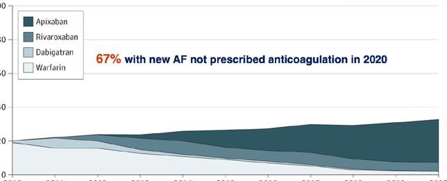
• Older age
• Anaemia
• Frailty
• Dementia
• CKD
12-month OAC initiation and direct OAC uptake in the total OAC-eligible incident atrial fibrillation cohort. OAC initiation increased from 20.2% to 32.9% (odds ratio for OAC use per year: 1.06; 95% CI: 1.06–1.07; p<0.001).
AF: atrial fibrillation; CI: confidence interval; CKD: chronic kidney disease; OAC: oral anticoagulants.
Figure 1: Oral anticoagulant initiation and direct oral anticoagulant uptake in 2010–2020.
2010 0 20 40 60 80 100 2012 2014 Apixaban Rivaroxaban Dabigatran Warfarin 2017 2019 2020 2011 2013 2016 2018 2015 Year Proportion of patients (%) 67% with new AF not
in
Creative Commons Attribution-Non Commercial 4.0 ● September 2023 ● EMJ 21 Symposium Review
prescribed anticoagulation
2020
inhibitor) is 80% renally cleared which, Connors recalled, led to a surge in emergency department visits for bleeding when it was first introduced.17 This was due to unexpected declines in patients’ creatinine clearance, which precipitated a marked increase in plasma dabigatran levels due to bioaccumulation. With the other DOACs (Factor Xa inhibitors), renal clearance ranges from approximately 50% with edoxaban to approximately 27% with apixaban.17 But even patients on apixaban may have unforeseen decline in renal function that affects their plasma drug concentration, resulting in elevated anticoagulation intensity, and a heightened risk of bleeding. Thus, the co-existence of CKD, an escalating global health issue, significantly complicates the use of DOACs in people who need anticoagulation.
Potential Drug-Drug Interactions with Direct Oral Anticoagulants
A further concern when prescribing DOACs is the possibility of major drug–drug interactions. Some patients eligible for anticoagulation are at risk of such interactions with concomitant medications that affect cytochrome P450 3A4 or P-glycoprotein metabolism, which include antibiotics such as rifampicin, antifungal agents, antiseizure medications, and, importantly, many modern targeted anticancer drugs. Connors observed that patients with cancer in particular are frequently concerned about taking medications that might interfere with the efficacy or safety of their cancer treatment. A population-based Canadian study, looking at over 642,000 DOAC prescriptions in over 36,500 patients with AF, found concomitant prescribing of a P-glycoprotein- or cytochrome P450 3A4metabolised drug in a relatively small proportion of patients (11.2%), but when an adverse drug–drug interaction was reported, inappropriate DOAC dosing was noted in 63% of cases, with a 1.6-fold higher risk of death at 1 year.18 Thus, there are specific cohorts of patients for whom DOACs may not be appropriate for this reason. It is also worth noting that understanding of potential drug-drug interactions with DOACs is incomplete, and some individuals take numerous drugs for multiple conditions, where potential interactions could multiply with unpredictable results.
Inappropriate Dose Reduction of Direct Oral Anticoagulants
In an attempt to minimise the risk of bleeding, clinicians often prescribe inappropriately low doses of DOACs.19-23 A study conducted among inpatients at the Brigham and Women’s hospital, where Connors practices, found a reduced dose regimen in 13% of 224 patients receiving DOACs.19 Pharmacist-led assessment of the renal function of these patients, alongside the package label of the DOAC prescribed, found that the reduced dose was justified in fewer than 50% of these individuals. Importantly, those on an offlabel dose reduction still experienced a roughly equal number of thrombotic events (20%) and bleeds (25%), meaning that they experienced loss of anticoagulant efficacy without greater safety. This finding was borne out by a much larger analysis of 7,577 patients in the Outcomes Registry for Better Informed Treatment (ORBIT), whose bleeding risk scores were known. Of those receiving a standard dose of DOAC, only 4% were found to be inappropriate, but of those receiving a reduced dose, 57% were deemed inappropriate.20 Finally, in an analysis of almost 15,000 patients with AF, 4% had an inappropriate standard-dose DOAC prescription (i.e., their dose had not been reduced despite having a renal indication for dose reduction) but three times as many (12%) were found to have an inappropriately reduced dose, i.e., they did not meet package label criteria for dose reduction.21 Moreover, dose reduction in apixaban-treated patients in this study was found to be associated with an increased risk of stroke without a reduced risk of bleeding.21
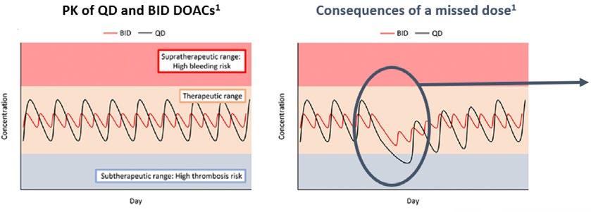
Implications of Poor Adherence with Daily Oral Drugs
In addition to undertreatment, the effectiveness of DOACs, which patients need to take orally on a daily basis due to the pharmacokinetic properties of these drugs, can be jeopardised by poor adherence.24-26 In a review of 48 realworld studies, patients with AF missed a DOAC dose once every 4 days.26 Poor adherence was associated with a 39% increased risk of thromboembolic events.26 Because of the short half-life of DOACs (5–14 hours), even one missed dose can place a patient in a ‘low protection’ zone (Figure 2).27-29
22 EMJ ● September 2023 ● Creative Commons Attribution-Non Commercial 4.0 Symposium Review
1 missed dose is expected to drop DOAC concentration into the low protection zone
This is especially true for DOACs dosed QD
Based on a systematic review of 48 RWE studies in AF
AF:
Summarising, Connors acknowledged the advantages of DOACs compared with previous anticoagulant options, but also noted that all of the prevailing concerns from the warfarin era have not been eliminated with DOACs. Many patients at risk of thromboembolism are still untreated or undertreated, and clinicians remain worried about how to manage DOAC prescribing in the elderly and those with reduced kidney function. As for adherence, this remains a problem for all daily oral drugs in preventative settings, and the DOACs for SPAF, which afford no symptom relief but may be associated with bruising and other bothersome bleeding events, are no exception.
The Promise of Factor XI Inhibition
Jeffrey Weitz
Jeffrey Weitz, Thrombosis & Atherosclerosis Research Institute (TAARI), McMaster University, Hamilton, Canada, began by reiterating current unmet needs in anticoagulation, particularly for SPAF, emphasising that the ultimate goal when prescribing an anticoagulant is to attenuate thrombosis risk without meaningfully increasing the risk of bleeding. Although the DOACs come closer to this goal than VKAs, the risk of bleeding with DOACs remains concerning, contributing to their systemic underuse. Ultimately, many patients who need
anticoagulant protection are not receiving it, highlighting the need to explore new approaches to SPAF.
Rationale for Factor XI as a New Target for Anticoagulation
Weitz went on to explain the rationale for focusing on Factor XI as a promising new target for anticoagulation, stating that the evidence to support this comes from several sources. Firstly, it has been observed that people with severe congenital Factor XI deficiency appear to be protected from thrombosis, but very rarely have serious or spontaneous bleeding.30 Secondly, large genetic epidemiology studies have shown that, compared with individuals with normal Factor XI levels, people with low Factor XI levels have a reduced risk of thrombosis, while people with high Factor XI levels are at increased risk of thrombosis.31,32 Finally, numerous animal models in rodents and non-human primates indicate that inhibition of Factor XI attenuates both venous and arterial thrombosis with no increase in bleeding.33 This is a very different outcome from that seen in animal models with DOACs where thrombosis was also attenuated, but the bleeding rate increased in line with increasing dosage.
atrial fibrillation; BID: twice per day; DOAC: direct oral anticoagulants; PK: pharmacokinetics; QD: once per day; RWE: real-world evidence.
Day Consequences of missed dose PK of QD and BID DOACs BID BID QD QD Concentration Concentration Day
Figure 2: Consequences of missed doses of direct oral anticoagulants.27,28
Supratherapeutic range: high bleeding risk Therapeutic range Supratherapeutic range: high thrombosis risk Creative Commons Attribution-Non Commercial 4.0 ● September 2023 ● EMJ 23 Symposium Review
The Prospect of ‘Uncoupling’ the Pathways of Thrombosis and Haemostasis
Weitz described our understanding that Factor XI inhibition appears able to prevent thrombosis without simultaneously impacting haemostasis, a finding inconsistent with traditional teaching about how the coagulation cascade works. The conventional depiction of the coagulation cascade suggests that the pathways leading to pathological thrombosis and physiological haemostasis are inextricably linked. Essentially, the thinking was that, by blocking fibrin, we inevitably intervene not only in harmful clotting, but also in helpful clotting, leading to the longheld belief that it is impossible to achieve effective anticoagulation without an appreciably increased bleeding risk.
However, a newer model of the coagulation cascade, informed by insights from genetic, epidemiological, and animal studies, has now emerged.34 It reveals two distinct pathways, with only one section in common: the downstream ‘common’ pathway. The pathway of physiological haemostasis, also known as the extrinsic or tissue factor pathway and measured readily by the prothrombin time, leads to the formation of extravascular haemostatic ‘plugs’ that seal leaks and injuries in vessel walls to prevent bleeding. In contrast, pathological thrombosis results from the generation of an intravascular clot that ultimately occludes the flow of blood within arteries, leading to heart attacks and strokes; or within veins, leading to deep vein thrombosis and pulmonary embolism. This process might also initiate with tissue factor, but the growth of pathologic thrombi, to an extent sufficient to occlude blood vessels (which occurs via the intrinsic pathway, and is measured by the partial thromboplastin time), depends on an amplification loop driven by Factor XI.
Weitz further explained that, if we consider the targets of currently available anticoagulants, it becomes clear that the vitamin K-dependent factors targeted by warfarin are located in both of these pathways, while Factor Xa and thrombin, targeted by DOACs, reside in the shared ‘common pathway’ (Figure 3A). This explains why these approaches to anticoagulation, while protecting against thrombosis, also undermine haemostasis, which can lead to bleeding. In contrast, Factor XI is
situated only in the amplification loop of the intrinsic, pathological thrombosis pathway. It is not involved in haemostasis, but is essential for the promulgation of harmful clotting that leads to thrombus growth and vessel occlusion (Figure 3A). Thus, by targeting Factor XI, we can conceptually ‘uncouple’ the haemostatic pathway from the pathologic thrombosis pathway (Figure 3B).34 Leaving the haemostasis pathway intact enables us, in theory, to inhibit thrombosis with a potentially minimal risk of bleeding, a vision that has long been seen as the ‘holy grail’ of anticoagulation therapy.
Current Approaches to Factor XI Inhibition
Weitz went on to review the various diverse modalities that have emerged to target Factor XI. The monoclonal antibodies abelacimab and osocimab both inhibit Factor XI but in different ways: abelacimab binds to zymogen Factor XI (the inactive precursor), blocking its activation to Factor XIa. In contrast, osocimab binds directly to the activated form, Factor XIa. Fesomersen, a second-generation antisense oligonucleotide, reduces the synthesis of Factor XI by targeting its messenger RNA in the liver. Finally, the small molecules asundexian and milvexian bind to the active site of Factor XIa to block its activity, much as the DOACs do with Factor Xa. All of these strategies have the potential to achieve anticoagulation with a lower risk of bleeding compared with current options. However, the monoclonal antibody Factor XI inhibitors may have additional benefits. These can be administered intravenously in acute care settings, to achieve a rapid onset of action, or as a once-monthly subcutaneous regimen for long-term community use, which is likely to be supportive of improved adherence. In addition, there is no dependence on renal clearance, and minimal risk of drug–drug interactions with these agents (Table 1).1,35
Phase II Data with Investigational Factor XI Inhibitors
Weitz then presented the Phase II data published to date with several of these investigational agents. With the exception of asundexian, all Factor XI inhibitors have been compared with standard of care enoxaparin for prevention of venous thromboembolism after knee replacement surgery. This is the gold standard
24 EMJ ● September 2023 ● Creative Commons Attribution-Non Commercial 4.0 Symposium Review
A B
Inhibiting Factor XI provides an opportunity to pharmacologically ‘uncouple’ the two pathways, effectively suppressing the pathological thrombosis pathway, while leaving the physiological haemostasis pathway largely unaffected.
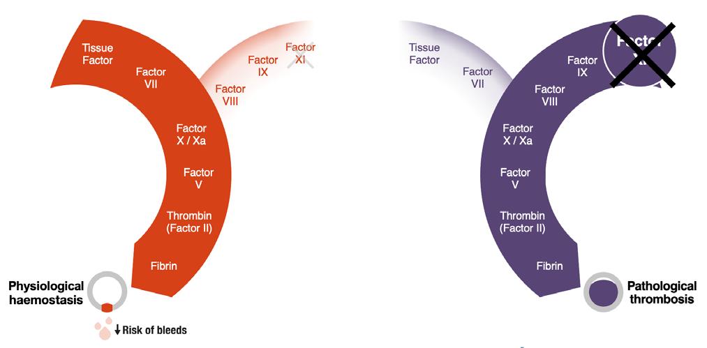
DOAC: direct oral anticoagulant.
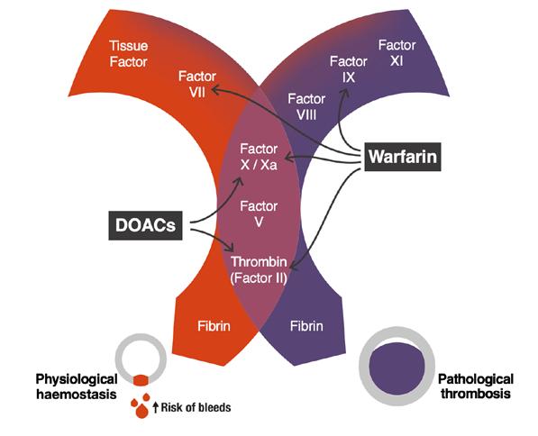 Figure 3: A new model of the coagulation cascade reveals the promise of Factor XI inhibition.34
Conventional anticoagulants all have targets located within the common pathway.
Figure 3: A new model of the coagulation cascade reveals the promise of Factor XI inhibition.34
Conventional anticoagulants all have targets located within the common pathway.
Creative Commons Attribution-Non Commercial 4.0 ● September 2023 ● EMJ 25 Symposium Review
model for evaluating the efficacy of potential new anticoagulants because patients undergoing knee replacement surgery are at high risk for venous thromboembolism, which can easily be measured, even when asymptomatic, through venography (X-ray of the veins of the operated leg). A meta-analysis of results of these Phase II studies showed an average 40% reduction in the rate of venous thromboembolism with the Factor XI inhibitors compared with enoxaparin, establishing the antithrombotic efficacy of the class.36 Although these studies were not designed to assess safety, since the absolute risk of bleeding is low with this type of surgical procedure, promising reductions in major and CRNM bleeding compared with enoxaparin were observed, and additional Phase II research is underway to investigate this further.
Phase III Evaluation of Investigational Factor XI Inhibitors
The promise of Factor XI inhibitor therapy has prompted the initiation of a range of Phase III trials, which in aggregate are planned to enrol 78,000 patients. The small molecules asundexian and milvexian are being compared with apixaban in patients with AF, and are also being studied in secondary stroke prevention (and, in the case of milvexian, in acute coronary syndrome) in combination with antiplatelet therapy. In contrast, the LILAC trial is studying abelacimab in a special subgroup of patients with AF: those deemed clinically unsuitable for any current anticoagulant, who have the most to gain from a potentially safer option. Abelacimab is also being studied in cancer-associated thrombosis, an area of growing importance and recognition.
In conclusion, Weitz reiterated that Factor XI is a promising new target that could revolutionise
Abelacimab
Agent Monoclonal antibody (fully human) Monoclonal antibody (fully human) Antisense oligonucleotide Small molecule Small molecule Mode of action Dual factor XI/XIa inhibition Factor XIa inhibition Decreases factor XI synthesis Factor XIa inhibition Factor XIa inhibition Administration SC or IV SC or IV SC Oral Oral Frequency of dosing Monthly, once Monthly, once Weekly to monthly Daily, once Daily, twice Onset of action Rapid Rapid Slow Rapid Rapid Offset of action Slow Slow Slow Rapid Rapid Renal clearance No No No Some Some Drug–drug interactions No No No Possible Possible CYP3A4 interaction No No No Yes Yes
Osocimab Fesomersen Asundexian Milvexian
CYP3A4: cytochrome P450 3A4; IV: intravenous; SC: subcutaneous.
26 EMJ ● September 2023 ● Creative Commons Attribution-Non Commercial 4.0 Symposium Review
Table 1: Mechanistic differences between investigational Factor XI inhibitors.1,35
the practice of anticoagulation, with abelacimab, asundexian, and milvexian undergoing Phase III evaluation. As a once-monthly, fully humanised antibody, abelacimab may improve adherence and eliminate concerns about drug–drug interactions or impaired kidney function in patients with atrial fibrillation at risk of stroke.35
The Burden of Bleeding from the Patient’s Perspective
Mellanie True Hills
Mellanie True Hills, American Foundation for Women’s Health, and StopAfib.org, both based
in Greenwood, Texas, USA, began by sharing insights on living with AF, and in particular, the challenges of being on anticoagulation for life. Hills announced that StopAfib.org had recently partnered with the National Blood Clot Alliance (NBCA) to run an online survey supported by Anthos Therapeutics, with the aim of investigating the impact of so-called ‘minor bleeding’ on patients’ quality of life.37 This was defined as bleeding that did not require medical attention, but may nonetheless have felt significant to the person experiencing it. More than 3,000 persons responded to the survey. It was shown that 59% of respondents had experienced bleeding and/or bruising since starting anticoagulation. Of these, almost half reported an emotional impact of these occurrences, ranging from embarrassment to
29% of patients confirm they have considered pausing, or did pause, their anticoagulant
7% did not consult their doctor at the time they stopped treatment
Have your bleeding problems made you feel like you want to stop taking anticoagulants?
Have asked their doctor to pause, stop, or reduce the dose of their anticoagulant (n=234)
Have stopped, paused, or reduced the dose of their anticoagulant without first consulting their physician (n=130)
Of those who stopped, paused, or reduced the dose of their anticoagulant themselves, later informed their doctor (n=83)
Reasons for not telling my doctor (Base: 1,815 who experienced bleeding problems)
“I'd be embarrassed. I feel that I can manage it myself.”
“I only stopped for a day or two whilst the bleeding stopped”
“I stopped for a few days because I was going to the dentist.”
“I have not seen my doctor since reducing my dosage. I intend to tell him soon.”
Base: 1,815 (59%) experiencing a bleeding problem on current treatment.
From Patient-Relevant Bleeding Events Among Patients Taking Anticoagulant Medication (Insocius, November 2022): A global survey of 3,000+ patients prescribed anticoagulants, conducted through StopAfib.org and the National Blood Clot Alliance (NBCA), to gain quantitative and qualitative insight on the impact of patient-relevant bleeding events, which were defined as bleeding not requiring medical intervention.37
Figure 4: Impact of bleeding problems on patient adherence with their anticoagulant.37
1 No 71% Yes
29%
13% 7% 5%
Creative Commons Attribution-Non Commercial 4.0 ● September 2023 ● EMJ 27 Symposium Review
anxiety and depression. In particular, the fear of a future serious bleed, especially if help is not close at hand, was expressed by numerous patients.
More than half of respondents said that they had changed their lifestyle, discontinuing sports and hobbies that carry even a small risk of injury, and limiting household tasks involving knives or sharp objects, including shaving and personal grooming, because they were taking an anticoagulant. Those who continued performing these activities reported having to take extra care, extra time, and sometimes extra expense to protect themselves. Many reported choosing clothing that covers up bruises to avoid the comments or judgement of others. In many cases, issues that may seem trivial to healthcare professionals emerged as major preoccupations for patients.
A further notable finding was that the impact on emotions and lifestyle was greater for younger people (aged 21–49 years; n=408) compared with older people (aged 50 years or over; n=1,407). Some younger respondents reported feeling socially isolated, as their peers could not meaningfully relate to their health issues. In fact, of all bleeding incidents, the greatest emotional impact came from heavy periods in
References
1. Fredenburgh JC, Weitz JI. Factor XI as a target for new anticoagulants. Hamostaseologie. 2021;41(2):10410.
2. Ruff CT et al. Comparison of the efficacy and safety of new oral anticoagulants with warfarin in pa-tients with atrial fibrillation: a meta-analysis of randomised trials. Lancet. 2014;383(9921):955-62.
3. Patel MR et al. Rivaroxaban versus warfarin in nonvalvular atrial fibrillation. N Engl J Med. 2011;365(10):883-91.
4. Majeed A et al. Management and outcomes of major bleeding during treatment with dabigatran or warfarin. Circulation. 2013;128(21):2325-32.
5. Granger CB et al. Apixaban versus warfarin in patients with atrial fibrillation. N Engl J Med. 2011;365(11):981-92.
6. Giugliano RP et al. Edoxaban versus warfarin in patients with atrial fibrillation. N Engl J Med.
premenopausal females, who were forced to stay home from work, and significantly curtail their usual activities while menstruating.
Overall, almost 30% of respondents reported that, as a result of their experiences with bleeding and/or bruising, they had considered pausing their anticoagulant, or actually did pause it. Some (7%) did so without first telling their doctor (Figure 4).37
This finding elevates the problem from a purely quality of life issue to a prognostic one. The bottom line, Hills stated, is that what doctors perceive as ‘minor bleeding’ is anything but to patients. Hills proposed that ‘minor bleeding’ should be reframed as “patient relevant bleeding,” with the recognition that these experiences can seriously jeopardise both quality of life and adherence to anticoagulation regimens. Many patients accept this situation as the cost they have to pay for avoiding strokes and other thromboembolic events, but huge interest was expressed in the possibility of a new anticoagulant with a lower bleeding risk. Thus, Hills concluded: “We hope that the emerging class of Factor XI inhibitors will be the answer for us.”
2013;369(22):2093-104.
7. Beyer-Westendorf J et al. Rates, management, and outcome of rivaroxaban bleeding in daily care: re-sults from the Dresden NOAC registry. Blood. 2014;124(6):95562.
8. Bhat A et al. Barriers to guidelinedirected anticoagulation in patients with atrial fibrillation: new approaches to an old problem. Can J Cardiol. 2023;39(5):625-36.
9. Lippi G et al. Global epidemiology of atrial fibrillation: an increasing epidemic and public health challenge. Int J Stroke. 2021;16(2):217-21.
10. Rahman F et al. Global epidemiology of atrial fibrillation. Nat Rev Cardiol. 2014;11(11):63954.
11. Gao X et al. Diagnostic accuracy of the HAS-BLED bleeding score in VKA- or DOAC-treated patients with atrial fibrillation: a systematic review and meta-analysis. Front Cardiovasc Med. 2021;8:757087.
12. Ko D et al. Trends in use of oral anticoagulants in older adults with newly diagnosed atrial fibrillation, 2010-2020. JAMA Netw Open. 2022;5(11):e2242964.
13. Rose AJ et al. Anticoagulant prescribing for non-valvular atrial fibrillation in the Veterans Health Ad-ministration. J Am Heart Assoc. 2019;8(17):e012646.
14. Mitchell A et al. Prescribing of direct oral anticoagulants and warfarin to older people with atrial fibrillation in UK general practice: a cohort study. BMC Med. 2021;19(1):189.
15. Washam JB et al. Pharmacotherapy for atrial fibrillation in patients with chronic kidney disease: insights from ORBIT-AF. J Am Heart Assoc. 2018;7(18):e008928.
16. Olesen JB et al. Stroke and bleeding in atrial fibrillation with chronic kidney disease. N Engl J Med. 2012;367(7):625-35.
17. Steffel J et al. The 2018 European Heart Rhythm Association practical guide on the use of non-vitamin K
28 EMJ ● September 2023 ● Creative Commons Attribution-Non Commercial 4.0 Symposium Review
antagonist oral anticoagulants in patients with atrial fibrillation. Eur Heart J. 2018;39(16):1330-93.
18. Sandhu RK et al. Concurrent use of P-glycoprotein or cytochrome 3A4 drugs and non-vitamin K an-tagonist oral anticoagulants in non-valvular atrial fibrillation. Eur Heart J Qual Care Clin Outcomes. 2022;8(2):195-201.
19. Barra ME et al. Evaluation of dose-reduced direct oral anticoagulant therapy. Am J Med. 2016;129(11):1198-204.
20. Steinberg BA et al. Frequency and outcomes of reduced dose non-vitamin K antagonist anticoagulants: results from ORBITAF II (the Outcomes Registry for Better Informed Treatment of Atrial Fibril-lation II). J Am Heart Assoc. 2018;7(4):e007633.
21. Yao X et al. Non-vitamin K antagonist oral anticoagulant dosing in patients with atrial fibrillation and renal dysfunction. J Am Coll Cardiol. 2017;69(23):277990.
22. Leef GC et al. Appropriateness of direct oral anticoagulant dosing in patients with atrial fibrillation: insights from the Veterans Health Administration. J Pharm Pract. 2020;33(5):647-53.
23. Fernández CS et al. The problem of underdosing with direct-acting oral anticoagulants in elderly patients with nonvalvular atrial fibrillation. J Comp Eff Res. 2020;9(7):509-23.
24. Andrade JG et al. Values and preferences of physicians and patients with nonvalvular atrial fibrilla-tion who receive oral anticoagulation therapy for stroke prevention. Can J Cardiol. 2016;32(6):747-53.
25. Yao X et al. Effect of adherence to oral anticoagulants on risk of stroke and major bleeding among patients with atrial fibrillation. J Am Heart Assoc. 2016;5(2):e003074.
26. Ozaki AF et al. Real-world adherence and persistence to direct oral anticoagulants in patients with atrial fibrillation: a systematic review and metaanalysis. Circ Cardiovasc Qual Outcomes. 2020;13(3):e005969.
27. Ido T et al. Twice- or once-daily dosing of direct oral anticoagulants and gastrointestinal bleeding in patient with atrial fibrillation. Am Heart J Plus Cardiol Res Practice. 2022;22:100203.
28. Paquette M et al. Methodological considerations for investigating oral anticoagulation persistence in atrial fibrillation. Eur Heart J Cardiovasc Pharmacother. 2021;7(3):251-60.
29. Gosselin RC et al. International Council for Standardization in Haematology (ICSH) recommendations for laboratory measurement of direct oral anticoagulants. Thromb Haemost. 2018;118(3):437-50.
30. Asselta R et al. Exploring the global
landscape of genetic variation in coagulation factor XI deficiency. Blood. 2017;130(4):e1-6.
31. Meijers J et al. High levels of coagulation Factor XI as a risk factor for venous thrombosis. N Engl J Med. 2000;342(10):696701.
32. Preis M et al. Factor XI deficiency is associated with lower risk for cardiovascular and venous throm-boembolism events. Blood. 2017;129(9):1210-5.
33. Gailani D, Gruber A. Factor XI as a therapeutic target. Arterioscler Thromb Vasc Biol. 2016;36(7):1316-22.
34. Hsu C et al. Factor XI inhibition to uncouple thrombosis from hemostasis: JACC review topic of the week. J Am Coll Cardiol. 2021;78(6):625-31.
35. Fredenburgh JC, Weitz JI. News at XI: moving beyond Factor XA inhibitors. J Thromb Haemost. 2023;21(7):1692-702.
36. Nopp S et al. Factor XI inhibitors for prevention and treatment of venous thromboembolism: a re-view on the rationale and update on current evidence. Front Cardiovasc Med. 2022;9:903029.
37. Bloomfield D et al. Patient-relevant bleeding events among patients taking anticoagulant medication. Abstract 1441167. ISTH 2023 Congress, 26 June, 2023.
FOR REPRINT QUERIES PLEASE CONTACT: INFO@EMJREVIEWS.COM Creative Commons Attribution-Non Commercial 4.0 ● September 2023 ● EMJ 29 Symposium Review
The Unspeakable Disease: A Tale of Two Siblings
Interviewees: Simon Jones,1-3 Dipak Ram,4 Ally Shaw5
1. St Mary’s Hospital, Manchester, UK
2. Honorary Manchester Academic Health Science Centre (MAHSC), UK
3. University of Manchester, UK
4. Royal Manchester Children’s Hospital, UK
5. Primary caregiver of patients mentioned
Disclosure:
Jones has declared no payment linked to this article, but has received honoraria/consulting fees from Orchard linked to past activities. Ram has declared no payment linked to this article, but has received honoraria/consulting fees from Orchard linked to past activities. Shaw has declared no payment linked to this article, and has nothing else to declare.
Disclaimer: The opinions expressed in this article belong solely to the named interviewees.
Support: The publication of this article was sponsored by Orchard Therapeutics.
Keywords: ARSA gene, gene therapy, lysosomal storage disease, newborn screening.
Citation: EMJ. 2023;8[3]:30-37. DOI/10.33590/emj/10304055. https://doi.org/10.33590/emj/10304055.
Interview Summary
The deficiency of arylsulfatase A due to the mutations in the ARSA gene is the cause of a rare inherited lysosomal storage disease, resulting in the accumulation of sulfatides in the central nervous system (CNS) and peripheral nervous system (PNS). This, in turn, leads to progressive demyelination, neuroinflammation, and neurodegeneration, and the accumulation in visceral organs.

Affected young children gradually lose the ability to walk, stand, talk, and swallow; they lose their independence and show a steady physical and cognitive regression resulting, ultimately, in their premature death at a younger age. This condition not only devastates young patients, but it also deeply affects their families and carers, both psychologically and economically.
In this interview, Simon Jones, Consultant in Paediatric Inherited Metabolic Disease at St Mary's Hospital, Manchester, Honorary Manchester Academic Health Science Centre (MAHSC), and Professor in Paediatrics and Senior Lecturer at the University of Manchester, UK; Dipak Ram, Royal College of Paediatrics and Child Health (RCPCH) and British Paediatric Neurology Association (BPNA) National Training Advisor for Paediatric Neurology, and Consultant Paediatric Neurologist at the Royal Manchester Children’s Hospital, UK; and Ally Shaw, primary caregiver of the patients mentioned in this article, explore how the disease impacts the normal development of predominantly young patients; consider its subtle evolution; and witness the stressful diagnostic odyssey families are experiencing, often leading to the wrong diagnosis, which is compounded by the lack of a national newborn screening.
30 EMJ ● September 2023 ● Creative Commons Attribution-Non Commercial 4.0 Interview
Jones and Ram speak directly about the diagnosis and management of these patients. This article also includes the experience of a mother of two affected children, one of whom was diagnosed thanks to their affected older sister, who gave the younger child the chance of receiving a timely treatment. In this disease, time is of the essence; often, patients are sadly diagnosed too late, and are destined to palliative care and premature death. It is hoped that this interview and testimony will help raise awareness on this disease, and give a chance to future patients and their families.
With a prevalence rate estimated to be around 1 in 40,000–160,000 worldwide, there is only a small chance that a physician will come across a patient with this devastating lysosomal storage disease during their career.1 This may lead some healthcare professionals to assume the condition is too rare to invest adequate time to familiarise themselves with its (early) symptoms, prioritising their focus on other diseases with greater incidence.
To complicate the matter, affected young children may not manifest any concerning symptoms until it is too late. For a period, they exhibit apparently healthy growth, meeting the developmental milestones of their nonaffected peers. However, this subtle illness is gradually and inexorably causing permanent and cumulative damage in multiple organs, eventually resulting in progressive motor and cognitive deterioration, with loss of motor and neurocognitive functions and, ultimately, death.
This unspeakable condition is a rare inherited lysosomal storage disease caused by deficiency of arylsulfatase A (ARSA), due to mutations in the ARSA gene. The reduced ARSA activity results in the accumulation of sulfatides in the CNS and PNS, leading to progressive demyelination, neuro-inflammation, and neurodegeneration, in addition to the accumulation in visceral organs, such as the gallbladder and kidneys.2
“The disease progression in the most severe form of the disease follows a typical pattern,” said Jones. “Affected young children gradually lose the ability to walk, stand, talk, [and] swallow; they lose their independence and show a steady physical and cognitive regression. Their ability to communicate, express themselves is progressively heavily compromised. They become detached from the world around them, unable to share their feelings, slowly becoming a shadow of their former selves, bedridden
until their premature death at a younger age. As this horrible condition devastates these children, it also deeply affects their families, and carers both psychologically and economically, a heartbreaking convergence of negative factors causing an incredible amount of stress for those looking after their sick child.”
A caregivers-reported impact on quality of life and disease burden in diagnosed patients provides a grim depiction of what these families experience. Some 94% of carers said that the illness has led to considerable change in their lifestyle, and 50% had seen their emotional relationship with their spouse/partner deteriorate to an extreme degree. In addition, 76% of carers had suffered a loss of income due to the cessation of their professional full- or part-time activity, due to the 15 hours a day spent on average caring for their child.3
“Unfortunately, the subtleness of this condition frequently results in a diagnostic odyssey; young patients are often referred to several specialists in an attempt to manage the symptoms appearing at different stages of disease progression, the result of a gradual accumulation of sulfatides in the tissues and organs,” explained Ram. “In the late infantile and most severe subtype, for example, the hypotonia, muscle weakness, gait disturbances, abnormal movement patterns, and frequent falls are often the first symptoms that parents or carers notice first, which worry them. They consult their general practitioner, who may or may not refer the young patient to a paediatrician or paediatric neurologist for further investigations. This takes time, and time for these patients is extremely precious. In addition, clinicians may adopt a watch-and-wait approach, hoping that some of these atypical symptoms self-resolve, or provide additional clues on what to do next. Unfortunately, even if this may be the right
Creative Commons Attribution-Non Commercial 4.0 ● September 2023 ● EMJ 31 Interview
rationale for other conditions, a late diagnosis in this scenario leads to fatal consequences.”
A study of caregivers in the USA who were caring for, or had cared for, a living or deceased patient classified as having late infantile or juvenile subtypes of the disease, confirmed this assessment.4 The most common initial symptoms reported in late infantile patients related to problems with gross motor function. Often, this was observed as a delay in developmental progression, particularly in walking, and nearly 70% of late infantile patients never learned to walk independently. In addition to motor function-related observations, such as losing balance, falling over, and relying on holding hands to be able to walk, fine motor or related symptoms were also commonly reported as pre-diagnosis symptoms in late infantile patients. This included problems with eye movement, eating, or swallowing, and hand tremors. In addition, nearly 44% of late infantile patients experienced pre-diagnosis speech problems. In comparison, patients with the juvenile subtype, as expected, exhibited initial symptoms later, and the first symptoms often related to changes in cognitive function (56.3%) or social/behavioural function (43.8%). The initial symptoms were usually noticed at school, following a decline in academic performance, difficulty focusing, or disruptive behaviour.
“The collective incidence of inherited disorders affecting the lysosomes is roughly 1 in 2,300–7,000 live births,” explained Jones, “and among the different lysosomal disorders, the general prevalence of this autosomal recessive condition varies within diverse populations, especially when looking at the different degrees of consanguinity.”5
This disease is a pan-ethnic lysosomal storage disease, with affected patients found in several populations, such as European, Iranian, Indian, Japanese, Jewish, Habbanite Jewish, Lebanese, Muslim Arab, South African, Polynesian, Algerian, Navajo Indian, Alaskan Eskimo, and Christian Arab, with phenotypes ranging from the mild to the severe.6
The mutations in the ARSA gene in homo- or heterozygosity is the triggering factor, with over 150 mutations (of which more than 70% are missense) reported in these patients.7 Missense
mutations in specific genes encoding for misfolded mutant lysosomal enzymes can cause lysosomal storage disorders. However, most of these misfolded mutant lysosomal enzymes are destroyed, and only a small amount of the mutant enzyme eventually reaches the lysosomal compartment and remains functionally active. This residual enzymatic activity ranges between 0–10% of the normal activity detected from the wild-type ARSA 8 The critical threshold of ARSA enzymatic activity (about 10% of that measured in the wild-type ARSA) is considered the minimum, above which the activity is sufficient to prevent the accumulation of sulfatides. In what is known as ARSA pseudo-deficiency, the levels of enzyme activity between 10–15% of wild-type ARSA levels result in no sulfatide accumulation, and no disease-related symptoms.
Sulfatides (a type of glycosphingolipid) are the main component of the myelin sheath in both the CNS and PNS. In the CNS, oligodendrocytes situated in the myelin surrounding neural axons are responsible for their synthesis. In the PNS, the neurolemmocytes are the myelin-producing cells.5 Sulfatides account for 4% of myelin composition, the most abundant sphingolipid. An excessive concentration of sulfatides and deficiency of cerebrosides can lead to an abnormal myelin composition, potentially affecting the development of a stable lipid bilayer.9 In addition, the sulfatide metabolite galactosylsphingosine can cause cytotoxicity, leading to the death and dysfunction of neuronal cells.10 The progressive accumulation of sulfatides causes dysfunction of the lysosomalendosomal system, triggering other secondary pathogenic cascades, which ultimately result in apoptosis.11 The progressive demyelination in both CNS and PNS correlates with the clinical manifestations of the disease.12
Sulfatides can also be found, albeit in low concentrations, in the respiratory tract, gastric mucosa, and uterine endometrium. They accumulate in the epithelium cells of the gallbladder, resulting in sludge, gallstones, haemobilia, cholecystitis, polyposis, papillomatosis, and a small or enlarged gallbladder.13-20 A retrospective cohort study performed at the Duke University Medical Center, Durham, North Carolina, USA, provided additional evidence on the incidence of gallbladder abnormalities.21 The small study, which used a prospectively maintained
32 EMJ ● September 2023 ● Creative Commons Attribution-Non Commercial 4.0 Interview
database, included 87 children who underwent haematopoietic stem cell transplant (HSCT) at the same institution between 1994–2015. Children were stratified into two groups: 58 patients with a diagnosis of adrenoleukodystrophy or Krabbe disease (Cohort 1), and the remaining 29 patients with the lysosomal storage disease subject of this article (Cohort 2). Children in Cohort 2 were more likely to show gallbladder abnormalities on imaging, the most common of which was sludge, compared with children with other lysosomal storage diseases.
“This neurological lysosomal storage disorder displays a broad and heterogeneous clinical spectrum combining age-based progression and a genotype/phenotype continuum. Homozygosity for 0 alleles (0/0) codifying for ARSA enzyme, is reported only in patients characterised as early-onset, whereas phenotypic variability is more evident in the juvenile and adult subtypes,” explained Jones. “This means that genotype/ phenotype correlation is more evident in those patients presenting with a 0/0 genotype, whereas predicting the phenotype associated with 0/R or R/R profile is much harder and more complex.”6,22
The three clinical forms are based on the age of symptom onset, the rate of progression of neurological symptoms, and their severity. The late infantile subtype is the most severe form, with the first symptoms developed at just a few months of age up to 30 months. The juvenile clinical form includes patients with symptom onset occurring between 30 months–16 years of age, and lastly patients with the adult clinical form see their symptoms manifest from 17 years onwards. The two late-onset forms, juvenile and adult, at times overlap, and they may present with a more insidious manifestation of a wide range of neurological symptoms, offering more opportunities for therapeutic interventions.
Patients with the late infantile form of the disease show delays in psychomotor development, characterised by impairment of speech, and gross and fine motor development. In addition, patients can present with muscular hypotonia, hyporeflexia, spasticity, and hyperreflexia. Other symptoms seen are ataxia, spastic paresis, and optic dystrophy, all of which may be common in other diseases, and lead to difficulties in differential diagnosis. A study including patients from two national cohorts at the University
Children’s Hospital, Tübingen, Germany, and Amsterdam University Medical Center, the Netherlands, found that paralytic strabismus was an early sign in patients presenting with the late infantile subtype, and hypothesised this could be the result of early cranial nerve involvement.23
In the juvenile form, patients develop the same symptoms, but less rapidly. Early juvenile children (symptom onset between 30 months–<7 years of age) typically display poor school performance, some behavioural disturbances, and peripheral neuropathies, which may mimic other conditions (e.g., attention deficit hyperactivity disorder [ADHD]). As the disease progresses, patients become debilitated, losing gait independence and speech. Seizures and recurrent pulmonary infections also occur frequently. Despite the slower pace of regression, once these children lose the ability to walk, the disease progresses at a comparable rate to the late infantile subtype.24
Symptoms of the late juvenile subtype become visible between 7–16 years of age, usually associated with behavioural or cognitive issues. Prognosis is variable, with a fast disease progression if motor symptoms are seen at disease onset, while patients with behavioural and cognitive issues at diagnosis tend to have a less severe condition, independently of age at onset.24 Often the deterioration of school performance, regression of verbal skills, behavioural or emotional disturbances, and fine motor functions damage are the initial symptoms in these patients. Initially, when motor function may still be intact, symptoms may be suggestive of other pathologies, such as schizophrenia, depression, ADHD, and autistic spectrum disturbances. About 61% of patients present only with cognitive symptoms at disease onset, whereas the remaining 39% exhibit only motor symptoms, or a combination of both motor and cognitive symptoms.24
Lastly, the hallmark of the adult form is a gradual impairment of intellectual function, emotional instability, behavioural/psychiatric disorders, and epileptic seizures. At a later stage, polyneuropathy occurs, and does not usually present symptoms. This form of the disease shows the first symptoms after puberty (around 16 years of age), but they have been noticed in patients older than 60 years of age. The first symptoms involve poor school or work
Creative Commons Attribution-Non Commercial 4.0 ● September 2023 ● EMJ 33 Interview
performance. Patients may show emotional instability, disorganised thoughts, and sometimes psychiatric symptoms, such as hallucinations or delirium, which may lead to incorrectly diagnosing schizophrenia, psychotic depression, or dementia.
“The difference in the pace of disease progression has implications in how patients are managed,” said Ram, and “it is important to diagnose the condition in a timely fashion to be able, where possible, to treat patients or anticipate their needs when they are not treatable, ensuring some optimised care. The sooner these patients are diagnosed, the better it is, and this is particularly important for the most severe subtype.”
The accumulation of excess sulfatides resulting from the absence of ARSA enzymatic activity, or a low level of activity, makes them a good starting point to determine the diagnosis.
“The detection of low ARSA levels in the blood, or raised urinary sulfatides, are the main rapid diagnostic tools in our possession,” continued Ram. “For the final diagnosis, we rely on additional procedures, which include molecular genetic tests, electrophysiological tests, and brain MRI.1,25,26 It is important to remember, however, that early-onset patients might have normal or non-characteristic, mildly affected MRIs when they are already symptomatic, which means that this investigative technique should not be used as the main tool to diagnose patient[s], but as a confirmatory one.”
Ram’s comments were reinforced by the results of a retrospective study in which the authors reviewed the medical records of patients followed at the Amsterdam Leukodystrophy Center, the Netherlands, or Tübingen University Hospital, Germany.27 They evaluated 104 MRI images, and discovered that nearly a quarter (n=10) of the late infantile patients (n=43) were initially misdiagnosed with chronic inflammatory demyelinating polyneuropathy, unclassified polyneuropathy, Dejerine–Sottas disease, Segawa syndrome, oculomotor apraxia, post-infectious gait ataxia, and spinocerebellar ataxia; and in all these cases, a normal appearing MRI was an important factor for the incorrect diagnosis. The authors stated that CNS symptoms, therefore, may precede MRI abnormalities in these children,
and recommended that clinicians pay attention to signs of upper motor neuron involvement, such as scissoring, Babinski signs, elevated muscle tone, and increased tendon reflexes during physical examination, to help avoid misdiagnosis as an isolated peripheral demyelinating polyneuropathy. Misdiagnoses of the juvenile and adult forms are not uncommon, and include ADHD for the former and schizophrenia for the latter.1,4
“The challenge we currently have is to find patients before it is too late”, said Jones. “Our team at the Royal Manchester Children’s Hospital has recently published the results of a 12-month study on the number of patients referred to us for treatment.28 The findings are chilling, with only four out of 17 UK patients asymptomatic at [the] time of diagnosis and therefore eligible for gene therapy, which is available in the UK after European Medicines Agency (EMA) approval in 2020. The rest could only receive supportive care. It is a particularly difficult situation; the window for treatment is extremely narrow, especially for early-onset children. For them to have a chance, they should be minimally or asymptomatic at diagnosis, but without a national newborn screening [programme], finding them early is a nearly impossible task. Unfortunately, the patients we were able to treat were identified thanks to an older affected sibling whose disease was too advanced to be eligible for treatment. In essence, the older sibling saved the younger one, a heartbreaking scenario for families to process and accept; it really becomes a story of two siblings.”
Until the EMA’s approval of a gene therapy, there was no sufficiently effective therapy for some patients. Symptoms can be treated pharmacologically, for instance, using antiepileptic drugs for seizures or myorelaxants for muscular spasms. Gastro-oesophageal reflux and constipation can be managed with targeted treatments. Pharmacological support may also be necessary to treat pain. Swallowing difficulties can be attenuated using supplements, modifying the consistency or volume of meals, and by placing a feeding tube to alleviate discomfort and help prevent aspiration pneumonia.
The gradual physical decline of patients will require the adoption of a wheelchairs, walkers, and electronic communication devices to optimise quality of life. Psychological support is often
34 EMJ ● September 2023 ● Creative Commons Attribution-Non Commercial 4.0 Interview
necessary for both the patient and their family. This supportive care may extend the lifespan of a patient by dealing with growing complications, such as nutritional challenges and pulmonary infections, but overall, quality of life remains poor and, despite the alleviation of symptoms, the rate of disease progression is not reduced.1
“In the past two decades, [allogeneic] allo-HSCT has been the only available treatment for the juvenile form of the disease,” said Jones. “The rationale of this approach is that monocytes derived from donor cells can cross the blood–brain barrier, becoming a local source of missing enzyme for the neighbouring cells in the CNS, preventing the process of demyelination and neurodegeneration.”
The results seen in late infantile and early juvenile subtypes have been limited in demonstrating a significant impact on motor and cognitive decline, possibly due to the inability of donorderived cells to produce supraphysiological levels of ARSA enzyme. Some patients have shown rapid disease progression following alloHSCT compared with non-transplanted patients, which may suggest the negative impact of this procedure as a trigger to accelerate disease progression. In addition, the benefits on late infantile patients appear to be extremely limited, even when patients are in the pre-symptomatic phase of the disease.
“It is likely that in these children, the adequate enzymatic levels needed are reached too late, and in the meantime the disease has already entered the rapidly progressive phase,” said Jones. “In most of these patients, motor and cognitive decline, including the ability to communicate verbally, was observed even after allo-HSCT, leading to the conclusion that this option is not particularly useful in symptomatic patients, or in those with the most severe disease subtype. In addition, engrafted donorderived microglial cells are only able to produce a physiological level of the ARSA enzyme, which [is] insufficient to cross-correct the patient’s defective neuronal cells.”
When considering the effects of allo-HSCT on neuropathic pain, the results have been disappointing, showing a slow worsening in the years following the transplant.29,30 The safety implications of allo-HSCT also need to
be considered, as the procedure is associated with a risk of severe complications, transplant rejection, graft-versus-host disease, and mortality risks due to the intense conditioning regimen used. Additional limitations include the availability of a compatible donor, which may delay treatment and compromise its efficacy. In current clinical practice, allo-HSCT is restricted to pre- or pauci-symptomatic patients showing a late disease onset and slow progression.
“Nala was just an absolute character, very theatrical, always singing, always dancing, had everyone laughing,” recounted Ally Shaw, the mother of two young girls. “When she was about 6 month[s] old, I started noticing something unusual; she would not put her feet flat, and as a first-time mother I thought that was not normal.
“I, therefore, consulted the doctors, and they would reassure me that nothing was out of the ordinary; after all, she was only 6 month[s] old; [there was] nothing they would do at this stage. They suggested to wait until she was walking, and then they could reassess. When she did start walking, I continued to have concerns, but I still wasn’t being taken very seriously by the specialists.
“Eventually, she was referred to physiotherapists. She went to them every few months to start with, and they just kept saying she would grow out of it. They did not have any worrying concerns, and she got discharged.
“Nala continued to walk a bit strange, and 6 months later I went back to a physiotherapist, but was not taken seriously until she started falling over a lot and develop tremors when she was about 2 and a half. And that is when I started to look online, and really think there was something more seriously wrong.
“Originally, I thought she had a brain tumour or cerebral palsy, since the symptoms were very similar. Eventually, I managed to get her seen by a paediatrician who ruled out the brain tumour, suspecting a diagnosis of cerebral palsy instead.
“I still had doubts. I did not believe that it was cerebral palsy because of how fast she was changing, [and] I was very much convinced it was a brain tumour. By this point, even with my massive concerns, it seemed like there was no rush to do anything, and she was put on a
Creative Commons Attribution-Non Commercial 4.0 ● September 2023 ● EMJ 35 Interview
waiting list in late February–early March of 2022, and by the April Nala had gotten to the point where she could not walk at all.
“At night-time she was screaming as if she was in great pain, and this went on for a couple of weeks until I phoned an ambulance that took her to the Newcastle Royal Victoria Infirmary [UK], a children’s hospital. Upon arrival, they were instantly concerned that something was seriously wrong, and within 2 hours we were seen by a team of neuro-doctors who thought she may have a brain tumour.
“She was rushed for an emergency MRI, and within about 45 minutes the doctor came to tell me and my husband, Jake, that they had found a leukodystrophy.
“I had never heard of metachromatic leukodystrophy (MLD); I do not think anyone’s ever heard of MLD unless it horribly gets thrown in your life. I did not have a clue what it was, [or] how bad it was. No idea.
“We were told instantly there was absolutely nothing to do because Nala had completely stopped walking, and no active treatment could be offered. When we were told that Teddi, Nala’s younger sister, had the same disease, we were in shock; however, they said that there might have been an available gene therapy treatment for her. As a parent, being told that only one child can be helped is almost crueller than being told that both cannot.
References
1. Shaimardanova AA et al. Metachromatic leukodystrophy: diagnosis, modeling, and treatment approaches. Front Med (Lausanne). 2020;7:576221.
2. Fumagalli F et al. Lentiviral haematopoietic stem-cell gene therapy for early-onset metachromatic leukodystrophy: long-term results from a nonrandomised, open-label, phase 1/2 trial and expanded access. Lancet. 2022;399(10322):372-83.
3. Sevin C et al. An international study of caregiver-reported burden and quality of life in metachromatic leukodystrophy. Orphanet J Rare Dis. 2022;17(1):329.
4. Harrington M et al. Insights into the
“MLD is probably just one of the most horrible diseases I have ever known. To watch a child be stripped of everything is just horrendous. Since Nala got diagnosed, she lost all her skills very rapidly, and within weeks she had lost the ability to speak, to eat, and to hold her own body up.
“When Teddi had to go into hospital for a long period of time for treatment, I could not expect my employers to keep my job just open, and I gave it up. It has been a massive struggle for me and Jake, and without the community support, I think we would struggle to even pay bills.
“What I am going through is the reason why I have become a great advocate of newborn screening. If MLD was screened at birth, my child would not be dying, and she would have potentially had the treatment that her sister had, changing our lives from watching a child slowly leaving us to a growing one. Knowing that she is going to die is the hardest thing.
“My advice to other parents is that if they strongly suspect something is wrong with their children, they must push and fight and fight, because unfortunately it is about who shouts the loudest.”
“MLD is [a] horrific disease,” concluded Jones, and “without newborn screening more children and families will suffer. We do have a chance to prevent this suffering if we act quickly, because we do have an approved treatment available. Now is the right time to act.”
natural history of metachromatic leukodystrophy from interviews with caregivers. Orphanet J Rare Dis. 2019;14(1):89.
5. Patil SA, Maegawa GHB. Developing therapeutic approaches for metachromatic leukodystrophy. Drug Des Devel Ther. 2013;7:729-45.
6. Von Figura K, “Metachromatic leukodystrophy,” Scriver C et al. (eds.) Metabolic and Molecular Basis of Inherited Disease (2001), New York: McGraw-Hill, pp.3696724.
7. Stenson PD et al. The human gene mutation database: 2008 update. Genome Med. 2009;1(1):13.
8. Leinekugel P et al. Quantitative correlation between the residual
activity of beta-hexosaminidase A and arylsulfatase A and the severity of the resulting lysosomal storage disease. Hum Genet. 1992;88(5):513-23.
9. O’Brien JS, Sampson EL. Myelin membrane: a molecular abnormality. Science. 1965;150(3703):1613-4.
10. Tanaka T et al. Ibotenic acidinduced nigral lesion and limbic seizure in cats. Brain Res. 1989;498(2):215-20.
11. Vitner EB et al. Common and uncommon pathogenic cascades in lysosomal storage diseases. J Biol Chem. 2010;285(27):20423-7.
12. Webster HD. Schwann cell alterations in metachromatic leukodystrophy: preliminary
36 EMJ ● September 2023 ● Creative Commons Attribution-Non Commercial 4.0 Interview
phase and electron microscopic observations. J Neuropathol Exp Neurol. 1962;21:534-54.
13. Krivan HC et al. Adhesion of Mycoplasma pneumoniae to sulfated glycolipids and inhibition by dextran sulfate. J Biol Chem. 1989;264(16):9283-8.
14. Sugano K et al. Localization of sulfatides in the epithelial lining of gastric mucosa: studies with a monoclonal antibody to sulfatides. J Clin Gastroenterol. 1995;21(Suppl 1):S98-103.
15. Takamatsu K. Phytosphingosinecontaining neutral glycosphingolipids and sulfatides in the human female genital tract: their association in the cervical epithelium and the uterine endometrium and their dissociation in the mucosa of fallopian tube with the menstrual cycle. Keio J Med. 1992;41(3):161-7.
16. Siegel EG et al. Repeated upper gastrointestinal hemorrhage caused by metachromatic leukodystrophy of the gall bladder. Digestion. 1992;51(2):121-4.
17. Ries M, Deeg KH. Polyposis of the gallbladder associated with metachromatic leukodystrophy. Eur J Pediatr. 1993;152(5):450-1.
18. Kim TS et al. Involvement of the gallbladder in childhood metachromatic leukodystrophy:
ultrasonographic findings. J Ultrasound Med. 1996;15(12):8215.
19. Oak S et al. Papillomatosis of the gallbladder in metachromatic leukodystrophy. Pediatr Surg Int. 1997;12(5-6):424-5.
20. Simanovsky N et al. Unusual gallbladder findings in two brothers with metachromatic leukodystrophy. Pediatr Radiol. 1998;28(9):706-8.
21. Jina Kim et al. Gallbladder abnormalities in children with metachromatic leukodystrophy. J Surg Res. 2017;208:187-91.
22. Gieselmann V, Krägeloh-Mann I. Metachromatic leukodystrophyan update. Neuropediatrics. 2010;41(1):1-6.
23. Beerepoot S et al. Acute-onset paralytic strabismus in toddlers is important to consider as a potential early sign of late-infantile metachromatic leukodystrophy. Eur J Paediatr Neurol. 2022;37:8793.
24. Kehrer C et al. Association of age at onset and first symptoms with disease progression in patients with metachromatic leukodystrophy. Neurology 2021;96(2):e255-66.
25. van Rappard DF et al. Metachromatic leukodystrophy:
disease spectrum and approaches for treatment. Best Pract Res Clin Endocrinol Metab. 2015;29(2):26173.
26. Gomez-Ospina N, “Arylsulfatase A deficiency,” Adam MP et al. (eds.), GeneReviews [Internet] (2020), Seattle: University of Washington. Available at: https://www.ncbi. nlm.nih.gov/books/NBK1130/. Last accessed: 21 August 2023.
27. Schoenmakers DH et al. Recognizing early MRI signs (or their absence) is crucial in diagnosing metachromatic leukodystrophy. Ann Clin Transl Neurol. 2022;9(12):1999-2009.
28. Horgan C et al. A retrospective cohort study of Libmeldy (atidarsagene autotemcel) for MLD: what we have accomplished and what opportunities lie ahead. JIMD Reports. 2023;1-7.
29. Beschle J et al. Early clinical course after hematopoietic stem cell transplantation in children with juvenile metachromatic leukodystrophy. Mol Cell Pediatr. 2020;7(1):12.
30. Boucher AA et al. Long-term outcomes after allogeneic hematopoietic stem cell transplantation for metachromatic leukodystrophy: the largest singleinstitution cohort report. Orphanet J Rare Dis. 2015;10:94.
UK-NoP-2300012 | DOP: Aug 2023 Creative Commons Attribution-Non Commercial 4.0 ● September 2023 ● EMJ 37 Interview
Insights From an Expert Roundtable Discussion: Navigating Intermittent CatheterisationAssociated Complications
This roundtable discussion brought together an advisory board of renowned international experts in the field of urology, urogynaecology, physical medicine, and rehabilitation, who convened in 2023
Chairpersons: Ased Ali,1,2 Diana Durieux3
Interviewees: Diane Newman,4 Linda Cardozo,5 Emmanuel Chartier-Kastler,6,7 Chris Harding,8,9 Andrei Krassioukov,10 Stefania Musco,11 Angie Rantell,5,12 Todd Linsenmeyer,13-15 Piet Eelen16
1. Medical Affairs, Continence Care, Convatec, Deeside, UK
2. Yorkshire Regional Spinal Injuries Unit, Mid Yorkshire Hospitals, Wakefield, UK
3. Medical Affairs, Continence Care, Convatec, Søborg, Denmark
4. Division of Urology, Perelman School of Medicine, University of Pennsylvania, Philadelphia, USA

5. Department of Urogynaecology, King’s College Hospital NHS Foundation Trust, London, UK
6. Department of Urology, Sorbonne University, Paris, France
7. Urology Department, University Hospitals Pitié-Salpétrière, Paris, France
8. Department of Urology, Freeman Hospital, Newcastle upon Tyne Hospitals NHS Foundation Trust, UK
9. Translational and Clinical Research Institute, Newcastle University, UK
10.International Collaboration on Repair Discoveries (ICORD), Division of Physical Medicine and Rehabilitation, University of British Columbia, Vancouver, Canada
11. Department of Neuro-Urology, Careggi University Hospital, Florence, Italy
12.Brunel University, London, UK
13.Department of Urology, Kessler Institute for Rehabilitation, West Orange, New Jersey, USA
14.Department of Physical Medicine and Rehabilitation, Rutgers New Jersey Medical School, Newark, New Jersey, USA
15.Department of Surgery (Urology), Rutgers New Jersey Medical School, Newark, New Jersey, USA
16.National Multiple Sclerosis Center, Melsbroek, Belgium
Disclosure:
Newman has received research funding from Convatec and National Institutes of Health; has been a consultant for Cosm and Urovant; and is Editor for Digital Science Press. Cardozo has been a consultant for Convatec and Merck Sharp & Dohme; and has been on the advisory board for Convatec. ChartierKastler has received funding, been a consultant, and/or invited speaker for AbbVie, B. Braun, Boston Scientific, Coloplast, Convatec, Medtronic, TBF Lab, and UroMems. Harding has been a consultant for Mylan Pharmaceuticals and Teleflex Medical; has received speaker honoraria from Allergan, Astellas Pharma, Convatec, and Medtronic; has received fellowship and travel grants from Medtronic; has received research funding from the
38 EMJ ● September 2023 ● Creative Commons Attribution-Non Commercial 4.0 Interview
National Institute for Health Research and The Urological Foundation; and is chair of the European Association of Urology (EAU) Guidelines Panel for Female Lower Urinary Tract Symptoms. Krassioukov has received research funding from the Canadian Institute for Health Research, Canadian Foundation for Innovation and BC Knowledge Development Fund, International Spinal Research Trust, PRAXIS Spinal Cord Institute, Wings for Life Research Foundation, and U.S. Department of Defense; has been a speaker/consultant for Coloplast; and has served on the advisory board for Coloplast, Convatec, and ONWARD. Musco has received funding, been a consultant, and/or speaker for Convatec, Hollister, Coloplast, and Medice (Arzneimittel Pütter). Linsenmeyer has been a consultant and served on the advisory board for Convatec. Rantell has been a consultant and/or speaker for AbbVie, B. Braun, Convatec, Coloplast, Hollister, Consilient Health, and Laborie. Eelen has been a consultant and received presenting fees from Biogen Idec, Convatec, Merck, and Novartis Pharmaceuticals.
Acknowledgements: Medical writing assistance was provided by Hannah Moir, EMJ, London, UK.
Disclaimer: The views and opinions expressed in this article are exclusively those of the expert panel and do not necessarily reflect those of Convatec. All interviewees received honoraria from Convatec for participation in this global advisory board.
Support: The roundtable panel discussion Advisory Board was initiated by Convatec. The publication of this article was funded by Convatec.
Keywords: Catheter, catheter-associated complications (CAC), catheter technology, FeelClean™ Technology, haematuria, intermittent catheterisation (IC), intermittent catheterisation-associated complications (ICAC), microhaematuria, urethral damage, urethral trauma, urinary tract infections (UTI).
Citation: EMJ. 2023;8[3]:38-48. DOI/10.33590/emj/10306793. https://doi.org/10.33590/emj/10306793.
Interview Summary
With a pre-specified aim of improving the standard of care of those living with intermittent catheter use, a roundtable discussion led by a panel of esteemed international experts convened in early 2023. The discussion provided valuable insights and recommendations regarding understanding the challenges associated with intermittent catheter use and catheter-associated complications (CAC). Key issues centred on the myriad of complications associated with intermittent catheterisation (IC), including urinary tract infections (UTI), discomfort, urethral trauma, haematuria, and their impact on patient-reported outcomes. The heterogeneity of patient groups included in IC research evidence, and discrepancies in current guidelines emerged as key concerns. The panel acknowledged the lack of consensus and clarity surrounding definitions and classification of several complications related to IC and the heterogenous range of reported outcome measures, highlighting the critical need for establishing unified definitions of ICassociated complications (ICAC), and better-defined patient groups in future research, in order to avoid these issues, and produce more definitive research conclusions.
Creative Commons Attribution-Non Commercial 4.0 ● September 2023 ● EMJ 39 Interview
To promote clarity and consistency in terminology and clinical practice, the roundtable discussion proposed an overarching consensus definition for catheterrelated complications of IC and associated endpoints, referring to these as “events that disrupt catheterisation.”
The panel also considered the potential of education and innovative catheter technology as an effective means to address these common issues. Recognising the importance of education, the experts highlighted the need for new definitions and descriptions to improve clarity and consistency in clinical practice, and more research involving the array of complications associated with intermittent catheter use. Furthermore, the discussion shed light on advancements in catheter technology, exploring the potential contributions of emerging innovations, such as next-generation catheter technology like FeelClean™ Technology (Convatec, Paddington, London, UK), in minimising complications and enhancing patient outcomes.
INTRODUCTION
Those living with catheters often face clinical challenges that impact overall health and wellbeing, and report having to “suffer in silence” due to a perceived lack of understanding and education.
Catheterisation is used for various conditions, such as urinary retention; postsurgical bladder management; trauma or urinary tract injury;1,2 neurogenic lower urinary tract dysfunction, such as spinal cord injuries (SCI);3,4 stroke; or multiple sclerosis.5 However, catheter use is associated with complications and risks, including UTIs,6,7 pain,8 haematuria,9 urethral trauma,10 autonomic dysreflexia in SCI,11 psychological trauma, and impaired quality of life.12 These complications may arise from the use of various urethral catheter types, such as indwelling catheters (including urethral and suprapubic), and intermittent catheters.
1,3,8,9,13
As a member of the expert panel, Diane Newman, Adjunct Professor of Urology in Surgery at the University of Pennsylvania, Philadelphia, USA, highlighted the growing number of individuals, “living longer and maintaining independence with devices such as urinary catheters.” Newman noted that clear or defined recommendations for healthcare professionals (HCP) on catheter use in international guidelines, such as those issued, for example, by the European Association of Urology (EAU),14 European Association of Urology Nurses (EAUN),15 Urology Nurses of Canada (UNC), Infection Prevention and Control Canada (IPAC),16 and the UK National Institute for Health and Care Excellence (NICE),17 are lacking.
Such guidelines should aid HCPs in the selection of the most appropriate catheters based on patient scenarios or situations, lifestyle, and medical conditions. Linda Cardozo, Professor of Urogynaecology and Consultant Gynaecologist at King’s College Hospital, London, UK, stated that such considerations vary for different populations, including females, children, those with SCI, or those who have experienced bladder overdistension following surgery or childbirth. Emmanuel Chartier-Kastler, Professor and Chief of Urology at Sorbonne University, Paris, France, and Urology Department, University Hospitals Pitié-Salpétrière, Paris, France, also highlighted the need to address the basics of IC, which they stated were not adequately covered in the international guidelines (although, it was noted that local-level guidelines and teaching recommendations may address this more effectively).14,17 In general, there is a lack of national and international guidance, as well as agreement/consensus among existing guidelines on catheters, and the management of associated complications. Therefore, the global advisory board emphasised the need for clear clinical recommendations and guidelines, to assist in selecting the appropriate catheter type based on patient characteristics and medical conditions.
The experts acknowledged the existence of disparities in inter-disciplinary practices and research, which are compounded by time constraints, the need to determine the appropriate catheter type, and financial costs. The importance of employing the best technique and ensuring consistency in practice was also recognised.3,13,18,19 The panel expressed concern regarding the heterogeneity of patient groups
40 EMJ ● September 2023 ● Creative Commons Attribution-Non Commercial 4.0 Interview
included in intermittent catheter studies.20-23 Notably, Cardozo, Newman, and Chris Harding, Professor in Urology at Newcastle University, UK, and Consultant Urological Surgeon at Freeman Hospital, Newcastle-upon-Tyne, UK, highlighted deficiencies in evidence-based research. They identified a lack of differentiation in indications and reasons for catheterisation, frequency of IC, as well as catheter types, or detailed information on the evolution of catheter technology in contemporary published research. Consequently, they identified the necessity for better-defined patient groups in clinical studies, which could mitigate these issues and yield more conclusive findings.20-23 Furthermore, different organisations rely on different sources, highlighting the lack of clear guideline recommendations resulting from a paucity of high-level conclusive evidence upon which recommendations can be based.
The roundtable discussion identified limited detailed baseline information regarding study populations and a lack of aligned outcomes specifically in association with catheter-related complications, noting the paucity of robust evidence and detailed information available for several clinical endpoints.
The panel highlighted the importance of providing guidance and education to effectively address the clinical needs associated with IC, and to establish an international consensus on ICACs.
UNDERSTANDING THE CLINICAL COMPLICATIONS AND CHALLENGES IN INTERMITTENT CATHETERISATION
The roundtable discussion revealed that the majority of research on catheter use is primarily focused on indwelling catheters.1,3,12 There is a limited emphasis on IC and its associated consequences,10,20-22 with most of the existing literature predominantly addressing UTIs as compared to other complications.5,19,23 Andrei Krassioukov, Professor in Physical Medicine and Rehabilitation Medicine, Clinician-Scientist at the University of British Columbia, Vancouver, Canada, identified that most clinical trials related to IC are primarily focused on UTIs as an endpoint, and rarely consider other complications or endpoints. Furthermore, Stefania Musco,
Consultant Urologist in Neurourology at Careggi University Hospital, Florence, Italy, raised the importance of differentiating between symptoms and complications associated with indwelling catheters versus IC.
The lack of consensus and clarity regarding the complications, outcome measures, clinical endpoints, and intermittent catheterrelated issues in the literature necessitates the establishment of unified definitions and recommendations for addressing ICACs. The panel identified that ICACs can encompass various clinical endpoints, including UTIs; trauma, which may be due to injury (visible lesions, tears, or stricture);5,24,25 or microhaematuria;20,21 as well as pain;9,26-29 discontinuation and compliance issues;13 and events that disrupt the ability to catheterise, or lead to end-stage complications like strictures and bladder stones.13,22 Indwelling catheterisation may also include catheter-related hypospadias, catheter blockage, transurethral leakage, bladder neck issues, autonomic dysreflexia in patients with SCI,11 and pericatheter leakage.12,29
Key Points
1. The roundtable discussion highlighted the lack of consistency in defining and measuring urethral trauma, as different terms or descriptions are often used interchangeably, or without specific definition. Furthermore, there is an emphasis on UTIs in the existing research, while overlooking the importance of other complications such as pain, urethral trauma, or damage.
2. To better guide HCPs in their decisionmaking, and to address the lived experiences of those performing catheterisation, the expert panel identified that it was imperative to establish clear consensus definitions for research to encompass a wider spectrum of complications, and the need for better-defined patient groups.
3. The roundtable discussion emphasised the significance of considering UTIs, pain, urethral damage, trauma, and complications such as gross haematuria (also termed visible, and formerly macroscopic) and microhaematuria (also termed non-visible haematuria, and formerly microscopic) in the context of ICACs.
Creative Commons Attribution-Non Commercial 4.0 ● September 2023 ● EMJ 41 Interview
Challenges in Defining Urethral Trauma, Urethral Damage, and Haematuria in Intermittent Catheterisation
The panel acknowledged the challenges in accurately defining and measuring urethral trauma or damage, as it can encompass visible and non-visible lesions, strictures, tears, or even full rupture and distraction. This lack of clarity poses difficulties in accurately capturing and reporting urethral trauma, and the necessity for a unified definition and identified outcome measures due to inconsistent use of these terms in the research, such as urethral trauma, urethral complications, urethral injuries, or urethral damage, without a clear consensus on the precise definition.21,26,29,30
Moreover, many studies primarily focusing on UTIs may not adequately measure trauma or damage. Urethral trauma is a significant concern associated with catheterisation, with Angie Rantell, Urogynaecology Consultant Nurse at King’s College Hospital, London, UK, and Senior Lecturer at Brunel University, London, UK, identifying that it encompasses both physical and psychological aspects. Chartier-Kastler discussed how such clinical consequences and events can impede or prevent catheterisation, and may include self-inflicted trauma, which can lead to a reduction in patients’ compliance with their intermittent catheter regimen. A study investigating urethral epithelial cells as an indicator of urethral trauma after IC in patients with SCI underscored the need for a precise definition of urethral trauma.30
The roundtable discussion debated whether measures of urethral trauma are sufficiently specific to the location, as haematuria, for instance, may also associate with other causes. Newman raised a valid point regarding the challenges in classification and assessing anatomical differences between males and females. Additionally, it was evident that there was a significant over-representation of males in research studies.20-22 There were questions
regarding the relative importance of trauma in the bladder versus the urethra, and how to categorise and identify existing gaps in knowledge. It was noted that damage is a longterm consequence, whereas trauma can be acute. However, Newman pointed out that the full extent of urethral damage and its clinical consequences remains unclear.
Todd Linsenmeyer, Professor in Urology and Physical Medicine and Rehabilitation at Rutgers New Jersey Medical School, and Director of Urology at the Kessler Institute for Rehabilitation, both in Newark, New Jersey, USA, noted that there are a number of classifications of urethral trauma. These are focused on significant blunt and traumatic urethral injuries, often involving traumatic disruptions due to pelvic fractures.24,25,31 Linsenmeyer mentioned that these classifications are not very useful when considering catheter-induced microhaematuria. Linsenmeyer proposed that it may be helpful to develop a microhaematuria classification to better characterise catheter-induced microhaematuria, particularly for research studies. This classification could be based on various degrees of microscopic haematuria (non-visible) up to gross (visible) haematuria. In those undergoing a cystoscopic examination, the actual location of the catheter-induced urethral trauma and any possible long-term urethral trauma sequalae, such as false passage, urethral stricture, and meatal stenosis, could be noted.32
The significance of microhaematuria and its association with ICACs is also less clear. Krassioukov acknowledged the difficulty of assessing patients with urethral trauma, where catheterisation was categorised based on cystoscopic examination on the basis of microhaematuria. Harding added: “The only time cystoscopy is carried out in a useful timeframe relative to urethral trauma, is if they are having a catheter inserted or changed, and the patient reports significant visible haematuria.” Harding also noted that currently, there is no clinical evidence on whether microhaematuria (nonvisible) contributes to longer-term CACs, such as urethral stricture, recurrent infection, reduced compliance with intermittent self-catheterisation regimens, or bladder stones. The variability of cystoscopy practices and the lack of clear guidelines for its use in relation to urethral trauma further complicates the matter. Linsenmeyer
42 EMJ ● September 2023 ● Creative Commons Attribution-Non Commercial 4.0 Interview
4. The expert panel highlighted the importance of providing guidance and education to effectively address the clinical needs associated with IC.
noted that the new American Urological Association (AUA)/Society of Urodynamics, Female Pelvic Medicine & Urogenital Reconstruction (SUFU) guideline on neurogenic bladder dysfunction as a clinical principle does not recommend routine cystoscopy surveillance on everyone with a neurogenic bladder.32 Cystoscopy is suggested for those with recurrent infections, suspected anatomical anomalies, haematuria, or false passage.33
The Complexities of Defining Urinary Tract Infections in Intermittent Catheterisation
Compliance with IC is crucial in reducing the risk of UTIs, and associated signs and symptoms. Harding discussed the relationship between urethral barrier function, cell damage, urethral damage, and the development of catheterassociated UTIs (CA-UTI).10 Ensuring compliance with IC is essential for minimising the risk of UTIs.
Chartier-Kastler criticised the lack of evidence linking technology, lubrication, and CA-UTIs, stating while there is a theoretical link between UTIs and IC, there is insufficient empirical evidence to support this connection with catheter technology. Studies comparing different catheter types and outcomes have yielded mixed results and have lacked clear definitions of UTI and haematuria.4,10,18,19
Differences in UTI definitions and practices between the USA and Europe were also noted, emphasising the need for a unified approach. Various organisations, including the International Spinal Cord Society (ISCOS), the American Spinal Injury Association (ASIA),34 the EAU,35 the National Institute on Disability, Independent Living, and Rehabilitation Research (NIDILRR),11,23 and the Infectious Diseases Society of America (IDSA),36 acknowledge the need to consider both bacterial presence and symptomatic UTIs. However, the clinical definition of a symptomatic UTI is non-specific, and often relies on the confirmation of an antibiotic prescription.6
Both Harding and Linsenmeyer pointed out the limitations of antibiotic prescription studies in terms of accurately discriminating between whether individuals actually had a UTI, or if there was inappropriate antibiotic use.18 Harding acknowledged the limitations of relying solely on microbiological tests as indicators of UTIs, and stated that recent study outcomes defined a UTI
as “presenting with a symptomatic UTI requiring treatment and being prescribed antibiotics,” and reported that urine cultures were negative in up to 50% of cases.37
Linsenmeyer noted the importance of confirmation of a UTI in a person presenting with complaints of a possible UTI. The criteria which have been recommended by a consensus conference and UTI data set are bacteria, pyuria, and the new onset of symptoms.11,34 The panel acknowledged the relatively high prevalence of background symptoms among those living with intermittent catheter use and highlighted that the diagnosis of symptomatic UTIs may be based on symptom exacerbation, which may not be accurate in terms of CA-UTI diagnosis. Other possible UTI symptoms based on the consensus guidelines include fever, autonomic dysreflexia in patients with SCI, increased spasticity, discomfort or pain over the kidney or bladder during micturition, onset and/or increase in incontinence episodes, cloudy urine with increased malodour, malaise, lethargy, or sense of unease.11 Additionally, a positive dipstick test for leukocyte esterase was mentioned.6 However, Krassioukov commented that dipstick urinalysis is more commonly used in Europe compared with the USA.
Rantell acknowledged the challenge in defining UTI symptoms, as there can be overlap between symptoms of a UTI and symptoms of urethral trauma. Harding added that those with neurological lower urinary tract dysfunction may not experience any symptoms due to their neurological deficit, despite having a UTI. Linsenmeyer noted the challenge of using different definitions to classify UTIs, highlighting the difficulty in accurately defining and categorising these infections. This indicates significant differences in community and international practices, which can present challenges in identifying a unified definition of CA-UTIs in IC. However, a unified definition would help to improve understanding and facilitate evidence-based research.
From a patient perspective, challenges associated with IC, including UTIs and discomfort, were highlighted by Cardozo, noting the impact on quality of life. Linsenmeyer noted that bladder overdistension is one of the most common causes of UTI, due to the
Creative Commons Attribution-Non Commercial 4.0 ● September 2023 ● EMJ 43 Interview
resulting bladder wall ischaemia.38 Linsenmeyer highlighted the importance of making selfcatheterisation more comfortable in order to improve patient compliance, and thereby reduce the incidence of UTIs. Additionally, they highlighted the significance of educating patients on fluid intake, and best catheterisation practices, thereby promoting the role of education in this context.
ADDRESSING THE LIVED EXPERIENCE OF THOSE USING INTERMITTENT CATHETERS
The discussion highlighted the importance of considering the lived experience of IC, with a focus on avoiding UTIs, urinary incontinence, and achieving patient satisfaction. Newman also mentioned that patients report observing visible blood in the catheter, which may be indicative of trauma, or gross haematuria. However, ChartierKastler commented that, currently, there is no evidence linking this gross haematuria to clinical consequences. Newman also highlighted the “challenge of the catheter sticking and the adhesiveness of certain coatings,” emphasising the impact this can have on catheter selection, and on compliance with frequency of catheterisation. Newman acknowledged the patient perspective, suggesting that patients are aware of catheters ‘sticking’ particularly during removal, with reports of the patient needing to use force during removal. Patients commonly use phrases such as “can’t pull it out,” or “have to be pulled out,” and express discomfort during catheter removal, indicating that these issues may be more prevalent than reported. The panel discussed the issues of HCPs being unaware that ‘sticking’ can be due to the catheter coating. The roundtable discussion emphasised the importance of HCPs understanding that certain hydrophilic-coated catheters can cause this ‘sticking’ sensation.9,27
Concerns were raised regarding the cellular repair time of urethral damage between ICs. Newman and Piet Eelen, Clinical Nurse Specialist in Multiple Sclerosis at the National Multiple Sclerosis Center, Melsbroek, Belgium, identified that insufficient time for repair before the next catheterisation has unknown consequences. Additionally, Rantell highlighted the need to investigate the acclimatisation of epithelial
barrier proteinisation, questioning the extent of damage, the potential for cellular death, and the unknown consequences that may arise as a result. These concerns highlight the need for further investigation in this area.
Rantell suggested that consideration of patient satisfaction and patient-reported outcomes was therefore just as crucial as clinical endpoints in terms of evaluating treatment success. They suggested the focus should extend beyond device-related complications, and consider factors like motivation, lifestyle, and psychological aspects that may impact patient satisfaction. Validated questionnaires and measures, such as patient global impression of improvement,39 and quality of life indicators, such as the IC Difficulty Questionnaire,40 and IC Satisfaction Questionnaire,41,42 should therefore be included as patient-reported outcomes. Incorporating mixed methods research, including qualitative and quantitative measures, and gaining insights from the patients’ lived experiences are crucial for a comprehensive understanding of catheterisation, and the factors contributing to complications. Follow-up studies and reflections on patients’ journeys can provide valuable insights for improving care.
Key Points
1. Patient adherence to catheterisation protocols was recognised as a challenge that needs to be addressed.
2. Success of IC should take into account the patient’s perspective, such as comfort, ease of use, and overall satisfaction.
3. The panel agreed that a successful single catheterisation episode should be characterised by comfort, ease, quickness, and be painless.
UNDERSTANDING INTERMITTENT CATHETERISATION-ASSOCIATED COMPLICATIONS TOWARDS CONSENSUS DEFINITIONS, AND OUTCOME MEASURES
The roundtable discussion acknowledged the interchangeable use of terminology across
44 EMJ ● September 2023 ● Creative Commons Attribution-Non Commercial 4.0 Interview
practices and scientific literature. Cardozo highlighted that this interchangeability can lead to “different interpretations among different audiences,” thus limiting the ability to draw meaningful comparisons between studies.
The roundtable discussion underscored the need for a comprehensive consensus definition for ICACs, that encompasses “events that disrupt catheterisation” (Table 1). Such a definition is crucial to ensuring clarity and consistency in clinical practice, and establishing a core outcome set is crucial for future studies. The roundtable discussion highlighted the importance of considering clinical factors in addition to UTIs, including urethral damage, urethral trauma, and other potential complications, when discussing IC (Table 1).
The panel was unified in their agreement that urethral trauma can lead to stricture, which refers
to the narrowing of the urethra as a result of scarring or inflammation. Microscopic haematuria refers to non-visible blood in the urine, while gross haematuria involves visible bleeding that can disrupt a patient’s ability to perform catheterisation, and lead to other CACs such as UTIs. However, what remains to be defined is discontinuation and/or compliance.
Reducing the Knowledge Gap in Catheter-Associated Complications
The discussion also identified the importance of enhancing education and empowering HCPs and patients regarding the risks and consequences of CACs.
Newman shared clinical experience from the USA, noting that nurses often miss opportunities for prescribing IC, as opposed to indwelling catheterisation, due to concerns about patient
Microhaematuria
Discontinuation or compliance issues
Autonomic dysreflexia
Patient-reported outcomes
• Patient satisfaction, e.g., PGI-I39
Previously well-defined (including sweating, bradycardia, blood pressure elevation)11
• Quality of life indicators, e.g., IC Difficulty Questionnaire,40 and IC Satisfaction Questionnaire41,42
• Pain
• Discomfort
CAC: catheter-associated complications; IC: intermittent catheterisation; PGI-I: Patient Global Impression of Improvement.
Clinical endpoints Definition Urinary tract infections CACs definition yet to be determined4,9,35-38 Urethral trauma CACs definition yet to be defined Gross
Visible haematuria
haematuria
Non-visible
haematuria
CACs
definition yet to be defined
Creative Commons Attribution-Non Commercial 4.0 ● September 2023 ● EMJ 45 Interview
Table 1: Clinical endpoints and patient-reported outcomes of catheter-associated complications in intermittent catheterisation.
harm and a lack of procedural training. Rantell acknowledged the challenges in the teaching of catheterisation and managing complications associated with IC. Newman added that nursing education in the USA often lacks comprehensive teaching and practical training around catheterisation. The use of a “checklist” when teaching a patient to self-catheterise can be helpful.43 Krassioukov also identified that adherence among patients was a challenge, with misconceptions regarding bladder infections caused by self-catheterisation, particularly among individuals with SCI. A study conducted in the USA demonstrated the benefits of a nurse-led education and training programme in reducing CA-UTIs.7 Harding said that the study “underlines the importance of having education programmes” to promote proper catheterisation practices and improve patient comfort. ChartierKastler stressed the need to educate HCPs on new approaches, training, and appropriate catheter selection based on clinical needs. Harding added the importance of the clinical teams’ initial catheter choice, but also highlighted the need for timely follow-ups to address any issues, to promote adherence to intermittent catheter protocols, and to explore different catheter products if necessary.
The gap in knowledge and competency potentially contributes to the reluctance to perform IC. This may lead to potential patients who would be candidates for IC being inappropriately managed with indwelling catheters. The discussion recognised the need to address this gap by providing adequate education and training, covering topics such as appropriate catheter choice, proper techniques, and ongoing relevant education to equip HCPs with the necessary skills and knowledge.
Advancements in Catheter Technology in Addressing Catheter-Associated Complications
One challenge linking CACs and catheters is their coating. Catheter sticking and the adhesiveness of coatings are significant challenges reported by patients, which can impact catheter choice and cause discomfort.8,9,27 Musco identified that there was no specific mention of the risk of infertility with IC in guidelines, although differences in catheter types, and catheter residues, could be amongst the factors that impact the quality
of sperm in patients with spinal cord injury.44 Newman added: “The dilemma in this area is the rapid changes in catheter technology, which continues to evolve.”
The panel discussion shed light on the advancements in catheter technology, such as technology where the hydrophilic properties are integrated and not adhered to the catheter surface as a coating. An example of this is the FeelClean™ Technology (Paddington, London, UK). This next generation technology may contribute to minimising CACs and improving patient outcomes.
Hydrophilic-coated catheters have been noted to become adhesive as they dry, which has been associated with sticking of the catheter, where increased force is required to withdraw the device.45-47 As such, the roundtable discussion proposed that HCPs need to be aware that hydrophilic-coated catheters can cause sticking issues,8,9,27,45-47 emphasising the importance of understanding in order to address patient concerns, and provide appropriate guidance to minimise discomfort and potential CACs. As mentioned by Newman, patients report challenges with catheters sticking during removal. This can impact catheter choice and cause discomfort, which Newman feels may negatively affect patient compliance. However, newer ‘next generation’ catheter technologies, such as those with integrated amphiphilic surfactant (FeelClean™ Technology), do not become sticky as they dry, and there is no coating, so no shedding of residue,45 and may reduce urethral microtrauma.46,47 Linsenmeyer noted that the FeelClean™ Technology is not a typical “uncoated” catheter, but a unique type of hydrophilic catheter. Such advances in catheter care may alleviate common issues such as CACs, including urethral trauma and potentially patient discomfort.45-47
SEEKING A CONSENSUS ON INTERMITTENT CATHETERISATIONASSOCIATED COMPLICATIONS
The experts agreed on key themes and challenges regarding the use of intermittent catheters, such as defining ICACs as events that disrupt catheterisation, and the expert panel emphasised the need for future research.
46 EMJ ● September 2023 ● Creative Commons Attribution-Non Commercial 4.0 Interview
The discussion highlighted the current reactive approach to symptoms, and the need to emphasise prevention of CACs and problems in catheter care. Proactive strategies to address complications before they become major issues were deemed necessary.
Measuring outcomes and understanding the implications of ICACs remains a challenge, particularly in relation to outcomes such as microscopic haematuria, and the relationship with complications such as strictures and discontinuation. Long-term epidemiological studies are needed to understand the cumulative effects and complications associated with IC and patient compliance with intermittent catheter regimens. To enable specific recommendations
References
1. Newman DK. Internal and external urinary catheters: a primer for clinical practice. Ostomy Wound Manage. 2008;54(12):18-35.
2. Jeong SH et al. Efficacy of urethral catheterisation with a hydrophilic guidewire in patients with urethral trauma for treating acute urinary bladder retention after failed attempt at blind catheterisation. Eur Radiol. 2012;22(4):758-64.
3. Agwu NP et al. Review article: urethral catheters and catheterization techniques. Niger J Med. 2022;31(5):497-508.
4. Li L et al. Impact of hydrophilic catheters on urinary tract infections in people with spinal cord injury: systematic review and meta-analysis of randomized controlled trials. Arch Phys Med Rehabil. 2013;94(4):782-7.
5. Shamout S et al. Outcome comparison of different approaches to self-intermittent catheterization in neurogenic patients: a systematic review. Spinal Cord. 2017;55(7):629-43.
6. Cardenas DD et al. Intermittent catheterization with a hydrophiliccoated catheter delays urinary tract infections in acute spinal cord injury: a prospective, randomized, multicenter trial. PM R. 2011;3(5):408-17.
7. Price D, McKeon L. Outcomes of a nurse-led difficult urinary catheter team in an academic medical center. J Nurs Care Qual. 2020;35(4):309-16.
8. Litherland AT, Schiøtz HA. Patient-
for IC, it is also necessary for research to strive for homogenous patient populations. Additionally, the underrepresentation of females and paediatric populations in research was noted.
In summary, the expert advisory board’s roundtable discussion offered valuable insights for HCPs on ICACs. By establishing unified definitions, improving education initiatives, patient comfort, and embracing technological advancements such as FeelClean™ Technology, significant progress may be made in reducing complications, and enhancing patient care in intermittent catheter management. However, it is important to note that further highquality research is necessary to confirm the effectiveness of these approaches.
perceived discomfort with two coated urinary catheters. Br J Nurs. 2007;16(5):284-7.
9. Hedlund H et al. Hydrophilic versus non-coated catheters for intermittent catheterization. Scand J Urol Nephrol. 2001;35(1):49-53.
10. Ye D et al. Catheters for intermittent catheterization: a systematic review and network meta-analysis. Spinal Cord. 2021;59(6):587-95.
11. Krassioukov A et al. Evaluation and management of autonomic dysreflexia and other autonomic dysfunctions: preventing the highs and lows: management of blood pressure, sweating, and temperature dysfunction. Top Spinal Cord Inj Rehabil. 2021;27(2):225-90.
12. Bhat AL et al. Catheter-induced urethral injury and tubularized urethral plate urethroplasty in such iatrogenic hypospadias. Afr J Urol. 2020;26(1):17.
13. Igawa Y et al. Catheterization: possible complications and their prevention and treatment. Int J Urol. 2008;15(6):481-5.
14. European Association of Urology (EAU). EAU guidelines. 2023. Available at: https://uroweb.org/ guidelines/. Last accessed: 4 July 2023.
15. European Association of Urology Nurses (EAUN). EAUN guidelines. 2023. Available at: https://nurses. uroweb.org/nurses/guidelines/. Last accessed: 4 July 2023.
16. Infection Prevention and Control Canada (IPAC). Clean intermittent
urethral catheterization in adults – canadian best practice recommendations for nurses. 2020. Available at: https://ipaccanada.org/photos/custom/ Members/pdf/Clean-IntermittentUrethral-Catheterization-Adultsfor-Nurses-BPR-May2020.pdf. Last accessed: 4 July 2023.
17. National Institute for Health and Care Excellence (NICE). Urinary tract infection (catheterassociated): antimicrobial prescribing. 2018. Available at: https://www.nice.org.uk/guidance/ ng113. Last accessed: 4 July 2023.
18. Chartier-Kastler E et al. A realworld data analysis of intermittent catheterization, showing the impact of prelubricated versus hydrophilic catheter use on the occurrence of symptoms suggestive of urinary tract infections. Eur Urol Open Sci. 2022;38:79-87.
19. Cardenas DD, Hoffman JM. Hydrophilic catheters versus noncoated catheters for reducing the incidence of urinary tract infections: a randomized controlled trial. Arch Phys Med Rehabil. 2009;90(10):1668-71.
20. Moore KN et al. Long-term bladder management by intermittent catheterisation in adults and children. Cochrane Database Syst Rev. 2007;(4):CD006008.
21. Liao X et al. Effects of hydrophilic coated catheters on urethral trauma, microtrauma and adverse events with intermittent catheterization in patients with bladder dysfunction: a systematic review and meta-analysis. Int Urol
Creative Commons Attribution-Non Commercial 4.0 ● September 2023 ● EMJ 47 Interview
Nephrol. 2022;54(7):1461-70.
22. Ivaz SL et al. Intermittent selfdilatation for urethral stricture disease in males: a systematic review and meta-analysis. Neurourol Urodyn. 2016;35(7):75963.
23. Christison K et al. Intermittent catheterization: the devil is in the details. J Neurotrauma. 2018;35(7):985-9.
24. Moore EE et al. Organ injury scaling. Surg Clin North Am. 1995;75(2):293-303.
25. Martínez-Piñeiro L et al. EAU guidelines on urethral trauma. Eur Urol. 2010;57(5):791-803.
26. Stensballe J et al. Hydrophiliccoated catheters for intermittent catheterisation reduce urethral micro trauma: a prospective, randomised, participant-blinded, crossover study of three different types of catheters. Eur Urol. 2005;48(6):978-83.
27. Fader M et al. Coated catheters for intermittent catheterization: smooth or sticky? BJU Int. 2001;88(4):373-7.
28. Walter M et al. Prevalence of selfreported complications associated with intermittent catheterization in wheelchair athletes with spinal cord injury. Spinal Cord. 2021;59(9):1018-25.
29. Clark C et al. Urinary catheter management: what neurologists need to know. Pract Neurol. 2021;21(6):504-14.
30. Biering-Sørensen F et al. Urethral epithelial cells on the surface on hydrophilic catheters after intermittent catheterization: crossover study with two catheters. Spinal Cord. 1999;37(4):299-300.
31. Goldman SM et al. Blunt urethral trauma: a unified, anatomical mechanical classification. J Urol. 1997;157(1):85-9.
32. Lindehall B et al. Complications of
clean intermittent catheterization in boys and young males with neurogenic bladder dysfunction. J Urol. 2004;172(4 Pt 2):1686-8.
33. Ginsberg DA et al. The AUA/SUFU guideline on adult neurogenic lower urinary tract dysfunction: diagnosis and evaluation. J Urol. 2021;206(5):1097-105.
34. Goetz LL et al. International spinal cord injury urinary tract infection basic data set. Spinal Cord. 2013;51(9):700-4.
35. European Association of Urology (EAU). EAU guidelines on urological infections. 2023. Available at: https://d56bochluxqnz.cloudfront. net/documents/full-guideline/ EAU-Guidelines-on-Urologicalinfections-2023.pdf. Last accessed: 4 July 2023.
36. Mermel LA et al. Clinical practice guidelines for the diagnosis and management of intravascular catheter-related infection: 2009 update by the Infectious Diseases Society of America. Clin Infect Dis. 2009;49(1):1-45.
37. Fisher H et al. Continuous lowdose antibiotic prophylaxis for adults with repeated urinary tract infections (AnTIC): a randomised, open-label trial. Lancet Infect Dis, 2018;18(9):957-68.
38. Lapides J et al. Clean, intermittent self-catheterization in the treatment of urinary tract disease. J Urol. 1972;107(3):458-61.
39. Viktrup L et al. Construct validation of patient global impression of severity (PGI- S) and improvement (PGI- I) questionnaires in the treatment of men with lower urinary tract symptoms secondary to benign prostatic hyperplasia. BMC Urol. 2012;12:30.
40. Guinet-Lacoste A et al. Intermittent catheterization difficulty questionnaire (ICDQ): a new tool for the evaluation of patient difficulties with clean intermittent
self-catheterization. Neurourol Urodyn. 2016;35(1):85-9.
41. Hervé F et al. Assessment of intermittent self- catheterization procedures in patients with neurogenic lower urinary tract dysfunction: Dutch translation and validation of the intermittent catheterization satisfaction questionnaire, intermittent catheterization acceptance test, intermittent self catheterization questionnaire and intermittent catheterization difficulty questionnaire. Urol Int. 2019;102(4):476-81.
42. Guinet-Lacoste A et al. Validation of the InCaSaQ, a new tool for the evaluation of patient satisfaction with clean intermittent selfcatheterization. Ann Phys Rehabil Med. 2014;57(3):159-68.
43. Newman DK. Intermittent selfcatheterization patient education checklist. Urologic Nursing. 2021;41(2):97-109.
44. Auger J et al. Effect of short-term exposure to two hydrophiliccoated and one gel pre-lubricated urinary catheters on sperm vitality, motility and kinematics in vitro. Minerva Urol Nefrol. 2007;59(2):115-24.
45. Pollard D et al. Evaluation of an integrated amphiphilic surfactant as an alternative to traditional polyvinylpyrrolidone coatings for hydrophilic intermittent urinary catheters. Biotribology. 2022;32:100223.
46. Carson L, Wylie M. Guide to intermittent catheterisation technology. 2022. Available at: https://pure.qub.ac.uk/en/ publications/guide-to-intermittentcatheterisation-technology. Last accessed: 4 July 2023.
47. Burns J et al. 18 - Evaluating the effects of intermittent urinary catheters on urethral microtrauma. Continence. 2023;7(Suppl 1):100736.
FOR REPRINT QUERIES PLEASE CONTACT: INFO@EMJREVIEWS.COM 48 EMJ ● September 2023 ● Creative Commons Attribution-Non Commercial 4.0 Interview
Interviews
Kelly Hirko discusses disparities in the treatment of breast cancer, and in the wider field of oncology during their exclusive interview with EMJ. Hirko also discusses their current research which focuses on modifiable lifestyle risk factors, particularly obesity and nutrition.
Kelly Hirko

Assistant Professor of Epidemiology and Biostatistics, Department of Epidemiology and Biostatistics, College of Human Medicine, Michigan State University, Traverse City, USA
Citation: EMJ. 2023; DOI/10.33590/emj/10305342. https://doi.org/10.33590/emj/10305342.
What initially sparked your interest to pursue a career in this field, and motivated you to continue researching?
Q1
My interest in cancer research was sparked during my experience in the Undergraduate Research Opportunity Program (UROP) as an undergraduate at the University of Michigan, in Ann Arbor, USA. Through that experience, I worked in a prostate cancer pathology laboratory focused largely on biomarker discovery. My work centred on creating a tissue microarray from prostate cancer tissue for immunohistochemical analysis. As a member of the lab, I enjoyed participating in the research team meetings to discuss interesting findings and future directions for the research.
During my time in the lab, I had the opportunity to read and discuss medical literature on prostate cancer aetiology, and to discover epidemiologic research describing patterns of prostate cancer occurrence and outcomes. At that time, I was particularly intrigued by several recently published migration studies showing the increased risk of prostate cancer among
individuals, after they moved from a low-risk to a high-risk country. These findings suggested that cancer risk is modifiable, and that cancer could be prevented. Through my experience working in this pathology laboratory, I discovered epidemiology, and this set me on my path to pursue my MPH and PhD in Epidemiology, focused on cancer prevention and disparities.
I am now a community-based researcher and epidemiologist at Michigan State University’s College of Human Medicine’s rural campus in Northern Michigan. Being on the ground in a rural community oncology setting has been particularly illuminating, given that most of my training was in high-resource academic medical centres. In this role as a community-based researcher, I can better understand the multiple factors that may contribute to rural cancer disparities. For example, there are many unique challenges and barriers to delivering cancer care in rural community oncology settings, which often serve expansive geographic regions. Individuals in rural regions tend to travel long distances to receive cancer care, and public transportation options are limited.
Creative Commons Attribution-Non Commercial 4.0 ● September 2023 ● EMJ 49
Interview
These circumstances can place an undue and excessive burden on rural residents seeking to receive guideline-concordant cancer care. Moreover, cancer care is complex and challenging to deliver, even in high-resource academic medical centres. These challenges can be compounded in rural oncology settings, which are often under-resourced, and serve populations that tend to be older, sicker, and poorer. I am extremely motivated to continue my research focused on addressing cancer disparities, by engaging clinical and community partners to identify optimal approaches and strategies to improve cancer outcomes in this under-served population where I live, work, and play.
Q2Your current research focuses on understanding the aetiology of cancer through the investigation of modifiable lifestyle risk factors, particularly obesity and nutrition. How big is the impact of these modifiable risk factors on the risk of developing cancer?
My research seeks to identify modifiable factors that contribute to the unequal burden of cancer, of which diet, physical activity, and obesity play an important role. Diet and physical activity are strongly linked with obesity, so we can think about these factors as interconnected, and each are related to cancer risk and prognosis.
Obesity-related cancers comprise around 40% of all the cancer diagnoses in the USA each year and, given the rising prevalence worldwide, obesity is a significant contributor to the overall cancer burden. When evaluating obesity-related factors in isolation, around 8% of cancers in the USA are attributed to obesity alone, another 3% are attributed to physical inactivity, and about 2% are attributed to low fruit and vegetable consumption. Therefore, consuming a healthy diet and living a healthy active lifestyle can reduce obesity, and have a dramatic impact on reducing cancer risk, and the risk of many other chronic diseases. More work is certainly needed at the policy level to ensure that the environments where people live support healthy lifestyle habits, including ensuring access to affordable healthy foods, and plentiful opportunities for physical activity.
Q3You also research the use of biomarkers, such as sex steroid concentrations and tumour marker characteristics, to explore how exposures to certain lifestyle factors may result in the development of breast cancer. What are some of the main findings of this research?
Much of this research was conducted during my postdoctoral fellowship, when I was working with data from the Nurses’ Health Study, a large prospective cohort study following study participants over several decades. Data from this study are extremely rich, with multiple measures of lifestyle factors assessed from surveys conducted every 2 years over a long period of time, as well as biospecimens collected for biomarker assessment. In the studies I worked on using the Nurses’ Health Study data, we sought to understand how alcohol consumption contributes to breast cancer risk. Alcohol intake has been consistently linked to increased breast cancer risk, but there is not a clear understanding of biological mechanisms underlying the observed association.
We were interested in examining whether alcohol may increase breast cancer risk by elevating sex steroid concentrations, including oestrogen, which is a known breast cancer risk factor. Using blood samples that were timed during a female’s menstrual cycle, we assessed whether sex steroid concentrations in both the follicular and luteal phase of the menstrual cycle were associated with breast cancer risk in pre-menopausal females. In this cross-sectional study, we observed positive associations between alcohol consumption and oestrogen concentrations measured during the luteal phase of the menstrual cycle, but not with oestrogen levels during the follicular phase. We did not observe any significant associations with androgen levels in either phase of the menstrual cycle. Findings from this study suggested that differences in pre-menopausal oestrogen levels may contribute to the association of alcohol and breast cancer.
We followed this up by looking at associations between alcohol and breast cancer molecular subtypes, with the understanding that the luminal subtypes of breast cancer have oestrogen receptors expressed. Our hypothesis was that if oestrogen pathways linked to alcohol
50 EMJ ● September 2023 ● Creative Commons Attribution-Non Commercial 4.0
Interview
were contributing to the increased risk of breast cancer, we would expect to see stronger positive associations between alcohol and the oestrogen receptor positive (luminal) subtypes. Our results from this study showed that alcohol was associated with an increased risk of luminal A breast cancer, but also an increased risk that was suggested to be stronger in human epidermal growth factor receptor 2 (HER2) breast cancer, which is oestrogen receptor negative. We did not observe significant associations between alcohol and the other breast cancer subtypes, including luminal B and triple negative. However, the sample size was quite small for the less common oestrogen receptor negative subtypes. Therefore, we cannot say that alcohol consumption is not associated with those subtypes. Given that alcohol consumption was significantly associated with both oestrogen receptor positive luminal A and oestrogen receptor negative HER2type breast cancer, our findings suggest that mechanisms other than hormonal pathways may play a role in the association between alcohol and breast cancer.
During my postdoctoral research fellowship, I also evaluated associations between dietary patterns and sex hormone concentrations, and those findings were not consistent. For example, the Alternative Healthy Eating Index (AHEI) was inversely associated with premenopausal oestrogen concentration, suggesting that adherence to this healthy dietary pattern may reduce breast cancer risk by lowering oestrogen concentrations in pre-menopausal women. However, the associations were not similarly observed in the other healthy dietary patterns that we examined, including the Dietary Approaches to Stop Hypertension (DASH) and the alternative Mediterranean dietary patterns. We then also looked at associations between adherence to dietary patterns with risk of breast cancer by molecular subtype, and did not observe consistent associations; however, we observed a suggested inverse association between the DASH diet and a lower risk of HER2 breast cancer.
Overall, this research suggested that hormonal pathways may play a role in explaining some of the observed association between alcohol and risk of oestrogen receptor positive breast cancer subtypes, but the role of dietary patterns and hormonal pathways in breast cancer risk were not substantiated.
Q4You wrote an article entitled, ‘The impact of race and ethnicity in breast cancer—disparities and implications for precision oncology’. What are the disparities in the treatment of breast cancer?
In breast cancer, we have seen an increased survival over the past several decades, due largely to improved treatment and early detection; however, disparities in survival across geographic regions and racial and ethnic groups have persisted. From a health equity perspective, it is imperative to conduct research to better understand what factors contribute to these disparities. I believe that differences in access to, and utilisation of, guideline-contribute breast cancer treatment plays an important role in these disparities. Breast cancer treatments have evolved and can be very effective, but the advances many not reaching all of the populations in need.
Globally, there are extreme disparities in terms of access to comprehensive cancer treatment, with comprehensive cancer treatment available in more than 90% of high-income countries, but less than 15% of low-income countries. Even within middle- and high-income countries, it is often the case that where a person resides unfortunately predicts likelihood of survival after cancer diagnosis. So, individuals diagnosed with the same tumour type and stage in different geographical locations may have varying access to care, and drastically different outcomes.
In our recent EMJ article, we discuss how the emphasis on precision oncology approaches and targeted therapies hold tremendous potential to improve outcomes, by creating treatment pathways based on specific tumour characteristics that vary across patient populations. However, these targeted drug therapies are costly, and often require additional testing and follow-up to determine eligibility based on the presence of specific mutations. This adds complexity and cost to the process, which disproportionately impacts underresourced populations.
Creative Commons Attribution-Non Commercial 4.0 ● September 2023 ● EMJ 51
Interview
"There are extreme disparities in terms of access to comprehensive cancer treatment."
Additionally, oncology workforce and infrastructure limitations in under-resourced settings create additional barriers for delivery of these effective therapies, often resulting in suboptimal care. Thus, the utilisation of many targeted cancer therapies is inequitable with lower access and uptake among many disadvantaged populations.
It is also important to consider that these targeted therapies were developed largely from clinical studies with little to no enrolment of minority and disadvantaged populations. Introducing cancer therapies that were developed in a select population of participants may inadvertently exacerbate disparities if disadvantaged populations are excluded from the research that demonstrated the therapy’s effectiveness. Many of these targeted therapies have proven extremely beneficial for those who can get them; however, we need to focus efforts on increasing access to these therapies, and including globally representative and underserved populations in clinical trials that develop these targeted therapies, so that the reach of the benefits are equitable across populations. We also need to learn more about how to effectively implement evidence-based cancer prevention and treatment programmes for all populations, and not just those who are treated in academic cancer centres.
Q5
What are the current disparities that are faced in the field of oncology care, and how do these healthcare disparities affect different patient populations?
One of the biggest challenges that I see is that some of the important cancer care advances are not accessible to everybody. Research is needed to develop effective strategies for implementing evidence-based cancer care in under-resourced settings, and to ensure that under-represented populations participate in research studies. This equity focus is critical to ensure that the scientific advancements reach the under-served populations who often face worse cancer outcomes, and may benefit most. There are many existing evidence-based cancer treatments available, but we need to focus efforts on ensuring that these interventions are acceptable, accessible, and feasible for delivery in under-resourced settings across the globe. Implementation science approaches and outcomes should be prioritised from the onset of research studies, to ensure broad reach and sustainability of cancer control and treatment programmes in multiple settings and contexts. To effectively mitigate disparities, it will also be critical to engage with community partners throughout the research process, including guiding research questions and approaches based on community input and priorities.

52 EMJ ● September 2023 ● Creative Commons Attribution-Non Commercial 4.0
Interview
It is important to consider the unique challenges and barriers to receiving quality cancer care across populations and settings. As a rural community-based researcher, transportation barriers are stark, with some individuals travelling 200 miles each way to receive cancer care, which often requires daily treatments over multiple weeks. This travel can be extremely costly and challenging for individuals, particularly with those who have other comorbidities, those with inflexible jobs, and other family responsibilities. At the rural cancer centre where I am based, this transportation barrier became a pressing issue, with patients parking their recreational vehicles in the cancer centre parking lot and camping out for the duration of treatment because they could neither commute back and forth from home, or afford to stay in a hotel for the treatment duration. Elevating the awareness around how these barriers impact cancer care delivery and contribute to disparities is critical, given that the bulk of cancer research reflected in the medical literature is conducted at academic medical centres, largely in urban settings. There are plentiful examples of specific challenges related to receiving quality guideline-concordant cancer care from across the globe, and these contextual barriers need to be addressed to ensure that scientific advances are equitably implemented across diverse settings.
Q6
Which initiatives exist to combat existing disparities?
The good news is that more emphasis is being placed on conducting research to address cancer disparities, and we are making progress. Much of the progress has been accomplished due to the tireless work of patient advocates, cancer survivors, and community organisations advocating for outreach efforts to promote cancer screening, and/or providing resources to address specific barriers to preventive and treatment services. Ongoing efforts to standardise the collection of social determinants of health information in the medical record, in order to enhance referrals to support services, and leveraging technology tools, such as telehealth, to overcome healthcare access barriers hold tremendous potential to reduce persistent cancer disparities. Additionally, the use of resource-stratified phased implementation to address cancer control efforts in the context
of available resources, similar to those developed by the Breast Health Global Initiative, are also promising, and potentially sustainable over the longer term.
Q7 Could you share some insights from your 2022 EMJ Innovations feature, entitled, ‘Addressing Global Cancer Care Inequities Using Implementation Science and Community-Engaged Research Approaches’?
Our feature article highlights the importance of using implementation science approaches to develop feasible, appropriate, sustainable, and affordable cancer care delivery pathways in resource-constrained settings, and to identify priorities that can ensure maximum gains with the limited resources available. This approach requires recognition of the practical considerations for implementing the healthcare advances that are the focus of clinical trials in real-world oncology settings (outside of academic medical centres), where the bulk of cancer care is delivered. Importantly, in this article, we also advocate for community-engaged research approaches, to ensure that cancer research is relevant and important for the communities we serve; and to develop and tailor our approaches for cancer care delivery, as needed, to make them work in real-world oncology centres. We cannot just assume that what works in a highlycontrolled clinical trial setting can automatically be translated into real-world settings, especially in resource-constrained settings. Researchers should recognise the multiple contextual factors that influence the implementation and dissemination of research findings into practice. To ensure that our approaches are equitable, we need to include the community voice, and that includes people who are treated for cancer in different cancer settings across the globe.
Q8 What changes do you hope to see in the future to fight disparities in healthcare?
The change that I would most like to see is to prioritise community-engaged research, including community members, patient advocates, and providers serving under-resourced populations, in research efforts to develop and test cancer care interventions. Community members most
Creative Commons Attribution-Non Commercial 4.0 ● September 2023 ● EMJ 53
Interview
burdened by the disease should have an elevated voice to raise concerns about what is working, what is not working, and how we can work together to create acceptable and appropriate interventions to reduce the burden of cancer in under-served populations. As such, it is critical to increase diversity in our clinical trials, and have global access to clinical trials for cancer. We need to move away from a system of self-selection in clinical trials, which often results in a uniform study population in terms of race, ethnicity, and socioeconomic status.
This selection bias into trials can influence the findings of these studies, and the treatments that are developed and approved for use. Increasing access and diversity of participants in clinical trials can help to ensure that our advancements are equitable, and have broad reach across all populations. ●

54 EMJ ● September 2023 ● Creative Commons Attribution-Non Commercial 4.0
Interview
"It is critical to increase diversity in our clinical trials."
Creative Commons Attribution-Non Commercial 4.0 ● September 2023 ● EMJ 55 EMJ Podcasts The EMJ Podcast aims to provoke conversations around the latest trends and innovations in healthcare, provide engaging and educational content for healthcare professionals, and hosts conversations with physician entrepreneur, Jonathan Sackier. Listen today www.emjreviews.com
Immune Benefits of HMO Supplementation in
The publication of this infographic was supported by Nestlé Institute of Health Sciences and Nestlé Health Science.
HMO are the preferred substrate for infant-type bifidobacteria
Human milk oligosaccharides (HMO):
A highly abundant and diverse group of complex carbohydrates that make up the 3 rd largest component of human milk solids.
Uniquely support gut and immune health via direct interactions with the gut epithelium and production of specific immune-modulating metabolites.
Act as the specific substrate for infant-type bifidobacteria and promote the establishment of other beneficial microbiota in early infancy.
The CINNAMON study
Cow’s milk protein allergy (CMPA) is globally one of the most prevalent food allergies in infants and young children.
Overgrowth of proteobacteria
Bifidobacterial deficiency
CMPA in non-breastfed infants is associated with intestinal microbial dysbiosis, characterised by the suppression of infant-type bifidobacteria and the enrichment of potentially pathogenic proteobacteria (e.g., E. coli or Klebsiella).
The CINNAMON study was a randomised, double-blind, controlled clinical trial in non-breastfed infants with CMPA. It assessed the effects of a w-EHF (Althéra HMO), containing lactose and supplemented with 2’-FL and LNnT, in regard to adequate growth, safety and tolerance. The effects of 2’-FL and LNnT on the infant’s microbiome, metabolome and infective morbidity were also assessed.
Nutritional and clinical outcomes Microbiome analysis
The w-EHF with 2’-FL and LNnT supported normal growth in infants with CMPA, and was effective in resolving symptoms of CMPA within one month
The HMO-supplemented w-EHF displayed immune-enhancing properties, with a protective effect against respiratory and ear infections in infants with CMPA
Breast milk-identical been added to hypoallergenic formula for infants with unable to breastfeed
98.4%
Results
HMO partially correct dysbiosis in infants with CMPA
Reduction of proteobacteria
Enrichment of bifidobacteria
The exploratory objective of the study was to assess the effects of 2’-FL and LNnT on the faecal microbial ecosystem in this population
Stool samples were collected at various timepoints during this study and shotgun metagenomics, as well as targeted metabolomic analyses were performed
Feeding with an HMO-supplemented hypoallergenic formula containing 2ʹ-FL and LNnT in infants with CMPA partially corrected the intestinal microbial dysbiosis by enriching infant-type bifidobacteria, and reducing the abundances of other bacteria such as proteobacteria.
In a randomised controlled whey-based, extensively formula (w-EHF, Althéra 2’-FL and LNnT was proven hypoallergenic, safe and 98.4% of infants with
Early bacterial the
This graph shows stages of early common gut bacteria, illustrating first year of life; Faecal Community
Effect of complementary diet
The overall effect of HMO on the microbiome and metabolome stronger in infants in the first 6 months of life.
There was a major shift in microbiome composition and metabolome profile 6 months of age, due to the effects of complementary diet and increased fermentation of dietary fibre. HMO shape the microbiome composition for and promote healthy early immune development.
100% FCT1 FCT2 FCT3 FCT4 75% 50% 25% 0%
infantile Mean relative abundance (%)
LNnT
2’-FL
Study visit schedule and stool collection VISIT 0 VISIT 1 VISIT 2 VISIT 3 VISIT 4 VISIT 5 VISIT 6 Baseline +1 month +2 months +3 months +4 months +6 months 12 months old
Abbreviations: ABC: ATP-binding cassette transporters; B.: Bifidobacterium; CMPA: cow’s milk protein allergy; EE: early enrollment; FCT: faecal community type; FOXP3+: Forkhead box P3+; HMO: human milk oligosaccharides; LNnT: lacto- N -neotetraose; NS: not significant; SCFA: short-chain fatty acid; T-reg: T regulatory cell; w-EHF: whey-based extensively hydrolysed formula; 2’-FL: 2′-Fucosyllactose. Bibliography: Boulangé C et al. An extensively hydrolyzed formula supplemented with two human milk oligosaccharides modifies the fecal microbiome Int J Mol Sci. 2023;24(14):11422. Milani C et al. The first microbial colonizers of the human gut: composition, activities, and health implications of the infant gut microbiota.
milk-identical HMO have hypoallergenic with CMPA breastfeed
HMO utilisation by infant-type bifidobacteria and related bacterial metabolites
Human milk
oligosaccharides
controlled study, a extensively hydrolysed Althéra HMO) with proven to be and tolerated by with CMPA
bacterial colonisation of infantile gut
ABC transporter
Glycosyl hydrolases
Sugar fermentation
Butyrate-producing bacteria
Butyrate
Biological effects of butyrate
Colonic epithelium:
• Improved mucosal barrier function
Tolerance development:
• •
Infant-type
The progression to an adult-type microbiome was different between formula groups
T-reg FoxP3+ cell stimulation
CD103+ tolerogenic dendritic cell maturation
FCT5
Veillonella spp
Bacteroides spp
Blautia spp
Streptococcus spp
Enterococcus spp
Klebsiella spp
Lachnoclostridium spp
Clostridium spp
Akkermansia spp
Faecalibacterium spp
Other
early colonisation with the 12 most illustrating 5 stages of colonisation in the Community Types (FCT) 1 to 5
metabolome was
profile at around increased bacterial for this transition
Supplementation with 2’-FL and LNnT delayed the transition of the microbiome composition towards an adult-like pattern (FCT5), which may prolong the window period for early immune modulation.
Clinical Implications
Feeding with an HMO-supplemented hypoallergenic formula containing 2ʹ-FL and LNnT in infants with CMPA partially corrected the intestinal microbial dysbiosis by enriching infant-type bifidobacteria and reducing the abundances of proteobacteria.
•
B-cell differentiation and IgG/IgA production
HMO regulate production of short-chain fatty acids and other metabolites
There was an early increase in acetate production in the HMO group, whereas in the control group, acetate decreased.
Other metabolomic effects of 2’-FL and LNnT included a decrease in conjugated bile acids, and reduced bacterial breakdown of amino acids via the Ehrlich pathway. The clinical implications of these findings are yet to be determined.
Supplementation with 2ʹ-FL and LNnT contributed to a healthier, age-appropriate gut microbiome and promoted immune-modulatory effects, including a lower rate of respiratory tract infections and otitis media.
in Infants
CMPA EMJ. 2023;8[3]:56-57. DOI/10.33590/emj/10304719. https://doi.org/10.33590/emj/10304719. Control HMO Control HMO Control HMO Control HMO Proportion of samples (%) Treatment 25 50 75 100 Visit 0 ns ns ns p=0.01 FCT clusters FCT1 Early Late FCT2 FCT3 FCT4 FCT5 Visit 1 Visit 3 Visit 6 0
spp
with
Bifidobacterium
Escherichia spp
Lactate Acetate
bifidobacteria B. longum subsp. infantis B. breve B. bifidum B. longum subsp. longum
Control Test (HMO) Visit 0 Visit 1 Visit 0 Visit 1 p=0.089 6 7 8 9 Log 2 ratio Acetic acid
microbiome and metabolome in infants with cow’s milk protein allergy. microbiota. Microbiol Mol Biol Rev. 2017;81(4). Nowak-Wegrzyn A et al. Confirmed hypoallergenicity of a novel whey-based extensively hydrolyzed infant formula containing two human milk oligosaccharides. Nutrients. 2019;11(7):1447. Vandenplas Y et al. Effects of an extensively hydrolyzed formula supplemented with two human milk oligosaccharides on growth, tolerability, safety and infection risk in infants with cow's milk protein allergy: a randomized, multi-center trial. Nutrients. 2022;14(3):530.
Assessing the Relationship Between Vitiligo and Cardiovascular Disease Risk Factors
Editor's Pick
This review article explores the association between vitiligo and cardiovascular disease, as well as its associated risk factors. Using the Preferred Reporting Items for Systematic Reviews and Meta-Analyses (PRISMA) guidelines, the authors explored 2,553 articles and identified six which suggested a significant association between vitiligo and increased cardiovascular disease risk.
Lászlo Vécsei Head of Neuroscience Research Group, Department of Neurology, University of Szeged, Hungary
Authors: Syed Minhaj Rahman,1 Matthew Wang,1 Fahad Ahmed,2 *Mohammad Jafferany3
1. College of Medicine, University of Rochester School of Medicine and Dentistry, New York, USA
2. College of Medicine, Perelman School of Medicine, University of Pennsylvania, Philadelphia, USA
3. Department of Psychiatry, Central Michigan University, Saginaw, USA
*Correspondence to mjafferany@yahoo.com
Disclosure: The authors have declared no potential conflicts of interest with respect to the research, authorship, and/or publication of this article.
Received: 20.07.23
Accepted: 07.08.23
Keywords: Autoimmune disorders, cardiovascular disease (CVD), cardiovascular risks, psychodermatology, vitiligo.
Citation: EMJ. 2023; DOI/10.33590/emj/10308019. https://doi.org/10.33590/emj/10308019.
Abstract
Vitiligo is an autoimmune disorder characterised by white depigmented cutaneous macules. Although vitiligo may generally be considered a cosmetic disease, literature has associated broader systemic comorbidities, including a higher risk for atherosclerotic events, dyslipidaemia, and cardiovascular risk. To the authors’ knowledge, this is the first systematic review that assesses the association between vitiligo and cardiovascular disease (CVD)/CVD-associated factors. Utilising the Preferred Reporting Items for Systematic Reviews and Meta-Analyses (PRISMA) guidelines, the authors searched PubMed and Scopus databases to identify studies available as of 30th June 2022, examining CVD and CVD-associated risk factors in patients with vitiligo. Of 2,553 articles, seven studies (four cross-sectional and three case-control), totalling 611 patients diagnosed with vitiligo (56.3% female), met the inclusion criteria for the authors’ review. Six studies suggested a significant association between patients with vitiligo and an increased risk for CVD via increased atherosclerotic events, constraint-induced movement therapy, plaque presence, dyslipidaemia, high-sensitivity C-reactive protein, oxidative stress, as well as
58 EMJ ● September 2023 ● Creative Commons Attribution-Non Commercial 4.0 Article
decreased levels of vitamin D, calcium, zinc, and antioxidants. However, one study found that patients with vitiligo presented with fewer cardiovascular risk factors and increased levels of high-density lipoprotein. Although few studies indicated an increase in atherosclerotic risk due to elevated low-density lipoprotein-cholesterol and total cholesterol, contradictory high-density lipoprotein and total cholesterol levels in additional studies indicate the need for further investigation. Lastly, the association between vitiligo severity and CVD risk also indicated conflicting results. The authors’ small sample size restrained their ability to compare populations and incorporate racial and ethnic diversity to generalise their conclusions. Additional studies are required to comprehensively understand the association between vitiligo and the risk of CVD.
Key Points
1. Despite being primarily seen as a cosmetic condition, vitiligo has systemic implications which are frequently overlooked. The existing research regarding vitiligo’s association with cardiovascular disease (CVD) reveals contradictory results. It is important for dermatologists to understand vitiligo’s associated risk factors to provide comprehensive care, improve risk assessment, and guide treatment approaches for patients with vitiligo.
2. This review provides a summary of the current understanding regarding the association between vitiligo and CVD, including CVD-related risk factors. In total, seven case-control and cross-sectional studies were included in the analysis, and the results are presented.
3. This review highlights a reported emerging link between vitiligo and CVD risk factors. However, few studies also reported contradicting support when analysing vitiligo’s association with hypertension and lipid profiles, two known CVD-related risk factors. Therefore, more randomised controlled studies and future research is warranted to further examine this relationship.
INTRODUCTION
Vitiligo is an autoimmune disorder characterised by the loss of melanocytes, resulting in white, depigmented cutaneous macules. This disorder affects approximately 0.76–1.11% of adults in the USA, and has an estimated prevalence of 0.5–2.0% of the population worldwide.1,2 Although understanding the pathogenesis is still under debate, many hypotheses suggest metabolic abnormalities, oxidative stress, generation of inflammatory mediators, cell detachment, and autoimmune responses.3 Although vitiligo may generally be considered a cosmetic disease, literature has associated broader systemic comorbidities. A variety of comorbidities have been associated with vitiligo, including thyroid disease, alopecia areata, diabetes, systemic lupus erythematosus, rheumatoid arthritis, psoriasis, and atopic dermatitis.4 Additionally, patients with vitiligo demonstrated a higher risk for developing atherosclerotic events, dyslipidaemia, and cardiovascular risk.5
Cardiovascular disease (CVD) is a leading cause of death globally, and is a significant contributor to healthcare expenditure. In 2019, 32% of all deaths worldwide were attributed to CVD, totalling an estimated 18.6 million people.6,7 Although factors such as high-density lipoprotein (HDL) cholesterol, diabetes, and smoking have been used extensively in calculating risk for cardiovascular events, recent work has identified additional factors associated with CVD. Specifically, dermatologic conditions, such as psoriasis and hidradenitis suppurativa, have been linked to increased cardiovascular risk.5 Other conditions, including vitiligo, have received attention as they may share similar pathophysiologic mechanisms to CVD, and also may be linked to traditional CVD risk factors.5
To the best of the authors’ knowledge, this is the first systematic review that assesses the association between vitiligo and CVD. Previous research suggests that patients with vitiligo are at a higher risk of developing dyslipidaemia, atherosclerosis, and a potential increase in cardiovascular risk.5 Further, a case-control study
Creative Commons Attribution-Non Commercial 4.0 ● September 2023 ● EMJ 59 Article
suggests that vitiligo is significantly correlated with a family history of CVD.8 A recent systematic review and meta-analysis demonstrated an association between vitiligo and metabolic disorders such as diabetes mellitus, hypertension, and obesity via metabolic syndrome.9 However, other studies indicate contradictory results, where patients with vitiligo displayed lower cardiovascular risk than controls.10-12 Therefore, the authors conducted a systematic review of available casecontrol and cross-sectional studies to synthesise the evidence regarding the association between vitiligo and CVD and CVD-associated risk factors. Understanding this association is important due to the potential systemic impact of vitiligo beyond its cosmetic manifestations. The authors hope that this review can help clinicians direct screening strategies, risk assessment tools, and tailored interventions for patients with vitiligo to mitigate the risk of CVD in this population.
MATERIALS AND METHODS
Study Identification
The authors conducted a systematic literature search using PubMed and Scopus databases to identify studies available as of 30th June 2022. The search consisted of the following medical subject heading terms: “vitiligo” OR “nonsegmental vitiligo” OR “segmental vitiligo” AND “cardiovascular disease” OR “cardiovascular risk” OR “CVD” OR “CVR” OR “atherosclerosis” OR “hypertension”. Additionally, the authors searched the reference lists of selected studies. This search was conducted adhering to the updated 2020 standards of the Preferred Reporting Items for Systematic Reviews and Meta-Analysis (PRISMA) statement.13
Eligibility Criteria
Two reviewers independently selected studies that met the following inclusion criteria: observational studies, including prospective cohort, retrospective cohort, cross-sectional, and case-control; studies that examined the association of vitiligo with CVD or risk; and studies reported in English. Studies were excluded if all participants had metabolic syndrome. Any disagreement was reviewed by a third reviewer, and the final decision was made unanimously between the three authors.
Data Extraction
A total of 2,553 studies were screened, and seven were included. Information was collected regarding study design, location, data year range, cohort groups, number of subjects, average mean or range, sex, and risk for outcome metrics. All included studies and data collection information is presented in Table 1
RESULTS
The authors’ search strategy is illustrated by the flowchart in Figure 1. Their initial search resulted in 2,553 studies, and ultimately seven studies that met all three inclusion criteria were used for data collection and analysis (Table 1). Overall, four cross-sectional studies with a level of evidence of four, and three case-control studies with a level of evidence of three were included. Of the 611 patients with a confirmed diagnosis of vitiligo, the ages ranged from 8–85 years, with 56.3% females and 43.7% males. Dates for incorporated studies ranged from 2011–2022.
Six out of the seven studies directly support the positive association between vitiligo and CVD or CVD-associated risk factors. Tang et al.’s14 crosssectional survey examined 83 non-segmental and segmental vitiligo patients for 3 months, and found that patients with vitiligo had an increased risk of coronary heart disease (adjusted odds ratio [OR]: 1.88; 95% confidence interval [CI]: 1.03–3.41.)14 In subgroup analyses, the increased risk was observed in participants ≥60 years (adjusted OR: 2.25; 95% CI: 1.13–4.47), and in overweight (BMI: ≥24 kg/m2) participants (adjusted OR: 2.45; 95% CI: 1.02, 5.88). In addition to CVD risk, Azzazi et al.’s12 recent case-control study utilised ultrasonography to demonstrate that patients with vitiligo had an increased carotid intima media thickness (CIMT; case: 0.6180±0.1900 mm versus control: 0.5250±0.2000 mm) and an increased presence of atherosclerotic plaques when compared to controls (p=0.001 and 0.006, respectively). Further, Namazi et al.15 measured a similar variable, mean intima media thickness of the common carotid artery (MIMT-CCA), and found that patients with vitiligo had a greater MIMT-CCA than healthy controls; however, this was not statistically significant.15 They also found no atherosclerotic plaques in any subjects, although significantly more patients with vitiligo displayed subclinical atherosclerosis (p=0.006).15
60 EMJ ● September 2023 ● Creative Commons Attribution-Non Commercial 4.0 Article
Table 1: Characteristics of included studies.
Study
and year published Study design Data collection (location and year ranges) Study group type Number of subjects Age (mean years±SD, range, or median range) Sex (male/female) (n) Risk type/data reported in patients with vitiligo Tang et al. 14 Cross- sectional survey China, (October 2009– January 2010) Vitiligo 83 51–60 44/39 Coronary HD: 1.88 (1.03–3.41) Control 9,031 51–60 4,244/4,787 Azzazi et al. 12 Case- control study Egypt (December 2018– January 2020) Vitiligo 50 40.0±14.6 23/27 CIMT: 4.827 (1.821–12.791); p=0.001 Control 50 Age-matched (NR) 24/26 Plaque presence: 4.235 (1.412–12.705); p=0.006 N/A Lipid profile (mean±SD) mg/dL Type Case Control p TC 214.60±73.91 160.74±37.88 <0.001 LDL 134.96±58.95 91.28±37.56 <0.001 HDL 49.44±20.59 39.04±11.15 0.020 Namazi et al. 15 Case- control study Iran (October 2016– February 2018) Vitiligo 70 37.61±12.27 37/33 Lipid profile (mean±SD) mg/dL Control 70 38.86±11.40 37/33 Type Case Control p N/A TC 180.20±31.74 160.57±25.87 <0.001 LDL 106.57±25.33 94.84±17.55 0.002 HDL 44.77±9.89 48.01±8.20 0.036 MIMT-CCA correlations VASI Disease duration r=0.482; p<0.010 r=0.386; p<0.010 Singh et al. 18 Case- control study India (dates NR) Vitiligo 35 NR NR hsCRP oxLDL HDL Control 30 NR NR p<0.010 p<0.050 p<0.050 Creative Commons Attribution-Non Commercial 4.0 ● September 2023 ● EMJ 61 Article
author(s)
BP: blood pressure; CI: confidence interval; CIMT: carotid intima media thickness; CVD: cardiovascular disease; HD: heart disease; HDL: high-density lipoprotein; hsCRP: high-sensitivity C-reactive protein; LDL: low-density lipoprotein; MIMT-CCCA: mean intima media thickness of the common carotid artery; N/A: not applicable; NR: not reported; OR: odds ratio; oxLDL: oxidised low-density lipoprotein; TC: total cholesterol; TG: triglycerides; SD: standard deviation; VASI: Vitiligo Area Scoring Index.
Table 1 continued. Martins et al. 17 Cross- sectional study Brazil (dates NR) Vitiligo 73 43.00±17.82 25/48 Systolic BP (mean±SD) mmHg Case Control p Control 57 49.35±17.71 17/40 124.57±18.01 121.19±18.50 0.010 Namazi et al. 16 Case- control study Iran (June– September 2019) Vitiligo 83 20.00–50.00 33/50 Essential hypertension Disease duration VASI Control 83 20.00–50.00 33/50 p<0.040 p<0.497 p<0.681 Rodríguez- Martín et al. 10 Case- control study Spain (January 2011– August 2011)) Vitiligo 105 44.40±17.40 (14.00–85.00) 50/55 HDL (higher in cases) p=0.001 Control 95 49.10±17.00 (16.00–87.00) 32/63 TG (higher in control) p=0.005
62 EMJ ● September 2023 ● Creative Commons Attribution-Non Commercial 4.0 Article
Records identified through PubMed and Scopus databases (n=260)
Records excluded without full text available (n= 27)
Full text records screened (n=233)
Full text records excluded due to article type (note, book chapter, editorial, letter, short survey, and conference paper (n=22)
Full text records assessed for eligibility (n=211)
Full text records excluded after title and abstract evaluation and according to including criteria and outcome reporting (n=204)
Total studies included (N=7)
Cross-sectional study (n=4)
Case-cohort study (n=3)
Further, four out of the seven included studies examined the association between hypertension, a CVD-associated risk factor, and vitiligo. Namazi et al.’s16 case-control study found that patients with vitiligo were diagnosed with hypertension more often than the normal population (p=0.040). In contrast, Tang et al.’s14 cross-sectional study found that the adjusted OR for hypertension in patients with vitiligo compared with healthy controls was 1.00 (95% CI: 0.61, 1.65).14 Similarly, Namazi et al.15 found no significant difference between systolic and diastolic blood pressures between patients with vitiligo and healthy controls (p value: 0.247 and 0.957, respectively). Although Martins et al.’s17 cross-sectional study showed no significant difference in hypertension between vitiligo and control patients, they did find significantly increased systolic blood pressures in patients with vitiligo (p=0.01).
Lastly, four out of the seven included studies examined the association between dyslipidaemia and vitiligo. Azzazi et al.’s12 case-control study found that patients with vitiligo had significantly higher low-density lipoprotein-cholesterol (LDL-C), high-density lipoprotein-cholesterol, and total cholesterol when compared with controls (p=<0.001, 0.020, and <0.001, respectively).12
Namazi et al.’s15 study also showed that LDL-C and total cholesterol levels were elevated in patients with vitiligo compared with controls (p=0.002 and p<0.001, respectively). Singh et al.’s18 case-control study found that patients with vitiligo had significantly increased levels of serum high-sensitivity C-reactive protein and serum oxidised LDL levels compared with healthy controls (3.9±2.3 mg/L versus 2.3±1.4 mg/L; p<0.01; 313.5±157.3 pg/dL versus 172.1±103.8 pg/dL; <0.05, respectively).18 Interestingly, only Singh et al.18 found elevated triglycerides
Figure 1: Flowchart for the selection of eligible studies included.
Included Eligibility Screening Identification
Creative Commons Attribution-Non Commercial 4.0 ● September 2023 ● EMJ 63 Article
levels in patients with vitiligo when compared to controls. Of note, both Namazi et al.16 and Singh et al.’s18 studies found that patients with vitiligo had significantly decreased levels of HDL compared with controls (p=0.036 and p<0.05, respectively),15 which is contrary to Azzazi et al.’s12 findings. Furthermore, another study by Rodríguez-Martín et al.10 indicated that patients with vitiligo had higher levels of HDL, lower levels of triglycerides, and no significant difference in LDL levels compared to healthy controls.10
DISCUSSION
Only Tang et al.14 directly supported the association between vitiligo with CVD risk, specifically among older and overweight individuals. The remaining six studies instead identified associations between vitiligo and CVDassociated risk factors, indicating that additional studies are needed to fully characterise the relationship between vitiligo and CVD.
Azzazi et al.’s12 study demonstrated an increased presence of atherosclerotic plaques compared with controls, concluding that patients with vitiligo displayed increased CIMT and a higher risk of developing clinical atherosclerosis, which is one of the predominant pathologic events predisposing patients to CVD. CIMT and an increased plaque presence are independent predictors of cardiovascular events,12,15 with studies demonstrating that an absolute increase of 0.1 mm in CIMT increases the future risk of myocardial infarction (MI) by 10–15%.19 Additionally, the presence of carotid plaques has been associated with an increased risk of coronary heart disease and CVD.20 Although Namazi et al.15 did not find a similar statistically significant result, they did demonstrate that patients with vitiligo displayed increased rates of subclinical atherosclerosis. Further studies with increased sample sizes and greater longitudinal data are required to draw conclusions regarding carotid intima thickness and its association with CVD outcomes in patients with vitiligo.
With postulations that vitiligo is a systemic disease, it is vital to assess whether there is a relationship between vitiligo severity and its systemic effects on CVD. Such a relationship could help providers optimise screenings for patients with vitiligo to minimise the risk for CVD.
Although both Azzazi et al.12 and Namazi et al.15 found an increase in thickness of the carotid arteries in patients with vitiligo, they found contradictory results regarding the relationship between disease severity and CVD. Azzazi et al.12 did not find a significant correlation between CIMT and Vitiligo Extent Score (VES), Vitiligo Area Scoring Index (VASI), or vitiligo activity. However, Namazi et al.15 found that the MIMTCCA correlated significantly with both the VASI score and disease duration in patients with vitiligo, suggesting that increased disease severity was associated with an increased risk for CVD (r=0.482; p<0.010, and r=0.386; p<0.01, respectively). Further studies that analyse the relationship between disease severity and duration are required to understand the severity of the systemic effects of vitiligo on CVD risk.
Additionally, Azzazi et al.’s12 study demonstrated significantly increased levels of oxidative stress, and decreased levels of total antioxidant capacity in patients with vitiligo compared with healthy controls. Since oxidative stress is deemed the common mechanism supporting atherosclerosis, the increased levels of reactive oxidative stress in patients with vitiligo may explain the increased risk of atherosclerosis.12,21 Further, vitiligo inflammatory markers including homocysteine, TNF-α, and IL-6 are mediators in atherosclerosis development, suggesting that vitiligo as a systemic disease could promote CVD risk.21
Patients with low levels of vitamin D and zinc have also been shown to be at an increased risk for CVD, including MI.22 Additionally, low ionised serum calcium levels can prolong the ST segment, a common manifestation of early MI. Rahman et al.22 found significantly decreased levels of vitamin D, zinc, and calcium levels in patients with vitiligo compared with healthy controls. Concurrently, levels of serum calcium showed a significant positive correlation with vitamin D levels, increasing the risk of MI in patients with vitiligo. Since these decreased levels of vitamin D, zinc, and calcium may make patients with vitiligo more prone to MI, it may be a valuable tool for providers to screen and manage patients with vitiligo to minimise their risk for MI.22 However, this would require further investigation to demonstrate its clinical utility in the management of patients with vitiligo.
64 EMJ ● September 2023 ● Creative Commons Attribution-Non Commercial 4.0 Article
Hypertension is a major contributor to the high prevalence of CVD.23 Four out of the seven included studies examined the association between hypertension and vitiligo. Of these studies, only Namazi et al.16 found a positive association, demonstrating that patients with vitiligo were diagnosed with hypertension more often than the normal population. As increased efflux of catecholamines has been proposed as a mechanism for vitiligo and hypertension, the authors suggested that the increase in plasma catecholamines in patients with vitiligo could lead to a higher predisposition to hypertension.16 However, they found no association between characteristics of vitiligo and hypertension, which may have been due to the presence of confounding factors. For example, the authors note that they did not examine factors such as nutrition and stress level in the study. Although Tang et al.,14 Namazi et al.,15 and Martins et al.’s17 studies showed found no significant association between hypertension and vitiligo, Martins et al.17 identified a significantly increased systolic blood pressure in patients with vitiligo. This trend was also demonstrated in an additional study.24 These conflicting results indicate the need for further studies to understand the complete impact vitiligo has on hypertension and ultimately CVD.
Dyslipidaemia is an established risk factor known to promote atherosclerosis and CVD. In patients with vitiligo, Singh et al.’s18 study found increased levels of oxidised LDL and highsensitivity C-reactive protein, while Azzazi et al.12 and Namazi et al.15 found elevated LDL-C and total cholesterol levels compared with healthy controls. Elevated levels of LDL-C are oxidised into atherogenic particles that, when accumulated, initiate an inflammatory response leading to potential atherosclerotic lesions.25 High levels of high-sensitivity C-reactive protein are shown to be indicators of increased cardiovascular risk, and have been associated with an increased risk of hypertension development.26 Additionally, elevated levels of total cholesterol are associated with increased CVD risk reinforcing the increased atherosclerotic risk in patients with vitiligo.27 Three of the reported studies indicated conflicting results regarding HDL levels. In patients with vitiligo, Azzazi et al.’s12 study showed significantly increased levels of HDL while both Namazi et al.15 and Singh et al.’s18 studies demonstrated significantly decreased levels of HDL compared with controls.15 HDL levels have been shown
to be inversely correlated with atherosclerotic events.28 Contrary to the antagonistic role of HDL against atherosclerotic events, many patients who experience cardiovascular events have also shown normal to high levels of HDL cholesterol.28 On the contrary, Rodríguez-Martín et al.’s10 study indicated that patients with vitiligo had higher levels of HDL, lower levels of triglycerides, and no significant difference in LDL levels compared with healthy controls.10 This study ultimately concluded that patients with vitiligo present fewer cardiovascular risks. Further studies measuring lipid profiles in patients with vitiligo are required to confirm the association between patients with vitiligo and lipid profiles, and its ultimate impact on cardiovascular risk.
Ways to Decrease Cardiovascular Disease Risk in Patients with Vitiligo
Bae et al.29 compared the effects of long-term narrowband ultraviolet B phototherapy on cardiovascular (ischaemic heart disease and MI) and cerebrovascular events (cerebrovascular infarction and haemorrhage) in patients with vitiligo.29 They found that the risk for both cardiovascular [hazard ratio: 0.682; 95% CI: 0.495–0.940] and cerebrovascular events (hazard ratio: 0.601; 95% CI: 0.470–0.769) was significantly lower in those who received longterm narrowband ultraviolet B. These findings were consistent with those from clinical trials and in vitro studies that suggest beneficial effects of phototherapy on the cardiovascular system, although the mechanism of action is still unclear. Importantly, this study demonstrated a potential added benefit to phototherapy for vitiligo patients. Future studies may investigate whether such a benefit is present in patients with varying vitiligo severity, and if fewer phototherapy sessions can produce a similar effect.
The authors’ study was limited by a small sample size, which posed challenges in comparing studies due to varying data parameters collected. This led to a focus on a systematic review rather than a meta-analysis, further limiting their study. Additionally, given the genetic and racial differences in vitiligo, the limited racial and ethnic diversity of included studies impacts the study’s generalisability. These highlight the need for additional research in this area. Nevertheless, the authors' review offers valuable insight into the elevated risk of CVD and cardiovascular events among patients with vitiligo.
Creative Commons Attribution-Non Commercial 4.0 ● September 2023 ● EMJ 65 Article
CONCLUSIONS
This systematic review provides a comprehensive summary of the association between vitiligo and CVD risk. Through a review of seven case-control and cross-sectional studies, positive associations were observed in CVD risk factors, such as atherosclerosis, dyslipidaemia, oxidative stress, and altered
References
1. Gandhi K et al. Prevalence of vitiligo among adults in the United States. JAMA Dermatol. 2022;158(1):43-50.
2. Bergqvist C, Ezzedine K. Vitiligo: a review. Dermatology. 2020;236(6):571-92.
3. Picardo M et al. Vitiligo. Nat Rev Dis Primers. 2015;1(1):15011.
4. Dahir AM, Thomsen SF. Comorbidities in vitiligo: comprehensive review. Int J Dermatol. 2018;57(10):1157-64.
5. Hojman L, Karsulovic C. Cardiovascular disease-associated skin conditions. Vasc Health Risk Manag. 2022;18:43-53.
6. Roth GA et al.; GBD-NHLBI-JACC Global Burden of Cardiovascular Diseases Writing Group. Global burden of cardiovascular diseases and risk factors, 1990–2019: update from the GBD 2019 study. J Am Coll Cardiol. 2020;76(25):2982-3021.
7. Birger M et al. Spending on cardiovascular disease and cardiovascular risk factors in the United States: 1996 to 2016. Circulation. 2021;144(4):271-82.
8. Arunachalam M et al. Autoimmune signals in non-segmental vitiligo patients are associated with distinct clinical parameters and toxic exposures. J Eur Acad Dermatol Venereol. 2013;27(8):9616.
9. Kang P et al. Association between vitiligo and relevant components of metabolic syndrome: a systematic review and meta‐analysis. J Deutsche Derma Gesell. 2022;20(5):629-41.
10. Rodríguez-Martín M et al. Patients with vitiligo present fewer cardiovascular risk factors: results from a case-control study. J Eur Acad Dermatol Venereol. 2013;27(1):124-5.
11. Sallam M et al. Metabolic syndrome in Egyptian patients with vitiligo: a case-control study. J Egypt Women’s Dermatol Soc. 2017;14(2):100-5.
vitamin D, calcium, and zinc levels. While most studies support the association between vitiligo and risk for CVD, conflicting findings were noted when comparing vitiligo’s association with hypertension and lipid profiles. Overall, although this review highlights the emerging link between vitiligo and CVD, future comprehensive research is required to elucidate this intricate relationship.
12. Azzazi Y et al. Support for increased cardiovascular risk in non‐segmental vitiligo among Egyptians: a hospital‐based, case-control study. Pigment Cell Melanoma Res. 2021;34(3):598604.
13. Page MJ et al. The PRISMA 2020 statement: an updated guideline for reporting systematic reviews. BMJ. 2021;372:n71.
14. Tang L et al. Prevalence of vitiligo and associated comorbidities in adults in Shanghai, China: a community-based, crosssectional survey. Ann Palliat Med. 2021;10(7):8103-11.
15. Namazi N et al. Increased risk of subclinical atherosclerosis and metabolic syndrome in patients with vitiligo: a real association or a coincidence? Dermatol Ther. 2021;34(2):e14803.
16. Namazi MR et al. Vitiligo and rise in blood pressure - a case-control study in a referral dermatology clinic in southern Iran. Clin Cosmet Investig Dermatol. 2020;13:425-30.
17. Martins CC et al. Evaluation of insulin resistance and risk factors for cardiovascular diseases in patients with vitiligo. Surg Cosm Dermatol. 2019;10(2):111-5.
18. Singh A et al. Pilot study on higher risk of atherosclerosis in vitiligo patients at a tertiary care centre in North India. Abstract 1566. World Congress of Dermatology, 10-15 June, 2019.
19. Lorenz MW et al. Prediction of clinical cardiovascular events with carotid intima-media thickness: a systematic review and meta-analysis. Circulation. 2007;115(4):459-67.
20. Mehta A et al. Association of carotid artery plaque with cardiovascular events and incident coronary artery calcium in individuals with absent coronary calcification: the MESA. Circ Cardiovasc Imaging. 2021;14(4):e011701.
21. Yang X et al. Oxidative stressmediated atherosclerosis: mechanisms and therapies. Front Physiol. 2017;8:600.
22. Ahmed Abdel Rahman SH et al. Are patients with vitiligo more prone to myocardial infarction?: a case-control study. J Clin Aesthet Dermatol. 2019;12(11):28-31.
23. Sowers JR et al. Diabetes, hypertension, and cardiovascular disease: an update. Hypertension. 2001;37(4):1053-9.
24. Karadag AS et al. Insulin resistance is increased in patients with vitiligo. Acta Derm Venereol. 2011;91(5):541-4.
25. Linton MF et al. The Role of Lipids and Lipoproteins in Atherosclerosis. [Internet] (2000) South Dartmouth: MDText.com, Inc. Available at: http://www.ncbi.nlm.nih.gov/books/ NBK343489/. Last accessed: 20 July 2022.
26. Pan L et al. The association between high-sensitivity C-reactive protein and blood pressure in Yi people. BMC Public Health. 2019;19(1):991.
27. Expert Panel on Detection, Evaluation, and Treatment of High Blood Cholesterol in Adults. Executive summary of the third report of the National Cholesterol Education Program (NCEP) expert panel on detection, evaluation, and treatment of high blood cholesterol in adults (Adult Treatment Panel III). JAMA. 2001;285(19):2486-97.
28. Navab M et al. HDL, and cardiovascular disease: atherogenic and atheroprotective mechanisms. Nat Rev Cardiol. 2011;8(4):222-32.
29. Bae JM et al. Both cardiovascular and cerebrovascular events are decreased following long‐term narrowband ultraviolet B phototherapy in patients with vitiligo: a propensity score matching analysis. J Eur Acad Dermatol Venereol. 2021;35(1):222-9.
66 EMJ ● September 2023 ● Creative Commons Attribution-Non Commercial 4.0 Article
Collaborating to Overcome the Barriers to Implementing Planetary Health Education for Medical Students: The International Medical Education Collaboration on Climate and Sustainability (IMECCS)
Authors:
*James H.J. Bevan,1 Kevin Ardon Casco,2 Nicolas Contento,3 Aditi Gadre,4 William Hancock-Cerutti,5 Chloé Jammes,4 Valentina Sedlacek,3 Perry Sheffield6,7
1. School of Primary Care, Population Science and Medical Education, Faculty of Medicine, University of Southampton, UK
2. Universidad Nacional Autónoma de Honduras, Francisco Morazán, Honduras
3. University of Rochester School of Medicine and Dentistry, New York, USA

4. Georgetown University School of Medicine, Washington D.C., USA
5. Yale School of Medicine, New Haven, Connecticut, USA
6. Department of Environmental Medicine and Public Health, Icahn School of Medicine at Mount Sinai, New York, USA
7. Department of Pediatrics, Icahn School of Medicine at Mount Sinai, New York, USA
*Correspondence to j.bevan@soton.ac.uk
Disclosure: All authors are founding members of the International Medical Education Collaboration on Climate and Sustainability (IMECCS). None of the authors declare competing interests.
Received: 26.04.23
Accepted: 29.09.23
Keywords: Climate change, curriculum, integrated, international medical education, planetary health, public health, sustainability.
Citation: EMJ. 2023;8[3]:67-75. DOI/10.33590/emj/10305307. https://doi.org/10.33590/emj/10305307.
Abstract
Medical education is lagging behind advances in planetary health knowledge due to the considerable barriers to introducing new topics into medical curricula. This potentially leaves doctors of the future ill-equipped to deal with the health challenges associated with environmental degradation. The recently conceived ‘infusion’ approach by the Icahn School of Medicine at Mount Sinai, New York, USA, represents a promising method for integrating planetary health topics into medical education. Adopting this approach, the International Medical Education Collaboration on Climate and Sustainability (IMECCS) was founded, with the goal of empowering healthcare students and faculty members worldwide to integrate planetary health education into their curricula.
IMECCS consists of medical students and faculty members at universities in the USA, UK, and Honduras with experience in introducing planetary health topics into
Creative Commons Attribution-Non Commercial 4.0 ● September 2023 ● EMJ 67 Article
medical curricula. Based on discussions of challenges and successes, the group created an online open-access resource bank designed to enable a medical student or faculty member, without prior experience, to implement a planetary health curriculum and infuse these topics into existing teaching sessions at their institution.
Key Points
1. Planetary health is underemphasised in medical school curricula. There are numerous barriers to implementing changes to existing curricula. This article was written by an international group of authors who collaborated to identify and tackle those barriers.
2. This article describes a streamlined approach and framework that medical schools across the globe can adopt to start narrowing this gap, and prioritising future physician education on these important topics.
3. The authors’ generalisable platform is aimed at empowering healthcare students and faculty members worldwide to implement changes to their medical school curricula, with a resource bank of educational content and guides. This content is designed to be easily 'infused' into already existing teaching sessions, allowing educators to teach planetary health topics without prior knowledge.
INTRODUCTION
The climate crisis is a health crisis that has the potential to cause significant morbidity and mortality to human populations, and to worsen health inequities across the globe.1,2 The healthcare sector itself is a major contributor to this crisis.3 To combat the present and forecasted threats of global environmental change on human health, it is important that we, as a global society, prepare doctors for a future with an unstable climate, equip them to serve patients given this changing landscape, and empower them to practice sustainably. The reality of this looming health disaster has only recently become widely appreciated within the medical community, and planetary health (PH) has only truly been conceived as a science and concept in the last decade.4,5
It is well recognised that medical curricula and practice tend to lag behind advances in scientific research.6 This, in part, has led to a paucity of teaching on PH topics at medical schools globally.7 Given the role that doctors will have in mitigating the worst of the health consequences of the approaching environmental crises, there is a certain urgency to educate students on PH topics, and we cannot afford the typical lag from research to curriculum. The slow dissemination of PH research into the classroom and clinical spaces has meant that educating current and
future doctors on PH topics is a major challenge, which now requires innovative solutions.
At the medical school level, solutions to this challenge must address fundamental barriers to change and implementation, which are common, to varying degrees, across all medical schools. Various methods and frameworks to overcome these barriers, and to integrate PH topics into the curricula, have been suggested in the literature, but uptake still appears slow from medical schools globally.8-11
The authors founded the International Medical Education Collaboration on Climate and Sustainability (IMECCS) in 2020, in response to discussions on the barriers to introducing comprehensive PH education to medical school curricula. The authors quickly began to identify common themes in their struggles. Now, with faculty and medical student representatives in eight medical institutions and across three continents, the authors have worked to develop a generalisable platform that supports others to successfully overcome these barriers. IMECCS’ resources include a framework for the successful integration of relevant and critical PH material into the already existing medical curriculum, as well as a publicly available database of readyto-use, well-cited slides covering an array of PH topics and content.
68 EMJ ● September 2023 ● Creative Commons Attribution-Non Commercial 4.0 Article
BARRIERS TO IMPLEMENTING PLANETARY HEALTH TOPICS INTO THE MEDICAL CURRICULUM
Through the authors’ work, the following common barriers to implementing a PH curriculum were identified.
Limited Time Within a Dense Medical Curriculum
As with introducing any new topics into the undergraduate medical curriculum, there is little room for additional content without removing existing teaching sessions, or risking overwhelming students who are already under considerable pressure. Understandably, these factors can lead to pushback from faculty and students when attempting to add new content to the curriculum.12
Variable Educator Awareness and Subject Knowledge About Planetary Health
Due to the longstanding dearth of education on PH topics, many educators lack the confidence or base knowledge to teach on these topics, even if relevant to their specialty.11,13,14 For example, respiratory physicians may have never been equipped with the knowledge that different inhalers have considerably different emissions profiles, or stroke physicians may not know that pollution has strong links to cerebrovascular disease.15,16 Therefore, these important topics are not covered in their teaching sessions.
Lack of Bandwidth Among Medical Faculty for Curriculum Change
Even with knowledgeable and eager faculty, finding adequate staff with capacity to change medical curricula can be challenging without additional specific PH education funding, mandates, or staff resources. Effectively creating a PH curriculum requires a significant amount of time in researching and generating materials, communicating with educators, delivering teaching sessions, and developing assessment tools.
Perceived Lack of Interest in Planetary Health Topics?
Another barrier the authors had anticipated was student and faculty appetite for the introduction of PH topics. However, in their experience, engagement in PH topics was high amongst both educators and students, and met mostly with great enthusiasm. This experience is supported by the literature exploring attitudes towards the climate crisis among medical professionals.8,17-20 It has been well documented that there is global demand for medical teaching on PH topics, which is unmatched in supply from medical curricula.
Strategies to Implement Planetary Health Education
Given the heterogeneity of institutional structures and cultural norms, it is difficult to overcome all barriers for all organisations without local expertise and institutional support. To make their approach as generalisable as possible, the authors sought input from medical students, and junior and senior faculty members from multiple institutions across the UK, the USA, and Honduras. This group, IMECCS, has the express goal of empowering healthcare students and faculty members worldwide to integrate climate and sustainability education into their curricula, by creating open source educational content and guides for implementation.21
A consensus was reached to develop a resource bank of materials that would enable relatively easy implementation of a PH curriculum in any undergraduate or graduate medical course. This bank would provide a roadmap from inception to full integration of a PH curriculum, so that any engaged individual, student, or faculty member could approach their organisation with a proposal to introduce PH into the curriculum.
In addition to referencing excellent preexisting educational resources, including the Planetary Health Report Card (PHRC), Medical Students for a Sustainable Future (MS4SF), and the Global Consortium on Climate and Health Education (GCCHE), the authors focused on leveraging the experiences of their multi-institutional working group, to identify specific topic areas within the existing medical curricula where collaborators had experienced success in introducing PH.11,22-24 These topic areas were separated into categories that
Creative Commons Attribution-Non Commercial 4.0 ● September 2023 ● EMJ 69 Article
would reflect teaching sessions common across all medical schools.
Curriculum Infusion
To provide a reliable framework for implementation, a robust overarching approach was required. The recently conceived ‘infusion’ approach to integrating PH topics represents a promising solution to introducing PH into the medical curriculum. The approach was developed by staff and students at the Icahn School of Medicine at Mount Sinai, New York, USA, who implemented a ‘Climate Change Curriculum Infusion Project’.8 This strategy was subsequently adopted by Southampton Medical School, UK, which in 2023 was one of only four universities globally to achieve an ‘A grade’ for curriculum in the PHRC.23,25
The approach attempts to overcome the aforementioned barriers by inserting PH topics into already existing lecture content, sparing the need to add separate lectures on climate topics, or for educators to spend additional time and energy researching the PH implications of their lecture topics. After an assessment of the medical curriculum, specific teaching sessions are identified, in which PH material could be organically inserted to maintain relevance to clinical topics. Medical educators are approached with ready-made material to ‘infuse’ into their teaching sessions (Figure 1). This approach is beneficial, as it allows for a relatively easy introduction of PH teaching across the medical curriculum, to which it is almost ubiquitously relevant.
Other ways of integrating environmental health topics include problem-based learning approaches and standalone distinct modules.22 Medical schools that appear to be leading the way in implementing PH curriculum, such as Keele University, UK, which was one of the top-ranking medical schools in the PHRC, tend to have multifaceted approaches, both infusing content but also delivering standalone teaching sessions and workshops.22 However, following collaborator discussions, the authors decided that the infusion approach offered the greatest generalisability as a starting point for curriculum development, and as such, was used as a basis upon which the resources were created.
CREATING AN OPEN-ACCESS RESOURCE BANK
How-To Guide and Model of Implementation
Based on the authors’ collective experiences in implementing PH content at their respective medical institutions, they distilled common steps for PH infusion. These steps were amalgamated to create the circular six step ‘IMECCS Model of Climate and Sustainability Implementation’ (Figure 2), and an associated how-to guide that it is anticipated individuals can utilise in their PH infusion process.
The six steps of this implementation model map out the process by which an individual can implement a PH curriculum at their medical school. The six steps are detailed in Figure 2, and are accompanied by an illustration of how these steps were carried out in practice at Southampton Medical School, where this model was implemented.
Identification
Identification refers to an initial assessment or audit of the undergraduate medical curriculum, to identify where there are potential opportunities to incorporate PH topics. An audit would involve identifying a relevant set of PH learning outcomes, as adopted by the institutions’ governing body, and assessing which learning outcomes are covered by the current curriculum. A recent study detailed a methodology whereby UK medical schools’ curricula were audited against learning outcomes referenced by the General Medical Council (GMC).27-29 A similar approach can be taken on an individual institution level. Additionally, the PHRC provides an excellent assessment of curricula. Following this assessment, individual teaching sessions or modules should be identified where there may be an opportunity for the introduction of PH topics.
This step also includes the process of identifying influential members of faculty who might be amenable to helping support the transformation of curricula. These faculty members can help advocate for change.
70 EMJ ● September 2023 ● Creative Commons Attribution-Non Commercial 4.0 Article
Long Term Air Pollution Exposure and Miscarriage
Statistically significant associations between chronic whole pregnancy average air pollutant exposures and time to pregnancy loss
• A US based cohort study between 2004 and 2009 found that pregnancy loss before 18 weeks was correlated with chronic exposures to PM2.5 and ozone
• Particulate matter can induce oxidative stress and systemic inflammatory markers that can cross the placenta and disrupt fetal growth and development
• These pollutants can also interfere with implantation and induce chromosomal anomalies which lead to early loss
Script for lecturer:
• Exposure to low to moderate levels of ambient air pollu�on may be associated with early pregnancy loss.
• This has been especially correlated with ozone (pollu�on arising from urban buildings and roads during hoter periods of weather) and par�culate mater which o�en arises from cars of fossil fuel plants.
• A proposed mechanism is that par�culate mater can cross the placenta and effect fetal growth and development and even cause chromosomal abnormali�es leading to pregnancy loss.
References: (Vancouver Style)
[1] Ha S, Sundaram R, Buck Louis GM, Nobles C, Seeni I, Sherman S, et al. Ambient air pollu�on and the risk of pregnancy loss: a prospec�ve cohort study. Fer�lity and Sterility. 2018Jan1;109(1):148–53.
IMECCS: International Medical Education Collaboration on Climate and Sustainability.

Reinforcement Infusion Improvement
Figure 2: International Medical Education Collaboration on Climate and Sustainability (IMECCS) Model of Climate and Sustainability Implementation.
Identification
Implementation Evaluation
Figure 1: Example of International Medical Education Collaboration on Climate and Sustainability (IMECCS) slide with script and reference.26
Pollutants Hazard Ratio (95%CI) Unadjusted Adjusted Adjusted and truncated O3 1.09 (1.06, 1.12) 1.12 (1.07, 1.17) 1.13 (1.08, 1.18) PM2.5 1.34 (1.24, 1.44) 1.13 (1.03, 1.24) 1.13 (1.03, 1.24) Sulfate compounds 1.22 (0.89, 1.67) 1.58 (1.07, 2.34) 1.68 (1.11, 2.53)
Creative Commons Attribution-Non Commercial 4.0 ● September 2023 ● EMJ 71 Article
Worked example at the University of Southampton
A junior doctor initially approached a Professor of Public Health regarding the implementation of a PH curriculum. The professor was very enthusiastic, and supported an audit of the curriculum against an agreed set of learning outcomes. Specific teaching sessions were identified and targeted for infusion of PH topics.
Infusion
Once specific teaching sessions or modules have been identified, the corresponding educators should be contacted to gauge their interest in including PH content in their teaching sessions. If they are amenable, they can then be provided with ready-made materials, as provided by IMECSS. For example, a lecture discussing miscarriage could include IMECCS’ ‘Miscarriage and Pollution’ slides; an example is shown in Figure 1.
Worked example at the University of Southampton
Following the audit, the team emailed educators who were teaching targeted sessions. Educators who showed an interest in including PH content were provided with slides appropriate for their specific teaching session(s). For example, slides regarding the environmental and health cobenefits of a plant-based diet were provided for a nutrition lecture.
Implementation
Prior to the teaching of these topics, educators may need to be provided with some support, for example, bulleted talking points, to increase their comfort in teaching the topic. All IMECCS slides are provided with extensive notes for the educator, to support them in delivering the new content.
Worked example at the University of Southampton
In some cases, the team arranged meetings with individual educators to discuss and agree on the topics for infusion, and where they might fit best in their teaching sessions.
Evaluation
The assessment of the current progress of implementation across the curriculum step can include formal student assessment, in the form of essays or exam questions; student and staff surveys; and qualitative feedback from educators.30 Similarly to the identification step, internal audits, or the PHRC can provide an evaluation of progress towards implementing a comprehensive PH curriculum.
Worked example at the University of Southampton
Some formal assessment questions were written and added to the end-of-year exams for preclinical students. In addition, the undergraduate curriculum went through a second audit phase to identify where there was inadequate coverage of the PH learning outcomes.
Improvement
The refining of current resources is based on the evaluation step, as is tailoring them to educator and student feedback. If gaps in the PH curriculum persist, further infusion and implementation may be required in additional teaching sessions across the curriculum.
Worked example at the University of Southampton
After the second audit, new material was added to existing infusion materials, and further educators were engaged from a wide variety of subject areas. This second round led to further coverage of learning outcomes.
Reinforcement
To ensure the sustained teaching of PH topics, it is important to regularly reinforce the importance with educators. As such, the authors recommend that after each academic year, educators are reengaged and, if necessary, provided with up-todate materials.
Worked example at the University of Southampton
The team compiled a list of educators who have been engaged previously. These educators are emailed on a yearly basis to ask if any further support is required, and if the content was used in their teaching sessions.
72 EMJ ● September 2023 ● Creative Commons Attribution-Non Commercial 4.0 Article
Creating Teaching Materials
The infusion approach can be utilised in curricula with both case-based and didactic teaching methods. While case-based teaching has become increasingly popular, lecture-based teaching remains one of the most common methods of educating medical students. As such, the authors chose to create teaching materials in the form of lecture slides, with the intention that this medium would be the most utilisable globally for infusion into medical curricula.
For effective and successful infusion, it was essential that the slides be relevant, concise, well-referenced and, most importantly, include a script for the educators. The slides were designed to be used by educators with no necessary prior experience in teaching PH topics, so a detailed script is essential to all materials.
The creation of the slides was divided amongst collaborators, with an independent reviewer for each topic nominated to ensure the quality of the slides and scripts, correct formatting, and adequate referencing.
Three categories of teaching materials were identified for the open access platform: ‘Climate and Health’, ‘Clinical Skills’, and ‘Sustainability in Practice’. Within these categories, further subcategorisation was made to reflect common education themes or topics in medical education, and so that materials could be easily navigated by the user.
Sharing Resources
A simple website was designed, so that any user could easily access materials. In April 2021, IMECCS.org21 was launched online, with open access to all materials. Users are required to complete a short survey to gain access.
Overcoming Barriers to Implementation
This project was born out of a need for free, easy access educational materials to improve coverage of PH topics in medical curricula worldwide. The aim was to create a ‘onestop shop’, complete with an implementation framework, and well-cited, ready-to-use lecture slides that a student or faculty member could use to introduce these topics into their medical school’s curriculum, with or without dedicated
institutional support and resources. This strategy addresses the barriers to implementing PH education for medical students.
The infusion approach was used as a basis for IMECCS resources, as it offers the possibility of overcoming two major barriers to implementation: limited time within a dense medical curriculum, and variable educator awareness and subject knowledge about planetary health. The approach does not overly burden the already denselypacked medical curriculum. Inserting PH content strategically across the course means that no additional lectures are required. Integrating the topics within existing teaching sessions also keeps the content relevant to medical students, who may not have otherwise engaged in elective or non-core PH modules, and is less likely to overwhelm them than adding standalone mandatory lectures.
The infusion approach does not require advanced educator knowledge. With this method, premade, well-cited content is provided to the educator, empowering them to teach the topic with minimal additional cognitive load, or time spent researching. This approach helps to reduce the burden on the educator to create novel material, and reduces anxiety about teaching outside of their core expertise.
The success of the initial curriculum infusion projects at the Icahn School of Medicine at Mount Sinai, and the University of Southampton, benefitted greatly from strong support from motivated faculty. The authors recognise that without such support from individuals in positions of power, creating resources and gaining educator buy-in can be significantly more challenging. Students and educators alike may lack the confidence and capacity to create quality material, and to integrate PH topics effectively into the curriculum. Though topdown support is ideal, the authors hope that their implementation framework and database of lecture slides will minimise effort, and facilitate bottom-up infusion of climate change topics by motivated students and faculty, even in the absence of institutional support, thereby addressing the third barrier: lack of bandwidth among medical faculty for curriculum change.
So far, the resource has been used globally, and has received anecdotal positive feedback.
Creative Commons Attribution-Non Commercial 4.0 ● September 2023 ● EMJ 73 Article
A limitation of the authors’ work was that they did not robustly collect this feedback from users. Although they initially collected data as to individuals’ motivations for accessing the platform, this was for quality improvement purposes, and, as such, relevant ethics were not obtained at the time, so this data cannot be published. It is important that this data is collected and published in the future, to understand the utility of open access resources.
There is still much room for growth and development in this space. Specifically, there is a need for assessment materials for educators, in the form of essays, and short answer and multiple-choice questions.31 In addition, the provision of other forms of educational material may be beneficial for both educators and students, such as problem-based learning cases.32 The GCCHE, in collaboration with Brigham and Women’s Hospital, Boston,
References
1. Romanello M et al. The 2021 report of the Lancet Countdown on health and climate change: code red for a healthy future. Lancet. 2021;398(10311):1619-62.
2. Zhao Q et al. Global, regional, and national burden of mortality associated with non-optimal ambient temperatures from 2000 to 2019: a three-stage modelling study. Lancet Planet Health. 2021;5(7):e415-25.
3. Lenzen M et al. The environmental footprint of health care: a global assessment. Lancet Planet Health. 2020;4(7):e271-9.
4. Whitmee S et al. Safeguarding human health in the Anthropocene epoch: report of The Rockefeller Foundation-Lancet Commission on planetary health. Lancet. 2015;386(10007):1973-2028.
5. McLean M et al. AMEE consensus statement: planetary health and education for sustainable healthcare. Med Teach. 2021;43(3):272-86.
6. Morris Z et al. The answer is 17 years, what is the question: understanding time lags in translational research. J R Soc Med. 2011;104(12):510-20.
7. Omrani O et al. Envisioning planetary health in every medical curriculum: an international medical student organization’s perspective. Med Teacher.
Massachusetts, USA; Massachusetts General Hospital, Boston, USA; University of California San Francisco, USA; and Emory University School of Medicine, Atlanta, Georgia, USA, are in the process of developing such resources through their ‘Climate Resources for Health Education Initiative’, which will be particularly useful for PH educators.24
CONCLUSION
There is a clear need for the integration of PH topics into medical curricula worldwide. There are various barriers to including new material into existing curricula. Through IMECCS, the authors have created a free online resource21 for medical students or faculty users to help overcome these barriers, and enable the introduction of PH topics into undergraduate curricula globally.
2020;42(10):1107-11.
8. Karwoska Kligler S et al. Climate change curriculum infusion project: an educational initiative at one U.S. medical school. J Clim Change Health. 2021;4:100065.
9. Moore A. A planetary health curriculum for medicine. BMJ. 2021;375:n2385.
10. Shaw E et al. AMEE consensus statement: planetary health and education for sustainable healthcare. Med Teach. 2021;43(3):272-86.
11. Shea B et al. Assessment of climate-health curricula at international health professions schools. JAMA Netw Open. 2020;3(5):e206609.
12. Slavin S, D'Eon M. Overcrowded curriculum is an impediment to change (Part A). Can Med Educ J. 2021;12(4):1-6.
13. Goldman RH et al. Developing and implementing core competencies in children's environmental health for students, trainees and healthcare providers: a narrative review. BMC Med Educ. 2021;21(1):503.
14. Wellbery C et al. It's time for medical schools to introduce climate change into their curricula. Acad Med. 2018;93(12):1774-7.
15. Janson C et al. Carbon footprint impact of the choice of inhalers for asthma and COPD. Thorax.
2020;75:82-4.
16. Verhoeven J et al. Ambient air pollution and the risk of ischaemic and haemorrhagic stroke. Lancet Planet Health. 2021;5(8):e542-52.
17. Ryan EC et al. Medical, nursing, and physician assistant student knowledge and attitudes toward climate change, pollution, and resource conservation in health care. BMC Med Educ. 2020;20(1):200.
18. Kotcher J et al. Views of health professionals on climate change and health: a multinational survey study. Lancet Planet Health. 2021;5(5):e316-23.
19. Kilpatrick N et al. The environmental history in pediatric practice: a study of pediatricians’ attitudes, beliefs, and practices. Environ Health Perspect. 2002;110:823-7.
20. Hathaway J, Maibach EW. Health implications of climate change: a review of the literature about the perception of the public and health professionals. Curr Environ Health Rep. 2018;5:197-204.
21. International Medical Education Collaboration on Climate and Sustainability (IMECCS). Homepage. 2022. Available at: https://www.imeccs.org/. Last accessed: 20 February 2022.
22. Planetary Health Report Card. Homepage. 2022. Available at:
Article 74 EMJ ● September 2023 ● Creative Commons Attribution-Non Commercial 4.0
https://phreportcard.org. Last accessed: 20 February 2022.
23. Medical Students for a Sustainable Future (MS4SF). Homepage. 2022. Available at: https://ms4sf.org. Last accessed: 20 February 2022.
24. Columbia Mailman School of Public Health. Climate resources for health education initiative. 2023. Available at: https://www.publichealth. columbia.edu/research/centers/ global-consortium-climatehealth-education/students/ climate-resources-healtheducation-initiative. Last accessed: 25 August 2023.
25. Bevan J, Roderick P. BMJ Opinion:
infusing climate change and sustainability into the medical school curriculum. 2021. Available at: https://blogs.bmj.com/ bmj/2021/06/07/infusing-climatechange-and-sustainability-intothe-medical-school-curriculum/. Last accessed: 20 February 2022.
26. Ha S et al. Ambient air pollution and the risk of pregnancy loss: a prospective cohort study. Fertil Steril. 2018;109(1):148-53.
27. General Medical Council (GMC). Outcomes for graduates 2018. Available at: https://www.gmc-uk. org/-/media/documents/dc11326outcomes-for-graduates-2018_ pdf-75040796.pdf. Last accessed: 25 August 2023.
28. Thompson T et al. 2014. Learning objectives for sustainable health care. Lancet. 384(9958):1924-5.
29. Bevan J et al. Planetary health and sustainability teaching in UK medical education: a review of medical school curricula. Med Teacher. 2022;45(6):623-32.
30. Norcini J et al. Criteria for good assessment: consensus statement and recommendations from the Ottawa 2010 conference. Med Teacher. 2011;33(3):206-14.
31. Yew EHJ, Goh K. Problem-based learning: an overview of its process and impact on learning. Health Prof Educ. 2016;2(2):75-9.
Article FOR REPRINT QUERIES PLEASE CONTACT: INFO@EMJREVIEWS.COM Creative Commons Attribution-Non Commercial 4.0 ● September 2023 ● EMJ 75
A Case Control Study of Mesoamerican Nephropathy in Farmers with Long-Term Exposure to Agrochemical Compounds in El Salvador
Authors: Raul Aguilar,1 Luis Mozo,1 Santiago Ceron,2 Jaime Sanchez3
1. Internal Medicine Department, Hospital Nacional Rosales, San Salvador, El Salvador
2. Internal Medicine Wards, Hospital Nacional Rosales, San Salvador, El Salvador
3. Peritoneal Dialysis Ward, Hospital Nacional Rosales, San Salvador, El Salvador *Correspondence to detectiveprivado2@yahoo.ca
Disclosure: The authors have declared no conflicts of interest.
Received: 21.11.22
Accepted: 26.05.23
Keywords: Chronic kidney disease (CKD), Mesoamerican nephropathy (MN), nephropathy, pesticides.

Citation: EMJ. 2023; DOI/10.33590/emj/10306306. https://doi.org/10.33590/emj/10306306.
Abstract
Introduction: Chronic kidney diseases (CKD) are very prevalent in Central America, particularly in El Salvador. Mesoamerican nephropathy (MN) is a CKD diagnosis that mostly affects male agricultural labourers; its aetiology is debatable. Prolonged contact with plague control pesticides is among the possible causes. To determine if there is any association between farming and long-term exposure to such chemical substances and MN in male agricultural labourers, a case control study was performed.
Methods: From January 2020–December 2021, the authors gathered a group of 143 male patients with CKD who met the MN criteria, as well as 572 male matched controls with no kidney disease. All were asked about any long-term exposure to agrochemical pesticides, and the odds ratio was calculated.
Results: A total of 715 individuals were included. There were 127 cases of MN in individuals who were exposed to agrochemicals and 16 cases in the non-exposed patients. Among the controls, the authors found that 348 had been exposed, while 224 had not. Exposure to pesticides was 5.2 times more likely to be associated with MN compared with individuals with no kidney disease.
Conclusion: MN could be a multifactorial disease, where heat stress and dehydration combine with direct long-term exposure to pesticides, causing damage to kidneys’ tubular network and leading to CKD. Further research is needed.
Article
76 EMJ ● September 2023 ● Creative Commons Attribution-Non Commercial 4.0
Key Points
1. Mesoamerican nephropathy (MN) is very prevalent among farmers in El Salvador, with approximately 50% of farmers having MN. Because of limited resources, it is almost 100% lethal in the short-term. Long-term exposure to agrochemicals, heat stress, and chronic dehydration are suspected to be associated factors.
2. In a group of 715 males (143 cases and 572 matched controls), work-related daily exposure to pesticides was 5.2-times more likely to be associated with MN compared with individuals with no kidney disease.
3. There are places at different latitudes in Latin America where MN has not been observed, despite documented poor handling of pesticides by farmers. Therefore, the authors speculate that it is a multifactorial disorder, where heat stress and protracted daily dehydration, combined with direct long-term exposure to pesticides, can damage the kidneys and lead to MN.
INTRODUCTION
Chronic kidney diseases (CKD) are very prevalent in Central America,1-6 particularly in El Salvador. Mesoamerican nephropathy (MN) is a CKD diagnosis that almost exclusively affects young male agricultural labourers.7 It was first reported over 20 years ago,8 and its aetiology has been a matter of debate since. Although other renal diseases with similar features have been described elsewhere,9,10 it is not clear yet whether they are part of a single entity. Longterm exposure to plague control pesticides is among the potential causes of MN.10,11
Pesticides are heavily applied in Central America, and high volumes of hazardous compounds are used. Researchers have estimated that approximately 400,000 poisonings occur per year (1.9% of population), where 76% are workrelated. Reportedly, the region has brought in about 33 thousand tons of active ingredients per year between 2000–2004.12 Out of 41 pesticides included in the Stockholm Convention on Persistent Organic Pollutants treaty, the Rotterdam Convention on Prior Informed Consent, the Montreal Protocol on Substances that Deplete the Ozone Layer, the Pesticide Action Network (PAN) Dirty Dozen, and the Central American Dirty Dozen, 16 (17% total volume) were imported.13
Local studies have found that almost all farmers never use any kind of protection to avoid direct contact with such chemicals when spraying them on corn, beans, and sugar cane crops. They do
not wear proper protective clothing, such as eyeglasses and gloves, and there is no adequate handling of hardware while spraying.14 Due to the lack of running water in most rural homes, very few agricultural labourers wash their hands or shower after a day’s work.
Cross-sectional investigations have calculated an average 51.9% CKD prevalence in a sample of 976 males and 1,412 females.15 Interestingly, in a follow-up analysis of this sample, 215 were involved in agricultural jobs, and 94 were diagnosed as having CKD with non-traditional causes, with an estimated prevalence of 43%.16 In the original study of 202 patients, there were 118 (58%) males with non-traditional CKD.7 A 2015 survey of 4,817 people (3,111 females and 1,706 males) found that among 877 farmers, nontraditional CKD prevalence was 2.5 times higher (7.5% versus 3.0%) compared with individuals in other occupations.17
The authors performed this case control study to find out if there is any association between farming and long-term exposure to plague control chemical substances and MN in male agricultural labourers.
METHODS
The investigation was developed in a referral hospital in San Salvador, El Salvador. From January 2020–December 2021, the authors gathered MN cases among patients with CKD, who had been admitted for dialysis, while
Article Creative Commons Attribution-Non Commercial 4.0 ● September 2023 ● EMJ 77
controls were taken from males who had been admitted for surgery and outpatients sent for pre-operative evaluation.
Cases were defined accordingly to the following profile: males aged between 12–55; patients who did not have diabetes; no history of obstructive urological disease of any kind; no chronic intake of over the counter non-steroidal anti-inflammatory analgesics, meaning less than 6 consecutive days18 and at most 4 times in any year; no history of previously known systemic arterial hypertension before the diagnosis of renal disease; and no history of autoimmune disease.
Kidney ultrasound imaging was not necessarily performed or required.
On the other hand, the control group was built up of patients who shared the same features, except renal pathology. They were also asked about exposure to pesticides and were matched by age within a 2-year range, up or down. Four controls for each case were collected.
Exposure was defined as having worked in agricultural labour and having mixed and sprayed chemical pesticides on crops for more than 2 years in a row, while carrying a backpack pump filled with agrochemical compounds and wearing no protection at all. As previously explained, this is considered ‘normal’ farming practice in El Salvador. As a result of poverty, most rural homes do not have running water and usually they do not shower after a day’s work.
Due to the study’s characteristics, all patients’ rights were fully granted, and Institutional Review Board permission was waived.
RESULTS
A total of 715 individuals were included (143 cases and 572 matched controls). Exposure to pesticides was 5.2-times more likely to be associated to MN compared with individuals with no kidney disease (Table 1). Every farmer reported exposure to agrochemical compounds. They treated their crops by applying pesticides while wearing a backpack pump without any kind of protection. There were 127 exposed cases: 58 worked on sugar cane farms near in
coastal areas, 37 worked on corn farms, and 32 grew bean crops. About half of the latter two crops were grown near the to the coast, with the remaining half growing crops in higher parts of the country. Their age distribution is shown in Table 2.
While some of the agricultural labourers had had 1–9 years of formal schooling, 22 were completely illiterate. None of the 16 patients with CDK who had not been exposed had ever worked in farming. They were taxi drivers, construction workers, street vendors, tailors, carpenters, and shoemakers, as well as one student. There were 400 control inpatients taken from surgical wards and 172 outpatients from surgical offices. All had a normal creatinine (<1.3 mg/dL) blood test. Out of the 572 individuals with no kidney disease, the authors found that 224 (39.1%) had never worked in any agriculture-related job. Again, their occupations were taxi drivers, bus drivers, construction workers, street vendors, mechanics, security guards, bartenders, accountants, welders, electricians, musicians, cooks, and gardeners, including one undertaker. Their age distribution is shown in Table 3
DISCUSSION
Over the last 50 years, CKD incidence has increased worldwide,19,20 and in Latin America it went up from 33.3 patients per million people in 1993 to 167 patients per million people in 2005.21 The most frequently identified risk factors are diabetes and arterial hypertension.22 Non-traditional risk factor-associated nephropathy with agricultural labours is almost absent in North and South America.
Epidemiological evidence suggests an association between farming and CKD.15,23-25 By doing a spatial regression analysis across Central America, researchers have found more support for heat stress as a risk factor than agricultural pesticide use.26 Nevertheless, it is noteworthy that there are places where MN is almost unknown, no matter the altitude that crops are grown at,27 and despite farmers who are handling agrochemicals inappropriately and without protection. The only noticeable difference that has been reported is that 76% of farmers do shower and change clothes after applying pesticides.28 However, a cross sectional study
Article 78 EMJ ● September 2023 ● Creative Commons Attribution-Non Commercial 4.0
95% confidence interval: 2.95–8.82; p<0.0001; z=5.851; odds ratio=(a/b)/(c/d)=127x224/16x348=28,448/5,568=5.10.
Cases Controls Total Exposed 127 348 475 Non-exposed 16 224 240 Total 143 572 715 Age (years) Number 12–20 64 21–30 248 31–40 116 41–50 76 51–55 68 Total 572 Ages (years) Number of exposed Number of non-exposed 12–20 12 4 21–30 57 5 31–40 26 3 41–50 18 1 51–55 14 3 Total 127 16
Table 1: Cases and controls.
Table 3: Age distribution of the control group.
Article Creative Commons Attribution-Non Commercial 4.0 ● September 2023 ● EMJ 79
Table 2: Cases and age distribution of agricultural workers.
performed in Nicaragua found the presence of CKD biomarkers among non-agricultural labourers (e.g., miners, construction, factory, and port workers), no specific profile tools to look for MN were implemented.29
Elsewhere in the Americas, neither farming nor any other occupation has been associated with CKD, except in Central America. However, poor handling of pesticides by agricultural labourers is also common practice in places like Mexico,30 Cuba,28 Peru,31 and Argentina.32
Central American agriculture workers’ living standards are identical to those of other working-class individuals, with similar incomes, housing, clothing, food, habits, education, and medical conditions. Diabetes and arterial hypertension, for example, are equally prevalent for everybody, regardless of their jobs. Nevertheless, MN is diagnosed almost exclusively in individuals involved in farming activities. The only identifiable toxic substances in this profession are the chemical compounds used to spray on crops.
Evidence concerning CKD being related to heat stress exposure by itself seems inconsistent.24,33-35 Repeated daily dehydration has been documented in farm labourers,35 and in mice it promotes tubular injury.36 However, it is not known whether these changes happen
References
1. Almaguer M et al. Chronic kidney disease of unknown etiology in agricultural communities. MEDICC Rev. 2014;16(2):9-15
2. Wesseling et al.; Program for Work, Environment and Health (SALTRA); Central American Institute for Studies on Toxic Substances (IRET). Mesoamerican nephropathy: report from the first international research workshop on MeN. 2012. Available at: http:// www.regionalnephropathy.org/ wp-content/uploads/2013/04/ Technical-Report-for-WebsiteFinal.pdf. Last accessed: 7 September 2022.
3. Sociedad LatinoAmericana de Nefrología e Hipertensión (SLANH). Declaración de roatán: definiendo un plan de salud renal para centroamérica y el caribe. 2013. Available at: https:// www.slanh.net/wp-content/
in humans. On the other hand, cross sectional analyses in farming communities have found that, when it comes to contact with agrochemicals, CKD prevalence is twice as high in males than it is in females (66.5% versus 33.1%).15 Meanwhile, CKD prevalence is almost the same in children of rural communities, for young males and young females.37
Because about 20% of patients with long-term hypertension silently develop renal disease,38,39 there is a chance that some subjects with MN profile had CKD related to systemic arterial hypertension. Furthermore, there could have been a few individuals with other types of nontraditional CKD diagnoses, among those who had no histological and kidney ultrasound tests.
CONCLUSION
Ultimately, this study leaves more questions than answers. The authors cannot explain why MN is absent in other Latin American regions, in spite of farmers not handling pesticides correctly either. For the time being, the authors speculate that MN is probably a multifactorial disease, where heat stress and dehydration combine with direct long-term exposure to pesticides, bringing about damage to kidneys’ tubular network and leading to CKD. Further research into this matter is required.
uploads/2014/07/Declaracion_ de_Roatan_definiendo_un_ Plan_de_Salud_Renal_para_ Centroamerica_y_el_Caribe.pdf. Last accessed: 23 May 2023.
4. Silva LC, Ordúñez P. Chronic kidney disease in central American agricultural communities: challenges for epidemiology and public health. MEDICC Review. 2014;16(2):66-71.
5. Pan American Health Organization (PAHO); World Health Organization (WHO). Chronic kidney disease in agricultural communities in Central America. Available at: https://www. paho.org/hq/dmdocuments/2013/ CD52-8-e.pdf. Last accessed: 30 May 2023.
6. Ordunez P, Martinez R. Belize population-based survey confirms the high prevalence of chronic kidney disease and its risk factors in Central America. Lancet Reg
Health Am. 2021;1:100035.
7. Sanchez Polo V et al. Mesoamerican nephropathy (MeN): what we know so far. Int J Nephrol Renovasc Dis. 2020;13:261-72.
8. Trabanino RG et al. [End-stage renal disease among patients in a referral hospital in El Salvador]. Rev Panam Salud Publica. 2002;12(3):202-6. (In Spanish).
9. Jayasumana C et al. Chronic interstitial nephritis in agricultural communities: a worldwide epidemc with social, occupational and environmental determinants. Nephrol Dial Transplant. 2017;32(2):234-41.
10. Negai K. Environment and chronic kidney disease in farmers. Ren Replace Ther. 2021;DOI:10.1186/ s41100-021-00377-1.
11. Vervaet BA et al. Chronic interstitial nephritis in agricultural
Article 80 EMJ ● September 2023 ● Creative Commons Attribution-Non Commercial 4.0
communities is a toxin-induced proximal tubular nephropathy. Kidney Int. 2020;97(2):350-69.
12. Murray D et al. Surveillance of pesticide-related illness in the developing world: putting the data to work. Int J Occup Environ Health. 2002;8(3):243-8.
13. Bravo V et al. Monitoring pesticide use and associated health hazards in Central America. Int J Occup Environ Health. 2011;17(3):258-69.
14. Mejia R et al. Pesticides handling practices in agriculture in El Salvador: an example from 42 patient farmers with chronic kidney disease in the Bajo Lempa region. Occup Environ Med. 2014;2(3):5670.
15. Orantes-Navarro CM et al. Epidemiology of chronic kidney disease in adults of Salvadoran clinical communities. MEDICC Rev. 2014;16(2):23-30.
16. Orantes-Navarro CM et al. Epidemiological characteristics of chronic kidney disease of non-traditional causes in women of agricultural communities of El Salvador. Clin Nephrol. 2015;83(1):24-31
17. Orantes-Navarro CM et al. The chronic kidney disease epidemic in El Salvador: a cross-sectional study. MEDICC Rev. 2019;21(23):29-37.
18. Baker M, Perazella MA. NSAIDs in CKD: are they safe? Am J Kidney Dis. 2020;76(4):546-57.
19. Jha V et al. Chronic kidney disease: global dimension and perspectives. Lancet. 2013;382(9888):260-72.
20. GBD Chronic Kidney Disease Collaboration. Global, regional, and national burden of chronic kidney disease, 1990-2017: a systematic analysis for the Global Burden of Disease Study 2017. Lancet. 2020;395(10225):709-33.
21. Cusumano AM, González Bedat
MC. Chronic kidney disease in Latin America: time to improve screening and detection. Clin J Am Soc of Nephrol. 2008;3(2):594600.
22. Cusumano A et al. End-stage renal disease and its treatment in Latin America in the twenty-first century. Ren Fail. 2006;28(8):6317.
23. Sanoff SL et al. Positive association of renal insufficiency with agriculture employment and unregulated alcohol consumption in Nicaragua. Ren Fail. 2010;32(7):766-77.
24. Correa-Rotter R et al. CKD of unknown origin in Central America: the case for Mesoamerican nephropathy. Am J Kidney Dis. 2014;63(3):506-20.
25. Herrera R et al. Clinical characteristics of chronic kidney disease of nontraditional causes in Salvadoran farming communities. MEDICC Rev. 2014;16(2):39-48.
26. VanDervort DR et al. Spatial distribution of unspecified chronic kidney disease in El Salvador by crop area cultivated and ambient temperature. MEDICC Rev. 2014;16(2):31-8.
27. Almaguer M et al. Chronic kidney disease in Cuba: epidemiological studies, integral medical care, and strategies for prevention. Ren Fail. 2006;28(8):671-76.
28. López Dávila E et al. [Knowledge and practical use of pesticides in Cuba]. Cienc Tecnol. 2020; 21(1):120. (In Spanish).
29. Laws RL et al. Changes in kidney function among Nicaraguan sugarcane workers. Int J Occup Environ Health. 2015;21(3):241-50.
30. Antonio J et al. [Use and management of pesticides in different strawberry production systems in Mexico]. Agron Sustain Dev. 2017;6:27-42. (In Spanish).
31. Guerrero Padilla AM, Ana M.
[Pesticide management in crops of Zea mays L. "maize" (Poaceae), Brassica cretica Lam. "broccoli" (Brassicaceae), Apium graveolens L. "celery", Coriandrum sativum L."cilantro" (Apiaceae), Allium fistulosum L. "chinese onion" (Amaryllidaceae) in the countryside of Moche, Trujillo, Peru]. Arnaldoa. 2018;(25):159-78. (In Spanish).
32. Landini FP et al. Use and management of agrochemicals in family farmers and rural workers in five Argentine provinces. Rev Salud Pública (Argentina). 2019;10(38):22-8.
33. Hansson E et al. An ecological study of chronic kidney disease in five Mesoamerican countries: associations with crop and heat. BMC Public Health. 2021;21(1):840.
34. Peraza S et al. Decreased kidney function among agricultural workers in El Salvador. Am J Kidney Dis. 2012;59(4):531-40.
35. Crowe J et al. Heat exposure in sugarcane workers in Costa Rica during the non-harvest season. Glob Health Action. 2010;DOI:10.3402/gha.v3i0.5619.
36. Roncal Jimenez CA et al. Fructokinase activity mediates dehydration-induced renal injury. Kidney Int. 2014;86(2):294-302.
37. Orantes-Navarro CM et al. Chronic kidney disease in children and adolescents in Salvadoran farming communities: NefroSalva Pediatric Study (2009–2011). MEDICC Rev. 2016;18(1-2):15-22.
38. Barri YM. Hypertension and kidney disease: a deadly connection. Curr Hypertens Rep. 2008;10(1):39-45.
39. Centers for Disease Control and Prevention (CDC). Chronic kidney disease in the United States, 2021. 2021. Available at: https:// www.cdc.gov/kidneydisease/pdf/ Chronic-Kidney-Disease-in-theUS-2021-h.pdf. Last accessed: 5 June 2023.
Article FOR REPRINT QUERIES PLEASE CONTACT: INFO@EMJREVIEWS.COM Creative Commons Attribution-Non Commercial 4.0 ● September 2023 ● EMJ 81
Diagnosis of Tuberculosis in Low-Resource Settings: Overcoming Challenges Within Laboratory Practice
Authors: Chavini K. Shaozae,1 Debjani Das,1 *Manoj Kumar1
1. Department of Microbiology, Lady Hardinge Medical College and Associated SSK and KSC Hospitals, New Delhi, India *Correspondence to drkumarrims@gmail.com

Disclosure: The authors have declared no conflicts of interest.
Received: 19.05.23
Accepted: 29.06.23
Keywords: Challenges, diagnosis, India, limited resource setting, tuberculosis (TB).
Citation: EMJ. 2023; DOI/10.33590/emj/10302558. https://doi.org/10.33590/emj/10302558.
Key Points
1. Tuberculosis has a mortality rate of 50% if treatment is not provided, but with timely detection and interventions, 85% of people can be cured.
2. India, being a resource-poor country, has one of the highest burdens of tuberculosis in the world, with an incidence of 210 out of 100,000 people in 2021.
INTRODUCTION
Tuberculosis (TB), rightly referred to as an ancient disease, has affected humans for thousands of years, the first drafted reference of which came from India and China around 3,300 and 2,300 years ago, respectively.1 TB, caused by a bacillus called Mycobacterium tuberculosis, is a deadly infectious disease that is transmitted through aerosol droplets, and is estimated to have infected one-quarter of the global population. It has a mortality rate of 50% if treatment is not provided; however, with timely detection and interventions, which include currently recommended anti-TB drugs, 85% of people can be cured.2
India, being a resource-poor country, has one of the highest burdens of TB in the world, with an incidence of 210/100,000 in 2021, according to the World Health Organization (WHO) Global TB report of 2022.2
The global impact of TB is marked, and it is estimated that nearly one-third of the human population is afflicted by it. In 2021, the WHO recorded a surge in the incidence of TB and drug-resistant TB, which marks the first upturn in approximately two decades. This upsurge is concurrent with an increase in mortality rates. It is worth noting that only eight countries account for approximately two-thirds of all cases, one of which is India.2
Article
82 EMJ ● September 2023 ● Creative Commons Attribution-Non Commercial 4.0
3. This article discusses the commonly used diagnostic methods in nations with limited resources, outlining the main challenges, whilst advocating for potential resolutions.
During the timeframe spanning 2016–2020, the WHO employed three distinct global lists of high-burden countries in relation to TB; TB coinfection with HIV; and multidrug-resistant TB (MDR-TB), respectively. Notably, India features in all the three lists. Therefore, India is contending not just with the incidence of TB, but also with MDR-TB and TB/HIV.
The primary objective of this article is to conduct an overview of the challenges encountered in the diagnosis of TB within a setting that possesses limited resources, whilst advocating for potential resolutions. Furthermore, a comprehensive analysis of the prevailing practices for the laboratory diagnosis of TB is provided.
DIAGNOSTIC MODALITIES OF TUBERCULOSIS
The diagnosis of TB is complex because, although the WHO has established guidelines for the disease's diagnosis and treatment, each country has employed an algorithm-specific approach. Here, the commonly used diagnostic methods in nations with limited resources are discussed.
Microscopy
In low-resource settings, the diagnosis of TB is still based on sputum smear microscopy, where samples are labelled ‘smear positive’ or ‘smear negative’, based on the results of Ziehl–Neelsen (ZN) stain.3 Only around 50% of all active TB cases are identified. Nonetheless, it is the most practical, simple, cheap, and accurate method of diagnosing the disease in TB-prevalent countries.4 Two samples (spot and early morning) are required for diagnosis under India's current National TB Elimination Programme (NTEP), as a result of which, the patient must make multiple visits to healthcare facilities in order to get screened for TB by sputum microscopy.5 The rise in diagnostic expenditures has caused a significant number of patients to discontinue follow-up care amidst ongoing diagnostic procedures. The new WHO criteria for smear positivity facilitates the use of a single smear for follow-up purposes.4 Relying solely on sputum microscopy for diagnosing TB may pose a public health risk, as smear negative cases, which account for 17%, are known to transmit the
disease.6 Routine microscopy cannot be used to detect non-tuberculous mycobacteria, or utilised as a follow-up diagnostic test because of its incapability to differentiate between live and dead bacilli. Despite numerous drawbacks, such as the requirement of a large number of bacilli for detection, quality of material, and expertise of the laboratory worker, it is a convenient tool in the rudimentary labs that are standard in underdeveloped countries.3,7,8
Light-Emitting Diode
Fluorescent Microscopy
Light-emitting diode fluorescent microscopy (FM) outperforms ZN microscopy in many aspects. The approach is more resilient; easily operated; has better battery life, with no ultraviolet (UV) light generation; and is low-maintenance, allowing areas of the world where there are a lack of resources to benefit from FM.3,9 The effectiveness and sensitivity of microscopy results of a smear are enhanced by using FM, the disadvantage being the fading of the stain over time. These microscopes provide a much broader view of the smear, allowing for a quicker analysis of the specimen (up to four-times faster), and easier bacilli counting.4,10,11 The NTEP, which received WHO sanction in 2009, has brought in the use of light-emitting diode FM to substitute the ZN technique in increased workloaddesignated microscopy centres across India.8
Culture Culture, including both conventional egg-based (Löwenstein–Jensen) and agar-based methods (Difco™ Middlebrook 7H10/11 Agar, Avantor Inc, Radnor, Pennsylvania, USA), allows for the detection of antimicrobial resistance, including new mutations. Growth in a Löwenstein–Jensen medium takes 4–8 weeks, with an additional 4 weeks for drug sensitivity using the conventional proportion approach. Consequently, standard culture methods require an average of 70 days to diagnose a case of MDR-TB.12
Few developing nations are able to conduct highquality drug-sensitivity tests (DST) on secondline medications, and even fewer on first-line drugs. Most countries reserve TB culture for treatment failure or patients who are drugresistant, and sending samples to distant labs might cause delays in the processing of samples
Article Creative Commons Attribution-Non Commercial 4.0 ● September 2023 ● EMJ 83
and, ultimately, in test findings. Biosafety cabinets are a prerequisite for performing both conventional and automated culture methods. While culture remains the gold standard method to diagnose TB with a 99.99% specificity, as summarised in Table 1, there is always a risk of contamination.3
Liquid Culture Media
NTEP has introduced the WHO-endorsed mycobacterial growth indicator tube (MGIT 960 System; BACTEC™ MGIT™, Becton, Dickinson and Company, Franklin Lakes, New Jersey, USA), a liquid culture using Middlebrook 7H9 broth that evaluates O2 consumption by fluorescence for detection of M. tuberculosis and DST, and is compatible with all types of clinical specimens except blood.4,8
When the O2 in the culture is used by the bacteria, it becomes luminescent when exposed to UV light. Similar to microbiological methods, DST uses two culture samples: one with and one devoid of the drug (a growth control), and is known as one percent proportion testing sensitivity method.19 The growth of tubercle bacilli and the fluorescence will be suppressed if the test medicine is effective against them. With the manual system, a technician uses a portable UV lamp-containing device to visually identify fluorescence. Automatic systems may handle up to 960 different cultures simultaneously.4
When inoculating decontaminated samples, an antibiotic cocktail of polymyxin B, amphotericin B, nalidixic acid, trimethoprim, and azlocillin (BBL™ MGIT™ PANTA, Becton, Dickinson and Company) is added to the culture tube containing oleic acid, albumin, dextrose, catalase (BBL™ MGIT™ OADC, Becton, Dickinson and Company), and 7 mL of Middlebrook 7H9 broth base. The degree of fluorescence, measured by a fluorescent sensor, is proportional to the amount of O2 used by the microorganisms in the inoculated samples which, in turn, correlates with the number of bacteria present, and is considered positive. The test is deemed superior to the standard egg-based solid culture approach due to its low turnaround time (TAT) and higher mycobacterial recovery; however, it has a higher contamination rate (6–8%) than the conventional method (4–5%). TAT for M. tuberculosis complex recovery is 10–14 days, with an additional 6–10 days on average
for DST results.8,20,21 This method can detect mycobacteria other than tuberculosis, and DST can be carried out for both first- and secondline anti-TB medicines.8,22 Each culture grown using these methods must be tested using an immunochromatographic assay for the detection of M. tuberculosis complex or ZN staining.8
Genotypic Method
Cartridge based nucleic acid amplification test
Clinical specimens can be tested for M. tuberculosis and rifampicin (RIF) resistance using the cartridge-based nucleic acid amplification test (CBNAAT), a semi-quantitative, nested, realtime PCR. In 2010, this approach was approved by the WHO for the detection of pulmonary and extrapulmonary TB, as well as for the detection of TB in children.3 However, in case of extrapulmonary TB, NTEP does not advocate the use of urine, stool, and blood. Using ultrasensitive hemi-nested PCR (by amplifying 81-base pairs hot spot region of rpoB gene of M. tuberculosis) and molecular beacon technology, the GeneXpert® (Cepheid®, Sunnyvale, California, USA) MTB/RIF assay may detect M. tuberculosis complex (MTBC) and related RIF susceptibility status directly from clinical samples.8 Current NTEP guidelines recommend it for drug resistant-TB (DR-TB) diagnosis in presumptive DR-TB, and for first TB diagnosis in high-risk groups, such as children, individuals with extrapulmonary TB, and those with HIV.
Sample type has an effect on how well tests perform with data from patients with extrapulmonary TB. More effective detection of M. tuberculosis has led to the development of a new version, Xpert® MTB/RIF Ultra (Cepheid®), which was released recently. The Ultra cartridge has enhanced performance, now detecting 16 bacilli per mL sputum sample, by including two new molecular targets for M. tuberculosis detection. Paucibacillary TB, such as those caused by HIV-TB co-infection, paediatric TB, extrapulmonary TB, and acid-fast bacilli smear-negative TB, benefit from this improved sensitivity.8,23 Specifically for the diagnosis of presumptive DR-TB cases, NTEP has updated the diagnostic algorithm to permit the use of CBNAAT instead of microscopy in a phased approach.23 It has a faster TAT and a much lower
Article 84 EMJ ● September 2023 ● Creative Commons Attribution-Non Commercial 4.0
•
•
•
CBNAAT: cartridge-based nucleic acid amplification test; DST: drug-sensitivity tests; MGIT: MGIT 960 System; BACTEC™ MGIT™ (Becton, Dickinson and Company, Franklin Lakes, New Jersey, USA); TAT: turnaround time; Truenat: Molbio Diagnostics, Goa, India; ZN: Ziehl–Neelsen.
contamination rate, but requires a constant power supply for it to function. Furthermore, its incapability to distinguish between live and dead bacilli makes this diagnostic modality unsuitable for follow-up cases.3
Truenat MTB test
The WHO has approved a portable chipbased nucleic acid test for the detection of M. tuberculosis 24 If the initial test for M. tuberculosis in a sample is positive, the extracted DNA can be re-tested for rifampicin resistance with the Truenat MTB-Rif Dx chip (Molbio Diagnostics, Goa, India). A TruLab® microprinter (Durham, North Carolina, USA) can be used to print the results. However, the analyser includes built-in connectivity, so the results can be sent over a wireless connection, Bluetooth, or a SIM card. There are three stages to the test: liquefaction and cell destruction (sample preparation); DNA
extraction and purification; and amplification and fluorescence probe-based detection.25 Truenat assays for detecting M. tuberculosis and RIF resistance were shown to be just as accurate as the Xpert MTB/RIF assay (Table 1).8
OTHER MODALITIES FOR DETECTION OF TUBERCULOSIS
Antigen Detection
MPT64, a TB-specific antigen released by MTBC during development, is detected by strip speciation test in positive cultures in 15 minutes. Most laboratories in India use this antigen detection for quick identification of MTBC in liquid culture.3
Diagnostic Modality Sensitivity Specificity Advantages3,8,13 Disadvantages8,13 Cost ZN microscopy 60.00–69.00%13 97.00–98.00%13 • Cheap • Rapid • Practical • Operatordependent • Low sensitivity 4.72 USD14 MGIT 86.00–93.00%15 99.99%15 • High specificity, sensitivity • DST can be performed • Time-consuming • Labour-intensive
Requires expertise, high-containment laboratories 12.00 USD 17.10 USD (MGIT with DST)16 CBNAAT 85.00%17 98.00%17 • Rapid TAT (<2 hours)
Low contamination rate
•
•
Rifampicin resistance can be detected
Does not require expertise
Expensive
Requires constant power supply
Cannot differentiate between dead and live bacilli 16.48 USD14 Truenat 73.00%15 98.00%15 • Rapid TAT (1 hour)
Portable
Can detect rifampicin resistance
•
•
•
•
•
Less technical expertise required
Limited number of samples can be processed
Cannot differentiate between dead and live bacilli
Expensive 13.20 USD18
•
•
•
Article Creative Commons Attribution-Non Commercial 4.0 ● September 2023 ● EMJ 85
Table 1: Costs of commonly used diagnostic tools for tuberculosis, with associated advantages and disadvantages.
Lipoarabinomannan
The detection of lipoarabinomannan, a glycolipid, takes around 4–6 hours and 20 minutes by ELISA (Creative Diagnostics®, Shirley, New York, USA) and dipstick, respectively.3,4,7 The WHO only recommends lipoarabinomannan detection in urine for TB diagnosis in patients with HIV with TB symptoms and a CD4 of count <100 cells/L.
Line Probe Assay
After DNA extraction and PCR amplification, this strip test can detect TB DNA and genetic alterations linked with treatment resistance in smear positive sputum specimens or culture isolates. Different species of Mycobacterium can be distinguished using this hybridisation experiment. Although the TAT is 4–6 hours in theory, the full process can take up to 72 hours in practice. When done on smear-positive and culture-isolate samples, its sensitivity and specificity are both high. NTEP at intermediate reference laboratories in India has implemented line probe assay, which is also recommended by WHO for MDR-TB.3
Loop-Mediated Isothermal Amplification for Detection of Mycobacterium tuberculosis (TB-LAMP)
TB-LAMP, a commercial version of LAMP (Eiken Chemical Company, Tokyo, Japan), is a nucleic acid amplification method. Eiken’s Loopamp MTBC Detection Kit (TB-LAMP) targets MTBC gyrB and insertion sequences regions, detects the presence of amplified product in less than 1 hour, and can be read with unaided eye or under UV light. The WHO suggests employing TB-LAMP in place of microscopy for detection in symptomatic people with signs of TB. In adults with pulmonary TB symptoms, it can be used as a follow-up to microscopy, especially for sputum smear-negative specimens.26
Whole Genome Sequencing
Over the past 10 years, whole-genome sequencing of M. tuberculosis has revolutionised tuberculosis research.27 Single nucleotide polymorphisms, identified by genome sequencing of the bacteria, can be used to predict susceptibility to first-line drugs,28 distinguish strains and lineages,29 or study outbreaks and transmission events.30-32 The
lengthy and laborious procedure of growing M. tuberculosis for DNA extraction has limited the use of this method in the routine diagnosis and surveillance of tuberculosis. Most high-burden countries cannot afford these procedures, and direct whole-genome sequencing is less sensitive than culturing.32
Detection of Extrapulmonary Tuberculosis
Extrapulmonary specimens can be divided into two groups: sterile body fluids like spinal, pleural, pericardial, synovial, peritoneal, blood, bone marrow, tissues (lymph node, tissue biopsies), and fine needle aspirates; and specimens contaminated by normal flora: gastric lavage, bronchial washings, urine, pus, and stool (in individuals who are HIV-positive and infants with disseminated TB).33
Due to the paucibacillary nature, extrapulmonary TB specimens have a poor sensitivity of about 10%. Although culture is the gold standard for detection of TB, the result depends upon the type of extrapulmonary TB specimen being processed.34-36 Xpert® Ultra has been used in extrapulmonary specimens in much research. The sensitivity ranges from 47.6–84.2% for pleural fluid to 50.0–100.0% for lymph nodes and cerebrospinal fluid, and has shown high performance in paediatric and TB/HIV groups.37
A useful biomarker, adenosine deaminase in peritoneal fluid, has been seen as a good tool for screening and diagnosing peritoneal TB in India, with a pooled sensitivity of 93% and specificity of 95%, according to a systematic review.38 However, Xpert® MTB/RIF is much more rapid in terms of diagnosis, with a high specificity, and can replace conventional microscopy, culture, and histopathology for testing specimens like lymph nodes and other tissues from patients suspected of extrapulmonary TB (conditional recommendation, very low-quality evidence).37,39
CHALLENGES IN THE LABORATORY DIAGNOSIS OF TUBERCULOSIS
In high-incidence countries like India, the laboratory diagnosis of TB is fraught with multiple obstacles. In addition to the lack of resources, the sheer volume of patient inflow
Article 86 EMJ ● September 2023 ● Creative Commons Attribution-Non Commercial 4.0
attributes to the challenges faced, amongst many others. The heavy reliance on ZN microscopy for the diagnosis of TB is a widely acknowledged fact, despite the numerous limitations. Due to the lack of access to sophisticated diagnostic tools and adequate healthcare facilities, diagnosis is often delayed, causing a greater burden on the patient and the healthcare system. This issue highlights the need for expanded access to advanced medical technologies and point-of-care diagnostic tools in poor-resource settings, to enhance the timely detection and management of TB. However, it is understood that implementation of such a solution requires a protracted timeline and is complex, thereby demanding a nuanced methodology. Here, the various problems that institutions in developing countries face and potential solutions are elaborated.
Inadequate Infrastructure
A crucial element of a good laboratory lies in its infrastructure. A laboratory infrastructure includes not just a well-designed building that is work-flow centric, but prioritises safety and adaptability as well. The National Accreditation Board for Testing and Calibration Laboratories (NABL), which is in alignment with International Standard (ISO 15189; 2012) mentions in detail the environmental conditions applicable in laboratories. They include the following requirements: sufficient space conducive to smooth operation, a pleasant environment, and measures that prevent cross-contamination; effective segregation of activities that are not compatible, for example, the autoclave for sterile articles and for decontamination should not be kept together; enough room for patient reception, sample collection, workstations, equipment, and the safe storage of potentially hazardous materials, such as volatile and inflammable reagents and biohazardous substances; provision of continuous and reliable power backup, electrical outlets, and adequate lighting; a seamless support system for all computers, peripherals, equipment, and communication devices to ensure uninterrupted service; and separation of the sample processing site from the area designated for testing, whenever feasible.40
TB, being a highly infectious disease, mandates the use of a biosafety laboratory that adheres to
the set guidelines. The majority of laboratories in developing countries do not have the funding or the means to obtain recognition as per national or international standards, which is the main cause behind the following commonly encountered obstacles. Outdated constructions of laboratories, characterised by antiquated design, with limited potential for adaptability or expansion, constitute most of the institutions. This proves to be challenging not only for the employees, but also creates difficulties for the patients' orientation, the bulk of whom belong to impoverished segment of the population. Another matter identified pertains to patients originating from geographically remote locations who, as a result of the obscure and indistinct infrastructure, experience significant delays in their treatment journey. This is commonly associated with misplacement of specimens or the need for repeated sample collection. There is a clear deficit of suitable biosafety-level laboratories, and the prevalent issue of accumulation of expired reagents and dysfunctional equipment, which results in unnecessary occupancy of space. Disrupted Internet connectivity is frequently faced, and hinders the processing of the Laboratory Information Management System (LIMS), which is vital for report dissemination and patient information.
Insufficient Manpower: Health Workers and Laboratory Staff
The vital role of laboratories in domestic healthcare systems is gaining recognition on a global scale. Despite the challenges faced by resource-limited nations, one major issue they face is the significant shortage of skilled professionals in laboratory settings.41
The paucity of human resources, coupled with a high volume of samples, restrict the potential of laboratory diagnosis of TB in numerous ways. Diagnostic modalities, such as ZN staining microscopy, are heavily operator-dependent, and a high sample load may adversely impact the accuracy of the report outcome. The issue of inadequate workforce in countries grappling with TB endemicity, poverty, and overpopulation is far from being a straightforward predicament. Rather, it is accompanied by various factors, like absence of secure working conditions, appropriate equipment, personal protective gear, training programmes, and regular medical evaluation.
Article Creative Commons Attribution-Non Commercial 4.0 ● September 2023 ● EMJ 87
During the period of 2011–2015, a survey was conducted in the Asia Pacific region on diagnostics laboratories. The survey indicated that India had a significantly high ratio of specialised personnel to laboratory when compared to developing and developed nations. It also states that the levels of computerisation and equipment are relatively lower in the Indian laboratories.42
In order to combat the shortage of human resources in financially-challenged nations, it is necessary to explore solutions beyond merely increasing funding.
Supply Chain Management
A diverse range of diagnostic modalities are available for detecting TB, as outlined within this review. Nonetheless, poor maintenance and upkeep of the equipment, and delayed repair or supply, consistently impede operational
efficiency (Figure 1). Some examples that can be cited would be: the delay in repair of modules of important molecular diagnostic tools like CBNAAT and Truenat by weeks and even months, which in turn results in a substantial reduction in testing capacity; interrupted supply of reagents and kits essential for the consistent functioning of laboratory equipment; and delayed restoration of malfunctioning biological safety cabinets, which is crucial to the secure operation of the laboratory and its personnel (Figure 1).
OVERCOMING CHALLENGES IN TUBERCULOSIS DIAGNOSIS
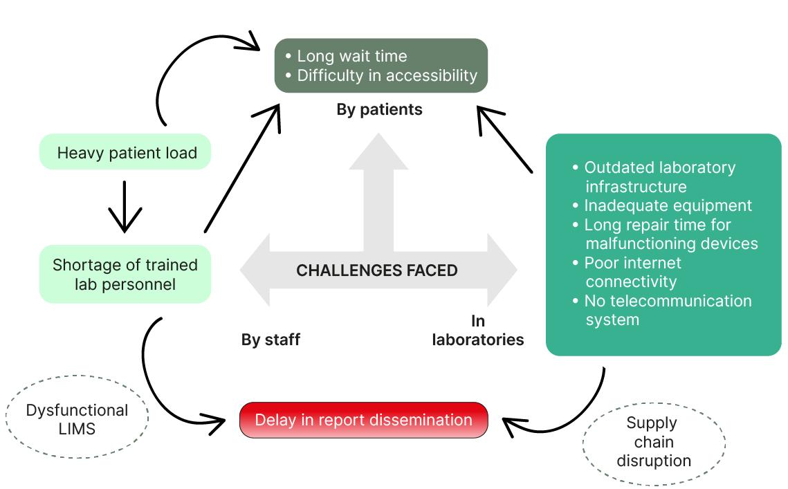
Technology-Enabled Services
A well-functioning LIMS should be made a standardised practice among all physicians and data operators. The benefits of a smoothly operating LIMS are several, including easier
LIMS: Laboratory Information Management System; TB: tuberculosis.
Article 88 EMJ ● September 2023 ● Creative Commons Attribution-Non Commercial 4.0
Figure 1: Schematic diagram of major challenges encountered in the laboratory diagnosis of TB in lowresource settings.
management, tracking, and profiling of samples; effortless generation of reports owing to realtime, advanced, and integrated dashboards; minimised paperwork; increased productivity; and efficient utilisation of time for health workers and patients. Also useful would be the use of mandatory hospital-generated patient identification, and the installation of computers and a telecommunication system within hospital wards and laboratory facilities.
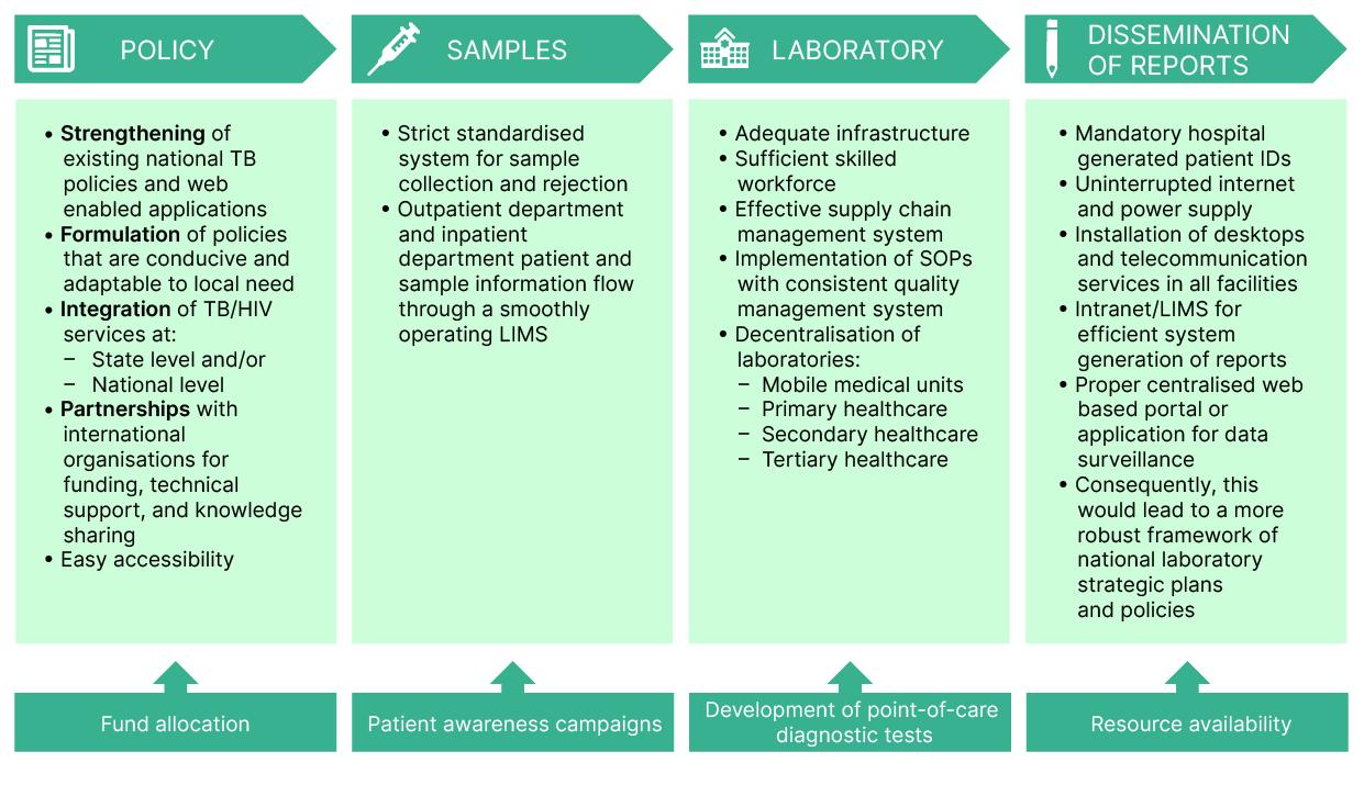
Laboratory Infrastructure and Management
Establishment of safe laboratory designs and environment that include well-ventilated and sufficiently spaced laboratories with minimal risk of cross-contamination and a straightforward and consistent setup, wherein the areas for sample collection and processing are well demarcated; the provision of adequate and
functional biosafety cabinets and protective equipment for the laboratory staff; and strict implementation of standard operating procedures in each laboratory.
Human Resources
More recruitment of skilled manpower, and training should be provided to the senior staff regarding the management of human resources, workload, and workplace organisation, forecasting and ordering of supplies, budgeting, and oversight of quality assurance.40 In this day and age of science and technology, the extension of training can be achieved through portals like telemedicine, videoconferencing, and webinars. A crucial, yet often overlooked, measure that would significantly improve the long-term outcome would be to ensure periodic medical check-ups of laboratory personnel, given their extensive exposure to contagious illnesses.
ID: identification; LIMS: Laboratory Information Management System; SOP: standard operating procedures; TB: tuberculosis.
Article Creative Commons Attribution-Non Commercial 4.0 ● September 2023 ● EMJ 89
Figure 2: Schematic diagram of strategies to overcome challenges in the diagnosis of tuberculosis in resource-limited countries.
Logistics
The appointment of a competent supply manager to oversee procurement and inventory operations within all laboratory settings should be made mandatory, which could include strict monitoring and maintenance of laboratory stock with documentation, and regular reports of the logistics data. Effective communication and collaboration is necessary among the pharmacy, supply management, and laboratory units.40
The task of creating the optimal conditions and circumstances in a country with limited resources is far from achievable in a short timeframe. However, we can continue to pursue endeavours towards enhancing our performance with the mentioned strategies, and broader goals involving the policy-makers and the government (Figure 2).41
CONCLUSION
The Indian government has implemented several measures to mitigate the impact of TB on patients and society. These measures comprise subsidised or cost-free treatment options, financial and nutritional assistance programmes, incentive-based initiatives, and thorough monitoring practices aimed at providing effective follow-up care for patients with TB.
However, being a resource-poor country, there are enormous challenges, like insufficient funding, high population density, increasing cases of MDR-TB and TB with HIV, and general lack of awareness. To overcome these challenges would require a lot of time and collective effort from various sectors, such as the government, the healthcare industry, and the general population. Given that the COVID-19 outbreak has impeded efforts to eliminate TB, there is a heightened sense of urgency to initiate measures to terminate this lethal and contagious disease using the assets and capabilities available presently. In this article, the authors have outlined some of the most common obstacles that are encountered during the laboratory diagnosis of TB, as illustrated in Figure 1. Although point-of-care diagnostic technologies are currently in the research and development phase, their availability remains distant, even after their completion. Therefore, certain strategies that can be employed to address this complex predicament have been elucidated, with emphasis on technologyenabled services. It is crucial to acknowledge that in order to progress, it will always be fundamental for healthcare providers to thoroughly evaluate the correlation between laboratory findings and clinical observations, so that accurate diagnosis and informed decisions can be made regarding treatment.
1. Centers for Disease Control and Prevention (CDC). History of World TB Day. 2023. Available at: https:// www.cdc.gov/tb/worldtbday/history. htm. Last accessed: 14 May 2023.
2. World Health Organization (WHO). Global Tuberculosis Report 2022. 2022. Available at: https://www. who.int/teams/global-tuberculosis-programme/tb-reports/global-tuberculosis-report-2022. Last accessed: 14 May 2023.
3. Oommen S, Banaji N. Laboratory diagnosis of tuberculosis: advances in technology and drug susceptibility testing. Indian J Med Microbiol. 2017;35(3):323-31.
4. World Health Organization (WHO) Stop TB Partnership; Retooling Task Force. New laboratory diagnostic tools for tuberculosis control. 2008. Available at: https:// apps.who.int/iris/bitstream/handle/10665/44036/9789241597487_ eng.pdf. Last accessed:
14 May 2023.
5. Mase SR et al. Yield of serial sputum specimen examinations in the diagnosis of pulmonary tuberculosis: a systematic review. Int J Tuberc Lung Dis. 2007;11(5):485-95.
6. Behr MA et al. Transmission of Mycobacterium tuberculosis from patients smear-negative for acid-fast bacilli. Lancet. 1999;353(9151):4449.
7. Gill CM et al. New developments in tuberculosis diagnosis and treatment. Breathe (Sheff). 2022;18(1):210149.
8. Yadav R et al. Laboratory diagnosis of tuberculosis: conventional and newer methods. IJSR. 2018;9(10):319-24.
9. Minion J et al. Comparison of LED and conventional fluorescence microscopy for detection of acid-fast bacilli in a low-incidence setting. PLoS One. 2011;6(7):e22495.
10. Minion J et al. Fading of auramine-stained mycobacterial smears and implications for external quality assurance. J Clin Microbiol. 2011;49(5):2024-6.
11. Sanborn WR et al. World Health Organization (WHO) Regional Publications, Eastern Mediterranean Series 28: Fluorescence microscopy for disease diagnosis and environmental monitoring. 2005. Available at: https://apps.who.int/iris/handle/10665/119734.
Last accessed: 14 May 2023.
12. Shah NS et al. Rapid diagnosis of tuberculosis and multidrug resistance by the microscopic-observation drug-susceptibility assay. Am J Respir Crit Care Med. 2011;183(10):1427-33.
13. MacGregor-Fairlie M et al. Tuberculosis diagnostics: overcoming ancient challenges with modern solutions. Emerg Top Life Sci. 2020;4(4):423-36.
Article 90 EMJ ● September 2023 ● Creative Commons Attribution-Non Commercial 4.0
14. Muniyandi M et al. Estimating TB diagnostic costs incurred under the National Tuberculosis Elimination Programme: a costing study from Tamil Nadu, South India. Int Health. 2021;13(6):536-44.
15. World Health Organization (WHO). Global tuberculosis report 2020. 2020. Available at: https://www. who.int/publications-detail-redirect/9789240013131. Last accessed: 15 May 2023.
16. Yadav RN et al. Laboratory cost analysis of conventional and newer molecular tests for diagnosis of presumptive multidrug-resistant tuberculosis patients. J Glob Infect Dis. 2022;14(3):93-8.
17. World Health Organization (WHO). WHO operational handbook on tuberculosis: module 3: diagnosis: rapid diagnostics for tuberculosis detection, 2021 update. 2021. Available at: https://www. who.int/publications-detail-redirect/9789240030589. Last accessed: 15 May 2023.
18. Lee DJ et al. Rapid, point-of-care diagnosis of tuberculosis with novel Truenat assay: cost-effectiveness analysis for India’s public sector. PLoS One. 2019;14(7):e0218890.
19. Rodrigues C et al. Evaluation of the bactec MGIT 960 TB system for recovery and identification of Mycobacterium tuberculosis complex in a high through put tertiary care centre. Indian J Med Microbiol. 2009;27(3):217-21.
20. Yu M-C et al. Evaluation of the rapid MGIT TBc identification test for culture confirmation of Mycobacterium tuberculosis complex strain detection. J Clin Microbiol. 2011;49(3):802-7.
21. Siddiqi S et al. Direct drug susceptibility testing of Mycobacterium tuberculosis for rapid detection of multidrug resistance using the Bactec MGIT 960 System: a multicenter study. J Clin Microbiol. 2012;50(2):435-40.
22. Kim H et al. Evaluation of MGIT 960 system for the second-line drugs susceptibility testing of Mycobacterium tuberculosis. Tuberc Res Treat. 2013;2013:108401.
23. World Health Organization (WHO). Molecular assays intended as initial tests for the diagnosis of pulmonary and extrapulmonary TB and rifampicin resistance in adults and children: rapid communication. 2020.
Available at: https://apps.who.int/
iris/handle/10665/330395. Last accessed: 14 May 2023.
24. Nikam C et al. Rapid diagnosis of Mycobacterium tuberculosis with Truenat MTB: a near-care approach. PLoS One. 2013;8(1):e51121.
25. Stop TB Partnership. Practical guide to implementation of Truenat tests. 2020. Available at: https://stoptb. org/assets/documents/resources/ wd/Practical%20Considerations%20 for%20Implementation%20of%20 Truenat.pdf. Last accessed: 17 June 2023.
26. World Health Organization (WHO). The use of loop-mediated isothermal amplification (TB-LAMP) for the diagnosis of pulmonary tuberculosis: policy guidance. 2016. Available at: https://www.who.int/publications/i/item/9789241511186. Last accessed: 16 June 2023.
27. Meehan CJ et al. Whole genome sequencing of Mycobacterium tuberculosis: current standards and open issues. Nat Rev Microbiol. 2019;17(9):533-45.
28. CRyPTIC Consortium and the 100,000 Genomes Project; AllixBéguec C et al. Prediction of susceptibility to first-line tuberculosis drugs by DNA sequencing. N Engl J Med. 2018;379(15):1403-15.
29. Lipworth S et al. SNP-IT tool for identifying subspecies and associated lineages of Mycobacterium tuberculosis complex. Emerg Infect Dis. 2019;25(3):482-8.
30. Jajou R et al. Epidemiological links between tuberculosis cases identified twice as efficiently by whole genome sequencing than conventional molecular typing: a population-based study. PLoS One 2018;13(4):e0195413.
31. Walker TM et al. Whole-genome sequencing to delineate Mycobacterium tuberculosis outbreaks: a retrospective observational study. Lancet Infect Dis. 2013;13(2): 137-46.
32. Goig GA et al. Whole-genome sequencing of Mycobacterium tuberculosis directly from clinical samples for high-resolution genomic epidemiology and drug resistance surveillance: an observational study. Lancet Microbe. 2020;1(4):e175-83.
33. Central Tuberculosis Division. Training module on extrapulmonary tuberculosis 2023. 2023. Available at: https://tbcindia.gov.in/WriteReadData/l892s/7702334778Training_
Module_on_Extrapulmonary_TB_-_ Book_24032023.pdf. Last accessed: 16 June 2023.
34. Chakravorty S et al. Diagnosis of extrapulmonary tuberculosis by smear, culture, and PCR using universal sample processing technology. J Clin Microbiol. 2005;43(9):4357-62.
35. Uddin MKM et al. Diagnostic performance of different laboratory methods for the detection of extrapulmonary tuberculosis. Microorganisms. 2023;11(4):1066.
36. Zürcher K et al.; International Epidemiology Databases to Evaluate AIDS (IeDEA) consortium. Diagnosis and clinical outcomes of extrapulmonary tuberculosis in antiretroviral therapy programmes in low- and middle-income countries: a multicohort study. J Int AIDS Soc. 2019;22(9):e25392.
37. World Health Organization (WHO). Automated real-time nucleic acid amplification technology for rapid and simultaneous detection of tuberculosis and rifampicin resistance: Xpert MTB/ RIF assay for the diagnosis of pulmonary and extrapulmonary TB in adults and children. Policy update. 2014. Available at: https:// apps.who.int/iris/bitstream/handle/10665/112472/9789241506335_ eng.pdf. Last accessed: 16 June 2023.
38. Zhou R et al. Diagnostic performance of adenosine deaminase for abdominal tuberculosis: a systematic review and meta-analysis. Front Public Health. 2022;10:938544.
39. Jha DK et al. Evidence-based approach to diagnosis and management of abdominal tuberculosis. Indian J Gastroenterol. 2023;42(1): 17-31.
40. National Accreditation Board for Testing and Calibration Laboratories (NABL) India. Specific criteria for accreditation of medical laboratories. 2019. Available at: https:// nabl-india.org/nabl/index.php?c=publicaccredationdoc&m=index&docType=both&Itemid=199. Last accessed: 16 May 2023.
41. Parsons LM et al. Laboratory diagnosis of tuberculosis in resource-poor countries: challenges and opportunities. Clin Microbiol Rev. 2011;24(2):314-50. 42.
42. Badrick TC et al. Diagnostic laboratories in India: investigating quality characteristics, productivity and time of reporting. Indian J Clin Biochem. 2018;33(3):304-13.
Article Creative Commons Attribution-Non Commercial 4.0 ● September 2023 ● EMJ 91 FOR REPRINT QUERIES PLEASE CONTACT: INFO@EMJREVIEWS.COM
Health-Economic Determinants of COVID-19 Pandemic and Countries' Efficiency
Authors:
*Reza Gharoie Ahangar,1 Victor R. Prybutok2
1. Department of Accountancy, Business Analytics, Economics, and Finance, College of Business, Lewis University, Romeoville, Illinois, USA
2. Department of Information Technology and Decision Sciences, Toulouse Graduate School, G. Brint Ryan College of Business, University of North Texas, Denton, USA *Correspondence to rgharoieahangar@lewisu.edu
Disclosure: The authors have declared no conflicts of interest. This research did not receive any specific grant from funding agencies in the public, commercial, or non-profit sectors. The data were collected from the trading economics website and will be available upon request; however, data is publicly available.
Received: 18.06.23
Accepted: 31.07.23
Keywords: COVID-19, efficiency score, inflation, macroeconomic factors, vaccine
Citation: EMJ. 2023; DOI/10.33590/emj/10301710. https://doi.org/10.33590/emj/10301710.
Abstract
This study examines the relationship between vaccination and inflation in battling the COVID-19 pandemic across nations. Data from 85 countries worldwide were collected from the Trading Economics (New York City, USA) website during the COVID-19 pandemic. First, a new theoretical model was proposed based on the economic and healthcare literature; then, a binary variable, inflation/vaccination% was developed according to the proposed theoretical model. The relationship between inflation/vaccination% and macroeconomic factors was examined using logistic regression. After that, the countries were ranked by minimising the inflation/ vaccination% rate that measures a country’s efficiency in fighting the COVID-19 pandemic, thereby permitting governments to compare the performance of different countries. The findings show that a country with a higher gross domestic product growth rate and competitiveness index during the COVID-19 pandemic has a lower inflation/vaccination% ratio. The results of this study provide strong evidence that countries should mitigate a pandemic’s economic impact by managing vaccination programmes to control global inflation.
Key Points
1. This research shows that managing vaccination programmes during a pandemic can help mitigate the impacts of rising global inflation. The findings are highly relevant for future pandemics, as this approach can help effectively plan for the pandemic, reducing the overall impact on society.
Article
92 EMJ ● September 2023 ● Creative Commons Attribution-Non Commercial 4.0
2. The research describes the interrelationship between the vaccination rate and global inflation during the COVID-19 crisis. The findings rank countries based on their economic and health sectors battling the COVID-19 pandemic and provide implications for future pandemics.
3. An important message from this research for clinicians is demonstrating how to help their patients resume their normal lives following a pandemic. Healthcare professionals can be key players in accelerating the vaccination process, helping to reduce the long-term impacts of this crisis. To achieve this goal, it is essential that they take proactive action and work together to make it happen
INTRODUCTION
In late 2019, a highly contagious virus from the family of severe acute respiratory syndromes originated from Wuhan, China, and spread around the world in 2020.1,2 This experience suggested the need to examine how countries can mitigate the economic effects of pandemics.
The World Health Organization (WHO) declared the COVID-19 pandemic on 11th March 2020. This disease is contagious and is caused by severe acute respiratory syndrome coronavirus 2.3 As of 26th July 2023, around 768 million cases and 6.95 million deaths were reported worldwide.3 Despite scientists having developed several vaccines, the number of COVID-19 infections and deaths is still increasing, with a negative acceleration slope worldwide.
Some scientists believe that the COVID-19 pandemic is one of the worst pandemics in the world,4 and some scientists have predicted a K-, L-, U-, or V-shaped economic recovery following the pandemic, depending on the amount of time needed to return to normalcy.5 However, it is still hard to find an absolute recovery model with the emergence of new variants of the virus, even after vaccine development.
One of the negative impacts of the COVID-19 pandemic is global inflation, which was caused by the disruption of the global supply.6-8 Inflation is one of the critical macroeconomic factors for households, which could cause a range of decisions that affect a society’s savings, borrowing, and consumption during the COVID-19 pandemic.6,7 Therefore, global inflation control requires that businesses and their supply chains practise overcoming lingering COVID-19 pandemic issues. To achieve this goal, the healthcare system can play a major role and help people get vaccinated faster, which will help restore normal life before the pandemic.
Almost 1 year after the start of the COVID-19 pandemic, health service scientists developed various types of vaccines (e.g., mRNA, inactivated virus, or non-replicating virus vector).9-12 However, people have not been vaccinated at the rates governments anticipated.13 For example, as of 2nd December 2022, only 69.2% of the world’s population, and 25.9% of low-income countries, have been vaccinated with one dose of COVID-19 vaccines.14
Deiana et al.15 and Li et al.16 indicated that reluctance to get vaccinated was partially due to a belief that the new vaccines were not adequately planned. Machingaidze and Wiysonge17 and Shrestha et al.18 reported that side effects, safety, competence, and effectiveness of the vaccines were yet to be clarified. Khatiwada et al.19 and Machingaidze and Wiysonge17 suggested that vaccine hesitancy could also be attributed to rapid vaccine development and media misinformation. Nemat et al.20 and Adebisi et al.21 suggested that people were concerned about the reliability of the vaccines in some countries, or that they may possess natural immunity against the COVID-19 virus. Armantier et al.6 and Detmers et al.7 found that hesitancy to get vaccinated and return to the normal job situation exacerbated supply chain disruption issues, which may be a cause of higher global inflation.
This paper seeks to add to the existing literature by developing a theory through a binary variable connecting economic factor, inflation, with healthcare variable, vaccine rates, during the COVID-19 pandemic. It then examines the interrelationships between macroeconomic factors and inflation/vaccination%, as well as ranking countries’ efficiency in combating the pandemic based on their health-economic factors.
Article Creative Commons Attribution-Non Commercial 4.0 ● September 2023 ● EMJ 93
INFLATION-VACCINE THEORY
The COVID-19 pandemic caused problems for both public and private sectors.22-25 Several sectors (e.g., households, businesses, and governments) have been affected by the COVID-19 pandemic. McKibbin and Fernando26 and Ahangar and Kim27 examined the effects of the pandemic on different macroeconomic sectors (i.e., labour supply, equity risk premium, consumption demand, government expenditure, production, and gross domestic product [GDP]). They found that the consumption demand shock on GDP was more influential than the other shocks in households’ lives. The inflation rate is one of the critical macroeconomic factors influencing households that may cause a consumption demand shock. Studies by Crump et al.,28 D’Acunto et al.,29 Detmers et al.,7 and Candia et al.30 have demonstrated a relationship between households’ economic behaviour and their expectations of future inflation during the COVID-19 pandemic. Wulandhari et al.25 argued that COVID-19 caused a supply shock, which could result in a higher inflation rate for households. Bobeica and Hartwig31 asserted that, in early 2021, the primary cause of high inflation was the increase in supply side and commodity prices. Moreover, Armantier et al.,6 Detmers et al.,7 and Gautier et al.8 suggested that both supply and demand shocks due to COVID-19 and the lockdown could be the reason for high inflation.
Various plans were implemented by governments, and particularly healthcare systems, during the COVID-19 outbreak, such as mandates for lockdowns to control disease's spread.32,33 These efforts can reduce the number of deaths caused by the disease but can also lead to social and political disruption and economic slowdown. Harland et al.,34 in their study about healthcare during the COVID-19 pandemic, showed that the development and implementation of plans in healthcare systems that can withstand future emergencies depend upon understanding their capabilities and the motivation to use them during a pandemic.
Finkenstadt and Handfield35 highlighted that the emergence of a new virus during the pandemic led to a significant vaccine shortage, and the lessons can improve the planning for healthcare and future pandemic vaccines. The idea of a social vaccine suggests that popular mobilisation
and advocacy can be encouraged to change social and economic structural conditions that make people and communities vulnerable to disease.36
During the COVID-19 pandemic, Dorn et al.37 developed a new theory examining the relationship between public healthcare and economic systems. They demonstrated that balancing interests between public health and the economy is essential during the pandemic. The monetary theory of inflation38 declares that the growth of cash flow or money supply is the primary cause of inflation. Thus, injecting cash flow (e.g., subsidy or stimulus check) into the market can lead to faster inflation in nations. In economics, disposable personal income, defined as money households retain after paying taxes or receiving government assistance (e.g., stimulus checks), is critical in understanding a society's economic security and inflationary challenges.39 For instance, Jorda et al.39 found that government stimulus checks distributed to households during the pandemic contributed to a higher inflation rate in the USA. Additionally, Barnichon et al.40 and Bianchi et al.41 have conducted research, showing the effects of COVID-19 pandemic on inflation.
The healthcare industry represents a considerable portion of economic and social activities worldwide, with countries spending over 8% of GDP on healthcare.42 Employee commitment and control theories propose that employee initiatives are critical to patient safety.43 As such, healthcare plays a vital role in economics, making the availability of effective COVID-19 vaccines for the public at high speed. To this end, it is optimal to start the downstream task concurrently in cyclical projects.44 In a study, Shahmanzari et al.33 showed that the structure of containment measures strongly relates to critical economic and disease parameters, such as recovery rate, containment capacity, and infection rate. Bjurling-Sjöberg et al.45 developed a practical theory of resilient performance during a pandemic, highlighting the vital role of working conditions and patient safety during a COVID-19 pandemic. In addition, Sarabia et al.46 showed that a higher annual expenditure could be related to a lower incidence of COVID-19 in the health system.
Article 94 EMJ ● September 2023 ● Creative Commons Attribution-Non Commercial 4.0
Therefore, this study develops a theory and proposes an interrelationship between inflation and vaccination using inflation/vaccination% ratio and other macroeconomic factors to help countries control global inflation appropriately. Vaccinating people as soon as possible may reduce the need for stimulus income, help the economy retain jobs, and contribute to business health. The rationale suggests that inflation and the vaccination rate of people are related to each other, and if both are improved, the ratio of inflation/vaccination% would be smaller. However, to achieve this goal, some key macroeconomic factors must be improved along with the vaccination rate.
RESEARCH THEORETICAL MODEL
After reviewing the literature and synthesising economic and pandemic theories, the authors
theorised that global inflation in a pandemic can be related to a nation's healthcare system through other macroeconomic factors. To test this interrelationship between vaccination and global inflation rates, the authors constructed a theoretical model and analysed it using various macroeconomic variables across multiple market sectors. Thus, their theoretical model can be seen in Figure 1.
This theoretical model indicates no direct relationship between the inflation rate and vaccination rate; however, the diagram indicates an interrelationship between this economic and health factor through other macroeconomic factors. The authors examined the direct and interrelation effects to answer the following research questions.
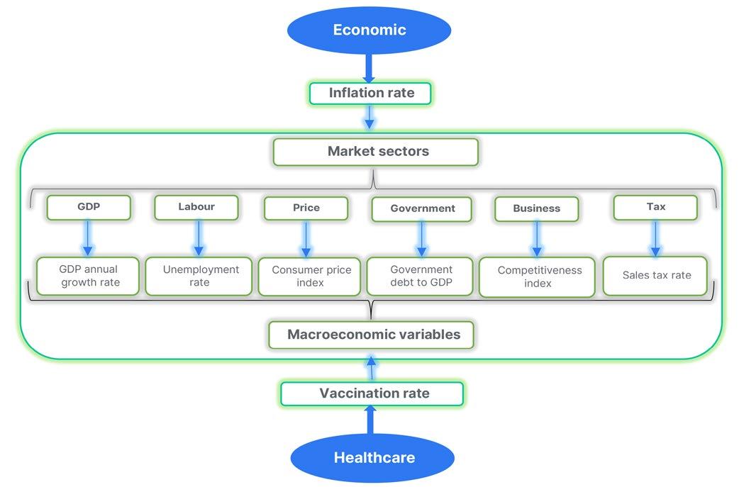 GDP: gross domestic product.
Article Creative Commons Attribution-Non Commercial 4.0 ● September 2023 ● EMJ 95
Figure 1: Theoretical model of inflation/vaccine ratio in this study.
Research Questions
The important research questions that provide insight and recommendations to mitigate the economic effects of a pandemic were:
• Is there any interrelationship between the vaccination rate and global inflation during the COVID-19 pandemic?
• How can countries mitigate a pandemic’s impact by managing vaccination programmes to control global inflation?
• How can countries be ranked based on their economic and health sectors in battling the COVID-19 pandemic?
DATA AND METHODOLOGY
Data from 85 countries worldwide collected from the Trading Economics (New York City, USA) website47 were used for this study, which focused on macroeconomic variables relevant to the COVID-19 pandemic. In the initial step, the data of more nations, including 115 countries, were selected. After that, a nation with missing values was removed from the list. For some political and monetary sanctions, the authors short-listed 95 countries whose economic fluctuations have an economic base. Then ten countries were removed from the list based on the Box–Tidwell test, which checks the linearity between the explanatory factors and their natural log in the model. In the end, 85 countries were selected, for which the data was available and public, and there were no outliers or political/ financial sanctions on their different sectors. Therefore, to ensure that the models were valid, various combinations of data preparation and tests were employed. High correlation variables were excluded, and no missing values were present. Furthermore, countries with extreme observations coming from a political rather than economic base were excluded.
First, the relationship between inflation/ vaccination% and macroeconomic factors is examined using logistic regression. After that, the data envelopment analysis (DEA) was applied to measure a country’s efficiency in fighting the COVID-19 pandemic, thereby permitting them to compare the performance of different countries. Given the focus of the
analysis on the inflation-vaccine ratio, a constant return to scale two-stage output-oriented DEA model was developed. The selection of input and output variables was based on the most influential variables identified in the logistic regression stage.
Dependent Variable
The dependent variable was the inflation/ vaccination%, with the numerator being the inflation rate in December 2021 and the denominator being the total number of vaccinated individuals per 100 people in December 2021. This is a newly developed factor related to healthcare during the COVID-19 pandemic.
In this study, the authors used the median value of this fraction as a point for data segmentation. Any value lower than the median was labelled as ‘low’, and any value higher than the middle point was labelled as ‘high’. Each country had a specific value for inflation and vaccination rates, making their ratio unique. The authors ordered the numerical values from the largest to the smallest and divided them into two equal samples by determining the median value. This procedure produced extreme polar segmentation,48 which requires ordering of the observations of a variable. Then, the data was parsed into two data subsets that each contained half the values to maximise variance within the inflation/ vaccination% variable.
Explanatory Variables
Previous studies have explored the various solutions for developing nations' health related to the COVID-19 pandemic. These findings were broken down into economic and health factors, with nations' economic health during the pandemic being measured using a set of economic indicators commonly reported by nations. Explanatory variables in this study have a ratio-level measurement scale, and they are selected from different sectors and their derivatives in the market (i.e., GDP, labour, price, government, business, and tax) to ensure that all aspects of a household affected by the COVID-19 pandemic shock are captured in the analysis.26,27
Article 96 EMJ ● September 2023 ● Creative Commons Attribution-Non Commercial 4.0
The variables selected for this study were the GDP annual growth rate from the GDP section, which shows a nation’s GDP change at market prices during the COVID-19 pandemic; the unemployment rate from the labour category, representing a portion of the labour force without a job but who are actively searching for a job during the pandemic; the consumer price index from price categories, which indicates an average change in prices paid by a household for goods and services during the COVID-19 pandemic; the government debt to GDP from the government section, to show a nation’s public debt to its GDP and a country’s ability to pay back its loans after the pandemic; the competitiveness index from the business section, to show a country’s productivity level during the pandemic; and the sales tax rate from the tax category, to show government aid in terms of subsiding during COVID-19. Additionally, the COVID-19 vaccination rate over time was chosen from the health side to indicate a nation’s health plan in batting against the COVID-19 pandemic.
RESULTS Correlation Analysis
The authors examined the correlation between the explanatory variables to avoid selecting variables with a high correlation that would result in multicollinearity.49
Table 1 presents the correlation matrix of all regressors, as well as mean and standard deviation. Results demonstrate that correlations between independent variables are relatively low. Additionally, a mean variance inflation factor of 1.16 indicates that multicollinearity was not an issue in this study. Subsequently, a pair correlation plot analysis was conducted to graphically visualise correlations between each pair of variables.
Logistic Regression Analysis
The authors performed logistic regression to examine the relationships between the binary variable, inflation/vaccination%, and the macroeconomic regressors. Table 2 shows the results from the logit model analysis, which models the probability of the ratio inflation/ vaccination% falling below the central value,
GDP: gross domestic product; N/A: not applicable; SD: standard deviation.
Variable Mean SD GDP annual growth rate Unemployment rate Consumer price index Government debt to GDP Competitiveness index Sales tax rates GDP annual growth rate 5.84 3.57 1.000 N/A N/A N/A N/A N/A Unemployment rate 8.52 7.47 -0.041 1.000 N/A N/A N/A N/A Consumer price index 410.18 1,113.05 -0.053 -0.063 1.000 N/A N/A N/A Government debt to GDP 69.65 41.38 0.043 0.059 -0.127 1.000 N/A N/A Competitiveness index 65.30 11.37 -0.120 -0.448 -0.077 0.152 1.000 N/A Sales tax rates 16.59 5.48 0.165 -0.029 0.098 -0.008 0.036 1.000
Table 1: Correlation matrix of macroeconomic explanatory variables in this study.
Article Creative Commons Attribution-Non Commercial 4.0 ● September 2023 ● EMJ 97
referred to as low. The likelihood ratio test (χ2: 105.15; p<0.001; pseudo-R2: 89.67%) supports that the overall model is significant at the 1% level.
Table 2 presents the estimated parameters, p values of independent variables, and the significant macroeconomic variables at 10% and 5% statistical levels. The intercept was also found to be statistically significant in this model. The odds estimates represent the log odds of the outcome for a one unit increase in predictor variables. According to the coefficients and odds estimates, the first two influential, statistically significant predictors are the GDP annual growth rate and the competitiveness index that help to decrease the inflation/vaccination% ratio.
The odds ratio estimates measure the association of variables. The odds ratio is the multiplicative change in the odds for one unit change in regressors. It reveals that an increase in the sales tax rate, unemployment rate, and consumer price index increases the log odds of having a greater inflation/vaccination%,
while an increase in the GDP growth rate, competitiveness index, and government debt to GDP decreases this ratio. The results show that the unemployment rate and consumer price index are not statistically significant at 5%.
COUNTRY RANKINGS AND EFFICIENCY SCORES
After identifying the most important factors that contribute to the development of the inflation/ vaccination% indicator, health authorities may wish to rank countries according to their performance. However, establishing a comprehensive ranking is not always possible due to various statistical and subjective issues.
Figure 2 demonstrates the inflation-vaccination ratio in relation to the competitiveness index of all countries. This figure shows that African countries generally experience higher inflation and lower vaccination rates, as well as a lower competitiveness index in comparison with other countries.
*Significant at p<0.05.
†Significant at p<0.10.
Sample size is 85. Government debt to GDP-S is the square of variable government debt to GDP, and competitiveness index-SR is the SR of the variable competitiveness index.
GDP: gross domestic product; GDP-S: square of the government debt to gross domestic product; SR: square root.
Parameter Co-efficient Odds estimate Standard error Z value p GDP annual growth rate -1.716 0.179 0.771 -2.23 0.026* Unemployment rate 0.267 1.306 0.152 1.75 0.080† Consumer price index 0.013 1.013 0.007 1.81 0.071† Government debt to GDP-S -0.002 0.998 0.001 -2.01 0.044* Competitiveness index-SR -27.092 1.71e-12 12.084 -2.24 0.025* Sales tax rate 2.733 15.383 1.333 2.05 0.022*
Table 2: A logit model analysis of maximum likelihood estimates for the variables in this study.
Article 98 EMJ ● September 2023 ● Creative Commons Attribution-Non Commercial 4.0
The smaller circles indicate a lower inflation/vaccination percentage, while the darker green colour represents a higher competitiveness index among countries.
Inf-vac: inflation/vaccine.

A more objective method known as DEA has been proposed for measuring a country's efficiency in fighting against the COVID-19 pandemic.50 This allows authorities to compare a country’s performance with its peers, and a health official can use the DEA's efficiency ranking system to compare countries and assess potential improvements.
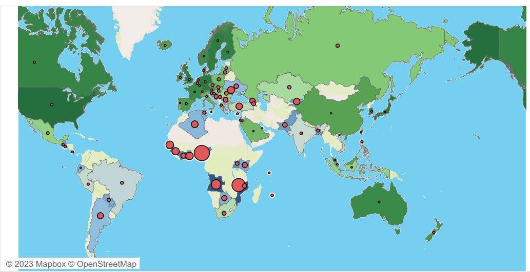
To minimise scaling problems with DEA, the data were converted to standard scores by subtracting each data point from its mean and dividing by the standard deviation of each variable to avoid negative signs in DEA. Then, a constant number equal to the maximum standard score of each variable was added to the converted standard scores of all observations, resulting in positive values (adding a constant number does not change the relative rankings of the observations).
The selection of appropriate inputs and outputs for the DEA was carried out according to the following considerations. To minimise efficiency discrimination power, the number of inputs and outputs was kept to a minimum.50 Moreover, the most relevant input and output variables were used to maximise efficiency of the operations.50 In order to improve efficiency of the operations
and facilitate country comparability, the most important explanatory variables were considered. The inflation/vaccine ratio was chosen as the output for the model; consequently, the input variables of GDP annual growth rate and competitiveness index were selected based on logit regression for the DEA model. As the focus of the analysis is on inflation/vaccine ratio, a constant return to scale two-stage outputoriented DEA model was developed based on the identified input and output variables.
The DEA model can be used in various ways, with one of its primary functions being to provide a view of how countries utilise their resources to combat the COVID-19 pandemic. Although wealthy nations generally have higher efficiency, some are still predicted to perform poorly. Table 3 displays the results, which suggest that some of these countries are surprisingly underperforming.
Table 3 shows the DEA efficiency and ranking of the standardised scores of the inflation/ vaccine ratio for each country. A larger positive number implies a worse index for the inflation/ vaccination ratio. This transformation changed the direction of the inflation/vaccination% rates from minimum to maximum outputs, which is
Figure 2: Inflation/vaccine ratio with respect to the competitiveness index of countries.
Inf-vac percentage Competitiveness index -0.2 50.0 100.0 160.0 38.08 84.78 Article Creative Commons Attribution-Non Commercial 4.0 ● September 2023 ● EMJ 99
Rank Country Efficiency score 1 Israel 0.419412 2 Ireland 0.422393 3 Malta 0.437015 4 Saudi Arabia 0.452526 5 Hungary 0.457426 6 Costa Rica 0.459342 7 Greece 0.460860 8 Philippines 0.460927 9 Poland 0.460943 10 UK 0.462992 11 Italy 0.463171 12 Peru 0.463867 13 Sweden 0.465536 14 Croatia 0.465897 15 Singapore 0.466702 16 Seychelles 0.466748 17 India 0.469105 18 Netherlands 0.469560 19 Iceland 0.471148 20 Portugal 0.471247 21 Cyprus 0.471479 22 Serbia 0.475360 23 France 0.477929 24 Ecuador 0.477989 25 Norway 0.478423 26 Vietnam 0.480029 27 Belgium 0.480804 28 Luxembourg 0.480960 29 Austria 0.481428 30 Mauritius 0.485453 31 USA 0.485470 32 Indonesia 0.485906 33 Spain 0.486561 34 Taiwan 0.487559 35 Guatemala 0.492360 36 Slovenia 0.493175 37 El Salvador 0.497226 38 Switzerland 0.500470 Article 100 EMJ ● September 2023 ● Creative Commons Attribution-Non Commercial 4.0
Table 3: Ranking and efficiency scores of countries based on the inflation/vaccination ratio.
Rank Country Efficiency score 39 China 0.500930 40 South Korea 0.502564 41 Bangladesh 0.503338 42 Australia 0.506915 43 Canada 0.507158 44 Bosnia and Herzegovina 0.509417 45 Ukraine 0.510006 46 Finland 0.512260 47 Malaysia 0.512560 48 Denmark 0.513545 49 Georgia 0.514395 50 Lithuania 0.515061 51 Brazil 0.517618 52 Russian Federation 0.518342 53 Kazakhstan 0.524187 54 Paraguay 0.526131 55 Czechia 0.526894 56 Latvia 0.527119 57 Botswana 0.529213 58 Uganda 0.530275 59 Türkiye 0.534092 60 Kenya 0.534493 61 Bulgaria 0.535076 62 Jordan 0.535089 63 Bahrain 0.540195 64 Argentina 0.542298 65 Pakistan 0.545365 66 Ivory Coast 0.555957 67 Japan 0.556848 68 South Africa 0.557587 69 Armenia 0.561594 70 Germany 0.564313 71 Moldova 0.567269 72 Romania 0.569517 73 Slovakia 0.578466 74 Mexico 0.584153 75 Mozambique 0.584634 76 Ghana 0.601491 Article Creative Commons Attribution-Non Commercial 4.0 ● September 2023 ● EMJ 101
Table 3 continued.
Table 3 continued.
*Significant at p<0.05.
†Significant at p<0.10.
Sample size is 85. Government debt to GDP-S is the square of variable Government debt to GDP, and competitiveness index-SR is the square root of the variable competitiveness index.
consistent with the DEA directive. For example, Nigeria has an index of 1.00 whereas Israel has an index of 0.42. It can be observed that other African countries, such as Malawi, Algeria, and Angola, have high scores, followed by Ireland, Malta, Saudi Arabia, and then Israel. Nations may be able to follow the health and economic plans of Israel, which would increase productivity levels in their countries. It is noteworthy that there is a minimal difference between the efficiency score of Israel (0.419412, the first rank country) and the score of the USA (0.485470, the 31st rank country). Nearly half of the countries have an efficiency score lower than 0.5, and only the aforementioned African countries have a score greater than 0.7, indicating that the people of these African countries are suffering from the COVID-19 pandemic and require special attention from authorities.
DISCUSSION AND LIMITATIONS
To create a robust model that can better capture the relationship between the vaccination rate and the inflation rate of countries, a binary variable inflation/vaccination% representing the combined
Article 102 EMJ ● September 2023 ● Creative Commons Attribution-Non Commercial 4.0
effect of vaccine and inflation rates was created. One important reason to examine this binary relationship is that it avoids the assumption that is required for linear regression and allows the examination of nonlinear relationships. The logistic model also allowed for the testing of the interrelationship between vaccination and inflation rates with a group of macroeconomic factors.
The findings show that the pseudo-R2 of logistic regression after including all variables is 89.67%. In the logistic model, annual GDP growth rate and competitiveness index are the most influential factors contributing to the inflation/vaccination% ratio for all countries during the COVID-19 pandemic. (Note: the sales tax rate is also playing an important role in this model, but the authors do not recommend this variable, since reducing the sales tax rate is inversely related to the competitiveness index, and a nation cannot stop using tax funds to build roads or bridges).
GDP: gross domestic product; GDP-S: square of the government debt to gross domestic product; SR: square root. Rank Country Efficiency score 77 Tunisia 0.605928 78 New Zealand 0.611007 79 Senegal 0.614758 80 Guinea 0.621319 81 Kyrgyzstan 0.691551 82 Angola 0.837729 83 Algeria 0.985291 84 Malawi 0.990429 85 Nigeria 1.000000
These results imply that a country with a higher rate of GDP growth and competitiveness index or lower sales tax rate during the COVID-19 pandemic has a lower inflation/vaccination%
ratio. This fraction becomes smaller with a lower inflation rate, a higher vaccination rate, or for both reasons. Another influential macroeconomic factor in this study contributing to the inflation/ vaccination% ratio is government debt to GDP. This macroeconomic factor shows the vital role of governmental help (e.g., stimulus checks) in mediating the effect of COVID-19 in a nation.
After finding the most influential variables in explaining the inflation/vaccination% ratio, a constant return to scale constant return to scale two-stage output-oriented DEA model based on the assurance region was used to help the authorities and healthcare systems to better understand the health economic situations of nations worldwide. The DEA inputs and outputs were chosen based on the most significant factors from the above statistics and data elements. The output includes the percentage of a nation’s inflation/vaccination rate, and inputs include the nation’s annual GDP growth rate and competitiveness index. The DEA’s complete dataset was then converted to an easily interpreted format to ensure that the result of the fight against the COVID-19 pandemic is not affected by the few inputs and outputs participating in optimal solutions.
These relationships show it is expected that a nation with a higher vaccination rate has a higher GDP growth rate because people can return to stable work situations,51 businesses are open, and people have improved mental health.52 Open businesses result in increased mobility,53 social capital,54 and global research and development,55 which consequently results in higher production rates. Additionally, avoiding support options such as stimulus checks removes disposable personal income, which feeds inflation for a postpandemic situation.39-41 Reopening all businesses and getting back to a pre-pandemic economy helps to balance demand and supply, where imbalances are likely to increase inflation.6-8,31
This study has some limitations. For example, a significant change in the consumption baskets of households during the pandemic may disrupt the supply and demand balance in the short and
medium terms. Such an imbalance contributes to an overall inflation rate where supply shortages during the lockdown period can result in excess demand due to the reopening and recovery of the economy. In fact, the household baskets change slowly year over year, but the lockdown and remote delivery of jobs created a sudden change in the consumption basket of households, and this abrupt shift affects the equilibrium point of demand and supply in the market; therefore, including other health and economic factors might have the potential to change the results. However, this study's policy recommendations can aid planners and authorities in creating long-term plans because the findings present strong evidence that countries should regulate vaccination programmes to counteract global inflation during pandemics.
CONCLUSIONS
This study shows that there is an interrelationship between the healthcare variable, vaccination rate, and the economic variable, inflation, during the COVID-19 pandemic worldwide. The findings indicate that inflation and vaccination rates are closely connected and related to each other through other macroeconomic factors. These findings are consistent with the lack of necessity for governments to provide external financial support (e.g., stimulus checks in the USA). An increase in disposable income increases the amount of cash flow in society and therefore leads to an increase in the inflation rate. Additionally, the lack of external support puts additional pressure on individuals to return to work, resulting in a job market boom due to higher GDP growth rate and competitiveness index that controls the inflation rate. The results from this study have the potential to provide invaluable insights to governments, healthcare systems, and policymakers in understanding the economic and health situations of countries and helping them make informed decisions in their fight against the COVID-19 pandemic.
Article Creative Commons Attribution-Non Commercial 4.0 ● September 2023 ● EMJ 103
References
1. Ahangar RG et al. Estimation and demographic analysis of COVID-19 infections with respect to weather factors in Europe. JBA. 2020;3(2):93-106.
2. Huang C et al. Clinical features of patients infected with 2019 novel coronavirus in Wuhan, China. Lancet. 2020;395(10223): 497-506.
3. World Health Organization (WHO). Novel coronavirus (2019-nCoV) situation reports. Available at: https://covid19.who.int/. Last accessed: 26 July 2023.
4. Ferguson N et al. Report 9: impact of non-pharmaceutical interventions (NPIs) to reduce COVID19 mortality and healthcare demand. Available at: https://spiral.imperial.ac.uk/ handle/10044/1/77482. Last accessed: 14 April 2023.
5. Guerrieri V et al. Macroeconomic implications of COVID-19: can negative supply shocks cause demand shortages? American Economic Review. 2022;112(5);1437-74.
6. Armantier O et al. How economic crises affect inflation beliefs: evidence from the Covid-19 pandemic. J Econ Behav Organ. 2021;189:443-69.
7. Detmers G-A et al. Understanding consumer inflation expectations during the COVID‐19 pandemic. Aust Econ Rev. 2022;55(1):141-54.
8. Gautier E et al. Inflation and households’ inflation expectations during the Covid‐19 pandemic. 2020. Available at: https:// blocnotesdeleco.banque-france. fr/en/blog-entry/inflation-andhouseholds-inflation-expectationsduring-covid-19-pandemic. Last accessed: 23 March 2023.
9. Fadhel FH. Vaccine hesitancy and acceptance: an examination of predictive factors in COVID-19 vaccination in Saudi Arabia. Health Promot Int. 2021;DOI:10.1093/ heapro/daab209.
10. Forman R et al. COVID-19 vaccine challenges: what have we learned so far and what remains to be done? Health Policy. 2021;125(5):553-67.
11. Gambichler T et al. Cutaneous findings following COVID-19 vaccination: review of world literature and own experience. J Eur Acad Dermatol Venerol. 2022;36(2):172-80.
12. Majid U et al. COVID-19 vaccine hesitancy and acceptance: a comprehensive scoping review of global literature. Health Promot Int. 2022;37(3):daac078.
13. Vasireddy D et al. Review of COVID-19 vaccines approved in the United States of America for emergency use. J Clin Med Res. 2021;12(4):204-13.
14. Our World in Data. Coronavirus (COVID-19) vaccinations. Available at: https://ourworldindata.org/ covid-vaccinations. Last accessed: 13 Mary 2023.
15. Deiana C et al. Perceived risk and vaccine hesitancy: quasiexperimental evidence from Italy. Health Econ. 2022;31(6):1266-75.
16. Li Y et al. A comprehensive review of the global efforts on COVID-19 vaccine development. ACS Cent Sci. 2021;7(4):512-33.
17. Machingaidze S, Wiysonge CS. Understanding COVID-19 vaccine hesitancy. Nat Med. 2021;27(8):1338-9.
18. Shrestha S et al. Adverse events related to COVID-19 vaccines: the need to strengthen pharmacovigilance monitoring systems. Drugs Ther Perspect. 2021;37(8):376-82.
19. Khatiwada AP et al. Paradigm shift of drug information centers during the COVID-19 pandemic. Drugs Ther Perspect. 2020;36(9):389-5.
20. Nemat A et al. Public willingness and hesitancy to take the COVID-19 vaccine in Afghanistan. Am J Trop Med Hyg. 2021;105(3):713-7.
21. Adebisi YA et al. When it is available, will we take it? Social media users’ perception of hypothetical COVID-19 vaccine in Nigeria. Pan Afr Med J. 2021;38:230.
22. Gupta S et al. Tracking public and private responses to the COVID-19 epidemic: evidence from state and local government actions. ASHEcon. 2021;7(4):361-404.
23. Koch J, Schermuly CC. Managing the crisis: how COVID‐19 demands interact with agile project management in predicting employee exhaustion. British Journal of Management. 2021;32(4):1265-83.
24. Sheng J et al. COVID-19 pandemic in the new era of big data analytics: methodological innovations and future research directions. British Journal of Management. 2020;32(4):1164-83.
25. Wulandhari NBI et al. Organizational resilience supply chain risks during the COVID-19 pandemic. British Journal of Management. 2022;34(3):1282-315.
26. McKibbin W, Fernando R. The global macroeconomic impacts of COVID-19: seven scenarios. Asian Economic Papers. 2020;20:1-30.
27. Ahangar RG, Kim M. The impact of COVID-19 shocks on business and GDP of global economy. American Business Review. 2022;25(2): 328-54.
28. Crump R et al. Subjective intertemporal substitution. 2021. Available at: https://www. newyorkfed.org/research/staff_ reports/sr734.html. Last accessed: 21 February 2023.
29. D’Acunto F et al. Managing household expectations with unconventional policies. The Review of Financial Studies. 2022;35(4):1597-642.
30. Candia B et al. Communication and the beliefs of economic agents. 2020. Available at: https://www. nber.org/papers/w27800. Last accessed: 17 March 2023.
31. Bobeica E et al. The COVID-19 shock and challenges for inflation modelling. Int J Forecast. 2023;39(1):519-39.
32. Pirrotta L et al. COVID-19 vaccinations: an overview of the Italian national health system's online communication from a citizen perspective. Health Policy. 2022;126(10):970-9.
33. Shahmanzari M et al. Managing disease containment measures during a pandemic. Prod Oper Manag. 2022;32(5):1362-79.
34. Harland CM et al. Practitioners’ learning about healthcare supply chain management in the COVID-19 pandemic: a public procurement perspective. International Journal of Operations & Production Management. 2021;41(13):178-89.
35. Finkenstadt DJ, Handfield RB. (2021), Tuning value chains for better signals in the post-COVID era: Vaccine supply chain concerns. International Journal of Operations & Production Management 202141(8):1302-17.
36. Baum F et al. Social vaccines to resist and change unhealthy social and economic structures: a useful metaphor for health promotion. Health Promot Int. 2009;24(4): 428-33.
Article 104 EMJ ● September 2023 ● Creative Commons Attribution-Non Commercial 4.0
37. Dorn F et al. The common interests of health protection and the economy: evidence from scenario calculations of COVID-19 containment policies. Eur J Health Econ. 2023;24:67-74.
38. Barro RJ, Fischer S. Recent developments in monetary theory. Journal of Monetary Economics. 1976;2(2):133-67.
39. Jordà O et al. Why is U.S. inflation higher than in other countries? 2022. Available at: https://www. frbsf.org/economic-research/ publications/economic-letter/2022/ march/why-is-us-inflation-higherthan-in-other-countries/. Last accessed: 14 March 2023.
40. Barnichon R et al. Is the American rescue plan taking us back to the ’60s? 2021. Available at: https:// www.frbsf.org/economic-research/ publications/economic letter/2021/ october/is-american-rescue-plantaking-us-back-to-1960s/. Last accessed: 14 Mach 2023.
41. Bianchi F et al. Some inflation scenarios for the American Rescue Plan Act of 2021. 2021. Available at: https://www.chicagofed. org/publications/chicago-fedletter/2021/453. Last accessed: 14 March 2023.
42. Fredendall LD, Smith JS. Editorial: delivering effective healthcare at lower cost: Introduction to the special issue. Journal of Operations Management. 2019;66(1-2):4-11.
43. Gowen III CR et al. Exploring the efficacy of healthcare quality practices, employee commitment, and employee control. Journal of Operations Management. 2006;24(6):765-78.
44. Murthy NN et al. Managing concurrency in cyclical projects under stochastic task environments: vaccine development projects during pandemics. Prod Oper Manag. 2022;DOI:10.1111/ poms.13907.
45. Bjurling-Sjöberg P et al. Resilient performance in healthcare during the COVID-19 pandemic (ResCOV): study protocol for a multilevel grounded theory study on adaptations, working conditions, ethics and patient safety. BMJ Open. 2021;11(12):e051928.
46. Sarabia M et al. Health, longevity, infrastructure and competitiveness: the four horsemen of COVID-19. Journal of Business Research. 2021;129:244-9.
47. Trading Economics. Available at: https://tradingeconomics.com. Last accessed: 9 December 2022.
48. George B, Prybutok V. Development of a polar extreme method for use in partial least squares SEM. Quality & Quantity. 2015;49:471-88.
49. Peng DX, Lai F. Using partial least squares in operations management research: a practical guideline and summary of past research. Journal of Operations Management. 2012;30(6):467-80.
50. Cooper WW et al., “The basic CCR model,” Introduction to Data Envelopment Analysis and Its Uses with DEA-Solver Software and References [Internet] (2006) New York: Springer Science+Business Media, Inc. Available at: https://link.springer.com/ chapter/10.1007/0-387-29122-9_2. Last accessed: 17 March 2023.
51. Adisa TA et al. Exploring the impact of COVID-19 on employees’ boundary management and work–life balance. British Journal of Management. 2022;33(4): 1694-709.
52. Serrano-Alarcón M et al. Impact of COVID-19 lockdowns on mental health: evidence from a quasinatural experiment in England and Scotland. Health Econ. 2022;31(2):284-96.
53. Kim D, Lee,YJ. Vaccination strategies and transmission of COVID-19: evidence across advanced countries. J Health Econ. 2022;82:102589.
54. Bartscher AK et al. Social capital and the spread of covid-19: insights from European countries. J Health Econ. 2021;80:102531.
55. Agarwal R, Gaule P. What drives innovation? Lessons from COVID-19 R&D. J Health Econ. 2022;82:102591.
Article Creative Commons Attribution-Non Commercial 4.0 ● September 2023 ● EMJ 105 FOR REPRINT QUERIES PLEASE CONTACT: INFO@EMJREVIEWS.COM
Lupus Enteritis: A Case Report
Authors: Lucas Zambiasi,1 Alícia Regina Zambiasi,2 Maria Eduarda Tomasetto,2 Patrick Bonacina,2 Matheus Augusto Eisenreich,3 Lísia Hoppe,1 Fernando Fornari,1 *Ricardo Valões1
1. Department of Gastroenterology, Hospital São Vicente de Paulo, Passo Fundo, Brazil

2. University Medical School of Passo Fundo, Brazil
3. Department of Rheumatology, Hospital São Vicente de Paulo, Passo Fundo, Brazil *Correspondence to rvaloes@hotmail.com
Disclosure:
The authors have declared no conflicts of interest. The patient signed a written informed consent form for the purpose of publication of the results of this case study.
Received: 21.01.23
Accepted: 19.04.23
Keywords: Abdominal pain, systemic lupus erythematosus (SLE), vasculitis.
Citation: EMJ. 2023; DOI/10.33590/emj/10308412. https://doi.org/10.33590/emj/10308412.
Abstract
Lupus enteritis is a rare presentation of systemic lupus erythematous, clinically manifested by abdominal pain, vomiting, and diarrhoea. Proper diagnosis and treatment are essential to avoid complications, including death. Here, the authors report a case of a 52-year-old White female who presented with abdominal pain and chronic diarrhoea, with diagnostic tests compatible with lupus enteritis. Such a condition is an uncommon manifestation of systemic lupus erythematosus, an autoimmune disease that affects young females, resulting from gastrointestinal involvement by small vessel vasculitis. Early detection and proper management of lupus enteritis are essential to improve long-term survival. The present case addresses the clinical characteristics of lupus enteritis, emphasising its pathophysiology, diagnosis, and treatment.
Key Points
1. The authors report a case of lupus enteritis as the first presentation of systemic lupus erythematosus, with a difficult diagnosis, emphasising the clinical manifestations, its pathophysiology, and its importance to differential diagnosis in cases with abdominal pain and chronic diarrhoea.
2. This is a case of a 52-year-old White female with abdominal pain and chronic diarrhoea, with diagnostic tests compatible with lupus enteritis. The present case addresses this clinical condition and aims to give importance to this differential diagnosis in chronic diarrhoea, emphasising the pathophysiology, clinical picture, diagnosis, and treatment.
Case Report
106 EMJ ● September 2023 ● Creative Commons Attribution-Non Commercial 4.0
3. Lupus enteritis is a rare presentation of systemic lupus erythematous, and proper diagnosis and treatment are essential to avoid relevant complications, including death. Its clinical presentation is somewhat non-specific and can be confused with several chronic gastrointestinal diseases, making it a challenge even for specialised care teams in gastroenterology, as its importance to differential diagnosis in cases of chronic diarrhoea.
INTRODUCTION
Lupus enteritis is a rare and poorly understood cause of abdominal pain in patients with systemic lupus erythematosus (SLE).1 It is characterised by intestinal wall inflammation secondary to the deposition of immune complexes and complement system activation, resulting in vasculitis and ischaemic injury.2 Patients may present with abdominal pain, vomiting, diarrhoea, and fever,3 with a risk of complications such as intestinal infarction, obstruction, and perforation.1 The authors report a rare case of lupus enteritis as the first manifestation of SLE and discuss other presentations available in the literature. The study of lupus enteritis is relevant since it can be confused with conditions of significant morbidity and mortality, including mesenteric or hepatic thrombosis, intestinal obstruction and perforation, and other common causes of abdominal pain.
CASE REPORT
A 52-year-old White female with no previous comorbidities was admitted to the emergency department with severe abdominal pain in the mesogastrium, obstipation, vomiting, and abdominal distention. They had lost 15 kg in weight over the past 4 months. On physical examination, the patient was pale, with dehydrated mucous membranes, afebrile, tachycardia (heart rate: 112); however, their other vital signs stable.
The abdomen had reduced bowel sounds, pitting, was tympani, and was painful on palpation, without peritonism. Initial laboratory tests showed anaemia, lymphopenia, increased C-reactive protein, and other abnormalities as hypokalaemia. Abdominal CT (Figure 1A and B) showed small bowel distension with air-fluid levels, signs of jejunal pneumatosis, wall thickening of the small intestine and right colon (target-like appearance), free fluid in the
peritoneal cavity, and portal vein gas, with no evidence of intestinal obstruction. The patient received intravenous fluids, antibiotics, gastric decompression with a nasogastric tube, and a parenteral diet.
Before this presentation, the patient was under medical investigation for chronic diarrhoea, which had started 4 months earlier, with three episodes a day of yellowish, large-volume stools (Bristol stool scale: 7).
During their hospital stay, the patient received a 3-day course of nitazoxanide for intestinal parasitosis, with no response. As the patient used antibiotic regimens with increased risk for Clostridium difficile proliferation, they received a 10-day course of oral vancomycin for pseudomembranous colitis, with no response. They were empirically treated for bacterial overgrowth with rifaximin 550 mg/day for 14 days, showing transient symptoms relief.
An upper digestive endoscopy raised the suspicion of duodenal atrophy; however, a colonoscopy showed flat erosions in the sigmoid and rectum, with normal terminal ileum. The anatomopathological examination of these endoscopic abnormalities was unrevealing.
The patient’s status worsened after an episode of intestinal sub-occlusion when serum tests suggested the presence of SLE: positivity for anti-nuclear antibody (1/160 with dotted nuclear pattern), thrombocytopenia, and sub nephrotic proteinuria (protein/creatinine ratio of 0.8). Lupus enteritis was confirmed after rheumatological evaluation and new tests showing low complement levels (C3 and C4) and positivity for anti-Sjögren’s-syndromerelated antigen A (Table 1). The patient denied other lupus symptoms in addition to mild diffuse non-scarring alopecia, totalling 14 points in the European League Against Rheumatism (EULAR)/ American College of Radiology (ACR) 2019 classification criteria for SLE.4
Case Report Creative Commons Attribution-Non Commercial 4.0 ● September 2023 ● EMJ 107
Treatment was started with methylprednisolone 125 mg intravenously daily for 5 days, with complete symptom resolution. Afterward, the patient received daily doses of prednisone 60 mg, hydroxychloroquine 400 mg, and azathioprine 50 mg. There was a prompt resolution of proteinuria and haematuria, with the protein/creatinine ratio changing from 0.8 to 0.1. The patient was discharged asymptomatic, with oral diet and outpatient follow-up.
DISCUSSION

To the authors’ knowledge, this is the first case report of lupus enteritis as the initial presentation of SLE. This case of lupus enteritis was hard to diagnose because it occurred without a previous diagnosis of SLE. Its clinical presentation, consisting of abdominal pain, vomiting, diarrhoea, and weight loss, is non-specific and can be confused with several chronic gastrointestinal diseases, making it a challenge to diagnose even for specialised care teams in gastroenterology. In rheumatological practice, SLE may present with overlapping symptoms and laboratory findings with other immunological and non-immunological diseases, making it difficult to manage.5
Approximately 50% of patients with SLE present with gastrointestinal symptoms, particularly non-painful palatal ulcers.5,6 Lupus enteritis is a rare complication of SLE, diagnosed in 1% of patients with lupus with abdominal pain, and accounting for up to 65% of cases of acute abdomen in the presence of SLE.7 It has considerable mortality, particularly after delayed recognition.1,5 Its pathophysiology is still uncertain, but many authors believe that immunocomplex deposits are responsible for microvascular lesions, possibly causing intestinal ischaemia.8,9 Although the lesion is due to small vessel arteritis and venulitis in most cases, blood vessel inflammation is not found in all patients. Therefore, such a condition was named ‘lupus enteritis’ rather than gastrointestinal vasculitis.10
The clinical picture includes abdominal pain of sudden or insidious onset, nausea, vomiting, and chronic diarrhoea. Bowel necrosis and perforation may occur, usually associated with gastrointestinal bleeding, resulting in high mortality. Although it can affect any area of the digestive tract, the supply territory of the superior mesenteric artery is the most implicated.11 Furthermore, the predominant sites of intestinal involvement are the ileum (85%) and the jejunum (80%).8
Case Report
B A 108 EMJ ● September 2023 ● Creative Commons Attribution-Non Commercial 4.0
Figure 1: Axial (A) and coronal (B) CT scan of the abdomen demonstrating diffuse thickening of the intestinal wall, small intestine, and right and transverse colon, as well as distension of the intestinal loops.
ANF: anti-nuclear factor; SSA: Sjögren’s-syndrome-related antigen A; SSB: Sjögren’s-syndrome-related antigen B; C3: complement component 3; C4: complement component 4; CRP: C-reactive protein; ds: double stranded; ESR: erythrocytes sedimentation rate; PCI: protein creatinine index.
Laboratory findings include haematological abnormalities, such as anaemia (52%), leukopenia and lymphopenia (40%), and thrombocytopenia (21%). The median C-reactive protein value was 2 mg/dL.1 About 88% of the patients had reduced complement levels, and proteinuria was present in almost half of them.1,3 In the present case, although the anti-double stranded DNA was negative, the diagnosis of lupus was made by positive anti-nuclear factor and anti-Sjögren’s-syndrome-related antigen
A, consumed complement component 3 and
complement component 4, thrombocytopenia, leukopenia with lymphopenia, sub-nephrotic proteinuria, and alopecia.
The abdominal X-ray is usually normal, but the ultrasound may demonstrate thickened intestinal walls and free fluid. The best image method is abdominal CT, which may reveal distended intestinal loops, focal or diffuse intestinal wall thickening, altered enhancement (halo sign), engorgement of mesenteric vessels (comb sign), and attenuation of mesenteric fat and ascites.
Result Normal values Haemoglobin (g/dL) 9.1 12.0–18.0 Leukocytes (/μL) 3,570 4,000–11,000 Lymphocytes (/mm³) 382 1,150–4,590 Platelet count (/mm3) 98,000 140,000–450,000 Direct Coombs test Non-reactive Non-reactive ESR (mm/h) 12 <16 CRP (mg/L) 36.3 <3.0 C3 (mg/dL) 30 87–200 C4 (mg/dL) 8 19–52 Urinalysis Protein++ and blood++ Negative PCI 0.8 <0.5 ANF 1:160 Non-reactive Anti-SSA antibody (U/L) 88 Non-reactive Anti-SSB antibody Non-reactive Non-reactive Anti-ds DNA antibody Non-reactive Non-reactive Anti-Smith antibody Non-reactive Non-reactive
Table 1: Laboratory findings.
Case Report Creative Commons Attribution-Non Commercial 4.0 ● September 2023 ● EMJ 109
The comb sign is present in approximately 70% of patients with lupus enteritis.12 Arteriography has limited value, since the disease affects small vessels.8 Endoscopy and colonoscopy serve to identify mucosal inflammation and perform biopsies, helping in the differential diagnosis.13
In cases with no evidence of intestinal perforation, the ischaemia is potentially reversible, favouring a conservative treatment. It includes high-dose corticosteroids (prednisone 1-2 mg/kg/day) or pulse therapy with methylprednisolone 1 g daily for 3 days. The authors’ patient improved quickly with a 5-day course of an intermediate dose of methylprednisolone (125 mg per day). Intestinal rest should be associated. Prokinetics can help by improving peristalsis and reducing intraluminal pressure.13,14 In patients resistant to corticosteroids, cyclophosphamide or mycophenolate can be used, but the latter presents a better safety profile.15 The authors treated their patient with azathioprine due to its accessibility and the rapid improvement that the patient had with corticosteroids, predicting a good response also to azathioprine. The mycophenolate was expensive, and cyclophosphamide, although potent, was left in the background due to its toxicity.16
References
1. Peter J et al. Lupus enteritis: from clinical findings to therapeutic management. Orphanet J Rare Dis. 2013;8:67.
2. Manuel BO et al. Lupus enteritis as initial manifestation of systemic lupus erythematosus. Report of one case. Rev Med Chil. 2017;145(10):1349-52.
3. Long C et al. Clinical features of lupus enteritis: a single-center retrospective study. Orphanet J Rare Dis. 2021;16(1):396.
4. Aringer M et al. 2019 EULAR/ACR classification criteria for systemic lupus erythematosus. Arthritis Rheumatol. 2019;71(9):1400-12.
5. Kröner PT et al. Gastrointestinal manifestations of rheumatological diseases. Am J Gastroenterol. 2019;114(9):1441-54.
6. Liu Z et al. A nomogram to predict the risk of lupus enteritis in systemic lupus erythematosus patients with gastroinctestinal involvement. EClinicalMedicine. 2021;36100900.
In patients refractory to cyclophosphamide or other immunosuppressants, tacrolimus can be helpful, as described in case reports.17
The prognosis is generally good, and recurrence is infrequent.1,5 However, some factors may be associated with a higher recurrence rate, such as colonic or urinary tract involvement, intestinal wall thickness >9 mm, and low cumulative dose of immunosuppressants.2,14
It is important to note that one of the main limitations observed in the authors’ case report would be the short follow-up for this patient, which was about 4 months.
CONCLUSION
In conclusion, the authors reported a case of lupus enteritis as the first presentation of SLE, with a difficult diagnosis but a remarkable response to immunosuppressive treatment. They emphasised the clinical manifestations of lupus enteritis, its pathophysiology, and its importance to differential diagnosis in cases with abdominal pain and chronic diarrhoea. The authors highlighted the complex diagnostic tools and the importance of proper management.
7. Fukatsu H et al. A case of systemic lupus erythematosus presenting with an acute abdomen: successful treatment with steroid. Case Rep Med. 2014;DOI:10.1155/2014/318939.
8. de Mathias Silvestre B et al. Mesenteric enteritis as a rare complication of systemic lupus erythematosus. Rev Soc Bras Clín Méd. 2021;19(2):116-9.
9. Williams Smith L, Petri M. Lupus enteritis an uncommon manifestation of systemic lupus erythematosus. J Clin Rheumatol. 2013;19(2):84-6.
10. Lee C-K et al. Acute abdominal pain in systemic lupus erythematosus: focus on lupus enteritis (gastrointestinal vasculitis). Ann Rheum Dis. 2002;61(6):547-50.
11. Coutinho M et al. Abdominal pain due to lupus enteritis: a rare cause for a frequent complaint. Acta Reumatol Port. 2009;34(2B):405-8.
12. Amouei M et al. Imaging of intestinal vasculitis focusing on MR and CT enterography: a two-way street between radiologic findings and clinical data. Insights Imaging. 2022;13:143.
13. Marinello DK et al. Systemic lupus erythematosus complicated by intestinal vasculitis and intestinal pneumatosis. Rev Bras Reumatol. 2010;50(5):596-602.
14. Kim YG et al. Acute abdominal pain in systemic lupus erythematosus: factors contributing to recurrence of lupus enteritis. Ann Rheum Dis. 2006;65(11):1537-38.
15. Al Balushi F et al. Mycophenolate mofetil inducing remission of lupus enteritis. Lupus 2012;21(5):556-8.
16. Voelcker G. Causes and possibilities to circumvent cyclophosphamide toxicity. Anticancer Drugs. 2020:31(6):61722.
17. Shirai T et al. The use of tacrolimus for recurrent lupus enteritis: a case report. J Med Case Reports. 2010;4:150.
Case Report 110 EMJ ● September 2023 ● Creative Commons Attribution-Non Commercial 4.0
Sensitivity And Specificity of FEF25–75/Forced Vital Capacity for Diagnosing Restrictive Lung Disease
Authors: Mohamad Reza Tarkhorani,1 Fateme Ziamanesh,1 Hosein Kazemi Zadeh,2 Maryam Edalatifard,2 *Besharat Rahimi2
1. School of Medicine, Tehran University of Medical Science, Iran
2. Advanced Thoracic Research Center, Tehran University of Medical Science, Iran *Correspondence to besharatrahimi@yahoo.com
Acknowledgements: The authors thank the study participants for their cooperation. This research did not receive any specific grant from funding agencies in the public, commercial, or not-for-profit sectors.
Disclaimer: The authors have declared no conflicts of interest.
Received: 18.04.23
Accepted: 17.07.23
Keywords: Body box, pulmonary function tests, restrictive lung disease, spirometry.
Citation: EMJ. 2023; DOI/10.33590/emj/10306372. https://doi.org/10.33590/emj/10306372.
Abstract
Introduction and objective: The role of spirometry in the diagnosis of restrictive lung diseases is unclear. This study investigated the sensitivity and specificity of the forced expiratory flow 25–75 (FEF25–75)/forced vital capacity (FVC) parameter in diagnosing restrictive lung disease.
Methods: In this study, the records of all restrictive patients who were referred to the pulmonary centre of Imam Khomeini Hospital, Tehran, Iran, from March 2021–March 2022 have been reviewed, and the indexes in the body box and spirometry have been recorded.

Results: A total of 527 people were included in the study. Among them, 134 people (25.4%) had restrictive lung disease. The average area under the graph of the FEF25–75/FVC index is 0.648±0.028. It can be said that the area under the graph for the FEF25–75/FVC index is between 0.594–0.703. The FEF25–75/FVC index at values above 79.90 has a sensitivity of 70.9% and a false positive rate (1- specificity) of 53.2%.
Conclusions: According to the result of this study, the ratio of FEF25–75/FVC index in spirometry at above 79.90 has a sensitivity of 70.9% and a false positive rate (1- specificity) of 53.2%. Therefore, it can be used as a screening test for restrictive lung diseases.
Article Creative Commons Attribution-Non Commercial 4.0 ● September 2023 ● EMJ 111
Key Points
1. A restrictive disease pattern is seen in approximately 20% of patients with pulmonary syndromes. Whilst the global prevalence of restrictive lung disease is unclear, studies from the USA suggest the overall prevalence rate is 3–6 cases per 100,000 people.
2. In this single-centre retrospective study of 527 patients, 134 had restrictive lung disease and 393 had non-restrictive lung disease. The FEF25–75/forced vital capacity (FVC) ratios were calculated for all patients included in the study, and the analysis revealed that a FEF25–75/FVC ratio with a cut-off value of 79.90 displayed a sensitivity of 70.9% and specificity of 53.2%.
3. The authors recommend that the FEF25–75/FVC ratio can be used as a screening test to identify restrictive lung disease earlier, and that patients should be referred for plethysmography if their FEF25–75/FVC ratio is >79.90.
INTRODUCTION
There are two major categories of lung functional disorders, including restrictive and obstructive lung diseases.1 Restrictive lung disease is characterised by a decrease in lung volume.2-4 Intrinsic causes, such as inflammation and toxins, as well as extrinsic conditions, including extraparenchymal conditions like kyphoscoliosis and obesity, can lead to destructive lung parenchyma and cause restrictive lung diseases.5-7 Approximately one-fifth of pulmonary syndromes have restrictive lung patterns, while the majority of cases (80%) have obstructive patterns.8 The worldwide prevalence of restrictive lung disease is challenging to determine due to the lack of large-scale studies. However, studies conducted in the USA regarding intrinsic lung disease indicate an overall prevalence rate of 3–6 cases per 100,000 people.9
Pulmonary function tests (PFT) are used to assess pulmonary restriction. The main tests of PFT include spirometry, body plethysmography, and gas diffusion.10-14 Spirometry is a beneficial screening test to evaluate lung function, and can classify results into obstructive and restrictive lung diseases.15-17 If spirometry suggests a restrictive pattern, to differentiate between restrictive and obstructive lung diseases, physicians, along with spirometry, may perform two specific assessments, including diffusing capacity for carbon monoxide (DLCO) and pulmonary plethysmography.18
In spirometry, the pattern of restrictive lung disease is opposite of obstructive disease.19,20 The primary findings that indicate restrictive lung disease are decreased total lung capacity (TLC) with a preserved forced expiratory volume in 1 second (FEV1) and forced vital capacity (FVC) ratio greater than 70%.21 FVC and TLC are reduced, while FEV1 is usually slightly reduced or normal.21 Forced expiratory flow (25–75; FEF25–75)/FVC is used in spirometry to detect the disproportionate size of the airways relative to the lung volume. As the diameter and capacity of the airway decrease or the lung volume increases, FEF25–75/FVC decreases.22
DLCO measures the exchange of O2 and CO2 between the alveoli and capillaries in the lungs. It might be lower in certain restrictive lung diseases due to the thickening of the membrane.18 DLCO is reduced in patients with intrinsic pulmonary restriction.23 Patients with extrinsic pulmonary restriction also show a restrictive pattern in PFT, but their DLCO is typically normal.24
Body box or plethysmography, unlike spirometry, offers characteristics such as residual lung volume, TLC, or airway resistance.25,26 Assessing TLC is crucial in diagnosing restrictive patterns, and TLC below the 5th percentile is indicative of this condition.27 If the vital capacity is decreased and the FEV1/FVC ratio is normal or increased, restrictive lung disease may be suspected during spirometry. However, this can only be confirmed by measuring TLC using plethysmography.27 Additionally, other findings of a restrictive pattern in plethysmography include a decrease in functional residual capacity and TLC,
Article
112 EMJ ● September 2023 ● Creative Commons Attribution-Non Commercial 4.0
normal resistance, and reduced or normal residual volume.28
While plethysmography is considered the gold standard for diagnosing restrictive lung diseases, it can be time-consuming and expensive compared to spirometry.29 Moreover, recent studies have placed less emphasis on this topic. Therefore, in this study, the authors aim to assess the sensitivity and specificity of the FEF25–75/FVC parameter in diagnosing restrictive lung disease.
MATERIALS AND METHODS
In this retrospective study, medical records of patients at the pulmonary center of Imam Khomeini Hospital in Tehran, Iran, from March 2021–March 2022 were analysed.
In this study, patients were divided into restrictive and non-restrictive groups according to the results of the plethysmography. Patients with TLC lower than 80 and FEV1/FVC greater than 70% were considered a restrictive group, and other patients were considered a nonrestrictive group. These criteria were applied according to the European Respiratory Society (ERS) and American Thoracic Society (ATS) guidelines.30,31 Then, the FEF25–75/FVC ratio was calculated in these groups.
The inclusion criteria were patients whose restrictive lung disease had been proven through PFTs such as spirometry and plethysmography during the aforementioned period. Spirometry and plethysmography results for each patient would be investigated and compared separately. The required variables were demographic characteristics, BMI, body surface, and spirometry and plethysmography parameters (FVC, TLC, FEF25–75, FEF25–75/FVC), which were gathered from the medical records of patients. The exclusion criteria were patients who did not perform lung tests correctly, as well as patients with restrictive lung disease who did not perform plethysmography during the diagnosis process. Ganshorn (Ganshorn Schiller Group, Niederlauer, Germany) equipment was utilised to perform the index and the standard tests. The authors collected data after conducting tests.
This study was a file study, and the patients were not present. All terms of confidentiality for the patients have been preserved, and the data was collected in coded form in accordance with the Declaration of Helsinki. This study was confirmed by the ethics committee of Imam Khomeini Hospital, and written consent forms were obtained from the participants.
All statistical analyses were conducted using IBM® (Armonk, New York, USA) SPSS version 22 software. The qualitative and quantitative data were assessed by χ2 test and t-test, respectively, and the test results were analysed by receiver operating characteristic (ROC) curve.32
RESULTS
A total of 527 people were recruited for the study. Among them, 134 people (25.4%) had restrictive lung disease. Demographic and pulmonary parameters in the restrictive and nonrestrictive groups are demonstrated in Table 1.
In the non-restrictive group, 167 (42.5%) were male, and in the restrictive group, 85 (63.4%) were male. The frequency of restrictive lung disease was significantly higher in males than in females (p=0.000).
The ROC curve is shown in Figure 1. The average area under the curve (AUC) of the FEF25–75/FVC index is 0.648±0.028. It can be said that the AUC for the FEF25–75/FVC index is between 0.594–0.703, which means that the FEF25–75/FVC index has low accuracy (Table 2). The overall performance and accuracy of the test depend on various factors, such as the specific context, the nature of the disease or condition being tested, and the predetermined threshold value. Based on these characteristics, this curve could be utilised for different purposes. The FEF25–75/FVC index in the patients of the present study at values above 79.90 has a sensitivity of 70.9% and a false positive rate (1- specificity) of 53.2%. Other values are demonstrated in Table 3
DISCUSSION
Until now, the gold standard diagnostic method for restrictive lung diseases has been
Article Creative Commons Attribution-Non Commercial 4.0 ● September 2023 ● EMJ 113
FEF25–75: forced expiratory flow 25–75; FVC: forced vital capacity; SD: standard deviation; TLC: total lung capacity.
Diagonal segments are produced by ties. ROC: receiver operating characteristic.
Non-restrictive group Restrictive group Male (%) 167 (42.5%) 85 (63.4%) Age (mean±SD) 53.71±14.25 54.33±16.33 BMI (mean±SD) 28.88±5.94 30.83±6.81 Height (mean±SD) 161.560±12.183 165.32±10.30 Weight (mean±SD) 75.78±16.67 84.52±22.35 Body surface (mean±SD) 1.81±0.25 1.91±0.26 TLC (mean±SD) 99.57±13.51 70.57±8.420 FVC (mean±SD) 2,806.44±899.17 2,312.99±878.59 FEF25–75 (mean±SD) 2,349.54±1,226.29 2,400.00±1,269.40 FEF25–75/FVC (mean±SD) 83.19±34.59 103.45±37.70
ROC Cur ve 1 - Specificity Sensitivit y 1.0 0.8 0.6 0.4 0.2 0.0 0.00.2 0.20.6 0.81.0
Table 1: Demographic and pulmonary parameters description in the restrictive and non-restrictive group.
Article 114 EMJ ● September 2023 ● Creative Commons Attribution-Non Commercial 4.0
Figure 1: Receiver operating characteristic curve.
AUC: area under the curve; CI: confidence interval; FEF25–75: forced expiratory flow 25–75; FVC: forced vital capacity.
plethysmography. Spirometry is more costeffective and accessible than plethysmography. As a result, the authors assessed the sensitivity and specificity of the FEF25–75/FVC ratio of spirometry.
According to the patients’ ROC curve, AUC, and the tables that described sensitivity and specificity, it could be concluded that if the ratio of FEF25–75/FVC is lower than the numbers in the table, it lacks specificity and sensitivity, and cuts the middle line of the curve below it (Figure 1). If the FEF25–75/FVC ratio is larger
than the numbers shown in Table 3, it is considered significant. Based on different applications, such as screening, this ratio value can be changed.
According to the examination of the appropriate point of the FEF25–75/FVC index, if a point of 79.90 is selected as the cut-point, the sensitivity would be 70.9%, and the false positive rate would be 53.2%, which is acceptable.
The FEF25–75/FVC ratio is a measure used in spirometry to identify any imbalances between
Index AUC (95% CI) p 0.000 0.648 (0.594–0.703) FEF25–75/FVC
Positive if greater than or equal to Sensitivity 1-specificity 3.7222 1.000 1.000 30.5583 1.000 0.936 54.6681 0.903 0.771 64.1310 0.806 0.697 79.9002 0.709 0.532 92.5902 0.612 0.389 103.0788 0.507 0.290 110.0186 0.410 0.201 122.6269 0.306 0.120 136.9062 0.201 0.059 202.2422 0.000 0.000
Table 2: Receiver operating characteristic curve information.
Article Creative Commons Attribution-Non Commercial 4.0 ● September 2023 ● EMJ 115
Table 3: Specific points of the receiver operating characteristic curve.
the size of the airways and lung volume. When the diameter and capacity of the airway decrease or the lung volume increases, the FEF25–75/FVC ratio decreases. However, in restrictive lung diseases, although the lung volume decreases, the increase in elastic recoil prevents airway collapse during the latter part of the FVC test. As a result, the FEF25–75/FVC ratio tends to show an increase.22
There are few studies on the FEF25–75/FVC ratio. The association of the FEF25–75/FVC ratio with several conditions has been assessed. These conditions include obstructive lung diseases, like asthma, methacholine airway responsiveness, ambient ozone, torquetenovirus, anthracofibrosis, trawler fuel exhaust, ataxiatelangiectasia (AT), left ventricular hypertrophy in patients with morbid obesity, and BMI.33-41 The relationship between this ratio and restrictive lung disease has not been directly studied in previous studies.
Mirsadraee et al. conducted a case-control study of the accuracy of FEF25–75/FVC for primary classification of the PFT in 2019, in 80 people with a clinical diagnosis of chronic obstructive pulmonary disease (COPD) and idiopathic pulmonary fibrosis. The FEF25–75 and FEF25–75/FVC showed the highest sensitivity and specificity. FEF25–75/FVC was useful in diagnosing difficult cases, such as the combined pattern in spirometry, or spirometry results that do not match clinical findings and require TLC measurement. Moreover, this index can classify pulmonary patients into four groups, and its confirmation with the clinical diagnosis was more than FEV1/FVC and residual volume.42
An observational study performed by Abston et al.37 in 2017 demonstrated that higher BMI was associated with higher FEF25-75/FVC and air trapping in COPD. BMI has a role in developing restrictive mechanisms. The ratio of FEF25–75/FVC appeared to quantify how obesity affects the phenotype of COPD. This ratio was independently associated with COPD exacerbations and mortality.37
A retrospective study by Vilozni et al.39 in 2015 was conducted on 37 patients with AT. Restrictive lung disease frequently occurs in individuals with AT, a rare and progressive multisystem disorder. The FEF25–75/FVC ratio may reveal a higher respiratory effort. Further
results show that TV/FVC and FEF25–75/FVC ratios are better added to the assessment of pulmonary function in patients with AT.39
Mead et al.43 made a ratio that involved multiplying lung elastic recoil with FEF25–75/FVC. The purpose was to indicate the size of the airway structure in relation to the volume of the lung. This ratio showed how variations in growth patterns between genders could impact the geometry of the tracheobronchial tree and lung parenchyma, which is referred to as dysanapsis.43
Several studies assessed the relationship of FEF25–75/FVC with obstructive lung disease. DeMeo et al.44 did a study that demonstrated non-smoking first-degree relatives of patients with early-onset COPD had lower levels of FEF25–75 and FEF25–75/FVC compared to controls.44 Rao et al.45 assessed the effectiveness of using forced expiratory flow rates between 25–75% of vital capacity as a means of predicting the severity and incidence of asthma in children. A study done by Litonjua et al.46 in 1998 found a significant link between FEF25–75/FVC and methacholine airway responsiveness. It can be a valuable marker for determining airway responsiveness, irrespective of any pre-existing respiratory conditions or risk factors.46
This study was conducted in a university hospital, and the results cannot be generalised to all hospitals. It is recommended to implement a similar study in other medical centers.
Also, it is suggested to perform this study by taking some drugs or interventions. It can be done on other spirometry indices to increase the sensitivity and specificity of the test by combining several indices.
CONCLUSION
The ratio of FEF25–75/FVC in spirometry at a cut-off point of 79.90 has a sensitivity of 70.9% and a specificity of 53.2%. Therefore, it can be used as a screening test for restrictive lung diseases. It is recommended that if the ratio is higher than 79.90, the patient be referred for plethysmography. Doing this can detect many lung diseases before they enter the terminal phase. Also, it is possible to avoid financial costs and morbidities.
Article 116 EMJ ● September 2023 ● Creative Commons Attribution-Non Commercial 4.0
References
1. Schroeder EB et al. Lung function and incident coronary heart disease: the Atherosclerosis Risk in Communities Study. Am J Epidemiol. 2003;158(12):1171-81.
2. Sperandio EF et al. Restrictive pattern on spirometry: association with cardiovascular risk and level of physical activity in asymptomatic adults. J Bras Pneumol. 2016;42(1):22-8.
3. Levitzky MG, Pulmonary Physiology, LANGE Physiology Series (2007) 7th edition, New York: McGraw-Hill Education.
4. Gold WM. Restrictive lung disease. Phys Ther. 1968;48(5):455-66.
5. Fiorentino G, Esquinas AM. Restrictive lung disease: Low EPAP - good ventilation. Is it real? Chron Respir Dis. 2017;14(3):321-2.
6. Thurlbeck WM, Macklem PT. Physiologic considerations in restrictive lung disease. Hum Pathol. 1970;1(2):259-64.
7. Kurth L, Hnizdo E. Change in prevalence of restrictive lung impairment in the U.S. population and associated risk factors: the National Health and Nutrition Examination Survey (NHANES) 1988-1994 and 20072010. Multidiscip Respir Med. 2015;10(1):7.
8. Mangera Z et al. Practical approach to management of respiratory complications in neurological disorders. Int J Gen Med. 2012;5:255-63.
9. Esposito DB et al. Idiopathic pulmonary fibrosis in United States automated claims. Incidence, prevalence, and algorithm validation. Am J Respir Crit Care Med. 2015;192(10):1200-7.
10. American Thoracic Society. Lung function testing: selection of reference values and interpretative strategies. Am Rev Respir Dis. 1991;144(5):1202-18.
11. De Maria A. [A pulmonary function test]. Archivio Chir Torace. 1955;12(1):103-13. (In Italian).
12. Paulet G. [The pulmonary function test]. Rev Corps Sante Armees Terre Mer Air. 1961;2:173-90. (In French).
13. Shiraishi T. [Significance of spirometry and flow-volume analysis as a routine pulmonary function test]. Rinsho Byori. 1979;27(4):292-8. (In Japanese).
14. Tweeddale PM, McHardy GJ. A standardised comment scheme for routine pulmonary function test results. Int J Biomed Comput. 1987;21(3-4):265-73.
15. Pellegrino R et al. Interpretative strategies for lung function tests. Eur Respir J. 2005;26(5):948-68.
16. Hyatt RE et al., Interpretation of pulmonary function tests (2004), 4th edition, Philadelphia: Lippincott Williams & Wilkins.
17. Ruppel GL. Spirometry. Respir Care Clin N Am. 1997;3(2):155-81.
18. Leader D. An Overview of Obstructive vs. Restrictive Lung Diseases, 2020. Available at: https://www.verywellhealth.com/ obstructive-and-restrictive-lungdiseases-914741. Last accessed: 19 July 2023.
19. Webster IW. Spirometry in assessing non-obstructive ventilatory impairment. Respiration. 1974;31(2):97-104.
20. Good JT Jr., Petty TL. Office spirometry. Am Fam Physician. 1980;21(4):111-4.
21. Lee DY, Nam SM. Association between restrictive pulmonary disease and type 2 diabetes in Koreans: a cross-sectional study. World J Diabetes. 2020;11(10): 425-34.
22. West JB. Respiratory Physiology: The Essentials (2012), Philadelphia: Lippincott Williams & Wilkins.
23. Johnson JD, Theurer WM. A stepwise approach to the interpretation of pulmonary function tests. Am Fam Physician. 2014;89(5):359-66.
24. Zammit C et al. Obesity and respiratory diseases. Int J Gen Med. 2010;3:335-43.
25. Criée C et al. [Recommendations on spirometry by Deutsche Atemwegsliga]. Pneumologie. 2006;60(9):576-84. (In German).
26. Wanger J et al. Standardisation of the measurement of lung volumes. Eur Respir J. 2005;26(3):511-22.
27. Criée C et al. Body plethysmography-its principles and clinical use. Respir Med. 2011;105(7):959-71.
28. Goldman M et al. Whole-body plethysmography. Eur Respir Mono. 2005;31:15.
29. Pedersen JT. [Body plethysmography. A review]. Ugeskr Laeger. 1972;134(33):17115. (In Danish).
30. Stanojevic S et al. ERS/ATS technical standard on interpretive strategies for routine lung function tests. Eur Respir J. 2022;60(1):2101499.
31. Carsin AE et al. Restrictive spirometry pattern is associated with low physical activity levels. A population based international study. Respir Med. 2019;146: 116-23.
32. Metz CE. Basic principles of ROC analysis. Semin Nucl Med. 1978;8(4):283-98.
33. Pifferi M et al. High torquetenovirus loads are correlated with bronchiectasis and peripheral airflow limitation in children. Pediatr Infect Dis J. 2006;25(9):804-8.
34. Shafiq I et al. Correlation between reduced FEF25-75% and a positive methacholine challenge test in adults with nonobstructive baseline spirometry. Pulm Med. 2021;2021:6959322.
35. Gilliland FD et al. Effects of early onset asthma and in utero exposure to maternal smoking on childhood lung function. Am J Respir Crit Care Med. 2003;167(6):917-24.
36. Mirsadraee M et al. Pattern of pulmonary function test abnormalities in anthracofibrosis of the lungs. Tanaffos. 2012;11(2): 34-7.
37. Abston E et al. Higher BMI is associated with higher expiratory airflow normalised for lung volume (FEF25–75/FVC) in COPD. BMJ Open Respir Res. 2017;4(1):e000231.
38. Müller PdeT. [Lung function and left ventricular hypertrophy in morbidly obese candidates for bariatric surgery]. J Bras Pneumol. 2015;41(5):427-32. (In Portuguese).
39. Vilozni D et al. FVC deterioration, airway obstruction determination, and life span in Ataxia telangiectasia. Respir Med. 2015;109(7):890-6.
40. Moitra S et al. Trawler fuel exhaust and respiratory impairments: a cross-sectional pilot study among Indian fishermen working in informal sectors. Int J Occup Environ Health. 2015;21(3):185-91.
41. Tager IB et al. Chronic exposure to ambient ozone and lung function in young adults. Epidemiology. 2005;16(6):751-9.
Article Creative Commons Attribution-Non Commercial 4.0 ● September 2023 ● EMJ 117
42. Mirsadraee M et al. The accuracy of FEF25-75/FVC for primary classification of the pulmonary function test. J Cardiothorac Med. 2019;7(4):509-17.
43. Mead J. Dysanapsis in normal lungs assessed by the relationship between maximal flow, static recoil, and vital capacity. Am Rev Respir Dis. 1980;121(2):339-42.
44. DeMeo DL et al. Familial aggregation of FEF(25-75) and FEF(25-75)/FVC in families with severe, early onset COPD. Thorax. 2004;59(5):396-400.
45. Rao DR et al. The utility of forced expiratory flow between 25% and 75% of vital capacity in predicting childhood asthma
morbidity and severity. J Asthma. 2012;49(6):586-92.
46. Litonjua AA et al. The FEF25–75/FVC ratio is associated with methacholine airway responsiveness: the normative aging study. Am J Respir Crit Care Med. 1999;159(5):1574-9.
Article 118 EMJ ● September 2023 ● Creative Commons Attribution-Non Commercial 4.0 FOR REPRINT QUERIES PLEASE CONTACT: INFO@EMJREVIEWS.COM
Assessment of Post-Traumatic Stress Disorder in Patients Who Recovered from COVID-19
Authors: Sachin Patidar,¹ Manjula Gupta,1 Ruchi Soni,2 Simmi Dube,1 Sarthak Verma1
1. Department of Medicine, Gandhi Medical College, Bhopal, India
2. Department of Psychiatry, Gandhi Medical College, Bhopal, India *Correspondence to patidar.sachin392@gmail.com
Disclosure: The authors have disclosed no conflicts of interest.
Received: 08.05.23
Accepted: 11.08.23
Keywords: Central India, comorbidities, COVID-19, mental health, post-traumatic stress disorder (PTSD).
Citation: EMJ. 2023;8[3]:119-125. DOI/10.33590/emj/10300241. https://doi.org/10.33590/emj/10300241.
Abstract
Background: It is essential to address psychological health, particularly posttraumatic stress disorder (PTSD), among patients who have recovered from COVID-19. The negative impacts on the psychological health of an individual have negative impacts on health-related quality of life. The authors aimed to assess PTSD in patients recovered from COVID-19, and COVID-19-related comorbidities.
Methodology: The present study was conducted as an observational crosssectional study on patients diagnosed with COVID-19 who were discharged from Gandhi Medical College and Hamidia Hospital, both in Bhopal, India, and returning to follow-up at the medicine/psychiatry outpatient department within 6 months after discharge, during the study period of 20 months. Detailed history regarding sociodemographic variables, previous medical history, comorbidities associated with COVID-19 (e.g., mucormycosis, etc.) were noted. PTSD was assessed using the PTSD Symptom Scale (PSS).
Results: A total of 120 cases, who recovered from COVID-19 infection and sought care at the authors’ centre, were included in this study, with mean age of 37.520±12.756 years. Mean PTSD score was 3.350±1.528, and PTSD was noted in 85% cases. Of these, 83.3% cases had mild, and 1.7% cases had moderate PTSD. The authors observed no significant association of sociodemographic variables with PTSD on univariate as well as multivariate analysis (p>0.05).
Conclusions: Though the wave of COVID-19 pandemic has subsided, the longterm morbidities, particularly due to the impact on psychological health, are still persistent. PTSD is a common consequence following recovery from COVID-19 infection. Thus, mental health services must be provided to patients recovered from COVID-19 infection, mainly targeted at prevention of PTSD.
Creative Commons Attribution-Non Commercial 4.0 ● September 2023 ● EMJ 119 Article
Key Points
1. Articles on post-traumatic stress disorder (PTSD) in patients with past COVID-19 infection are crucial, as they shed light on the psychological aftermath of the pandemic. Understanding and addressing this issue is vital for healthcare professionals to provide appropriate support, to minimise long-term mental health consequences, and to develop effective intervention strategies.
2. This manuscript describes the potential psychological effects, such as PTSD, in patients who have recovered from COVID-19.
3. Clinicians addressing PTSD in patients post-COVID infection should recognise the potential for psychological distress. Early identification, empathetic communication, and tailored interventions can mitigate long-term effects, promoting holistic recovery and improved mental wellbeing.
INTRODUCTION
An outbreak due to a novel strain of severe acute respiratory syndrome coronavirus 2 was reported in China in December 2019, which was observed in all parts of the world rapidly. The World Health Organization (WHO) termed the condition as novel COVID-19, and on 11th March 2020, the WHO declared COVID-19 a pandemic.1 Though the infection had a significant impact on physical health, particularly respiratory illness, in the form of mild-to-severe symptoms, almost all the dimensions of health were affected.2 Patients were kept in isolation away from their family and relatives in intensive care units, highdependency units, and COVID care centres, depending upon the severity of infection, to prevent the spread of infection. As the mortality rate of the infection was high, especially in the presence of certain risk factors, knowledge regarding such risk factors had significant impact on the person’s perception of the likelihood of death from COVID-19. The impact on mental health was reported not only in COVID-19 patients with risk factors, but also in patients without risk factors.3,4 Also, the outbreak created a state of panic among the general population, and fuelled psychological problems in the form of fear, stress, anxiety, depression, insomnia, irritability, confusion, and stigma associated with quarantine.4-7 Thus, COVID-19 may have been a traumatising experience, which may be associated with psychological and psychiatric symptoms in patients post-COVID-19 infection.3
According to previous literature, fear, stress, anxiety, panic, etc., are natural reactions to any sudden outbreak or global health crisis.8-10 However, recent studies suggest that epidemics and pandemics may have been traumatic experiences for certain individuals, and may result in post-traumatic stress disorder (PTSD), or long-term sequlae in the form of chronic psychological symptoms.11,12 PTSD is a stressrelated psychological condition that occurs immediately following traumatic experience, such as severe accident, near-death experience, and sexual or physical violence.13 This occurs when the memories, emotions, or thoughts experienced during the trauma or event reoccur, leading to restrictions and inconvenience in the individual’s day-to-day life.13 Overall, the prevalence of PTSD among individuals who have recovered from COVID-19 infection has been documented to range from 7.0–41.3%.12,14 In addition, the prevalence of PTSD is documented to be higher among residents of highly affected areas compared to individuals living in surrounding areas.11
The impact of COVID-19 infection on the psychological health of an individual in the general population, and health workers, has been explored previously.3,4 It is essential to address the psychological health, particularly PTSD, among recovered patients, as the negative impact on psychological health of an individual has negative impacts on health-related quality of life.14,15 The data regarding the prevalence of PTSD is scarce in recovered cases. The present study was therefore conducted at tertiary care centre to assess PTSD in patients recovered from COVID-19, and COVID-19-related comorbidities.
120 EMJ ● September 2023 ● Creative Commons Attribution-Non Commercial 4.0 Article
METHODOLOGY
The present study was conducted as an observational, cross-sectional study on patients diagnosed with COVID-19, who were discharged from Gandhi Medical College and Hamidia Hospital, both in Bhopal, India; and who returned to follow-up at the medicine/psychiatry outpatient department within 6 months after discharge, during the study period of 20 months between January 2021–September 2022. People with documented COVID-19-positive status, who were willing to participate in the study were included, whereas those who already had psychiatric disease or a mental health issue, those not giving consent, and those with prolonged medical and surgical comorbidities were excluded from the study.
Initially, ethical clearance was obtained from the Institute’s ethical committee. Following this, all patients fulfilling inclusion criteria were enrolled, and written consent was obtained. Detailed history regarding sociodemographic variables, such as age, sex, residence, marital status, education, occupation, socioeconomic status, etc., was obtained, and entered in proforma. Each patient was assigned a case code and date of enrolment in the study, and a date of positive report was documented. History regarding comorbidities associated with COVID-19 (e.g., mucormycosis), and social factors associated with illness (COVID-19), etc., were also noted.
PTSD was assessed using PTSD Symptom Scale (PSS). This is a 17-item scale that helps in assessing the presence, as well as the severity of, PTSD.16 For each item, the score ranges from 0–3, depending upon the severity. The global PTSD score is calculated by adding the score of all items. Based upon the score, patients can be categorised as16 having mild PTSD (score 1–13), moderate PTSD (score 14–26), or severe PTSD (score 27 and above).
STATISTICAL ANALYSIS
Data was compiled using Excel (Microsoft, Redmond, Washington, USA), whereas analysis of data was done with the help of SPSS Statistics software (IBM, Armonk, New York, USA), version 20. All categorical variables were represented
as frequency (percentage), and continuous variables as mean (standard deviation). Predictors of PTSD were evaluated using univariate and multivariate analysis. P value of less than 0.05 was considered statistically significant.
RESULTS
A total of 120 cases recovered from COVID-19 infection and seeking care at the authors’ centre were included in the study, with a mean age of 37.520±12.756 years.
Approximately 39.2% cases belonged to age range of <30 years and male predominance was observed, with a male:female ratio of 1.1:1.0. About 74.2% cases were married, whereas 25% cases were unmarried, and 0.8% cases were widowed. The majority of patients were university graduates (44.2%), followed by 15.0% postgraduates. About 27.5% cases were unemployed, and 72.5% cases were employed. Comorbidities and social factors were absent in 100% of patients.
Mean PTSD score was 3.350±1.528, and PTSD was noted in 85.0% of cases; of these, 83.3% cases had mild and 1.7% cases had moderate PTSD.
In the present study, the authors observed no significant association of sociodemographic variables with PTSD (p>0.05; Table 1). As documented in Table 2, the authors reported no significant association of PTSD with sociodemographic variables (p>0.05). Multivariate analysis revealed no significant association of sociodemographic variables with PTSD (p>0.05; Table 3).
DISCUSSIONS
COVID-19, the recent dreadful pandemic, has affected almost all the dimensions of health.2 Though short-term effects of COVID-19 have been studied in various research, in the form of morbidity and mortality, long-term effects are yet to be explored. Patients are finding significant effects on their mental health.3,4 The pandemic has effects on the mental health of an individual,
Creative Commons Attribution-Non Commercial 4.0 ● September 2023 ● EMJ 121 Article
PTSD: post-traumatic stress disorder.
in the form of sleep disturbances, PTSD, and in the form of various psychological problems.11,12 The effect of COVID-19 on psychological health has been addressed among healthcare workers and general population. However, the person experiencing the infection may experience higher psychological harm as compared with the general population. Thus, it is essential to address the psychological health among patients recovered from COVID-19 infection.14,15
The present study was conducted on a total of 120 cases recovered from COVID-19 infection, and seeking care at the authors’ centre. They aimed to assess PTSD among these patients.
COVID-19 infection affected individuals of all age groups, and any gender. All socioeconomic groups of people were affected. However, mortality was documented to be higher in elderly individuals.17 In the authors’ study, mean age of
Baseline variables PTSD Absent Mild Moderate p n % n % n % Age (years) ≤30 12.0 66.7 33.0 33.0 2.0 100.0 0.214 31–40 3.0 16.7 30.0 30.0 0.0 0.0 41–50 1.0 5.6 18.0 18.0 0.0 0.0 51–60 1.0 5.6 13.0 13.0 0.0 0.0 >60 1.0 5.6 6.0 6.0 0.0 0.0 Sex Male 8.0 44.4 53.0 53.0 1.0 50.0 0.79 Female 10.0 55.6 47.0 47.0 1.0 50.0 Marital status Unmarried 7.0 38.9 22.0 22.0 1.0 50.0 0.54 Married 11.0 61.1 77.0 77.0 1.0 50.0 Widowed 0.0 0.0 1.0 1.0 0.0 0.0 Education Illiterate 1.0 5.6 4.0 4.0 0.0 0.0 0.76 Primary school 0.0 0.0 5.0 5.0 0.0 0.0 Middle school 0.0 0.0 15.0 15.0 0.0 0.0 High school 3.0 16.7 8.0 8.0 0.0 0.0 Higher secondary school 2.0 11.1 11.0 11.0 0.0 0.0 Graduate 10.0 55.6 42.0 42.0 1.0 50.0 Postgraduate 2.0 11.1 15.0 15.0 1.0 50.0 Occupation Unemployed 5.0 27.8 28.0 28.0 0.0 0.0 0.49 Unskilled 1.0 5.6 10.0 10.0 0.0 0.0 Skilled 2.0 11.1 14.0 14.0 0.0 0.0 Semi-professional 3.0 16.7 24.0 24.0 0.0 0.0 Professional 7.0 38.9 24.0 24.0 2.0 100.0
Table 1: Association of post-traumatic stress disorder with sociodemographic variables.
122 EMJ ● September 2023 ● Creative Commons Attribution-Non Commercial 4.0 Article
OR: odds ratio; SE: standard error.
CI: confidence interval; OR: odds ratio.
patients recovered from COVID-19 infection was 37.520±12.756 years; the majority belonged to the <30 years of age group. Only 5.8% cases were elderly in the authors’ study. They reported slight male predominance, with 51.7% males and 48.3% females. However, about 44.2% cases were graduates, and the majority were employed in professional work (27.5%).
The authors’ study findings were supported by findings of Fu et al.,18 in which 29.6% cases belonged to the 31–40 years of age group, about 56.3% cases were females, 43.2% cases achieved education up to college level or above, and 40.2% were engaged in full-time employment. However, the mean age of patients in a study of Sun et al.19 was higher as compared with the present study (55.7±13.7 years), with
Sociodemographic variables OR SE p Age >60 years 0.670 0.218 0.066 Sex Male 1.642 0.573 0.387 Marital status Unmarried/widowed 1.035 0.338 0.918 Education Illiterate 1.062 0.209 0.773 Occupation Unemployed 0.599 1.000 0.609
Table 2: Univariate analysis for factors associated with post-traumatic stress disorder.
Factors OR 95% CI p Lower Upper Age ≤30 Reference 31–40 5.042 0.912 27.881 0.064 41–50 12.925 0.710 235.296 0.084 51–60 9.062 0.382 214.741 0.172 >60 2.769 0.174 44.044 0.471 Sex Male Reference Female 0.617 0.137 2.773 0.529 Marital status Unmarried/ widowed 0.888 0.198 3.975 0.877 Married Reference Education Illiterate 0.042 0.001 1.944 0.105 Literate Reference Occupation Unemployed 1.334 0.124 14.341 0.812 Employed Reference
Table 3: Multivariate analysis to determine factors affecting post-traumatic stress disorder.
Creative Commons Attribution-Non Commercial 4.0 ● September 2023 ● EMJ 123 Article
male predominance similar to the authors’ study. Mean age of patients recovered from COVID-19 infection was 57.67±11.42 years, and the majority (60.6%) of cases were females in a study of Huang et al.20 The majority of cases in a study of Pérez-Carbonell et al.21 were females, who were middle aged (median age: 52.0 years).
PTSD is characterised by psychiatric symptoms development, particularly following a traumatic event.22 The cause of PTSD among survivors has been linked with bad experience such as near-death experiences, delirium, and trauma.23 Hypoxic brain injury may be linked with PTSD.24 Further, altered circadian rhythm, intensive care admission, intensive care psychosis, and poor sleep quality may also be factors associated with PTSD, even after discharge.25 PTSD was assessed using PSS. The authors documented PTSD in 85.0% of cases, with mean PTSD score of 3.350±1.528. All the cases had mild PTSD, except two (1.7%) who had moderate PTSD.
Sun et al.19 observed PTSD in 22.6% survivors of COVID-19. Tu et al.26 documented higher proportions of PTSD among survivors of COVID-19 as compared to controls. Janiri et al.27 reported PTSD in 30.2% cases. Huang et al.20 documented much lower prevalence of PTSD (11.5%).
In the present study, the authors observed no significant association of sociodemographic factors, such as age, sex, marital status, occupation, and education with PTSD (p>0.05). Boyraz et al.28 suggested that sociodemographic variables, such as age, sex, fear of job loss, and financial loss may be associated with PTSD. Patients who developed symptoms of PTSD
References
1. World Health Organization (WHO). Archived: WHO Timeline – COVID-19. 2020. Available at: https://www.who.int/news-room/ detail/27-04-2020-who-timeline--covid-19. Last accessed: 9 August 2022.
2. Serafini G et al. The psychological impact of COVID-19 on the mental health in the general population. QJM. 2020;113(8):531-7.
3. Shaukat N et al. Physical and mental health impacts of COVID-19 on healthcare workers: a scoping review. Int J Emerg Med.
reported a significantly lower health-related QoL compared to patients without PTSD.29 The main factors associated with the development of PTSD-related symptoms were a higher education level, a lower monthly income, and more than two comorbidities.29 Female sex was associated significantly with high risk of PTSD using the Impact of Event Scale – Revised (IES-R) in a study of Pappa et al.30 Janiri et al.27 also documented female sex to be significantly associated with PTSD (p<0.05). Female sex was associated with higher risk of PTSD in a study of Huang et al.20 Social support had a weak association with mental disorder symptoms during the COVID-19 pandemic, while the association with loneliness was moderate. Strategies to address loneliness could be highly effective in reducing the impact of the pandemic on social relationships and mental health.31
The authors’ study had certain limitations; first, the sample size was small, and the majority of cases presented in this study suffered from mildto-moderate disease. However, the incidence of PTSD might have been higher in cases with severe or critical disease.
CONCLUSIONS
Though the wave of the COVID-19 pandemic is subsided, the long-term morbidities, particularly due to the impact on psychological health, are still persistent. PTSD is a common consequences following recovery from COVID-19 infection. Thus, mental health services must be provided to patients who have recovered from COVID-19 infection, mainly targeted at the prevention of PTSD.
2020;13(1):40.
4. Brooks SK et al. The psychological impact of quarantine and how to reduce it: rapid review of the evidence. Lancet. 2020;395(10227):912-20.
5. Jeong H et al. Mental health status of people isolated due to Middle East Respiratory Syndrome. Epidemiol Health. 2016;38:e2016048.
6. Altena E et al. Dealing with sleep problems during home confinement due to the COVID‐19 outbreak: practical
recommendations from a task force of the European CBT‐I Academy. J Sleep Res. 2020;29(4):e13052.
7. Hong X et al. Posttraumatic stress disorder in convalescent severe acute respiratory syndrome patients: a 4-year follow-up study. Gen Hosp Psychiatry. 2009;31(6):546-54.
8. Lau JTF et al. SARS-related perceptions in Hong Kong. Emerg Infect Dis. 2005;11(3):417-24.
9. Mak IW et al. Risk factors for chronic post-traumatic stress
124 EMJ ● September 2023 ● Creative Commons Attribution-Non Commercial 4.0 Article
disorder (PTSD) in SARS survivors. Gen Hosp Psychiatry. 2010;32(6):590-8.
10. Liu N et al. Prevalence and predictors of PTSS during COVID-19 outbreak in China hardest-hit areas: gender differences matter. Psychiatry Res. 2020;287:112921.
11. Sun L et al. Prevalence and risk factors of acute posttraumatic stress symptoms during the COVID-19 outbreak. J Affect Disord. 2021;283:233-9.
12. Casagrande M et al. The enemy who sealed the world: effects quarantine due to the COVID-19 on sleep quality, anxiety, and psychological distress in the Italian population. Sleep Med. 2020;75:12-20.
13. Tang L et al. Prevalence of post-traumatic stress disorder symptoms among patients with mental disorder during the COVID-19 pandemic. BMC Psychiatry. 2022;22:156.
14. Wu C et al. Health-related quality of life of hospitalized COVID-19 survivors: an initial exploration in Nanning city, China. Soc Sci Med. 2021;274:113748.
15. Valent A et al. Three-month quality of life in survivors of ARDS due to COVID-19: a preliminary report from a French academic centre. Anaesth Crit Care Pain Med. 2020;39(6):740-1.
16. Forman-Hoffman V et al. Psychological and pharmacological treatments for adults with posttraumatic stress disorder: a systematic review update. Table B-1, Instruments used to measure outcomes of PTSD trials. 2018. Available at: https://www.ncbi. nlm.nih.gov/books/NBK525129/ table/appb.tab1/. Last accessed: 9 August 2022.
17. National Institutes of Health (NIH) COVID-19 Treatment Guidelines. Clinical Spectrum of SARS-CoV-2 Infection. 2023. Available at: https://www. covid19treatmentguidelines.nih. gov/overview/clinical-spectrum/. Last accessed: 6 March 2023.
18. Fu L et al. Pre-hospital, inhospital and post-hospital factors associated with sleep quality among COVID-19 survivors 6 months after hospital discharge: cross-sectional survey in five cities in China. BJPsych open. 2021;7(6):e191.
19. Sun L et al. PTSD symptoms and sleep quality of COVID-19 patients during hospitalization: an observational study from two centers. Nat Sci Sleep. 2021;13:1519-31.
20. Huang L et al. Post-traumatic stress disorder symptoms and quality of life of COVID-19 survivors at 6-month follow-up: a cross-sectional observational study. Front Psychiatry. 2021;12:782478.
21. Pérez-Carbonell L et al. Impact of the novel coronavirus (COVID-19) pandemic on sleep. J Thorac Dis. 2020;12(Suppl 2):S163-75.
22. Di Crosta A et al. Individual differences, economic stability, and fear of contagion as risk factors for PTSD symptoms in the COVID-19 emergency. Front Psychol. 2020;11:567367.
23. Fudalej S et al. Association between tryptophan hydroxylase 2 gene polymorphism and completed suicide. Suicide Life Threat Behav. 2010;40(6):553-60.
24. Schultz IZ et al. Anoxia-hypoxia in forensic neuropsychological assessment: cognitive impact of pulmonary injuries, respiratory distress, cerebral
blood hypoperfusion, and major surgeries. Psychol Inj and Law. 2018;11(2):153-70.
25. Lucidi L, Di Muzio I. Posttraumatic stress disorder (PTSD) and the COVID-19 pandemic. Evidence-based Psychiatric Care. 2021;7:100-11.
26. Tu Y et al. Post-traumatic stress symptoms in COVID-19 survivors: a self-report and brain imaging follow-up study. Mol Psychiatry. 2021;26(12):7475-80.
27. Janiri D et al.; Gemelli Against COVID-19 Post-Acute Care Study Group. Posttraumatic stress disorder in patients after severe COVID-19 infection. JAMA Psychiatry. 2021;78(5):567-9.
28. Boyraz G, Legros DN. Coronavirus disease (COVID-19) and traumatic stress: probable risk factors and correlates of posttraumatic stress disorder. J Loss Trauma. 2020;25(6-7):503-22.
29. Miori S et al. Incidence, risk factors, and consequences of post-traumatic stress disorder symptoms in survivors of COVID19-related ARDS. Int J Environ Res Public Health. 2023;20(8):5504.
30. Pappa S et al. Depression, insomnia and post-traumatic stress disorder in COVID-19 survivors: role of gender and impact on quality of life. J Pers Med. 2022;12(3):486.
31. Gabarrell-Pascuet A et al. The association of social support and loneliness with symptoms of depression, anxiety, and posttraumatic stress during the COVID-19 pandemic: a metaanalysis. Int J Environ Res Public Health. 2023;20(4):2765.
Creative Commons Attribution-Non Commercial 4.0 ● September 2023 ● EMJ 125 Article FOR REPRINT QUERIES PLEASE CONTACT: INFO@EMJREVIEWS.COM


Receive our free newsletters and alerts Get the latest updates on all our upcoming journals and receive first-class insights into ground-breaking news and advancements in medicine across multiple therapeutic areas. www.emjreviews.com Join our mailing list