14 minute read
5) Image Receptors


Image Receptors
Dental x-ray film composition & latent image
Advertisement
- In dental radiography, after the x-ray beam passes through teeth & adjacent structures, it reaches the x-ray film. - The dental x-ray film serves as a recording medium, or image receptor; the term image refers to a picture or likeness of an object, & the term receptor refers to something that responds to a stimulus. - Images are recorded on the dental x-ray film when the film is exposed to a stimulus—specifically, energy in the form of x-radiation or light. - To understand how these images result, an understanding of film composition and latent image formation is necessary.
Film Composition
The x-ray film used in dentistry has four basic components:
(1) A film base, (2) An adhesive layer, (3) Film emulsion, & (4) A protective layer
Film Base
The film base is a flexible piece of polyester plastic 0.2 mm in thickness that is constructed to withstand heat, moisture, & chemical exposure.
The film base is transparent & exhibits a slight blue shade that is used to emphasize contrast & enhance image quality.
The primary purpose of the film base is to provide a stable support for the delicate emulsion. The base also provides strength.
Adhesive Layer
The adhesive layer is a thin layer of adhesive material that covers both sides of the film base.
The adhesive layer is added to the film base before the emulsion is applied and serves to attach the emulsion to the base

Film Emulsion
The film emulsion is a coating attached to both sides of the film base by the adhesive layer to give the film greater sensitivity to x-radiation.
Gelatin.
The gelatin is used to suspend & evenly disperse millions of microscopic silver halide crystals over the film base.
During film processing, the gelatin absorbs the processing solutions & allows the chemicals to react with the silver halide crystals.
Halide Crystals.
A halide is a chemical compound that is sensitive to radiation or light.
The halides used in dental x-ray film are made up of the element silver plus a halogen (bromine or iodine).
Silver bromide (AgBr) and silver iodide (AgI) are two types of silver halide crystals found in the film emulsion; the typical emulsion is 80% to 99% silver bromide and 1% to 10% silver iodide.
The silver halide crystals absorb radiation during x-ray exposure & store energy from the radiation.

Protective Layer
The protective layer is a thin, transparent coating placed over the emulsion.
It serves to protect the emulsion surface from manipulation as well as mechanical & processing damage.
Latent Image Formation
Silver halide crystals absorb x-radiation during x-ray exposure & store the energy from the radiation.
Depending on the density of the objects in the area exposed, silver halide crystals contain various levels of stored energy.
For example, the silver halide crystals on the film that are positioned behind an amalgam restoration receive almost no radiation.
Amalgam is dense & absorbs the x-ray energy. As a result, the silver halide crystals are not energized.
In contrast, the silver halide crystals that correspond to air space (no density) receive more radiation & are highly energized.
The stored energy within the silver halide crystals forms a pattern & creates an invisible image within the emulsion on the exposed film.
This pattern of stored energy on the exposed film cannot be seen & is referred to as a latent image.
The latent image remains invisible within the emulsion until it undergoes chemical processing procedures. When the exposed film with latent image is processed, a visible image result.


TYPES OF DENTAL X-RAY FILM
Three types of x-ray film may be used in dental radiography:
(1) Intraoral film, (2) Extraoral film, (3) Duplicating film.
Intraoral X-RAY FILM
Intraoral Film Packaging
Each intraoral film is packaged to protect it from light & moisture.
The film & its surrounding packaging are referred to as a film packet.
Intraoral film packets are typically available in quantities of 25, 100, or 150 films per container.
Film packets are packaged in convenient plastic trays or cardboard boxes that can be recycled
Boxes of intraoral film are labeled with the
1. Type of film, 2. Film speed, 3. Film size, 4. Number of films per individual packet, 5. Total number of films enclosed, 6. The film expiration date.

An intraoral x-ray film packet is made up of four separate items:
(1) X-ray film, (2) Paper film wrapper, (3) Lead foil sheet, (4) Outer film wrapping


Intraoral film

X-Ray Film.
The intraoral x-ray film is a double-emulsion film (emulsion on both sides).
Double-emulsion film is used instead of single-emulsion film (emulsion on one side) because it requires less radiation exposure to produce an image.
A film packet may contain 1 film (one-film packet) or 2 films (two-film packet).
A two-film packet produces two identical radiographs with the same amount of exposure necessary to produce a single radiograph.
The two-film packet is used when a duplicate record of a radiographic examination is needed (e.g., for insurance claims, patient referrals, etc.)
A small, raised bump known as the identification dot is located in one corner of the intraoral x-ray film. The raised bump is used to determine film orientation.
After the film is processed, the raised identification dot is used to distinguish between the left & right sides of the patient. The dot is important in film mounting & interpretation

Paper Film Wrapper.
The paper film wrapper within the film packet is a protective sheet that covers the film & shields the film from light.
Lead Foil Sheet.
The lead foil sheet is a single piece of lead foil within the film packet that is located behind the film wrapped in protective paper.
The thin lead foil sheet is positioned behind the film to shield the film from backscattered (secondary) radiation that results in film fog
The manufacturer-placed embossed pattern on the lead foil sheet is visible on a processed radiograph → if the film packet is mistakably positioned in the mouth backward & then exposed.
Outer Package Wrapping.
The outer package wrapping is a soft-vinyl or paper wrapper that seals the film packet, protective paper, & lead foil sheet. This outer wrapper serves to protect the film from exposure to light & saliva.
The outer wrapper of the film packet has two sides:
(1) The tube side. (2) The label side
Tube Side
The tube side is solid white & has a raised bump in one corner that corresponds to the identification dot on the x-ray film.
When placed in the mouth, the white side (tube side) of the film packet must face the teeth & the tube-head.
Label Side
The label side of the film packet has a flap used to open the film packet & remove the film before processing.
The label side is color-coded to identify films outside of the plastic packaging container; color codes are used to distinguish one-film & two-film packets & film speeds.
When placed in the mouth, the color-coded side (label side) of the packet must face the tongue. It may be easier to remember that “the white side of the film faces the white teeth”

The following information is printed on the label side of the film packet:
• A circle or dot that corresponds with the raised identification dot on the film
• The statement “opposite side toward tube”
• The manufacturer’s name
• The film speed
• The number of films enclosed
Intraoral Film Types Three types of intraoral films are available:
(1) Periapical,
(2) Bite-wing,
1- Periapical Film
The periapical film is used to examine:
The entire tooth (crown & root) and Supporting bone.
This type of film shows the tip of the tooth root & surrounding structures as well as the crown.


2- Bite-Wing Film
The bite-wing film is used to examine the crowns of both maxillary & mandibular teeth on one film.
The bite-wing film is particularly useful in examining interproximal, or adjacent, tooth surfaces.
The bite-wing film has a “wing,” or a tab, attached to the tube side of the film
The patient “bites” on the wing to stabilize the film. Bite-wing films may be purchased with tabs attached to the film or may be constructed from a periapical film & bite-wing loop.


3- Occlusal Film
The occlusal film is used for examination of large areas of the maxilla or the mandible.
The occlusal film is so named because the patient “occludes,” or bites on, the entire film.
The occlusal film is larger than periapical or bite-wing films

Intraoral Film Sizes
Periapical Film.
Three sizes (0, 1, & 2) of the periapical film are available.
Size 0 The smallest intraoral film available & is used for very small children. Size 1 Used primarily to examine the anterior teeth in adults Size 2 Also known as the standard film, is used to examine the anterior & posterior teeth in adults.
Bite-Wing Film.
Three sizes (0, 2, & 3) of the bite-wing film are available. With the exception of the size 3 film, the size & shape of the bitewing film are identical to the size & shape of the periapical film.
Size 0 Used to examine the posterior teeth in small children. Size 2 Used to examine the posterior teeth in adults. This is the most frequently used bite-wing film Size 3 Is longer & narrower than the standard size 2 film & is used only for bite-wing images. This bite-wing film shows all the posterior teeth on one side of the arch in one radiograph
Occlusal Film.
The occlusal film is the largest intraoral film & is almost four times as large as a standard size 2 periapical film.
The size-4 occlusal film is used to → show large areas of the maxilla or the mandible

Intraoral Film Speed
Film speed refers to the amount of radiation required to produce a radiograph of standard density.
Film speed, or sensitivity, is determined by the following:
1. Size of the silver halide crystals 2. Thickness of the emulsion 3. Presence of special radiosensitive dyes
Film speed determines how much radiation & how much exposure time are necessary to produce an image on a film.
For example, a fast film requires less radiation exposure because the film responds more quickly; a fast film responds more quickly because the silver halide crystals in the emulsion are larger.
The larger the crystals, the faster is the film speed.
An alphabetical classification system is used to identify film speed. X-ray films are given speed ratings ranging from A speed (the slowest) to F speed (the fastest).
Only the D-speed film & the F-speed film are used for intraoral radiography; the E-speed film has been discontinued by Kodak.
The American Dental Association (ADA) & the American Academy of Oral & Maxillofacial Radiology (AAOMR) currently recommend the use of the F-speed film.
The F-speed film requires 60% of the exposure time of the D-speed film and has comparable image contrast and resolution.
Use of the F-speed film results in less radiation exposure of the patient.
The F-speed film is a faster film than the D-speed film because of the larger crystals and the increased amount of silver bromide in the emulsion.
Current F-speed films not only reduce radiation dose to the patient but also provide stable contrast characteristics under various processing conditions.
The speed of a film is clearly indicated on the label side of the intraoral film packet as well as on the outside of the film box or container.
Extraoral X-RAY FILM
An extraoral film is placed outside the mouth during x-ray exposure.
Extraoral films are used to examine large areas of the skull or jaws.
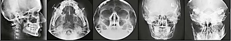
Common extraoral films include panoramic and cephalometric films.
A panoramic film shows a panoramic (wide) view of the maxilla & the mandible & surrounding structures on a single radiograph.
A cephalometric film exhibits the bony and soft tissue areas of the facial profile.


Extraoral Film Packaging
Extraoral films are designed for use outside the mouth and therefore are not enclosed in moisture proof packets.
Extraoral films used in dental radiography are available in 5 × 7 inch & 8 × 10-inch sizes as well as in the panoramic 5 × 12 inch & 6 × 12-inch sizes.
Extraoral films are boxed in quantities of 50 or 100.
Extraoral Film Types
Two types of film may be used in extraoral radiography:
(1) Screen film (2) Non-screen film

Screen Film
The majority of extraoral films are screen films.
A screen film is a film that requires the use of a screen for exposure (see later discussion). A screen film is placed between two special intensifying screens in a cassette.
When the cassette is exposed to x-rays, the screens convert the x-ray energy into light, which, in turn, exposes the screen film. The screen film is sensitive to fluorescent light rather than to direct exposure to x-radiation.
Non-screen Film

A non-screen film is an extraoral film that does not require the use of screens for exposure.
A non-screen extraoral film is exposed directly to x-rays; the emulsion is sensitive to direct x-ray exposure rather than to fluorescent light.
A non-screen extraoral film requires more exposure time than does a screen film and is not recommended for use in dental radiography
Extraoral Film Equipment
In extraoral radiography, screen films are used in combination with two special equipment items:
(1) Intensifying screens (2) Cassettes
Intensifying screens
An intensifying screen is a device that transfers x-ray energy into visible light; the visible light, in turn, exposes the screen film.
These screens intensify the effect of x-rays on the film.
With the use of intensifying screens, less radiation is required to expose a screen film, and the patient is exposed to less radiation.
Cassette
A cassette is a special device that is used to hold the extraoral film and the intensifying screens.
Cassettes are available in a variety of sizes that correspond to film and screen sizes.
A cassette may be flexible or rigid; most cassettes are rigid, although the panoramic cassette may be flexible


Duplicating X-RAY FILM
A duplicate radiograph is one that is identical to the original x-ray film.
In dentistry, duplicate radiographs are used for patient referrals to specialists, for insurance claims, and as teaching aids.
A special film, or duplicating film, is required to make a duplicate radiograph.
In dental radiography, a duplicating film is a type of photographic film used to make an identical copy of an intraoral or extraoral radiograph.
Unlike intraoral and extraoral films, the duplicating film is used only in a darkroom setting and is not exposed to x-rays.
When examined in the darkroom under safe light conditions, duplicating film has an emulsion on one side only.
The emulsion side of the film appears dull, whereas the side without the emulsion appears shiny.
The emulsion side of the film must contact the radiograph during the duplication process.
Packaging
Duplicating films are boxed in sets of 50 sheets and are available in three sizes:
5 × 12 inch,
6 × 12 inch,
8 × 10 inch
FILM STORAGE & PROTECTION
Films are adversely affected by heat, humidity, & radiation & must be stored away from sources of radiation at 50° to 70° F & with a relative humidity level of 30% to 50%.
Dental films should always be used before the expiration date printed on the label.

FILL IN THE BLANK
Exercise (2)
1. The component of an x-ray film described as “a thin transparent coating that is placed over the emulsion” is termed:
2. The component of the x-ray film described as “a flexible piece of plastic that withstands heat, moisture, and chemical heat” is termed:
3. The chemical compounds that change when exposed to radiation or light are termed:
4. The invisible pattern of stored energy on the exposed film is termed:
IDENTIFICATION
Identify the items indicated on the intraoral film packet illustrated in Figure

5. 6.
7.
9.
11. 8.
10.