12 minute read
9) The Bisecting Technique


Advertisement
Theory:
The theoretical basis of the bisected angle technique can be summarized as follows:
1. The image receptor is placed as close to the tooth under investigation as possible without bending the packet.
2. The angle formed between the long axis of the tooth & the long axis of the image receptor is assessed & mentally bisected.

P a g e | 59 3. The X-ray tube-head is positioned at right angles to this bisecting line with the central ray of the X-ray beam aimed through the tooth apex.
4. Using the geometrical principle of similar triangles, the actual length of the tooth in the mouth will be equal to the length of the tooth on the image.
Vertical angulation of the X-ray tube-head
The angle formed by continuing the line of the central ray until it meets the occlusal plane determines the vertical angulation of the X-ray beam to the occlusal plane.
Note: Vertical angles are often quoted but inevitably they are only approximate. Patient differences including head position, and individual tooth position and inclination mean that each positioning should be assessed independently. The vertical angulations suggested should be taken only as a general guide.
Horizontal angulation of the X-ray tube-head
Positioning techniques
The bisected angle technique can be performed either by:
1) Using an image receptor holder to support the image receptor in the patient’s mouth, or 2) by asking the patient to support the image receptor gently using either an index finger or thumb. - It is, however, good practice that the image receptor should be held by the patient only when it cannot otherwise be kept in position.
In summary:
1. The image receptor is pushed securely into the chosen holder.
- Either a large or small size of image receptor is used so that the particular tooth being examined is in the middle of the receptor. - When using a film packet the white surface faces the X-ray tube-head & the film orientation dot is opposite the crown.
2. The X-ray tube-head is positioned using the beam--aiming device if available OR the operator has to assess the vertical & horizontal angulations by observation & then position the tube-head without a guide.
3. The exposure is made.

Using the patient’s finger:
1- The appropriately sized image receptor is positioned & orientated in the mouth with about 2 mm extending beyond the incisal or occlusal edges, to ensure that all of the tooth will appear on the image.
*The patient is then asked to gently support the image receptor using either an index finger or thumb.
2. The operator then assesses the vertical & horizontal angulations by observation and positions the tube-head without a guide.
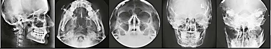
The effects of incorrect tube-head position.
3. The exposure is made.
The specific positioning for different areas of the mouth, using both simple holders & the patient’s finger to support the image receptor.


Comparison of the paralleling and bisected angle techniques:
The advantages & disadvantages of the two techniques can be summarized as follows:
Advantages of the PARALLELING technique
● Geometrically accurate images are produced with little magnification.
● The shadow of the zygomatic buttress appears above the apices of the molar teeth.
● The periodontal bone levels are well represented.
● The periapical tissues are accurately shown with minimal foreshortening or elongation.
● The crowns of the teeth are well shown enabling the detection of approximal caries.
● The horizontal & vertical angulations of the X-ray tube-head are automatically determined by the positioning devices if placed correctly.
● The X-ray beam is aimed accurately at the center of the image receptor – all areas of the image receptor are irradiated & there is no coning off or cone cutting.
● Reproducible radiographs are possible at different visits & with different operators.
● The relative positions of the image receptor, teeth & X-ray beam are always maintained, irrespective of the position of the patient’s head. This is useful for some patients with disabilities.
Disadvantages of the PARALLELING technique:
● Positioning of the image receptor can be very uncomfortable for the patient, particularly for posterior teeth, often causing gagging.
● Positioning the holders within the mouth can be difficult for inexperienced operators particularly when using solid--state digital sensors.
● The anatomy of the mouth sometimes makes the technique impossible, e.g. a shallow, flat palate.
● The apices of the teeth can sometimes appear very near the edge of the image.
● Positioning the holders in the lower third molar regions can be very difficult.
● The technique cannot be performed satisfactorily using a short focal spot to skin distance (a short spacer cone) because of the resultant magnification.
● The holders need to be autoclavable or disposable.
Advantages of the BISECTED ANGLE technique:
● Positioning of the image receptor is reasonably comfortable for the patient in all areas of the mouth.
● Positioning is relatively simple & quick.
● If all angulations are assessed correctly, the image of the tooth will be the same length as the tooth itself & should be adequate (but not ideal) for most diagnostic purposes.
Disadvantages of the BISECTED ANGLE technique:
● The many variables involved in the technique often result in the image being badly distorted.
● Incorrect vertical tube head angulation will result in foreshortening or elongation of the image.
● The periodontal bone levels are poorly shown.
● The shadow of the zygomatic buttress frequently overlies the roots of the upper molars.
● The horizontal & vertical angles have to be assessed by observation for every patient & considerable skill is required.
● It is not possible to obtain reproducible views.
● Coning off or cone cutting may result if the central ray is not aimed at the center of the image receptor, particularly if using rectangular collimation.
● Incorrect horizontal tube head angulation will result in overlapping of the crowns & roots.
● The crowns of the teeth are often distorted, thus preventing the detection of approximal caries.
● The buccal roots of the maxillary premolars & molars are foreshortened.
Conclusion
The diagnostic advantages of the accurate, reproducible images produced by the paralleling technique using image receptor holders & beam-aiming devices ensure that this technique should be regarded as the technique of choice for periapical radiography.
It is good practice that whenever practicable, techniques using image receptor holders with beam--aiming devices should be adopted.
QUESTIONS
MATCHING: For questions 1 to 8, refer to Figure. Match the letters (A to H) of the appropriate items with the descriptions below:
1. _______ No.1 size receptor
2. _______ No.2 size receptor
3. _______ XCP aiming ring, posterior
4. _______ XCP aiming ring, anterior
5. _______ XCP indicator arm, posterior
6. _______ XCP indicator arm, anterior
7. _______ XCP bite-block, posterior
8. _______ XCP bite-block, anterior
FILL IN THE BLANK
1. What happens to the image when the object–receptor distance is increased?
2. What piece of equipment is required to hold the receptor parallel to the long axis of the tooth in the paralleling technique?
3. What do the letters X, C, and P refer to?

4. What size receptor is typically used with the anterior XCP instrument?
5. What size receptor is used with the posterior XCP instrument?
6. Which beam alignment devices are recommended for use with the paralleling technique to reduce radiation exposure of the patient?
7. How is the patient’s head positioned before exposing receptors?
MULTIPLE CHOICE
Why is an increased target–receptor distance required in the paralleling technique? a. to avoid image magnification b. to avoid distortion c. to reduce scatter radiation d. to improve receptor placement
Which of the following describes the relationship of the central ray to the receptor in the paralleling technique? a. 20 degrees to the long axis of the tooth b. 90 degrees to the receptor and long axis of the tooth c. 75 degrees to the long axis of the tooth d. 15 degrees to the receptor and the long axis of the tooth
Which of the following definitions is incorrect? a. parallel: always separated by the same distance b. intersecting: to cut through c. right angle: formed by two parallel lines d. central ray: central portion of the x-ray beam
Which of the following describes the relationship between the receptor and the long axis of the tooth in the paralleling technique? a. The receptor and the tooth are parallel to each other. b. The receptor and the tooth are at right angles to each other.
c. The receptor and the tooth are perpendicular to each other. d. The receptor and the tooth are intersecting each other.
Which of the following describes the distance between the receptor and the tooth in the paralleling technique? a. The receptor is placed as close as possible to the tooth. b. The receptor is placed away from the tooth and toward the middle of the oral cavity. c. Either a or b. d. None of the above.
Which of the following about receptor placement is correct? 1. Anterior receptors are placed horizontally. 2. Anterior receptors are placed vertically. 3. Posterior receptors are placed horizontally 4. Posterior receptors are placed vertically a. 1, 2, and 3 b. 2, 3, and 4 c. 2 and 3 d. 1 and 4
Which of the following about the exposure sequence for periapical receptors is incorrect? a. Anterior receptors are always exposed before posterior receptors. b. Either anterior or posterior receptors may be exposed first. c. In posterior quadrants, the premolar receptor is always exposed before the molar receptor. d. When exposing anterior receptors, work from the patient’s right to left in the maxillary arch, and then work from left to right in the mandibular arch.
Which of the following about the lack of parallelism between the receptor & the long axis of the tooth is correct? a. If the lack of parallelism is greater than 30 degrees, the image is generally acceptable. b. If the lack of parallelism is less than 20 degrees, the image is generally acceptable. c. If the lack of parallelism is less than 50 degrees, the image is generally acceptable. d. If the lack of parallelism is greater than 50 degrees, the image is generally acceptable.
Which of the following are advantages of the paralleling technique? 1. increased accuracy 2. simplicity of use 3. ease of duplication 4. ease of receptor placement a. 1, 2, 3, and 4 b. 1, 2, and 3 c. 2, 3, and 4 d. 1, 3, and 4
The advantages of the paralleling technique outweigh the disadvantages. a. true b. false
ESSAY
• State the basic principle of the paralleling technique.
• Describe why a beam alignment device must be used in the paralleling technique.
• State the five rules of the paralleling technique.
• Discuss the patient & equipment preparations that must be completed before using the paralleling technique.
• Discuss the exposure sequence for 15 periapical receptor placements using the paralleling technique.
• Describe each of the 15 periapical receptor placements that are recommended for usewiththeXCPinstruments.
• Summarize the guidelines for periapical receptor positioning with the paralleling technique.
• Explain the modifications in the paralleling technique that are used for a shallow palate, bony growths, or a sensitive premolar region.
MATCHING
For questions 1 to 4, refer to Figure.
Match the letter (A to D) of the item shown with the description below.
1. _____Plane of the receptor
2. _____Long axis of the tooth
3. _____Imaginary bisector
4. _____Central ray

IDENTIFICATION
For questions 5 to 10, refer to Figures 1, 2, and 3. Write in the letter of the item defined in each question.
5. _____In Figure (1), identify the angle that is bisected correctly.

Figure (1)
6. _____ In Figure (2), identify the central ray that is correctly positioned perpendicular to the imaginary bisector.
7. _____ In Figure (3), identify the position-indicating device (PID) that is aligned correctly.
8. _____In Figure (3), identify the vertical angulation that results in foreshortening.
9. _____In Figure (3), identify the vertical angulation that results in elongation.
10. _____In Figure (3), identify the correct vertical angulation.

Figure (3)
FILL IN THE BLANK
11. What happens to the dental image when a short (8-inch) PID is used?
12. Which size receptor is used with the bisecting technique?
13. Which beam alignment device is recommended for use with the bisecting technique because it aids in the alignment of the PID and reduces patient exposure?
14. How is the patient’s head positioned before exposing maxillary periapicals with the bisecting technique?
15. How is the patient’s head positioned before exposing mandibular periapicals with the bisecting technique?
MULTIPLE CHOICE
16. Which of the following describes the proper direction of the central ray in the bisecting technique?
a. 90 degrees to the long axis of the tooth
b. 90 degrees to the receptor and long axis of the tooth
c. 90 degrees to the receptor
d. 90 degrees to the imaginary bisector
17. Which of the following describes the distance between the receptor & the tooth in the bisecting technique?
a. The receptor is placed as close as possible to the tooth.
b. The receptor is placed away from the tooth and toward the middle of the oral cavity.
c. The receptor is placed parallel to the tooth.
d. None of the above.
18. Which of the following are advantages of the bisecting technique?
1. increased accuracy, 2. simplicity of use, 3. shorter exposure time
a. 1, 2, and 3
b. 1 and 2
c. 2 and 3
d. 3 only
19. The disadvantages of the bisecting technique outweigh the advantages.
a. true
b. false
ESSAY
Discuss the significance of the shaded areas in Figure (4).
State the five rules of the bisecting technique.
Figure (4)

P a g e | 74 Discuss the patient & equipment preparations that must be completed before using the bisecting technique.
Discuss the exposure sequence for the 14 periapical placements using the bisecting technique.
Describe each of the 14 periapical placements recommended for use with the bisecting technique.
Describe correct and incorrect horizontal angulation.
State the recommended vertical angulations for each maxillary periapical exposure using the bisecting technique.
P a g e | 75 State the recommended vertical angulations for each mandibular periapical exposure using the bisecting technique.