
9 minute read
18) Digital Imaging

Basic Concepts
- Digital imaging is a technique used to record radiographic images. Unlike conventional dental radiography techniques, there is no film or processing chemistry is used. - Instead, digital imaging uses an electronic sensor as well as a computerized imaging system that produces radiographic images almost immediately on a computer monitor.
Advertisement
- Before the dental radiographer can use digital imaging technique competently, a thorough understanding of the terminology & fundamentals of digital imaging is necessary. - Knowledge of radiation exposure, equipment & types of digital imaging is also required.
Uses:
• o detect lesions, diseases, conditions of teeth and surrounding structures
• o confirm or classify suspected disease
• o provide information during dental procedures (root canal therapy instrumentation & surgical placement of implants)
• o evaluate growth and development
• o illustrate changes secondary to caries, periodontal disease, or trauma
• o document the condition of a patient at a specific point in time
TYPES OF DIGITAL IMAGING
Two methods of obtaining a digital image currently exist:
(1) Direct digital imaging (2) Indirect digital imaging.
Direct Digital Imaging
▪ The essential components of a direct digital imaging system include an x-ray machine, an intraoral sensor, & a computer monitor.
▪ A sensor with a fiberoptic cable linked to the computer is placed into the mouth of the patient & exposed to x-radiation. The sensor captures the radiographic image & then transmits the image to a computer monitor. ▪ Within seconds of exposing the sensor to radiation, an image appears on the computer screen. Software is then used to enhance & store the image.


Indirect Digital Imaging
1. Scanning Traditional Radiographs 2. Storage Phosphor Imaging
Scanning Traditional Radiographs
− The essential components of an indirect digital imaging system include a charge-coupled device “CCD” camera & a computer. − In the scanning method, an existing radiograph is digitized using a CCD camera. − The CCD camera scans the image on the film, digitizes or converts the image, & then displays it on the computer monitor.
Storage Phosphor Imaging: Photo-stimulable Phosphor Imaging (PSP) In this system, a reusable imaging plate coated with phosphors is used instead of a sensor with a fiberoptic cable. The phosphor-coated plate is flexible & is placed into the mouth in the same way as intraoral film.


- A phosphor-coated plate resembles an intensifying screen used to expose an extraoral film in that it converts x-ray energy into light.
- The images are cleared from the plates by exposure to view-box light for several minutes, or the images may be erased from the plates immediately after the scanning process. Once the image is erased, the plates may be wrapped in plastic & sterilized for reuse.
S t e p B y S t e p P r o c e d u r e s
❑ Step-by-step procedures for the use of digital imaging systems vary from manufacturer to manufacturer.
❑ It is critical to refer to the manufacturer’s instruction booklet for information on the operation of the system, equipment preparation, patient preparation, & exposure. ❑ Only general guidelines concerning sensor preparation & placement are provided next.
Sensor Preparation
• Digital imaging involves the placement of the intraoral sensor in the mouth of the patient, using the same technique as in conventional film placement. • Whether using indirect or direct digital imaging, it is important that the individual sensors are kept sterile. • Storage phosphor sensors are sealed & waterproofed. • For infection control purposes, sensors used in direct digital imaging must be covered with a disposable barrier or sleeve because they cannot withstand heat sterilization.
Sensor Placement:
• The sensor is held in the mouth by bite-block attachments or devices that aim the beam & sensor accurately. • The paralleling technique is the preferred exposure method because of the dimensional accuracy of images produced & the ease of standardizing such images.
• Paralleling technique beam alignment devices must be used to stabilize the sensor in the mouth. As with conventional intraoral film, the sensor is centered over the area of interest.


A d v a n t a g e s & D i s a d v a n t a g e s
Advantages of Digital Imaging:
1. Superior gray-scale resolution. “Digital imaging uses up to 256 shades of gray compared with the 16 to 25 shades f ff f ” 2. Reduced exposure to x-radiation “T u f 50% to 90% less than that required for E- f u ”
3. Increased speed of image viewing. “ f & u ”


4. Lower equipment and film cost. Long-term digital imaging eliminates the need for purchasing conventional film, costly processing equipment, and processing solutions. With digital imaging, darkroom and processing solutions and maintenance are unnecessary. Also, environmental costs are reduced because the disposal hazards of processing chemicals, silver salts in film emulsion, and lead foil sheets are avoided. The elimination of darkroom processing errors is also an advantage.
5. Increased efficiency.
6. Enhancement of diagnostic image. Features such as colorization and zooming allow users to highlight conditions such as bone resorption caused by periodontal disease or to help detect small areas of decay. Another feature that can be used to enhance a diagnostic image is digital subtraction. With digital subtraction, the gray-scale is reversed so that radiolucent images (normally black) appear white and radiopaque images (normally white) appear black
7. Effective patient education tool. “The size of the digitized image on the 15-inch or 17-inch computer screen (compared with a 2- f f k u ”
Disadvantages of Digital Imaging:
1. Initial setup costs. The initial cost of purchasing a digital imaging system is a significant disadvantage. The cost depends on the manufacturer, the level of computer equipment currently in the office, and auxiliary features such as the intraoral camera. Maintenance and repairs must also be considered.
2. Image quality. Many studies have reported on the ability of the digital image to capture early caries, bone loss, periapical radiolucencies, and so on. The majority of the research has shown that the digital imaging performs at least as well as, and at times even better than, traditional radiography.
3. Sensor size and thickness.
Patients may complain about the bulkiness of the sensor, which may cause discomfort or elicit the gag reflex
4. Infection control. Some digital sensors cannot withstand heat sterilization. Therefore, these sensors require complete coverage with disposable plastic sleeves that must be changed between patients every time to prevent crosscontamination.
5. Wear and tear. The receptors used in the PSP system are vulnerable to wear and tear and may have a limited lifespan. The phosphor plates are not designed to have their edges bent or softened to accommodate individual patient anatomy. If bending or scratching of the plates occurs, permanent defects will appear on all images exposed, which may obscure diagnostic information.

6. Legal issues.
MATCHING QUESTIONS
For questions 1 to 9, match each term with its corresponding definition. a. charge-coupled device b. digital radiography c. digital subtraction d. digitize e. direct digital imaging f. indirect digital imaging g. pixel h. sensor
i. storage phosphor imaging 1. A small detector that is placed intraorally to capture the radiographic image. ________________________ 2. An image receptor found in the intraoral sensor. ________________________ 3. A form of indirect digital imaging in which the image is recorded on phosphor-coated plates and then placed into an electronic processor, where a laser scans the plate and produces an image on a computer screen.
4. To convert an image into digital form that, in turn, can be processed by a computer. _____________________ 5. A method of obtaining a digital image in which an intraoral sensor is exposed to x-rays to capture a radiographic image that can be viewed on a computer monitor. ________________________ 6. A discrete unit of information; a picture element. ________________________ 7. A method of obtaining a digital image, in which an existing radiograph is scanned and converted into a digital form using a CCD camera. ________________________ 8. A method of reversing the gray scale as a digital image is viewed. ________________________ 9. A filmless imaging system; a method of capturing a radiographic image using a sensor, breaking the image into electronic pieces, and presenting and storing the image using a computer. ________________________
TRUE OR FALSE
In digital imaging, the term used to describe the picture that is produced is radiograph. _______________ Digital imaging requires more x-radiation than does conventional radiography. _______________ The x-radiation source used in most digital imaging systems is a conventional dental x-ray unit. _____________
P a g e | 170 Compared with film emulsion, the pixels used in digital imaging are structured in an orderly arrangement. ___ All intraoral sensors can be heat-sterilized after use. _______________
The preferred exposure method for intraoral digital imaging is the paralleling technique. _______________ One advantage of a digital imaging system is the superior gray-scale resolution that results. _______________ Digital subtraction is an advantage in digital imaging because distracting background information is eliminated from the image. _______________ The manipulation of the original digital images can be considered a legal issue. _______________
MULTIPLE CHOICE
Digital imaging was introduced to dentistry in:
a. 1967
b. 1977
c. 1987
d. 1997
Digital imaging can be used for:
a. detecting conditions of teeth and surrounding structures b. evaluating the growth and development of jaws c. confirmation of suspected disease d. all of the above
Digital imaging requires less radiation than does conventional radiography because:
a. the sensor is larger b. the sensor is more sensitive to x-rays c. the exposure time is increased d. the pixels sense transmitted light quickly
The method of obtaining a digital image similar to scanning a photograph to a computer screen is termed:
a. direct digital imaging b. indirect digital imaging c. storage phosphor imaging d. CMOS/APS
The image receptor found in the intraoral sensor is termed:
a. CCD
b. pixel c. semiconductor chip d. software
Digital imaging systems can be used for:
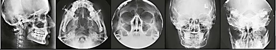
a. bite-wing images b. panoramic images c. cephalometric images d. all of the above
All of the following are advantages of digital imaging except:
a. digital subtraction b. the ability to enhance the image c. size of the intraoral sensor
d. patient education


