
COLOPHON
Production and Publication
Research & Training, Dept. of Radiology & Nuclear Medicine, Erasmus MC
Editor
Lieke Visser
Assistant Editor
Ouidad Oujjit
Design & Photography
Steven Ensering
Frank van der Panne
Vincent Blinde
Maartje de Sonnaville
Printing GROENPRINT
Aristotelesstraat 20 3076 BD Rotterdam
The Netherlands
For this scientific report, GROENPRINT and Trees for All plant several trees to restore the tropical rainforest
Visiting Address
Department of Radiology & Nuclear Medicine
Erasmus MC
Dr. Molewaterplein 40 3015 GD Rotterdam
The Netherlands
Telephone: +31 10 703 5372 research.radiology@erasmusmc.nl
Post Address
Department of Radiology & Nuclear Medicine
Erasmus MC
P.O. Box 2040 3000 CA Rotterdam
The Netherlands
Website
Radiology & Nuclear Medicine – Department –Erasmus MC

Cover photo
A signpost within the Intervention Complex at department Radiology & Nuclear Medicine, Erasmus MC –Rotterdam, the Netherlands.
2024 Scientific Report department
of radiology & nuclear medicine
In 2024, we have faced significant challenges in securing robust funding for biomedical research at local, national, and international levels. Locally, rising personnel costs and necessary investments in infrastructure have forced us to carefully balance our budget. Nationally and globally, trust in scientific institutions is eroding. As public and political confidence declines, so does the willingness to invest in research and innovation. Additionally, competing priorities such as climate change and defense are diverting funds away from healthcare and biomedical research.
Despite these threats, I firmly believe that revising the Erasmus MC research strategy and adapting our focus to address pressing societal concerns – such as aging populations, rising healthcare costs, and workforce shortages – will position us to make a meaningful impact. By investing in technological solutions, we can contribute to solving critical healthcare challenges.
Yet, beyond the quality of our research, I place my trust in the scientists within our department. Their remarkable skills, dedication, creativity, open-mindedness, and commitment to improving the well-being of patients and society are what truly drive progress. Ultimately, it is not technology but the human spirit that will continue to inspire us.
Over the past year, 30 PhD students successfully defended their theses. While it may seem like a routine milestone, I recognize the dedication and perseverance required to achieve this accomplishment. I hope these students continue to invest their knowledge and passion in imaging – whether in academia or industry – creating meaningful impact for patients.
The talent development plan implemented in 2023 is beginning to yield results. This initiative fosters an environment where young researchers can thrive, offering support to advance their work. Our newly adopted approach to recognition now considers not only publications and grants but also academic leadership, education, and societal impact. Outstanding achievements in these domains have led to several well-deserved promotions.
Astrid van der Veldt, medical oncologist with a joint appointment in the departments of Medical Oncology and Radiology & Nuclear Medicine, was appointed Associate Professor. Her research strengthens the field of molecular imaging, focusing on immunotherapy evaluation with specific radionuclides and radionuclide therapy. Erik de Blois, clinical radiochemist, was appointed Assistant Professor to advance the development and implementation of new therapeutic radionuclides. Jukka Hirvasniemi was appointed Assistant Professor to implement AI in musculoskeletal image analysis.
Rianne van der Heijden returned to Rotterdam after a two-year stay at the University of Wisconsin’s Radiology Department with a prestigious VENI grant and was appointed Assistant Professor.
In 2023, we initiated a collaboration with the Department of Ophthalmology and the Rotterdam Eye Hospital, forming the Eye Image Analysis Group Rotterdam (EyeR). A year later, Danilo Andrade de Jesus and Luisa Sánchez Brea, serving as principal investigators, were appointed Assistant Professors.
This year, we further strengthened our collaboration with TU Delft and Erasmus University:
In partnership with the Department of Orthopedics & Sports Medicine (Erasmus MC) and Biomechanical Engineering (TU Delft), we established the Motion Biomechanics & Imaging Lab (MOBI). This lab enables researchers to measure joint loading during movement in an unprecedented way – a breakthrough that opens new avenues for early osteoarthritis detection and accelerated treatment development.
With the support of Erasmus University, we replaced an aging 3T-MRI scanner with a state-of-the-art model. This neuroscience lab will primarily support groundbreaking research by the Erasmus School of Social and Behavioural Sciences, Rotterdam School of Management, and the Department of Child and Adolescent Psychiatry/Psychology.
Research is a collective effort, and our team is both resilient and committed. I also extend my gratitude to our collaborators – departments within Erasmus MC, universities in the Netherlands and abroad, and partners in industry – whose contributions help drive innovation forward.
Enjoy reading this annual report.
Aad van der Lugt, Professor and Chairman
May 2025
HIGHLIGHTS 2024
Francis Baffour first visiting Gabriel P. Krestin Visiting Professor
The Gabriel P. Krestin Visiting Professorship is a new annual tradition to mark Gabriel Krestin's retirement as the former chairman of our department at the end of 2021. To honor his exceptional contribution to the department, a visiting professorship grant program in his name was installed, providing the opportunity to international talents in the field of Radiology & Nuclear Medicine to come to Erasmus MC for a visiting professorship of two to four weeks. With his program we honor Gabriel Krestin’s dedication towards nurturing talent, building world-wide collaborations, and moving the field of radiology and nuclear medicine forward.
From 26 August through 6 September 2024, Francis Baffour visited our department as the first Gabriel P. Krestin Visiting Professor. Francis Baffour is a renowned musculoskeletal radiologist and associate professor at the Mayo Clinic in Rochester, USA. He is also a pioneer in the field of photon-counting CT for musculoskeletal applications.
The primary aim of Francis Baffour's visit was to strengthen our scientific collaboration with the Mayo Clinic in the area of photon-counting CT for musculoskeletal applications, already initiated with Prof. Edwin Oei and Dr. Ronald Booij, and to build similar collaborations in other radiological subspecialties. To this end, he gave several presentations on his scientific work and of others at the Mayo Clinic in the field of photon-counting CT. In addition, he had many discussions with our researchers and clinical radiologists active in the field of photon-counting CT. We expect that this will lead to multiple new collaborations on various applications of this exciting new technology in the near future.
Dr. Baffour also shared his clinical knowledge on musculoskeletal and acute radiology with our residents and radiologists during several case-based teaching sessions. He concluded his very successful and productive visit with a keynote lecture on clinical applications of wholebody MRI during a regional educational symposium, which was also attended by Gabriel Krestin.

Appointments
Astrid van der Veldt was appointed as Associate Professor.
Stijn Koolen was appointed as Associate Professor.
Jifke Veenland was appointed as Associate Professor.
Julia Neitzel was appointed as Assistant Professor.
Erik de Blois was appointed as Assistant Professor.
Rianne van der Heijden was appointed as Assistant Professor.
Jukka Hirvasniemi was appointed as an Assistant Professor.
Luisa Sanchez Brea was appointed as Assistant Professor.
Danilo Andrade De Jesus was appointed as Assistent Professor.
Martijn Starmans was appointed as Assistant Professor.
Ivo Schoots was appointed as Co-chair PI-RADS steering committee, on prostate MR imaging.
Frank Wolters was appointed principal investigator of the neuroepidemiology research section.
David Hanff became a board member of the Musculoskeletal Radiology section of the Dutch Society of Radiology (NVvR).
Daniel Bos was appointed as Associate Programme Director for the MSc-programme in Clinical Epidemiology for the Netherlands Institute of Health Sciences.
Edwin Oei became the President of the European Society for Magnetic Resonance in Medicine and Biology.
Rianne van der Heijden became Junior Fellow of the International Society for Magnetic Resonance in Medicine.
Jacob Visser was appointed as member of the board of the section Techniek of the Dutch Society of Radiology.
Meike Vernooij was appointed as program planning chair for neuroradiology for the ECR 2026 conference.
Julia Neitzel became PI of the ORACLE Study, which includes extensive data on brain health, including neuroimaging, cognitive testing and motor functions, from 2,000 parents of the Generation R Study.

Frank Wolters was elected on the executive committee of VasCog (the International Society of Vascular Behavioural and Cognitive Disorders).
Frank Wolters was appointed principal investigator of Neuroepidemiology for the Rotterdam Study.
Julia Neitzel was elected as the first non-Dutch board member of VENA (Vrouwen binnen Erasmus MC Netwerk voor Academici).
Contribution to Guidelines
François Willemssen contributed to two Dutch guidelines for diagnostic abdominal imaging for HCC and Cholangiocarcinoma.
Maarten Thomeer contributed to the Dutch guidelines for Ovarian, Endometriual and Cervical Carcinoma.
Ivo Schoots is member of the European Association Urology (EAU) Prostate Cancer guideline panel and member PI-RADS steering committee.
Tessa Brabander contributed to the ESMO guideline Gastroenteropancreatic neoplasms.
Astrid van der Veldt is chair of the Dutch Melanoma Guideline.
Marion Smits was involved in the following guidelines:
– PET-based response assessment criteria for diffuse gliomas (PET RANO 1.0): a report of the RANO group. Lancet Oncol 2024;25:e29-e41.
– A Neuroradiologist's Guide to Operationalizing the Response Assessment in Neuro-Oncology (RANO) Criteria Version 2.0 for Gliomas in Adults. AJNR Am J Neuroradiol 2024;45:1846-1856.
– Standardized reporting for Head CT Scans in patients suspected of traumatic brain injury (TBI): An international expert endeavor. Neuroradiology 2024;66:1513-1526.
Sophie Veldhuijzen van Zanten contributed to international guideline for the use of theranostics in brain tumors; a joint effort of the Response Assessment in Neuro-Oncology (RANO) Working Group for PET and the European Association for Neuro-Oncology (EANO).
Erik de Blois contributed to the IAEA TecDoc publication on Production and Quality Control of Actinium-225 Radiopharmaceuticals.
Societal Impact
The randomized phase III trial entitled 'Neoadjuvant nivolumab and ipilimumab in resectable stage III melanoma' by Blank CU, Lucas MW, ....., van der Veldt A and Long GV was published in the New England Journal of Medicine and resulted in the reimbursement of ipilimumab for patients with stage III melanoma in the Netherlands.
Patrick Tang contributed to the public understanding of research as one of the KNAW (Royal Netherlands Academy of Arts and Sciences) ‘Faces of Science’.
The patient organization Hyponews conducted an interview with S ophie Veldhuijzen van Zanten's group to discuss the findings of the scientific publication “[18F] FET PET/MRI: An Accurate Technique for Detection of Small Functional Pituitary Tumors”, aiming to inform the broader public about the importance of these findings for patients with small pituitary tumors.
Frank Wolters organized a public session on dementia prevention at the National Dementia Conference (organized by the Ministry of Health).
Awards
Jessica de Jong received the “Excellent Research Presentation Award” at the Landelijke Werkgroep Neuro-Oncologie (LWNO) meeting in March 2024.

Esther Droogers was selected for the “Best abstract presentation” at the Erasmus MC Cancer Retreat in April 2024.


Nina Overdevest won the “Best Poster Award” at the EMC
Sophie Veldhuijzen van Zanten won the “Innovative Protocol Award” during the 25th Workshop on Methods in Clinical Cancer Research, organized by the European Organisation of Research and Treatment of Cancer, European Society for Medical Oncology, and American Association for Cancer Research.

Marcella Zijta won the Best Oral Presentation award in category AI at the ISUOG World Congress on Ultrasound in Obstetrics and Gynecology, for her work on detection of congenital brain anomalies on 3D first-trimester ultrasound.
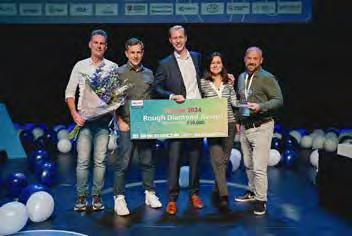
Lyla (previously AlphaPace), TU Delft spin-off company in which Erik de Blois is involved, proudly won the Philips Innovation Award 2024 in the Rough Diamond category, selected from 175 entries. This recognition highlights his innovative contribution to advancing healthcare and radiopharmaceutical quality testing.

Anouk de Jong received the Alavi–Mandell Award by the SNMMI for her publication entitled, '68Ga-PSMA PET/CT for Response Evaluation of 223Ra Treatment in Metastatic Prostate Cancer' in the Journal of Nuclear Medicine.
Anouk de Jong received the Fred Guurink award for her excellent thesis.
David Hanff was awarded the Erasmus MC MORE Award of Master Teacher of the Year 2023.
Kaouther Mouheb won the first price in the MICCAI Educational Challenge 2024 with a tutorial on denoising diffusion models, that she developed together with two researchers from the University of Girona, Spain.
Mahlet Birhanu was awarded in the Euro-BioImaging Job Shadowing program and visited the Medical Research Institute of the Hospital La Fe in Valencia, Spain in October 2024.
Patrick Tang was awarded the Best Power Pitch award during the annual meeting of the ISMRM Benelux in January 2024.
Stijntje Dijk received the Young Scholar Award in Health Policy in honor of Sandy Schwartz at the Society for Medical Decision Making meeting.
Circe van der Heide won the EANM Young Investigator Award.
Esther Droogers was selected for the “Best abstract presentation” at the Erasmus MC Cancer Retreat in April 2024.
Niels Dur received a Young Investigator Award during the International Workshop on Osteoarthritis Imaging (IWOAI) held in Marrakech, Morrocco, from 25 to 28 June 2024.

Marijn Mostert won the first prize in the clinical trainee abstract competition of the Musculoskeletal MR imaging Study Group during the ISMRM annual meeting in Singapore from 4-9 May 2024.
Meetings
Ryan Muetzel hosted the Raynor Cerebellum Project kickoff meeting in Rotterdam in April, with more than 15 invited speakers and 40 attendees, in order to foster collaboration, brainstorm future directions, and initiate a funded project to further our understanding of cerebellum development.
David Hanff organized the Sandwich Course Musculoskeletal Radiology for the Dutch Society of Radiology (NVvR) in Ede, the Netherlands, on 6-7 November 2024. He also organized and chaired the Dutch and Belgian MSK meeting of the NVvR in Rotterdam, the Netherlands on 22 June 2024.

Marjolein Dremmen was a co-chair and member of several committees for development of national guidelines (e.g. imaging in trauma, epilepsy, craniosynostosis).
Pierluigi Ciet together with Professor Emeritus Harm Tiddens organized the first Academy of Pediatric Chest Imaging course in Rotterdam.
Simone Dalm was a jury member for the Sanjiv Sam Ghambir Young Investigator Award.
Ivo Schoots was co-chair of the 3-day Conference on Prostate Cancer Imaging of the European Society Urogenital Radiology, 2024, Zeist, the Netherlands.
Ivo Schoots was co-chair of a 2-day lecturing program on Prostate Cancer Imaging – RSNA, 2024, Chicago, USA.
Grants 2024
Personal Grants / Fellowships
Dutch Research Council VENI Grant
Rianne van der Heijden
Title: ‘Towards better care for patients with chronic low back pain using advanced imaging’.
Dutch Research Council VIDI Grant
Astrid van der Veldt
Title: ‘Unravelling the tumour escape in melanoma survivors after stopping immunotherapy’.
National Grants
Dutch Research Council XS Open Competition
Ilva Klomp
Title: ‘Identifying the tumor stroma as a key player in resistance to internal radiation treatment’.
Dutch Research Council XS Open Competition
Joana Campeiro
Title: ‘Tackling triple-negative breast cancer: Development of a novel nuclear medicine-based strategy to create a personalized medicine approach for a biomarkernegative cancertype’.
Ministry of Education, Culture & Science Starting Grant
Martijn Starmans
Title: ‘RadPathRI: Research Infrastructure for AI for Integrated Diagnostics joining forces of radiology and pathology’.
Ministry of Education, Culture & Science Starting Grant
Julia Neitzel
Title: ‘Multimodal Assessment of Brain Health Using Blood-Based and Imaging Markers’.
Ministry of Education, Culture & Science Starting Grant
Rianne van der Heijden
Title: ‘Pain Imaging’.
Health Holland
Juan Hernandez Tamames, Marion Smits, Dirk Poot & Laura Nunez Gonzalez
Title: ‘Gadolinium Free Enhancement in MRI (GEM)’.
Health Holland
Stefan Klein
Title: ‘Towards Fully automated Anomaly Screening in the first Trimester of pregnancy using Artificial Intelligence (FAST-AI)’.
Heath Holland
Theo van Walsum & Kay Pieterman
Title: ‘X-ray vision for surgeons: tumor localization and visualization using magnetic seed tracking and augmented reality - The Inside Project’.
Health Holland
Erik de Blois
Title: ‘Phase-I dose escalation study to evaluate the tolerability and safety of 161Tb-PSMA in patients with metastatic, castration resistant prostate cancer’.
Dutch Research Council Open Technology Program
Stefan Klein, Maarten Thomeer & Martijn Starmans
Title: ‘The Liver Artificial Intelligence (LAI) consortium: a benchmark dataset and optimized machine learning methods for MRI-based diagnosis of solid appearing liver lesions’.
Dutch Research Council Open Technology Program
Yann Seimbille
Title: ‘Molecular oncology twins advancing treatment and innovative cancer evaluation (MOTIVATE)’.
Dutch Research Council Dutch Research Agenda
Ryan Muetzel
Title: ‘Why are there more men than women with autism? Sex differences in Autism: Genes, Brain, and Healthcare’.
Dutch Research Council Venture Challenge
Laura Mezzanotte
Title: ‘Radigene-Reporter gene technology for imaging cell and gene therapies’.
Dutch Research Council Perspective
Stefan Klein
Title: ‘Artificial Intelligence for Accessible Medical Imaging (AI4AI)’.
Dutch Research Council Perspective
Juan Hernandez-Tamames
Title: ‘Development of personalized MR-guided thermochemotherapy for breast conserving surgery (CARES) Conserving the breast by heating the tumour.’
Dutch Society for Gastroenterology
Kay Pieterman
Title: ‘Continuous periprocedural portal pressure measurements using pressure microwires to study effect of sedation on portal pressure and evolution of portal pressure in the hours following TIPS – a pilot study’.
International Grants
The European Network on Osteoarthritis
Wouter Schallig
Title: 'Dynamic fluoroscopy to assess knee pathomechanics'.
EU Horizon IHI
Theo van Walsum
Title: ‘Unleashing a CoMprehensive, Holistic and Patient Centric Stroke Management for a Better, Rapid, AdvancEd and PersonaLised Stroke Diagnosis, TreAtment and Outcome Prediction (UMBRELLA)’.
EU Horizon IHI
Mark Konijnenberg & Erik Verburg
Title: 'Theranostics ecosystem for personalised care (Thera4Care)’.
EU Horizon IHI
Yann Seimbille
Title: ‘Elevating the future of cancer care with alpha theranostics (Accelerate)’.
EU Horizon DIGITAL
Jan-Jaap Visser, Ilva van Houwelingen, Martijn Starmans & Stefan Klein
Title: ‘Supporting Health Data Access Bodies to establish AI pathways enabling Deployment of AI as medical device tools (SHAIPED)’.
ERC Synergy Grant
Astrid van der Veldt (co-applicant)
Title: ‘Enchanced treatment and sustainable care: 3DPrinting of BRAF/MEK inhibitors’.
EU MSCA Doctoral Networks
Juan Hernandez-Tamames
Title: ‘AI in Parkinson Disease (AIPD)’.
EU MSCA Doctoral Networks
Dirk Poot, Stefan Klein & Juan Hernandez Tamames
Title: ‘Improving QMRI by realizing trustworthy integration of AI in Neuro-imaging (IQ BRAIN)’.
Charitable Organisations
Dutch Cancer Foundation
Yann Seimbille
Title: ‘First-in-human assessment of a FAP-targeted probe for fluorescence guided surgery of pancreatic cancer’.
Stichting Astma Bestrijding
Pierluigi Ciet & Daan Caudri
Title: ‘Developing and validating an AI-supported chest CT score to diagnose Post-infectious Bronchiolitis Obliterans (PiBO) in children’.
Alzheimer Nederland Biomedical Research
Frank Wolters
Title: ‘The APOE- ε 2 paradox: role of APOE in lipid metabolism, vascular injury and amyloid deposition’.
Stichting bevordering onderzoek Franciscus
Eva Bocharewicz & Kay Pieterman
Title: ‘Anastomotic Leakage Prevention by Endovasculair Stenting of the Superior Mesenteric Artery (ALPrES2MA Study)’.
Vaillant grant
Kay Pieterman
Title: ‘Fine needle aspiration versus core biopsy of suspected metastatic liver lesions’.
Raynor Cerebellum Project
Ryan Muetzel
Title: ‘Normative growth models of the human Cerebellum’.
Institutional Grants
Stichting Erasmus MC Pijnfonds
Rianne van der Heijden
Title: ‘Richting betere zorg voor patienten met chronische lage rugpijn met geavanceerde beeldvorming’.
Erasmus MC Research Innovation Grant
Dianne van Dam-Nolen
Title: ‘Photon-counting CT for the detection of intraplaque hemorrhage in carotid atherosclerosis? An innovative pilot-study for optimizing stroke work-up’.
Erasmus MC Research Innovation Grant
Maarten Leening & Daniel Bos
Title: ‘Old dog, new trick - Unravelling the effects of Low-Dose Colchicine on coronary plaque regression and stabilzation (LoDoCo-Plaque)’.
Medical Delta 3.0
Edwin Oei
Title: ‘Early Rheumatoid Arthritis identification’.
Medical Delta 3.0
Marion Smits & Sophie Veldhuijzen van Zanten
Title: ‘Cancer Diagnostics for Sustainable Health CareTheranostic work package (CARES)’.
Medical Delta 3.0
Meike Vernooij & Frank Wolters
Title: ‘Applying advanced brain imaging for efficient dementia diagnosis and prediction’.
Investigator initiated industry sponsored grants
Qure AI
Jan-Jaap Visser
Title: ‘Validation of AI in stroke patients’.
Qure AI
Jan-Jaap Visser
Title: ‘AI to improve nodule detection on chest X-ray’.
AstraZeneca
Jan-Jaap Visser
Title: ‘Pulmonary Incidental Nodules: Improve Detection and Follow-up by integrating Artificial Intelligence (PINPOINT)’.
Sanofi
Maarten Leening & Daniel Bos
Title: ‘Unlocking the Preventive Potential of Routine Clinical Imaging: Implementation of the KALK Project’.
Bracco
Ricardo Budde
Title: ‘Assessment of coronary stents with PCCT’.
Siemens Healthineers
Ricardo Budde
Title: ‘Evaluation of PCCT’.
Medtronic
Aad van der Lugt
Title: ‘CONTRAST2.0, consortium for new treatments for acute stroke’.
New facilities
Bucky Room
The Bucky Room in the emergency department was renovated in 2024. The Siemens Ysio has been replaced by the Philips DigitalDiagnost Flexroom. This system offers the advantage of providing the Radiology Technician with increased workspace in the X-ray room for patient care. The Bucky table can be tilted 90 degrees, thereby expanding the working area. Additionally, the detectors are of the latest generation, enhancing the quality of the images.
PCCT scanner

In 2024, the CT scanner at the emergency department had to be replaced. The CT (Siemens Drive) was moved from Sophia Children’s Hospital to the emergency department, which resulted in an upgrade compared to the older system. Additionally, a new PCCT was installed at Sophia Children hospital. The PCCT represents a significant improvement for the current clinical practice for all patients at the
Sophia Children's Hospital and simultaneously offers new research opportunities. In 2021, one of the first PCCTs in Europe was already installed at Erasmus MC for adults.
This second PCCT is the first PCCT in a children's hospital in Europe and offers significant advantages for imaging children. For example, there is a greatly improved resolution, allowing for much more detail and the visualization of smaller anatomical structures. Spectral imaging also provides more information about tissue composition and can be used to visualize, for instance, the effect of lung perfusion. Additionally, spectral imaging can more accurately map metal medical devices, such as those used in surgeries for children with scoliosis. Furthermore, it is expected that the PCCT scanner will not only reduce radiation exposure but also decrease the amount of contrast agent required, without compromising image quality.

3T MRI Scanner
In collaboration with the Generation R Study and Erasmus University, Erasmus MC is excited to announce the successful development of a state-of-the-art MRI facility designed to support and enhance a wide range of functional MRI (fMRI) studies. This initiative marks a significant milestone in our commitment to advancing innovative research within the field of neuroimaging.
Since December 2024, the new MRI scanner has been operational, accompanied by cutting-edge equipment designed to cater to the diverse needs of researchers. The facility now boasts a comprehensive suite of tools, including specialized button press devices for fMRI tasks, a high-resolution screen for visual stimuli, high-quality MRI headphones to deliver auditory stimuli, and even an eye-tracking system to capture visual attention dur-
Motion Biomechanics and Imaging laboratory (MOBI-LAB)
On 17 October 2024, after several years of preparation, the Motion Biomechanics and Imaging (MOBI) laboratory was officially opened by Prof. Stefan Sleijfer (Chair of the Board of Directors and Dean of Erasmus MC) and Prof. Fred van Keulen (Dean of the Faculty of Mechanical

ing experiments. These advanced tools ensure that fMRI research aligns with international standards, offering an ideal platform for a wide range of experimental designs.
Engineering of Delft University of Technology). This new facility, located in the Department of Radiology & Nuclear Medicine, is the first in the Netherlands and amongt the first in Europe, that combines fluoroscopy and motion analysis technologies to assess dynamic joint func-
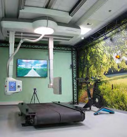
tion and loading in a clinical setting. In the next years, the MOBI-lab will be used mainly in interdisciplinary clinical research projects on osteoarthritis and other musculoskeletal disorders affecting joint stability, leveraging the capability of this lab to explore dynamic joint health to an extent that was not possible before. We also will link joint loading and stability measurements obtained in the MOBIlab to findings on imaging, in particular using advanced quantitative MRI and PET/MRI studies, in order to advance our understanding the pathophysiology of joint disorders. We envision that, in the longer term future, the MOBIlab could also be applied in clinical care, to facilitate personalized treatment strategies.
Being fully embedded in the Convergence Health and Technology program, it is supported by both Erasmus MC and TU Delft, and serves as a showcase for the Convergence program as the first joint facility between these two institutions.
CONVERGENCE


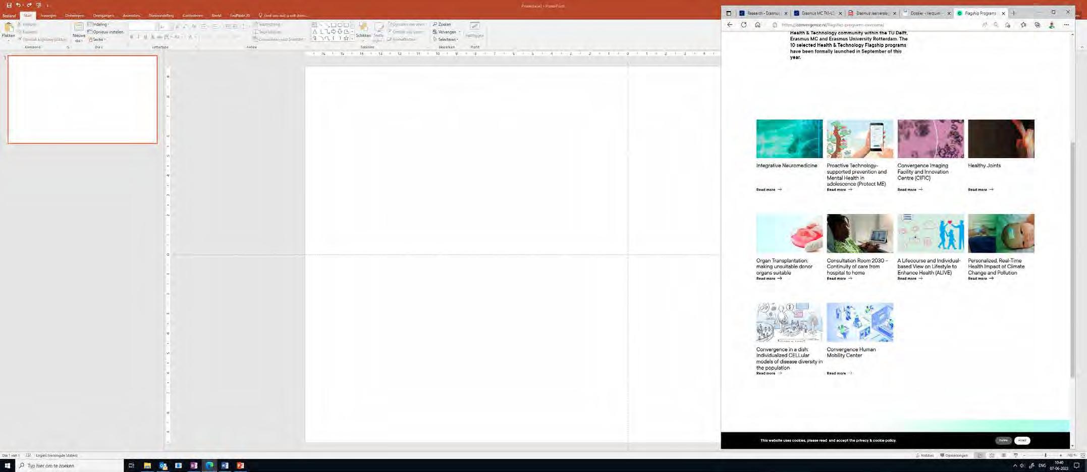

Figure: Ten Flagships started in 2022.
Brain Tumors – CONVERGENCE
Erasmus MC
Martin van den Bent
Juan Hernandez-Tamames
Stefan Klein
Pieter Kruizinga
Alejandra Mendez Romero
Dirk Poot
Marion Smits
Krishnapriya Venugopal
Esther Warnert
Karen van der Werff
Expertise
TU Delft
Chi-Hsien Tseng
Frans Vos
Jeroen Kalkman
Miriam Menzel
Erasmus University Rotterdam
Justien Dingelstad
Iris Wallenburg
Brenda Leeneman
Hedwig Blommestein
Seamus Kent
Contribution and Added Value
Cross-pollination of clinical, technical and social sciences, health technology assessment, and use of specific equipment (e.g., PET-MRI at Erasmus MC, 7T at LUMC, proton therapy at HPTC).

Marion Smits co-initiated and was main lead of the Convergence Flagship ‘Deep Medical Imaging of Structure, Physiology and Function’, in which brain tumor imaging features prominently. She stepped down in Summer 2023, and handed over to Juan Hernandez-Tamames to lead the Flagship on behalf of Erasmus MC. Marion Smits was also scientific lead of the Medical Delta Cancer Diagnostics 3.0 scientific program, which focused primarily on brain tumor diagnostics. This program reached the end of its term in 2024, and continues with a new angle with Cancer Diagnostics for Sustainable Health Care (CARES), focusing on theranostics, intra-operative imaging, and early skin cancer detection. Radiology & Nuclear Medicine prominently features in these scientific programs providing expertise on the full spectrum from image acquisition and image analysis to data management and diagnostic clinical imaging. See:
Grants and funding

Figure: Brain tumour with high vascularisation imaged with perfusion MRI.
2019 Convergence: Quantitative Susceptibility MRI: Deep insights in cardio- and neuro-vasculature
2021 Convergence Open mind call: O2-Sense, converging on wearable oxygen monitoring for brain tumor patients
2021 Convergence Open mind call: Neurodegeneration beyond DTI
2022 ICAI lab ROBUST: Trustworthy AI for MRI – brain tumors
2022 Convergence Incentive Grant PIRL: Real-world assessment of ‘PrognosAIs’ for measuring, typing and grading of presumed adult-type diffuse glioma
2022 KWF: Early detection of brain tumor progression with amide proton transfer weighted CEST MRI
2023 ZonMW Vici: Virtual biopsy: paving the way towards reality
Musculoskeletal Imaging – CONVERGENCE
Erasmus MC
Sita Bierma
Jaap Harlaar
Rianne van der Heijden
Jukka Hirvasniemi
Stefan Klein
Joyce van Meurs
Edwin Oei
Gerjo van Osch
Gennady Roshchupkin
Jos Runhaar
Dieuwke Schiphof
Expertise
TU Delft
Samantha Copeland
Jaap Harlaar
Jesse Krijthe
Marco Loog
Marcel Reinders
Amir Zadpoor Ajay Seth
Erasmus University Rotterdam
Inge Merkelbach
Sandra Sülz
Jukka Hirvaniemi, jointly appointed at the Department of Radiology & Nuclear Medicine of Erasmus MC and the Department of Biomechanical Engineering of TU Delft, has advanced expertise in the field of musculoskeletal image analysis using artificial intelligence and radiomics. As example, the extraction of quantitative imaging biomarkers for assessment of osteoarthritisis depicted in the figure below. We also contribute using advanced image acquisition techniques: MRI and PET/ MRI, linking with biomechanics measurements in the new MOtionBiomechanics & Imaging (MOBI) lab.
Contribution and Added Value
The new MOtionBiomechanics & Imaging (MOBI) lab opened in October 2024 in the Department of Radiology & Nuclear Medicine as a joint initiative between Erasmus MC (Oei, Bierma-Zeinstra) and TU Delft (Harlaar) is considered a showcase for the Convergence program as it unites the expertise of technical and medical sciences with the need of collaboration between scientists from various disciplines (engineering, biomechanics, imaging physics, image analysis, clinical orthopedics, radiology).

Grants and funding
2019 ZonMW Open: Biomechanical precision diagnostics in osteoarthritis
Figure: Schematic presentation of a quantitative imaging biomarker extraction pipeline.
2020 Dutch Research Agenda Research along routes by Consortia (NWA-ORC): Healthy Loading to combat osteoarthritis: Leveraging molecular variations in load bearing capacity for individualized movement aDvice: The LoaD project
2022 Convergence Flagship: Healthy Joints
Image-guided therapy – CONVERGENCE
Erasmus MC
Tessa van Ginhoven
Aad van der Lugt
Kees Verhoef
Theo van Walsum
Bart Cornelissen
Eppo Wolvius
Expertise
TU Delft
Nandini Bhattacharya
Jenny Dankelman
Frank Gijssen
Benno Hendriks
Ricardo Guerra Marroquim
Aimee Sakes
Theresia van Essen
The success of integrating smart instruments with augmented navigation is leveraged by the complementary expertise of the project members, that covers the domains of all aforementioned challenges. Integrating smart instruments with augmented navigation leads to novel solutions that cannot be accomplished with only one of the groups. To develop and implement the SMART Surgical knife in clinical practice, expertise of building surgical instruments with incorporated optical fibers and analysis of the signals (Biomechanical Engineering, TU Delft) has to be combined with surgical expertise on safe removal of tumor tissue (Surgical Oncology group, Erasmus MC). Moreover, to augment navigation real time in an intuitive way preoperative information needs to be adapted to the surgical setting (Biomedical Imaging Group Rotterdam, Erasmus MC) and transferred back to the AR environment (Computer Graphics Group, TU Delft). Combining these approaches will provide a more robust and safer way to enhance the surgical procedure, as the visualization can be finely aligned with the surgical procedure using the guidance of the smart instrument, and the feedback from the smart instrument can be enhanced through visualization.

Grants and funding
Erasmus University Rotterdam
Sandra Sülz
Martina Buljac
Contribution and Added Value
Two flagships from 2021, entitled I-GUIDE: Image guided minimally invasive interventions and Smart Surgery in Smart OR, were not granted in the first round. However, both collaborations are still ongoing, collaborative projects are being established and potential subsidies identified. The research group of Theo van Walsum focuses on improving image guidance by integrating preoperative image information in various interventional procedures. Challenges addressed are the modeling and tracking of motion, and deformation of the anatomy and the instruments. Such trackerless navigation approaches have been implemented for ultrasound and x-ray guided procedures such as TIPS, TACE and ablation of liver lesions. Currently, this research is extended with the integration of augmented reality devices to integrate the information in the direct view of the clinician. Many students from TU Delft, from Technical Medicine as well as from engineering disciplines such as Computer Science and Biomedical Engineering, are involved in projects involved in improving image guidance.
More recently, the application of AI in image guided therapy is being investigated, in the ICAI Stroke Lab, where Erasmus MC collaborates with EUR, and in the Smart OR 2030 project, which a.o. addresses automated virtual planning and intraoperative assistance.
2019 Flagship Augmenting Humans – Smart instruments and interventions: Combining the smart Knife with Augmented Reality
2019 Koers23 TUD-EMC grant: Smart Surgery Lab
2019 Flagship Augmenting Humans – Smart instruments and interventions: Optically guided endovascular thrombectomy in patients with large-vessel ischemic stroke
2022 ICAI lab ROBUST: Stroke
Figure: a projection of vessels and structures in the brain (via AR), aligned with a skull phantom.
Theranostics – CONVERGENCE
Erasmus MC
Julie Nonnekens
Yann Seimbille
Laura Mezzanotte
Simone Dalm
Erik Verburg
Gerard van Rhoon
Miranda Christianen
Mark Konijnenberg
Expertise
TU Delft
Freek Beekman
Antonia Denkova
Marlies Goorden
Antonia Denkova
Kristina Djanashvili
Rienk Eelkema
Elisabeth Carroll
Alina Rwei
Zoltan Perko
In a project together with TU Delft to develop a system allowing to image alpha-labeled radiopharmaceuticals TU Delft was working on the development of the detector and software, while we provided actinium-labeled peptides and tissue samples. The data will be used as pilot data for a new grant application.
TU Delft and Erasmus MC have worked on a project to develop a scanning confocal nuclear microscope for improved radiopharmaceutical imaging. TU Delft was providing technical input and physically building the collimator for higher resolution imaging, and we provided biological samples to be used during the testing phase and we will in the future implement the new technology in our experimental work.
Erasmus University Rotterdam
Lucas Goossens
Esther de Bekker-Grob
Ken Redekop
The group at the TU Delft reactor institute produces radioactive isotopes that we use for biological assays. For the production, some optimization has been done upfront and we are currently in the phase of receiving biweekly radioactive compounds to perform the biological experiments.
Contribution and Added Value
By collaborating with TU Delft, it is possible to advance the technological side of our medical oriented work and to have access to facilities that we do not have at the Erasmus MC. By sharing students and facilities, such a collaboration will be a perfect example of convergence between technology and clinics, while accounting for economic and societal aspects.
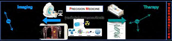
Figure: Theranostics, a concept in which a molecule can be used sequentially as an imaging agent and a therapeutic, has recently revolutionized nuclear medicine.
Grants and funding
2021 Convergence Open Mind call: Scanning Confocal Nuclear Microscope for improved Radiopharmaceutical Imaging
2021 Convergence Open Mind call: Advancing cancer treatment with CERN technology
Erasmus University and Erasmus MC Collaboration on Advanced fMRI Facility – CONVERGENCE
The collaboration between Erasmus University (EUR) and Erasmus Medical Center (Erasmus MC) has led to the successful establishment of a cutting-edge MRI facility, equipped with advanced functional MRI (fMRI) technology. This partnership aims to drive forward innovative research in the fields of neuroscience, psychology, and medical science, creating a hub for groundbreaking studies. The newly set-up MRI facility serves as a prime location for both current and future research initiatives.
Ongoing Projects at Radiology Department Erasmus MC
Several exciting and impactful projects are currently underway at Erasmus MC, showcasing the breadth of research benefiting from this state-of-the-art MRI equipment:
1. GenR – A pioneering study aimed at understanding the genetic and environmental factors contributing to human health and disease across multiple generations. This study will recruit a new cohort in June 2025.
2. PANDA – This project explores neurodevelopmental processes and their implications for mental health, utilizing advanced neuroimaging techniques to observe brain development in real-time.
3. OPPER – This study investigates the neurobiology and longitudinal outcomes of severe postpartum mood disorders (PPMD), such as depression, mania, and psychosis, with a focus on identifying biomarkers and predictive factors for disease course. It aims to distinguish between classical bipolar disorder and postpartum-specific mood disorders. The study uses blood sampling and neuroimaging to explore pathophysiology and predict long-term outcomes.
4. BRIDGE – This study aims to develop brain growth charts for children and adolescents aged 6-20, based on neuroimaging data from the Generation R cohort, which includes over 7,000 brain scans. These charts will help identify deviations in brain development and assist in clinical assessment. The study collaborates internationally with cohorts from the U.S. and China to ensure the robustness and clinical applica-
bility of these charts. By comparing individual brain scans to population norms, the study seeks to improve the detection of brain development abnormalities. The goal is to bridge the gap between advanced neuroimaging data and everyday clinical practice in pediatric neuroradiology.
EUR Research Initiatives
From the EUR, multiple significant studies have already commenced, further strengthening the potential of the MRI facility:
1. Braintime – The Braintime study was completed in 2024 and was the first step in the collaboration between the SYNC lab at the EUR and de department of radiology. In this study, we tested the neural correlates of well-being in young adults. This study resulted in the first joint publication using fMRI: Green, K.H., van de Groep, S., van der Cruijsen, R., Warnert, E., & Crone, E.A. (in press). Neural Correlates of Wellbeing in Young Adults. Emotion , which will appear in 2025.
2. Growing Up Together in Society (GUTS) – The GUTS study is a 10-year longitudinal program in which we study the conditions for growing up successfully in a complex society. In three brain imaging waves, the GUTS team examines structural brain development, the functional neural correlates of self-regulation and trust, and a behavioral development in adolescents (10-20-yrs) from a wide range of socio-economic backgrounds. The project is part of a national Gravitation-funded program.
3. SocCRED – This research explores how social credit scores, which influence individuals' treatment by governments, companies, and communities, impact neural activity during trust-based decisions. The study examines whether these systems intensify or mitigate existing social biases, potentially deepening discrimination. By studying the effect of social credit scores on neural responses, the study aims to provide insights into their societal impact and guide policy recommendations on their implementation or regulation. The findings may also open avenues for future research into related areas of social bias and decision-making.
4. GMG Study – The "Neural Prediction of Multiattribute Giving" study examines how individuals make charitable decisions when exposed to dynamic narratives from donation requests. Participants in the scanner are shown donation appeals from a Dutch television show, where financially distressed candidates ask for donations. The study investigates the neural activations triggered by these narratives to identify key factors influencing donation decisions. It also explores how participants' evaluations evolve over time during exposure to the request. The goal is to understand the neural mechanisms behind charitable giving decisions.
A Vision for Future Research
The success of these projects is just the beginning. Both Erasmus MC and Erasmus University are enthusiastic about the potential for future studies to be conducted at the MRI facility, as this collaboration continues to grow and evolve. The advanced fMRI technology is poised to support a wide range of interdisciplinary research, from neuroscience to psychology, and beyond.
To further streamline and facilitate these research efforts, an fMRI User Community has been established. The commission is led by Eveline Crone, Maarten Boksem, Ryan Muetzel, Carolina Deurloo-Mendez Orellana, Monique de Waard, and Muhammet Sahan, who are dedicated to ensuring the optimal functioning and expansion of the facility. Their leadership will help guide the implementation of future studies, making the most of the technology and resources available.

RESEARCH STAFF
Maarten Leening, MD, PhD
Marcel van Straten, PhD
Martijn Starmans, PhD
Pierluigi Ciet, MD, PhD
Rianne van der Heijden, MD, PhD
Ryan Muetzel, PhD
Simone Dalm, PhD
Sophie Veldhuijzen van Zanten, MD, PhD
Tessa Brabander, MD, PhD
Full Professors
Aad van der Lugt, MD, PhD
Edwin Oei, MD, PhD
Frederik Verburg, MD, PhD
Juan Hernández Tamames, PhD
Marion Smits, MD, PhD
Marleen de Bruijne, PhD
Meike Vernooij, MD, PhD
Myriam Hunink, MD, PhD
Ricardo Budde, MD, PhD
Wiro Niessen, PhD
Associate Professors
Alexander Hirsch, MD, PhD
Astrid van der Veldt, MD, PhD
Daniel Bos, MD, PhD
Frans Vos, PhD
Ivo Schoots, MD, PhD
Jifke Veenland, PhD
Julie Nonnekens, PhD, ius promovendi
Laura Mezzanotte, PhD
Stefan Klein, PhD, ius promovendi
Stijn Koolen, MD, PhD
Theo van Walsum, PhD, ius promovendi
Yann Seimbille, PhD
Assistant Professors
Daan Caudri, MD, PhD
Danilo Andrade de Jesus, PhD
Dirk Poot, PhD
Esther Warnert, PhD
Esther Bron, PhD
Erik de Blois, PhD
Frank Wolters, MD, PhD
Gennady Roshchupkin, PhD
Gyula Kotek, MD, PhD
Henri Vrooman, PhD
Jan-Jaap Visser, MD, PhD
Jukka Hirvasniemi, PhD
Julia Neitzel, PhD
Luisa Sánchez Brea, PhD
Post-Docs & Junior Researchers
Arlette Odink, MD, PhD
Ties Mulders, MD, PhD
Eline Vinke, PhD
Erik Vegt, MD, PhD
Fariba Tohidinezhad, PhD
Giulia Tamborino, PhD
Hanyue Ma, PhD
Hyunho Mo, PhD
Hoel Kervadec, PhD
Ilva Klomp, PhD
Jan-Willem Groen,PhD
Joana Campeiro, PhD
Justine Perrin, PhD
Kay Pieterman, MD, PhD
Laura Nunez Gonzalez, PhD
Maarten Thomeer, MD, PhD
Mariangela Sabatella, PhD
Mark Konijnenberg, PhD
Mark de Wolf, MD, PhD
Maryana Handula, PhD
Renske Gahrmann, MD, PhD
Rob van de Graaf, MD, PhD
Ronald Booij, PhD
Roy Dwarkasing, MD, PhD
Sandra Cornelissen, MD, PhD
Shuai Chen, PhD
Wenjie Kang, MSc
PhD Students
Including partially appointed to department
Abdullah Thabit, MSc
Adnane Zerguit, MSc
Ahmad Alafandi, MD, MSc
Aikaterini Tziotziou, MSc
Alexander Wakker, MD, MSc
Alireza Samadifardheris, MSc
Angelina Pieters, MD, MSc
Anna Streiber, MSc
Anna Lavrova, MD, MSc
Anouk de Jong, MD, MSc, PhD 2024
Arno van Hilten, MSc
Asabi Leliveld, MSc
Bart-Jan Boverhof, MSc
Bas Dille, MSc
Bianca Dijkstra, MSc
Bina Tariq, MD, MSc
Bo Li, MSc
Boudewijn Willems, MSc
Bram Roumen, MSc
Brian Berghout, MSc
Bridget Schoon, MD, MSc
Brigit van Dijk, MD, MSc
Britt van Dijk, MSc
Camiel Box, MD, MSc
Carolline Ntihabose, MSc
Céline van de Braak, MSc
Ching Khan, MD, MSc
Chintan Chawda, MSc
Christina Cretu
Circe van der Heide, MSc
Daniek van der Kaaij, MSc
Danny Feijtel, MSc, PhD 2024
David Hanff, MD, MSc
Desirée de Vreede, MD, MSc
Dianne van Dam-Nolen, MD, MSc PhD 2024
Dorottya Papp, MSc
Douwe Spaanderman, MSc
Duscka Kleijn, MSc
Duygu Harmankaya, MD, MSc
Duygu Kilinc, MSc
Dylan Chapeau, MSc
Eefje Dalebout, MD, MSc
Eline Hooijman, MSc
Eline Zoetelief, MSc
Emanoel Sabidussi, MSc
Erik Kemper, Msc
Érika Murce Silva, MSc, PhD 2024
Esther Droogers, MD, Msc
Eva Bocharewicz, MD, MSc
Eveline Molendijk, MSc
Fatemehsadat Arzanforoosh, MSc, PhD 2024
Federico Mollica, MSc
Felipe Gama Franceschi
Frank te Nijenhuis, MSc
Frederik Hartmann, MSc
Gerda Verduijn, MD, MSc
Gigi Vissers, MSc
Gonzalo Mosquera Rojas, MSc
Hannelore Coerts, MSc
Hazel Zonneveld, MD, MSc
Huib Ruitenbeek, MSc
Ieva Aliukonyte, MSc
Ilanah Pruis, MSc, PhD 2024
Ilaria Neri, MSc
Imren Ozdamar, MD, MSc
Ingrid Bakker, MSc
Jacqueline Claus, MD, MSc
Jamie Verwey, MSc
Jan van der Voet, MD,MSc, PhD 2024
Jarno Steenhorst, MSc
Jasika Paramasamy, MSc
Jessica de Jong, MD, MSc
Jie Deng, MD, MSc
Jing Yu, MSc
Jochem Wolfert, MSc
Joep van de Sanden, MSc
Joost Verschueren, MD, MSc, PhD 2024
José Castillo Tovar, MSc
Josephine Janssen, MSC
Joyce van Arendonk, MSc, PhD 2024
Juancito van Leeuwen, MSc
Judith van der Bie, MSc
Julie Hamm, MSc
Justien Dingelstad, MSc
Kaouther Mouheb, MSc
Karen van der Werff, MSc
Karin van Garderen, MSc, PhD 2024
Karlijn de Joode, MD, MSc
Karthik Prathaban, MSc
Katrien Bracké, MD, MSc
Krishnapriya Venugopal, MSc
Laura Kemper, MSc
Laurens Topff, MD, MSc
Lennard Wolff, MD, MSc
Le Li, MSc
Lisa Bokhout, MSc
Luca Bontempi, MSc
Luke Terlouw, MD, MSc, PhD 2024
Mara Veenstra, MSc
Marchella Zijta, MSc
Matthew Marzetti, MSc
Marguerite Faure, MD, MSc
Mariana Silva Pereira Fialho de Piedade, MSc
Marijn Mostert, MSc
Marjolein Dremmen, MD, MSc
Marjolein Verhoeven, MSc, PhD 2024
Mark van den Dorpel,MD, MSc
Marleen van den Heuvel, MD, MSc
Mathijs Rosbergen, MSc
Matthijs van der Sluijs, MD, MSc
Megan van de Veerdonk, MSc
Mirthe Kamphuis, MSc
Meedie Ali, MSc
Merel de Vries, MSc
Mohamed Benmahdjoub, MSc, PhD 2024
Myrthe van Haaften, MSc
Nadinda van der Ende, MD, MSc, PhD 2024
Natalia Oviedo Acosta, MSc
Neslisah Seyrek, MD, MSc
Niels Dur, MD, MSc
Nienke Sijtsema, MSc, PhD 2024
Nikki Boodt, MD, MSc
Nikki van der Velde, MD, MSc, PhD 2024
Nina Becx, MSc
Nina Overdevest, MSc
Noemi Sgambelluri, MSc
Patrick Tang, MSc
Peter van Hulst, MSc
Pinar Yilmaz, MD, MSc
Pleun Engbers, MSc
Pranali Raut, MSc
Priciana Paraiso, PharMD
Qianting Lv, MSc, PhD 2024
Riwaj Byanju, MSc, PhD 2024
Robin Camarasa, MSc, PhD 2024
Roisin McMorrow, MSc
Rosemarijn Paassen, MSc
Ruben Niemantsverdriet, MSc
Ruisheng Su, MSc, PhD 2024
Sanne Boeren, MD, MSc
Sanne Steltenpool, MSc
Shishuai Wang, MSc
Simran Sharma, MD, MSc
Sophie Derks, MD, MSc
Sonja Katz, MSc, PhD 2024
Sterre de Jonge, MSc
Stijntje Dijk, MD, MSc
Subhradeep Kayal, MSc
Sven Luijten, MD, MSc, PhD 2024
Swaaij Ling, MD, MSc
Tareq Abdel Alim, MSc, PhD 2024
Theresa Feddersen, MSc, PhD 2024
Thom Reuvers, MSc, PhD 2024
Thuy Nguyen, MSc
Tijmen van Zadelhoff, MD, MSc
Tijmen de Wolf, MSc
Tiny Cox, BSc
Tong Wu, MD, MSc, PhD 2024
Tyrillshall Damiana, MSc
Wenjie Kang, MSc
Wietske Bastiaansen, MSc, PhD 2024
Wytse van den Bosch, MD, MSc, PhD 2024
Xi Li, MSc
Xianjing Liu, MSc
Xinyi Wan, MSc
Yahong Wu, MSc
Yulun Wu, MSc, PhD 2024
Yuxin Chen, MD, MSc, PhD 2024
Zoë Keuning, MSc
Unit Research & Training
Monique C de Waard – Director of Research & Training
Lieke Visser – Secretary Research & Training
I maging Trialbureau and Imaging Office
Amos Pomp – Student Assistant
Carolina Méndez-Deurloo – Research Assistant
Daan van der Velden – Post Processing CT
Gaia Hermans – Student Assistent
Ilva van Houwelingen – Process coordinator Imaging Office
Isabelle Klapwijk – Student Assistent
Ivar Jole – Research Assistant
Jessica Wijngaarden – Data Manager
Laurens Groenendijk – Data Manager, Research Assistant
Leontien Heiligers – Coordinator Imaging Trial Office
Miranda Slotboom – Trial Monitor
Mohamed Sheikh – Student Assistant
Nicole Vos van Avezathe – Research Assistant
Renée Broeren – Foekens – Research Assistant
Sharida Ibrahim – Administrative Assistant
Viktoria Ehret – Data Manager
IT Architects and Research Software Engineers
Adriaan Versteeg
Alexander Harms
Hakim Achterberg
Ivan Bocharov
Mahlet Birhanu
Marcel Koek
Ruben van Oosterhoudt
Student Assistant MRI ERGO/GenR
Anne-Sterre Schutter
Akin Sonmezdag
Celine Tuik
Esra Hemmelder
Fengli Bottema
Gaia Hermans
Hafsa Tozkoparan
Hajar el Moussati
Hoa Nguyen
Issrae Affani
Jill Liu
Levy Schimmel, Team Leader
Lieke Bouvy
Lucas de Groot
Martijn van der Meer
Mehdi Badaoui
Michiel van den Akker
Ouidad Oujjit
Paula Rijs Alonso, Team Leader
Suheda Yuce
Technicians
Amber Piet – Research Technician
Corrina de Ridder – Biotechnican
Debra Stuurman – Biotechnican
Jan de Swart – Imaging Specialist
Lilian van den Brink – Research Technician
Lisette de Kreij-de Bruin – Research Technician
Marcel Dijkshoorn – Research Technologist CT
Rob Verhagen – Research Technician
Departmental Operational Staff
Including partially appointed to research
Anita Harteveld – Technical Physician
Britt Gulpen – Staff Advisor
External Support Staff
Mika Vogel – MRI Scientist GE Healthcare
Dennis Kuijper – Nuclear Medicine technologist, Coördinator Research & Innovation
Ivan Dudurych – Research employee
Jean-Baptiste van Aarsen – Nuclear Medicine technician, Coördinator Research & Innovation
Jeffrey Langerak – System Administrator
Jip Holtzer – Staff Advisor
Joël de Groen – Computer Tomography, Coördinator Research & Innovation
Luud Rijnen – Magnetic Resonance Imaging, Coördinator Research & Innovation
Mart Rentmeester – System Administrator
Maureen van Duin – Staff Advisor
Maurice Cats – Staff Advisor
Michelle de Bloeme - Hus – Intervention, Coördinator Research & Innovation
Piotr Wielopolski – MR Physicist
Rachida Hadouch – Radiology Assistant MRI Ommoord
Sylvia Bruininks – Magnetic Resonance Imaging, Coördinator Research & Innovation
Thom Korthals – Student Assistant
Yoelle Kilsdonk – Staff Advisor
Erasmus MC Support
Daphne Jerphanion – Legal Counsel
Fenna de Kruif – Financial Administrator
Fridjof Berdowski – Financial Advisor
Karin Ter Meulen – Boer – Business Controller
Lyda Kramp – Financial Administrator
Marjolein van Laere – Legal Counsel
Melissa Taylor – HR Officer
Natasja Gouweleeuw – Business Controller
Selma de Vries – Advisor HRM, Health & Absence Coach
Sonja Anker – HR officer
Tim Malherbe – Advisor HRM
RESEARCH SUPPORT
The department Radiology & Nuclear Medicine contains two large sections, Patient Care and Research & Education. Monique de Waard is director of Research & Training and is responsible for managerial, financial, and strategic issues and responsible for research support, the main contact point for advice regarding research content and legal matters. She provides management reports for several output overviews and plays an important role in project management. Lieke Visser works as her secretary and has a huge role in supporting Monique, but she also supports researchers with organizational issues. Britt Gulpen, Jip Holtzer, Maureen van Duin, Maurice Cats and Yoëlle Kilsdonk are staff advisors and support projects when needed. Fenna de Kruif, Fridjof Berdowski, Lyda Kramp, Natasja Gouweleeuw and Tim Malherbe , staff from the management office of Theme Diagnostics & Advice, support us regarding project management, financial administration, and human resource management. The research staff office provides individual researchers with top-quality support for organizational, management, legal, ethical, financial, administrative, or other research

issues. This way our researchers can focus fully on their research projects.
The Research Committee forms the center of all research activities of the department and meets once every two months. Members of the committee are full professors, associate professors and assistant professors and are leading a research group as Principal Investigator. In 2024 32 research groups were organized within four main research focus areas.
A research group is defined as a distinct research topic within a focus area with its own strategic plan, coordinated by a Principal Investigator in a tenured position at the level of assistant professor or above, with substantial external funding and a group of at least two PhD students. The research committee discusses new research opportunities and strategies and monitors the quality of research within the department. To encourage collaboration within the department, a member of the committee presents his/ her long and short-term research plans during Research Committee meetings. The committee consists of several subcommittees and working groups like research strategy, data management, scientific integrity and communication. The committee gets advice from several working groups, who, for example, prepare policy documents, communication items or analyze output factors.
Our PhD students have a hierarchal appointment within the section Research & Training. Their operational appointment is within the research group they work in. PhD student review meetings are organized regularly with a sub-committee of the Research Committee. The students are asked to present their research, education and thesis planning. The subcommittee advices, asks questions related to research integrity and data management, and observes whether the student complies with the departmental and institutes procedures and policies. Once a year, the Research Committee invites all PhD students for the PhD Student Dinner. This dinner aims to bring PhD students and members of the Research Committee closer together. In 2024 this dinner was held at the restaurant ‘Humphreys’ in Rotterdam.
Monique de Waard
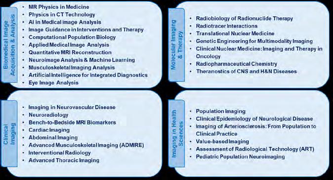
Figure: The individual research lines (32) are organised within four main research focus areas.

The following groups of employees have a role in research support:
Imaging Trial Office
The Imaging Trial Office (ITO) provides high-quality scientific research support to all researchers from the department and from other departments. The ITO employees prepare Institutional Review Board (IRB) protocols and function as the primary contact point for the IRB. They provide study volunteers, take oral questionnaires, liaise with the clinic to arrange logistics, manage data, anonymize images and perform image analysis. They also advise on laws and regulations and perform quality controls to assure performance levels, monitor projects and they manage all aspects of service projects, freeing our researchers and radiologists of this burden. The data manager specializes in data safety and privacy, and development of (clinical trial) databases, which extends the level and range of support offered. The clinical trial monitor oversees the conduct of clinical trials and ensures that these trials are conducted according to protocol, Good Clinical Practice, Standard Operating Procedures and regulatory requirements.
Research technicians
Research technicians at our department work within the pre-clinical research groups. They support and execute fundamental research and animal experiments and carry out histological, radiochemical, molecular and imaging techniques.
Research Radiographer
Research radiographers are (specialized) radiographers and medical nuclear technicians executing data collecting at the different modalities. They guide the introduction of new technologies. They scan study participants for diagnostic- or therapeutic research projects, collect data for scientific projects and provide post processing of radiologic images. This involves, for example, volumetric measurements of liver and lung measurements on CT images and a variety of other services for patient care as well as research projects.
Coordinators Research & Innovation
Each Imaging Modality has its own Coordinator Research & Innovation who is responsible for the organization of research support within their own units as well as the translation of research results into clinical practice. Together with colleagues like researchers, PhD students, ITO, but also research radiographers, radiologists and clinical physicists they take care of development and optimization of research protocols and give advice on the use of the protocols. In 2024 six coordinators for the units MRI, CT, Intervention and Nuclear Medicine were available.
ICT administrators
ICT support staff, part of the Unit Technical Support, maintain our Picture Archiving and communication System (PACS) 24/7. They are also responsible for other software, varying from general office programs to medical software to specific research applications, and maintain and troubleshoot the hundreds of laptops, desktops, workstations, servers, and other computer equipment used in our department. Large scale medical studies pose technical and administrative challenges.
Biomedical engineers
Our biomedical engineers, part of the Unit Technical Support, play an important role in the acquisition and installation of imaging equipment, both for clinical work and research. The technical support team tests and validates new equipment before it is used for patient care or research, assuring image quality and patient safety. Their work allows researchers to acquire validated and reliable data for their research projects.
Medical Students
At the ERGO center in Ommoord MRI scans for the Rotterdam study are performed. The medical student team of Generation R and ERGO support our research organization. For the Generation R study, they make MRI scans of children and their parents. For the ERGO study they assist with the acquisition of MRI scans. After the MRI they are responsible for taking movement tests to screen for Parkinson, a walking test and a polyneuropathy screening including an EMG and a questionnaire.
Research strategy and targets
The focus of Erasmus MC’s research in the coming years will be on socially driven research. Four goals have been formulated, all of which are in line with Koers28 and the core values of Erasmus MC: connecting, responsible and entrepreneurial.
Strategic research goal 1:
The Erasmus MC will develop innovative strategies to promote health by preventing disease, disease progression, and the consequences of disease.
Strategic research goal 2:
The Erasmus MC will unravel the mechanisms that are associated with a healthy life course and involved in disease, and applies this knowledge in new interventions.
Strategic research goal 3:
The Erasmus MC will take the lead in the development of strategies for dealing with emerging health threats.
Strategic research goal 4:
The Erasmus MC will develop innovative methods and technologies that contribute to tailored healthcare, inclusive accessibility and sustainable healthcare.
Based upon Erasmus MC’s research strategy the department defined seven research targets for the coming years.
1
Target 1. Development and validation of new imaging techniques that result in rapid and accurate diagnostics which are cost-effective and sustainable.
The department will develop and validate innovative imaging techniques, focusing on MRI, CT, and nuclear radiotracers. Key efforts include novel MRI pulse sequences for faster scans or enhanced diagnostics, and a contrastreducing MRI method. Two new radionuclide tracers targeting specific molecules will be created to improve early disease detection, monitoring disease progression, and potentially facilitate targeted radionuclide therapy. Imaging strategies with these tracers using PET-CT and PET-MRI will also be developed. The department expects innovations from industrial partners and anticipates early access to cutting-edge technology for testing the clinical value of five new techniques. Cost-effectiveness studies, in collaboration with the EUR Health Technology Assessment group, will be conducted to evaluate the clinical and economic value of developed imaging techniques.
2
Target 2. Development and validation of new quantitative imaging biomarkers which will be used for grading disease, monitoring disease progression, and assessment of the effects of treatment.
Imaging-derived biomarkers can indicate normal or pathological processes or responses to interventions. We will optimize and validate ten novel quantitative imaging biomarkers across MRI and PET-MRI (for musculoskeletal tissue composition, dementia, Parkinson disease, and oncological applications), and photon-counting CT techniques (bone quality, vascular disease and cancer). Emphasis will be placed on accuracy, repeatability, and reproducibility. Diagnostic accuracy will be benchmarked against histopathology and established imaging methods. Clinical relevance will be assessed through correlation with outcomes, and impact on decision-making will be evaluated. These biomarkers will be integrated into multi-center (clinical) trials to assess disease activity, progression, and treatment response, supporting their practical use in routine healthcare.
3
Target 3. Development, validation and implementation of artificial intelligence for capacity planning, image acquisition, automated image analysis and interpretation, and creation of image reports aiming at increasing productivity and reducing costs.
AI will enhance radiology planning by selecting the optimal modality, protocol, and preparation. MRI acquisition will improve through automated adaptive planning and deep learning-based image reconstruction, reducing scan time and correction of motion artifacts reducing the likelihood of scan failures. Eight AI algorithms will be developed for disease detection, diagnosis, subtyping, and quantification across the body, targeting conditions like fractures, tumors, dementia, osteoarthritis, atherosclerosis, and lung disease. Novel AI approaches will be made to increase reliability in challenging clinical scenarios like limited or biased data. Structured reporting based on AI analysis will be developed and tested to support faster, more consistent reporting. Fifteen new industrydeveloped AI tools will be evaluated for diagnostic accuracy, clinical impact, cost, and cost-effectiveness in value-based healthcare settings.
Target 4. Development of novel personalized targeted therapies for oncological diseases by studying cellular and molecular targets for diagnosis of disease and radionuclide therapy in a non-invasive manner.
Personalized targeted radionuclide therapies offer promising benefits for cancer patients by improving effectiveness and quality of life. The department will identify disease biomarkers and develop innovative drugs, exploring cellular and molecular mechanisms to optimize treatment safety and efficacy. This includes discovering two new biomarkers and creating two strategies with improved outcomes and fewer side effects. New clinical guidelines based on dosimetry models are being implemented. A research program using AI and combinatorial drug discovery will develop novel therapies targeting key biomarkers. Three clinical studies, including a phase 1 trial combining radionuclide therapy with a PARP1 inhibitor, will also be launched.
Target 6. Develop robust imaging biomarkers to elucidate disease etiology and to identify targets for early (lifestyle) interventions for disease prevention.
Using large-scale quantitative imaging and automated analysis, we aim to unravel etiology and pathophysiology of common age-related diseases over the next six years, focusing on dementia, arteriosclerosis, and osteoarthritis. For dementia, we will study how modifiable risk factors may protect against genetic predisposition and neuropathology. In arteriosclerosis, we focus on intracranial disease as a modifiable risk factor for dementia and stroke, identifying lifestyle-based intervention targets and developing advanced MRI and Photon Counting CT techniques. For osteoarthritis, we use population-based imaging to study genetic risk factors, joint development, disease subtypes, progression patterns, and links to other diseases using longitudinal joint imaging.
5 7
Target 5. Develop and validate tools and platforms for integration of radiological imaging, pathologic imaging and laboratory exams utilizing the multi-modal data to predict outcomes and treatment responses for personalized medicine.
We will develop multimodal AI algorithms to bridge clinical modalities and departments, focusing on integrating radiology and pathology data (RadioPathomics). RadioPathomics models will enhance glioma typing in the Vici-funded Virtual Biopsy project and stratify sarcoma treatment in the AiNed-funded AIID program. These models will support generalizable AI methods for liver, colorectal, breast cancer, and melanoma. Validation will include in-silico and planned randomized trials for primary brain tumors and sarcoma. Collaborating across departments, we aim to merge imaging with multi-omics and genetics. Projects include digital pathology, spatial transcriptomics in infectious diseases, and building infrastructure for distributed multi-omics AI to advance precision diagnosis and care.
Target 7. Development, implementation and improvement of (minimally invasive) imageguided treatments in clinical pathways to treat or reduce the effects of disease.
The department aims to develop, implement and improve minimally invasive image-guided treatments for neurovascular disease, focusing on endovascular thrombectomy (EVT) for ischemic stroke. We will develop image analysis methods to quantify EVT effects using perfusion-based metrics in digital subtraction angiography (DSA). Integrating pre-operative 3D imaging with X-ray and DSA during procedures will improve real-time decision-making. Accurate EVT assessment will help refine devices and strategies, improving outcomes. Additionally, we will pioneer tumor-targeting imaging probes for fluorescence-guided surgery (FGS), focusing on fibroblast activation protein and fatty acid metabolism. We will also set up the first-in-human studies with fatty-acid indocyanine green (FA-ICG) and a FAP-targeted probe for FGS of glioblastoma and pancreatic cancer in the coming years to support optimal resection of tumor tissue.
Imaging Office
In the dynamic landscape of medical imaging, access to the latest imaging technologies and expertise is crucial. This is where the department’s Imaging Office comes to play. The office provides a comprehensive range of services in the medical imaging domain. From data storage to (software for) (automatic) image analysis to implementation in the clinic, the Imaging Office acts as a gateway to the combined knowledge and skills of the department.
Another aim of the Imaging Office is to ease the workload of healthcare professionals working in the imaging field, like radiologists. By getting a better insight in the needs of these professionals we can look for solutions in the direction of software for automated analysis or having certain types of radiological measurements (e.g. liver volume, aorta diameter) performed by trained technicians.
To decrease the gap between research and clinical practice, the Imaging Office is also actively working on incorporating the requirements from regulations like the MDR and AI act in an early (i.e. research) stage of imaging software development.
Projects
For service projects, the Imaging Office works with a service request form (which is available on ServiceNow). After an intake, it will be decided if the request is feasible and if a project is started.
The Imaging Office service portfolio can roughly be divided into Digital Image Processing , Data services (e.g. storage), and Software Development. In 2024, the Imaging Office actively worked on 16 projects, with requests originating from a variety of locations.
2024 Highlights
To be able to showcase the work being performed at the Imaging Office outside of the Erasmus MC, a public website was created, see QR code. The website showcases the full service portfolio, that also provides some examples of projects that have been worked on in 2024.
In collaboration with the ICAI stroke lab, a symposium was organized on the topic: “What should researchers who develop software know about the MDR (Medical Device Regulations)”. The symposium took place on the 4 th of June 2024 and triggered a lot of interest throughout the Erasmus MC. It initiated collaborations and working groups on the topic of MDR.
Collaborations
Imaging Trial Bureau
The Imaging Office is in close connection with the Imaging Trial Bureau (ITB). The ITB supports researchers in administrative tasks related to research projects, like drafting study protocols, METC submissions, agreements, monitoring, working with Castor/PaNaMa, etc. They also play a role in setting up databases and data transfer. Because of the close connection, a request will always end up in the right place, even if it was initially not clear if it was a request for the Imaging Office or the ITB.
Infrastructure Team
Large scale medical studies pose technical and administrative challenges. The Infrastructure team designs and develops an IT infrastructure to solve these challenges and make medical imaging research reproducible, more robust and more consistent. We are applying our infrastructure and knowledge in local Erasmus MC projects (e.g. Rotterdam Scan Study, Generation R, Research Suite), national projects (e.g. CVON CONTRAST, several ICAI labs, Health-RI), and international projects (EUCAIM, Euro-BioImaging, EuCanImage, EOSC4Cancer, PATH2XNAT). Additionally, the team is also responsible for hosting the medical imaging archive XNAT in Erasmus MC and Health-RI. We deliver software and infrastructure that support researchers, as such we work together with a long list of researchers in and out of our department to create the best possible solutions.
We have created a reference IT infrastructure using a modular approach, so we can suit all projects and studies that need to deal with medical imaging data and data analysis. The modules can be rearranged and configured to fit the specific needs. In the figure below a schematic overview of the infrastructure is given.
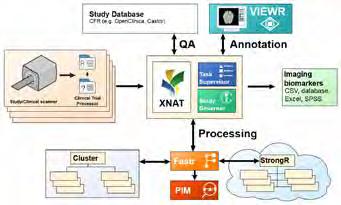
Reference infrastructure for handling data and analysis in projects and studies involving medical imaging data.
Notable achievements
– We have run image analysis pipelines, consulted on study design and data management plans, and performed data management tasks for the Imaging Office for 13 projects.
– Developed metadata model for imaging data in catalogs in collaboration with Health-RI to make data more findable, HealthDCAT-NL. [EuCanImage, EUCAIM, (local) Health-RI]
– Developed tool to populate Fair Data Points with data stored in our XNAT instances. [EUCAIM, Health-RI]
– Developed XNAT to Galaxy tool, for being able to process data stored on XNAT with pathology and genetics analysis pipelines. [PATH2XNAT]
– Proof of concept of CTP manager, used in anonymisation of medical imaging data. [for Trial Office]
– Developed a connection between cBioPortal and XNAT to link cancer genomics with cancer imaging data. [EOSC4Cancer]
– Build DICOM Data ingestion systems from different data sources e.g. CMRad, PACS, VNA and various other DICOM based archives. [EuCanImage]
– Development of data models that allow us to link imaging (XNAT) and non-imaging data. (EGA-CRG) [EuCanImage]
Infrastructure Team
Adriaan Versteeg Ivan Bocharov
Alexander Harms Mahlet Birhanu Hakim Achterberg Marcel Koek
Henri Vrooman
Population Imaging Flagship Node R otterdam
The European imaging community Euro-BioImaging offers open access to biological and biomedical imaging technologies, training and data services across 41 Nodes, comprised of 237 facilities in 18 countries. One of these nodes is the Population Imaging Flagship Node Rotterdam which is embedded within the department of Radiology & Nuclear Medicine of the Erasmus MC. The Node is at the forefront of developments in infrastructure for medical imaging research, tackling the challenges related to collecting, anonymizing, cleaning-up & structuring, storing, sharing, inspecting & annotating, processing & analyzing, and integrating imaging data. Via the Population Imaging Flagship Node, the Imaging Office is able to provide its services on a transnational level. Furthermore, the Node has/had a leading role in European projects that aim to develop infrastructure for medical imaging research, such as EuCanShare, EuCanImage, EOSC4Cancer, and EUCAIM.
Figure:
IMAGING FACILITIES
Magnetic Resonance Imaging
Brand
GE
7.0T Discovery MR901 (pre-clinical)
3.0T Discovery MR750W
3.0T Signa Premier
3.0T Signa Premier
1.5T Signa Explorer
1.5T Signa Artist
1.5T Discovery MR450W
1.5T Signa Artist
1.5T Signa Artist
1.5T Signa Explorer
X-Ray Computed Tomography
Brand
Siemens Naeotom Alpha Photon Counting CT
Definition Edge Twinbeam
Somatom Definition DRIVE
Somatom Definition Edge
Somatom Definition Edge Plus
Naeotom Alpha Photon Counting CT
Somatom On.Site
Angiography, Interventional Radiology, and Fluoroscopic Imaging
Brand
Philips Allura Xper FD 20
Allura Xper FD 20/10
Siemens Axiom Artis Zee MP
Luminos Lotus Max
Artis Q-Ceiling
Artis Icono Biplane
Adora DRFi
Single Photon Emission Computed Tomography (SPECT)-based Imaging
Siemens Symbia T16 SPECT-CT
Starguide
Population Imaging Center
Positron-Emission Tomography (PET)-based Imaging
Mammography
Ultrasonic Imaging
Support Equipment
Photo Camera Equipment
DEXA systems
Brand Equipment
Year of acquisition Location
GE iDEXA Dual-Energy X-ray Absorptiometry System 2014 Central Hospital
Information & Communication Technology
Brand Equipment
Medis Medis Suite MR
Year of acquisition Location Associated modality
2016Central HospitalMRI
Philips IntelliSpace Portal 2015All CT, MRI
Scintomics Labeling software
2014Central HospitalRobotica Robot Synthesizer Labeling software
GE Healthcare AW Server
2015RadiochemistryRobotica Robot Synthesizer
2012All MRI Siemens SyngoVia 2012All CT
TEMA Sinergie Dispensing software
2011Central HospitalPET
2011Central HospitalDispensing robot and Dose calibrators Dispensing software
Hermes Application Server
2024Central HospitalDispensing robot and Dose calibrators
2011Central HospitalSPECT Application Server
2022Central HospitalSPECT Gold3 PACS
Comecer IBC Holtlab Management Software
Merge CADSTREAM
Hologic Softcopy Workstation
PMOD Technologies LLCPMOD
2011Central HospitalSPECT
2011Cancer InstituteSPECT
2008Central HospitalDose calibrators
2006Central HospitalMRI
2016Central HospitalMammography
2017Central HospitalPET
GE Xeleris Server 2023Central HospitalSPECT
Conventional X-Ray Imaging
Brand Equipment
Siemens Mobilett MiraMax
Ysio XPree
Year of acquisition Location
2016Central Hospital
2016Central Hospital 2016Sophia
2024Central Hospital Ysio Max
Cios Alpha
Carestream DRX Revolution
Philips
2018Central Hospital
2018Central Hospital
2018Central Hospital
2017Central Hospital
2017Central Hospital
2017Central Hospital
2017Central Hospital
2012Central Hospital
2001Central Hospital
C-arm Veradius 2011Sophia
C-arm Pulsera
C-arm Unity
2009Central Hospital
1998Sophia
Digital Diagnost C90 2020Sophia
Digital Diagnost C90 Flexroom
Oldelft Benelux Triathlon Trauma DR
Hologic Insight FD Flex
2020Sophia
2024Central Hospital
2014Central Hospital
2009Central Hospital
2009Central Hospital
2022Central Hospital
Insight FD Flex 2021Sophia
Laboratory Facilities
tography


Focus Area
BIOMEDICAL IMAGE ACQUISITION & ANALYSIS
Its focus is to develop advanced image acquisition, image reconstruction, image processing, and machine learning techniques to optimize both the acquisition and analysis of biomedical imaging data with the aim to develop novel diagnostic, prognostic, therapy planning and therapy monitoring tools, and to develop techniques to support image-guided interventions and surgery.
APPOINTMENT IN RADIOLOGY AND NUCLEAR MEDICINE (ERASMUS MC) AND IMAGING PHYSICS (TU DELFT)
Prof. Juan Hernandez-Tamames received his MSc degree in Physics from Complutense University (Madrid, Spain) in 1992. He received his PhD degree (cum laude) from Polytechnic University (Madrid, Spain) in 1999 with a dissertation about Wavelet Transforms in fMRI. Between 1999 and 2002 he obtained several academic positions as Assistant Professor at Complutense University and at Rey Juan Carlos University in Madrid. In 2000 he was visiting professor at the Institute of Psychiatry in London (King’s College of London).
In 2002 he was appointed as Associate Professor at Rey Juan Carlos University. From 2004 to 2015 he was the Head of Medical Image Analysis and Biometry Lab at Rey Juan Carlos University. From 2007 to 2014 he was the head of the Electronics Department at Rey Juan Carlos University. From 2008 to 2014 he was the director of the MR Physics Group at the Queen Sofia Research Center for Alzheimer’s Disease in Madrid. From 2010 to 2015 he was faculty of the MIT program M+Vision for medical imaging training and mentoring. Since 2020 he has a double appointment in the department of Imaging Physics in TU Delft. j.hernandeztamames@erasmusmc.nl
MAGNETIC RESONANCE PHYSICS IN MEDICINE
Juan A Hernández Tamames, PhD
full professor

Context
Magnetic Resonance physics in medicine is continuously evolving, improving and offering new techniques and biomarkers to be able to make substantial progress in clinical diagnosis research. This research line tries to keep the Radiology and Nuclear Medicine department updated to the latest MR techniques to facilitate clinical research and the best patient care at Erasmus MC.
The primary role of the MR Physics group in the Radiology and Nuclear Medicine department is to implement and develop novel MR imaging techniques. Secondly, to support other research groups in the use of advanced MR physics. To improve reproducibility and sensitivity it is necessary to take MR beyond morphology-based diagnosis. The underlying physical parameters and their connection to biological processes and pathologies offer the potential for making MRI a quantitative diagnostic tool. We explore new quantitative MR techniques to establish pathology specific cut-off values and to improve the performance of Radiomics and AI with more accurate quantitative biomarkers. AI “per se” does not surpass the performance of our radiologists. New information from advanced MR Physics is what can make the difference.
Top Publications 2024
Moya-Sáez E, R de Luis-García, L Nunez-Gonzalez, C Alberola-López, JA Hernández-Tamames. Brain tumor enhancement prediction from pre-contrast conventional weighted images using synthetic multiparametric mapping and generative artificial intelligence. Quantitative Imaging in Medicine and Surgery 2024; 15:42-54.
Feddersen TV, JA Hernández-Tamames, MM Paulides, M Kroesen, GC van Rhoon, DH Poot. Magnetic resonance thermometry for hyperthermia in the oropharynx region. International Journal of Hyperthermia 2024; 41:2352545.
Fokkinga E, JA Hernández-Tamames, A Ianus, M Nilsson, CM Tax, R Perez-Lopez, F Grussu. Advanced Diffusion-Weighted MRI for Cancer Microstructure Assessment in Body Imaging, and Its Relationship With Histology. Journal of Magnetic Resonance Imaging 2024; 60:1278-1304.
Research Projects: Objectives & Achievements
Projects:
– MRI-guided hyperthermia for precision treatment of advanced head and neck tumors ( Theresa Feddersen )
– Development of Imaging-Based Response Predictors for Personalized Radiotherapy in Head and Neck Cancer (Nienke Sijtsema)
– Fast Multi-parametric Acquisition Methods for Quantitative Brain MRI ( Laura Nuñez Gonzalez)
– High resolution images of structural lung changes using Zero Echo Time MRI (Dorottya Papp)
– Vascular Signature Mapping in Brain Tumors ( Krishnapriya Venugopal )
– Myelin Quantification and Multiparametric Mapping ( Ilaria Neri)
– Gadolinium-free MRI enhancement using advanced MRI and artificial intelligence ( Noemi Sgambelluri)
MR Physics and Artificial Intelligence
AI is becoming a revolution in Medical Imaging and aid diagnosis. However, oncologists, neurologists, radiologists, specialists in general, need to fill the gap between AI and physiology.
The MR Physics group wants to contribute to answer an important question for our clinical researchers: to what extent quantitative MR can provide reliable physiological information from the microscopic level.
The MR Physics group is leading several initiatives on that regard.
The GEM Project
This project belongs to the Dutch Top Sector Life Science and Health (“Topconsortium voor Kennis en Innovatie” or TKI Life Sciences and Health).
It is a consortium including GE Healthcare, TU Delft and Erasmus MC.

The main objective is to detect Blood Brain Barrier (BBB) breakdown in brain tumors and multiple sclerosis without the necessity of gadolinium contrast agents.
A new PhD student has been incorporated to this project, Noemi Sgambelluri (page 49).
ICAI LAB: Smart, adaptive MR protocols for precision diagnosis
The MR physics group is actively participating in two of the five workpackages of the ICAI-LAB “MRI trustworthy AI” in collaboration with GE Healthcare. We contributed in 2 PhD theses: Alireza Samdifardheris (work package 1, page 90) and Shishuai Wang (work package 2, page 90).
After one year of work we already have obtained synthetic high resolution quantitative maps in some experimental conditions from low-resolution maps, which opens a new avenue for quantitative MR.
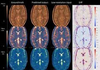
CARES
Project: Development of personalized MR-guided thermochemotherapy for breast conserving surgery
The integration of Magnetic Resonance (MR) imaging in the development of personalized thermo-chemotherapy for breast conserving surgery using liposomes is a transformative advancement in breast cancer treatment. MR imaging plays a pivotal role in this innovative approach, offering unparalleled precision in targeting and monitoring the treatment directly at the tumor site. By leveraging the detailed imaging capabilities of MR, clinicians can visualize the tumor in real-time, ensuring that the liposome-delivered chemotherapy and heat treatments are administered with utmost accuracy.
Figure 2. AI in acquisition and reconstruction (ICAI-LAB).
Figure 1. Logo of the GEM project consortium.
MR imaging's critical role extends beyond treatment delivery; it also enhances the effectiveness of breast conserving surgery. By providing detailed insights into the tumor's response to the thermo-chemotherapy, MR imaging facilitates informed decisions on the therapy regimen, optimizing outcomes while minimizing side effects. This emphasis on MR-guided delivery not only elevates the precision of breast cancer treatment but also significantly improves patient outcomes by reducing the need for more invasive procedures. The marriage of MR imaging with targeted thermo-chemotherapy heralds a new era in personalized, less invasive cancer care, underscoring the indispensable role of MR in advancing breast cancer treatment.
The MR Physics group collaborates with the Hyperthermia group (Led by Dr. Sergio Curto) in the Radiotherapy department to be able to monitor the temperature along the treatment. This collaboration is embedded in a large Dutch consortium including academic center such as AUMC, Utrecht UMC, TU Eindhoven and companies such as GE Healthcare, Philips, Sensius, etc.
EU Marie Slodowska-Curie Doctoral Networks
IQ-Brain: Quantitative Imaging in Brain AIPD: Artificial Intelligence in Parkinson Disease
The MR Physics group is participating in two prestigious Marie-Slodowska Curie doctoral networks.
IQ-Brain, in collaboration with Dirk Poot (page 86), Stefan Klein (page 76), and AIPD in collaboration with the Neurology Department (Dr. Agnita Boom), Anke van der Eerden (Neuroradiologist), Samy Abo (MR Physics PostDoc) and Laura Nunez (page 48), have implemented an innovative diagnosis tool using advanced MRI biomarkers to provide earlier diagnoses of parkinsonisms. The next figure shows the criteria to differentiate among Parkinson, parkinsonisms and healthy subjects.

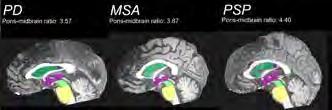
Figure 4. Pons-to-midbrain ratio to differentiate Parkinson from parkinsonisms. This figure shows the automatic segmentation of relevant anatomical features to distinguish between Parkinson and Parkinsonisms.
Lung MRI Project
In collaboration with Dr. Pier Luigi Ciet (Radiologist, page 236), Piotr Wielopolski, PhD (Medical Physics) and the PhD student Cristina Cretu (page 242), we are implementing and optimized protocol for Lung with MRI.

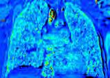
5. Lung T1 mapping in free breathing.
Expectations & Directions
In MR, the ambition is to grow, attract further talented PhD students and post-doctoral fellows in order to widen and solidify the technology related expertise in the group. The group needs to play two main roles: a) provide service for clinical researchers; b) contribute to MR technology through innovation in novel imaging techniques. The group needs to increase the technical support staff to guarantee further developments and the maintenance of the current ones. The growing number of collaborations with researchers from different fields requires competent MR Physicists who channel their knowledge to the respective medical or technical fields. The knowledge in the field of MR Physics is quite broad ranging from handson electronics to theoretical physics through computer simulation and experimental skills. Sub-specialization is already established for pulse sequence programming, image reconstruction, and AI in MRI.
Funding
Curto Ramos, Sergio, Marteen Paulides, Clemens Bos, Juan Hernández Tamames , et al. NWO Perspective: ‘Development of personalized MR-guided thermo-chemo-
Figure 3. Biomarkers involved in differentiating parkinsonisms from Parkinson and Healthy subjects at early stages.
Figure
therapy for breast conserving surgery (CARES) Conserving the breast by heating the tumour’. 2024-2028
Hernández Tamames, Juan, Dirk Poot , Marion Smits , and Frans Vos TKI Match 2024: ‘GEM Project: Gadolinium Free Enhancement in MRI’. 2024-2028
Poot , Dirk, Stefan Klein , and Juan Hernández Tamames EU Marie Slodowska-Curie Doctoral Networks: ‘IQ-Brain’. 2024-2027
Hernández Tamames, Juan , Anke van Eerden , Elise Dopper, and Agnita Boom EU Marie Slodowska-Curie Doctoral Networks: ‘APID’. 2024-2027
Invited Lectures
Juan Hernández Tamames . ‘Gadolinium-free MRI in brain tumors’. 9th ESMRMB-GREC Pre-Congress meeting, Barcelona, Spain. Oct 2024.
Additional Personnel
Mika Vogel – ASL Scientist and Team Leader Europe, GE Healthcare
Ella Fokkinga – MSc student, TU Delft
Emma de Rooij – BSc student, TU Delft
Nadja van Loon – BSc student, TU Delft
Ahmad Thias – MSc student, TU Delft
Elisa Moya – Internship, Valladolid University, Spain
Carmen Sanchez Albardiaz – Internship, Pamplona University, Spain
Assistant Professor Gyula Kotek, PhD

Email g.kotek@erasmusmc.nl
IMAGING PHYSICS AND TECHNOLOGY, PET/MRI CENTER OF COMPETENCE
Gyula Kotek received his MSc in Physics at Eötvös Loránd University, and his PhD in Physics at University of Szeged. He did post-graduate work at various research institutes and universities in Hungary, USA and Germany. From 2003 he works in MR Imaging research and medical physics. He joined Erasmus MC in 2008. His expertise is in MRI and PET/MR Imaging Physics, from theoretical work to engineering and applications in the clinical environment.
PET-MRI Center of Competence
From January 2023 Gyula is leading a new unit – the PET/ MR Center of Competence (CoC). He set up a committed core team. Internal awareness was raised and new external cooperation was triggered: with GE Healthcare, members of the PET/MR user community, and with academic centers. In cooperation with our clinicians and clinical researchers the CoC initiated and started new research projects with technical focus such as: motion correction of PET images, anatomy-guided PET reconstruction, synthetic patient cohort, advanced PET and MR image reconstruction techniques, dynamic PET, and multi-modal and multi-parametric image processing. The CoC hosted and supervised more than ten MSc students for internship and thesis work in 2024.
Technical research
Gyula’s current interest in technical research is advanced PET imaging with a focus on the quantitative aspect, deep learning methods to address imaging problems of challenging anatomies e.g. near metal implants, synthetic patient cohort, and virtual clinical trials.
Forward in 2025
The PET/MRI CoC has two major challenges in 2025: extending its focus and growing its team. Our focus will be extended to PET/CT with the arrival of the new total body scanner. This requires extending our expertise in the respective state of the art techniques, updating our core team and infrastructure. We are looking forward to a boost to our internal and external cooperations.
We will continue to host MSc students and our ambition is to secure research funds in cooperation with clinical and technical researchers and provide opportunities for PhD students.

Figure 1. The organizational structure of the CoC ensures that it is smoothly integrated into clinical research and clinical practice. It adds focus on technical aspects required in the diverse PET/MR field.
The CoC channels demands and requirements from clinical research to technical staff, and technical capabilities to clinical research and practice. The core team from left to right: Anita Harteveld, Marcel Segbers, Esther Warnert, Gyula Kotek and Rianne van der Heijden
Post-doc Post-docs

Laura Nuñez Gonzalez, PhD
Project funding Parkinson NL
Email l.nunezgonzalez@erasmusmc.nl
Atypical Parkinsonisms and MRI sequences
I continued the project started by Samy Abo Seada in collaboration with Agnita Boon and Anke van der Eerder for detecting atypical Parkinsonism. The difficulty of this project is the similarity of the symptoms and the MRI images of patients with Parkinson’s Disease, Multiple System Atrophy and Progressive Supranuclear Palsy. I analyzed the data of almost hundred patients and applied Bayesian classification to the features extracted from the images to differentiate between the 3 different pathologies. The results are promising and we are working on using Artificial Intelligence to create models more accurate classifying them.
PhD Students

Theresa Feddersen, MSc


Advisors Juan Hernandez Tamames, Dirk Poot, Gerard van Rhoon & Maarten Paulides
Project Funding KWF project number 11368: “Precision treatment of advanced head and neck tumors using MRI-guided hyperthermia”
Email t.feddersen@erasmusmc.nl
PhD Obtained 23-01-2024
MR Thermometry for hyperthermia in the Head and Neck
MR thermometry (MRT) can visualise the temperature during hyperthermia treatments non-invasively and in 3D. Our group has developed a MR-compatible head and neck hyperthermia applicator, and we have selected the most promising sequence for MRT: 3DME-FGRE, which will now be optimized further.
Also, with the recent upgrade of the MRI systems, there is the necessity of updating the MRI sequences used in research and are not available as a product. I updated them to facilitate the continuation and progress of the research projects.
Dorottya Papp, MSc
Advisors Juan Hernandez Tamames
Project Funding Erasmus MC
Email d.papp@erasmusmc.nl
Zero echo Time MRI in Lung
MRI has recently emerged as a potential clinical tool that can produce high resolution images of structural lung changes similar to Computed Tomography (CT) scans, thanks to the use of ultrashort TE readouts, but without using ionizing radiation. Thanks to these developments, pediatric patients with chronic lung disease, such as cystic fibrosis (CF), can undergo routine monitoring with CT-like images.

Nienke Sijtsema, MSc

Advisors Juan Hernandez Tamames, Steven Petit, Mischa Hoogeman & Dirk Poot
Project Funding Elekta AB, Stockholm, Sweden
Email n.sijtsema@erasmusmc.nl
PhD Obtained 21-05-2024
Response Assessment of Head and Neck Cancer
In my PhD project we optimized, implemented and evaluated Non-Gaussian IVIM and multi-delay pCASL for use in the head and neck. Currently, a clinical study is ongoing to assess the value NGIVIM in response assessment of oropharyngeal cancer patients. Additionally, we developed a local dose-response model for osteoradionecrosis in the mandible.

Ilaria Neri, MSc

Advisors Juan Hernandez Tamames, Paola Scifo, Dirk Poot & Laura Nuñez Gonzalez
Project Funding IRCCS San Raffaele Hospital, Milan, Italy
Email i.neri@erasmusmc.nl

Krishnapriya Venugopal, MSc

Advisors Juan Hernandez Tamames, Matthias van Osch, Dirk Poot & Esther Warnert
Project Funding NWO Domain AES
Email k.venugopal@erasmusmc.nl
Multi-echo-based Hybrid-EPI (HEPI) technique for measuring R2’
Measuring magnetic susceptibility changes in the brain using MRI is invaluable in the study of normal brain physiology and tumors. R2' is an important susceptibility measurement, sensitive to the deoxyhaemoglobin of brain, enabling information about oxygenation. We propose a new R2’ measurement method using multi-echo based HEPI (that combines both GRE and SE) and study its sensitivity to changes in brain oxygenation using a respiratory challenge MRI technique.

Noemi Sgambelluri, MSc

Advisors Juan Hernandez Tamames, Marion Smits, Frans Vos & Dirk Poot
Project Funding GE Healthcare Fellowship: “Gadolinium-Free Enhancement with Magnetic Resonance Imaging Synthesis”
maps
A multicenter investigation to assess the reproducibility and repeatability of quantitative MRI multiparametric
Multiparametric (MP) quantitative MRI (qMRI) provides simultaneous acquisition of multiple qMR maps in a shorter scanning time compared to the common qMR techniques. In collaboration with the IRCCS San Raffaele Hospital (Milan, IT), we are evaluating the myelin water fraction (MWF) maps in patients affected by leukoencephalopathy obtained from the 3D GRASE and MP-qMR sequences (such as MRF, QTI and MAGIC). Our aim is to optimize and evaluate these sequences in terms of reproducibility and repeatability in different study centers.
Email n.sgambelluri@erasmusmc.nl
Advanced MRI and AI algorithms for Gadolinium-Free Enhancement
For more than 30 years, invasive intravenous gadolinium-based contrast agents have been used in magnetic resonance imaging (MRI) to detect brain lesions. This project aims at eliminating the need for gadolinium, exploiting recent years advancements in MRI technology and Artificial Intelligence.
Marcel van Straten (1974) studied Applied Physics at Delft University of Technology. His MSc project was on contrast agents in MRI. He investigated the relationship between magnetization relaxation times and concentration of contrast agents. His PhD project at the Academic Medical Center in Amsterdam focused on the application of image registration techniques in spiral CT. One of the applications provided a fully automatic technique to remove obscuring bone
structures and calcifications from CT angiography images. After that he was a postdoctoral researcher at the Institute of Medical Physics of the University of Erlangen-Nuremberg. He worked on the optimization and dosimetric aspects of CT. In 2008 he joined the Department of Radiology of Erasmus MC. In 2013 he has been granted certification as a medical physics expert. marcel.vanstraten@erasmusmc.nl
Marcel van Straten, PhD assistant professor PHYSICS IN CT TECHNOLOGY

Context
In CT, image quality is influenced by many factors. It starts with the scanner’s hardware and acquisition protocol and ends with the reconstruction technique and post-processing techniques applied. In clinical practice, the radiologist would like to have the best possible image quality at the lowest radiation dose possible for a specific diagnostic task.
Besides the need for optimization of the acquisition and reconstruction technique, there is a need for a smooth introduction of these optimized techniques into clinical practice. Our research focuses on the standardization and optimization of image quality in x-ray computed tomography (CT) based on the laws of physics and driven by technological innovations.
Top Publications 2024
Van der Bie J, D Bos, ML Dijkshoorn, R Booij, M van Straten. Thin slice photon-counting CT coronary angiography compared to conventional CT: Objective image quality and clinical radiation dose assessment. Medical Physics 2024; 51:2924-2932.
Van der Bie J, SP Sharma, M van Straten, A Hirsch, PA Kamila, D Bos, ML Dijkshoorn, R Booij, RPJ Budde. Image quality assessment of coronary artery segments using ultra-high resolution dual source photon-counting detector computed tomography. European Journal of Radiology 2024; 171:111282.
Aliukonyte I, D Caudri, R Booij, M van Straten, ML Dijkshoorn, RPJ Budde, EHG Oei, L Saba, HAWM Tiddens, P Ciet. Unlocking the potential of photoncounting detector CT for paediatric imaging: a pictorial essay. BJR Open 2024; 6:tzae015.
Research Projects: Objectives & Achievements
Evaluation of technological innovations
New acquisition hardware or new reconstruction methods always claim to improve the image quality of CT scans or to reduce the dose without affecting the image quality. In order to assess the value of newly introduced acquisition and reconstruction techniques, it is of utmost importance to use objective evaluation methods to bring forth both benefits and limitations of these developments. Our research focuses on the development and application of such evaluation methods. In 2024, research continued on the dual-source photon-counting detector-based CT scanner (NAEOTOM Alpha, Siemens Healthineers). Post-doc Ronald Booij focused on musculoskeletal imaging with this scanner (see his section for details). PhD-student Thom van der Laan started working in the field of both musculoskeletal and cardiovascular photon-counting CT.
Building a knowledge base
After a successful evaluation and optimization of the acquisition and reconstruction technique, there is the need for a smooth introduction into clinical practice. This can be a complex optimization process. Changing system properties, acquisition parameters, or reconstruction parameters, will influence both radiation dose and image quality. Optimization via a human interface might be time consuming and error prone. We use the knowledge generated in our research group to build a knowledge base on the performance of a CT scanner in various situations that we use for the automation of the CT operating procedure. Our goal is that the automated procedure makes the best use of the latest technological innovations for a given diagnostic question.
Standardization of CT imaging of the lung s
CT has utility in Cystic Fibrosis (CF) research if it is sensitive enough to detect changes with therapy or disease progression. We work on the standardization and optimization of CT which is a prerequisite for unbiased automated analyses and reduced observer variability. In 2024, we published on the potential of photon-counting CT for paediatric (lung) imaging.
Expectations & Directions
In CT, objective quantification of the performance of new technology and algorithms will allow us to determine their impact on the image quality and thus on the diagnostic performance. The knowledge obtained by the studies described above will allow for building a knowledge base that can be used for the development of a ‘knowledgeable CT scanner’ and for the automation of the CT operating procedure.
Invited lectures
Marcel van Straten . 'Experiences of a (Medical) Physicist in Radiology in the Netherlands'. British Institute of Radiology Annual Congress, London, UK. Nov 2024.
Marcel van Straten . 'Photon Counting CT at Erasmus Medical Centre: Clinical and Research Experience'. British Institute of Radiology Annual Congress, London, UK. Nov 2024.
Ronald Booij . 'Photon-counting CT for musculoskeletal applications'. ESSR Webinar, online. Nov 2024.
Ronald Booij . 'Technological advances in musculoskeletal imaging: where are we in 2024 – Photon-counting CT: what can we see?'. European Congress of Radiology, Vienna, Austria. March 2024.
Highlights
Emma ten Hoor successfully defended her thesis on cardiac CT angiography protocol optimization for neonates.
Together with Siemens Healthineers and Delft University of Technology, a phantom (Figure 1) was developed and scanned to assess the impact of, for example, a tube voltage change on image quality and radiation dose.

Figure 1. Setup of a dynamic anthropomorphic neonatal heart phantom including contrast-enhanced arteries.

Ronald Booij, PhD
Email r.booij@erasmusmc.nl

Bone microarchitecture assessement using photon-counting detector CT
Accurate measurements of trabecular bone microarchitecture are required for a proper assessment of bone fragility. Photon-counting detector CT (PCD-CT) has different technical properties than conventional CT, resulting in higher resolution and thereby potentially enabling in-vivo measurement of trabecular microarchitecture. High-resolution peripheral quantitative CT (HR-pQCT) is considered the standard technique for in-vivo assessment of bone microarchitecture; yet, its clinical application is currently restricted by the limited availability of the scanner. PCD-CT is expected to become more available during the coming years and has potential for clinical assessment of bone microarchitecture. We compared trabecular microarchitecture measurements of cadaveric distal radii and tibiae from PCD-CT at various radiation doses with HR-pQCT and concluded that PCD-CT can
Additional Personnel
Marcel Dijkshoorn – Research Technologist CT
Emma ten Hoor – MSc student, TU Delft
accurately quantify trabecular bone microarchitecture at clinically relevant radiation doses.

Figure 1. The figure demonstrates an example of the distal radius imaged by HR-pQCT and PCD-CT at a CTDIvol(32) of 2.5 mGy (minimum radiation dose) and 20 mGy. Note that the original slices are shown, without 3D registration applied, for which reason the HR-pQCT and PCD-CT do not show exactly the same volume and view.
PhD Student
Thom van der Laan, MSc

Advisors Edwin Oei, Ricardo Budde, Ronald Booij & Marcel van Straten Project Funding Erasmus MC PCCT Grant: Research Collaboration with Siemens in the Field of PCCT Email t.vanderlaan@erasmusmc.nl
The Influence of PCCT on Cardiovascular and Musculoskeletal Imaging
Photon-Counting Computed Tomography (PCCT) enhances cardiovascular and musculoskeletal imaging by providing superior spatial resolution and tissue differentiation at reduced radiation doses. PCCT enables improved visualization of vascularization, stent patency, and atherosclerotic plaques, as well as detailed assessment of bone quality and implant interfaces, potentially advancing diagnostic accuracy and patient outcomes.
JOINT APPOINTMENT IN UNIVERSITY OF COPENHAGEN
Marleen de Bruijne is Professor of AI in Medical Image Analysis at Erasmus MC and University of Copenhagen. She received an MSc degree in physics (1997) and a PhD degree in medical imaging (2003), both from Utrecht University. Before joining the University of Copenhagen (2007) and Erasmus MC (2008) she was assistant/associate professor at the IT University of Copenhagen. Marleen has (co-)authored ca 250 peer-reviewed papers in international conferences and journals, holds 7 patents, is the recipient of the NWO-VENI, NWO-

VIDI, NWO-VICI, and DFF-YDUN awards, and is elected fellow of the MICCAI Society. She has been program (co)chair and general co-chair of several international conferences, including MIDL, MICCAI, and IPMI. She is currently a member of the IPMI board and of the editorial board of Medical Image Analysis. Her research is in machine learning for quantitative analysis of medical images and computer aided diagnosis, with applications in pulmonary-, neuro-, and cardiovascular imaging. She has supervised 34 PhD theses in these areas. marleen.debruijne@erasmusmc.nl
AI IN MEDICAL IMAGE ANALYSIS
Marleen de Bruijne, PhD full professor

Context
The “AI in Medical Image Analysis” research line develops novel techniques for quantitative analysis of medical images, with a focus on machine learning – and especially deep learning –techniques and on large-scale image-based studies. An important theme is the development of machine learning techniques to predict disease directly based on imaging data. Using prediction models derived from a database of images for which the diagnosis has already been established or for which the future course of the disease is known from clinical followup, such techniques are more widely applicable and often more sensitive and robust than conventional image analysis methods. Another important theme is the development of robust and fair image analysis models based on clinically realistic situations. Machine learning techniques often work well on large, well-curated, fully annotated datasets, but how do we learn reliable models if datasets are small or heterogenous and have few, weak, or noisy annotations?
Currently our main application areas are in neuro-, vascular-, and pulmonary image analysis.
Top Publications 2024
Sudre CH, K van Wijnen, F Dubost, H Adams, D Atkinson, F Barkhof, MA Birhanu, EE Bron, R Camarasa, N Chaturvedi, Y Chen, Z Chen, S Chen, Q Dou, T Evans, I Ezhov, H Gao, M Girones Sanguesa, JD Gispert, B Gomez Anson, AD Hughes, MA Ikram, S Ingala, HR Jaeger, F Kofler, HJ Kuijf, D Kutnar, M Lee, B Li, L Lorenzini, B Menze, JL Molinuevo, Y Pan, E Puybareau, R Rehwald, R Su, P Shi, L Smith, T Tillin, G Tochon, H Urien, BHM van der Velden, IF van der Velpen, B Wiestler, FJ Wolters, P Yilmaz, M de Groot, MW Vernooij, M de Bruijne. Where is VALDO? VAscular Lesions Detection and segmentatiOn challenge. Medical Image Analysis 2024; 91:103029.
Dudurych I, GJ Pelgrim, G Sidorenkov, A GarciaUceda, J Petersen, DJ Slebos, GH de Bock, M van den Berge, M de Bruijne, R Vliegenthart. Low-Dose CT-derived Bronchial Parameters in Individuals with Healthy Lungs. Radiology 2024; 311:232677.
Petersen J, I Oguz, M de Bruijne. Graph cut-based segmentation. Medical Image Analysis – a volume in The MICCAI Society book Series, Academic Press 2024; 247-273.
Research Projects: Objectives & Achievements
We develop novel methods for quantitative analysis of medical images. The focus is on fully automatic analyses, which makes our techniques ideally suited for large-scale imaging studies. We aim to develop techniques that are generic and that will often have multiple applications. For instance, the segmentation approach that we originally developed to segment the airway tree from CT images was later successfully applied to segment the lumen and outer wall of carotid arteries in MR and in ultrasound images and the aorta and pulmonary artery from CT; and the appearance models we developed to analyze lung texture are also used in brain structure segmentation and to derive imaging biomarkers of dementia. The different research lines are described briefly below.
Projects:
– Learning imaging biomarkers: Machine learning techniques for data-driven disease prediction ( Hoel Kervadec )
– Advanced deep learning for data-efficiency and performance improvement of medical image segmentation ( Shuai Chen )
– Data-efficient and self-supervised Convolutional Neural Networks (CNNs) for biomedical image segmentation ( Subhradeep Kayal )
– Interpretable and uncertainty-aware deep learning models in medical imaging to counter the 'black box' phenomenon ( Robin Camarasa )
– Artificial intelligence-based airway segmentation and automated measurements for obtainment of bronchial biomarkers to enable early detection of pulmonary disease (Ivan Dudurych)
Pulmonary image analysis
Accurate and reproducible quantification of abnormalities in lung images is crucial to improve our understanding of development and progression of lung diseases, to assess the effect of treatment, and to determine prognosis in individual patients. Pulmonary image analysis at BIGR focuses on measuring structural lung damage in patients with cystic fibrosis (CF)—both in very early and in advanced stages—and on quantifying chronic obstructive pulmonary disease (COPD) from CT images. In close collaboration with researchers at the University of Copenhagen and with LungAnalysis we have developed techniques to segment and measure the dimensions of lungs, airways, and vessels, texture based methods to quantify parenchymal abnormalities, model-based image registration techniques to monitor localized changes, and motion analysis in dynamic MR and CT sequences of the breathing lungs to study respiratory insufficiency in patients with Pompe disease.
Vascular image analysis
Within this research line, we develop imaging biomarkers of atherosclerosis from different imaging modalities, with the aim to improve our ability to identify patients who have a high risk of suffering a (recurring) stroke and who need surgical treatment. We worked with in- and ex-vivo MRI, CT, ultrasound and histology images, and developed automated methods to segment the carotid arteries as well as different components of atherosclerotic plaque including calcification, lipid, fibrous tissue, and intra-plaque hemorrhage. This provides measures of plaque volume and plaque composition, which are known predictors of whether a plaque is likely to rupture or not. We also investigate the value of more advanced imaging features as potential biomarkers; for instance, we found that ultrasound image texture characteristics correlate with future ischemic vascular events.
Neuro image analysis
Within neuro image analysis we develop techniques to quantify different aspects of neurodegenerative diseases, to facilitate clinical and epidemiological research in this area. We are working on techniques to automatically and robustly quantify markers of cerebral small vessel disease, including microbleeds, enlarged perivascular spaces, lacunes of presumed vascular origin, and white matter hyperintensities. We previously developed techniques to segment MR images of the brain intro gray matter, white matter and cerebrospinal fluid as well as techniques to perform brain structure segmentation as a starting point for volume and shape analysis. We have for instance shown that hippocampal shape derived from MRI scans is predictive for dementia years before clinical symptoms arise and that this provides additional predictive value over hippocampal volume.
Image analysis across imaging protocols
Software to analyze medical images often stops working properly when one switches to a new scanner type or changes the imaging protocol. This limits the applicability of such software and makes it difficult to compare data from different sites. In several projects we therefore investigate how learned models can be adapted to new types of image data and how image representations can be derived that are invariant to certain changes in scan protocols, such that multi-center or multi-scanner imaging data can be analyzed more robustly.
Learning from weak labels
Most machine learning approaches to quantitative image analysis need a large number of carefully, manually annotated images for model training. This requires that a) experts are not only able to assess the images visually, but also to indicate boundaries reliably, which may be problematic, for example, in case of diffuse abnormalities; and b) resources are available to perform annotation for the sole purpose of developing image analysis systems. Much more training data would be readily available if weaker labels that indicate for instance the presence, but not the location, of an abnormality could be exploited as well. In several projects we investigate how to learn from “weak” labels such as an estimate of the relative lung volume that is affected by disease as visible in CT, the number of observed lesions in a single slice in a brain MRI scan, annotated vessel diameters, or patient outcome measures.
Expectations & Directions
In the past few years we have turned to deep learning approaches in all our application areas. With these highly flexible models, attention to proper interpretation of which image features drive model predictions, understanding of the model’s failure modes, estimating model bias, and correction for possible confounding factors has become even more important than with more conventional learning techniques. Furthermore, in several of our studies longitudinal imaging data, where the same patient was scanned at multiple points in time, is becoming available. Although such data can be analyzed in the same way as is done for cross-sectional studies, by quantifying aspects in each individual image and then comparing the results of different time points, joint analysis of data of all time points may be more suitable to detect subtle changes.
Funding
De Bruijne, Marleen with University of Copenhagen and consortium partners EU: 'Artificial Intelligence for early detection of non-communicable disease risk in people with breast cancer (ARTILLERY)'. 2023-2027
Duijts, Liesbeth, and Marleen de Bruijne Dutch Foundation for Asthma Prevention: 'Origins of childhood asthma: focus on the developmental lung structure pathway using nonradiant imaging and artificial intelligence'. 20232024
De Bruijne, Marleen NWO VICI: 'Learning imaging biomarkers: Machine learning techniques for data-driven disease prediction'. 2019-2028
Wendelboe, Nielsen, Olav (University of Copenhagen), Marleen de Bruijne , and consortium partners RegionH: 'Breath-CT: Diagnosing Patients Admitted with Breathlessness - Development and Validation of Machine Learning Algorithms based on Images from Computed Tomography'. 2019-2024
Tiddens, Harm , Eva van Rikxoort, and Marleen de Bruijne Netherlands CF foundation: 'Computer assisted diagnosis for monitoring CF airway Disease'. 2019-2024
Oudkerk, Matthijs, Rozemarijn Vliegenthart, Marleen de Bruijne , and consortium partners ZonMW Innovative Medical Devices Initiative – Technology for Sustainable Healthcare: 'B3CARE'. 2018-2024
Highlights
We published the results of a large-scale validation of segmentation techniques to quantify imaging markers of cerebral small vessel disease, the Vascular Lesions DetectiOn and Segmentation (Where is VALDO?) challenge. This challenge aimed to promote the development of methods for automated detection and segmentation of small and sparse imaging markers of cerebral small vessel disease, namely enlarged perivascular spaces, cerebral microbleeds, and lacunes of presumed vascular origin.
Marleen de Bruijne was guest editor for the Special Issue on Advancements in Foundation Models for Medical Imaging from IEEE Transactions on Medical Imaging.
Robin Camarasa, Qianting Lv, and Ivan Dudurych successfully defended their PhD thesis in 2024.
Additional Personnel
Silas Orting – PhD, affiliated Postdoc, University of Copenhagen
Shengnan Liu – PhD, associated researcher, Department of Cardiology, Erasmus MC
Qianting Lv – PhD student with prof. Harm Tiddens and dr. Pierluigi Ciet
Ruben van Oosterhoudt – Scientific programmer
Post-docs
Hoel Kervadec, PhD
Project Funding NWO TTW DLMedIA: Deep Transfer Learning; NWO VICI: Learning imaging biomarkers: Machine learning techniques for data-driven disease prediction
Email h.kervadec@erasmusmc.nl

High-level supervision for image semantic segmentation through shape descriptors
Deep learning methods for image semantic segmentation have made tremendous progress in the past few years, but still require large and well-annotated datasets to be effective. Those annotations take the form of pixelwise masks, indicating exactly the boundary of the object to segment (be it an organ, blood vessel, tumor, …). As such, the most popular method to train neural networks is simply to learn “by-heart” the correct assignment for each pixel, disregarding high-level characteristics such as object shape or location.
Recent works have demonstrated the feasibility of using “shape-descriptors” to fully supervise segmentation neural networks, without resorting to pixel-wise labels.

This type of supervision is closer to the way a human would describe an object and has potential to generalize better: some shape descriptors could be invariant to the patient or scan. At the same time, this more natural way of describing objects can be a way to embed anatomical knowledge directly into the training process, without requiring new annotations.
Moreover, improved methods to measure and characterize 2D/3D objects precisely can be valuable biomarkers, which could also be used diagnosis or risk-assessment.
Shuai Chen, PhD
Project Funding NWO TTW Perspective Programme DLMedIA: Deep Transfer Learning
Email s.chen.1@erasmusmc.nl
Advanced deep learning for medical image segmentation: Towards global and data-efficient learning
Shuai was both a PhD student and postdoc in our group. His thesis tackled two challenges in medical image segmentation with deep learning: learning global information and learning from small sets of training data. The thesis proposes novel methods for global and semi-supervised learning that improve segmentation performance. In his postdoc project, Shuai extended these techniques to learn models for automated quantification of enlarged perivascular spaces based on mixed supervision by a combination of dot annotations, visual scores, and full contour annotations.
PhD Students

Subhradeep Kayal, MSc
Advisors Marleen de Bruijne & Hoel Kervadec
Email s.kayal@erasmusmc.nl
Self Supervision and Data Efficiency in Biomedical Image Segmentation
Convolutional Neural Networks (CNNs) have been particularly useful in medical image analysis. CNNs can be trained relatively easily to perform many kinds of image segmentation tasks, ranging from isolating abnormal tissue in a brain image to segmenting entire airway trees in a lung scan. However, learning tasks in the biomedical domain are often constrained by the lack of substantial annotated data, which is often difficult and time-consuming to obtain. In our project, we try to tackle this problem by proposing solutions on the lines of data augmentation and self-supervised learning. Recently we proposed a new self-supervision task, which is inspired by the classic blind source separation problem.

Robin Camarasa, MSc

Advisors Marleen de Bruijne & Daniel Bos
Project Funding Netherlands Organisation for Scientific Research (NWO) VICI project VI.C.182.042
Email r.camarasa@erasmusmc.nl
PhD Obtained 18-10-2024
Uncertainty and interpretability of deep learning in medical imaging
To counter the "black box" effect of Deep Learning, designing interpretable and uncertainty-aware models is needed. This project develops such models for medical imaging and validates them in magnetic resonance images of the carotid artery, to help improve assessment of the risk of stroke.
Ivan Dudurych, MD, MSc
Advisors Rozemarijn Vliegenthart & Marleen de Bruijne
Project Funding B3CARE | ZonMW Innovative Medical Devices Initiative
Email i.dudurych@erasmusmc.nl
This PhD is part of the B3CARE project with a focus on bronchial wall measurements on low-dose Thorax CT. The bronchial tree has a vast number of branches sequentially reducing in size. Research shows the small airways are the first to undergo remodeling in a diseased state, however manually quantifying these changes is incredibly time consuming and unreliable. Thus this project will collaborate with the BIGR to adapt an AI airway segmentation tool, and apply automated measurements to obtain bronchial biomarkers from ImaLife scans. These biomarkers will be matched to participant parameters to provide reference values, which may then be used to screen the general population for early signs of pulmonary disease.
Theo van Walsum graduated in Informatics at the TU Delft in 1990. In 1995 he received his PhD on flow visualization at the Scientific Visualization Group of TU Delft. After one year at the LKEB (LUMC), where he picked up his interest in image processing, he became a Post-doc at the Image Sciences Institute (UMC Utrecht). There he developed his interest in improving image guidance, and worked on several projects where rotational X-ray imaging was used for guidance in minimally invasive interventions. He became assistant professor at the BIGR group at the Erasmus MC in February 2005, and associate
professor in 2013. He is heading the “Image Guidance in Interventions and Therapy” theme group. This group focusses on improving image guidance by integrating pre-operative image information in the interventional situation, and recently started a research line in agumented reality navigation. He is also involved in the use of AI for stroke treatment, and initiated the SWITCH workshop series at MICCAI. He also is one of the PIs of the Smart Surgery Lab, and Scientific Director of the ICAI Stroke Lab. t.vanwalsum@erasmusmc.nl
IMAGE GUIDANCE IN INTERVENTIONS AND THERA PY
Theo van Walsum, PhD associate professor

Context
Minimally invasive interventions are interventions where small incisions are made to diagnose or treat patients. These interventions are beneficial for the patients and society: the minimal trauma reduces recovery time and required care compared to open surgery. These benefits come at the physician's expense: direct eyesight on the anatomy of interest, such as in conventional surgical procedures, is lacking, and tactile feedback. Interventional imaging, such as fluoroscopy and ultrasound is used to guide the physician during the intervention. These modalities come with disadvantages: ultrasound is often only available as 2D, is operator-dependent and hard to interpret, fluoroscopy only provides 2D projection images, does not have soft tissue contrast and uses ionizing radiation. They lack the wealth of information that is available from 3D diagnostic imaging, such as in CT and MRI. It is our goal to improve image guidance in these procedures, for which we follow three strategies: 1) multimodal image guidance, i.e. reliable integration of pre-operative information during interventions, 2) improved visualization in guidance, by projecting information in the physician’s field of view using modern Augmented Reality headsets, and 3) image-based support in therapeutic and interventional decision making.
Top Publications 2024
Van der Sluijs PM, R Su, S Cornelissen, ACGM van Es, GJ Lycklama, A Nijeholt, PJ van Doormaal, WH van Zwam, DWJ Dippel, T van Walsum, A van der Lugt. Assessment of Automated TICI Scoring during Endovascular Treatment in Patients with an Ischemic Stroke. Journal of NeuroInterventional Surgery 2024; 021892.
Ambrosini P, S AzizianAmiri, E Zeestraten, T Van Ginhoven, R Marroquim, T Van Walsum. 3D Magnetic Seed Localization for Augmented Reality in Surgery. International Journal of Computer Assisted Radiology and Surgery 2024; 723-733.
Wakker A, MHJ Verhofstad, JJ Visser, MG van Vledder, T van Walsum. Talus-derived reference coordinate system for 3D calcaneal assessment: A novel approach to improve morphological measurements. Journal of Orthopeadic Research 2024; 2216-2227.
Research Projects: Objectives & Achievements
Multimodal image guidance
Navigation approaches have become state-of the art in brain surgery and orthopedics. Application of this technology in e.g. cardiac and abdominal interventions is hampered by continuous tissue motion and deformation. Our aim is to develop robust techniques for integrating pre-operative information that can be used in cases where tissue motion and deformation may occur. To this end, we are developing and evaluating methods to build pre-operative models for interventional planning, and for alignment of these pre-operative models with the interventional situation, by using interventional images, position tracking information and motion/deformation models.
Projects:
– CTA-DSA Image registration (Charles Downs)
– Automated occlusion detection In DSA Images (Anushka Kore)
Augmented reality
Conventional navigation approaches, in which surgical instruments are shown in relation to pre-operative imaging on a 2D computer screen, result in continuous switching of focus (from screen to surgical field), and difficulties in hand-eye coordination. Our objective is to develop and assess mixed reality approaches that allow integration of 3D imagery in the surgical field of view. Main challenges are obtaining and maintaining accurate alignment of the images with the patient. On this topic we collaborate with the departments of oral and maxillofacial surgery, neuro surgery, trauma surgery, plastic and reconstructive surgery and oncological surgery. We have developed an Augmented Reality extension of the BrainLab navigation system, that allows visualization of the tools and anatomy aligned with the patient in the surgeon's field of view. In addition, a system for incision planning for craniosynostosis, using a Hololens 2 and an electro-magnetic tracking system has been developed and assessed in a phantom study. We are currently working towards assessment at the operating room.
Projects:
– Augmented reality for Le Fort 1 surgery ( Mohamed Benmahdjoub )
– Device-agnostic framework for surgical guidance using augmented reality ( Abdullah Thabit )
– Augmented reality for EVD placement (Emma de Bruin)
– Manual alignment of virtual and physical models (Jiaqi Tang)
– Model-to-patient alignment using facial landmarks (Xiang Gao)
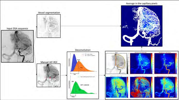
Figure 1. Pipeline of perfusion DSA, Illustrating the deconvolution approach and resulting perfusion Images.

Therapeutic decision making in Stroke
In 2015, mechanical thrombectomy was proven to be an effective treatment for patients with a stroke caused by a large vessel occlusion, and thrombectomy has become the therapy of choice for stroke patients with a large vessel occlusion. Unfortunately, not all patients recover well from the stroke, even after a seemingly successful intervention. Our aim here is to use imaging (and other data) to optimally support therapeutic decision making. In the Q-Maestro project, we focus on interventional imaging, investigating whether, and to what extent, Digital Subtraction Angiography images can be used to better determine treatment effect and predict patient outcome. We are also working on approaches to integrate 3D baseline imaging data in the intervention.
Projects:
– autoTICI to clinic (Lotte Strong, Matthijs van der Sluijs, Ellen Hu)
– Quantitative persufusion DSA ( Ruisheng Su, Matthijs van der Sluijs )
– Assessment of quantitative perfusion DSA with MR Perfusion (Mouad Allaoui)
– Regional perfusion quantification in DSA ( Frank te Nijenhuis, Matthijs van der Sluijs )
– Multimodal functional outcome prediction in EVT ( Frank te Nijenhuis )
– Automated imaging biomarkers assessment in acute ischemic stroke ( Lennard Wolff )
– Clinical decision support and EVT optimization ( Xi Li )

3. Brain regions determined from CTA projected on DSA, after aligning the CTA to the DSA Image.
Decision making In Surgery
Fracture detection, quantification, and detection of abnormalities is important both for clinical research (e.g. to assess the added value of various surgical approaches) and for therapeutic decision making. Since a few years, there is a strong collaboration in this area with the Department of Trauma Surgery, focusing on calcanous, distal radius and rib fractures, and with the Department of Cranio-Maxillofacial surgery, on automating 3D cephalometric analysis.
Projects:
– 3D quantitative analysis of the calcaneous (Alexander Wakker)
– automating 3D cephalometric analysis ( Bram Roumen )
– Quantitative analysis of fractures in the posterior talocalcaneal joint (Cile van Holthe)
– Quantitative analysis of extra-articular distal radius fractures (Josefien van den Berg)
– Detection and classification of rib fractures (Victoria Marting)
Figure 2. Augmented Reality for surgical navigation: projecting a virtual rib cage model on a phantom. (Photo courtesy Ivar Pel.)
Figure
Expectations & Directions
We developed several multi-modal image guidance approaches. One direction that I would like to explore is the integration of the results in the augmented reality line with the multi-modal image guidance results.
Augmented reality applications in medicine is a quickly evolving field. In this research line, we will continue developing technology for AR guidance in interventions, such as fast and reliable registration of the image to the patient. Based on this technology, we intend to build prototype systems for using augmented reality in various surgical and minimally invasive interventions.
Our stroke research is strongly linked to the clinical trials within the CONTRAST consortium, and the MR Clean Registry. The ICAI Stroke Lab, part of the ROBUST program for AI in the Netherlands, has started in 2023. In this lab, we will further develop AI approaches for therapy and rehabilitation of stroke patients in a multidisciplinary setting.
Several quantifications for fractures have been developed in strong collaboration with the Department of Trauma Surgery. We expect to further enhance these methods, extend their application, and validate these on larger dataesets.

Funding
Van Walsum, Theo , Ad van Es, and Danny Ruijters Health Holland TKI Call: ‘Q-Maestro: Quantitative Microvasculature AssEssment in projection angiography of ischemic STROke patients’. 2019-2024
Van Walsum, Theo , Caroline Klaver, Nicolas Chateau, Danilo Andrade De Jesus, and Luisa Sanchez Brea Health Holland TKI Call: ‘AO-Vision -- Adaptive Optics imaging: A guiding star to save vision’. 2022-2026
Van der Lugt, Aad, Wiro Niessen, and Theo van Walsum NWO LTP ROBUST program: ‘ICAI Stroke Lab: from 112 to Rehabilitation’. 2022-2027
Molina, Carlos, … Hester Lingsma, Bob Roozenbeek, Ruud Selles, Theo van Walsum Horizon Europa IHI: 'UMBRELLA (Unleashing a CoMprehensive, Holistic and Patient Centric Stroke Management for a Better, Rapid, AdvancEd and PersonaLised Stroke Diagnosis, TreAtment and Outcome Prediction)'. 2024-2029
Invited Lectures
Theo van Walsum . ‘AI and stroke’. Neurology Seminar, Rotterdam, the Netherlands. June 2024.
Highlights
Ruisheng Su, Frank te Nijenhuis and Theo van Walsum organized the 4th SWITCH workshop at MICCAI, bringing together clinicians and engineers to discuss medical imaging and stroke.
Theo van Walsum and Ilva van Houwelingen organized a workshop on the MDR and Research Software.
For the autoTICI assessment study, in the context of the MSc project of Lotte Strong , Erasmus MC was the first in the world to have the Philips CloudCast installed, allowing a seamless connection to the Open Innovation Platform, and enabling application of in-house developed software on live images from the X-ray systems.
Ruisheng Su obtained his PhD degree Cum Laude on April 2, with a thesis on the development of quantification approaches for DSA Images.
Mohamed Benmahdjoub obtained his PhD degree on June 26, on a thesis on techniques for Augmented Reality for guidance and navigation.
A picture of Abdullah Thabit demonstrating an Augmented Reality application for rib fractures was on the cover of I/O magazine, the magazine of the ICT Research Platform the Netherlands.
Abdullah Thabit and Mohamed Benmadjoub organized and hosted the 2nd Dutch AR for Surgery meeting on January 10.
Ruisheng Su received the IEEE TMI Distinguished Reviewer Award for 2023-2024.
Figure 4. Quantification of the posterior talocalcaneal joint.
Additional Personnel
Alexander Wakker – PhD student
Adriaan Versteeg – Research Software Engineer for CONTRAST 2 project. Jan 2024 – Dec 2024.
Lotte Strong – 3rd year MSc student Technical Medicine, TU Delft, Erasmus University, Leiden University. Feb 2024 – Oct 2024. Daily supervisors Matthijs van der Sluijs and Ruisheng Su.
Victoria Marting – 3rd year MSc student Technical Medicine, TU Delft, Erasmus University, Leiden University. Feb 2024 – Oct 2024. Daily supervisors Theo van Walsum and Mathieu Wijffels.
Xiang Gao – 2nd year MSc student Computer Science, TU Delft. Feb 2024 – Oct 2024. Daily supervisors Mohamed Benmahdjoub and Abdullah Thabit.
Jiaqi Tang – 2nd year MSc student Computer Science, TU Delft. Feb 2024 – Oct 2024. Daily supervisors Mohamed Benmahdjoub and Abdullah Thabit.
Yvonne Veltman – 2nd year MSc student Medicine, Erasmus University. Feb 2024 – Aug 2024. Daily supervisors Matthijs van der Sluijs and Theo van Walsum.
Anushka Kore – 2nd year MSc student Biomedical Engineering TU Delft. May 2024 – Oct 2024. Daily supervisor Frank te Nijenhuis.
Cile van Holthe – 3rd year MSc student Technical Medicine, TU Delft, Erasmus University, Leiden University. Feb 2024 – Nov 2024. Daily supervisor Alexander Wakker.
Emma de Bruin – 2nd year MSc student Biomedical Engineering, TU Delft. Sept 2024 – Nov 2024. Daily supervisor Mohamed Benmahdjoub.
Mouad Allaoui – 2nd year MSc student Biomedical Technology and Physics, Vrije Universiteit Amsterdam. Sept 2024 – Dec 2024. Daily supervisor Frank te Nijenhuis and Matthijs van der Sluijs.
Charles Downs – 2nd year MSc student Computer Science, TU Delft. Feb 2024 – Oct 2024. Daily supervisors Matthijs van der Sluijs and Ruisheng Su.
Josefien van den Berg – 2nd year MSc student Technical Medicine, TU Delft, Erasmus University, Leiden University. Dec 2023 – Feb 2024. Daily supervisors Theo van Walsum and Mathieu Wijffels.
PhD Students

Mohamed Benmahdjoub, MSc

Advisors Theo van Walsum, Wiro Niessen & Eppo Wolvius
Project Funding Erasmus MC
Email m.benmahdjoub@erasmusmc.nl
PhD Obtained 26-06-2024
Augmented
Reality Navigation for Craniomaxillofacial Surgery
The goals of this project are to develop intraoperative solutions that would help integrate an external augmented reality device into the current workflow of the navigation systems, and to conduct experiments (phantom studies and case studies) to investigate the feasibility, usability and accuracy of the solutions in the context of craniomaxillofacial surgery.

Frank te Nijenhuis, MD, MSc

Advisors Theo van Walsum, Danny Ruijters & Sandra Cornelissen
Project Funding ICAI Stroke Lab
Email f tenijenhuis@erasmusmc.nl
Applications of Artificial Intelligence in Image Guided Interventions for Acute Ischemic Stroke
The aim of this project is to investigate how AI and computer vision techniques can be employed to enhance stroke interventions. The main focus is on improving image guidance by including pre-operative imaging information. Additionally, collaboration within the lab facilitates integration of AI methods along the entire stroke care pathway.
Ruisheng Su, MSc

Advisors Theo van Walsum, Wiro Niessen, Aad van der Lugt & Danny Ruijters Project Funding Q-MAESTRO: a Health Holland project funded by Philips Healthcare and ErasmusMC
Email r.su@erasmusmc.nl
PhD Obtained 02-04-2024
Image analysis of cerebral angiography in ischemic stroke
This project aims to develop AI methods for automated analysis of cerebral digital subtraction angiography (DSA) for enhanced diagnosis, prognosis, and interventional guidance in endovascular treatment for patients with ischemic stroke. Relevant clinical applications include automatic TICI, adverse event detection, blood flow analysis, multi-modal information fusion, and functional outcome prediction.

Matthijs van der Sluijs, MD, MSc

Advisors Theo van Walsum, Aad van der Lugt & Sandra Cornelissen
Project Funding Q-MAESTRO: a Health Holland project funded by Philips Healthcare and ErasmusMC
Email p.vandersluijs@erasmusmc.nl
Quantitative Microvasculature AssEssment in projection angiography of ischemic STROke patients
This project aims to develop imaging parameters that quantify perfusion restoration after reperfusion therapy using Digital Subtraction Angiography (DSA). Through this, clinical decision making of the neuro-interventionalist performing the treatment could potentially be influenced, as supplementary locoregional therapeutic action in the intervention stage might still be available.

Abdullah Thabit, MSc

Advisors Theo van Walsum, Wiro Niessen & Eppo Wolvius
Project Funding Koers 23 Smart Surgery Lab
Email a.thabit@erasmusmc.nl
Augmented Reality image-guidance in surgery
Navigation has become standard of care for several surgical areas, such as neurosurgical and orthopedic procedures. However, conventional navigation systems suffer from a few drawbacks, such as the poor hand-eye coordination and the need to switch focus (from the operative field to the screen). The objective of my PhD is to investigate the use of AR with head mounted displays as an alternative to conventional systems in surgical navigation.

Bram Roumen, MSc

Advisors Eppo Wolvius, Theo van Walsum & Bart Cornelissen
Project Funding SMARTOR 2030
Email b.roumen@erasmusmc.nl
Automation
of preoperative planning and intraoperative assistence.
By integrating digital tools to support surgical teams in preoperative preparations and intraoperative guidance, we aim to streamline OR procedures. Our goal is to reduce OR times, minimize the need for revisional surgeries, and maximize the utilization of OR resources, while improving the OR staff's preceived workload.

Lennard Wolff, MD, MSc

Advisors Aad van der Lugt & Theo van Walsum
Project Funding Dutch Heart Foundation, Dutch Brain Foundation, Stryker, Medtronic and Ceronovus. Collaboration for New treatments of Acute Stroke (CONTRAST): WP7 Imaging Biobank.
Email l.wolff.1@erasmusmc.nl
Automated imaging biomarkers in acute ischemic stroke
Several imaging biomarkers predict outcome in patients with acute ischemic stroke and effects of endovascular treatment (EVT). Automated analysis tools might decrease the variability in the evaluation of imaging biomarkers with a subsequent improvement of prediction tools.

Xi Li, MSc

Advisors Hester Lingsma, Bob Roozenbeek, Theo van Walsum & Daniel Bos
Project Funding ICAI Stroke Lab Email x.li.1@erasmusmc.nl
In-hospital clinical decision support and treatment optimization for stroke
This project aims to develop and validate traditional and AI models using pre- and peri-procedural clinical and imaging variables to estimate stroke patient outcomes after treatment. Additionally, we aim to create models for individualized prediction of treatment effect to optimize patient selection for endovascular treatment.
JOINT APPOINTMENT IN EPIDEMIOLOGY
Gennady Vasilievich Roshchupkin received his MSc from Moscow State University in Physics Department with specialization in stellar astronomy and astrophysics. After that he spent 3 years as a senior research engineer at a network security company. In 2014 he started his PhD and graduated (cum laude) in 2018 at Department of Radiology and Department of Epidemiology at Erasmus MC. He established his research line and was appointed as Assistant Professor in 2022. Gennady received several personal grants (e.g. VENI, NIH, Erasmus MC Fellowship).

His research focused on developing and application of methods for the integrative analysis of large-scale biological, epidemiological and clinical data. Gennady has a broad background in statistics, computer science, machine learning, deep learning, medical image analysis and genomics. Since 2019 Gennady is chairing Machine Learning working group in the CHARGE Consortium. Since 2021 Gennady is also leading Bioinformatics Working group in Genomics of MusculoSkeletal traits Translational Network. Gennady is co-founder of Erasmus MC squAIre (Society for Quantitative Artificial Intelligence Research). g.roshchupkin@erasmusmc.nl
COMPUTATIONAL POPULATION BIOLOGY
Gennady Roshchupkin, PhD
assistant professor
Context
The Computational Population Biology group is melding computational science with biological, epidemiological, and clinical data analysis to address critical health issues facing society today. Through the sophisticated analysis of large-scale datasets, our team seeks to deepen the understanding of omics' role in complex traits, aiming to revolutionize the prevention, diagnosis, and treatment of diseases. This research is not just about scientific discovery; it's about responding to a societal need for better healthcare solutions, reducing the burden of disease worldwide, and enhancing the quality of life for individuals.
Leveraging the latest in genomics, medical imaging, computer science, statistics, and machine learning, we are uniquely positioned to sift through the rich and complex data that modern healthcare generates. Our work is crucial in an era where personalized medicine and targeted therapies are becoming the cornerstone of healthcare, offering new hope and possibilities for patients.
Top Publications 2024
Liu X, TE Sangers, T Nijsten, M Kayser, LM Pardo, EB Wolvius, GV Roshchupkin, M Wakkee. Predicting skin cancer risk from facial images with an explainable artificial intelligence (XAI) based approach: a proof-ofconcept study. EClinicalMedicine 2024; 71:102550.
van Hilten A, J van Rooij, MA Ikram, WJ Niessen, JB van Meurs, GV Roshchupkin. Phenotype prediction using biologically interpretable neural networks on multicohort multi-omics data. NPJ systems biology and applications 2024; 10:81.
Van Hilten A, S Katz, E Saccenti, WJ Niessen, GV Roshchupkin. Designing interpretable deep learning applications for functional genomics: a quantitative analysis. Briefings in Bioinformatics 2024; 25:bbae449.
Research Projects: Objectives & Achievements
Key Research Areas:
– Multi-Omics analysis and data integration using AI algorithms
– Neuroimaging – AI-powered analysis of structural MRI, DTI and resting-state fMRI to map brain structure and function.
– Facial Morphology – Exploring 2D and 3D facial features as phenotypic biomarkers linked to genetic and health indicators.
– Musculoskeletal Health – Investigating genetic and biomechanical factors that influence bone and joint health.
– 3D Imaging in Clinical Research – Advancing precision diagnostics through AI-driven analysis of 3D medical imaging.
Projects:
– Enhancing brain imaging power and genetic discovery using genetic data and high-dimensional, multimodal brain MRI measurements ( Jing Yu )
– Precision medicine using explainable deep learning models ( Sonja Katz )
– Advancing knowledge of genetic diseases by unraveling complex mechanisms with interpretable machine learning techniques ( Arno van Hilten )
– Variational autoencoder-based deconfounding techniques using multi-omics data for meaningful patient subgrouping ( Zuqi Li )
– Quantitative morphometric models and AI-driven shape analysis for prediction of and insights into craniofacial dysmorphologies ( Tareq Abdel Alim )
– Multimodal imaging techniques for the identification of brain features associated with (chronic)pain and comorbidities ( Xianjing Liu )
Circular AI-Driven Pipeline for Precision Medicine
Our research group develops and implements a circular AI-driven pipeline that integrates clinical genetics, population studies, and drug discovery to transform precision medicine. By combining advanced AI, federated learning, and multi-omics approaches, we bridge fundamental research with real-world applications, accelerating insights into disease mechanisms and therapeutic innovations.
Closed-Loop AI for Clinical Genetics, Population Studies & Drug Discovery
Clinical Genetics – We leverage AI to analyze genomic data, identifying critical motifs and pathways that reveal underlying disease mechanisms.
Population Studies – Using federated learning, we enable secure, large-scale collaboration across institutions, harmonizing diverse datasets while preserving privacy.
Drug Discovery – AI and multi-omics integration facilitate the identification of therapeutic targets and optimize treatment strategies, advancing precision medicine.
This closed-loop system ensures that population-level insights refine genetic discoveries, while breakthroughs in genetics and drug development fuel personalized interventions, creating a continuous cycle of innovation.
Federated Learning & Industry Collaborations
We are at the forefront of federated learning research, developing privacy-preserving AI models that enable multi-institutional collaboration without sharing raw data. Our group works with:
– NVIDIA, driving advancements in AI infrastructure for healthcare;
– OASYS NOW and other startups, developing innovative AI solutions for clinical and biomedical applications; – Global research consortia, integrating AI-driven insights into large-scale, multi-center studies.
Multi-Modal Data Analysis & Endophenotype Research
We focus on advanced phenotyping and endophenotype analysis, integrating imaging, omics, and clinical data to unravel the etiology of complex traits.
Open Science & AI for Research Accessibility
We are committed to open science and the development of open-source tools that enhance data visualization, AI model accessibility, and collaborative research platforms. By fostering an open and transparent research environment, we empower scientists and clinicians to explore, validate, and apply AI-driven insights to real-world challenges.
Through this AI-driven circular pipeline, we are shaping the future of precision medicine, biomedical AI, and mul-
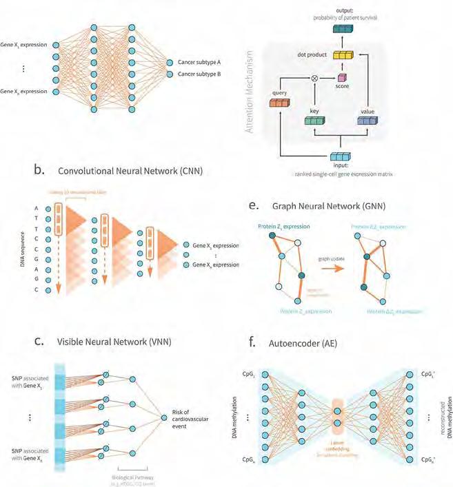
ti-institutional research, making healthcare innovations more accessible, efficient, and impactful.
Collaboration
The group is actively involved in various collaboration projects within Erasmus MC: Departments of Epidemiology Radiology and Nuclear Medicine, Neurosurgery, Plastic Surgery, Internal Medicine, Psychiatry, Neuroscience, Craniomaxillofacial surgery, Genetic Identification, Dermatology, Clinical Genetics and AI Accelerator.
Also, nationally and internationally: Netherlands Cohorts Consortium (NCC), CHARGE consortium (the Cohorts for Heart and Aging Research in Genomic Epidemiology), ENIGMA consortium, eQTLGen consortium, and EADB consortium (A European DNA bank for deciphering the missing heritability of Alzheimer's disease).
The group members contribute as AI experts in several EU COST actions networks.
Figure 1. Overview of the most popular neural network architectures in genomics applications.
Expectations & Directions
Our research group is dedicated to pushing the boundaries of AI in precision medicine by:
– Advancing federated learning to enable scalable, privacy-preserving multi-institutional AI research.
– Enhancing explainability in AI models, making genomic and biomedical AI more interpretable and actionable.
– Expanding multi-omics integration, bridging genomics, imaging, and clinical data for deeper insights.
– Strengthening collaborations with industry partners, startups, and research consortia to translate AI innovations into clinical applications.
– Promoting AI accessibility & open science, ensuring fair and ethical AI applications in global healthcare. omics, text, and voice analysis, among others, to create a holistic understanding of health and disease.
Funding
Roshchupkin, Gennady Erasmus MC-TKI-LSH: 'The oral microbiome as modifiable risk factor for caries lesions'. 2020-2024
Yu, Jing , Arno van Hilten , and Xianjing Liu NWO Rekentijd: 'Dutch supercomputer computational grant'. 20232024
Roshchupkin, Gennady Erasmus MC-TKI-LSH: 'Seeing is Believing: Virtual & Augmented Reality For Enhancing Personalized Patient Experiences'. 2021-2025
Roshchupkin, Gennady ZonMW VENI: 'Explainable Artificial Intelligence to unravel genetic architecture of complex traits'. 2021-2024
Roshchupkin, Gennady NIH R01: 'An integrative computational interrogation of circuit dysfunction in schizophrenia via neural timescales'. 2022-2027
Roshchupkin, Gennady, and Daniel Bos Erasmus MC, TU Delft & EUR Convergence Flagship: 'ALIVE: A Lifecourse and Individual-based View on Lifestyle to Enhance Health'. 2022-2026
Roshchupkin, Gennady Erasmus MC Fellowship: 'GenetiX: Decoding the Genetic Puzzle of Complex Traits with Federated learning and Explainable AI'. 2023-2028
Roshchupkin, Gennady Dutch Ministry of Education, Culture and Science (OCW): 'Starting Grant'. 2023-2029
Roshchupkin, Gennady European Union's Horizon 2020 research and innovation programme: ‘Identifying molecular mechanisms of pain related disorders’. 2022-2026
Roshchupkin, Gennady and Tareq Abdel Alim Sophia Stichtingen: 'Machine learning for postoperative shape prediction in craniosynostosis treatment using 3D models'. 2024-2025
Invited Lectures
Gennady Roshchupkin . ‘The Art of AI-Driven Healthcare Navigating through Modern Medicine with Ancient Wisdom’. Tannet, Amsterdam, the Netherlands. Feb 2024.
Gennady Roshchupkin . ‘House of Cards The Fragile Foundation of AI in Medical Research’. Rotterdam Square, Rotterdam, the Netherlands. Feb 2024.
Gennady Roshchupkin . ‘MEGA Make ERGO Great Again’. ERGO MT, Rotterdam, the Netherlands. Feb 2024.
Gennady Roshchupkin . ‘AI's Journey to Beat Colorectal Cancer’. Gastroenterology PhD day, Rotterdam, the Netherlands. May 2024.
Gennady Roshchupkin . ‘Zero to Hero: Unlocking Research Excellence with ChatGPT’. Alzheimer Center Dementia Day, Rotterdam, the Netherlands. June 2024.
Gennady Roshchupkin ‘Explainable AI interpretability is not the same as explainability’. University College Dublin, online. July 2024.
Gennady Roshchupkin . ‘From Silos to Synergy Harnessing Data Science and Infrastructure for National Cohort Coalition’. NCC cohort meeting, Rotterdam, the Netherlands. Sept 2024.
Gennady Roshchupkin . ‘AI in Tumor Immunology: Disruptive Innovation or Overhyped Promise?’. Immunology Symposium, Rotterdam, the Netherlands. Oct 2024.
Gennady Roshchupkin . ‘Econometrics student pitch about research career’. Erasmus University Rotterdam, Rotterdam, the Netherlands. Nov 2024.
Gennady Roshchupkin. ‘AI in Microbiology: Game-Changer or Just Another Sci-Fi Fantasy?’. Microbiology Department Erasmus MC, Rotterdam, the Netherlands. Nov 2024.
Additional Personnel
Kiefer Comasi – MSc student, ‘Advancing Targeted Therapies: Bridging the Chemical Space Gap through Deep Learning for Drug Discovery’.
Sara Okhuijsen – MSc student, 'Evaluating the Privacy of Confidential Multi-Party AI (CoMPAI) on Genomic Data'.
Rafael Campos-Martin – Visiting PostDoc, 'Breaking down the genetic factor responsible for the progression from MCI to AD using Deep Learning and multi-cohort study'.
Ruizhi Deng – Visiting PhD student, Clinical Genetics Erasmus MC
Yasna Afshari – MSc student, ‘Web-Based Viewer for Population-Based Studies’.
Xinge Guo – Visiting master student, ‘GWAS on AI-based phenotypes derived from structural brain MRI in UKBB cohort’.
Cliodhna Gartland – Visiting PhD student, Ireland
Semyon Galichenko – Linux system administrator and data scientist
Lennart Karssen – Senior Linux administrator
Post-docs

Tareq Abdel Alim, PhD
Project Funding Sophia Fonds Grant 2022
Email t.abdelalim@erasmusmc.nl

Predictive modeling and shape analysis of craniofacial dysmorphologies
The human skull is a complex biological structure shaped by interactions between bone growth, brain development, and genetic regulation. In patients with craniosynostosis, where cranial sutures fuse prematurely, these processes become disrupted. This leads to craniofacial dysmorphologies that affect both function and aesthetics. Understanding and predicting these shape variations is essential for improving diagnostics, treatment planning, and surgical outcomes.
Our research develops quantitative morphometric models to assess skull shape variability, integrating 3D photogrammetry, CT, and AI-driven shape analysis to evaluate dysmorphologies in both normal and pathological states.
Refining computational methods to simulate growth and intervention outcomes enhances understanding of biomechanics, aiding treatment planning and clinical decision-making. These models help clinicians determine whether surgery is needed, which technique to use, and the optimal timing.
Future research will explore the relationship between cranial morphology, intracranial structures such as the brain and cerebrospinal fluid, and their biomechanical interplay, leading to a deeper understanding of the intricacies of skull development, the treatment of its abnormalities, and the refinement of personalized treatment strategies.
PhD Students

Xianjing Liu, MSc

Advisors Gennady Roshchupkin & Eppo Wolvius
Project Funding The Generation R Study and the Rotterdam Study.
Email x.liu.1@erasmusmc.nl
3D facial shape analysis
Unravel the complexity involving the human face, genetic factors, environmental factors, and their connection with health outcomes, by developing and applying advanced artificial intelligence-based techniques with a special focus on addressing challenges of confounder and interpretability in deep learning models.

Sonja Katz, MSc

Advisors Gennady Roshchupkin
Project Funding EuCanImage, Horizon 2020 Marie Skłodowska-Curie grant (860895, TranSYS) , Simon Stevin Meester
Email s.katz@erasmusmc.nl
PhD Obtained 20-09-2024 * Cum Laude
Explainable deep learning for precision medicine
My PhD research focuses on the development of artificial intelligence models to capture patient characteristics using a variety of medical information. To bridge the gap between research and clinical practice we prioritize the explainability and fairness of our models, fostering close collaborative relationships with clinicians.

Jing Yu, MSc
Advisors Gennady Roshchupkin & Arfan Ikram
Project Funding Erasmus University Rotterdam -China Scholarship Council (EUR CSC) scholarship
Email j.yu@erasmusmc.nl
High-dimensional brain MRI measurements for genetic analysis
Incorporating modern techniques including deep learning, federated learning, enrichment analysis for high-dimensional multi-model brain MRI measurements and genetic data to increase the power of brain imaging and genetics analysis discovery, in addition to facilitate brain disease understanding and prediction.

Tareq Abdel Alim, MSc
Advisors Gennady Roshchupkin, Marie-Lise van Veelen, Clemens Dirven & Wiro Niessen
Project Funding Sophia Fonds Grant 2022
Email t.abdelalim@erasmusmc.nl
PhD Obtained 22-10-2024
3D Imaging and Craniofacial Shape Analysis
My research is focused on 3D shape analysis of dysmorphologies, specifically common craniofacial conditions in children like craniosynostosis. Utilizing 3D photogrammetry, I develop mathematical shape models to assess and predict surgical outcomes. These models aim to enhance our understanding of treatment effectiveness and facilitate patient and parent involvement in decision-making.

Arno van Hilten, MSc

Advisors Wiro Niessen & Gennady Roshchupkin
Project Funding Simon Stevin Meester
Email a.vanhilten@erasmusmc.nl
Interpretable Machine Learning in Genetics
Interpretable Machine Learning in Genetics is a crucial approach in understanding the complex genetic mechanisms underlying various traits and diseases. It involves applying machine learning techniques to genetic data in a way that allows for clear interpretation of the models' decisions. This can lead to insights about the significance and impact of specific genes or genetic variations. These insights can enhance our understanding of genetic diseases, enable personalized treatment strategies, and potentially facilitate the discovery of new therapeutic targets.

Zuqi Li, MSc

Advisors Kristel Van Steen, Peter Claes, Nataša Pržulj & Bertram MüllerMyhsok
Project Funding EuCanImage, Horizon 2020 Marie Skłodowska-Curie grant (860895, TranSYS)
Email zuqi.li@kuleuven.be
Novel multi-omics deconfounding variational autoencoders can obtain meaningful disease subtyping
With the availability of large-scale multi-omics data, clustering algorithms can be empowered by deep learning models to better exploit the heterogeneity between individuals. We proposed four variational autoencoder-based deconfounding approaches utilizing multi-omics data. We demonstrated our models effectively mitigated the artificially introduced confounder effect, and unveiled meaningful patient subgrouping.
Dr. ir. Stefan Klein is Associate-Professor in Applied Medical Image Analysis, and is General Chair of the Biomedical Imaging Group Rotterdam (BIGR). His interests span a wide range of domains: in the last 5 years he has worked on fundamental technology for accelerated magnetic resonance image (MRI) acquisition and quantification, multi-scale and multi-modal retina imaging, novel spatiotemporal models of the developing and aging brain, and AI-supported diagnosis and prediction methods for various types of cancer, neurodegenerative disorders, osteoarthritis, and major eye diseases. Besides performing research,

Stefan is also active in setting up infrastructures that facilitate research in medical imaging, and he has for instance initiated a national Health-RI research archive for medical imaging data, currently used by numerous multi-centre imaging studies in the Netherlands, he is Imaging Community manager at Health-RI, and director of the EuroBioImaging Population Imaging node. He strongly believes in the power of open science to promote research reproducibility: sharing code, sharing data, and collaborating rather than competing. s.klein@erasmusmc.nl
APPLIED MEDICAL IMAGE ANALYSIS
Stefan Klein, PhD associate professor

Context
The Applied Medical Image Analysis research line focuses on the development and validation of novel medical image analysis methods using advanced computational methods based on numerical mathematics, signal processing, and artificial intelligence (AI) including (deep) machine learning. We bring state-of-the-art techniques from computer science to the medical imaging domain, further developing, optimizing and rigorously validating them. This research line is part of the Biomedical Imaging Group Rotterdam (BIGR), a collective of principal investigators at the forefront of medical image analysis & AI research, working together to develop a joint strategy for research and infrastructure and create a thriving and dynamic working atmosphere. BIGR is rooted and embedded in Department of Radiology & Nuclear Medicine, and has many collaborations with other research groups within and beyond Erasmus MC. An overview of the BIGR staff (PI's plus support staff) is shown in Figure 1 – more information can be found at https://www.bigr.nl.
Top Publications 2024
Spaanderman DJ, MPA Starmans, G Van Erp, DF Hanff, JH Sluijter, ARW Schut, GJLH van Leenders, C Verhoef, DJ Grünhagen, WJ Niessen, JJ Visser, S Klein. Minimally interactive segmentation of soft-tissue tumors on CT and MRI using deep learning. European Radiology 2024; 39560714.
Spaanderman DJ, SN Hakkesteegt, DF Hanff, ARW Schut, LM Schiphouwer, M Vos, C Messiou, SJ Doran, RL Jones, AJ Hayes, L Nardo, YG Abdelhafez, AW Moawad, KM Elsayes, S Lee, TM Link, WJ Niessen, GJLH van Leenders, JJ Visser, S Klein, DJ Grünhagen, C Verhoef, MPA Starmans. Multi-center external validation of an automated method segmenting and differentiating atypical lipomatous tumors from lipomas using radiomics and deep-learning on MRI. EClinicalMedicine 2024; 76:102802.
Wang S, H Ma, JA Hernandez-Tamames, S Klein, DHJ Poot. qMRI Diffuser: Quantitative T1 Mapping of the Brain Using a Denoising Diffusion Probabilistic Model. Lecture Notes in Computer Science – 4th MICCAI Workshop on Deep Generative Models 2024; 15224:129-138.
Research Projects: Objectives & Achievements
Prenatal Image Analysis
We have a fruitful collaboration with the Dept. of Obstetrics and Gynaecology (Dr. Rousian & Prof. Steegers-Theunissen), aimed at the analysis of 3D ultrasound images of the embryo during pregnancy. In 2024, Wietske Bastiaansen defended her PhD thesis and continued as a postdoctoral researcher, focusing on modeling embryonic, fetal, and placental development using AI. Her research aims to identify adverse growth patterns and congenital anomalies. Furthermore, she explores the impact of lifestyle behaviors and various maternal and paternal factors—both modifiable and non-modifiable, such as diet, smoking, and alcohol consumption—during the periconceptional period. The prenatal image analysis team has been extended by Marcella Zijta, Nikolai Herrmann, and Abdullah Thabit. Marcella Zijta started her PhD project in 2023, in a collaboration with Bernadette de Bakker from the Amsterdam Medical Center, who is the founder of the Dutch Fetal Biobank. In this project, the unique data from the biobank will be used in conjunction with the in-vivo ultrasound data of the Rotterdam Periconceptional Cohort, with the ultimate aim to improve the detection of congenital anomalies based on first-trimester 3D ultrasound. In 2024 she presented her
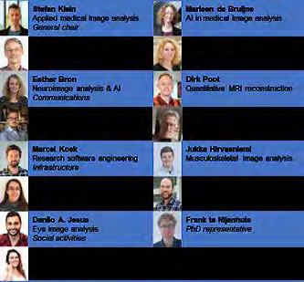
work at two important international conferences. PhD student Nikolai Herrmann and Research Software Engineer Abdullah Thabit started in 2024, funded by two new projects in collaboration with GE Healthcare: FAST-AI and AI4AI. In these projects, we will focus especially on improving the robustness and explainability of AI methods for automated analysis of first-trimester 3D ultrasound images.
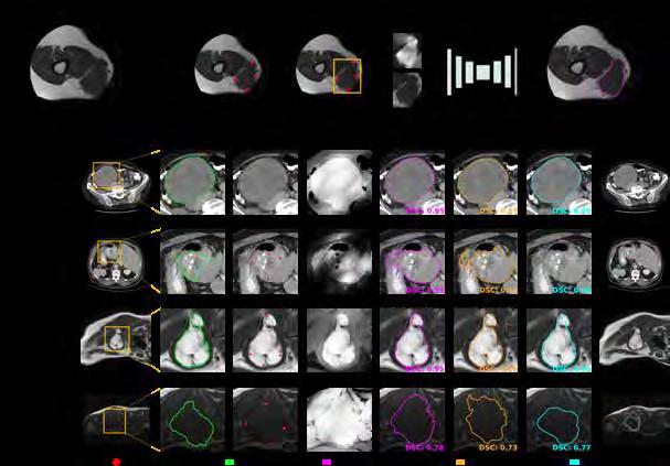
Figure 1. Biomedical Imaging Group Rotterdam (BIGR).
Figure 2. InteractiveNet – our interactive method for segmenting soft-tissue tumours.
Trustworthy AI for MRI
Our research on AI methods for the acquisition, reconstruction, processing, and analysis of MRI has received a major boost thanks to the launch of an Innovation Center for Artificial Intelligence (ICAI), in close collaboration with GE Healthcare. Dr. Dirk Poot is lab manager, Stefan Klein and Aad van der Lugt are scientific directors. In this ICAI lab, all research revolves around improving and evaluating the trustworthiness of AI methods. Four PhD students were appointed at our department: Alireza Samadi and Shishuai Wang work on the improvement of MRI acquisition and reconstruction methods (with Prof. Tamames and Dr. Poot, see p. 86 ); Xinyi Wan and Gonzalo Mosquera Rojas work on the development of AI methods to support diagnosis and prediction in patients with bone, soft-tissue, and brain tumors (see p. 106 and p. 83, respectively, with Prof. Oei, Dr. Visser, Dr. Starmans, and Prof. Smits). A fifth PhD student, Jamie Verweij, has been appointed at Erasmus University Rotterdam, studying the trustworthiness of AI methods from a social sciences perspective. In 2024, first results of this research have been published at various conferences; for example, the exciting work by Shishuai Wang on the qMRI Diffuser: a method for quantitative T1 mapping of the brain using a denoising diffusion probabilistic model. On top of that, Xinyi Wan published her first journal paper on preoperative classification of peripheral nerve sheath tumors on MRI using radiomics.
AI in Cancer Imaging
The concept of personalized medicine has been fully embraced in oncology already for years. Medical imaging plays a crucial role here, and many studies have shown the promise of AI to support image interpretation, disease monitoring, diagnosis and prediction. Yet few methods have reached clinical practice. Our research in this domain includes applications to multiple cancer types, and aims accelerate the translation to clinic by tackling major barriers to adoption, in particular the lack of trustworthiness, explainability, and generalizability of AI methods.
In collaboration with Prof. Smits (p. 184), we have a strong research line on AI methods for glioma (brain cancer). In 2024, Karin van Garderen defended her PhD thesis, entitled ‘Signs of Progression: MR image analysis for the management of adult low-grade glioma’. Moreover, four new PhD students are continuing this line of research (Van der Werff, Mosquera Rojas , Dille, Van Leeuwen), all focused on development of image analysis methods for tumor segmentation, MRI quantification, vascular fingerprinting, molecular classification, tumor grading, and prognosis.
Soft-tissue tumors form a second major area of our interest. PhD student Douwe Spaanderman developed a novel minimally interactive method to accurately contour such tumours in a time-efficient way (see Figure 2). This method showed excellent generalizability, for both CT and MRI scans, in 14 different types of soft-tissue tumors. Also, he performed a comprehensive external validation of a previously developed radiomics model classifying lipoma and well-differentiated liposarcoma on MRI. Both studies were published in 2024. A major highlight in 2024 was the awarding of a prestigious NGF AiNed Fellowship to Dr. Martijn Starmans, allowing him to establish his own AI for Integrated Diagnostics (AIID) research line, in which we will further expand our research on AI methods for soft-tissue tumors (see p. 106) .
Finally, in collaboration with Dr. Thomeer (see p. 212) and Dr. Starmans a third major area of interest was launched in 2024: the Liver Artificial Intelligence (LAI) consortium. The mission of the LAI consortium is to fast-forward the development, validation, and implementation of machine learning methods that could support MRI-based diagnosis of liver lesions. A dedicated website was launched: https://lai-consortium.org/. Two PhD students started on this project: Frederik Hartmann and Ruben Niemantsverdriet
Health Data Science Infrastructure
With our team of research software engineers led by Marcel Koek, the Imaging Office led by Ilva van Houwelingen, and strong involvement of Dr. Bron and Dr. Starmans, we are actively participating in EUCanImage, EOSC4Cancer, and EUCAIM: large-scale international projects funded by the European Commission to build secure and federated infrastructure for next-generation artificial intelligence in oncology. On national level we are contributing strongly to Health-RI, managing the imaging community and jointly establishing an infrastructure for data management, sharing, analysis and reuse of medical imaging data. On a European level, we are hosting the EuroBioImaging Population Imaging node, offering our tools and services in this domain. A dedicated section on these developments can be found on page 34.
Expectations & Directions
In the upcoming years, we will further expand both the fundamental research on development of new image analysis algorithms, and the applied research, in which we test the newly developed methods in clinical applications. Moreover, we expect to continue our activities in building infrastructure to facilitate image data management, data sharing, and data re-use for research, both in
national and international initiatives. Last but not least, we expect to intensify our contributions to education on AI in medicine, as highlighted on page 82 by Dr. Jifke Veenland .
Funding
Lekadir, Karim (Universitat de Barcelona), Aad van der Lugt , Wiro Niessen , Stefan Klein, Daniel Bos , and consortium partners EU Horizon2020: ‘EUCanImage – A European Cancer Image Platform Linked to Biological and Health Data for Next-Generation Artificial Intelligence and Precision Medicine in Oncology’. 2020-2025
Klein, Stefan , Jan-Jaap Visser, Dirk Grunhagen, Kees Verhoef, Stefan Sleijfer, Wiro Niessen , Arno van Leenders, and Martijn Starmans Hanarth Fonds: ‘Automatic grading and phenotyping of soft-tissue tumors through machine learning to guide personalized cancer treatment’. 20212025
Valencia, Alfonso (Barcelona Supercomputing Center), Stefan Klein, Aad van der Lugt, Wiro Niessen, Marcel Koek , and consortium partners EU HORIZON-INFRA-2021-EOSC-01: ‘EOSC4CANCER: A European-wide foundation to accelerate Data-driven Cancer Research’. 2021-2024
Marti-Bonmati, Luis (La Fe Polytechnic and University Hospital), Esther Bron, Martijn Starmans, Marcel Koek, Jan-Jaap Visser, Wiro Niessen , Stefan Klein , and consortium partners EU HORIZON DIGITAL-2022-CLOUD-AI-02: ‘EUCAIM: European Federation for Cancer Images'. 20232027
Niessen, Wiro, Aad van der Lugt , Stefan Klein, Juan A Hernandez-Tamames, Edwin Oei, Dirk Poot, Marion Smits, Jan-Jaap Visser, and consortium partners NWO, Min EZK and GE Healthcare: ‘ROBUST consortium: Trustworthy AI for MRI ICAI lab’. 2022-2027
Rousian, Melek Stichting Sophia Wetenschappelijk Onderzoek (SSWO): ‘A deep learning solution for congenital anomaly detection in early pregnancy’. 2022-2026
Hirvasniemi, Jukka, Stefan Klein, and Edwin Oei (co-applicants) , Sita Bierma-Zeinstra (General Practice), and Jaap Harlaar (TU Delft) (main applicants) TU Delft-Erasmus MC Convergence Flagship: ‘Healthy Joints’. 20222027
Klein , Stefan, Marcel Koek, Hakim Achterberg, Adriaan Versteeg, Martijn Starmans, and consortium partners ISIDORe JRA PROGRAMME: ‘PATH2XNAT: Covid19 Pathomics meets XNAT’. 2023-2025
Klein, Stefan, Maarten Thomeer , and Martijn Starmans NWO Open Technology Programme: ‘The Liver Artificial Intelligence (LAI) consortium: a benchmark dataset and optimized machine learning methods for MRI-based diagnosis of solid appearing liver lesions’. 2024-2028
Rousian, Melek, Wietske Bastiaansen, Stefan Klein , Régine Steegers-Theunissen, Martin Mienkina, and Stephan Anzengruber Erasmus MC-TKI-LSH Health Holland: ‘Towards Fully automated Anomaly Screening in the first Trimester of pregnancy using Artificial Intelligence (FASTAI)’. 2024-2028
Išgum, Ivana, Clarisa Sánchez, Marius Staring, Rob van der Geest, Keelin Murphy, Mitko Veta, Josien Pluim, Stefan Klein , and Mathias Prokop NWO Perspectief: ‘AI4AI: Artificial Intelligence for Accessible Medical Imaging’. 20242029
Invited Lectures
Stefan Klein. ‘Connecting with Health-RI: exposing NCC imaging collections in the national health data catalogue’. Netherlands Cohorts Consortium Symposium, Rotterdam, the Netherlands. Sept 2024.
Shishuai Wang ‘qMRI Diffuser: Quantitative T1 Mapping of the Brain Using a Denoising Diffusion Probabilistic Model’. MICCAI Workshop on Deep Generative Models, Marrakech, Morocco. Oct 2024.
Stefan Klein. ‘Health-RI Imaging’. Dutch National Cancer Research Data Node Meeting, Utrecht, the Netherlands. April 2024.
Stefan Klein. ‘Biomedical imaging group Rotterdam (BIGR)’. Computational Imaging Group seminar, Utrecht, the Netherlands. Feb 2024.
Wietske Bastiaansen . ‘Accessible AI for ultrasonography in the future: embryonic growth markers and detection of fetal growth restriction’. BEN Symposium, Utrecht, the Netherlands. May 2024.
Wietske Bastiaansen ‘AI today and in the future’. Congenital Anomaly symposium Erasmus MC, Rotterdam, the Netherlands. Feb 2024.
Douwe Spaanderman and Martijn Starmans . ‘AI in medical imaging for soft tissue and bone sarcoma’. Dutch Sarcoma Group, Utrecht, the Netherlands. May 2024.
Highlights
The Biomedical Imaging Group Rotterdam (BIGR) was featured in I/O Magazine, a publication by ICT Research Platform Nederland, including interviews with Stefan Klein , Luisa Sanchez , and Theo van Walsum
Tong Wu , Karin van Garderen , Riwaj Byanju , and Wietske Bastiaansen successfully defended their PhD theses.
The Musculoskeletal Image Analysis research has been recognized as an independent research line within the department, led by assistant-professor Jukka Hirvasniemi, see page 100
The new AI for Integrated Diagnostics research line was launched, led by assistant-professor Martijn Starmans , see page 106, who was awarded a prestigious NGF AiNed Fellowship, thanks to which we could greatly extend our research group on AI in Cancer Imaging.
Marcella Zijta won the Best Oral Presentation award in category AI at the ISUOG World Congress on Ultrasound in Obstetrics and Gynecology, for her work on detection of congenital brain anomalies on 3D first-trimester ultrasound.
Stefan Klein organized three vibrant Health-RI Imaging Community meetings, each time with 30-40 attendants, exchanging knowledge and experiences on infrastructure for imaging research.
Additional Personnel
Wenjie Kang – PhD student, see p. 99
Kaouther Mouheb – PhD student, see p. 99
Riwaj Byanju – PhD student, see p. 89
Shishuai Wang – PhD student, see p. 90
Alireza Samadifardheris – PhD student, see p. 90
Tong Wu – PhD student, see p. 224
Mirthe Kamphuis – PhD student, see p. 104
Xinyi Wan – PhD student, see p. 112
Matthew Marzetti – PhD student , see p. 112
Natalia Oviedo Acosta – PhD student, see p. 112
Karthik Prathaban – PhD student, see p. 112
Karin van Garderen – PhD student , see p. 190
Karen van der Werff – PhD student, see p. 90
Bas Dille – PhD student, see p. 190
Juancito van Leeuwen – PhD student, see p. 191
Manon Verburg – MSc student
Jette Slettenhaar – MSc student
Danchen Ge – MSc student
Abdullah Thabit – Research Software Engineer
Associate Professor Jifke Veenland, PhD

Project Funding TKI-LSH Prostate-X: “An MRI-based diagnostic and prognostic tool for improved prostate cancer management”; EU-Digital-2023-Skills: “Sustainable Healthcare with Digital Health Data Competence”.
Email j.veenland@erasmusmc.nl / j.f.veenland@tudelft.nl
APPOINTMENT IN RADIOLOGY
Jifke Veenland has a MSc degree both in medicine and in computer science, and a PhD in medical image processing and pattern recognition. The focus of her research is on tissue characterization and quantification of heterogeneity in MRI tumor images by applying Machine Learning and/or Deep Learning.
The main part of her activities are in the field of education: she is coordinator of the MSc track Imaging & Interventions of Technical Medicine, the joint degree program of the Erasmus MC, LUMC and the TU Delft. Next to that she is project leader of the ErasmusArts 2030 Technology group to develop a curriculum for digitally skilled doctors. She is coordinator of the modules Imaging and Image Processing for the BSc Clinical Technology and for the MSc Technical Medicine, she coordinates the modules Advanced Image Processing and Machine Learning.
Machine and Deep Learning
Machine and deep learning techniques are increasingly used for medical imaging analysis. These methods help segment lesions, such as tumors in prostate MRIs, and predict treatment outcomes, like chemotherapy response in breast cancer patients. Training technical medicine students, data scientists, and AI-smart doctors is crucial for advancing this field.
Research Projects
Prostate Cancer Segmentation
(In collaboration with the department of pathology)
In collaboration with pathology, Convolutional Neural Networks (CNNs) were developed to detect and segment clinically significant prostate cancer in MR images. A multi-class segmentation model was trained to classify different tumor aggressiveness levels, which can guide urologists in biopsy procedures.
Breast Cancer Outcome Prediction
( In collaboration with the department of pathology) Triple-negative breast cancer (TNBC) patients often have poorer outcomes. Neoadjuvant chemotherapy (NAC) reduces tumor size, but not all patients respond effectively. Using deep learning models on DCE-MRIs at two time points, we aim to predict pathologic complete response, improving personalized
Blastocyst Segmentation in IVF
(In collaboration with the department of gynaecology)
IVF treatment involves selecting viable embryos based on their growth in the EmbryoscopeTM. We developed a deep learning model to segment blastocysts from time-lapse images, generating growth curves. By combining these with clinical features, a machine learning model achieved a ROC-AUC of 0.70, this can improve embryo selection and IVF success rates.
Education Projects
Digital Technology in Medical Education
With the 2024 renewal of the Erasmus MC BSc Medicine curriculum, digital literacy is a key focus. Medical students are introduced to AI, e-health, VR/AR, and smart predictive algorithms. Learning goals and materials have been developed to train future doctors in identifying opportunities and ethical challenges in digital healthcare.
Participation in the SUSA Project
The Sustainable Healthcare with Digital Health Data Competence (SUSA) project, funded by the EU Digital Europe Programme, enhances digital competencies in healthcare. Integrating digital skills into 20 bachelor's and 26 master's programs, along with lifelong learning modules, SUSA aims to train over 6,500 graduates and upskill 600 professionals, aligning with EU goals for sustainable and data-driven healthcare.
Post-docs

Wietske Bastiaansen, PhD
Project Funding Erasmus MC-TKI-LSH Health Holland: “Towards Fully automated Anomaly Screening in the first Trimester of pregnancy using Artificial Intelligence (FAST-AI)”; NWO Perspectief: “AI4AI: Artificial Intelligence for Accessible Medical Imaging”; Sophia Fonds.
Email w.bastiaansen@erasmusmc.nl
AI for Prenatal Image Analysis
Every child deserves the best possible start in life, and proper prenatal growth monitoring is essential. Impaired growth, such as fetal growth restriction (FGR) or small-for-gestational-age (SGA) infants, raises perinatal risks and lifelong health challenges. Parental health and lifestyle factors further impact development, but 2D ultrasonography, the current standard, is limited in assessing these influences. While 3D ultrasonography offers richer insights, it demands expert training and is time-intensive. To address this, I focus on AI-based
PhD Students

Wietske Bastiaansen, MSc

Advisors Stefan Klein, Melek Rousian, Anton Koning, Wiro Niessen & Régine Steegers-Theunissen Project Funding Erasmus MC Research Grant Email w.bastiaansen@erasmusmc.nl PhD Obtained 02-07-2024
4D
spatiotemporal
atlas of the embryonic brain
To study brain development, semi-automatic measurements are performed. These are time-consuming and lack overview. The availability of an atlas, which consists of a template of the entire brain for a range of gestational ages, could overcome this by offering a unified framework to study brain development. This will offer unique insight into this crucial period in life, which ultimately will lead to an earlier detection, prevention and treatment of neuro-developmental disorders.

methods to automate 3D ultrasound analysis of the embryo, fetus, and placenta.
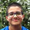
Gonzalo Mosquera Rojas, MSc

Advisors Stefan Klein & Marion Smits Project Funding ROBUST consortium: "Trustworthy AI for MRI" ICAI-lab Email g.mosquerarojas@erasmusmc.nl
Trustworthy AI for glioma diagnosis
This project focuses on the development of trustworthy AI models for glioma diagnosis using MR imaging, which has the potential to improve the treatment decision making process. The design of the models has a strong focus on explainability and uncertainty quantification, aiming to ensure interpretable results in clinical settings.
LinkedIn
LinkedIn

Marcella Zijta, MSc

Advisors Stefan Klein, Melek Rousian, Bernadette de Bakker & Wietske
Bastiaansen
Project Funding Stichting Sophia Wetenschappelijk Onderzoek (SSWO) 2022: “A deep learning solution for congenital anomaly detection in early pregnancy”
Email m.zijta@erasmusmc.nl
Congenital anomaly detection in early pregnancy
The project focuses on developing AI-driven methods for anomaly detection, organ segmentation, and growth assessment in embryos and fetuses using 3D ultrasound and Micro-CT scans. Key objectives include estimating developmental age, detecting congenital anomalies, and automating first-trimester anomaly screening to enhance early diagnosis and monitoring.
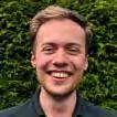
Ruben Niemantsverdriet, MSc

Advisors Stefan Klein, Martijn Starmans & Maarten Thomeer
Project Funding NWO Open Technology Program (OTP)
Email r.niemantsverdriet@ erasmusmc.nl
The Liver Artificial Intelligence (LAI) Consortium
Differentiating solid liver lesions on MRI is challenging due to their variety, different protocols, and rare subtypes. Accurate methods are needed to guide care and treatment. The LAI consortium aims to develop and implement machine learning tools for MRI-based diagnosis of liver lesions while promoting open science through public data sharing. Twelve hospitals worldwide are collecting MRI scans and clinical data from over 3,000 patients to support this effort.

Nikolai Herrmann, MSc
Advisors Stefan Klein, Régine SteegersTheunissen, Wietske Bastiaansen & Melek Rousian
Project Funding NWO AI4AI: “Artificial Intelligence for Accessible Medical Imaging”
Email n.herrmann@erasmusmc.nl
Novel, robust and resource-efficient volumetric biomarkers for first-trimester 3D ultrasound during pregnancy
To develop failure-aware and resource efficient AI methods for automated embryonic growth monitoring systems in three-dimensional (3D) ultrasound. This includes automatically performing embryonic body-part segmentations and from these, derive quantitative biomarkers for monitoring development. A strong focus is put on explainability and efficiency to facilitate implementation in clinical practice.
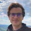
Frederik Hartmann, MSc
Advisors Martijn Starmans, Maarten Thomeer & Stefan Klein
Project Funding NWO Open Technology Programme
Email f.hartmann@erasmusmc.nl
The
Liver Artificial Intelligence (LAI) Consortium
Differentiating solid liver lesions on MRI is challenging due to their variety, different protocols, and rare subtypes. Accurate methods are needed to guide care and treatment. The LAI consortium aims to develop and implement machine learning tools for MRI-based diagnosis of liver lesions while promoting open science through public data sharing. Twelve hospitals worldwide are collecting MRI scans and clinical data from over 3,000 patients to support this effort.

Douwe Spaanderman, MSc

Advisors Stefan Klein, Martijn Starmans, Dirk Grünhagen & Wiro Niessen Project Funding Hanarth Fonds: “Automatic grading and phenotyping of softtissue tumors through machine learning to guide personalized cancer treatment”
Email d.spaanderman@erasmusmc.nl
AI-driven Soft Tissue Tumor Management
Soft tissue tumors (STTs) represent a rare and diverse group of lesions characterized by a wide spectrum of differentiation. These subtypes vary significantly in clinical behavior, aggressiveness, molecular profiles, and optimal treatment strategies. To address this complexity, we are leveraging advanced machine learning techniques to analyze medical imaging, such as computed tomography (CT) and magnetic resonance imaging (MRI), for improved tumor detection and prognosis prediction.

Dirk Poot is asssistant professor and heading the quantitative MRI reconstruction research line. He is affiliated with the Biomedical Imaging Group Rotterdam (BIGR, http://www.bigr.nl) and the MR physics group. In 2005, Dirk received his MSc degree from the faculty of Applied Physics at the Delft University of Technology, Delft/NL. In 2010 he obtained his PhD degree at the Visionlab, University of Antwerp, Antwerp/BE, for his research on reconstruction and statistical processing of Magnetic Resonance Images. He is lab manager of the new ICAI lab Trustworthy AI for Magnetic Resonance Imaging. His current research interests include MR image acquisition, reconstruction, quantification, and motion compensation. d.poot@erasmusmc.nl
QUANTITATIVE MRI RECONSTRUCTION
Dirk Poot, PhD assistant professor

Context
Quantiative MRI is becoming increasingly relevant in the era of precision medicine. Current clinically used MRI protocols are still mostly limited to weighted images, such as T1-weighted or T2-weighted. This delivers images optimized for visual inspection by a radiologist that is looking for structural abnormalities. However, these images do not provide measurements of the actual magnetic resonance properties of the tissue; e.g. the T1 or T2 relaxation time, or the diffusion or perfusion rate. Also, there might be substantial variability in the images between scanners, or even from the same scanner at different moments in time. This lack of standardization hampers the detection of subtle diffuse, disease induced, changes in the tissues. The key objective of quantitative MRI is to complement the qualitative images with quantitative measurements of tissue properties. We have a substantial number of projects in which different properties are measured.
Top Publications 2024
Feddersen TV, JA Hernandez-Tamames, MM Paulides, M Kroesen, GC van Rhoon, DHJ Poot. Magnetic resonance thermometry for hyperthermia in the oropharynx region. International Journal of Hyperthermia 2024; 41:2352545.
Sijtsema ND, I Lauwers, GM Verduijn, MS Hoogeman, DHJ Poot, JA Hernandez-Tamames, A van der Lugt, ME Capala, SF Petit. Relating pre-treatment non-Gaussian intravoxel incoherent motion diffusion-weighted imaging to human papillomavirus status and response in oropharyngeal carcinoma. Physics and Imaging in Radiation Oncology 2024; 30:100574.
Van Dorth D, K Venugopal, KN van der Werff, M Smits, EAH Warnert, JA Hernandez-Tamames, MJP van Osch, DHJ Poot. Exploring the need for a preload on the estimation of permeability, vessel radius, and relative cerebral blood volume in MR vascular fingerprinting–based dynamic susceptibility. Magnetic Resonance in Medicine 2024; 10.1002.
Research Projects: Objectives & Achievements
My research line focusses on quantitative MRI reconstruction, in close collaboration with the MR physics group of J.A. Hernandez-Tamames (page 42) as well as the image registration group of S. Klein (page 76). The aim of quantitative MRI is to objectively measure tissue properties such as for example the T1, T2(*) relaxation times, temperature or tissue perfusion. Traditionally, this is done by acquiring several images with specific differences in their acquisition settings such as echo time or inversion. The intensity of the acquired images is fitted to a model that is derived from the MR physics of the acquisition method.
Current projects:
– Diffusion model for qMRI (Shishuai Wang)
– Resolution enhancement of qMRI by using weighted images (Alireza Samdifardheris)
– Vascular signature mapping (Karen van der Werff)
Acceleration of acquisition
Within the new Trustworthy AI ICAI lab one of the projects is to further accelerate the image acquisition by severely under-sampling the k-spaces, such that normal image reconstruction fails. By using the known MR physics as well as deep learning approaches we aim to still obtain highquality images and tissue property maps from the short scans. The trustworthiness of these resulting images to correctly reflect the specific patient is key in this research.

Figure 1. From Sabidussi et al. NeuroImage 269, figure 13. This figure shows for two acquisition setups ( ) one of the performance metrics that we use for two reference quantification methods (MLE, IWLLS) and our newly proposed method (dtiRIM). Especially for the reduced, and hence faster, acquisition ( ) the error (RMSE) of the fraction anisotropy (FA) is substantially lower, demonstrating the benefit of our method.
Motion compensation
Subject motion is a major cause of low quality or failed MRI exams. For quantitative MRI acquisitions this situation is even worse, as typically they are longer than the acquisition of individual traditional weighted images. Additionally, in the analysis, images are combined, increasing the sensitivity to motion. Hence a major focus of my research line is to compensate for subject motion. By exploiting the known relations among the images as well as by adjusting the acquisition to acquire some reference data to allow identification and subsequent compensation of unavoidable subject motion.
Acceleration of reconstruction
The advanced methods that we develop for high quality reconstruction from highly accelerated scans may have long computation times. Even though computers get faster every year, innovative methods to improve computation time are needed. With novel work on deep learning based image reconstruction we are accelerating the reconstruction process to achieve clinically acceptable reconstruction times for the advanced methods.
Vascular properties
For tumor growth vascularity Is highly relevant. With the development of novel acquisition and analysis methods we aim to extract more information on the vasculature, which we aim to develop into biomarkers.
Expectations & Directions
In the upcoming years, we aim to further develop the quantitative MR image acquisition and reconstruction methods. In parallel the novel developments will be applied in clinical research projects for further evaluation and in collaboration with industry we aim to bring the developments to actual clinical use.
Funding
Poot, Dirk, Juan Hernandez Tamames, Stefan Klein, Aad van der Lugt, Edwin Oei, Marion Smits, Jan-Jaap Visser, and consortium partners NWO, Min EZK and GE HealthCare: ‘ROBUST consortium: Trustworthy AI for MRI ICAI lab’. 2022-2027
Poot, Dirk, Juan Hernandez Tamames, Stefan Klein, and consortium partners Marie Curie: 'IQ-BRAIN'. 2024-2028
Hernandez Tamames, Juan, Dirk Poot and Frans Vos TKI
Health~Holland and GE Healthcare: 'Gadolinium free enhancement with magnetic resonance imaging synthesis'. 2024-2028
Invited Lectures
Dirk Poot . 'Advanced techniques & clinical use'. Workshop on Quantitative multiparametric MRI as inversion problem, Utrecht, the Netherlands. Nov 2024.
Dirk Poot . 'Machine learning in MRI’. ESMRMB comprehensive course on practical MR physics, online. June 2024.
Dirk Poot . 'Quantitative MR reconstruction’. Computation imaging group meeting, UMC Utrecht, Utrecht, the Netherlands. Feb 2024.
PhD Students

Riwaj Byanju, MSc
Advisors Stefan Klein & Dirk Poot
Project Funding H2020 MSCA ITN – B-Q Minded
Email r.byanju@erasmusmc.nl
PhD Obtained 26-09-2024
Optimal parameter estimation from intra-scan modulated MR data
Riwaj developed methods to in-silico evaluate the efficiency of sampling patterns. Using these techniques he developed a Myelin water fraction mapping method that is faster than traditional approaches yet avoids artifacts created by previously used GRASE acquisitions. Additionally he worked on extending a novel multi-parametric acquisition method, developed within the MR physics group, to 3D acquisitions.
Highlights
The newly funded Marie-Curie ITN project IQ-BRAIN is starting, there are vacancies for 2 PhD students.
Additional personnel
Ilaria Neri – PhD student, see p 49
Noemi Sgambelluri – PhD student, see p 49

Emanoel Sabidussi, MSc
Advisors Stefan Klein & Dirk Poot
Project Funding H2020 MSCA ITN – B-Q Minded
Email e.ribeirosabidussi@ erasmusmc.nl
Advanced deep learning methods for Quantitative MRI
With Recurrent inference machines Emanoel could quantify relaxometry as well as diffusion properties. With this model based technique the strengths of the knowledge of the MR physics are combined with the power of deep learning in a way that allows training the deep learning method with simulated data. State of the art results are obtained.
Alireza Samadifardheris, MSc

Advisors Juan Hernandez-Tamames, Stefan Klein & Dirk Poot
Project Funding TAI-MRI ICAI Lab, part of the ROBUST program
Email a.samadifardheris@erasmusmc.nl
Trustworthy AI for adaptive and precision MR protocols
In the first project, Alireza applies deep learning to superresolve low-resolution MR quantitative maps using high-resolution weighted images. This is the first step towards obtaining fast multiparametric maps for abnormality detection and subsequent online protocol adjustment.

Karen van der Werff, MSc

Advisors Marion Smits, Stefan Klein, Frans Vos & Dirk Poot
Project Funding Vascular Signature Mapping of Brain Tumor Genotypes, projectnumber 17079
Email k.vanderwerff@erasmusmc.nl
Brain tumor genotyping based on multicomponent vascular fingerprinting
Karen is developing a MR fingerprinting technique for multi-component estimation of vascular properties. This will result in quantitative maps for different vascular parameters. The performance of these maps in terms of glioma genotyping will be analyzed, with the aim of non-invasively characterizing brain tumours.

Shishuai Wang, MSc
Advisors Stefan Klein, Juan HernandezTamames & Dirk Poot
Project Funding TAI-MRI ICAI Lab, part of the ROBUST program
Email s.wang@erasmusmc.nl
End-to-end deep learning quantitative MR reconstruction
Shishuai developed deep-learning based methods to directly reconstruct multiple high-quality quantitative MR maps (e.g. T1, T2 and proton density) from highly under sampled k-space data acquired with a Quantitative Transient Imaging protocol. Shishuai is also investigating quantitative MR mapping by using diffusion model.
Esther Bron is Assistant Professor in Medical Image Analysis and is heading the Neuroimage Analysis and Machine Learning research line. She is affiliated with the Biomedical Imaging Group Rotterdam (BIGR, http://www.bigr.nl). In 2011, Esther received her MSc degree in Medical Natural Sciences – specialization Medical Physics, cum laude – at the VU University, Amsterdam/NL. Esther successfully defended her doctoral thesis in 2016 at the Erasmus University Rotterdam, which focused on the development and validation of advanced image analy-

sis techniques for computer-aided diagnosis of dementia. Since then, she has successfully built her research line and has been appointed assistant professor in 2020. Esther has received several (young investigator) awards and is management board member in several (inter)national consortia (e.g. TAP-Dementia, EUCAIM). In addition, she leads the imaging working group of Health-RI, the national health data infrastructure in the Netherlands. e.bron@erasmusmc.nl
Esther E Bron, PhD assistant professor NEUROIMAGE ANALYSIS & MACHINE LEARNING

Context
Neurodegenerative and cardiovascular diseases impose an enormous burden to the individual and to society. As a consequence, there is an urgent need to develop effective preventive and therapeutic strategies, especially in dementia. Early detection and accurate prediction of the progression of at-risk subjects are key in this development. Artificial Intelligence (AI) has high potential for aiding medical decision making and creating a learning healthcare system, where current patients are diagnosed and treated based on knowledge gained from previous patients. Especially in the domain of neuroradiology, AI has proven very successful in gaining new knowledge by extracting patterns related to neurodegenerative diseases from large sets of MRI data. My mission is to translate AI to clinical practice, so future patients can be diagnosed and treated based on knowledge gained from previous patients.
Top Publications 2024
Mouheb K, M Elbatel, S Klein, EE Bron. Evaluating the Fairness of Neural Collapse in Medical Image Classification. Lecture Notes in Computer Science – 27th International Conference on Medical Image Computing and Computer Assisted Intervention (MICCAI) 2024; 15010:286-296.
Mo H, D Bos, M Kavousi, M Leening, EE Bron. Coronary Artery Calcium Scoring from Non-Contrast Cardiac CT Using Deep Learning With External Validation. MICCAI workshop on Statistical Atlases and Computational Modeling of the Heart (STACOM) 2024.
Mateus P, S Garst, J Yu, D Cats, AGJ Harms, M Birhanu, M Beekman, PE Slagboom, M Reinders, J van der Grond, A Dekker, JFA Jansen, M Beran, MT Schram, PJ Visser, J Moonen, M Ghanbari, G Roshchupkin, D Vojinovic, I Bermejo, H Mei, EE Bron. MRI-based and metabolomics-based age scores act synergetically for mortality prediction shown by multi-cohort federated learning. ArXiv 2024; 2409.01235.
Research Projects: Objectives & Achievements
AI for image-based diagnosis and prediction
I develop with my team novel image analysis and prediction methodology for neurodegenerative and cardiovascular diseases and strongly collaborate with clinicians and clinical researchers to validate these methods for clinical application. This entails AI for automatic analysis of multimodal MRI brain scans, including perfusion MRI and vascular imaging biomarkers. We developed a novel disease progression model and evaluated this in several neurodegenerative diseases (i.e. Alzheimer’s disease, frontotemporal dementia). Current work focuses on developing diagnostic AI that can be translated to clinical practice and its validation on large-scale clinical data.
Deep learning methods have large potential for early detection in dementia. However, there are some limitations of deep learning methods that have prevented them from being routinely used in clinical practice. A key problem is the limited interpretability of the predicted results, because deep learning models often operate as a black box. Wenjie Kang finished his novel method Glo&Loc-EBM and submitted the journal paper (Figure. 1). His novel me-

Figure 1. The novel method Glo&Loc-EBM selects relevant regions based on an occlusion map, computes deep learning features and performs classification based on an explainable boosting machine (EBM). The method achieves higher performance for classifying Alzheimer's disease patients from controls than previous methods (Kang at al., submitted).
thod combines the interpretability of Explainable Boosting Machines with deep learning techniques exploiting the high-dimensional information captured by imaging. Another challenge in dementia diagnosis is the etiological diagnosis of different diseases underlying dementia. Myrthe van Haaften works on several project related to this. Based on the initial work of three MSc students, she used hierarchical clustering to improve understanding of imaging patterns in memory clinical patients related to their diagnosis. Using this approach, four clusters of patients are identified are related to diagnosis, neuropsychological test results and other outcomes (Figure. 2). Post-doctoral researcher Hyunho Mo works on the prediction of cardiovascular events in the general population. Current work includes a novel approach for coronary artery calcium scoring from non-contrast cardiac CT. The work was presented at the 2024 MICCAI-STACOM workshop and will be extended in 2025 with external validation as well as long-term outcome prediction.
Current projects:
– Explainable machine learning methods ( Wenjie Kang, Bo Li )
– AI for etiological diagnosis of dementia ( Myrthe van Haaften, Kaouther Mouheb , Nathalie Koorn, Nora Ali, Beatriz Domingos, Joana Dias de Oliviera)
– Subtypes of healthy aging and early detection of cognitive decline ( Sterre de Jonge )
– Predictive modeling in cardiovascular disease ( Hyunho Mo , Julien Triacca)
– Disease onset prediction in genetic FTD ( Wenjie Kang , Myrthe van Haaften , Jon Bregman)
– Image-based prediction in children after cardiac arrest (Wilco van der Velde, Hyunho Mo )
Figure 2. Four clusters of patients visiting the Alzheimer Center Erasmus MC based on their brain volume percentiles. The average volume of each brain regions within the clustered group of patients is shown by the colored line. A volume of 0.5 (bold line) represents the average of a cognitively normal population. lh = left hemisphere, rh = right hemisphere. We find different clusters representing a mix of diagnostic groups. (Van Haaften et al., submitted).
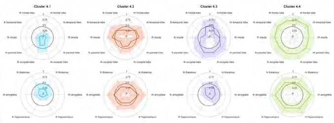
Imaging data accessibility
Medical AI development needs large and heterogeneous datasets. A key bottleneck here is clinical data sharing, which is difficult -or sometimes impossible- because of privacy and safety issues. The novel technology of federated learning provides a new angle to solving this universal problem. Opposed to bringing all data to a central place for training a machine learning algorithm (central learning), federated learning works with several computer nodes that train an algorithm collaboratively without exchanging the data. In biomedical imaging, federated learning has recently been applied to multiple research tasks, including whole-brain segmentation and brain-age prediction. While the technique cannot be applied in a routine way to clinical data yet, I aim with my team to set a major step in overcoming methodological (e.g. different data distributions, quality control) and organisational (e.g. privacy and security, workflow) challenges. I consider federated analyses to be essential for the success of AI in medicine and together with my team we have the expertise to boost this.
Related to this, deep learning are known to be susceptible to biases in the data. Especially, biases against specific groups may hinder clinical applicability. A recently discovered phenomenon, Neural Collapse (NC), has shown potential in improving the generalization of state-of-theart deep learning models. Nonetheless, its implications on bias in medical imaging remain unexplored. Kaouther Mouheb presented at the 2024 MICCAI conference a study that investigated deep learning fairness through the lens of neural collapse and analyzed its training dynamics. We found that biased training initially results in different neural collapse configurations across subgroups, before converging to a final solution by memorizing all data samples (Mouheb et al, 2024).
I work as Image Data Coordinator at the Dutch National Research Infrastructure: Health-RI. Here, I lead (together with Dr. Stefan Klein) the Health-RI Imaging Working group. In 2024, key milestones were the onboarding of the first 5 imaging datasets in the National Healthdata Catalogue, an associated software library (Img2catalog) and an advisory document to the Health-RI nodes on the recommended approach for onboarding imaging data. In addition, we wrote an document for Health-RI to advise general hospitals that aim to participate in multi-center studies on using CTP as software for image data de-identification.
Current projects:
– Federated learning for imaging data ( Kaouther Mouheb , Hyunho Mo , Alexander Harms, Mahlet Birhanu)
– Large-language models (LLMs) for extracting neuroradiology ratings ( Kaouther Mouheb , Antoine Manenti)
– Meta-data models for imaging data (Alexander Harms, Mahlet Birhanu, Hakim Achterberg)
– Cancer Imaging Europe dashboard and catalogue (Alexander Harms, Mahlet Birhanu, Hakim Achterberg)
– Imaging Office / Euro-BioImaging Population Node (Ilva van Houwelingen)
Expectations & Directions
In coming years we continue to study novel artificial intelligence (AI) methods for image analysis and outcome prediction in the field of neurological and cardiovascular diseases with a focus on dementia, addressing challenges related to clinical translation of these methods. In 2025, new projects will start (CHIME, Predict-FTD). In addition, through national and European research infrastructure initiatives, we work on pushing the field forward in making imaging and health data accessible for research and innovation in an efficient yet secure manner.
Funding
Daemen, Mat, Geert Jan Biessels, Wiro Niessen, Esther Bron, and consortium partners CardioVasculair Onderzoek Nederland (CVON): ‘HBCx: Heart-Brain Connection Crossroads’. 2019-2024
Niessen, Wiro, Frans Vos, Mark van Buchem, Esther Bron, and Jeroen de Bresser Medical Delta: ‘Medical Delta Diagnostics 3.0: Dementia and Stroke’. 2019-2024
Van der Harst, Pim, Folkert Asselbergs, Michiel Rienstra, Lotte Krabbenborg, Wiro Niessen, Daniel Bos, and Esther Bron NWO Commit2Data Big data & health grant: ‘MyDigiTwin: Early recognition and prevention of cardiovascular diseases’. 2022-2026
Kang, Wenjie, and Esther Bron China Scholarship Counsil (CSC) Fellowship: ‘Interpretable machine learning for diagnosis and prognosis in neurodegenerative disease’. 20222026
Bron, Esther Erasmus MC Fellowship grant: ‘Etiological diagnosis of dementia using federated artificial Intelligence’. 2023-2027
Vernooij, Meike, Esther Bron, Hugo Kuijf, Geert Jan Biessels, Vikram Venkatraghavan, and Betty Tijms ZonMW TAPDementia grant: ‘Timely Accurate Personalized Diagnosis using Artificial iNtelligence to Classify dementia Etiologies (TAP-DANCE)’. 2023-2027
Marti-Bonmati, Luis, .. , Wiro Niessen, Esther Bron, Stefan Klein, and Martijn Starmans Digital Europe Programme (Horizon Europe) grant: ‘EUropean Federation for CAncer IMages (EUCAIM)’. 2023-2027
Klomp, Dennis, Mariëlle Emmelot-Vonk, Geert Jan Biessels, Esther Bron, and Meike Vernooij Health~Holland LSH-match multicenter PPP: ‘Scan2go: autonomous MRI for large scale diagnostics of brain integrity’. 2023-2027
Zwanenburg, Jacobus, Natalia Petridou, Thijs van Osch, Geert Jan Biessels, Esther Bron, Meike Vernooij, et al. NWO Applied and Engineering Sciences Perspectief Grant: 'CHIME: Cerebral HemodynamIcs, Metabolism and clearancE: A comprehensive, non-invasive brain imaging approach to characterize key biological processes in dementia'. 2024-2028
Vernooij, Meike, Esther Bron, Frank Wolters, Eline Vinke, Jeroen de Bresser, Frans Vos, Thijs van Osch, Simon Mooijaart, and Harro Seelaar Medical Delta: 'Medical Delta Dementia 3.0: Applying advanced brain imaging for efficient dementia diagnosis and prediction: when, what, and for whom?'. 2024-2028
Seelaar, Harro, Esther Bron, Barbare Borroni, Ruta Gostautaite, Gianluigi Zanusso, Raquel Sanchez, Anja Schneider, Eino Solje, Mireia Jofre-Bonet, Jonas Meirer, Angela Bradshaw, Markus Otto, Yolande Pijnenburg, Evelyn Moreno, and Alessio Fasano Horizon Europe (HORIZON-HLTH2024-DISEASE-03-two-stage): 'PREDICTFTD: Accelerating the Validation of Predictive Liquid Biomarkers for Frontotemporal Dementia Diagnosis and Subclassification'. 2024-2028
Invited Lectures
Esther Bron. 'Demonstration of the EUCAIM platform: usability, challenges and a future outlook'. BioFIT workshop, Lille, France. Dec 2024.
Esther Bron. 'Sharing images: The why and how of medical imaging analysis research'. Data stewards course - HealthRI, Utrecht, the Netherlands. June 2024.
Myrthe van Haaften. 'Artificial intelligence and dementia'. Department of Neurology seminar, Erasmus MC, Rotterdam, the Netherlands. June 2024.
Esther Bron and Myrthe van Haaften. 'De toekomst van beeldvormende diagnostiek in de geheugenkliniek'. National Dementia Congress, Ministry of Health, Welfare and Sport, Nieuwegein, the Netherlands. April 2024.
Esther Bron. 'Unveiling the Future: The Impact of EUCAIM's First Platform Release on AI in Cancer Research'. European Conference on Radiology: Studio Session, Vienna, Austria. March 2024.
Esther Bron. 'Building a compliant data registry for AI research: the way we do it'. European Conference on Radiology: The AI Theatre, Vienna, Austria. March 2024.
Highlights
Esther Bron worked at Health-RI as Coordinator Imaging Data for the Architecture team, where she coordinates the Imaging Working Group.
Myrthe van Haaften, Esther Bron, Meike Vernooij and TAP-Dementia collaborators organised a public outreach symposium with the topic 'The Future of Image-based diagnostics in the memory clinic' at the Nationaal Dementie Congres on 15 April, targeting care professionals, policy makers, people with dementia and care givers.
Kouther Mouheb won the first prize in the MICCAI Educational Challenge 2024 with a tutorial on denoising diffusion models, that she developed together with two researchers from the University of Girona, Spain.
Mahlet Birhanu was awarded in the Euro-BioImaging Job Shadowing program and visited the Medical Research Institute of the Hospital La Fe in Valencia, Spain in October 2024.
The Neuroimage analysis & Machine learning research line was expanded by a new member: Sterre the Jonge started her PhD program in February 2024.
Myrthe van Haaften was selected to present a poster at the Dutch Dementia Researchers conference and the Dementia Day of Erasmus MC.
Myrthe van Haaften joined the Alzheimer Center stand at the Brain Awareness Week (organized by the Erasmus University, open to the general public) to provide information about brain MRI in dementia.
Additional Personnel
Mahlet Birhanu, MSc – Research Software Engineer
Alexander Harms, MSc – Research Software Engineer
Ilva van Houwelingen, MSc – Process Coordinator Imaging Office
Nathalie Koorn, BSc – MSc Student
Joana Dias de Oliveira Coelho – BSc Student
Beatriz Domingos – BSc Student
Antoine Manenti, BSc – MSc Student
Julien Triacca, BSc – MSc Student
Wilco van der Velde, BSc – MSc Student
Jon Bregman, BSc – MSc Student
Nora Ali, BSc – MSc Student
Assistant Professor Henri Vrooman, PhD

Email h.vrooman@erasmusmc.nl
Henri A. Vrooman received the Ph.D. degree in physics (1991) from Delft University of Technology (TU-Delft), the Netherlands. From 1986 to 1990, he was a Research Scientist at the Department of Image Processing and Pattern Recognition of the TU-Delft. From 1990 until 2000 he was involved in several image processing projects in cooperation with the Department of Radiology at the Laboratory for Clinical and Experimental Image Processing of Leiden University Medical Center. Since April 1st, 2000, Henri is Assistant Professor at the Erasmus MC - University Medical Center - Rotterdam, the Netherlands, where he initiated the Biomedical Imaging Group Rotterdam (BIGR). His research interests include digital image processing, pattern recognition, biomedical image processing techniques and diagnostic radiology. The recent focus of his work is on neuroimaging and on the development of infrastructures for the processing of large data sets, especially important for Population Imaging.
Providing image processing as a service
In 2024, the Imaging Office, embedded in the Department of Radiology & Nuclear Medicine, received more and more requests for processing and annotation tasks. The office targets parties that need access to medical imaging data and analysis tools. This includes support for image acquisition, data storage and analysis of imaging data with several pipelines.
Examples of services we provided in 2024 were the processing of brain MRI images of children with rare diseases to extract relevant volumetric biomarkers, the extension of an annotation tool to perform 3D measurements of the pineal and annotate present cysts, and the automated detection of white matter lesions, using an advanced neural network approach.
Advanced processing in craniosynostosis
Another interesting project we are Involved in for years now, is the processing of images from children with craniosynostosis. Metopic synostosis patients are at risk for neurodevelopmental disorders despite a negligible risk of intracranial hypertension. To understand underlying pathophysiology, we extended our portfolio with more advanced processing techniques, such as brain tissue segmentation, computation of the local gyrification Index (LGI), and arterial spin-labeling to measure regional cerebral blood flow.
For tissue segmentations, scans were processed with Infant FreeSurfer. The scans of children younger than one year differ from adult imaging data. By giving age as a parameter, a different brain atlas is used to compute the

Figure 1. Processing of brain images from patients with nongenetic metopic craniosynostoses. The native scan of a patient (top-left), the Desikan cortex parcellation (top-right), and the surfaces for computing the LGI (bottom-left) are shown. At the bottom right, an example of segmented ventricles of another patient.
Desikan cortex parcellation. The LGI of the cortex is computed by smoothing the pial surface and taking the local distances between the two surfaces. Finally, we were working on methodology to segment brain ventricles In MRI scans. Metopic patients may have strangely shaped ventricles with an increased size. Standard processing techniques are unusable. We are working on a neural network approach to detect and properly segment the diseased ventricles. So far, the training set Is based on manual segmentations but will be extended with automatically segmented ones.

Bo Li, PhD
Project Funding Heart Brain Connection – crossroads (HBCx)
Email b.li@erasmusmc.nl
Translatable AI in Neuroimage Analysis
Information technology, such as Artificial Intelligence (AI), has the potential to transform the field of radiology by improving diagnostic accuracy, increasing efficiency and reducing healthcare costs. While in order to integrate AI tools into clinical workflows, it is critical that we design and validate them to be not only accurate, but also effective, unbiased, and transparent.
Therefore, in my role of developing AI algorithms for brain MRI analysis, we focused on 1) simplifying and effectively supporting the imaging assessment of cerebral lacunes by additionally predicting the presence and a burden score of the lesion, besides normal segmentations (2nd place at VALDO challenge in MIC-


CAI 2021; OHBM 2022); 2) constructing neuroimaging endophenotypes for population studies that enables unsupervised dimensionality reduction at high-resolution, and allows for confounder controlling during feature construction (OHBM 2022, oral); and 3) extending our novel AI algorithms for group-wise mean-space image registration to handle subject-wise longitudinal brain MRIs with gliomas, which is shown to serve as a fast (CPU run time: 0.1 mins) alternative to the stateof-the-art conventional toolboxes, such as ANTs (26 hrs), NiftyReg (23 mins), and Elastix (33 mins) (ISMRM 2023).
Hyunho Mo, PhD
Project Funding MyDigiTwin
Email h.mo@erasmusmc.nl

Cardiovascular Disease Risk Prediction using Deep Learning Algorithms
Cardiovascular disease (CVD) is a leading cause of morbidity and mortality worldwide. Prevention of CVD requires accurate identification of high-risk individuals. Various CVD risk prediction models have been developed to identify persons at increased risk of CVD. However, those are commonly based on traditional linear regression models that are limited to using a small number of well-established risk factors typically assuming that there is no interaction between the different factors and each factor has a linear relationship with the CVD outcome. To this end, we are developing deep learning (DL) based CVD risk prediction models that can leverage associations between a larger number of predictors and their non-linear effects on the outcome.
The use of DL algorithms is advantageous for developing end-to-end prediction models on datasets comprising medical imaging data as well as structured data for large populations. The datasets used in our work are made up of anthropometric measures, some risk factors, and cardiac/head CT scans that have associations with the risk of CVD. We build a DL-based framework that can process the multimodal data input. In this framework, we employ a convolutional neural network (CNN) to extract high-level image features that can be considered as predictors from imaging data. The CNN is followed by a DLbased CVD risk prediction model that can combine the obtained predictors (from 3D image) with tabular data to make CVD risk predictions. We aim to improve predictive accuracy in CVD risk prediction using DL algorithms.
PhD
Students

Wenjie Kang, MSc

Advisors Esther Bron, Bo Li & Stefan Klein
Project Funding CSC Fellowship: “Interpretable ML for diagnosis and prognosis in neurodegenerative disease”
Email w.kang@erasmusmc.nl
Interpretable deep learning for dementia
Deep learning methods have large potential for early detection and prediction of dementia using high-dimensional data such as neuroimaging. A key problem is the poor interpretability of the predicted results, because deep learning models often operate as a black-box. In my PhD research, we aim to solve this problem by combining deep learning with interpretable machine learning methods to improve the explainability of deep learning model in the diagnosis and prediction of dementia.

Sterre de Jonge, MSc

Advisors Esther Bron, Eline Vinke & Meike Vernooij
Project Funding Health~Holland LSH-match multicenter PPP: “Scan2go: autonomous MRI for large scale diagnostics of brain integrity”; Erasmus MC Memory Walk
Email s.dejonge@erasmusmc.nl
Early diagnostics in individuals with cognitive complaints using MRI & AI
Early detection of dementia is challenging due to its preclinical phase, heterogeneity, and aging variations. Advances in AI and increasing availability of imaging provide new insights into brain patterns. I work on predicting dementia in individuals with cognitive complaints using disease progression models and AI, based on MRI brain markers.

Myrthe van Haaften, MSc

Advisors Esther Bron & Meike Vernooij
Project Funding ZonMW Onderzoeksprogramma Dementia: “Timely, Accurate and Personalised Diagnosis of Dementia”
Email m.f.vanhaaften@erasmusmc.nl
Deep learning for dementia diagnosis
Etiological diagnosis of dementia is a difficult task, complicated by disease heterogeneity within the etiologies and overlap between etiologies. Deep learning can provide datadriven insights in assigning the most probable etiology. In my PhD research, we will develop a deep learning model for etiological diagnosis of dementia based on multimodal input data, e.g. neuroimaging and neuropsychological exams.

Kaouther Mouheb, MSc

Advisors Esther Bron & Stefan Klein
Project Funding Erasmus MC Fellowship
Email k.mouheb@erasmusmc.nl
Federated learning for dementia diagnosis
Diagnosing Dementia is challenging for clinicians. AI offers a potential solution, especially with the growing availability of radiological data. However, the sensitive nature of medical imaging data often prevents its sharing, presenting a significant obstacle for training effective AI models. Federated learning (FL) enables decentralized model training without data sharing. However, it faces challenges due to interclient data heterogeneity. My PhD research focuses on developing novel FL techniques to train fair and accurate deep learning models for dementia diagnosis in collaboration with three Dutch Alzheimer's centers.
JOINT APPOINTMENT IN TU DELFT (BIOMECHANICAL ENGINEERING)
Jukka Hirvasniemi is an assistant professor with a joint appointment in Biomechanical Engineering at TU Delft. He leads the research line in Musculoskeletal Image Analysis. Jukka obtained his MSc degree in Biomedical Engineering in 2011 and his PhD in 2015, both from the University of Oulu, Finland. He received a 3-year postdoctoral research grant from Academy of Finland in 2017 and conducted a oneyear research visit to UMC Utrecht in 2018 funded by that grant. In 2018, he was awarded a 2-year Marie Skłodowska-Curie COFUND postdoctoral fel-
lowship at Erasmus MC. He received a title of Docent (Adjunct Professor) in Medical Image Analysis from University of Oulu in 2019. Besides research, he is involved in various academic activities including lecturing for Technical Medicine students, editing and refereeing for various journals, and serving as a member of an international working group dedicated to advancing open and reproducible musculoskeletal imaging research. His research interests include development of artificial intelligence (AI) and advanced image analysis techniques to improve diagnosis and prediction of musculoskeletal diseases, such as osteoarthritis. j.hirvasniemi@erasmusmc.nl
MUSCULOSKELETAL IMAGE ANALYSIS
Jukka Hirvasniemi, PhD assistant professor

Context
Musculoskeletal disorders have a tremendous impact on the quality of life of an individual and cause a large economic burden to a society. Novel imaging, image processing, and image analysis techniques have a great potential to enable earlier diagnosis and deeper understanding of musculoskeletal disorders and pathophysiological processes related to them. Additionally, AI techniques have a great potential to enhance each component in the imaging chain. The Musculoskeletal Image Analysis research line focuses on using AI and advanced image analysis techniques to improve diagnosis and prediction of musculoskeletal diseases. This research line is part of the Biomedical Imaging Group Rotterdam (BIGR, https://www.bigr.nl/). We are closely collaborating with the ADMIRE research group (Prof. Oei) and Applied Medical Image Analysis research line (Associate Prof. Klein). Our research projects include automatic segmentation of structures on MRI, imaging biomarker extraction from medical images, detection and diagnosis abnormalities, and prediction of onset and progression of musculoskeletal diseases such as osteoarthritis.
Top Publications 2024
Casula V, S Saarakkala, J Hirvasniemi. Advances in musculoskeletal imaging. Front. Physiol. 2024; 15:1535622.
Kamphuis MA, EHG Oei, J Runhaar, D Hanff, SMA Bierma-Zeinstra, S Klein, J Hirvasniemi. Enhancing model performance in hip joint segmentation by leveraging multiple image outputs from Dixon MRI. Osteoarthritis Imaging 2024; 4:100193.
Research Projects: Objectives & Achievements
To reach our research goals, we are closely collaborating with the ADMIRE research group (PI: Edwin Oei) and Applied Medical Image Analysis research line (PI: Stefan Klein). We are also actively collaborating with researchers from the departments of General Practice, Orthopedics, Internal Medicine, and Epidemiology within Erasmus MC. External collaborations include TU Delft, UMC Utrecht, and University of Oulu.
Analysis of musculoskeletal imaging data from the Generation R
We are actively involved in the analysis of musculoskeletal imaging data from the Generation R and Rotterdam Study, two large population studies in Erasmus MC.
In 2023, PhD student Mirthe Kamphuis has started on the HIPSTAR project, investigating the impact of hip dysplasia on young adult joint integrity. She has developed automated AI tools for segmentation and morphology assessment of hip joint from MRI scans of the Generation R cohort (Figure 1). This project involves collaborations with departments of General Practice (Sita Bierma-Zeinstra, Jos Runhaar) and Orthopedics (Rintje Agricola, Jaap Tolk).

Analysis of musculoskeletal imaging data from the Rotterdam Study
In the Rotterdam Study cohort, we have developed deep learning-based automated MRI segmentation tools to assess bone and meniscus in the knee joint (Figure 2). We have extracted radiomic features of bone from MRI to assess and predict osteoarthritis, T2 relaxation time values of articular cartilage to assess osteoarthritis, and meniscus volume to identify risk factors for osteoarthritis. Future analyses include extraction of 3D knee bone shape a potential biomarker for osteoarthritis. These projects are conducted in close collaboration with researchers from the Department of General Practice (Sita BiermaZeinstra, Jos Runhaar, Dieuwke Schiphof).

Figure 2. Automatically segmented MRI scan of a knee joint.
We have also developed tools to automatically extract bone texture features from plain knee radiographs in the Rotterdam Study to assess osteoarthritis. In another project, in collaboration with researchers from the Department of Epidemiology (Joyce van Meurs, Cindy Boer, Yahong Wu), University of Oulu, and BIGR (Jing Yu, Wenjie Kang, Gennady Roshchupkin), we are developing an automated deep learning tool for grading chondrocalcinosis on plain knee radiographs.
Another project utilizing Generation R MRI scans aims to automatically segment and extract biomarkers from upper leg muscles in adolescents. In this project we are collaborating with researchers from Tübingen, Germany. We are also actively involved in a project assessing knee shape in adolescents. This research project is a collaboration with the department of General Practice (Rosemarijn van Paassen, Marienke van Middelkoop).
Image analysis of advanced imaging techniques
We are collaborating closely with PET CoC (Gyula Kotek) and ADMIRE group (Edwin Oei, Rianne van der Heijden) to develop automated tools for detecting abnormal regions in PET-MRI. This project involves automated segmentation of spine MRI, synthetic PET lesion generation to expand the training dataset, and the development of machine learning and deep learning-based detection algorithms.
Figure 1. Automated segmentation and morphology assessment of the hip joint from MRI scans of the Generation R cohort.
Another project involving advanced imaging modalities aims to assess bone characteristics using photon counting CT. The improved image resolution and quality of photon counting CT compared to conventional CT allow for detailed assessment of bone microarchitecture. This project is a collaboration with ADMIRE group (Edwin Oei) and Ronald Booij.
Miscellaneous
Quantitative imaging is an essential tool for evaluating musculoskeletal conditions. However, the diversity in imaging modalities, sites, and vendors makes comparison and pooling of data impossible without standardized, open-source post-processing pipelines. I am a member of Open and Reproducible Musculoskeletal Imaging Research (ORMIR) community, which is dedicated to advancing open and reproducible musculoskeletal imaging research. As part of this effort, I actively contribute to the ORMIR-MIDS initiative, which aims to establish an open standard for curating and sharing musculoskeletal imaging data.
Expectations & Directions
In the coming years, we aim to further develop AI-based techniques for analyzing PET-MRI and photon-counting CT images, with a focus on musculoskeletal applications. With automated AI-based image analysis tools, we can extract a variety of information from population studies at Erasmus MC. This enables investigation of biomarkers and disease processes to enhance our understanding of musculoskeletal diseases. The opening of the Motion Biomechanics and Imaging Lab will open many new research directions and further strengthen our collaboration with TU Delft.
Funding
Hirvasniemi, Jukka , Edwin Oei , and Rianne van der Heijden (co-applicants) TU Delft-Erasmus MC Convergence Flagship: ‘Healthy Joints’. Main applicant: S.M.A. BiermaZeinstra (General Practice), J. Harlaar (TU Delft). 20222027
Hirvasniemi, Jukka and Edwin Oei (project team members, WP leaders) European Research Council (ERC) Advanced grant: ‘Biomechanical precision diagnostics in osteoarthritis Modelling trajectories and mechanisms of childhood hip dysplasia’. Main applicant: S.M.A. BiermaZeinstra (General Practice/Orthopedics). 2023-2028
Invited Lectures
Jukka Hirvasniemi. ‘KNOAP Challenge and data management with XNAT’. Euro-BioImaging User Forum, online. March 2024.
Mirthe Kamphuis. ‘Automatic hip joint segmentation using Dixon MRI’. 18th International Workshop on Osteoarthritis Imaging, Marrakech, Morocco. June 2024.
Highlights
Jukka Hirvasniemi was appointed as an assistant professor in Musculoskeletal Image Analysis.
Additional Personnel
Yijie Fang – MD, guest postdoctoral researcher from China
Tobias Haueise – visiting PhD student from Tübingen, Germany
Rosemarijn van Paassen – PhD student
Netanja Harlianto – MSc student Medicine, University of Utrecht
Lucas Bronder – MSc student NIHES
Chris Willemsen – MSc student Mechanical Engineering, TU Delft
Ties Wolterbeek – MSc student Mechanical Engineering TU Delft
Sofia Spinthaki – MSc student Biomedical Engineering, TU Delft
Margarida Costa dos Santos – MSc student Biomedical Engineering, TU Delft
Nick Hanenberg – MSc student Biomedical Engineering, TU Delft
Martijn Brouwer – MSc student Applied Physics, TU Delft, and Mathematical Sciences, Utrecht University
Floris Rupert – MSc student Applied Physics, TU Delft
Wesley de Reus – MSc student Technical Medicine, TU Delft
Astrid Stijlen – MSc student Biomedical Engineering, TU Delft
Chris van Straaten – MSc student Biomedical Engineering, TU Delft
PhD Students

Mirthe Kamphuis, MSc LinkedIn
Advisors Edwin Oei, Stefan Klein, Jukka Hirvasniemi & Jos Runhaar
Project Funding ERC
Email m.kamphuis@erasmusmc.nl
The Impact of Hip Dysplasia on the Young Adult Hip Joint
Hip dysplasia significantly influences joint health and resilience. This study examines the role of hip dysplasia in combination with osteoarthritis (OA) risk factors, including genetic predisposition, obesity, and physical activity levels. Through morphological biomarkers and radiomics, the research aims to uncover early markers of joint deterioration.

JOINT APPOINTMENT IN PATHOLOGY
Dr. ir. Martijn Starmans is Assistant Professor with a joint appointment at the department of Pathology and is heading the AI for Integrated Diagnostics (AIID) research line. His vision is that we can and should learn more from previous studies, which he pursues by working on meta-level methods across clinical applications. His current research interests include radiomics, pathomics, multimodal machine learning, meta-learning, and trustworthy AI, with a focus on application in oncology. In 2022, Martijn obtained his PhD degree “cum laude” with his thesis on Streamlined Quantitative Biomarker Development at

the Erasmus MC. He is recipient of an NGF AINed Personal Fellowship 2023, and co-principal investigator of the funded Sarcoma AI (SAI) consortium and the Liver AI (LAI) consortium. He has a strong affinity with research infrastructure, being work-package lead in the Horizon EuCanImage and EOSC4Cancer projects. Additionally, he was part of the MICCAI 2024 Conference organization as one of the first Open Data chairs to promote and facilitate open science. Martijn actively collaborates with the BCN-AIM group of prof. Lekadir at the University of Barcelona, with whom he created the FUTURE-AI guideline for trustworthy AI. m.starmans@erasmusmc.nl
ARTIFICIAL INTELLIGENCE FOR INTEGRATED DIAGNOSTICS
Martijn PA Starmans, PhD assistant
professor

Context
Artificial Intelligence (AI) has made substantial advances in predictive models for precision medicine. Within this field, medical imaging has gained an increasingly important role, with two of the most important domains being radiology (“radiomics”) and pathology (“pathomics”). While these often have similar goals and contain complementary information, these research fields are largely separated and rarely combined. Additionally, AI models are typically developed from scratch, considering only disease-specific datasets: knowledge on what worked well in previous AI applications is largely ignored.
In the AI for Integrated Diagnostics (AIID) we develop and evaluate novel multimodal machine learning methods to create quantitative biomarkers, with a focus on medical imaging and application in oncology. By developing pan-cancer methods on a metalevel across diseases, we facilitate generalization of our methods to other clinical domains. We strongly collaborate with clinicians and external partners in inter-disciplinary consortia to ensure trustworthy biomarkers that can truly aid clinicians.
Top Publications 2024
Spaanderman DJ, SN Hakkesteegt, DF Hanff, ARW Schut, LM Schiphouwer, M Vos, C Messiou, SJ Doran, RL Jones, AJ Hayes, L Nardo, YG Abdelhafez, AW Moawad, KM Elsayes, S Lee, TM Link, WJ Niessen, GJLH van Leenders, JJ Visser, S Klein, DJ Grünhagen, C Verhoef, MPA Starmans. Multi-center external validation of an automated method segmenting and differentiating atypical lipomatous tumors from lipomas using radiomics and deep-learning on MRI. EClinicalMedicine 2024; 76:102802.
Jansma CYMN, X Wan, I Acem, DJ Spaanderman, JJ Visser, D Hanff, W Taal, C Verhoef, S Klein, E Martin, MPA Starmans. Preoperative Classification of Peripheral Nerve Sheath Tumors on MRI Using Radiomics. Cancers 2024; 16:2039.
Starmans MPA, RL Miclea, V Vilgrain, M Ronot, Y Purcell, J Verbeek, WJ Niessen, JNM Ijzermans, RA de Man, M Doukas, S Klein, MG Thomeer. Automated Assessment of T2-Weighted MRI to Differentiate Malignant and Benign Primary Solid Liver Lesions in Noncirrhotic Livers Using Radiomics. Academic Radiology 2024; 31:870-879.
Research Projects: Objectives & Achievements
Novel multimodal machine learning methods
Together Dr. Klein (page 76), we established a successful collaboration between the department of Radiology & Nuclear Medicine and Pathology, uniting BIGR and the PHANTOM group in the AIID research line. Our mission is to enable research on biomarkers integrating various sources of data using artificial intelligence, with a focus on radiological and pathological imaging. A major highlight in 2024 was the awarding of a prestigious NGF AiNed Fellowship to Dr. Starmans, allowing him to establish this research line. Fariba Tohidinez had started her PostDoc on multimodal machine learning methods for radiopathomics in oncology. She is currently working on fusion strategies for multimodal AI in gastrointestinal stromal tumors. Natalia Oviedo Acosta started her PhD on automated machine learning and pan-cancer metalearning to translate knowledge across oncological diseases for improved diagnostics and prognostics. She is currently working on automated method design in medical deep learning through differential evolution. Karthik Prathaban started his PhD on AI for histopathological image analysis for soft tissue tumors. He Is currently working on automated phenotyping and grading of soft tissue tumors on whole slide imaging using AI.
Quantitative imaging biomarkers in oncology
In collaboration with Dr. Klein, our sarcoma research line has expanded and made substantial advancements. Douwe Spaanderman, Xinyi Wan , and Matthew Marzetti performed a systematic review on AI for bone and soft tissue tumors with a focus on trustworthiness, utilizing the FUTURE-AI guideline, to create a roadmap for this
research line and the translation of these methods to clinical practice. Douwe Spaanderman developed a novel minimally interactive method to accurately contour such tumors in a time-efficient way. Also, he performed a comprehensive external validation of a previously developed radiomics model classifying lipoma and well-differentiated liposarcoma on MRI. Both studies were published in 2024. Xinyi Wan published her first journal paper on preoperative classification of peripheral nerve sheath tumors on MRI using radiomics.
In collaboration with Dr. Thomeer (page 212) and Dr. Klein, the Liver Artificial Intelligence (LAI) consortium was officially launched in 2024. The mission of the LAI consortium is to fast-forward the development, validation, and implementation of machine learning methods that could support MRI-based diagnosis of liver lesions. A dedicated website was launched: https://lai-consortium. org/. Two PhD students started on this project: Frederik Hartmann and Ruben Niemantsverdriet.
Together with the department of Surgical Oncology, Zhen Qian developed a method to predict the colorectal liver metastases’ histopathological growth patterns on whole slide imaging using pathomics. He is currently working on an automated growth pattern segmentation method on whole slide imaging.
Finally, Eline van Lange started her PhD on AI in thermography images for patients with complex regional pain syndrome (CRPS) at the Erasmus MC Center for Pain in collaboration with the AIID research line. She started working on an automated and objective method for CRPS diagnosis and treatment effect quantification.

ing methods to integrate primary radiology and pathology data to develop trustworthy biomarkers, focused on oncology.
Figure 1. In the artificial intelligence for integrated diagnostics (AIID) research line, we develop novel multimodal machine learn-
Trustworthy and value-based AI for clinical practice
Besides biomarker development, we focus on trustworthiness to enable transition of AI models to clinical practice, which includes our involvement in the Trustworthy AI for MRI ICAI lab of Dr. Poot (page 86) through the work of Xinyi Wan . In collaboration with Dr. Visser (page 270), Erik Kemper published a position paper on the role and need for early health technology assessment (eHTA) for value-based radiology AI. He started working on an eHTA method for radiology AI utilizing the FUTURE-AI guideline.
Infrastructure for cancer research
To perform our research, infrastructure for e.g., data collection, storage, annotation, harmonization, integration, and analysis is essential. To this end, Dr. Starmans received an Erasmus MC Starting Grant in 2024 to develop radiology-pathology research Infrastructure to facilitate AIID, with a focus on FAIR data storage and multimodal data linkage.
We are also involved in research infrastructure on a larger scale in strong collaboration with Dr. Klein, Dr. Bron (page 92), and Marcel Koek (page 35). In the H2020 EuCanImage project, we are finalizing the biobanking of imaging and clinical data of 25.000 patients across Europe, led by research software engineers Ivan Bocharov and Alexander Harms. In the H2021 EOSC4Cancer project, we are working on the harmonization and integration of various data types (e.g., clinical, radiology, pathology, genomics) for cancer research. We published a deliverable on standard operating procedures and guidelines for the access, data models, and harmonization of these datatypes.
Lastly, dr. Starmans was part of the organization of MICCAI 2024, the largest medical imaging conference worldwide, as one of the two first ever Open Data chairs to facilitate and promote the sharing of medical imaging datasets (https://conferences.miccai.org/2024/en/ OPEN-DATA.html). As part of the track, they organized a main event session, a journal special issue, effectively quadrupled the number of publicly available medical imaging datasets from Africa which was this year’s focus area, and will continue these efforts in the future.
Expectations & Directions
We will further expand both the fundamental method development side, as well as the applied research on developing disease-specific biomarkers in our current and "new" clinical applications. As we envision further integration of additional omics data besides radiology and pathology, ultimately, we are aiming for an integrated diagnostics approach, using AI to combine all relevant data in the most optimal way. Hence, we aim for additional experts to join our multi-disciplinary AIID research line.
Funding
Lekadir, Karim, Aad van der Lugt, Wiro Niessen , Stefan Klein, Daniel Bos , and consortium partners EU Horizon2020: 'EUCanImage – A European Cancer Image Platform Linked to Biological and Health Data for Next-Generation Artificial Intelligence and Precision Medicine in Oncology'. 2020-2025
Klein, Stefan, Jan-Jaap Visser, Dirk Grunhagen, Kees Verhoef, Stefan Sleijfer, Wiro Niessen , Arno van Leenders, and Martijn Starmans Hanarth Fonds: 'Automatic grading and phenotyping of soft-tissue tumors through machine learning to guide personalized cancer treatment'. 20212025
Valencia, Alfonso, Stefan Klein, Aad van der Lugt, Wiro Niessen , Marcel Koek, and consortium partners EU HORIZON INFRA-2021-EOSC-01: 'EOSC4CANCER: A Europeanwide foundation to accelerate Data-driven Cancer Research'. 2022-2025
Marti-Bonmati, Luis, Esther Bron, Martijn Starmans, Marcel Koek, Jan-Jaap Visser, Wiro Niessen , Stefan Klein, and consortium partners EU HORIZON DIGITAL-2022-CLOUDAI-02: ‘EUCAIM: European Federation for Cancer Images'. 2023-2027
Marzetti, Matthew NIHR Research Design Service for Yorkshire and Humber Public Involvement Grant: ‘Novel Applications for Sarcoma Assessment (NASA)’. 20232026
Visser , Jan-Jaap, Martijn Starmans, Ken Redekop, and Frans Vos Health-Holland LSH-TKI PPP: ‘An artificial intelligence (AI)-based model for detection of incidental pulmonary embolism in chest CTs’. 2023-2027
Niessen , Wiro , Aad van der Lugt, Juan Hernandez-Tamames, Stefan Klein, Edwin Oei, Dirk Poot, Marion Smits, Jan-Jaap Visser, and consortium partners NWO Min EZK and GE healthcare: 'ROBUST consortium: Trustworthy AI for MRI ICAI lab'. 2022-2027
Klein , Stefan, Marcel Koek, Hakim Achterberg, Adriaan Versteeg, Martijn Starmans, and consortium partners ISIDORe JRA PROGRAMME: ‘PATH2XNAT: Covid19 Pathomics meets XNAT'. 2023-2025
Klein, Stefan, Maarten Thomeer and Martijn Starmans NWO Open Technology Programme: ‘The Liver Artificial Intelligence (LAI) consortium: a benchmark dataset and optimized machine learning methods for MRI-based diagnosis of solid appearing liver lesions’. 2024-2028
Starmans, Martijn NGF AiNed Fellowship Grant: ‘Radiology and pathology join forces through Artificial Intelligence for Integrated Diagnostics (AIID)’. 2024-2029
Visser, Jan-Jaap, Ilva van Houwelingen, Martijn Starmans, and Stefan Klein EU Horizon DIGITAL-2024-CLOUDDATA-06: ‘SHAIPED: Supporting Health Data Access Bodies to establish AI pathways enabling Deployment of AI as medical device tools’. 2024-2027
Invited Lectures
Douwe Spaanderman and Martijn Starmans . ‘AI in medical imaging for soft tissue and bone sarcoma'. The annual Dutch Sarcoma Group (DSG) meeting, Utrecht, the Netherlands. May 2024.
Martijn Starmans . ‘AI and image analysis of liver metastases’. The Liver Metastases Research Network (LMRN) Annual Meeting 2024, Rotterdam, the Netherlands. June 2024.
Martijn Starmans. ‘Radiology and pathology join forces through artificial intelligence for integrated diagnostics (AIID)’. The annual Erasmus MC Cancer Retreat, Rotterdam, the Netherlands. May 2024.
Martijn Starmans. ‘Radiology and pathology join forces through artificial intelligence for integrated diagnostics (AIID)’. The TIIM congress 2024, Utrecht, the Netherlands. May 2024.
Martijn Starmans . ‘Technical considerations for the design and construction of trustworthy AI in multi-country oncology imaging’. EACR EuCanImage workshop, Rotterdam, the Netherlands. June 2024.
Highlights
As of January 2024, Martijn Starmans was appointed as Assistant Professor.
Martijn Starmans was awarded a prestigious NGF AiNed Fellowship thanks to which we could establish the AI for Integrated Diagnostics research line.
Martijn Starmans , Douwe Spaanderman , Xinyi Wan , and Matthew Marzetti together presented an invited keynote and two accepted abstract presentations on our AI research in soft tissue tumors at the Dutch Sarcoma Group Annual Congress.
The Liver Artificial Intelligence (LAI) consortium, led and founded by Martijn Starmans , Maarten Thomeer, and Stefan Klein , officially started their research in 2024 with the start of PhD students Ruben Niemantsverdriet and Frederik Hartmann.
Martijn Starmans was awarded an Erasmus MC Starting Grant in December 2024 for a project entitled "RadPathRI: Research Infrastructure for AI for Integrated Diagnostics joining forces of radiology and pathology".
Xinyi Wan and Eline van Lange both were awarded an Erasmus MC ErasSupport grant on their respective projects "Trustworthy and explainable AI models for differentiating benign and malignant bone tumors on radiological imaging" and "Detecting Complex Regional Pain Syndrome and quantifying treatment success using a trustworthy artificial intelligence model based on video thermography".
Additional Personnel
Stavros Makrodimitris – PostDoc
Douwe Spaanderman – PhD student
Zhen Qian – PhD student
Erik Kemper – PhD student
Ruben Niemantsverdriet – PhD student
Frederik Hartmann – PhD student
Eline van Lange – PhD student
Ivan Bocharov – Research Software Engineer
Mahlet Birhanu – Research Software Engineer
Alexander Harms – Research Software Engineer
Jette Slettenhaar – Internship student
Michael de Leeuw – Internship student
Yin Tai (Diane) Wang – Internship student
Natalia Oviedo Acosta – Internship student
Souparno Chattopadhyay – Internship student
Yizhou Liu – Internship student
Faber Pas – Internship student
Danchen Ge – Internship student
Qiang Zhao – Internship student
Jialin Song – Internship student
Post-docs
Fariba Tohidinezhad, PhD
Project Funding NGF AiNed Fellowship: “Radiology and pathology join forces through Artificial Intelligence for Integrated Diagnostics (AIID)”
Email f.tohidinezhad@erasmusmc.nl

Multimodal machine learning for integrated radiomics and pathomics
Integrating the combined potential of radiomics and pathomics through multimodal machine learning offers a transformative approach to understanding cancer biology and improving prediction of patient outcomes. Radiomics extracts quantitative features from medical imaging, while pathomics derives high-dimensional data from histopathological slides. Individually, these modalities provide valuable insights into tumor characteristics, but their integration can capture complementary information critical for precise diagnosis and prognosis.
Multimodal learning enables the fusion of these data types by leveraging advanced machine learning techniques, such as attention mechanisms, to model complex relationships between radiology and pathology.
This project focuses on developing and validating robust multimodal frameworks using sarcoma as a use case, aiming to enhance the predictive performance of diagnostic and prognostic models. The approach seeks to bridge the gap between radiology and pathology, addressing the fragmented nature of current data analyses. Ultimately, we aim to set a foundation for scalable applications of multimodal learning across diverse clinical scenarios, advancing precision oncology.
PhD Students

Xinyi Wan, MSc

Advisors Stefan Klein, Martijn Starmans, Edwin Oei & Jan-Jaap Visser
Project Funding ROBUST consortium: Trustworthy AI for MRI ICAI lab
Email x.wan@erasmusmc.nl
Develop Trustworthy AI methods for Improved Diagnosis of Bone and Soft-tissue lesions on MRI
MRI plays a pivotal role in the diagnosis of bone and soft-tissue lesions. However, the wide variety of lesions share clinical manifestations. To address this, we propose the implementation of AI-based imaging analysis algorithms to aid healthcare. Additionally, we will prioritize the trustworthiness of AI models to ensure interpretable results.

Karthik Prathaban, MEng

Advisors Martijn Starmans, Farhan Akram & Stefan Klein
Project Funding NGF AiNed Fellowship: “Radiology and pathology join forces through Artificial Intelligence for Integrated Diagnostics (AIID)”
Email k.prathaban@erasmusmc.nl
Advancing Digital Pathology for Soft Tissue Tumors (STTs)
Digital pathology studies for soft tissue tumors (STTs) are limited and often constrained by small datasets and unreliable computational frameworks. This project aims to develop comprehensive, preoperative pathology-based models, leveraging AI, traditional computer vision methods, or both, to enhance diagnostic accuracy and support treatment planning for better clinical outcomes.

Matthew Marzetti, MSc
Advisors David Buckley, Andrew Scarsbrook, Martijn Starmans & Stefan Klein
Project Funding National Institute for Health and Care Research (NIHR) Doctoral Clinical and Practitioner
Academic Fellowship
Email m.marzetti@erasmusmc.nl
Novel Applications for Sarcoma Assessment
This project aims to develop AI models to help differentiate benign and malignant soft tissue masses using both routinely acquired clinical MR images and quantitative MR images. If successful, this research has the potential to reduce the number of patients requiring soft tissue biopsies for the diagnosis of soft tissue tumours.

Natalia Oviedo Acosta,
MSc

Advisors Martijn Starmans & Stefan Klein
Project Funding NGF AiNed Fellowship: “Radiology and pathology join forces through Artificial Intelligence for Integrated Diagnostics (AIID)”
Email n.oviedoacosta@erasmusmc.nl
Pan-Cancer Meta-Learning: Translating Model Knowledge Across Diseases with Automated Machine Leaning
AI models for medical imaging are often developed from scratch with limited data, making them costly and prone to overfitting. To address this, we propose pan-cancer meta-learning to translate knowledge across clinical domains, integrating AutoML to tackle challenges in architecture selection, imaging adaptation, and computational cost, creating generalizable solutions.

JOINT APPOINTMENT IN OPHTHALMOLOGY
The Eye Image Analysis Group Rotterdam (EyeR) is a collaboration between the departments of Radiology & Nuclear Medicine and Ophthalmology of Erasmus MC, together with the Rotterdam Eye Hospital. Led by Principal Investigators Luisa Sanchez Brea and Danilo Andrade De Jesus, the group specializes in applying artificial intelligence and advanced image processing techniques to ophthalmology.
The overall ambition of the EyeR group is to advance the diagnosis and therapeutic follow-up of retinal diseases by creating innovative methodologies that leverage the capabilities of multi-scale and multi-modal

imaging. By integrating advanced imaging data with novel analytical approaches, the group aims to extract deeper insights into retinal microstructures and disease mechanisms.
Key research interests of the group include motion correction and alignment in multi-modal ophthalmic data, compensation for imaging artifacts in Optical Coherence Tomography data, the development of end-to-end models for the staging of retinopathy of prematurity using clinical data, and the application of cutting-edge technologies such as Adaptive Optics to advance the study of rare conditions like inherited retinal dystrophies.
m.sanchezbrea@erasmusmc.nl
d.andradedejesus @erasmusmc.nl
EYE IMAGE ANALYSIS
Luisa Sanchez Brea, PhD Assistant professor
& Danilo Andrade De Jesus, PhD Assistant professor

Context
The human eye serves a dual purpose: it provides a visual reference to our surroundings while also acting as a window into our overall health. Its unique optical structure enables direct, non-invasive observation of blood vessels, nerves, and tissues, offering insights into ophthalmic, systemic, and neurological conditions. This makes the eye interesting for diagnosing and monitoring diseases such as diabetes, hypertension, and neurodegenerative disorders.
High-resolution imaging technologies like optical coherence tomography (OCT), OCT angiography (OCTA), and adaptive optics (AO), are becoming increasingly common in both research and clinical settings. Subtle changes in the light reflectivity, attenuation, and scattering can serve as early indicators of disease and help monitor its progression over time.
To process and analyze such data in an objective, repeatable, and efficient manner, artificial intelligence (AI) and advanced image processing play a major role. Automated analysis of retinal images has proven effective in detecting diseases, grading their severity, and predicting progression. Moreover, retinal image analysis assisted by AI holds the potential to uncover novel mechanisms underlying various conditions, driving advancements in both diagnostics and treatment strategies.
Top Publications 2024
Wooning S, PAT Heutinck, K Liman, S Hennekam, M van Haute, F van den Broeck, B Leroy, DM Sampson, D Roshandel, FK Chen, DM Pelt, LI van den Born, VJM Verhoeven, CCW Klaver, AAHJ Thiadens, M Durand, N Chateau, T van Walsum, D Andrade De Jesus, L Sanchez Brea. Automated Cone Photoreceptors Detection in Adaptive Optics Flood-Illumination Ophthalmoscopy. Ophthalmology Science 2024; 100675.
Driessen SJ, KA van Garderen, D Andrade De Jesus, L Sanchez Brea, J Barbosa-Breda, B Liefers, HG Lemij, D Nelson-Ayifah, A Ampong, PWM Bonnemaijer, AAHJ Thiadens, CCW Klaver. CNN-Based Device-Agnostic Feature Extraction From ONH OCT Scans. Translational Vision Science & Technology 2024; 13:12.
Heutinck PAT, S Wooning, K Liman, M Durand, L Sanchez Brea, CCW Klaver, VJM Verhoeven, D Andrade De Jesus, AAHJ Thiadens. Acute retinal pigment epitheliitis using adaptive optics imaging: a case report. BMC ophthalmology 2024; 24:1.
Torm MEW, M Pircher, S Bonnin, J Johannesen, ON Klefter, MF Schmidt, JL Frederiksen, N Lefaudeux, J Andilla, C Valdes, P Loza-Alvarez, L Sanchez, D Andrade De Jesus, K Grieve, M Paques, M Larsen, K Gocho. Detection of capillary abnormalities in early diabetic retinopathy using scanning laser ophthalmoscopy and optical coherence tomography combined with adaptive optics. Scientific Reports 2024; 14:1.
Research Projects: Objectives & Achievements
Analysis of OCTA images of the retina
Optical Coherence Tomography Angiography (OCTA) is a non-invasive imaging modality that enables the observation of the blood flow through the retina. The retina has a vital role in our vision, and many systemic and ophthalmic diseases, such as diabetes, glaucoma, or optic neuritis, have associated damage in the retinal vasculature. Therefore, studying the information in the OCTA images can help in monitoring these diseases, such as in finding better treatment windows, but also to understand disease mechanisms better, such as in detecting preclinical changes.
However, OCTA images are very detailed, and visual assessment is not sufficient to detect the small variations, which are the most interesting to ensure that we can intervene timely. That is, we need to objectively "summarize" the information from the image. With the help of artificial intelligence, we can create automatic approaches to measure specific biomarkers, such as microvascular density, in any part of the retina (around the foveal avascular zone or the papilla) and on a specific region of the image (superior, inferior, temporal, nasal). These biomarkers can then be used to model the disease course, and to detect variations from what is expected in a normal, healthy retina.
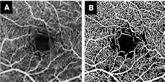
Adaptive Optics correction for detailed analysis of retinal structures
The retina is a highly complex tissue composed of multiple layers, each formed by distinct cellular and structural components. Adaptive Optics (AO) is a novel technique that lets us observe retinal structures that cannot be assessed in conventional images, such as cone photoreceptors, vessel walls, or the cells in the retinal pigment epithelium. AO also provides us a more detailed view of disease markers such as drusen, microaneurysms, or atrophic regions in the retina. AO has been applied frequently in age-related macular degeneration, hypertension, and diabetes; but there is a growing interest in glaucoma and in several inherited retinal dystrophies. Although the technology is mainly used in research settings, recent studies are collecting patient data at the clinic, in particular to monitor the effect of treatment, such as during genetic therapies.
AO offers exciting new avenues for looking at the retina, and a huge potential in detecting changes earlier and more accurately, which in turn can contribute to personalized treatment and better follow-up. However, to make use of these advantages, we need automatic analysis tools to measure specific image characteristics (number of cells, thickness of a vessel wall) so that small changes do not go unnoticed.

Figure 1. OCTA image of the superficial vascular plexus of the retina. (A) Original image, (B) Result of a model trained for automatic segmentation of the vasculature.
Figure 2. Adaptive Optics image of a healthy human retina. Adaptive Optics enables visualization of individual cones.
Analysis of specular microscopy images of the corneal endothelium
The cornea is the transparent, dome-shaped outer layer of the eye that helps focus light as it enters the eye, contributing to the eye's overall focusing power Its innermost layer, the corneal endothelium, has a regulatory function to maintain transparency, ensuring proper vision. Unlike other cells, corneal endothelial cells do not significantly regenerate and, if many of them are damaged, it will lead to consequences for our vision. Some cells are lost naturally with age, but there is also a number of factors such as diseases, direct injuries, or incorrect wear of contact lenses, which can cause alterations and cell loss. Furthermore, common surgeries, such as in cataract or glaucoma, are also known to affect corneal endothelial cells. Therefore, it is important to monitor them to ensure timely clinical intervention, preventing permanent damage.
Specular microscopy is an advanced imaging technique that provides a high-definition view of the corneal layers, including the endothelium. While these images are routinely acquired in clinical practice, and the acquisition device provides itself some basic measurement tools, these tools are inaccurate and show large discrepancies when we compare the number of automatically detected cells with manual counts by a human expert. Also, there are more structures that can be measured in the images, such as corneal guttae, edema, or lesions. These are especially important for tracking the progression of certain diseases, such as Fuchs' Endothelial Corneal Dystrophy.

Figure 3. Specular microscopy image of corneal endothelium of a patient. The prediction of an automatic model for guttata detection and the manual annotations of a human grader are overlapped on the image.
Expectations & Directions
EyeR's PIs have set a worldwide network of collaborators, counting among them clinical centres, AI experts, and companies. In the short term, the group is working with these collaborators to expand their current research lines, targeting multi-centre studies (collection of normative data of Adaptive Optics and OCTA), large research studies (collaboration with KU Leuven for the analysis of the Leuven Eye Study), population studies (analysis of the OCTA and AO ophthalmic data in the Maastricht Study), and real-world data from clinical settings (through the collaboration with the Rotterdam Eye Hospital). Furthermore, EyeR is pioneering the research in Adaptive Optics in the Netherlands, leading a collaboration with UMC Maastricht and Radboud University on the topic. Finally, EyeR has an interest in expanding the posibilities of ophthalmic imaging further than the scope of ophthalmology and, in particular, in linking retinal and cardiovascular health.
Funding
Van Romunde, Saskia, and Danilo Andrade De Jesus Stichting Wetenschappelijk Onderzoek het Oogziekenhuis: ‘Adaptive optics in patiënten na netvliesloslating’. 2023-2024
Derks, Lizanne, Jarinda Poppe, Angela Arends -Tjiam, Rob Taal, Danilo Andrade De Jesus , and Luisa Sanchez Brea Wishdom Foundation: ‘Voorspellen van ernstige prematurenretinopathie met behulp van kunstmatige intelligentie: uitbreiding en klinische validatie van een werkend model’. 2023-2026
Van Walsum , Theo , Caroline Klaver, Nicolas Chateau, Danilo Andrade De Jesus , and Luisa Sanchez Brea Health Holland TKI Call: ‘AO-Vision -- Adaptive Optics imaging: A guiding star to save vision’. 2022-2026
Invited Lectures
Danilo Andrade De Jesus. ‘AI for geeks: from basic principles to application in Glaucoma’. Portuguese Society of Ophthalmology, Lisbon, Portugal. March 2024.
Danilo Andrade De Jesus. 'Fast protocol workflows for prospective clinical studies'. AO retinal imaging expanded & streamlined webinar, online. July 2024.
Danilo Andrade De Jesus. ‘Standardization and good practices for AO imaging’. RTX1 users e-symposium, online. Oct 2024.
Luisa Sanchez Brea. ‘Automated Photoreceptor Detection in AO-FIO images’. RTX1 users e-symposium, online. Oct 2024.
Luisa Sanchez Brea . ‘Cutting edge automated quantification of photoreceptors in adaptive optics imaging’. ERNEYE Scientific Workshop, Madrid, Spain. Nov 2024.
Highlights
Luisa Sanchez Brea and Danilo Andrade De Jesus became Assistant Professors on 01-05-2024.
The BSc project "A model to distinguish vestibular migraine from other vertigo disorders based on characteristics of the hippus" by Joep de Waart, Eline Meulepas, Wouter van Wijck, Fleur Leenheer won the Medical Delta KTO-WOW Award.
Additional Personnel
Aniek Sips – MSc thesis
Afonso Pedrosa – MSc thesis
Aaron Sam – MSc thesis
Sander Wooning – MSc thesis
Leticia Lessa – MSc thesis
Catarina Carvalho – MSc thesis
Ophelia Urbing – MSc internship
Killian Perrin – MSc internship
Sebastian Koninkx – MSc internship
Noud van Ruremonde – MSc internship
Sem Hennekam – MSc internship
Joep de Waart, Eline Meulepas, Wouter van Wijck, Fleur Leenheer – BSc thesis (KTO group)
Donna de Leur, Laura Verdujin, Magriet Zwaal, Mehak Katarya – BSc thesis (KTO group)
Piotr van Dijk, Lisanne de Bruin, Isis Hendriks, Bernadet Aantjes – BSc thesis (KTO group)


Focus Area
MOLECULAR IMAGING & THERAPY
The aim is to study molecular and cellular events in a non-invasive manner and to develop new treatment modalities for cancer. The research focuses on the development of contrast agents, reporter genes, radiopharmaceuticals and multimodality agents for MRI, optical and/ or radionuclide imaging and therapy, as well as their functioning within the cell and/or whole organism, their preclinical validation, and their clinical translation, to ultimately improve the cure rate and quality of life of patients.
JOINT APPOINTMENT MOLECULAR GENETICS
Julie Nonnekens received her MSc in Biotechnology at Wageningen University in 2009 and completed her PhD in cancer biology, focusing on DNA repair mechanisms, at the University of Toulouse (France) in 2013. Following, she was a postdoc at the Hubrecht Institute working on ribosome biogenesis in cancer and longevity. Since 2014, Julie has been at Erasmus MC, holding a dual appointment in Radiology & Nuclear Medicine and Molecular Genetics. Her research integrates DNA damage repair and

nuclear medicine to explore the radiobiology of targeted radionuclide therapy, aiming to optimize treatment strategies.
Julie has received multiple recognitions, including young investigator awards, and serves as principal investigator on prestigious grants, such as the ERC starting grant. She chairs the Netherlands Society of Radiobiology and co-founded the European working group on Radiobiology of Molecular Radionuclide Therapy. Her work fosters interdisciplinary collaboration to advance cancer treatment through innovative approaches in radiobiology and nuclear medicine. j.nonnekens@erasmusmc.nl
RADIOBIOLOGY OF RADIONUCLIDE THERAPY
Julie Nonnekens, PhD
associate professor

Context
Targeted radionuclide therapies (TRT) are transforming the treatment of metastasized cancers by delivering radiolabeled compounds directly to tumor cells via specific receptors. Once bound, the radionuclides emit ionizing radiation, causing DNA damage leading to cancer cell death. Despite growing clinical application, challenges remain, as some patients experience over-treatment (toxicity) or under-treatment (insufficient tumor regression), emphasizing the need for optimization. Advancing our understanding of the radiobiology of TRT – the biological effects of ionizing radiation – and integrating this knowledge into improved dosimetric models are critical steps toward enhancing therapeutic outcomes. By providing evidence to guide treatment methods and regimens, these efforts aim to refine TRT approaches, reducing toxicity and improving efficacy.
Our overarching goal is to bridge radiobiological research with clinical practice, driving the evolution of TRT from a primarily palliative to a potentially curative treatment. For more information about our research, visit www.nonnekenslab.com.
Top Publications 2024
Reuvers TGA, V Grandia, RMC Brandt, M Arab, SLN Maas, EM Bos, J Nonnekens. Investigating the Radiobiological Response to Peptide Receptor Radionuclide Therapy Using Patient-Derived Meningioma Spheroids. Cancers (Basel) 2024; 16:2515.
Cheng J, J Zink, E O'Neill, B Cornelissen, J Nonnekens, L Livieratos, SYA Terry. Enhancing [177Lu]Lu-DOTATATE therapeutic efficacy in vitro by combining it with metronomic chemotherapeutics. EJNMMI Res. 2024; 14:73.
Dijkstra BM, QCF Cordia, J Nonnekens, GJ Meersma, VS Donthu, WB Nagengast, S Kruijff, WFA den Dunnen, FAE Kruyt, RJM Groen. Bevacizumab-IRDye800CW for tumor detection in fluorescence-guided meningioma surgery (LUMINA trial): a single-center phase I study. J Neurosurg. 2024; 5:1-12.
Research Projects: Objectives & Achievements
Cellular effects of TRT in tumor cells
Our research focuses on radiolabeled compounds, particularly those using the β -emitter lutetium-177, such as somatostatin analogue DOTA-(Tyr3)octreotate ((177Lu)Lu-DOTA-TATE) for neuroendocrine tumors (NET) and prostate-specific membrane antigen (PSMA) compounds ((177Lu)Lu-PSMA) for prostate cancer (PCa). Lutetium-177’s β -particles induce DNA damage, leading to tumor cell death with minimal harm to healthy tissues, significantly improving progression-free survival and quality of life. However, further insights into local treatment effects are essential for optimization.
To enhance understanding, we analyze the DNA damage response (DDR) and immune response induced by TRT in cell lines, ex vivo tumor slices, and xenografted mice using live-cell imaging, molecular biology, and histology. We have demonstrated diverse DNA damage patterns in tumor and normal tissue cells. Additionally, RNA expression analysis, drug screens, and CRISPR-Cas9 editing are helping elucidate the broader cellular responses. A collaborative project with Dr. Sophie Veldhuijzen van Zanten focuses on identifying TRT strategies for pediatric neuro-oncology.
• Tumor radiobiology of NET TRT ( Danny Feijtel, Pleun Engbers, Joke Zink, Giulia Tamborino, Tijmen de Wolf )
• Tumor cell radiobiology of PCa TRT ( Mariangela Sabatella, Txema Heredia Genestar )
• Pathway activation analysis of NET TRT ( Thom Reuvers, Mariangela Sabatella )
• Immune responses activated by TRT ( Justine Perrin, Rob Verhagen)
• Radiobiological assessment of blood of NET TRT patients ( Nina Becx, Renata Brandt)
• Novel TRT options for pediatric neuro oncology ( Nina Overdevest )
Radiobiology and dosimetry of different radiation qualities
Beyond lutetium-177, we study other radionuclides such as terbium-161 ( β - and conversion electron emitter), actinium-225 ( α -emitter), holmium-166, and yttrium-90 (both β -emitters). These radionuclides vary in decay type, half-life, and range, influencing their cellular effects. In collaboration with Dr. Erik de Blois, we compare their radiobiological effects through in vitro studies and in silico dose simulations to match radionuclides with clinical indications effectively.
Accurate dosimetry remains a challenge in TRT. With Dr. Mark Konijnenberg, we are developing advanced dosimetric models to predict biological responses based on (micro)dosimetric quantities. These models are vital for understanding dose effects on diverse cellular targets and heterogeneous tumor regions, paving the way for their integration into TRT treatment planning systems.
• Live cell imaging of DNA repair dynamics by TRT ( Pleun Engbers, Tijmen de Wolf, Justine Perrin, Rob Verhagen)
• Radiobiological comparison of lutetium-177 and actinium-225 for PCa TRT ( Mariangela Sabatella )
• Radiobiological comparison of lutetium-177 and terbium-161 ( Mariangela Sabatella, Joke Zink)
• Micro- and macrodosimetry of TRT ( Giulia Tamborino )
• Automated image analysis methods for TRT fluorescent microscopy ( Tijmen de Wolf )
• Radiobiological comparison of holmium-166 and yttrium-90 for radioembolization ( Justine Perrin )
• Radiobiological comparison of non-targeted radionuclides in 2D systems ( Duschka Kleijn )

Figure 1. Different types of radionuclides being used in our research and a schematic scheme on DNA damage induction.

Figure 2. Analysis of the dose-effect relationship. Linking biological effects to absorbed dose calculated using dosimetric simulations.
Radiosensitization to improve radionuclide therapy outcome
Our work highlights that TRT efficacy can be enhanced by combining it with radiosensitizing compounds. Specifically, DDR inhibitors impair DNA repair mechanisms, amplifying TRT-induced cell death. For instance, combining TRT with the PARP-1 inhibitor olaparib for NET tumors has led to clinical trials globally, including our phase I trial in collaboration with Dr. Hans Hofland.
We also explore additional radiosensitizers, such as DNA-PKcs and HSP90 inhibitors. DNA-PKcs inhibition has shown a significant increase in therapeutic efficacy without detectable toxicity in preclinical models. We are now investigating the mechanisms behind HSP90’s radiosensitizing properties to refine these combinatory regimens further.
• Preclinical radiosensitization to improve TRT outcome ( Thom Reuvers , Danny Feijtel, Mariangela Sabatella, Renata Brandt, Txema Heredia Genestar )
• Clinical phase 1 trial of NET TRT in combination with PARP inhibitors ( Nina Becx, Renata Brandt)
Expectations & Directions
Our team combines cutting-edge technological and radiobiological expertise to enable the clinical implementation of optimized therapeutic strategies. Our research aims to deepen understanding of the radiobiological effects of TRTs, a field with significant knowledge gaps. By uncovering the mechanisms underlying TRT-induced cellular effects, we aim to build a foundation for novel research opportunities, as current efforts only scratch the surface of TRT radiobiology.
Future directions will expand on key areas, such as how the physical and biological properties of radiolabeled compounds influence radiation dose delivery and treatment outcomes. Additionally, we will explore the role of the tumor microenvironment and systemic responses in shaping TRT efficacy. These insights will not only improve our understanding of TRT but also facilitate the development of tailored therapeutic regimens, moving closer to personalized and curative cancer treatment.
Funding
Nonnekens, Julie KWF Young Investigator Grant 2018: 'A radiant future: Improving targeted radionuclide therapy through modulation of DNA damage in the tumor'. 20192024
Nonnekens, Julie Erasmus MC Fellowship 2019: 'RADIANT: cellular RADIAtion exposure effects of molecular radioNuclide Therapies'. 2020-2024
Kanaar, Ronald, Julie Nonnekens, Hans Hofland, Ferry Eskens, Wouter de Herder, Tessa Brabander, Astrid van der Veldt, Mark Konijnenberg, and Stijn Koolen Oncode clinical proof of concept study: ‘Improving Peptide Receptor Radionuclide Therapy with PARP inhibitors: the PRRT-PARPi study’. 2021-2025
Nonnekens, Julie Investigator initiated research collaboration with Quirem Medical, Terumo: 'Radiobiological effects of holmium-166 and yttrium-90'. 2022-2024
Nonnekens, Julie Investigator initiated research collaboration with POINT Biopharma: 'Radiobiology of alpha and beta-PSMA targeted radionuclide therapy'. 2022-2024
Nonnekens, Julie ERC starting grant 2021: 'RADIOBIO: Deciphering the radiobiology of targeted radionuclide therapy: from subcellular to intra-tumoural analyses'. 2022-2027
Veldhuijzen van Zanten, Sophie, and Julie Nonnekens Research grant Cure Starts Now Foundation: 'Development and optimization of targeted radiopharmaceutical therapies for pediatric brain tumors; a world-first translational study'. 2023-2027
Perrin, Justine MSCA postdoctoral fellowship: 'Impact of BRCA2 deficiency on the DNA damage response and immunogenicity of prostate cancer after radioligand therapy'. 2024-2026
Nijsen, Frank, Julie Nonnekens, Sandra Heskamp, Antonia Denkova, and consortium partners NWO Perspectief consortium: 'Understanding the radiobiology of therapeutic medical radionuclides'. 2023-2028
Invited Lectures
Giulia Tamborino. 'Microdosimetry and biological effects'. Austrian Society of Nuclear Medicine, Bad Ischl, Austria. Jan 2024.
Justine Perrin. 'Radionuclide therapy: dose-effect relationship, focus on radiobiology and immunology'. Seminar at CRCINA, Nantes, France. Feb 2024.
Julie Nonnekens. 'PRRT mechanisms of action: known and unknown'. European Neuroendocrine Tumor Society (ENETS) annual conference, Vienna, Austria. March 2024.
Julie Nonnekens. 'Research and clinical trials: radiobiology'. Innovation Mission Nuclear Medicine & Medical Isotopes, London, UK. March 2024.
Justine Perrin. 'Evaluate the dose-dependent effect between radionuclide therapy and the immune response'. NVRO/NVRB scientific meeting, Amersfoort, the Netherlands. March 2024.
Julie Nonnekens. 'Radiobiologie: potentie voor verbetering van PRRT'. Nuclear medicine symposium, Delft, the Netherlands. April 2024.
Giulia Tamborino. 'Introduction to dosimetry for radiopharmaceutical development and treatment optimization'. NKRV symposium, Delft, the Netherlands. June 2024.
Julie Nonnekens. 'Stralingsbiologie, essentieel voor het begrijpen van de dosis-effect relatie bij radio(nucliden)therapie'. Future of medical imaging and radiotherapy (FMIR) –annual congress, Almere, the Netherlands. June 2024.
Julie Nonnekens. 'Radiobiology of radionuclide therapy'. Erasmus MC School of Lu-177 based radiopharmaceutical therapy, Rotterdam, the Netherlands. June 2024.
Julie Nonnekens. 'Radiobiology in radionuclide therapy: necessity when determining the dose-effect relationship'. European Federation of Organisations for Medical Physics (EFOMP) scientific meeting, online. June 2024.
Giulia Tamborino. 'Biologically driven dosimetric modelling: bridging the gap between radionuclide therapy and radiotherapy'. EFOMP scientific meeting, online. June 2024.
Julie Nonnekens. 'Radiobiology: dose-effect relationship of radionuclide therapy'. Gordon Research Conference –Radionuclide Theranostics for the Management of Cancer, Maine, USA. July 2024.
Julie Nonnekens. 'Optimizing radionuclide therapy: unraveling radiobiological insights for enhanced treatment strategies'. Rudbeck Laboratory, University of Uppsala, Uppsala, Sweden. Sept 2024.
Julie Nonnekens. 'The promise of radionuclide therapy in medicine'. Dutch Medicines Days, Oss, the Netherlands. Oct 2024.
Julie Nonnekens. 'What is/could be the contribution of radiobiology to clinical practice?'. European Association of Nuclear Medicine annual congress, Hamburg, Germany. Oct 2024.
Julie Nonnekens. 'Radiobiological comparison of different radiation qualities: alpha vs beta vs external beam radiation'. Netherlands Society for Radiobiology annual meeting, Utrecht, the Netherlands. Nov 2024.
Justine Perrin. 'Interplay between radionuclide therapy efficiency and tumor microenvironment'. Targeted Radiopharmaceutical Summit, Amsterdam, the Netherlands. Dec 2024.
Julie Nonnekens. 'Radionuclide Therapy Improvement Using Radiosensitizers: Preclinical Studies & Future Outlook'. Targeted Radiopharmaceutical Summit, Amsterdam, the Netherlands. Dec 2024.
Highlights
January 31st 2024 Danny Feijtel successfully defended his PhD thesis entitled: ‘Improving therapeutic outcome of patients with neuroendocrine cancer: understanding both the target and the bullet’.
Nina Overdevest won the best poster award at the Erasmus MC PhD symposium 2024.
November 26th 2024 Thom Reuvers successfully defended his Phd thesis entitled: ‘Improving peptide receptor radionuclide therapy outcomes: a radiobiological approach'.
Additional Personnel
Renata Brandt – Research technician
Rob Verhagen – Research technician
Joke Zink – Research technician
Samuel Odro – intern, 4th year BSc student Biologie en medische laboratoriumonderzoek, Avans Hogeschool Breda. Feb – June 2024. Daily supervisor Nina Overdevest.
Michelle Valk – intern, 3rd year BSc student Nanobiology, Erasmus University and TU Delft. Feb – July 2024. Daily supervisor Pleun Engbers.
Vincent Ribbe – intern, 3rd year BSC student Nanobiology, Erasmus University and TU Delft. Feb – July 2024. Daily supervisor Tijmen de Wolf.
Julian van Gerwen – intern, 2nd year MSc student Molecular life sciences Wageningen University. Feb – Sept 2024. Daily supervisor Mariangela Sabatella.
Daria Roman – intern, 1st year MSc student Nanobiology, Erasmus University and TU Delft. June – Nov 2024. Daily supervisor Tijmen de Wolf.
Doris Houtzager – intern, 2nd year MSc student Nanobiology, Erasmus University and TU Delft. Sept 2024 – June 2025. Daily supervisor Justine Perrin.
Stefan van Alen – intern, 2nd year MSc student Nanobiology, Erasmus University and TU Delft. Sept 2024 – June 2025. Daily supervisor Pleun Engbers and Tijmen de Wolf.
Ana Gebara Garcia de Lima – intern, 2nd year MSc student Molecular medicine, Erasmus University. Sept 2024 – June 2025. Daily supervisor Mariangela Sabatella.
Mimi Welte – intern, 3rd year BSc student University college, Erasmus University. Nov 2024 – May 2025. Daily supervisor Nina Overdevest.
Post-doc Post-docs

Justine Perrin, PhD
Project Funding Marie Curie Europe post-doctoral fellowship Email j.perrin@erasmusmc.nl
Impact of BRCA2 deficiency in TRT of prostate cancer
TRT has shown promising results in the treatment of metastasized prostate cancer. Prostate specific antigen (PSMA) overexpression in these cancers enables targeted therapy using a lutetium-177 radiolabeled peptide. The VISION phase III trial showed notable improvement in overall survival compared to standard care.
However, not all patients respond to TRT, highlighting the need for prediction of good responders early to avoid unnecessary side effects.
My research investigates the role of BRCA2 deficiency, a common feature in the most aggressive phenotype of prostate cancer. BRCA2 is critical for homologous recombination repair of DNA double-strand breaks, and its absence may serve as a biomarker for increased sensitivity to TRT.
My research focus on the impact of BRCA2 deficiency on TRT efficacy in prostate cancer cell lines, to identify if this deficiency makes cells more sensitive to TRT, and how it impacts the immunogenicity.
Mariangela Sabatella, PhD
Project Funding Investigator initiated industry project with POINT biopharma
Email m.sabatella@erasmusmc.nl
Radiobiology of alpha and beta-PSMA targeted radionuclide therapy
TRT uses radionuclides ( β - or α -emitters) linked to tumor-specific inhibitors. In metastasized prostate cancer (mPCa), prostate-specific membrane antigen (PSMA) is the main target. β -emitter-based PSMA-TRT has shown to improve overall survival in mPCa patients. However, not all patients respond to treatment, and some experience severe side effects, highlighting the need for improved therapies and novel PSMA inhibitors.
Recently, α -emitters have gained attention due to their ability to cause more complex and irreparable DNA damage, potentially enhancing therapeutic ef-


ficacy. Despite these promising characteristics, limited knowledge exists about the type of DNA damage induced by β - or α -emitters and the DNA damage response (DDR) mechanisms that tumor cells activate to survive the therapy.
My research focuses on unraveling the molecular mechanisms underlying the effects of β - and α -emitters in PSMA-TRT and identifying DDR targets. This knowledge may lead to combination therapies with DDR inhibitors, improving outcomes for mPCa patients.
Giulia Tamborino, PhD
Project Funding ERC Starting Grant 2021: “Deciphering the radiobiology of targeted radionuclide therapy: from subcellular to intra-tumoural analyses”
Email g.tamborino@erasmusmc.nl
Cellular Radiation exposure effects of molecular radioNuclide Therapies
TRT offers a promising treatment for systemic cancers, but its effectiveness can be enhanced by tailoring treatment to individual patient needs. Dosimetry, or radiation dose calculations, plays a crucial role in optimizing radiation-based treatments by customizing regimens to reduce toxicity and improve tumor responses. However, TRT’s radiobiological mechanisms differ from traditional external beam radiotherapy (EBRT), particularly at low absorbed doses or low dose rates. Understanding these differences at the cellular and molecular levels is essential for advancing TRT.

To improve current dosimetry methods, we aim to develop accurate computational models that correlate microdosimetry with biological effects in both in vitro and in vivo settings.
The creation of such computational frameworks and the investigation of the radiobiology of dose-response in TRT in cells and in small animals can ultimately lead to a better understanding of this treatment modality and to increase the predictive power of dosimetry.

Txema Heredia Genestar, PhD
Project Funding NWO Perspectief project: “UNderstanding the RAdiobiology of therapeutic medical radioNUclides (UNRANU)”
Email j.herediagenestar@erasmusmc.nl
Leveraging bioinformatics to understand the response to TRT
TRT is a promising treatment for patients with metastasized cancers. TRT comes in many forms, with differences in radioligand, isotope, and type-and-energy of the emitted radiation. The project aims to systematically characterize the effects of different radionuclides across different tissues and tumor types. Multiple Dutch institutes collaborate in the study of TRT’s effects in cell cultures, organoids, and the interactions with the immune system in in vivo studies.
Our group focuses on the in vitro effects of TRT in cell culture as well as the analyses of cellular response using bioinformatic tools. We aim to explore how cells
PhD Students

Advisors Julie Nonnekens, Hans Hofland & Roland Kanaar
Project Funding Oncode
Email m.becx@erasmusmc.nl
Improving Peptide Receptor Radionuclide Therapy with PARP inhibitors
Peptide receptor radionuclide therapy (PRRT) with the beta-emitting radiopharmaceutical 177LuDOTATATE is an effective and safe treatment option for patients with metastatic neuroendocrine tumors (NETs). Response rates are still limited, so there is a need for improvement. The PARP inhibitor olaparib has the potential to improve this treatment because of its radiosensitizing effects.
respond to therapy short after treatment and how the different radiation types affect the cellular repair pathways involved in overcoming DNA damage.
When observing cells under the microscope, we can only look at a limited number of proteins at once. However, bioinformatic tools allow us to explore the larger transcriptomic response, in hope to find targets that will improve therapy.

Advisors Julie Nonnekens, Erik Verburg & Mariangela Sabatella
Project Funding ERC Starting Grant: “RADIOBIO: Deciphering the radiobiology of targeted radionuclide therapy: from subcellular to intra-tumoural analyses”
Email p.engbers@erasmusmc.nl
Subcellular & intra-tumoural radiobiology of TRT
In this project, we aim to identify biological parameters that are important in the radiation effect of TRT. In addition, we perform live cell imaging in vitro and in vivo, to determine subcellular and intratumoral localiziation of 177Lu-DOTATATE. Finally, we aim to evaluate DNA damage induction and repair kinetics after TRT.
Nina Becx, MSc
Pleun Engbers, MSc

Danny Feijtel, MSc

Advisors Julie Nonnekens, Roland Kanaar & Erik Verburg
Project Funding Daniel den Hoed fellowship and EUR fellowship
PhD Obtained 31-01-2024
My project focusses on deepening the current understanding of both molecular and radiobiological principles in the fight against neuroendocrine tumors (NETs). We use a variety of in vitro and in vivo systems to study the effects of radionuclide therapy on NETs, the possibility of NET radiosensitization using additional chemotherapeutics, and look at genetic mutations and their effect on DNA maintenance to uncover new NET Achilles’ heels. These results will benefit fundamental, translational and, potentially, clinical studies.
Tijmen de Wolf, MSc
Advisors Julie Nonnekens, Ihor Smal, & Roland Kanaar
Project Funding ERC Starting grant. “Deciphering the radiobiology of targeted radionuclide therapy: from subcellular to intra-tumoural analyses“
Email t.dewolf@erasmusmc.nl
Image analysis for PRRT
This project aims to understand the radiobiology of peptide receptor radionuclide therapy (PRRT). Live cell microscopy is used to capture the dynamics of the radionuclides and DNA damage over time. Image analysis is an essential step required for the quantification. We develop novel algorithms to improve and automate the analysis.

Thom Reuvers, MSc

Advisors Julie Nonnekens, Roland Kanaar, Erik Verburg & Eelke Bos
Project Funding KWF Young Investigator Grant: “A radiant future: Improving targeted radionuclide therapy through modulation of DNA damage in the tumor
PhD Obtained 26-11-2024
Improving PRRT by DNA damage repair modulation
In this project we try to understand the cellular response to TRT as compared to external beam radiotherapy and use this knowledge to develop combination therapies with compounds aimed at DNA repair. In addition, we are developing patient derived 3D culture models to test TRT preclinically.

Duschka Kleijn, MSc

Advisors Julie Nonnekens, Roland Kanaar & Justine Perrin
Project Funding NWO perspectief grant: “Understanding the radiobiology of therapeutic medical radionuclides” (UNRANU)
Email d.kleijn@erasmusmc.nl
In vitro 2D radiobiological effects of radionuclides with different physical properties
In this project, we aim to map the direct radiobiological response of various liver and prostate cancer cells to a set of radionuclides in vitro. We will specifically examine tumor cell survival, DNA damage repair capacity, and the activation of specific cellular pathways.


Nina Overdevest, MSc LinkedIn
Advisors Julie Nonnekens, Sophie Veldhuijzen van Zanten & Justine Perrin Project Funding 2022 DIPG/DMG Collaborative and Cure Starts Now grant Email n.overdevest@erasmusmc.nl
Development and optimization of targeted radiopharmaceutical therapies for pediatric brain tumors; a world-first translational study
Currently, there is a lack of effective treatments available for pediatric medulloblastoma and diffuse intrinsic pontine glioma/diffuse midline glioma (DIPG/DMG). Therefore, the goal of this project is to determine through preclinical research whether targeted radionuclide therapy (TRT) is a potent treatment option for medulloblastoma and DIPG/DGM.
Simone Dalm has a BSc in Biomedical science and a MSc in Oncology, which she obtained at the VU University in Amsterdam. In 2012 she started her PhD at the department of Nuclear Medicine of the Erasmus MC and in 2017 she graduated cum laude. She then continued with a postdoc at the same department (by that time the department of Radiology and Nuclear Medicine had joined forces). During her postdoc she established her own research line and started her research group: The Radiotracer Interactions Group. In September 2019 she was promoted to assistant professor.

She received multiple personal and industry sponsored grants (e.g. KWF Young Investigator’s Grant and ZonMw Veni) and is involved in several national and international research projects. She also received multiple awards for her achievements such as the Editors’ choice award of the Society of Nuclear Medicine and Molecular Imaging, multiple Alavi Mandell awards and the Research Prize of the Erasmus University. Her scientific interests include molecular biology, targeted therapy, and nuclear imaging and therapy.
s.dalm@erasmusmc.nl
RADIOTRACER INTERACTIONS
Simone Dalm, PhD
assistant professor

Context
Target-mediated radionuclide imaging and treatment are anti-cancer interventions successfully applied in the clinic. For this, radiotracers are applied directed against molecules that are overexpressed on tumor cells. Depending on the radionuclide connected, the same tracer can be used for imaging (ß+ and γ -emitting radionuclides) and treatment (ß- and α -emitting radionuclides). Our studies focus on (but are not limited to) the application of radiotracers targeting the gastrin releasing peptide receptor (GRPR) (overexpressed on e.g. prostate- and breast cancer), the prostate specific membrane antigen (PSMA) (overexpressed on e.g. prostate cancer and the neovasculature of various solid tumors), the somatostatin receptor subtype 2 (SSTR2) (overexpressed on neuroendocrine- and breast cancer) and fibroblast activation protein (FAP) expressed on cancer associated fibroblasts (CAFs). The latter are present in the tumor stroma of ~90% of all solid cancers.
Our objective is to develop and evaluate novel radiotracers and application strategies, and to optimize the use of radiotracers in order to achieve more cure, fewer side effects and a better quality of life for cancer patients. This includes studies to identify patient groups best suited for application of a specific radiotracer, the development and the application of novel strategies to improve tumortargeting and minimize off-target organ toxicity, and studies to better understand the mechanism of action of radiotracers. To achieve this, we unravel the interaction of radiotracers with the human body on a molecular, cellular, tissue, organ, organ-system and patient level.
Top Publications 2024
Gaspar N, M Handula, MCM Stroet, K Marella-Panth, J Haeck, TA Kirkland, MP Hall, LP Encell, S Dalm, C Lowik, Y Seimbille, L Mezzanotte. A novel luciferase-based reporter gene technology for simultaneous optical and radionuclide imaging of cells. Int J Mol Sci. 2024; 25:8206.
Van der Heide CD, H Ma, MWH Hoorens, JD Campeiro, DC Stuurman, CMA de Ridder, Y Seimbille, SU Dalm. In vitro and in vivo analyses of eFAP: A novel FAP-targeting small molecule for radionuclide theranostics and other oncological interventions. EJNMMI Radiopharm Chem. 2024; 9:55.
Dalm S, H Duan, A Iagaru. Gastrin releasing peptide receptors-targeted PET diagnostics and radionuclide therapy for prostate cancer and management: Preclinical and clinical developments of the past 5 years. PET Clin. 2024; 19:401-415.
Research Projects: Objectives &
Achievements
Personalized treatment
For targeted radionuclide imaging and therapy to be successful high expression level of the target in the tumor is a prerequisite. Next to this, whether the target is expressed by the tumor cells themselves or by surrounding cells present in the tumor microenvironment, and how target expression is distributed (heterogeneous vs homogeneous), are also important aspects determining the success of these interventions. Furthermore, during cancer development and progression the expression of the target can vary, and additionally, different cancer subtypes can have different molecular characteristics, including different target expression levels. Additionally, it is most probable that novel developed targeted radionuclide treatments will be applied in late stage diseases. In this case patients have often been treated with other types of anti-cancer treatments such as hormone therapy, chemotherapy or a combination of the two. These treatments can influence target expression as well as radiosensitivity of cancer cells, the latter being another key component for successful radionuclide treatment. In our studies, we evaluate the expression level and distribution of novel targets in healthy tissues and during the development and progression of cancer. In addition, we also study how other anti-cancer drugs can affect target expression and radiosensitivity of cancer cells. Ultimately our goal is to identify specific patient groups, with respect to disease stage, disease subtype and treatment history, that can benefit from specific targeted radionuclide imaging and treatment strategies. See Figure 1.

Figure 1. Target expression and radiosensitivity can vary between different disease stages and as a consequence of prior treatment. Identification of patient groups best suited for specific target-mediated imaging and therapy strategies is essential for the success of these interventions.
Projects: – PSMA vs GRPR in prostate cancer progression ( Marjolein Verhoeven )
– The effect of treatment history on the success of GRPRdirected radionuclide therapy ( Tyrillshall Damiana ) – GRPR expression in relation to pathological and clinical properties of prostate cancer ( Tyrillshall Damiana )
Novel therapeutic strategies, combination treatment and the tumor stroma
Although targeted radiotracers are successfully applied clinically, complete response in patients is rare. Moreover, since healthy organs can also express the respective target and because of the physiological clearance of the radiotracers, healthy organs are often also exposed to radiation which can cause side effects. The above indicates that there is a need for novel developments to increase the efficacy and safety of targeted radionuclide treatment. In line with this, our focus is to develop novel and improved therapeutic strategies for targeted radionuclide treatment of cancer. This includes the development of novel radiotracers (Figure 2), the development of methods to improve tumor-to-background ratio of radiotracer uptake, targeting the tumor stroma, combining radionuclide therapy with other anti-cancer therapies or with strategies to sensitize cancer cells to radionuclide therapy, and tandem radionuclide/radiotracer treatment strategies.
Projects:
– Novel developments for GRPR-targeted theranostic interventions (Marjolein Verhoeven)
– Development and evaluation of NTS1-targeting radiopharmaceuticals (Tyrillshall Damiana)
– Targeting FAP-expressing CAFs in the tumor stroma for radionuclide theranostics (Circe van der Heide)
– Targeting proteoglycans with radiolabeled cell-penetrating peptides: a novel approach for detection and treatment of cancer (Joana Campeiro)
– Tandem GRPR- and PSMA-targeting radionuclide treatment for prostate cancer management (Lisa Bokhout)
– Novel biomarkers for targeting neuroendocrine prostate cancer (Lisa Bokhout)
– The combination of radioembolization and immunotherapy (Debra Stuurman - in collaboration with Dr. Vegt and the department of Gastroenterology and Hepatology)
– Novel developments for GRPR-targeted theranostic interventions (in collaboration with Dr. Seimbille and the department of Medical Oncology)
– Targeted radionuclide theranostics for sarcomas (in collaboration with Dr. Veldhuijzen van Zanten and the department of Radiology & Nuclear Medicine of the LUMC)

Figure 2. Targeting highly sulfated heparan sulphate proteoglycans (HSPGs) on triple negative breast cancer (TNBC). Compared to healthy tissues and other breast cancer subtypes, TNBCs have an increased expression of highly sulphated HSPGs on their cell surface. We aim to exploit this aberrant expression pattern to develop a targeted strategy for radionuclide imaging and treatment of TNBC. Crotamine, a cell penetrating peptide, has shown high affinity to highly sulfated HSPGs and therefore provides the ideal basis for exploring this strategy. ER=estrogen receptor, PR=progesterone receptor, HER2=human epidermal growth factor receptor 2.
Understanding the mechanism of action of radiotracers
In another attempt to improve the efficacy and safety of targeted radionuclide therapy, we aim to gain more understanding of the mechanisms of action of radiotracers. The obtained knowledge is subsequently used to develop novel strategies to improve the therapeutic index of radionuclide therapy. Examples include comparing radiotracers with agonistic and antagonistic properties, studying the difference in binding of radiotracers to cancer cells vs binding to healthy cells with physiological expression of the target, developing more accurate models for studying radiotracers (e.g. 2D vs 3D models) (Figure 3), and studying the effect of radiotracer treatment on the immune system and cancer immunity. The ultimate goal of these studies is to use the obtained knowledge to develop strategies to improve targeted radionuclide imaging and treatment.
Projects:
– Development of a clinically representative co-culture model for FAP-targeted radionuclide therapy ( Circe van der Heide )
– Unravelling resistance to PSMA-targeted radionuclide therapy in advanced prostate cancer patients ( Joana Campeiro )
– 2D vs 3D models for preclinical radiopharmaceutical evaluation ( Ilva Klomp )
– The role of the tumor microenvironment in targeted radionuclide therapy responses ( Ilva Klomp )

Figure 3. 2D vs 3D cell culture for studying radiotracers. Amongst other factors, the increased cell-to-cell interactions and the recapitulation of the tumor microenvironment in 3D models offers a more representative and accurate system for radiotracer evaluation.
Expectations & Directions
Our ultimate goal is to develop and improve novel therapeutic strategies using targeted radiotracers in a personalized setting to achieve more cure, fewer side effects and a better quality of life for cancer patients (Figure 4). We aim to achieve this by introducing novel radiotracers and application strategies for cancer imaging and treatment into the clinic. This also includes combination with other anti-cancer treatments and combinations of radionuclides. In addition, we will keep performing studies to better understand the interaction between radiotracers and their target at different levels of organization in the human body. This will provide opportunities to develop and apply novel radiotracers and application strategies.

Figure 4. Overview of research goals.
Funding
Klomp, Ilva NWO ENW XS: ‘Identifying the tumor stroma as a key player in resistance to internal radiation treatment’. 2024-2025
Campeiro, Joana NWO ENW XS: ‘Tackling triple-negative breast cancer: Development of a novel nuclear medicinebased strategy to create a personalized medicine approach for a biomarker-negative cancer type’. 2024-2025
Dalm, Simone Daniel den Hoed Award: ‘Taking Prostate Cancer Theranostics To The Next Level: PSMA- and GRPRtargeted Tandem Radionuclide Therapy for More Cure and Less Side Effects’. 2022-2026
Dalm, Simone Commercial collaboration Ratio Therapeutics: ‘Long circulating FAP tracers’. 2022-2025
De Jong, Marjon, and Simone Dalm Erasmus MC Mrace: ‘Breaking the tumour stroma barrier: A new way to hit cancers using a novel universal targeted radionuclide therapy strategy’. 2020-2024
Dalm, Simone Veni ZonMw: ‘Better understanding leads to better decisions: Evaluating the effect of anti-hormone therapy and chemotherapy on GRPR-targeting'. 2019-2024
Dalm, Simone KWF Young Investigator Grant/Bas Mulder Award: ‘Click on Target: Developing a safe drug with enhanced therapeutic potential for prostate cancer treatment’. 2018-2024
Highlights
Simone Dalm was a jury member for the Sanjiv Sam Ghambir Young Investigator Award.
Simone Dalm became associate editor for the British Journal of Radiology.
Simone Dalm was guest editor for EJNMMI reports, Theme: Role of Radiopharmaceutical Agents in Cancer Theranotics.
Circe van der Heide won the EANM Young Investigator Award.
Additional Personnel
Lilian van de Brink – BSc, Research Technician
Lisette W. de Kreij-de Bruin – MSc, Research Technician
Debra Stuurman – Biotechnician
Corrina de Ridder – Biotechnician
Jason Zhang – Intern
Eloise Vermunt – Intern
Post-doc Post-docs

Targeting
Joana Campeiro, PhD
Email j.campeiro@erasmusmc.nl
proteoglycans with radiolabeled cell-penetrating

peptides: a novel approach for detection and treatment of cancer
Triple-negative breast cancer (TNBC) is the most malignant breast cancer subtype with (extremely) poor prognosis, despite treatment options such as surgery and chemo/radiotherapy. Proteoglycans (PGs) have shown to play a role in cell proliferation and invasive growth in different tumor entities, and their dysregulated and overexpression is associated with poor prognosis in TNBC. Currently, few anti-cancer therapies targeting PGs have been developed using antibodies or inhibitors, but the clinical translation of these compounds remains unsuccessful. Alternatively, we propose to exploit PGs as a target to actively and specifically deliver imaging and/ or cytotoxic radionuclides to TNBC cells for targeted

radionuclide theranostic (TRT) purposes. Cell-penetrating peptides (CPPs) have a high affinity for PGs expressed on the cancer cell membrane, making them ideal candidates for the proposed PG-targeting interventions.
Our aim is not only to generate and evaluate the first class of radiolabeled CPPs for PG-targeted TRT, but also to use the developed molecules to get a better understanding of the role of PGs in TNBC. Moreover, PGs are overexpressed on multiple other cancer types as well, and thus the proposed strategy can be beneficial for a wide range of cancers.
Ilva Klomp, PhD
Project Funding Ratio Therapeutics
m.j.klomp@erasmusmc.nl

The role of the tumor microenvironment in targeted radionuclide therapy responses
Targeted radionuclide therapy (TRT) is clinically applied for the treatment of metastasized prostate cancer and neuroendocrine tumors. TRT has proven its clinical value, however, response rates are variable and a significant number of patients do not respond to TRT despite qualifing for treatment based on target expression observed on nuclear scans. Thus, there is a clear and urgent need for strategies to improve treatment outcomes.
Next to target expression, the tumor cells' radiosensitivity is essential for cancers' response to TRT. Several studies have demonstrated that specific components of the tumor microenvironment (TME) can induce epitheli-
al-to-mesenchymal transition (EMT) in cancer cells. EMT is associated with an aggressive cancer cell phenotype, and, very important for TRT, radioresistance towards other forms of radiation therapy. The aim of our project is to unravel the exact role of EMT in TRT responses, with the ultimate goal of improving TRT outcomes by interfering with EMT-induced radioresistance.
In addition, to adequately study the role of the TME in TRT response, we aim to develop clinically relevant three-dimensional models and accompanying standard operation procedures for accurate analysis of the interaction between TRT, the TME and TRT response.
PhD Students

Marjolein Verhoeven, MSc

Advisors Simone Dalm & Frederik Verburg
Project Funding Erasmus MC Grant, KWF
Email m.verhoeven.1@erasmusmc.nl
PhD Obtained 14-5-2024
Novel developments for GRPR-targeted theranostic interventions
The gastrin-releasing peptide receptor (GRPR) is overexpressed on many solid cancers, making it an interesting target for radionuclide theranostics. We aimed to improve GRPR theranostics by (1) demonstrating the efficacy and safety of the radiopharmaceutical NeoB; (2) exploring a novel pre-targeting strategy; (3) developing probes for image-guided surgery; and (4) by providing information for optimal clinical positioning of GRPR-targeting (radio) pharmaceuticals.
Lisa Bokhout, MSc

Advisors Simone Dalm, Frederik Verburg & John Martens
Project Funding Daniel den Hoed Award
Email l.bokhout@erasmusmc.nl
Improving radiocuclide treatment for PCa
Prostate cancer (PCa) treatment with prostate specific membrane antigen (PSMA)-targeting radiotracers is effective, but comes with side effects. Other promising radiotracers, with a more favorable safety profile, for PCa targeted radionuclide therapy (TRT) are gastrin-releasing peptide receptor (GRPR)-targeting radiotracers. However the prevalence of GRPR is lower than that of PSMA. We therefore aim to improve TRT for PCa by studying the efficacy and safety of tandem PSMA- and GRPR-TRT by performing preclinical in vitro and in vivo studies in clinically relevant models.

Tyrillshall Damiana, MSc

Advisors Simone Dalm & Frederik Verburg
Project Funding Health Research and Development (ZonMw): Veni
Email t.damiana@erasmusmc.nl
The effect of treatment history on the success of targeted radionuclide therapy
Gastrin-releasing peptide receptor (GRPR)-targeted radionuclide treatment (TRT) has shown promising results in preclinical and initial clinical studies. However, little is known about the influence of treatment history on the success of GRPR-TRT. We aim to unravel the effect of priorly received chemotherapy and hormone therapy on GRPR-TRT, with a main focus on the effect on cancer cells' radiosensitivity and target expression.

Circe van der Heide, MSc

Advisors Simone Dalm & Frederik Verburg
Project Funding Erasmus MC MRACE grant
Email c.vanderheide@erasmusmc.nl
FAP-targeted radionuclide therapy
Fibroblast activation protein (FAP) is expressed by cancer-associated fibroblasts (CAFs) present in the stroma of 90% of solid tumors, making it a relevant biomarker for targeted interventions, with potential pan-cancer application. The aim of our project is to increase understanding of the interaction between FAP-targeted radiotracers, CAFs, and cancer cells, and to develop clinically relevant models to study and evaluate (novel) FAP-TRT strategies.


2022-present: Director, Cyclotron Rotterdam B.V.
2020-present: Professor of Translational Nuclear Medicine and consultant in Nuclear Medicine, Erasmus MC.
2016-2020: Professor of Experimental Nuclear Medicine and deputy head of the Department of Nuclear Medicine, Philipps-University Marburg and University Hospital Gießen-Marburg, Marburg, Germany.
2010-2016: Consultant in Nuclear Medicine and Assistant Professor of Nuclear Medicine, RWTH Aachen University Hospital, Aachen, Germany.
2008: PhD, Utrecht University, Utrecht, the Netherlands.
2005-2010: Training in nuclear medicine, University Medical Center Utrecht, Utrecht, the Netherlands, and University Hospital Würzburg, Würzburg, Germany.
1997-2004: Medical studies at the Catholic University Leuven, Leuven, Belgium, and Utrecht University, Utrecht, the Netherlands. f.verburg@erasmusmc.nl
TRANSLATIONAL NUCLEAR MEDICINE
Frederik A Verburg MD, PhD
full professor

Context
The discipline of nuclear medicine has a rich tradition of successful translational research at the Erasmus MC. Notable examples to come out of this tradition are the commercial imaging product Octreoscan® and more recently the radionuclide therapeutic agent Lutathera®. The preclinical research group of our department is world-renowned.
The convergence with the TU Delft offers exciting new opportunities to integrate technical research into e.g. detector technologies or novel isotopes as well as various developments in medical informatics, such as artificial intelligence.
In order to facilitate and optimize the integration of new preclinical, technical and multimodality imaging concepts into clinical research and the transfer of these concepts from research to clinical practice, the Department of Radiology and Nuclear Medicine has newly created the Chair of Translational Nuclear Medicine in 2020. The year 2024 was characterized by many activities aimed at further building up the research line with numerous PhDs starting within the line and associated lines from assistant- and associate professors. In September 2023, the new GE Starguide SPECT/CT camera was installed – the first of its kind in the Netherlands and one of the first in the world. This new, full-ring, 12-head, CZT detector based SPECT/CT camera offers completely new options for e.g. completely dynamic SPECT imaging and opens up new avenues of clinical SPECT/CT research. The optimal use of this novel technology is an ongoing focus of research in Translational Nuclear Medicine. In 2024, the tender process for two innovative, world-first elongated axial field-of-view PET/CT scanners was completed. These machines will be installed from 2025 onward and their deployment in clinical work will be another central focus of research in Translational Nuclear Medicine.
Top Publications 2024
Veenstra MMK, E Vegt, M Segbers, S Franssen, BG Koerkamp, FA Verburg, MGJ Thomeer. Intra-arterial PSMA injection using hepatic arterial infusion pump in intrahepatic cholangiocarcinoma: a proof-of-concept study. Eur Radiol Exp. 2024; 8:90.
Pruis IJ, FA Verburg, RK Balvers, AA Harteveld, RA Feelders, MW Vernooij, M Smits, SJCMM Neggers, SEM Veldhuijzen van Zanten. [18F]FET PET/MRI: An Accurate Technique for Detection of Small Functional Pituitary Tumors. J Nucl Med. 2024; 65:688692.
Cox CPW, T Brabander, E Vegt, QG de Lussanet de la Sablonière, LH Graven, FA Verburg, M Segbers. Reduction of [68Ga]Ga-DOTA-TATE injected activity for digital PET/MR in comparison with analogue PET/CT. EJNMMI Phys. 2024; 11:27.
Research Projects: Objectives & Achievements
Projects:
– Safety and efficacy of dosimetry-based individualized treatment planning of Ho-166 RE for the maximization of treatment effectiveness while minimizing toxicity ( Erik Vegt )
– FDG-PET/CT for the detection of gastric cancer metastases ( Erik Vegt )
– Radiolabeling of inhaled pulmonary medication delivery systems for PET/CT-based airway deposition assessment ( Jochem Wolfert )
– A novel terrestrial molluscan model for medical imaging ( Joep van de Sanden )
– [18F]Tetrafluoroborate PET/CT scan for effectively detecting thyroid cancer metastases ( Hannelore Coerts )
Objectives
• Establish internal and external cooperation between Erasmus MC Academic Centers of Excellence, the TU Delft and clinical and preclinical groups with the Department of Radiology and Nuclear Medicine to further developments in tracer-based medical imaging and radionuclide therapy.
• Introduce novel tracers into clinical research and clinical practice for radionuclide imaging and therapy.
• Introduce novel radionuclides into clinical research and clinical practice.
• Establishment of novel imaging techniques such as PET/MRI, very high resolution dynamic SPECT/CT in clinical research and clinical practice.
• Establish novel non-vertebrate animal models to further improve the process of translational tracer development.
Achievements
• The Chair of Translational Nuclear Medicine has driven the process for replacement of existing nuclear medicine cameras. In 2024 the tender process for two innovative, world-first elongated axial field-of-view PET/CT scanners was completed.
• We are proud to be part of the European Thera4Care consortium, which has been awarded a €10 million grant (Erasmus MC's share: €400,000) for research into novel concepts for radionuclide therapy.
Expectations & Directions
It is expected to further expand the field of translational nuclear medicine in the near future:
• The Chair of Translational Nuclear Medicine has driven the process for replacement of existing nuclear medicine cameras. Installation of a revolutionary new SPECT/CT was completed in 2023 and procurement of two revolutionary new longer axis field-of-view PET/ CT scanners was completed in 2024. Both machines were expected to generate new projects.
• The Chair of Translational Nuclear Medicine was named Director of the Cyclotron Rotterdam BV in 2022; clinical and scientific integration of the Cyclotron Rotterdam B.V. and associated facilities within the Department of Radiology and Nuclear Medicine is being effectuated in the 2023-2025 timeframe.
Funding
Konijnenberg, Mark, and Erik Verburg EU IHI Grant: ‘Theranostics ecosystem for personalised care (Thera4Care)’. 2024-2029
Invited Lectures
Erik Verburg. 'How does individualization of benign thyroid therapy with 131I improve treatment?'. EANM annual conference, Hamburg, Germany. Oct 2024.
Erik Verburg. 'Theranostics in Erasmus MC'. Annual conference of the Norwegian Society of Nuclear Medicine, Fredrikstad, Norway. May 2024.
Erik Verburg. 'Post-Operative I-131 in DTC: Do!'. Annual conference of the Polish Society of Nuclear Medicine, Poznan, Poland. June 2024.
Highlights
Translational Nuclear Medicine is part of the European Thera4Care consortium which secured a €10 Million grant for research into novel concepts for radionuclide therapy (share Erasmus MC: €400.000).
Post-doc Post-docs
Erik Vegt, MD, PhD
Project Funding ZonMW grant ‘Doelmatigheid van Zorg’
Email e.vegt@erasmusmc.nl

Nuclear medicine diagnostics and therapy for gastric cancer and liver cancer
Liver cancer and gastric cancer are the second and third leading causes of cancer-related death worldwide. In our research into these diseases, we closely collaborate with UMC Utrecht and Leiden UMC.
Transarterial radioembolization (RE) is an effective treatment for locally advanced hepatocellular carcinoma (HCC), but optimizing treatment effect while minimizing side effects is challenging. In the iHepar study, we are going to investigate the safety and efficacy of dosimetry-based individualized treatment planning of Ho-166 RE, hoping to maximize treatment effect, while minimizing toxicity. In addition, we are working on a novel preclinical model of RE, in which we are going to study the immune effects of RE and possibilities for combining RE with immunotherapy.
For gastric cancer, the accuracy of gastroscopy and CT for detecting metastases is limited. Thus, some patients incorrectly undergo curative intent treatment, with risk of complications and mortality. In our multicenter study “evaluation of FDG-PET/CT and LAparoscopy in STagIng advanced gastriC cancer (PLASTIC)”, we evaluate the value of FDG-PET/CT and SL in patients with locally advanced gastric cancer. First results show that FDG-PET/CT detected distant metastases in 3% of patients, and SL detected peritoneal or locally nonresectable disease in 19% of patients. We concluded that FDG-PET/CT has limited added value, but SL has considerable impact. Data about quality of life and cost-effectiveness are still being collected and analysed. In addition, a follow-up study is being planned, investigating the value of the new PET tracer FAPI.
Mark Konijnenberg, PhD
Project Funding Pianoforte 2024 call: Lutadose project
KWF-11960: Phase I dose escalation study 225Ac-PSMA
KWF-13338: Retreatment with 225Ac-DOTATATE for metastatic NET
KWF-12567: Radionuclide therapy with alpha-emitters renal cancer
IHI: Thera4Care: Theranostic Ecosystem for Personalised Care
Email m.konijnenberg@erasmusmc.nl
Interaction of dosimetry and radiobiology in targeted radionuclide therapy TRT
New TRT modelities are increasingly being introduced for various metastatized forms of cancer. Personalisation of TRT Is sparcely performed by using dosimetry based on patient-specific uptake and distribution kinetics. Traditional TRT with beta-emitting radionculides like 131I and 177Lu can benefit from personalisation by maximising the absorbed dose to tumours while keeping the absorbed dose to organs at risk within acceptable dose constraints. These constraints are, however, not well known for TRT and were transposed from external beam radiotherapy experiencee. Radiobiology needs to be studied specifically for TRT, as radiation exposure
in TRT is very different. DNA damage repair is taking place while the absorbed dose is still cumulating and dosimetry should take this specific radiobiology Into account when setting up calculational models. Specificly dosimetry for alpha-particel emitter 225AcPSMA has been developped to report its absorbed doses In the phase I clinical trials and preclinical experiments yield knowledge on the relative biological effectiveness of the alpha-emitter 225Ac in comparison to 177Lu and external beam radiotherapy. All projects are aimed at gathering data on dosimetry and TRT radiation effects in order to build treatment planning tools for TRT.
PhD Students

Joep van de Sanden, MSc

Advisors Erik Verburg & Monique Bernsen
Project Funding Erasmus MC
Email j.vandesanden@erasmusmc.nl
Developing a new terrestrial molluscan model for medical imaging
Setting up a facility for Achatina Fullica and Limax Maximus culturing. Determinating the function of Thyroid hormones in these 2 species. Assembling a whole genome sequence for both species. Using the medical imaging facilities to validate the use of snails and snugs as novel animal models in medical imaging.

Hannelore Coerts, BSc

Advisors Erik Verburg, Tessa van Ginhoven, Bart de Keizer & Menno Vriens
Project Funding KWF Kankerbestrijding / Alpe d'Huzes grant: “[18F]Tetrafluoroborate PET/CT for Detection of Thyroid Cancer”
Email h.coerts@erasmusmc.nl
[18F]Tetrafluoroborate PET/CT for Detection of Thyroid Cancer
[18F]Tetrafluoroborate PET/CT has the potential to diagnose lymph node metastases and RAI-refractory differentiated thyroid cancer. Our aim is to introduce the [18F]Tetrafluoroborate PET/CT scan in the Netherlands, and to prospectively investigate whether this technique can detect thyroid cancer effectively.

Adnane Zerguit, MSc

Advisors Erik Verburg & Mark Konijnenberg
Project Funding Erasmus MC & SCK CEN research agreement: “LutADose project”
Email a.zerguit@erasmusmc.nl
Prediction of 225Ac dose–response based on 177Lu pharmacokinetics
The project investigates the predictive value of 177Lu pharmacokinetics for 225Ac behavior in targeted alpha therapy. It combines quantitative imaging, preclinical dosimetry, and radiobiological analysis to study radioligand distribution, the impact of recoiling daughters, and differences in relative biological effectiveness between 225Ac- and 177Lubased radiopharmaceuticals.

APPOINTMENT IN RADIOLOGY AND NUCLEAR MEDICINE
Laura Mezzanotte obtained her MS degree in pharmaceutical biotechnology and PhD in Pharmaceutical Sciences from the University of Bologna in 2007 and 2011, respectively. During her PhD she was awarded the Marlene De Luca young investigator prize for outstanding contribution in the field of Bioluminescence and Chemiluminescence. She carried out postdoc research at Leiden University Medical Center from 2011 to 2015, applying multimodal molecular imaging for cell tracking of immune and stem cells in cancer, neuroscience, and immunology related projects. She joined the department as Assistant Professor in May 2015 and was appointed Associate Professor in 2021. She is a visiting professor at Massachusetts General Hospital, Harvard Medical School. She has successful-

ly participated in several national and international projects as PI. She is an advocate for interdisciplinarity and internationalization of scientific research. She is member of the international society of Bioluminescence and Chemiluminescence, the World Molecular Imaging Society, and member elect of the council of the European Society for Molecular Imaging, where she is also program chair in probes and reporter genes category. She is a founding member of the ESMI study group on Oncoimmunology and Therapy. She is co-author of 70 peer reviewed papers, 33% as corresponding author, H index (ISI) 26 and holds two patents. She is responsible teacher of the Molecular Biology techniques courses at Nanobiology, a joint bachelor program of Erasmus MC and TU Delft.
l.mezzanotte@erasmusmc.nl
GENETIC ENGINEERING FOR
MULTIMODALITY
IMAGING
Laura Mezzanotte, PhD
associate professor

Context
Gene reporters have a long history in preclinical research but only recently, with the clinical approval of different cell-based therapies for cancer treatment or regenerative medicine, they have been used to track cells in patients. Knowing location of cells and their functional status helps patient stratification and evaluation of early clinical efficacy and decision making. If artificial intelligence (AI) is set to revolutionize medicine in the next decade, then so are genetic engineering, genome editing, and immunotherapies. My group is the first that will generate novel reporter genes for preclinical and translational imaging research and combine it with different injectable contrast agents to refine experiments and obtain a better picture of the distribution and function of the cells. The group is focused on developing models for imaging T cells and macrophages, and to assess the functional status (Exhaustion and Activation) of TCR or CAR T and CAR M cells ,and of Tumor associated macrophages (M2 like-Macrophages) as targets of anticancer combination therapies, and to study the biology of T cells and macrophage in healthy and disease state (cancer) in vivo. Moreover, the group is interested in development, preclinical validation, and clinical translation of targeted probes for image guided surgery and photodynamic therapy.
Top Publications 2024
McMorrow R, HS de Bruijn, I Que, DC Stuurman, CMA de Ridder, M Doukas, DJ Robinson, L Mezzanotte, CWGM Lowik. Rapid Assessment of Bio-distribution and Antitumor Activity of the Photosensitizer Bremachlorin in a Murine PDAC Model: Detection of PDT-induced Tumor Necrosis by IRDye® 800CW Carboxylate, Using Whole-Body Fluorescent Imaging. Mol Imaging Biol. 2024; 26:616-627.
Gaspar N, M Handula, MCM Stroet, K Marella-Panth, J Haeck, TA Kirkland, MP Hall, LP Encell, S Dalm, C Lowik, Y Seimbille, L Mezzanotte. A Novel Luciferase-Based Reporter Gene Technology for Simultaneous Optical and Radionuclide Imaging of Cells. Int J Mol Sci. 2024; 25:8206.
McMorrow R, HS de Bruijn, S Farina, RJL van Ardenne, I Que, PG Mastroberardino, DJ Robinson, L Mezzanotte, CWGM Löwik. Combination of Bremachlorin PDT and immune checkpoint inhibitor antiPD-1 shows response in murine immunological Tcell high and T-cell low PDAC models. Mol Cancer Ther. 2024; 39704624.
Research Projects: Objectives & Achievements
Novel reporter genes and substrates for multimodal and multiplexed imaging
The development of new reporter genes for imaging comprehends both the creation of mutants that allow enhance detection and fusion reporter for multimodality imaging (optical; photoacoustic and PET/SPECT). In this regard, the research focus is on developing new luciferase mutants and modified luciferin substrates for multicolor bioluminescence. In addition, luciferase reporters fused to fluorescent proteins (BRET probes), or PET reporter genes are under development in the laboratory. The new reporter genes are cloned in vectors that allow co-expression of different reporters at the same time in cells and animals.
Projects:
– Genetically engineering resistant bacterial species to express bioluminescent and fluorescent proteins to access novel antibiotics efficacy ( Felipe Gama Franceschi )
Molecular Imaging in Infection and Immunity
Since the beginning BLI was applied for imaging cells of the immune system and especially in transplantation studies. Nowadays with the increasing interest on cellular and antibody immunotherapies and use of biologics for chronic inflammatory disease, elucidating the role of different immune cells in vivo becomes of extreme importance to design and employ better therapies. There are transgenic mice that allow multimodal imaging of naïve T cells, NK T cells, and dendritic cells. However, most of the tools allow cell tracking of adoptively transferred cells while strategies to image endogenous immune response and cell function are still lacking. Our group is actively involved in the development of new transgenic mice models, reporter gene constructs and nanoparticle-based imaging probes for evaluation of T cells and macrophages. In addition, the group is focusing on multiplex immunofluorescence and other spatial biology approaches to analyze TME of tumors in preclinical models and clinical samples.
Projects:
– Gaining insight into Epithelial-to-Mesenchymal Transition using bioluminescence imaging and visualization of macrophage migrational dynamics ( Chintan Chawda )
– Molecular imaging tools for responses to combination therapy in pancreatic ductal adenocarcinoma ( Roisin McMorrow )
Translation of probes for image guided surgery of cancer
Fluorescence guided surgery is a growing field of research with translational potential. Improved intraoperative camera systems and standardization procedures have been crucial to increase the number of probes that reach the clinic as contrast agents. One of the important aspects is the development of PAN-cancer probes which can be used for different cancer types. With the recent acquisition of funding for clinical translation of a fatty acid-based probe, our group is moving in that direction. We are bringing a new investigational drug from the bench to the clinic and gaining experience in the field by collaborating with different industrial partners.
Projects:
– A novel fluorescent dye for maximizing surgical resection for gliobastomy therapy ( Meedie Ali )
Targeted photodynamic therapy (PDT) of Glioblastoma
PDT has a long history in oncology and it uses a photosensitizer (PS) drug to generate reactive oxygen species that directly induce cellular damage that leads to apoptosis, necrosis, or autophagy. In contrast to radiotherapy and chemotherapy that can be immunosuppressive, PDT induces immunogenic cell death, inflammation and triggers immune cell recruitment in tumors with minimal systemic side effects. The approval of PpIX induced 5-ALA for image guided surgery (IGS) in the brain has awakened the interest of neurooncologists and neurosurgeons to PDT. Seminal studies have shown that 5-ALA PDT in glioblastomsa is safe and feasible, although affected by poor light penetration due to the excitation/emission of PpIX at short wavelengths, variable distribution of PpIX in tumor cells and non-targeted uptake in surrounding normal tissues. Interestingly, its combination with other treatments has yielded enhanced results and the potential of PDT treatment to induce local immune response has been widely demonstrated. The combination of PDT induced immune stimulation with other anticancer immunotherapies represents a promising area of research for further improvement of glioblastoma therapy and its clinical adoption for future treatment of glioblastoma.
Expectations & Directions
In the focus area of Molecular Imaging and therapy the research line on genetic engineering for multimodal imaging will continue to develop novel reporter genes for multimodality imaging in order to go beyond state-ofthe-art imaging in organotypic culture slices; organs on
a chip and in vivo for cancer and infection diseases. Better models and methods are needed to improve translational potential of preclinical research. A new direction is on application of spatial biology methods to organotypic tissue cultures of patient tumors in order to study effect of immunotherapy on tumor microenvironment. Moreover, the research will expand on clinical translation of theragnostic optical agents for image guided surgery and photodynamic therapy, with special focus on NIR-II (near infrared II) window emission.
Funding
Mezzanotte, Laura Venture Challenge fall 2024-NWO subsidy: ‘Radigene-Reporter gene technology for imaging cell and gene therapies’. 2024
Mezzanotte, Laura, Rutger Balvers, and Clemens Dirven KWF-Dutch Cancer Foundation: ‘First in man assessment of FA-ICG for image guided surgery of Glioblastoma’. 2022-2025
Unger, Wendy, Laura Mezzanotte, John Hays, and partners HORIZON-MSCA-DN-01: ‘STOP SPREAD BAD BUGS’. 2022-2026
Lowik, Clemens, Laura Mezzanotte, and partners H2020MSCA-ITN-2019-PAVE: ‘A nanovaccine Approach for the treatment of Pancreatic Cancer’. 2020-2024
Karperien, Marcel, Liliana Moreira Teixeira, Laura Mezzanotte , and Clemens Lowik Health–Holland-TKI: ‘OABiodetects-CHIPs-Towards osteoarthritis fingerprinting –combining imaging biomarkers and multi-organ-on-chip technology for improved in vitro models’. 2021-2024
Katsikis, Peter, Stephen Shoenberger, Ken Ishii, Christopher Schliehe, and Laura Mezzanotte KWF-Dutch Cancer Foundation: ’Improving Checkpoint Blockade Therapy with Highly Immunogenic Personalized Neoepitope Vaccines’. 2020-2024
Invited Lectures
Laura Mezzanotte . ‘Multicolor bioluminescence imaging across scales: novel tools and developments’. XXII Symposium on Bioluminescence and Chemiluminescence, Foz De Iguazu, Brazil. June 2024.
Laura Mezzanotte . ‘Oa-biodetech-CHIPs highlights of results and beyond’. The 4th Human Measurement Models Program Day, Utrecht, the Netherlands. Nov 2024.
Highlights
Laura Mezzanotte and Felipe Gama Franceschi participated in the Venture Challenge as founder and CSO of RADIGENE with the scope to image live CAR T cells in patients. The program ended with the presentation of the venture to a jury at Loyens & Loeff in Amsterdam on December 4th and a pitch at the Dutch Life Science conference on December 12th, 2024.
The paper “Evaluation of NanoLuc substrates for bioluminescence imaging of transferred cells in mice” by Gaspar N et al. is the most downloaded paper in the journal of photochemistry and photobiology B: biology.
Additional Personnel
Debra Stuurman – Biotechnician
Julia Bielinska – BSc student Molecular Cellular biology, Erasmus University College.
Lorenzo Boscarini – MSc student Biotechnology, Erasmus+ scholarship, University of Piedmont Oriental, Italy.
Miriam Roberto – visiting PhD student, University of Torino, Italy
Alessandra Beelen – Guest PhD researcher from UC-Irvine, USA
Yue Wang – MSc student Medicine, Erasmus University
PhD Students
Meedie Ali, MSc

Advisors Laura Mezzanotte, Rutger Balvers & Clemens Dirven Project Funding KWF Grant: "Phase-1 trial to determine optimal dose of FAICG as probe for image-guided surgery of glioblastoma"
Email m.ali@erasmusmc.nl
In glioblastoma therapy, surgery has a key role by delivering the first hit. This project aims to maximize surgical resection through the introduction of a new fluorescent dye. Moreover, the combination with novel intraoperative therapies will be explored to not only target the tumor better but also hit it harder.
Felipe Gama Franceschi, MSc
Advisors Laura Mezzanotte & Erik Verburg
Project Funding Eu-MSCA-DN-SSBB: "STOP SPREAD BAD BUGS" Email f.gamafranceschi@erasmusmc.nl
Bacterial resistance to antibiotics is a growing problem, predicted to be the major cause of deaths in our society by 2050. Our goal with my project is to genetically engineer three of the most resistant species known: E. coli, S. aureus and P. aeruginosa. We intend to be able to make those species express constitutively bioluminescent and fluorescent proteins that can be used to access novel antibiotics efficacy in vivo and in vitro.
Advisors Laura Mezzanotte & Clemens Löwik
Project Funding Eu-MSCA-ITN-PAVE: "A nanovaccine for pancreatic cancer"
Email c.chawda@erasmusmc.nl
PDAC is a devastating malignancy, known for its aggressive nature and limited treatment. One of the key processes implicated in PDAC progression is the Epithelial-to-Mesenchymal Transition (EMT), which leads to metastasis. To gain deeper insights into these dynamics, we adapt in vivo bioluminescence (BL) imaging in orthotopic PDAC mouse model. Additionally, using the same BL reporter approach a transgenic macrophage reporter mouse was developed that allowed in vivo visualization of macrophage migrational dynamics over time.

Roisin McMorrow, MSc Chintan Chawda, MSc
Advisors Clemes Lowik & Laura Mezzanotte
Project Funding Erasmus Foundation Fund, H2020-MSCA-ITN-pHionic
Email r.mcmorrow@erasmusmc.nl
The research is aimed to investigate the complexity of pancreatic cancer (PDAC) by studying in vivo responses to combination therapies using molecular imaging tools.
Optical in vivo imaging and in vitro spatial biology approaches are employed to investigate tumor microenvironment changes upon treatment with photodynamic therapy (PDT) and/or immunotherapies. Discovery of potential liquid biomarker of response to PDT is performed using proteomic profiling of blood with novel Olink technology.

JOINT APPOINTMENT IN MEDICAL ONCOLOGY
Astrid van der Veldt is a medical oncologist at Erasmus MC. After obtaining her cum laude medical degree, she completed two PhDs in medical oncology and nuclear medicine, resulting in a double dissertation with cum laude degree in 2012 at the VU University Medical Center in Amsterdam. In 2016, she completed her training in internal medicine and medical oncology at the VU University Medical Center and The Netherlands Cancer Institute-Antoni van Leeuwenhoek Hospital, respectively. Since 2017 she works at the Departments of Medical Oncology and Radiology & Nuclear Medicine at Erasmus MC. In clinical oncology, her main research interests include immunotherapy and the treatment of urothelial cell cancer, renal cell cancer, and melanoma. In addition, she supervises research collaborations between medical oncology and imaging, including radiology and nuclear medicine. a.vanderveldt@erasmusmc.nl
Tessa Brabander is a nuclear medicine physician and radiologist at the Erasmus MC. In 2011, she graduated in medicine at the Erasmus University. After graduation, she completed training in nuclear medicine in 2016 and started working as a staff member at the Department of Radiology & Nuclear Medicine. During her residency, she started research at the Department of Nuclear Medicine under supervision of prof dr DJ Kwekkeboom. In October 2017, she received her PhD. Currently, she works as a nuclear medicine physician and radiologist at the department. Her main research interests are imaging, therapy of neuroendocrine tumors, and other radionuclide therapy with alpha emitting radionuclides. t.brabander@erasmusmc.nl
CLINICAL NUCLEAR MEDICINE: IMAGING AND THERAPY IN ONCOLOGY
Astrid van der Veldt,
MD, PhD medical oncologist & Tessa Brabander, MD, PhD nuclear medicine physician/radiologist

Context
Nuclear medicine is rapidly changing as a result of the ongoing development of new radiopharmaceuticals. A variety of radiopharmaceuticals is currently available for imaging and their number is still increasing. In the last decade, many new tracers were developed for both diagnostics and radionuclide therapy. In clinical practice, nuclear medicine plays a pivotal role in the diagnosis of many diseases, within for example cardiology, neurology, and medical oncology. The concept of “theranostics” refers to compounds that can be used for both imaging and therapy. To this end, different radionuclides are labeled to the same compound. After injection, compounds labeled with a short-lived radionuclide (e.g. 68Gallium) provide the biodistribution of the compound in the body, which enables the prediction of clinical response to the same compound when labeled with a therapeutic radionuclide such as 177Lutetium (177Lu). Peptide receptor radionuclide therapy (PRRT) with [177Lu-DOTA,Tyr3]octreotate (177Lu-DOTATATE) is an example of a very successful theranostic, as this treatment improves progression-free survival in patients with advanced progressive, somatostatin receptor positive neuroendocrine tumors (NETs).
Top Publications 2024
Cox CPW, T Brabander, E Vegt, QG de Lussanet de la Sablonière, LH Graven, FA Verburg, M Segbers. Reduction of [68Ga]Ga-DOTA-TATE injected activity for digital PET/MR in comparison with analogue PET/CT. EJNMMI Phys. 2024; 11:27.
Ling SW, AAM van der Veldt, M Konijnenberg, M Segbers, E Hooijman, F Bruchertseifer, A Morgenstern, E de Blois, T Brabander. Evaluation of the tolerability and safety of [225Ac]Ac-PSMA-I&T in patients with metastatic prostate cancer: a phase I dose escalation study. BMC Cancer 2024; 24:146.
Derks SHAE, EL van der Meer, A Joosse, MJA de Jonge, C Slagter, JW Schouten, EO Hoop, M Smits, MJ van den Bent, JLM Jongen, AAM van der Veldt. The development of brain metastases in patients with different therapeutic strategies for metastatic renal cell cancer. Int J Cancer 2024; 155:1045-1052.
Research Projects: Objectives & Achievements
Projects:
– The influence of new developments on the quality of PET imaging ( Tiny Cox )
– Peptide receptor radionuclide therapy using novel radionuclides for metastatic neuroendocrine tumor patients ( Eline Zoetelief )
– A tailored survivorship care plan for metastatic melanoma survivors ( Imren Özdamar )
– 225Ac-PSMA I&T for treatment of metastatic castration-resistant prostate cancer patients ( Sui Ling )
– Gaining insight into the immune response to radium-223 therapy ( Anouk de Jong )
– Novel MRI techniques for the detection of brain metastases ( Sophie Derks )
Diagnostics
18F-FDG is the most well known PET tracer for clinical diagnostics. This tracer is frequently applied for staging in oncology, but it is also evaluated for response evaluation during therapy, which is illustrated by the IMPACT-study in colorectal cancer. As prostate cancer is not 18F-FDG avid, other tracers, like 68Ga-(prostate specific membrane antigen)PSMA, have been introduced for clinical evaluation of prostate cancer. As 68Ga-PMSA PET provides high image quality, it is increasingly applied for staging of prostate cancer. In 2021, 68Ga-PMSA PET was evaluated to guide bone biopsies in patients with metastatic prostate cancer (see project Anouk de Jong). In addition, 68Ga-PMSA PET will also be investigated for response evaluation in patients with metastatic prostate cancer. In the IMPACT consortium (IMaging PAtients for Cancer drug selecTion), new tracers are evaluated in collaboration with VUmc, Radboudumc, and UMCG. In the IMPACT trial, the impact of PET using 18F-fluoroestradiol (18F-FES) and 89Zr-trastuzumab, is investigated for treatment planning of patients with metastatic breast cancer. In addition, PET using 89Zrgirentuximab, a monoclonal antibody targeting the glycoprotein CAIX, is evaluated during watchful waiting in patients with metastatic renal cell cancer.
Radionuclide therapy
For decades, the Department of Nuclear Medicine has been focused on radionuclide therapy. Together with the NETTER-1 trial, the results of our large phase 2 trial has resulted in FDA and EMA approval of 177Lu-DOTATATE for gastroenteropancreactic neuroendocrine tumors (GEPNETs). Although 177Lu-DOTATATE has proven efficacy in
patients with NET, there is a need to further improve tumor response to PRRT. Therefore, we will investigate the combination of 177Lu-DOTATATE with different systemic therapies, e.g. chemotherapy or PARP-inhibitors (see chapter Julie Nonnekens). This may improve tumour response rates and hopefully progression free survival. In addition, epigenetic drugs are under investigation to improve the number of somatostatin receptors in NETs. For therapy of prostate cancer, the peptide of first choice is PSMA. In addition, Erasmus MC is currently participating in a phase I clinical trial with 177Lu-NeoBomb, targeting GRPR, which is expressed in several cancers.
The department has invested in a new laboratory which is completely focused on the labeling of alpha emitting radionuclides. Recently, this laboratory has received a full GMP licence for the production of radiopharmaceuticals. These features are unique in Europe and will give us the opportunity to perform studies with alpha emitting radionuclides in patients. The first study that has started is the 225Ac-PSMA in a phase-1 clinical trial in patients with metastatic prostate cancer (see project Sui Wai Ling), funded by KWF/Maarten van der Weijden foundation. Also, in 2021 a KWF Young Investigator grant was awarded to start with alpha emitting radionuclides in neuroendocrine tumor patients ( 225Ac-DOTATATE). Besides alpha emitters, the department also focuses on other new radionuclides for therapy. The wordwide shortages of Lutetetium-177 makes it necessary to search for new therapeutic options. One of these options is the use of Terbium-161, which has similar characteristics as Lutetium-177. The use of 161Tb-dotatate in patients will be made possible with the Erasmus MC fellowship.
Expectations & Directions
To apply imaging from bench to bedside, collaborations with the preclinal imaging group will be further intensified. For diagnostics, future directions include the development and clinical implementation of new tracers. As the PET-MRI has been installed in 2019, more research collaborations between nuclear medicine and radiology (e.g. radiomics) are planned for e.g. prostate cancer (see project Sui Wai Ling ) and melanoma brain metastases (see projects Sophie Derks and Imren Özdamar ). The integrated PET-MRI combines a 3.0T MRI with the newest PET technology. This state-of-the-art scanner with simultaneous acquisition of PET and MRI creates many opportunities for imaging. The combination of these two techniques enables visualization of cellular changes by PET and localization by MRI. The PET-MRI can be used as a one-stop scan for staging of different tumor types. In the upcoming years, we will focus on new indications for PET-MRI and its additional value compared to PET-CT or MRI alone.
Funding
Brabander, Tessa, and Astrid van der Veldt KWF grant: ‘Phase I dose escalation study to evaluate tolerability and safety of 225Ac-PSMA in patients with metastatic prostate cancer’. 2019-2024
Van der Veldt, Astrid EMC fellowship: ‘Reducing toxicity and improving outcomes in immunotherapy treated melanoma patients’. 2019-2026
Van der Veldt, Astrid DDH Award: ‘Early detecting and understanding treatment failure in melanoma brain metastases’. 2019-2026
Van der Veldt, Astrid KWF Young Investigator Grant Bas Mulder Award: ‘Safe Stop-QoL: impact of early discontinuation of PD-1 blockade on quality of life (QoL) of patients with advanced melanoma’. 2019-2028
Brabander, Tessa Advanced Accelerator Applications Grant: ‘Expanding the indication of Lutathera’. 20202024
Van der Veldt, Astrid Trustfonds Erasmus: ‘Genomic landscape and actionable targets as identified by whole genome sequencing in metastases from patients with renal cell carcinoma’. 2021-2026
Brabander, Tessa KWF Young Investigator Grant: ‘Salvage therapy with 225Ac-DOTATATE for patients with metastatic neuroendocrine tumors’. 2022-2026
Brabander, Tessa Erasmus MC fellowship: '161Tbdotatate for neuroendocrine tumors'. 2023-2027
Van der Veldt, Astrid Transformation deal NFU: ‘Safe Stop IPI-NIVO trial: Early discontinuation of nivolumab upon achieving a (confirmed) complete or partial response in patients with irresectable stage III or metastatic melanoma treated with first-line ipilimumab-nivolumab’. 20222026
Van der Veldt, Astrid : KWF grant: ‘In-depth study to provide safe long-term survivorship care to survivors of metastatic melanoma (SURVIVOR)’. 2023-2029
Van der Veldt, Astrid (co-applicant) Research Synergy Grant: ‘Enhanced treatment and sustainable care: 3DPrinting of BRAF/MEK inhibitors’. 2024-2028
Van der Veldt, Astrid ZonMw Vidi grant: ‘Unravelling the tumour escape in melanoma survivors after stopping immunotherapy’. 2024-2029
Invited Lectures
Tessa Brabander. 'What is next - New isotopes and novel targets?'. ENETS annual meeting, Vienna, Austria. March 2024.
Tessa Brabander. 'How to set up a theranostic center'. ESMO 2024 - Novartis Webinar series, online. Sept 2024.
Tessa Brabander. 'RLT in NENS: state of the art and challenges in clinical practice'. Oncosur, online. Sept 2024.
Tessa Brabander. 'Peptide receptor radionuclide therapy in combination trials'. EANM annual meeting - CME session, Hamburg, Germany. Oct 2024.
Tessa Brabander. 'Alpha-PRRT'. Peer to peer day, UMCU, Utrecht, the Netherlands. Nov 2024.
Tessa Brabander. 'How to run a theranostic unit and beyond'. Peer to peer day, UMCU, Utrecht, the Netherlands. Nov 2024.
Tessa Brabander. 'Imaging based patient selection for PRRT'. UKINETS Annual meeting, Cardiff, UK. Dec 2024.
Tessa Brabander. 'Recent developments in PRRT'. UKINETS Annual meeting, Cardiff, UK. Dec 2024.
Tessa Brabander. 'Recent developments in PSMA therapy'. DUOS annual meeting, Soesterberg, the Netherlands. Dec 2024.
Astrid van der Veldt. 'Update urological cancers'. PostASCO NVMO, Driebergen, the Netherlands. June 2024.
Astrid van der Veldt. 'Optimal treatment duration: when is it safe to stop?'. ESMO, Singapore, Singapore. Dec 2024.
Highlights
Anouk de Jong defended her thesis entitled 'Imaging and biomarkers in prostate cancer' and received the Fred Guurink award for her excellent thesis.
Anouk de Jong received the Alavi-Mandell Award by the SNMMI for her publication entitled, '68Ga-PSMA PET/CT for Response Evaluation of 223Ra Treatment in Metastatic Prostate Cancer' in the Journal of Nuclear Medicine.
The randomized phase III trial entitled 'Neoadjuvant nivolumab and ipilimumab in resectable stage III melanoma' by Blank CU, Lucas MW, ....., van der Veldt A and Long GV was published in the New England Journal of Medicine and resulted in the reimbursement of ipilimumab for stage III melanoma in the Netherlands.
Imren Özdamar presented the abstract entitled 'Followup brain imaging in patients with melanoma brain metastasis and immune checkpoint inhibitors' at ESMO 2024.
Astrid van der Veldt received a Vidi grant.
PhD Students
Additional Personnel
Karlijn de Joode – PhD student, Department of Medical Oncology
Brigit van Dijk – PhD student, Department of Medical Oncology
Joséphine Janssen – PhD student, Departments of Medical Oncology and Surgical Oncology
Robert Stassen – PhD student, Department of Surgical Oncology
Tiny Cox, BSc Eline Zoetelief, MSc
Advisors Erik Verburg, Tessa Brabander, Mark Konijnenberg & Marcel Segbers
Email c.cox@erasmusmc.nl
Preparing for PET
PET image quality is of key improtance for optimal lesion detectability and interpretation of scans. Expansion of knowledge is requested about influence on image quality of new developments such as: new tracers, adapted patient preperation, digital PET/MRI and reconstruction algorithms to optimize PET image quality with adapted protocols and dosage regimen.


Advisors Tessa Brabander, Hans Hofland & Erik Verburg
Project Funding KWF Kankerbestrijding
Email e.zoetelief@erasmusmc.nl
Novel radionuclides for peptide receptor radionuclide therapy
Improve and secure peptide receptor radionuclide therapy (PRRT) for patients with metastatic tumors (NETs) with new radiopharmaceuticals 225Actinium(alfa-emitting) or 161Tb- ( beta-emmiting ) DOTATyr3, octreotate (225Ac-/161Tb-DOTATATE). For both radiopharmaceuticals the tolerability and saftey are clinically evaluated in metastatic NET patients.
Sui Ling, MD, MSc

Advisors Tessa Brabander, Astrid van der Veldt & Erik Verburg
Project Funding KWF Kankerbestrijding
Email s.ling@erasmusmc.nl
225Ac-PSMA I&T in patients with metastatic castration-resistant prostate cancer
Preclinical and preliminary clinical studies have shown that actinium-225 prostate specific membrane antigen (225Ac-PSMA) appears to be a promising radiopharmaceutical for therapy of metastatic castration-resistant prostate cancer (mCRPC). Our phase 1 dose-escalation study has already started and is currently still ongoing.

Imren Özdamar, MD, MSc

Advisors Astrid van der Veldt, Henk Verheul & Marjolein Luchtenberg Project Funding KWF Kankerbestrijding (www.kwf.nl) Email i.ozdamar@erasmusmc.nl
Long-term survivorship care for survivors of metastatic melanoma
The introduction of immune checkpoint inhibitors has significantly improved the survival of patients with metastatic melanoma.
For a tailored survivorship care plan, we will investigate the quality of life of these melanoma survivors. To develop the optimal follow-up schedule for radiological imaging, we will analyze residual lesions on radiological imaging over time.

Anouk de Jong, MD, MSc

Advisors Astrid van der Veldt, Martijn Lolkema & Ronald de Wit
Project Funding Bayer Dutch Uro-Oncology Study Group. Running Stairs for Cancer
Email a.c.dejong@erasmusmc.nl
PhD Obtained 21-06-2024
Insight in response to radium-223
Guidance of patients with metastatic prostate cancer during treatment with radium-223 is complicated by the lack of reliable biomarkers. In the Radium223Insight study, novel biomarkers, including liquid biopsies, whole genome sequencing of the tumor and 68GaPSMA PET/CT are studied. In addition, we hypothesized that radium-223 could sensitize prostate cancer for treatment with immunotherapy. To gain insight in the immune response to radium-223, we evaluate immune cells in blood and tumor tissue and visualize PD-L1 signaling with 89Zr-atezolizumab PET/CT.

Sophie Derks, MD

Advisors Astrid van der Veldt, Martin van den Bent & Marion Smits
Project Funding Daniel den Hoed Award 2018, Erasmus MC Foundation 2018
Email s.derks@erasmusmc.nl
Brain metastases in real-world practice
Patients with brain metastases (BMs) often have a poor survival. However, novel systemic therapies (i.e. targeted therapies and immune checkpoint inhibitors) have improved the overall survival for patients with cancer. We will develop novel magnetic resonance imaging (MRI) techniques for the detection of BMs. Since patients with BMs are often excluded from benchmark trials, we investigate real-world cohorts of patients with BMs of melanoma, renal cell cancer and non-small cell lung cancer. This data can provide helpful new insights about the effect of daily clinical practice decisions for clinicians at the Erasmus MC.
APPOINTMENT IN NUCLEAR MEDICINE
Yann Seimbille obtained his PhD in Radiopharmaceutical Sciences from the University of Sherbrooke under the supervision of Professors Johan van Lier and François Bénard. Following a postdoctoral fellowship at the University of California Los Angeles (UCLA) in the laboratories of Professors Daniel Silverman and Johannes Czernin, he assumed the role of Assistant Professor in the Department of Molecular & Medical Pharmacology. Subsequently, he joined the division of Nuclear Medicine & Molecular

Imaging at the University of Geneva and spent two years at Canada's particle accelerator centre (TRIUMF) in Vancouver before joining Erasmus MC in July 2017. His research group is dedicated to advancing theranostics and multimodality imaging probes. His translational research program relies on cutting-edge (radio) chemistry to promote the application of functional imaging and targeted (radio)therapy in biomedical sciences. y.seimbille@erasmusmc.nl

RADIOPHARMACEUTICAL CHEMISTRY
Yann Seimbille, PhD associate
Context
Tprofessor
he RadioPharmaceutical Chemistry group is actively engaged in the advancement of radiopharmaceuticals tailored for the diagnosis and treatment of cancer. Diagnostic radiopharmaceuticals are commonly administered to patients, and their distribution in the body is tracked using specialized imaging techniques, such as positron emission tomography (PET) or single-photon emission computed tomography (SPECT). It enables the non-invasive visualization and identification of pathophysiological processes. Therapeutic radiopharmaceuticals are designed to deliver targeted radiation dose to specific tissues or cells within the body, while minimizing damage to healthy organs.
Our research program is centered on theranostics and multimodality imaging probes, with a key focus on the clinical translation of these compounds. Design of these radiopharmaceuticals involves considerations of the chemical and biological properties of the compounds (e.g., binding affinity, specificity, stability, pharmacokinetics), as well as the characteristics of the radionuclide used (e.g., half-life, decay properties), to warrant effectiveness and safety. The following provides a concise overview of our ongoing research endeavors.
Top Publications 2024
Chapeau D, S Beekman, A Piet, L Le, C de Ridder, D Stuurman, Y Seimbille. eSOMA-DM1, a maytansinoid-based theranostic small-molecule drug conjugate for neuroendocrine tumors. Bioconjugate Chemistry 2024; 35:1823-1834.
Chapeau D, S Beekman, M Handula, E Murce, C de Ridder, D Stuurman, Y Seimbille. eTFC-01: a dual-labeled chelate-bridged tracer for SSTR2-positive tumors. EJNMMI Radiopharm Chem. 2024; 9:44.
Hooijman E, V Radchenko, S Ling, M Konijnenberg, T Brabander, S Koolen, E de Blois. Implementing Ac-225 labelled radiopharmaceuticals: practical considerations and (pre-)clinical perspectives. EJNMMI Radiopharm Chem. 2024; 9:9.
Research Projects: Objectives
& Achievements
Theranostics (Dx + Tx)
Peptides and small molecules are highly attractive carriers for radiopharmaceuticals, owing to their rapid pharmacokinetics, high affinity, and effective tissue penetration. Recently, we developed innovative compounds that target specific biomarkers, including somatostatin receptor subtype 2 (SSTR2), gastrin-releasing peptide receptor (GRPR), prostate-specific membrane antigen (PSMA), fibroblast activation protein (FAP), and human epidermal growth factor receptor 2 (HER2) (Figure 1).
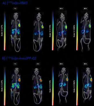
Figure 1. SPECT/CT images of T-47D tumor bearing mice after i.v. administration of: A) [111In]In-RM2; and B) [111In]In-hmdPP-02.
The amalgamation of these compounds into homo - or hetero-multimeric structures holds particular promise for enhancing tumor uptake and retention. Additionally, this approach addresses the intricate and diverse nature of primary tumors and metastases. Our ongoing preclinical assessments aim to pinpoint lead candidates for translation into clinical applications.
Projects:
– Development of theranostics for breast cancer ( Priciana Paraïso )
– FAP-targeted theranostic agents ( Hanyue Ma , Le Li , Amber Piet, Savanne Beekman)
– Imaging and treatment of NET tumors with novel theranostics ( Dylan Chapeau , Maryana Handula , Yozlem Chalashkan, Savanne Beekman, Amber Piet)
Targeted Alpha Therapy (TAT)
Recent clinical investigations have revealed a notable enhancement in the therapeutic efficacy of peptide receptor radionuclide therapy (PRRT) by substituting the conventional β -emitters (90Y, 177Lu) with alpha-emitting radionuclides (212Pb, 225Ac). Our recent work involved the labeling of somatostatin receptor subtype 2 agonists and antagonists with lead-212 and actinium-225, respectively. Comprehensive biological studies were conducted to assess the impact of chemical modifications and metal complexation on the binding properties of the peptides to SSTR2. Subsequently, we conducted evaluations on stability, biodistribution, pharmacokinetics, and dosimetry in tumor-bearing mice. While [225Ac]Ac-DOTA-JR11 showed a higher absorbed dose in the kidneys compared to [177Lu]Lu-DOTA-JR11, which may limit further clinical studies with this Ac-225 labeled peptide, our ligand labeled with 212, eSOMA-01, showed great potential for targeted alpha therapy of NETs. In fact, [ 212Pb]Pb-eSOMA-01 displayed a higher absorbed dose to the tumor, as well as a reduction in absorbed dose to the kidneys compared to the clinical reference DOTAM-TATE.
Projects:
– TAT of NET tumors ( Maryana Handula , Dylan Chapeau , Savanne Beekman)
– TAT of solid tumors ( Hanyue Ma , Le Li , Savanne Beekman, Amber Piet)
– Clinical implementation of Ac-225 labelled Somatostatin and PSMA radiopharmaceuticals (Eline Hooijman)
Theranostic Small-Molecule Drug Conjugates (T-SMDCs)
The linkage of a cytotoxic drug to a targeting vector, such as an antibody, peptide, or small molecule, enhances the effectiveness of chemotherapy while minimizing toxicity to normal organs. Recently, we have developed compounds that target PSMA and FAP, coupled with mertansine (DM1). DM1 is commonly used in antibody-drug conjugates (ADCs), such as the approved T-DM1 (trastuzumab-emtansine). In contrast to ADCs, our theranostic small-molecule drug conjugates (T-SMDCs) also incorporate a chelator, enabling labeling with a gamma emitting radionuclide to produce a companion diagnostic or a therapeutic radionuclide for combined targeted chemotherapy and radionuclide therapy.
Our studies indicated that our SSTR2-targeted T-SMDC, eSOMA-DM1 (Figure 2), showed promising characteristics in vitro, and tumor uptake in H69-xenografts higher at all-time points compared to [111In]In–DOTA-TATE. However, prolonged blood circulation led to increased accu-
mulation of [111In]In–eSOMA-DM1 in highly vascularized tissues, such as the lungs, skin, and heart. Therefore, our results suggest the need for adjustments to optimize the distribution of eSOMA-DM1 prior performing therapeutic efficacy studies.
Projects:
– SSTR2-targeted T-SMDCs ( Dylan Chapeau , Savanne Beekman, Amber Piet)
– Development of SMDCs and T-SMDCs targeting the tumor microenvironment ( Hanyue Ma , Le Li , Amber Piet, Gijs Louwerens)

Figure 2. Chemical structure of eSOMA-DM1. The SSTR2-targeting octreotate peptide (green) is conjugated to the cytotoxic drug DM1 (red) via a chelate-bridged linkage (blue). The drug is released by cleavage of the linker (black) by glutathione.
Fluorescence-Guided Surgery (FGS)
Recently, there has been an emergence of intraoperative imaging tools designed for real-time visualization of tumors. One such approach, termed fluorescenceguided surgery (FGS), has exhibited promising outcomes in aiding surgeons in accurately delineating tumor margins. This capability is particularly crucial for ensuring thorough tumor resection, reducing the risk of disease recurrence, and minimizing damage to healthy tissues. Despite these advancements, the availability of fluorescent imaging probes with optimal properties remains a challenge.
To address this gap, we synthesized fluorescent molecules targeting biomarkers (e.g., SSTR2, GRPR, FAP) prevalent in cancer. Notably, eFAP-24 demonstrated encouraging results in preclinical pancreatic tumor models (Figure 3).
Projects:
– FAP-targeted imaging probes for precision fluorescence-guided surgery ( Hanyue Ma , Savanne Beekman, Amber Piet, Khaled Al Kayal, Saguna Balesar)
– Dual-labeled tracer for SSTR2-positive tumors ( Dylan Chapeau , Savanne Beekman, Maryana Handula, Erika Murce Silva)
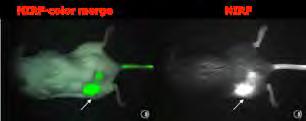
Figure 3. NIRF-color merge and NIRF images of tumor bearing mice at 8 h post-injection of eFAP-24. Images were captured using the clinical Artemis NIRF imager. White arrows indicate tumor localization.
Clinical Radiochemistry
Radionuclide therapy is gaining considerable attention, and traditionally, the theranostic paradigm is using a molecule (e.g., DOTA-TATE) labeled with a gamma-emitting radionuclide (e.g., 68Ga) for imaging, and subsequently with a therapeutic radionuclide (e.g., 177Lu) for PRRT. However, the availability of the main therapeutic radionuclide, 177Lu, is limited. This necessitates exploring alternatives, but the chemical and physical properties of these new radionuclides differ. It is, therefore, crucial to assess how labeling with these new radionuclides (e.g., 161Tb) impacts radiolabeling efficiency, stability, and the biodistribution of the radiotracer.
Projects:
– Radiolabeling of orally inhaled drug products to assess lung deposition (Sean Tsai, Jochem Wofert )
– Tb-161 labelled Somatostatin and PSMA radiopharmaceuticals ( Carolline Ntihabose )
– The ‘Plug-and-Play Radionuclide Generator’ ( Erik de Blois )
– Ho-166 radiopharmaceuticals ( Carolline Ntihabose )
Expectations & Directions
The increasing prominence of radiopharmaceuticals in personalized medicine is anticipated to lead to a continued surge in their clinical utilization for both diagnosis and therapy. There is a growing demand for novel radiopharmaceuticals in research, driven by the need to deepen our understanding of human disease biology, improve diagnostic and therapeutic capabilities, and facilitate drug development. This heightened interest in radiopharmaceuticals motivates our group to actively pursue the development of new theranostics and imaging probes, with the potential to significantly impact the management of cancer patients.
Funding
Seimbille , Yann, Carolien van Deurzen, and Agnes Jager Erasmus MC grants: ‘Theranostics hitting the Achilles’ heels of breast cancer: pointing the arrows at HER2 and GRPR’. 2021-2025
Ma, Hanyue Erasmus MC Research Innovation Grant: 'A universal approach to image and treat cancer with novel promising fibroblast activation protein inhibitors'. 20232024
De Blois, Erik TKI Health Holland grant: 'Phase-I dose escalation study to evaluate the tolerability and safety of 161Tb-PSMA in patients with metastatic, castration resistant prostate cancer'. 2024- 2028
Seimbille , Yann , Alexander Vahrmeijer, and Albert Windhorst Dutch Cancer Foundation Grant: ‘First-in-human assessment of a FAP-targeted probe for fluorescence guided surgery of pancreatic cancer’. 2024-2028
Seimbille , Yann Horizon IHI: ‘Accelerate.eu Elevating the future of cancer care with alpha theranostics’. 2024-2029
Seimbille , Yann, and Philip Elsinga NOW-OTP: ‘MOTIVATE: Molecular oncology twins advancing treatment and innovative cancer evaluation’. 2024-2029
Invited Lectures
Yann Seimbille . ‘Pb-212 Labeling / In vitro / In vivo’. NKRV Meeting, Petten, the Netherlands. Feb 2024.
Yann Seimbille . ‘Beyond 177Lu, emerging isotopes in targeted radionuclide therapy (TRT)’. French Society of Radiopharmacy, Saint-Malo, France. June 2024.
Erik de Blois ‘Radiopharmaceutical aspects of PRRT’. Summer School of Neuroendocrine Tumor Management, Rotterdam, the Netherlands. June 2024.
Erik de Blois ‘Design a Nuclear Pharmacy: sharing Experience in meeting requirement for Alpha & Beta Radiopharmaceuticals’. Malaysian Society of Nuclear medicine & Molecular Imaging, Kuala Lumpur, Malaysia. May 2024.
Erik de Blois . ‘Production and Quality control of Theranostic radiopharmaceuticals using alpha emitters: From basic radiochemistry to Clinical studies’. Malaysian Society of Nuclear medicine & Molecular Imaging, Kuala Lumpur, Malaysia. May 2024.
Highlights
Erika Murce Silva successfully graduated on October 29th, 2024. Her PhD thesis was entitled: “Towards the improvement of diagnosis and treatment of prostate cancer: optimization of PSMA-targeted radiopharmaceuticals”.
Our paper entitled “First preclinical evaluation of [225Ac] Ac-DOTA-JR11 and comparison with [177Lu]Lu-DOTA-JR11, alpha versus beta radionuclide therapy of NETs” published in EJNMMI Radiopharmacy and Chemistry (2023, 8:13) received the EANM Springer Prize for the Best Paper 2024
The presentation of Hanyue Ma at the World Molecular Imaging Congress (WMIC) on revolutionizing solid tumor surgery with fibroblast activation protein targeted imaging probes for precision fluorescence-guided surgery was selected in the highlights.
A patent was filed by Hanyue Ma , Le Li, and Yann Seimbille on dimeric FAP targeting agents. Erik de Blois is one of the applicants of a patent on alpha spectrometry detector by Lyla systems (previously AlphaPace).
Additional Personnel
Amber Piet, MSc – Research assistant
Savanne Beekman, BSc – Research assistant
Roan Claus – 4th year Chemistry student, Institute of Engineering and Applied Science, Hogeschool Rotterdam. Jan 2024-June 2024. Daily supervisor Maryana Handula.
Nilenska Martina – 4th year Chemistry student, Institute of Engineering and Applied Science, Hogeschool Rotterdam. Dec 2023-May 2024. Daily supervisor Maryana Handula.
Yozlem Chalashkan – 4th year Chemistry student, Institute of Engineering and Applied Science, Hogeschool Rotterdam. Dec 2023-May 2024. Daily supervisor Maryana Handula.
Kiefer Comassi – 2nd year MSc student Technical Medicine, TU Delft. Nov 2023-Oct 2024. Daily supervisor Gennady Roshchupkin and Yann Seimbille.
Khaled Al Kayal – 2nd year MSc student Drug Discovery & Safety, Vrije Universiteit Amsterdam. March 2024-Aug 2024. Daily supervisor Le Li and Hanyue Ma.
Saguna Balesar – 4th year Chemistry student, Institute of Engineering and Applied Science, Hogeschool Rotterdam. Sept 2024-Feb 2025. Daily supervisor Le Li and Hanyue Ma.
Gijs Louwerens – 2nd year MSc student Bio-Pharmaceutical Sciences, Leiden University. Sept 2024-Feb 2025. Daily supervisor Amber Piet.
Iris van der Merwe – 4th year Chemical-Physical Analyst student, Techniek College Rotterdam. Sept 2024-June 2025. Daily supervisor Erik de Blois.
Negin Eskandari – 4th year Chemical-Physical Analyst student, Techniek College Rotterdam. Sept 2024-June 2025. Daily supervisors Eline Hooijman and Carolline Ntihabose.
Sean Tsia – 2nd year MSc student Nanobiology, TU Delft. Sept 2024-Feb 2025. Daily supervisor Jochem Wolfert.
Nyah Rook – 4th year Chemical-Physical Analyst student, Techniek College Rotterdam. Sept 2023-June 2024. Daily supervisor Erik de Blois.
Louise van Dalen – 4th year Chemical-Physical Analyst student, Techniek College Rotterdam. Sept 2023-June 2024. Daily supervisor Carolline Ntihabose.
Assistant Professor

Erik de Blois, PhD
Email r.deblois@erasmusmc.nl

CLINICAL RADIOCHEMIST: CLINICAL RADIOCHEMISTRY & IMPLEMENTATION
Erik de Blois (1981, Zeist) started his MLO training at Zadkine Rotterdam and graduated in 2001 in biotechnology. He then studied part-time at HLO at the HvU Utrecht and graduated in 2005. In the following year he continued his part-time study in pharmaceutical sciences at the University of Utrecht in the direction of “Drug innovation”. He graduated in 2009, after which he started his PhD program part-time at Erasmus MC. In 2014 he successfully defended his thesis: Radiochemical Aspect of receptor Scintigraphy: labeling with radiometals, optimization and radiochemical purity and obtained his doctorate. In the meantime, he started in 2002 as a research analyst in the radiochemistry group at the Nuclear Medicine department at Erasmus MC. After obtaining his master's degree, his position changed to Clinical Radiochemist and for the past 4 years he has also been Head of Quality Control and responsible for the release of radiopharmaceuticals made in-house and started his own research group Clinical Radiochemistry & Implementation (radiochemical aspects of radiopharmacy). Within his position he contributed to the implementation of various (therapeutic) radiopharmaceuticals, including the first registered therapeutic radiopharmaceutical Lu-177 labeled DOTA-TATE (Lutathera).
Clinical implementation of [225Ac]Ac labeled radiopharmaceuticals
Targeted alpha therapy with Ac-225 showed to be effective in treating metastatic cancers. However, its complex decay chain requires optimized radiolabeling and quality control conditions. We identified the critical parameters of optimal radiolabeling and established accurate measurement techniques for radiochemical yield and purity for Ac-255 labeled DOTA conjugated radiopharmaceuticals.
Therapy with PSMA labeled with actinium-225 can be very effective in patients with metastatic castration resistant PCa (mCRPC). The use of alpha emitting radionuclides will result in more DNA double strand breaks than treatment with beta emitting radionuclides. A limited number of patients have been treated with [225Ac]Ac-PSMA therapy in Germany, and there is still a lack of a good clinical trial. Therefore, a phase I study is necessary to calculate the recommended dose that is both safe and effective. With the treatment of up to 30 patients, we expect to determine the most appropriate cumulative dose for [225Ac]Ac-PSMA treatment. Dose escalation was successfully performed, and more evidence will be collected with a 10 MBq dose of [225Ac] Ac-PSMA.
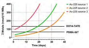
Figure 1. Test and application of 3 different suppliers of Ac225. Metal impurities were measured over time to determine whether the source can be clinically used.
Clinical implementation of [161Tb]Tb-PSMA
Therapy with [161Tb]Tb-PSMA is proven to be very effective in patients with metastatic castration resistant PCa in comparison to [177Lu]Lu-PSMA. Radiochemistry is compared to Lu-177 and in vitro and in vivo comparison experiments are recently performed. Additionally, a phase I trial is planned in the near future. Before implementation into cleanroom environment, radiolabeling and stability of the Tb-PSMA have to be optimized. Reaction conditions and stability were optimized in downscaled form in research labs. The final mixture was formulated and was finally full GMP prepared. For clinical implementation, related equipment, methods and quality controls were calibrated and validated. 3 test runs were performed in the cleanroom and showed that the product was according to all release criteria.

Maryana Handula, MSc

C-term modified DOTA-JR11 analogs for enhanced treatment of NETs
Due to the sensitivity of DOTA-JR11 to chemical modifications at the N-terminus, we introduced an albumin binder (AB) at the C-terminal position. Dansylglycine (DG) was reported as a potential AB to extend blood residence time of the radioligand. Prolonged blood circulation could increase the tumor uptake and therapeutic index. The preliminary data confirmed the potential of DG as a promising AB. Further research is ongoing to optimize the tumor uptake.

Hanyue Ma, PhD
Email h.ma@erasmusmc.nl

A universal approach to image and treat cancer with novel promising fibroblast activation protein inhibitor
Fibroblast activation protein (FAP) is highly expressed in cancer-associated fibroblasts and is abundant in more than 90% of epithelial tumors. FAP is involved in cancer progression, invasion, survival, and treatment resistance, while being barely expressed in normal organs. In recent years, FAP inhibitors based on smallmolecules have been reported as powerful imaging probes for the diagnosis of many types of cancers due to their rapid uptake and impressive tumor-to-background ratios. This further demonstrates the high potential for their use in cancer treatment.
We recently developed a series of novel quinolinebased FAP inhibitors (eFAPs) with a broad scope of application in oncology, (i.e., molecular imaging, radionuclide therapy, targeted chemotherapy, imageguided surgery). eFAP analogues showed strong FAP inhibitory activity, high binding affinity, selectivity, and good stability. Both radiolabeled and fluorescent eFAP conjugates showed high and rapid tumor uptake in xenograft tumor models. We believe that our lead candidates can be rapidly translated into clinical trials for imaging and treatment of cancer.
PhD Students
Dylan Chapeau, MSc

Advisors Yann Seimbille, Mark Konijnenberg & Erik Verburg
Project Funding Kansen voor West
Email d.chapeau@erasmusmc.nl
Synthesis of a trifunctional chelate for multimodality approches
Multimodality imaging and/or therapy in a single molecule can be challenging. Therefore, we synthetized new compounds containing a trifunctional linker to combine all modalities to the targeting vector. This synthetic approach avoids the alteration of the biochemical properties of the tracers (e.g., binding affinity). Our strategy relies on a chelate that serves as a bridge between all components of the tracers.

Eline Hooijman, MSc

Advisors Erik Verburg, Hugo van der Kuy, Erik de Blois & Stijn Koolen
Project Funding Unit Radiopharmacy
Email e.hooijman@erasmusmc.nl
Development of Targeted Alpha Therapy
Promising anti-tumor effects were observed in patients treated with small molecules labelled with different types of radioisotopes. α - particle emission provides an advantage over β - emission due to the localized radiation effects and high local cytotoxicity. Subsequently, [225Ac]Ac-PSMA-I&T and [225Ac] Ac-DOTATATE are currently clinically implemented according to full GMP. The first phase I dose-escalation trial with [225Ac]Ac-PSMA-I&T is ongoing with encouraging first results.

Priciana Paraïso, PharmD

Advisors Carolien van Deurzen, Yann Seimbille & Erik Verburg
Project Funding Erasmus MC Fellowship
Email p.paraiso@erasmusmc.nl
Peptide-based theranostics for breast cancer
Functional noninvasive nuclear imaging (i.e., SPECT, PET) can provide accurate assessment of receptor status in primary and metastatic breast cancer lesions. In this project, we aim at developing a new generation of monomeric and dimeric peptidebased theranostics pointing at two major Achilles’ heels of breast tumors, namely the gastrin releasing peptide receptors (GRPRs) and the human epidermal growth factor (HER2).
Le Li, MSc
Advisors Yann Seimbille & Aad van der Lugt
Project Funding CSC-Erasmus MC Fellowship
Email l.li.2@erasmusmc.nl
Strategies to improve the theranostic potential of fibroblast activation protein (FAP)-targeted radiotracers
The goal is to develop novel radiopharmaceuticals that can be potentially beneficial in the diagnosis and treatment of cancers. A new generation of FAPinhibitors will be designed, synthesized and evaluated in preclinical models to deliver new tracers with improved efficacy and safety profile for imaging and radionuclide therapy.

Erika Murce Silva, MSc

Advisors Erik Verburg & Yann Seimbille
Email e.murcesilva@erasmusmc.nl
PhD Obtained 29-10-2024
A theranostic platform for imaging and treatment of prostate cancer
Theranostic small-molecule drug conjugates (T-SMDCs) are a promising emerging class of molecules used for tandem targeted radionuclide therapy and targeted chemotherapy. We are currently investigating T-SMDCs for imaging and therapy of prostate cancer by targeting the prostate specific membrane antigen (PSMA).

Carolline Ntihabose, MSc

Advisors Erik Verburg, Harry Hendrikse, Erik de Blois & Stijn Koolen Email c.ntihabose@erasmusmc.nl
The (Pre)Clinical Potential of Targeted Radionuclide Therapy with Novel radionuclides
Targeted radionuclide therapy (TRT) has revolutionized the treatment of metastasized cancer patients. TRT containing the beta-emitter Lu-177, has shown improved results in cancer treatment. New radionuclides (Ho-166, Tb-161, Pb-212, etc.) are necessary to improve current treatments. For example, Tb-161, which has near-identical radiochemical properties. Tb-161 shows pre-clinically more therapeutic effect than Lu-177 TRT, potentially caused by its additional conversion and auger electrons.

Jochem Wolfert, PharmD

Advisors Harry Hendrikse, Erik Verburg, Liesbeth Ruijgrok, Ties Mulders & Erik de Blois
Project Funding Federal Drug Administration; FLUIDDA
Email j.wolfert.1@erasmusmc.nl
Radiolabeling of orally inhaled drug products to assess lung deposition
We are aiming to develop novel radiolabeling methods to assess lung deposition of orally inhaled drug products using PET-CT. These imaging methods can then be employed to improve knowledge of treatments used in pulmonary disease.
APPOINTMENT IN RADIOLOGY & NUCLEAR MEDICINE (ERASMUS MC) AND PEDIATRIC ONCOLOGY (PRINCESS MÁXIMA
CENTER)
Sophie Veldhuijzen van Zanten holds MSc degrees in Medicine (2011) and Epidemiology (2016). She began her scientific career at the molecular neurooncology lab of Dana-Farber Cancer Institute/Harvard Medical School. In 2017, she earned her PhD in Pediatric Oncology from VU University (Amsterdam). Following, she completed her medical specialty training in Radiology and Nuclear Medicine at Spaarne Gasthuis (Haarlem, 2016-2019) and Erasmus MC (Rotterdam, 2019-2023). In 2022, she was appointed Assistant Professor at Erasmus MC. Her research group integrates radiology and nuclear medicine, with a
focus on the central nervous system and head-andneck tumors.
Sophie has received several young investigator awards and is the principal investigator on multiple research grants. She currently supervises three PhD students at Erasmus MC, with three former students having successfully completed their PhDs at Amsterdam UMC, PMC/UMC Utrecht, and Erasmus MC. Sophie is an active board member of the Dutch Society for Nuclear Medicine (NVNG) and chairs a European registry collecting clinical, imaging, and biological data of pediatric glioma patients. This registry facilitates international collaborative research within the European and International Societies of Pediatric Oncology. s.veldhuijzenvanzanten@erasmusmc.nl
THERANOSTICS OF CNS AND H&N DISEASES
Sophie Veldhuijzen van Zanten, MD, PhD assistant
professor
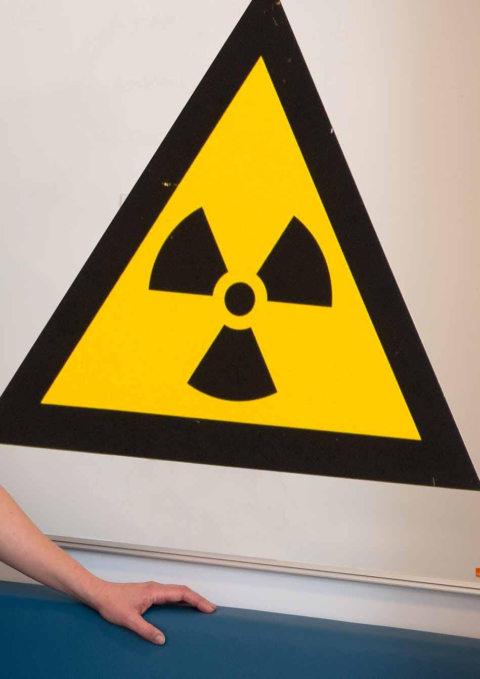
Context
This research line focuses on advancing theranostics for central nervous system and head-and-neck diseases, with an emphasis on hard-to-diagnose, hard-to-reach, and hard-to-treat tumors. Theranostics combines diagnostic and therapeutic applications by using alternating radionuclides for molecular imaging and targeted radionuclide therapy (TRT). This approach enables non-invasive exploration of disease molecular biology, allowing for the quantification of target expression across multiple sites and time points. It facilitates image-guided diagnosis, staging, and longitudinal disease monitoring. Additionally, it can be used to assess the biodistribution of drugs, quantify target binding, and enable the selection of the most effective (radio) pharmaceuticals, dosing, administration routes, and appropriate patient populations, ensuring that treatment is tailored to those most likely to benefit.
Top Publications 2024
Pruis IJ, PJ van Doormaal, RK Balvers, MJ van den Bent, AA Harteveld, LC de Jong, MW Konijnenberg, M Segbers, R Valkema, FA Verburg, M Smits, SEM Veldhuijzen van Zanten. Potential of PSMA-targeting radioligand therapy for malignant primary and secondary brain tumours using super-selective intra-arterial administration: a single centre, open label, non-randomised prospective imaging study. Lancet eBioMedicine 2024; 102:105068.
Pruis IJ, FA Verburg, RK Balvers, AA Harteveld, RA Feelders, MW Vernooij, M Smits, SJCMM Neggers, SEM Veldhuijzen van Zanten. [18F]FET PET-MRI; a novel and accurate technique for detection of small functional pituitary tumors. J Nucl Med. 2024; 65:688-692.
Droogers E, MP Hendriks, SEM Veldhuijzen van Zanten, namens het studieteam FAPI for CUP. [18F]F-FAPI PET-CT voor de identificatie van onbekende primaire tumoren. Nederlands Tijdschrift voor Oncologie 2024; 21:29-31.
Research Projects: Objectives & Achievements
In this research line, we investigate a range of diagnostic and therapeutic radiopharmaceuticals for various CNS and H&N diseases.
Primary brain tumours and Brain met astases
In a clinical proof-of-concept study led by Ilanah Pruis , we explored the expression and binding of prostate-specific membrane antigen (PSMA) in patients with adulttype diffuse glioma or brain metastasis. All patients showed uptake of the diagnostic radioligand [68Ga]GaPSMA at the tumor site, with minimal to acceptable uptake in healthy brain tissue and other organs. This study provided the first quantified evidence and modelled predictions for the potential of PSMA-based TRT in malignant brain tumors.
Additionally, both in this proof-of-concept study and clinical practice, we demonstrated that selective intraarterial (IA) administration of radioligands leads to significantly higher tumor uptake compared to intravenous administration. IA delivery of [68Ga]Ga-PSMA (in glioma/ brain metastasis) or [68Ga]Ga-DOTA-TATE (in meningioma) resulted in significantly improved tumor-to-liver uptake ratios, qualifying all patients for TRT. A prospective radionuclide therapy trial for glioma patients, based on these findings, will be led by Jessica de Jong . To further optimize the effectiveness of our treatments, we analyze tumor blood supply and microvascular dynamics using multimodal imaging in all of our patients (Figure 1).
Jessica de Jong is also investigating the clinical value of O-(2-[18F]-fluoroethyl)-L-tyrosine ([18F]FET) PET-MRI to differentiate progressive disease from treatment-related changes in glioma patients. This novel technique provides complementary and correlative information, improving diagnostic accuracy and clinical decision-making in a subset of patients. Sophie Veldhuijzen van Zanten contributed to the "Update to the RANO Working Group and EANO recommendations for the clinical use of PET imaging in gliomas", based on the Erasmus MC experience with this multiparametric diagnostic tool.
In parallel, in the laboratory, Nina Overdevest (co-supervised by Julie Nonnekens and Justine Perrin) is investigating novel targets for glioma theranostics.

Pituitary adenoma
For the detection of pituitary neuro-endocrine tumors (PitNETs, formerly known as microadenomas), we have introduced a novel diagnostic approach using [ 18F]FET PETMRI. In the first clinical series of patients with Cushing’s disease and negative or inconclusive prior MRI(s) (n=22, published by Ilanah Pruis ), we demonstrated 100% sensitivity and high accuracy in localizing ACTH-secreting PitNETs. This method offers several key advantages, including shorter diagnostic timelines, improved treatment planning for surgery or Gamma-/Cyber Knife radiation therapy, and the potential for partial hypophysectomies, sparing patients from lifelong hormone replacement therapy. Additionally, it leads to enhanced remission rates, reduced patient burden, improved well-being, and lower healthcare costs. As a result, [18F]FET PET-MRI is now the standard of care for patients at Erasmus MC with negative or inconclusive MRI results. In the next phase, we aim to disseminate our findings by collaborating with colleagues from various centers across Europe who are interested in adopting this approach.
H&N tumours and Cancer of Unknown Primary origin
Esther Droogers conducted a retrospective study to evaluate the accuracy of [18F]FDG PET-MRI and [18F]FDG PET-CT for the assessment of lymph nodes in patients with H&N squamous cell carcinoma (HNSCC) in routine clinical practice at Erasmus MC. Both imaging techniques demonstrated high accuracy in distinguishing malignant from benign lymph nodes. [18F]FDG PET-MRI showed a slight advantage in detecting suspicious lymph nodes compared to PET-CT, but challenges remain in accurately classifying these nodes. This necessitates additional invasive procedures, such as fine needle aspiration cytology or neck dissection, for further confirmation in a significant number of patients. These findings highlight the
Figure 1. Glioma tumor vessel density in relation to MRI T1 contrast-enhancing area (purple) and T2 hyperintense area (red), based on CBCT vessel segmentation.
need for ongoing improvements in diagnostic accuracy. To address this, advanced radioligands, such as [18F]/ [68Ga]-labelled fibroblast activation protein inhibitor (FAPI), which targets cancer-associated fibroblasts, shows promise for improving image-based lymph node evaluation in HNSCC.
Esther Droogers is also gaining experience in [18F]FAPI PET imaging (Fig.2) as part of the FAPI for Cancer of Unknown Primary (CUP) study, supported by KWF Dutch Cancer Society. This year, the first patients were enrolled in this multicenter study, developed in collaboration with Missie Tumor Onbekend.

Figure 2. PET scans following administration of [18F]FDG (left) and [18F]-FAPI (right) of a patient with CUP, showing a significantly higher yield with FAPI diagnostics.
Paragangliomas
In a retrospective cohort study conducted by Esther Droogers (co-supervised by Renske Gahrmann) we evaluated the tumor growth and characteristics of extraadrenal paragangliomas (PGLs) in patients with various Succinate Dehydrogenase (SDHx) mutations, using [68Ga]Ga-DOTATATE PET-MRI/CT. The study confirmed the predominantly indolent growth of PGLs in SDHx mutation carriers, with a notably higher risk of tumor progression, malignant transformation, and poor treatment response in patients with SDHB mutations, compared to those with other SDHx mutations.
Expectations & Directions
The advancement of radionuclide-based diagnostics and therapy for (oncological) diseases in the CNS and H&N regions is pioneering. This research line is distinguished by its unique focus on hybrid PET-MRI technology, providing some of the most detailed and informative images of anatomy and molecular (patho)physiology available today. Through our work, we challenge established paradigms and contribute to the development of improved diagnostic and therapeutic guidelines. Additionally, our IA delivery approach has shown promise in enhancing treatment effectiveness, opening new possibilities for innovative therapeutic strategies, including IA-TRT in the management of CNS tumors. With the rapid expansion of available radionuclide compounds, this research line has the potential for significant growth in the coming years.
Funding
Veldhuijzen van Zanten, Sophie, and consortium partners Medical Delta: ‘Cancer Diagnostics for Sustainable Health Care - Theranostic work package. Acronym: CARES’. 20242027
Veldhuijzen van Zanten, Sophie KWF Dutch Cancer Society: ‘Introduction of [18F]F-FAPI for diagnostics of carcinoma of unknown primary origin’. 2023-2027
Veldhuijzen van Zanten, Sophie KWF Dutch Cancer Society: ‘Introduction of 18F-FAPI for diagnostics of carcinoma of unknown primary origin’. 2023-2027
Veldhuijzen van Zanten, Sophie The Cure Starts Now Foundation: ‘Preclinical development of targeted radionuclide therapy for DIPG/DMG and medulloblastoma’. 2023-2027
Veldhuijzen van Zanten, Sophie Erasmus MC Foundation, Daniel den Hoed Young Scientific Talent Award: ‘Intraarterial PSMA-based targeted radionuclide therapy for recurrent and progressive malignant glioma’. 2022-2026
Veldhuijzen van Zanten, Sophie Stichting Semmy: ‘Introduction of theranostics for central nervous system tumours’. 2019-2024
Veldhuijzen van Zanten, Sophie, and SIOPE brain tumour group consortium partners The DIPG/DMG collaborative: ‘European DIPG Registry’ (several starting and maintenance grants). 2014-2024
Invited Lectures
Sophie Veldhuijzen van Zanten . ‘Hybrid imaging for brain tumors’. 7th European Society for Hybrid, Molecular and Translational Imaging (ESHI) Conference, Vienna, Austria. Nov 2024.
Sophie Veldhuijzen van Zanten . ‘FAPI imaging in the Netherlands’. FAPI featuring session at the 37th Annual Congress of the European Association of Nuclear Medicine (EANM), Hamburg, Germany. Oct 2024.
Sophie Veldhuijzen van Zanten . ‘Sanjiv Sam Gambhir competition session’. 37th EANM Congress, Hamburg, Germany. Oct 2024.
Highlights
Jessica de Jong received the ‘Excellent Research Presentation Award’ at the Landelijke Werkgroep Neuro-Oncologie (LWNO) meeting in March 2024.
Esther Droogers was selected for the ‘Best abstract presentation’ at the Erasmus MC Cancer Retreat in April 2024.
Nina Overdevest won the ‘Best Poster Award’ at the EMC PhD symposium in May 2024.
Sophie Veldhuijzen van Zanten won the ‘Innovative Protocol Award’ during the 25th Workshop on Methods in Clinical Cancer Research (MCCR), organized by the European Organization of Research and Treatment of Cancer (EORTC), European Society for Medical Oncology (ESMO), and American Association for Cancer Research (AACR).
Ilanah Pruis successfully defended her thesis entitled: ‘Theranostic PET-MRI for brain tumours’ and obtained her PhD degree in July 2024.
The publication titled ‘[18F]FET PET/MRI: An Accurate Technique for Detection of Small Functional Pituitary Tumors’ by Ilanah Pruis was highlighted by the Nederlandse Hypofyse Stichting with an interview in Hyponieuws in November 2024.
The thesis by Ilanah Pruis , titled ‘Theranostic PET-MRI for Brain Tumours’, was highlighted by the Nederlandse Vereniging voor Medische Oncologie with an interview in Tijdschrift voor Medische Oncologie in October 2024.
Additional Personnel
Lotte van Dijk, BSc – 4th year MSc student Medicine, Erasmus University Rotterdam. Oct 2023-March 2024. "Multimodal imaging of blood supply and microvascular dynamics in primary and secondary malignant brain tumors". Supervisors Ilanah Pruis, Sophie Veldhuijzen van Zanten.
Wouter Bron, BSc – 4th year MSc student Medicine, Erasmus University Rotterdam. Oct 2023-March 2024."18F-FET PET-MRI for glioma follow-up'". Supervisors Jessica de Jong, Sophie Veldhuijzen van Zanten.
Evy van Daalen, BSc – 4 th year MSc student Medicine, Erasmus University Rotterdam. Oct 2023-March 2024. "Extra-adrenal paragangliomas: a retrospective combined assessment of structural MRI and [68Ga]Ga-DOTATATE uptake in SDHx mutation carriers [68Ga]Ga-DOTATATE uptake in SDHx mutation carriers". Supervisors Esther Droogers, Sophie Veldhuijzen van Zanten.
Mouad Allaoui, BSc – 1st year MSc student Biomedical Technology and Physics, VU University. Sept 2024-Dec 2024. "Quantitative DSA validation using perfusion MRI". Supervisors Frank te Nijenhuis, Ilanah Pruis.
Romy van der Groef, MSc – 2nd year PhD student, Department of Internal Medicine / Endocrinology, Erasmus Medical Center. Ongoing project. "Pasireotide Induces Longterm Cystic Degeneration of Somatotrophic Pituitary Neuroendocrine Tumors (PitNETs)". Supervisors Sophie Veldhuijzen van Zanten, Julie Refardt, Sebastian Neggers.
Samuel Odro – 4th year BASc student Biology and Medical laboratory Research, Avans Hogeschool. Feb 2024-July 2024. "BBB models". Supervisors Nina Overdevest, Julie Nonnekens.
Mimi Welte – BSc student Erasmus University College. Nov 2024-May 2025. "TRT in glioma cell lines". Supervisors Nina Overdevest, Julie Nonnekens.

Ilanah Pruis, PhD
Project Funding Stichting Semmy Email i.pruis@erasmusmc.nl
Theranostic PET-MRI for Brain Tumours
Despite advancements in precision cancer medicine, patients with brain tumours still face a one-size-fits all approach. Patient outcomes are dismal and have not improved for decades, indicating the significant need for innovation. I aim to integrate advanced hybrid imaging with targeted radionuclide-strategies for precise diagnosis and therapy, referred to as theranostics, in neuro-oncology. The potential of theranostics for brain tumour management has recently gained significant interest. My research bridges clinical molecular imaging with the underlying pathophysiology of brain tumours, prioritizing (radio)biological validation and clinical implementation focused on delivering personalized, hybrid image-based approaches for patients with a brain tumour. My expertise lies

in implementing hybrid PET-MRI in neuro-oncology.
Key projects of my research include:
1. Initiating molecular imaging projects to validate novel theranostic targets in brain tumours;
2. Launching translational clinical studies to assess the diagnostic and therapeutic potential of preclinically validated tracers such as PSMA-based tracers for glioma and brain metastasis and FET-based tracers for pituitary adenomas (PitNETs);
3. Proof-of-concept investigations into advanced delivery techniques such as super-selective intra-arterial delivery.
PhD Students
Ilanah Pruis, MSc

Advisors Sophie Veldhuijzen van Zanten & Marion Smits
Project Funding Stichting Semmy (The Semmy Foundation)
Email i.pruis@erasmusmc.nl
PhD Obtained 05-07-2024
Theranostics for brain tumours

Jessica de Jong, MD, MSc

Advisors Sophie Veldhuijzen van Zanten, Erik Verburg & Marjolein Geurts
Project Funding Stichting Semmy | Erasmus MC Foundation - Daniel den Hoed Fonds
Email j.dejong@erasmusmc.nl
Intra-arterial targeted radionuclide therapy for brain tumours
PhD Students
I study the combined application of advanced MR imaging techniques, molecular imaging by PET and possibilities for molecular radiopharmaceutical therapy, conjoinedly forming a novel theranostic approach for brain tumor patients. We performed a first proof-ofconcept PET-MRI study for patients with glioma and brain metastases, using [68Ga]Ga-PSMA-11, aimed at non-invasive quantification of the expression of prostate-specific membrane antigen (PSMA), a possible target for therapy located at the tumor vasculature. In addition, we compared uptake in tumour after intraarterial versus intravenous injection to determine the optimal route of administration.
My research focuses on the use radiopharmaceuticals for both diagnostic and therapeutic purposes, i.e. theranostics, in the management of central nervous systems tumours. We aim to explore potential new treatments for glioma patients, who still face a dismal prognosis. Currently, we are setting up a first phase 1 clinical trial applying [177Lu]Lu-PSMA-I&T via super-selective intra-arterial administration as a salvage therapy for diffuse intracranial glioma.

Esther Droogers, MD, MSc

Advisors Sophie Veldhuijzen van Zanten, Debbie Robbrecht & Erik Verburg
Project Funding Dutch Cancer Society (KWF Kankerbestrijding)
Email e.droogers@erasmusmc.nl
Introduction of the novel radioligand [18F]-FAPI for diagnostics of carcinoma of unknown primary origin
I run a multicenter study in which patients with carcinoma of unknown primary origin (CUP) will undergo a PET-CT using the novel radiotracer [18F]-fluoro fibroblast activation protein inhibitor ([18F]F-FAPI). [18F]-FAPI targets cancer associated fibroblasts, which are specifically found in the tumor microenvironment. [18F]-FAPI PET-CT is therefore a promising novel diagnostic tool for patients with CUP.


Focus Area
CLINICAL IMAGING
The aim is to investigate the clinical value of (new) imaging technologies and imaging biomarkers, following a structured order of investigations. The robustness of imaging biomarker extraction is assessed with a focus on accuracy, repeatability and reproducibility. The diagnostic accuracy of new imaging technology is investigated by comparing this imaging data to reference standards. The clinical relevance is assessed by evaluating diagnostic confidence regarding clinical decision-making and impact on treatment planning. The prediction of outcome or treatment response based on quantitative imaging biomarkers, radiomics features and deep-learning algorithms is evaluated. Multi-center (clinical) trials are used to assess and evaluate imaging biomarkers of disease activity and response to treatment.
Aad van der Lugt graduated from Erasmus Medical School in 1988. Before specializing as a neuroradiologist at Erasmus Medical Center, he worked as a Junior Doctor in Surgery and Intensive Care in Tilburg and Eindhoven, respectively. His doctoral research at Erasmus University Rotterdam (1996) explored “Intravascular Ultrasound – Validation and Clinical Application.” Over the years, he played a leading role in neuroradiological research, chairing the program from 2002 to 2015 and becoming Professor of
Neuroradiology and Head & Neck Radiology in 2010. Since 2022, he has served as chairman of the Department of Radiology & Nuclear Medicine. He represents the Netherlands on the EuroBioimaging Board.
His research primarily focuses on imaging and treatment of acute stroke. Additionally, he leads research efforts within the Dutch acute stroke CONTRAST Consortium.
a.vanderlugt@erasmusmc.nl
Aad van der Lugt, MD, PhD
full professor IMAGING IN NEUROVASCULAR DISEASE

Context
This research program is focused on the role of imaging biomarkers in neurovascular diseases with a strong emphasis on ischemic stroke. Imaging in acute stroke aims to support in the diagnosis, assessment of the severity and reversibility of ischemia. Patient selection for treatments like intravenous thrombolysis or endovascular thrombectomy relies on imaging biomarkers. These markers guide decisions by identifying eligible patients. In the sub-acute phase, imaging can assist in evaluating the underlying cause of ischemic stroke. Recently, there’s been increased attention to atherosclerosis beyond the carotid bifurcation. Optimal secondary preventive measures can be better tailored by subdividing patients based on their presumed etiology of ischemic stroke. This approach aims to enhance patient outcomes. This research program encompasses technical development, evaluation of image analysis algorithms, and clinical validation of imaging biomarkers.
Top Publications 2024
Huijberts I, FME Pinckaers, SGH Olthuis, SMJ van Kuijk, AA Postma, HD Boogaarts, YBWEM Roos, CBLM Majoie, A van der Lugt, DWJ Dippel, WH van Zwam, RJ van Oostenbrugge. MR CLEANLATE investigators. Collateral-based selection for endovascular treatment of acute ischaemic stroke in the late window (MR CLEAN-LATE): 2-year follow-up of a phase 3, multicentre, open-label, randomised controlled trial in the Netherlands. Lancet Neurol. 2024; 23: 893-900.
Cahalane RME, JMH Cruts, HMM van Beusekom, MPM de Maat, M Dijkshoorn, A van der Lugt, FJH Gijsen. Contribution of Red Blood Cells and Platelets to Blood Clot Computed Tomography Imaging and Compressive Mechanical Characteristics. Ann Biomed Eng. 2024; 52: 2151-2161.
Samuels N, RA van de Graaf, YBWM Roos, D Dippel, A van der Lugt. MR CLEAN and CONTRAST investigators. Advancements in diagnostic and interventional radiology for stroke treatment: the path from trial to bedside through the pre-MR CLEAN, MR CLEAN, and MR CLEAN II eras. Insights Imaging 2024; 15: 30.
179
Research Projects: Objectives & Achievements
Endovascular treatment in patients with acute stroke: beyond the MR CLEAN studies
Treatment with intravenous (IV) alteplase, aimed at early reperfusion, has proven effective for patients with acute ischemic stroke. Approximately 25% of patients experiencing acute anterior circulation ischemic stroke exhibit symptoms caused by proximal occlusion of a major intracranial artery. Endovascular treatment (EVT) significantly increases the likelihood of recanalization in patients with acute ischemic stroke resulting from proximal intracranial arterial occlusion. The MR CLEAN Study demonstrated that intra-arterial treatment, administered within 6 hours after stroke onset, is both effective and safe. Since our landmark paper, multiple studies have further highlighted the beneficial aspects of EVT. Within the HERMES Collaboration, individual patient data have been leveraged to investigate additional research questions. These inquiries have focussed on optimal patient selection for treatment and improvement of overall outcomes.
In 2017 the CONTRAST-consortium (www.contrast-consortium.nl) was established creating a biobank infrastructure for stroke research in the Netherlands. This infrastructure included data, blood samples, thrombus and imaging. Three new trials have been conducted 1) to evaluate the effect of peri-procedural medication (MR CLEAN-MED), 2) to evaluate the effects in patients presenting between 6 and 12 hours after the event (MR CLEAN-LATE) and 3) to evaluate the benefits of direct IAT without prior IVT (MR CLEAN-NoIV). All trials have been completed and their results have been published. In 2023 the Consortium was extended by 5 years and secured new funding. The CONTRAST 2.0 Consortium aims to enhance the chances of a good recovery for a large group of patients with cerebral infarction, brain hemorrhage, or subarachnoid hemorrhage. This will be achieved by developing new treatments in the laboratory, improving treatment logistics, and initiating six large clinical trials. Each study will test one or more new treatments or answer important research questions. All studies will be benefit from a central core lab assessment of CT and MRI scans of the brain along with a central database and standardized statistical analysis.
Role of Thrombus
With EVT extracted thrombi have become available for histopathologic analysis providing a unique opportunity to study several critical relationships. We aimed to 1) explore how imaging features of the thrombus relate to its composition; 2) Investigate how thrombus composition influences biomechanical properties and 3) understand the impact of thrombus composition on successful recanalization.
In general, stroke thrombi consist of four main components: red blood cells, fibrin, platelets and white blood cells. In a MR CLEAN Registry biobank study, we have shown that the amount of red blood cells and fibrin/ platelets are related to the origin of the thrombus: red blood cell-rich clots were more likely to originate from large artery atherosclerosis, while fibrin/platelet-rich clots were more likely to have a cardioembolic source.
CT imaging at admission allows us to assess various thrombus characteristics, including attenuation, length, distance from the internal carotid artery terminus, and permeability (or “perviousness”). We have demonstrated that thrombus CT features correlate with its composition (e.g., red blood cells and fibrin/platelets). Importantly, these CT characteristics also align with the thrombus’s origin, validating our hypothesis based on histological studies. This is crucial, as histological analyses can be time-consuming, costly, and subject to bias.
In collaboration with the biomechanical department at Technical University Delft, we conducted the firstever study to mechanically characterize human stroke thrombi. Our findings confirmed that fibrin/platelet-rich thrombi are mechanically stiffer.
A highly debated topic in the field of EVT revolves around device selection: stent retrievers (SR) have long been the standard, but contact aspiration (CA) has rapidly gaining popularity. Researchers and clinicians speculate that thrombus type could serve as a guide for choosing the optimal first-line EVT device. Specifically, they believe that fibrin/platelet-rich thrombi, which are stiff and resistant to thrombectomy, might benefit more from CA. However, a MR CLEAN Registry substudy revealed a different outcome. Contrary to expectations, we found that this correlation did not hold true.
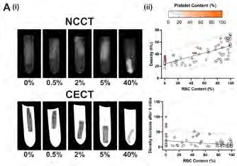
Figure 1. Clot Computed Tomography (CT) characteristics. (i) Representative red blood cell (RBC) group CT images for a noncontrast (NCCT) and contrast-enhanced (CECT) scan (all 90 x 103 platelets/μl). (ii) Scatterplots of RBC content versus NCCT density and CECT density increase. Points are coloured according to platelet content.
Project
– EVT for acute ischemic stroke: lessons learned from the occluding thrombus ( Nikki Boodt )
Socioeconomic status
Socio-economic status (SES) has previously been identified as affecting both stroke incidence and functional outcome after stroke. Although the disparity in stroke morbidity and mortality between low- and higher-income countries is well-known, there is evidence that patients with relatively low SES in high-income countries are also disproportionately affected. Few studies have focused on patients treated with endovascular thrombectomy (EVT). we are interested in the association between the neighborhood socio-economic status (nSES) and outcome after thrombectomy.
Project – Socioeconomic status and outcome of endovascular treatment ( Bridget Schoon )
Expectations & Directions
The expected results of CONTRAST 2.0 are:
1) Faster and better treatment through improved healthcare organization.
2) Expanding the possibilities for treating cerebral infarction with a catheter and medication.
3) Greater chance of success in treating cerebral infarctions and subarachnoid haemorrhages with a catheter.
4) New effective treatments for brain haemorrhage.
5) Better advice on how to guide patients to further recovery and better functioning after a stroke.
6) Prospects for new treatments in the future through animal experiments with more knowledge about the development of cerebral infarctions and ways to slow it down with medication.
Funding
Van der Lugt, Aad , Yvo Roos, and CONTRAST Consortium Partners: Medtronic: ‘CONTRAST2.0, consortium for new treatments for acute stroke’. 2024-2027
Van der Lugt, Aad , Yvo Roos, and partners: IMPULSE program Dutch Heart Foundation, Brain Foundation Netherlands in collaboration with the Dutch Cardio-Vascular Alliance: ‘CONTRAST2.0, consortium for new treatments for acute stroke’. 2023-2028
Van der Lugt, Aad , and Diederik Dippel: Thrombolytic Science, LLC: ‘DUal thrombolytic therapy with Mutant prourokinase (m-pro-urokinase, HisproUK) and low dose Alteplase for ischemic Stroke’. 2020-2024
Van der Lugt, Aad, Wiro Niessen, Stefan Klein, and Daniel Bos H2020: ‘An European Cancer Image Platform Linked to Biological and Health Data for Next-Generation Artificial Intelligence and Precision Medicine in Oncology (euCanImage)’. 2020-2024
Invited Lectures
Aad van der Lugt. ‘The EIBALL perspective on imaging biomarkers: where are we?’. European Congress of Radiology, Vienna, Austria. Feb 2024.
Aad van der Lugt. ‘Technical Advances in (Non-Contrast) CT images’. European Stroke Organisation Conference, Basel, Switserland. May 2024.
Aad van der Lugt. ‘CONTRAST as an example of successful consortium building? Dutch CardioVascular Alliance, Utrecht, the Netherlands. June 2024.
Highlights
Ruisheng Su defended his PhD thesis “Image Analysis of Cerebral Angiography in Ischemic Stroke” on April 2nd 2024
Nadinda van der Ende defended her PhD thesis “Reperfusion therapy for Ischemic stroke: Analysis of research methods and outcomes” on April 23th 2024.
Sven Luijten defended his PhD thesis “Imaging in Ischemic Stroke” on May 1st 2024
Nienke Sijtsema defended his PhD thesis “Development of imaging-based response predictors for personalized radiotherapy in head and neck cancer” on May 21st 2024.
Post-doc

Rob van de Graaf, PhD
Email r.a.vandegraaf@erasmusmc.nl
Evaluation of microcirculatory disfunction following stroke
Every year, over 1500 patients in the Netherlands presenting with an occlusion of a major supplying artery receive EVT with or without prior thrombolysis. Despite the substantial beneficial effect of current EVT on patient functional outcomes, still about 54% of patients do not recover to functional independence and die or are severely disabled during life. Currently, procedural success of EVT strategies is scored by visual evaluation using digital subtraction angiography (DSA), which displays the vessel status at the clot location and the distal vessel bed. Although macrovascular distal vessel bed reperfusion may intuitively be linked to reperfusion in the microvasculature the underlying pathophysiology of reperfusion in the microcirculation is different and currently not evaluated.
In my work I aim to evaluate the intracranial microcirculation in the setting of stroke. One of the projects I am working on is whether an established technique from the field of interventional cardiology to measure the absolute flow and vessel bed resistance can be applied to the neurovasculature. Using a simple and accurate (operator independent) technique of thermodilution technique in combination with a dedicated temperature and pressurewire, both flow and vesselbed resistance can be measured. Ultimately these measurements will guide treatment decisions in the setting of both ischemic and hemorrhagic stroke.
PhD Students

Nikki Boodt, MD, MSc

Advisors Aad van der Lugt, Diederik Dippel & Hester Lingsma
Project Funding Horizon 2020: IN-Silico trials for treatment of acute Ischemic Stroke (INSIST)
Email n.boodt@erasmusmc.nl
EVT for acute ischemic stroke: Lessons learned from the occluding thrombus
My research aims to evaluate the role of the occluding thrombus in achieving successful reperfusion. I focus on the histological, mechanical
and imaging characteristics of intracranial large vessel thrombus, and their relationship with procedural and clinical outcomes, to help improve EVT efficacy.

Bridget Schoon, MD, MSc

Advisors Aad van der Lugt, Diederik Dippel & Bob Roozenbeek
Project Funding The Dutch Heart Foundation, the Dutch Brain Foundation, Stryker, Medtronic and Cerenovus
Email b.schoon@erasmusmc.nl
Socio-economic status and outcome of endovascular treatment
Although the disparity in stroke morbidity and mortality between low- and higher-income countries is well-known, there is evidence that patients with relatively low SES in high-income countries are also disproportionately affected. Mu research aims to investigate the association between the socio-economic status and outcome after mechanical thrombectomy.

Nadinda van der Ende, MD, MSc

Advisors Aad van der Lugt, Diederik Dippel & Bob Roozenbeek
Project Funding Thrombolytic Science (DUMAS study)
Email n.vanderende@erasmusmc.nl
PhD Obtained 23-04-2024
Improving safety and clinical outcomes of reperfusion therapy for ischemic stroke
In DUal thrombolytic therapy with Mutant pro-urokinase and low dose Alteplase for ischemic Stroke (DUMAS; phase II RCT), we assess the safety and efficacy of dual thrombolytic treatment against usual treatment with alteplase in patients presenting with ischemic stroke. Dual thrombolytic treatment might reduce the occurrence of intracranial hemorrhage.
Marion Smits is an internationally active Neuroradiologist who combines clinical work with scientific research. She is Medical Delta Professor and also holds an appointment as full professor of Neuroradiology at the TU Delft. Marion studied Medicine at Maastricht University and worked as a junior doctor in the United Kingdom before specializing as a Neuroradiologist at Erasmus MC. She obtained her PhD cum laude from Erasmus University Rotterdam in 2008.

Marion is chair of Research on the board of the the Radiological Society of the Netherlands, past-chair of the Brain Tumor Group Imaging Committee of the European Organization for Research and Treatment in Cancer, and active in key national and international organizations. She also serves on the ESR Executive Council as chair of the Research Committee. marion.smits@erasmusmc.nl
NEURORADIOLOGY
Marion Smits, MD, PhD
full professor

Context
This research line is focused on the human brain’s function and (micro)structure under physiological and particularly under pathological conditions. Physiological and functional MR neuroimaging techniques combined with artificial intelligence are uniquely suited to study the human brain in vivo. These techniques include functional MRI (fMRI), diffusion, metabolic and perfusion imaging. The clinical applicability of these various imaging techniques and their findings are an important aspect of this research line. The research is performed in a continuous interplay between fundamental imaging research and clinical practice, with a primary focus on Neuro-Oncology. This means that there is a close collaboration with clinically as well as technically oriented researchers, in particular within the Erasmus MC Brain Tumor Center and the Medical Delta. To ensure true clinical value and implementation of this research, there is also a strong link with the Erasmus School of Health Policy & Management for health technology assessment in the context of the Convergence.
Top Publications 2024
Smits M, A Rockall, SN Constantinescu, F Sardanelli, L Martí-Bonmatí. Translating radiological research into practice-from discovery to clinical impact. Insights Imaging 2024; 15:13-22.
Alafandi A, S Soloukey Tbalvandany, F Arzanforoosh, SR van der Voort, F Incekara, L Verhoef, EAH Warnert, P Kruizinga, M Smits. Probing the glioma microvasculature: a case series of the comparison between perfusion MRI and intraoperative high-frame-rate ultrafast Doppler ultrasound. Eur Radiol Exp. 2024; 8:13.
Van der Vaart T, MJM Wijnenga, K van Garderen, HJ Dubbink, PJ French, M Smits, CMF Dirven, JM Kros, AJPE Vincent, MJ van den Bent. Differences in the prognostic role of age, extent of resection and tumor grade between astrocytoma IDHmt and oligodendroglioma: a single center cohort study. Clin Cancer Res. 2024; 30:3837-44.
Research Projects: Objectives & Achievements
Clinical validation
Advanced MR neuroimaging techniques are developed together with the MRI Physics group led by Prof. Juan Hernandez Tamames (page 42). Further technical development is achieved through intense collaboration within the Medical Delta, in particular with the MRI physics group at Leiden University Medical Center (LUMC, Prof. M.P.J. van Osch) and Imaging Physics at Delft University of Technology (TU Delft, Dr. F.M. Vos). Such techniques are explored for their potential to provide imaging markers of disease, within the research line led by Dr. Esther Warnert (page 192). Here, we focus on the transition from these (technical) labs to clinical practice.
One such novel techniques is chemical exchange saturation transfer (CEST). CEST is still mostly in the preclinical research stage. Together with King’s College London (Prof. G. Barker, Dr. T. Wood) we implement amide proton transfer (APT)-CEST in clinical practice and imaging genomics studies. We furthermore validate measurements of APT signal with tissue analyses, using histopathology and proteomics, in collaboration with Prof. Max Kros, Dr. Niek Maas and Dr. Theo Luider (page 197).
Quantitative physiological MR imaging of the brain is of great interest both for research and clinical applications, and is becoming a realistic possibility with the availability of several imaging sequences with (clinically) acceptable scanning times. Quantitative measures of brain structure and function allow for reference values to distinguish normal from abnormal conditions, follow-up studies sensitive to subtle changes over time, and the exchange or pooling of data across centers. Arterial spin labeling (ASL) is one such quantitative imaging techniques, having shown to produce robust cerebral blood flow (CBF) measurements in single center group studies. Implementation into multicenter studies seems to be the next step, which may eventually lead to the use of ASL as a clinical biomarker. Other techniques that are being developed and assessed in a clinical environment are more advanced vascular and metabolic imaging markers (page 197) tissue relaxation measurements, and MR fingerprinting approaches (page 90).
The clinical implementation and value of existing imaging techniques also requires scientific scrutiny, especially where there is heterogeneity and variation throughout hospitals. For instance, there is a wide variety in the use of perfusion MRI for brain tumor diagnosis and follow-up, even within The Netherlands. In the PERISCOPE project we collaborate with Dr. Brenda Leeneman and Dr. Hed-
wig Blommestein (Erasmus School of Health Policy & Management ) to assess the value of perfusion MRI from a clinical and cost-effectiveness perspective in a large multicenter observational study.
Diagnostics: Virtual biopsy
For diagnosis, imaging genomics of brain tumors has gained substantial relevance with the recently updated classification of brain tumors by the World Health Organization which relies heavily on tumor genetics. The non-invasive assessment of tumor genotypes is important for treatment decisions and follow-up. We develop and use physiological MR imaging and advanced post-processing techniques for imaging genomics of adult-type diffuse glioma in a multicenter setting, to obtain a so-called virtual biopsy (pages 191, 190, 83) with advanced image analysis techniques developed by the Biomedical Imaging Group Rotterdam led by Dr. S. Klein. This multidisciplinary project is performed in close collaboration with the departments of Neurology (Prof. M. van den Bent, Dr. M. Geurts), Pathology (Dr. Niek Maas), Neurosurgery (Prof. A.J.P.E. Vincent, Dr. E.M. Bos), as well as the Radboudumc Nijmegen (Prof. G. Litjens, Dr. D. Henssen, Dr. B. Kusters).
To study the interaction of clinicians with such diagnostic artificial analysis tools, we also work together with the Erasmus School of Health Policy & Management (J. Dingelstad, Prof. I. Wallenburg). For real-world validation we work together with the National Hospital for Neurology and Neurosurgery of University College London (Dr. M. Grech Sollars, Prof. T. Yousry) and Nottingham University Hospitals NHS Trust (Dr. S. Thust).
Surrogate markers & surveillance strategies
Especially in the context of newly developed treatments, accurate diagnosis and response assessment is of the utmost importance. With the aim of more effectively quantifying and predicting the course of low-grade glioma, Karin van Garderen developed MRI image analysis methods. In patients with brain metastasis from melanoma, we investigate novel MRI techniques and PET tracers, exploiting the combined imaging technology the PET-MRI scanner offers (pages 152 and 157). One target of interest is prostate specific membrane antigen (PSMA), which is overexpressed not only in prostate cancer, but also in highly aggressive brain tumors such as glioblastoma and brain metastasis. The combined assessment of physiological MR imaging and PSMA on the PET-MRI scanner is expected to provide insights into tumor biology and targets for treatment (pages 168 and 173 ). Renske Gahrmann primarily contributes to head and neck imaging research, with an emphasis on squamous cell
carcinoma and paraganglioma.
Furthermore, through collaboration with the European Organization for Research and Treatment of Cancer (EORTC) imaging markers of outcome after treatment are investigated. This now also includes an investigation into the role of advanced MRI and PET imaging.
MR imaging based assessment of brain tumors is traditionally heavily reliant on contrast-enhanced acquisition. Especially in patients with long survival times, this results in large cumulative contrast-agent exposure. Concerns with Gadolinium-based contrast agents are increasing, not only in terms of patient safety, but also due to their burden on the environment. Together with Amsterdam UMC (Dr. V.C. Keil, Prof. F. Barkhof, Prof. P. de Witt Hamer, Dr. M. Vries) we investigate alternative strategies of MRI-based surveillance in long-term survivors after glioma diagnosis. Additionally, we investigate whether contrast-agent administration can be obviated in patients with meningioma in collaboration with Neurosurgery (Dr. E. Bos).
Image guidance for invasive tumor tre atment
Brain tumor treatment through surgery or targeted radiation therapy aims to balance maximal reduction of tumor burden versus minimal damage to eloquent brain structures. Thus, both precise tumor delineation and reliable identification of functionally important tissue at risk is required pre-treatment. Several imaging techniques are developed and evaluated for this purpose.
With functional MRI (fMRI) we aim to gain insight in motor, language, and memory processing both under physiological and pathological conditions. With the department of Neurosurgery we study the effect on language and cognition of brain tumors and tumor surgery (Dr. D. Satoer, Prof. A.J.P.E. Vincent). Our collaboration with the functional Ultrasound group within CUBE (Dr. P. Kruizinga, Dr. S. Soloukey) opens opportunities to correlate intra-operative findings with pre-operative fMRI in terms of functional imaging characteristics and validity. Additionally tumor vascularization can be assessed in great detail with so-called microDoppler ultrasound, providing important information for MRI based assessment of tumor vascularization (Ahmad Alafandi) . Through her research, Mégan van de Veerdonk aims to create a standardized turn-key system to improve daily neurosurgical workflow for functional and oncological neurosurgery.
In a broad collaboration of brain tumor and imaging researchers in the context of proton therapy (HollandPTC) imaging techniques are developed within the RIGEL study (lead: Dr. A. Mendez Romero) to gain insight in radiation induced tissue damage at the micro-architectural level, with the aim to identify differences in tissue sensitivity and provide a proxy measure of long-term cognitive deficits after radiation therapy. As well as from Radiotherapy and HollandPTC, involved Medical Delta researchers are from LUMC (Prof. M.J.P. van Osch) and TU Delft (Dr. F.M. Vos). Optimization of the tumor target volume using advanced MRI is the aim of the project entitled ‘Hitting the mark’ (page 197) in a close collaboration with Radiotherapy (Prof. R. Nout, Dr. A. Mendez Romero).
Implementation
Through leading roles in European organizations the clinical aspects of imaging protocols and protocol harmonization are assessed and disseminated. Despite the fact that many physiological MR neuroimaging techniques have already been available for decades, some even being extensively used for fundamental research, their application in patient studies is still relatively limited. Clinical implementation is even less frequent, due to the fact that imaging findings from group studies commonly fail to make the essential transition to the individual patient level. It is the ultimate aim of this research line to provide imaging markers of brain physiology and disease that are directly clinically applicable and widely accessible.
Expectations & Directions
My additional appointments as Medical Delta Professor and full professor of Neuroradiology at TU Delft provide strong avenues for multicenter, multidisciplinary collaboration in health-tech development. Further development of Medical Delta projects work towards non-invasive tumor characterization through imaging as part of the Cancer Diagnostics 3.0 Program (together with Prof. M.J.P. van Osch and Dr. J. Kalkman) for which funding was obtained from NWO-TTW (page 49). This program continues with a new theme ‘CARES’ in Medical Delta 3.0 focusing on intra-operative diagnostics, photonics and theranostics. Additionally, we established the Convergence Flagship Deep medical imaging of function, structure and physiology, in which we aim to combine new developments in the field of ultrasound with those from MRI. Through this Flagship, new collaborations with social sciences and health policy & management at the Erasmus University Rotterdam (EUR) have already been established (Dr. B. Leeneman, Dr. H. Blommestein, Dr. R. Wehrens, Prof. I. Wallenburg).
These multidisciplinary collaborations provide the context for advancing and expanding our current studies, in a continuous interplay between physiological imaging research and highly expert clinical practice. With active positions in and connections with the EORTC brain tumor and imaging groups, the ESR, the European Association for NeuroOncology (EANO), the EU COST Action on Glioma MR imaging (glimr.eu), ISMRM and the Quantitative Imaging Biomarker Alliance (QIBA) working group on ASL, future efforts are directed at furthering the role of physiological neuroimaging in clinical research and practice on an international level.
Funding
De Geus-Oei, Lioe-Fee, Jeroen Kalkman, Sophie Veldhuijzen van Zanten , Myriam Menzel, Thijs van Osch, and Marion Smits Medical Delta 3.0: 'Cancer Diagnostics for Sustainable Health Care (CARES)'. 2024-2028
Hernandez-Tamames, Juan, Marion Smits, Dirk Poot, and Laura Nunez Gonzalez TKI-LSH: 'Gadolinium-Free Enhancement with Magnetic Resonance Imaging Synthesis – GEM'. 2024-2028
Florack, Luc, Geert-Jan Rutten, and Marion Smits NWO KIC: 'Bringing tractography into daily neurosurgical practice’. 2023-2028
Smits, Marion Vici: 'Virtual biopsy: paving the way towards reality’. 2023-2028
Smits, Marion, Thijs van Osch, Dirk Poot, Stefan Klein, and Juan Hernandez Tamames NWO-TTW Open Technology Programme: 'Vascular Signature Mapping of Brain Tumor Genotypes’. 2019-2025
Hötte, Gijsbert and Renske Gahrmann SWOO: 'The use of [18F]F-FAPI-PET/MRI for assessing disease activity in patients with Graves’ orbitopathy'. 2024-2028
Invited Lectures
Marion Smits, ‘Brain metastases: screening, imaging and follow-up’. ESOR Visiting Professorship Programme, Skopje, North Macedonia. Nov 2024.
Marion Smits. ‘Primary brain tumours’. ESOR Visiting Professorship Programme, Skopje, North Macedonia. Nov 2024.
Marion Smits. ‘Non-contrast perfusion imaging in brain tumours’. EANO annual meeting, Glasgow, UK. Oct 2024.
Marion Smits. ‘Neuroimaging of radionecrosis’. EANO annual meeting, Glasgow, UK. Oct 2024.
Marion Smits. ‘The road to independence’. ESMRMB annual meeting, Barcelona, Spain. Oct 2024.
Marion Smits. ‘Eureka! Let’s make science matter’. ESNR annual meeting, Paris, France. Sept 2024.
Marion Smits. ‘What is the future of imaging for brain tumours’. British Neuro-Oncology Society annual meeting 2024, Cambridge, UK. July 2024.
Marion Smits. ‘Advanced MRI for individualised patient management’. British Neuro-Oncology Society annual meeting 2024, Cambridge, UK. July 2024.
Marion Smits. ‘MRI biomarkers in neuro-oncology’. Japanese Congress of Radiology, Yokohama, Japan. April 2024.
Marion Smits. ‘Current brain tumour radiology - practical implementation, AI, harmonisation’. Society for Neuro-Oncology SubSaharan Africa (SNOSSA) Education Series, online. March 2024.
Marion Smits. ‘How to make the most of research opportunities’. ECR 2024 annual meeting, Vienna, Austria. March 2024.
Marion Smits. ‘Brain tumours: new developments in imaging and treatment’. ECR 2024 annual meeting, Vienna, Austria. March 2024.
Marion Smits. ‘Scales and numbers in brain tumour imaging’. ECR 2024 annual meeting, Vienna, Austria. March 2024.
Marion Smits. 'The future of brain tumour imaging’. Cambridge University, Cambridge, UK. Jan 2024.
Highlights
Wouter Teunissen (PhD obtained 2023) received the third prize for the Frederik Philipsprijs best thesis award.
Patrick Tang received a travel grant from the Royal Netherlands Academy of Arts and Sciences for a research visit to Cambridge University.
Additional Personnel
F. Arzanforoosh – Post-doc (page 197)
G. Mosquera Rojas – PhD student (page 83)
K. van der Werff – PhD student (page 90)
P. Tang – PhD student (page 197)
Y. Wu – PhD student (page 197)
S. Derks – PhD student (page 157)
L. Kemper – PhD student (page 197)
M. Mannil – PhD student, UMCN St. Radboud
C. Tseng – PhD student, TU Delft
D. van Dorth – PhD student, LUMC
J. Dingelstad – PhD student, EUR
S. Salih – MSc student Clinical Research, Erasmus MC
B. Monkel – MSc student Medicine, Erasmus MC
Post-doc

Renske Gahrmann, MD, PhD
Email r.gahrmann@erasmusmc.nl
Head and Neck Imaging Research
My primary focus is on head-and-neck tumor imaging, particularly squamous cell carcinoma (HNSCC) and paraganglioma (PGL). I lead several projects, including the retrospective analysis of dual-energy CT and PET-CT/ MRI scans to determine cervical lymph node status in HNSCC. Additionally, I uptake clinical and research protocols on (PET-)MRI in both PGL and HNSCC. I supervise students working on data analysis pipelines to extract quantitative information from advanced imaging techniques. I aims to enhance diagnostic accuracy, improve treatment response evaluation, and reduce costs.
I am a board member of the Rotterdam Head and Neck tumor working group and collaborate on research projects with clinicians within the Erasmus MC, Leiden, Utrecht and the Rotterdam Eye Hospital. In 2025, a feasability study on FAPI-PET-MRI in Graves Orbitopathy will commence, and I will continue working on different photon-counting CT projects as well as PET-CT/ MRI.
PhD Students

Bas Dille, MSc

Advisors Marion Smits, Stefan Klein & Geert Litjens
Project Funding ZonMW Vici
Email b.dille@erasmusmc.nl
Virtual Biopsy: predicting the pathology of gliomas using multimodal deep learning
While deep learning methods in radiology have allowed for improvements in the diagnosis and treatment approach of diffuse gliomas, this project explores the development of an advanced histopathology-based segmentation and grading model. By combining this with the radiology-based work from Juancito van Leeuwen, we could allow histopathomics to "help" the radiology models and explore how such a multimodal implementation can further improve detection and classification of diffuse gliomas. This will then enable us to generate histopathological "landscapes", allowing for the transition from surgical to virtual biopsies.
Ahmad Alafandi, MSc

Advisors Marion Smits & Pieter Kruizinga
Project Funding NWO Hestia ‘The sound of flow’
Email a.alafandi@erasmusmc.nl
Perfusion of brain tumors
In my project I assess the glioma microvasculature using intra-operative high frame rate micro-Doppler ultrasound in comparison to dynamic susceptibility contrast (DSC) MRI. Additionally I am evaluating the diagnostic accuracy of DSC-MRI using the multicenter PERISCOPE project data for distinguishing tumor progression from treatment-related changes.

Mégan van de Veerdonk, MD, MSc

Advisors Marion Smits , Geert-Jan Rutten & Bart Brouwers
Project Funding NWO KIC 'Key enabling technologies for minimally invasive interventions in healthcare'
Email m.vandeveerdonk@erasmus.nl
Bringing tractography into daily neurosurgical practice
Tractography is still rarely used in clinical neurosurgical routine due to a variability in technical and conceptual analyses of diffusion MRI and in functional-anatomical definitions of white matter tracts. In this research project, we aim to create a standardized turn-key system to improve daily neurosurgical workflow for functional and oncological neurosurgery.

Karin van Garderen, MSc

Advisors Marion Smits & Stefan Klein
Project Funding KWF EMCR 2017-11026: Glioma
Longitudinal AnalySiS in the Netherlands (GLASS-NL); Medical Delta Cancer Diagnostics 3.0
Email k.vangarderen@erasmusmc.nl
PhD Obtained 13-03-2024
Longitudinal analysis of low-grade glioma
Much is unknown about the development of lowgrade glioma. They may remain stable for many years or quickly progress to an aggressive type. By developing MR image analysis methods for glioma management, I worked on more effectively quantifying and predicting the course of the disease.

Juancito van Leeuwen, MSc

Advisors Marion Smits, Stefan Klein & Pim French
Project Funding ZonMW Vici
Email j.c.c.vanleeuwen@erasmusmc.nl
Virtual Biopsy: In vivo MRI glioma characterization
This project aims to circumvent the need for invasive brain tumor biopsies by predicting tumor genotype and grade from MRI scans using AI. My main focus is on improving the performance of pre-existing AI models by including advanced, physiological MRI modalities during training as well as the prediction of additional genetic information.

Esther Warnert is a medical engineer and Assistant Professor at the Department of Radiology & Nuclear Medicine. As a full-time researcher she is Principal Investigator of her research line “Bench-to-bedside MR Imaging Biomarkers” in which she focusses on development and translation of novel MRI techniques to assess the brain’s physiology for clinical application. Esther obtained her MSc degree in medical engineering from the Technical University of Eindhoven in 2011. In 2015 she obtained her PhD degree from Cardiff
University (Cardiff, United Kingdom) for her research into the development of non-invasive cerebrovascular MRI measurements. Esther is chair of "Glioma MR Imaging 2.0" an official Working Group of the European Society for Magnetic Resonance in Medicine and Biology, Junior Fellow of the International Society of Magnetic Resonance in Medicine, and board member of VENA, the network for women in academia at the Erasmus MC. e.warnert@erasmusmc.nl
BENCH-TO-BEDSIDE MR IMAGING BIOMARKERS
Esther Warnert, PhD, MSc
asisstant
professor
Context
This research line focuses on translating advanced physiological MRI techniques from the research domain into clinical practice. This includes development, validation, and application of novel MRI biomarkers that assess function and physiology of healthy and pathological brain tissue. Currently, I mainly focus on glioma for the application of these novel imaging biomarkers. However, the imaging technologies in my research line are applicable to other pathologies of the brain (e.g. stroke, dementia) and potentially beyond the brain, where I am currently exploring tissue oxygenation imaging of the liver.
Top Publications 2024
Woods JG, ... M Smits, .... EAH Warnert, … M Chappell. Recommendations for quantitative cerebral perfusion MRI using multi-timepoint arterial spin labeling: Acquisition, quantification, and clinical applications. Magnetic Resonance in Medicine 2024; 92:469–495.
Tang PLY, A Mendez-Romero, RA Nout, C van Rij, C Slagter, AT Swaak-Kragten, M Smits, EAH Warnert. Amide proton transfer-weighted CEST MRI for radiotherapy target delineation of glioblastoma: a prospective pilot study. Eur Radiol 2024; Exp 8, 123.
Clement P, ...., F Arzanforoosh, ... EAH Warnert, G Hangel. A guide to advanced MRI processing for clinical glioma research. SearchRxiv . 2024; 10.1079.
Research Projects: Objectives &
Achievements
Development & validation: Oxygen delivery to the brain
Cerebral hypoxia is a devastating pathophysiological state that can occur in a plethora of diseases, including stroke, chronic hypertension, and brain tumours. The consequences of impaired delivery of oxygen to the brain are severe, from increased resistance to therapy in brain tumours such as gliomas to irreparable tissue damage and cell death in stroke. However, despite these dramatic consequences, there currently is no rapid and non-invasive assessment of hypoxia across the whole brain available in the clinic because of the complex interplay of processes involved in oxygen delivery. Physiological models assessing oxygen delivery to tissue are becoming more and more advanced, including measurements of cerebral blood flow (CBF), oxygen extraction fraction (OEF) and macro- and microvascular structure to encompass the process of oxygen delivery. Additionally, I collaborate with Prof. Dr. Marion Smits, Prof. Dr. Matthias (Thijs) van Osch (LUMC), Prof. Dr. Juan Antonio Herandez Tamames, and Dr. Dirk Poot in the supervision of Krishnapriya Venugopal, PhD candidate, on the development of novel MRI approaches to do cerebrovascular signature mapping. PhD candidate Fatemehsadat Arzanforoosh focused on the assessment of cerebral hypoxia for glioma patients through a novel MRI framework.
In collaboration with the Department of Neurosurgery (Prof. Dr. Dirven, Dr. Bos, Dr. Vincent, Dr. Schouten) a pipeline is now operational in which targeted biopsies, guided by the advanced physiological MR images from my research line, are obtained at the start of resection surgery of patients treated for brain tumours. In collaboration with the Pathology Department (Prof. Dr. Max Kros) hypoxia and vessel size measurements done with MRI are matched to their immunohistochemistry counterparts, which is part of Fatemeh Arzanforoosh's PhD research (page 197).
I initiated the Oxygen Axis in 2021, which is a collaboration with Dr. Sebastian Weingartner and Dr. Alina Rwei (both from TU Delft), and Dr. Marleen de Mul (Erasmus University) with the aim to match the MRI-based measurements of oxygen delivery to the brain to the equivalent measured with a wearable device. Ultimately, this may lead to reduced burden for the patient when monitoring oxygen metabolism of the brain is of importance. The development of this work is ongoing and expected to continue in 2025.
Development & validation: Protein measurements in the brain
Malignant transformation occurs in practically all low grade diffuse gliomas. The processes leading up to tumour progression, and ultimately malignant transformation, are invisible with conventional magnetic resonance imaging (MRI) techniques used in routine clinical practice, which are mostly focused on obtaining structural T1- and T2-weighted images rather than on accurately mapping in vivo glioma biology. Hence, malignant transformation is usually established at a late stage. As aggressive cell proliferation and migration are underlying tumour growth and vessel formation, biomarkers of these processes can be used for early detection of tumour progression, eventually leading to malignant transformation. Chemical Exchange Saturation Transfer (CEST) imaging is a novel MRI technique with great potential for measuring molecular biomarkers of cell proliferation and migration within gliomas.
In collaboration with Dr. Tobias Wood (King's' College London, UK) and Dr. Thomas Booth (King's College Hospital), CEST MRI was implemented in 2022 in London to measure APTw-CEST in brain tumours and stroke patients. This included validation of APTw-CET MRI in brain tumours through collaboration with the Department of Neurology (Dr. Theo Luider). Biomarkers from CEST MRI are being matched with state of-the-art proteomics measurements of targeted biopsies of brain tumours of patients recruited at both King's College London and the Erasmus MC. This work was carried out by Yulun Wu (PhD candidate at Erasmus MC, page 197) and Rick Bezemer (final year MSc student of Biology and Business Studies at the university of Amsterdam).
Additionally, I am leading a national multi-centre effort to further the use of CEST MRI as an early biomarker for detecting true tumour progression in patients who have undergone treatment for glioblastoma. This is a collaboration between the Erasmus MC and Amsterdam UMC (PI: Elsmarieke van der Giessen, PhD, MD), UMC Utrecht (PI: Evita Wiegers, PhD, MSc) and Leiden UMC (Prof. Matthias van Osch and Chloé Najac, PhD, MSc). This trial received funding from the Dutch Cancer Association (KWF) in December 2022. Preparations for this trial are currently under way and being led by Laura Kemper (PhD candidate at Erasmus MC, page 197). The trial is currently on going an open for patient recruitment.
Development: Oxygenation of liver tissue
In 2024, I have extended the work on quantitative BOLD for measuring the oxygen extraction factor in the liver. After showing feasibility of performing qBOLD in the liver in 2023, work is currently underway to develop a freebreathing measurement of fitting R2'. This parameter is of key interest to allow modelling of the oxygen extraction fraction. This work is the focus of the ongoing final year project of Inge Bosch (MSc Technical Medicine), and is done in collaboration with Dr. Roy Dwarkasing, Prof. Dr. Juan Hernandez-Tamames, and GE Healthcare.
Application: Advanced MRI in pathology of the brain
We are currently working on the translation of advanced MRI biomarkers of oxygenation and protein content into the treatment planning and follow-up of patients diagnosed with glioblastoma and treated with radiotherapy. In collaboration with Prof. dr. Marion Smits, and Dr. Alejandra Mendez-Romero and Prof. dr. Remi Nout (both from the Department of Radiotherapy at the Erasmus MC) integration of biomarkers resulting from advanced MRI techniques in radiation therapy planning in patients with glioblastoma has been done by Patrick Tang (PhD candidate at Erasmus MC, page 197), who obtained a Mozaïek fellowship of the Dutch Research Council to pursue his PhD in both mine and Dr. Alejandra Mendez-Romero's research lines. In 2024, the initial pilot of including advanced MRI was finalized. Patrick presented and published the inclusion of APTw-CEST MRI in the radiotherapy treatment plan for patients with glioblastoma. Work to include perfusion-based MRI in the radiotherapy treatment plan is being finalized. In addition, the prospective MOSAIC trial is currently ongoing, in which the inclusion of advanced MRI in treatment planning for patients with glioblastoma is further established.
International collaboration: Glioma MR
Imaging 2.0
In 2024 I continued as chair for Glioma MR Imaging 2.0. A powerful tool for advancing imaging diagnostics and bringing new MRI biomarkers towards clinical application is connecting researchers and clinicians. The European network “Glioma MR Imaging 2.0” (www.glimr.eu) is doing just that, via hosting virtual, hybrid and onsite meetings and network events. It brings together over 200 researchers, clinicians and patient organisations from 30 countries and is still open to new members. A highlight of this year was the Training School in January in Padova,
Italy. Moreover, we have now officially become a working group of the European Society for Magnetic Resonance in Medicine and Biology.
Expectations & Directions
In 2025, the focus will be on consolidating the validation of advanced MRI biomarkers in patients diagnosed with brain tumours, further development of novel imaging approaches to assess cerebral and hepatic oxygenation status and advancing the use of novel MRI-based biomarkers of physiology in clinical practice.
Funding
Warnert, Esther KWF Onderzoek & Implementatie: ‘Early detection of brain tumour progression with amide proton transfer weighted MRI’. 2023-2027
Warnert, Esther COST Action CA18206: Glioma MR Imaging 2.0 Grant Period 4 & 5. 2019-2024
Tang, Patrick NWO Mosaic Fellowship: ‘Hitting the Mark: Introducing Artificial Intelligence and state-of-the-art MRI techniques for Precision Radiotherapy of Glioblastoma’. 2022-2026
Tang, Patrick KNAW Van Leersum grant: 'Hitting the mark: Exploring the potential of deuterium metabolic imaging for precision radiotherapy of gliobastoma'. 2024
Invited Lectures
Esther Warnert. 'CEST MRI for brain tumour imaging'. ISMRM Cancer Study Group, Virtual Meeting, online. Oct 2024.
Esther Warnert. 'Advanced MRI: Perfusion weighted imaging & CEST'. MR for Everyone Seminar Series, Rotterdam, the Netherlands. April 2024.
Esther Warnert. 'Development and validation of advanced MRI for brain tumour imaging in the ITEM study'. BIGR Seminar Series, Rotterdam, the Netherlands. Feb 2024.
Fatemeh Arzanforoosh. 'Post-processing of DSC MRI for brain tumour imaging'. GliMR Training School, Padova, Italy. Jan 2024.
Highlights
Patrick Tang was awarded the Best Power Pitch award during the annual meeting of the ISMRM BeNeLux in January 2024.
Laura Kemper won the Best Methodology Award during the international CEST Workshop in September 2024.
Laura Kemper was nominated for GliMR's Young Investigator Award during the annual meeting of GliMR in October 2024.
Additional Personnel
Philipp Wallimann – PhD candidate, Medical Physics at University of Zurich / University Hospital of Zurich, International exchange.
Rick Bezemer – MSc student, Biology and Business Studies, University of Amsterdam, Final year research project.
Ahmad Thias – MSc student Bioengineering, Delft University of Technology, Final year research project.
Inge Bosch – MSc student Technical Medicine (Delft University of Technology. Final year research project.
PhD Students
Fatemehsadat Arzanforoosh, MSc

Advisors Marion Smits & Esther Warnert
Project Funding NWO Veni 016.196.121: Food for thought – Oxygen delivery to the brain
Email f.arzanforoosh@erasmusmc.nl
PhD Obtained 14-05-2024
A novel MRI framework for assessing cerebral hypoxia
Hypoxia occurs at a certain point during the tumor growth and it plays a central role in tumor development, angiogenesis and tumor cell migration and invasion. In this study, we created and validated a clinically applicable framework with MRI for measuring oxygen delivery to the human brain that can be applied in patients with a glioma brain tumor.

Patrick Tang, MSc

Advisors Marion Smits, Esther Warnert, Remi Nout & Alejandra Méndez Romero
Project Funding NWO Mosaic 2.0: Email p.l.y.tang@erasmusmc.nl
Hitting the mark: Introducing artificial intelligence and state-of-the-art MRI for precision radiotherapy of glioblastoma
Glioblastomas are notorious for extensive tumor infiltration; therefore, a 1.5-cm safety margin is employed to define the target area for radiotherapy. With advanced MRI and AI comes an opportunity to more accurately define the target area for each patient and thus minimize the risk of radiation-induced side-effects.

Yulun Wu, MSc

Advisors Marion Smits & Esther Warnert
Project Funding The Brain Tumour Charity (GN000540)
Email y.wu@erasmusmc.nl
PhD Obtained 10-12-2024
In vivo mapping of biomarkers of active tumour tissue with CEST MRI
Development of acquisition and post-processing tools for amide proton transfer (APT), Nuclear Overhauser Effect (NOE) and Glucose (Gluco) CEST MRI. Research in collaboration with King's College London (UK) and the departments of Neurosurgery and Neurology of the Erasmus MC.

Laura Kemper, MSc

Advisors Marion Smits, Elsmarieke van der Giessen & Esther Warnert
Project Funding KWF Onderzoek & Implementatie, “Early detection of brain tumour progression with amide proton transfer weighted MRI”.
Email l.kemper@erasmusmc.nl
Harmonization of APTw-CEST as a biomarker for pseudoprogression in glioblastoma
A multi-center clinical trial for the harmonization of amide proton transfer (APT) weighted CEST MRI for the detection of (pseudo)progression of glioblastoma will be conducted in the Erasmus MC, UMC Utrecht, LUMC and Amsterdam UMC. The findings will be used to develop APTw-CEST as a reliable and repeatable biomarker.
JOINT APPOINTMENT IN CARDIOLOGY
Ricardo Budde obtained both his MD and an MSc in Medical Biology from Utrecht University. His PhD thesis concerned epicardial ultrasound in (minimally invasive) coronary artery bypass surgery. Training as a Radiologist at the University Medical Center Utrecht and registration as a radiologist was completed in 2013. He completed a fellowship in cardiovascular radiology as well as successfully passed the European Diploma in Cardiac Imaging examination in 2014. Subsequently, he joined the Erasmus MC as a staff radiologist specializing in cardiovascular and thoracic radiology. He leads the cardiovascular CT and photon counting research at EMC. He has (co)-authored over 285 publications and supervises multiple PhD students. His main research interests include photon counting CT, imaging of (prosthetic) heart valves, ischemic and structural heart disease. Ricardo is an executive board member of the European Society of Cardiovascular Radiology and Fellow of the Society of Cardiovascular Computed Tomography. He also serves as associate editor on the board of Radiology: Cardiothoracic Imaging. He is course director of the Hands-on cardiac CT course and organizer of the Erasmus Photon Counting CT meeting.
r.budde@erasmusmc.nl
JOINT APPOINTMENT IN CARDIOLOGY
Alexander Hirsch studied Medicine at the University of Amsterdam and received his PhD degree (cum laude) in 2010 from the same University with a dissertation about “Clinical and functional outcomes after revascularization strategies in acute coronary syndromes”. He trained as a cardiologist at the Academic Medical Center in Amsterdam and registered as a cardiologist in 2013. He specialized further in non-invasive imaging with the focus on cardiovascular magnetic resonance imaging (CMR) and obtained his level 3 certification in CMR from the European Society of Cardiology in 2014. He joined the department of Cardiology and Radiology of the Erasmus MC in 2016 and his main focus is CMR.
He is actively involved in scientific research and has (co)-authored over 160 publications published in peer-reviewed journals (Hirsch-index 36). His main research interests include cardiac CT and CMR in ischemic and non-ischemic cardiomyopathy. He is currently supervising multiple Phd students at the department of cardiology and radiology.
a.hirsch@erasmusmc.nl
CARDIAC IMAGING
Ricard o Budde, MD, PhD
full professor
& Alexand er Hirsch, MD,
P
hD associate professor
Context
CT and MRI are instrumental techniques for cardiac assessment. Technical developments have occurred at a tremendous pace over the last two decades, increasing diagnostic confidence, potential applications, and (for CT) a significant decrease in radiation dose. Cardiac CT has its role in the assessment of coronary disease including functional information (e.g., CT derived fractional flow reserve (FFR) and detailed coronary plaque assessment and quantification. Furthermore, its role in guiding cardiovascular interventions continues to expand at a rapid pace. Alongside cardiac CT, Cardiovascular Magnetic Resonance Imaging (CMR) has become an important imaging technique for patients with a wide variety of heart diseases, including ischemic heart disease, cardiomyopathies, valvular heart disease, and congenital heart disease.
Cardiac Imaging Group
The cardiac imaging group represents a close collaborative effort by the departments of Radiology & Nuclear Medicine and Cardiology and consists of staff members, fellows, and PhD students from both disciplines. During 2024, we also continued our collaboration with the departments of Thoracic Surgery, Experimental Cardiology, and Pediatric Cardiology on various projects.
Top Publications 2024
Bourque JM, U Birgersdotter-Green, PE Bravo, RPJ Budde, W Chen, VH Chu, V Dilsizian, PA Erba, C Gallegos Kattan, G Habib, F Hyafil, YM Khor, J Manlucu, PK Mason, EJ Miller, MR Moon, MW Parker, G Pettersson, RD Schaller, RHJA Slart, JB Strom, BL Wilkoff, A Williams, AE Woolley, BA Zwischenberger, S Dorbala. 18F-FDG PET/CT and Radiolabeled Leukocyte SPECT/ CT Imaging for the Evaluation of Cardiovascular Infection in the Multimodality Context: ASNC Imaging Indications (ASNC I2) Series Expert Consensus Recommendations From ASNC, AATS, ACC, AHA, ASE, EANM, HRS, IDSA, SCCT, SNMMI, and STS. JACC Cardiovasc Imaging 2024; 17:669-701.
Sharma SP, J van der Bie, M van Straten, A Hirsch, D Bos, ML Dijkshoorn, R Booij, RPJ Budde. Coronary calcium scoring on virtual non-contrast and virtual non-iodine reconstructions compared to true noncontrast images using photon-counting computed tomography. Eur Radiol. 2024; 34:3699-3707.
Minderhoud SCS, A Arrouby, AT van den Hoven, LR Bons, RG Chelu, I Kardys, D Rizopoulos, SA Korteland, AE van den Bosch, RPJ Budde, JW Roos-Hesselink, JJ Wentzel, A Hirsch. Regional aortic wall shear stress increases over time in patients with a bicuspid aortic valve. J Cardiovasc Magn Reson. 2024; 26:101070.
Steenhorst JJ, WA Helbing, WJ van Genuchten, DJ Bowen, A van den Bosch, N van der Velde, LS Kamphuis, D Merkus, IKM Reiss, A Hirsch. Cardiac dysfunction during exercise in young adults with bronchopulmonary dysplasia. ERJ Open Res. 2024; 10:00501-2023.
Research Projects: Objectives & Achievements
Coronary Imaging and Photon Counting CT
Traditionally, coronary CT angiography (CCTA) imaging has a central role in our cardiac imaging research. In 2024, we installed the second photon-counting CT (PCCT) scanner in our hospital. The improvements in spatial resolution, as well as the abilities of spectral imaging, will improve cardiac CT imaging. Important improvements in coronary imaging are seen regarding reducing calcification blooming artifacts, improved ability for quantitative assessment of coronary plaques, and better coronary luminal assessment.

We continue our efforts in assessing CT derived FFR as a tool to add functional information to the anatomical assessment of stenosis severity in patients with stable chest pain and patients after heart transplantation. In the multicenter randomized controlled FUSION trial, we assess FFRct in stable chest pain patients that have a >50% - <90%stenosis on CT. The primary endpoint will be the rate of unnecessary invasive coronary angiographies. Multiple Dutch hospitals participate in the FUSION trial and inclusion will be completed in 2025.
We also assessed FFRct analyses in patients after heart transplantation and especially the temporal changes in FFRct values. Transplant patients develop accelerated coronary wall thickening and atherosclerosis (so-called cardiac allograft vasculopathy (CAV)) and are screened at regular intervals. FFRct can serve as an additional tool to perform a comprehensive coronary assessment in these patients.
PCCT provides improved image quality of coronary CT scans using ultra-high resolution acquisition mode with slice thickness of 0.2mm. We thoroughly evaluated the advantages and effects of this acquisition on image quality, stenosis grading and the ability to rule out stenosis prior to transcatheter valve insertion. In addition, a prospective study comparing PCCT to quantitative invasive coronary angiography for coronary stent patency assessment was completed in 2024.
We also participated in multiple other studies on coronary CT imaging, including the HARMONY study that looks at coronary calcification in patients with BRCA1/2 gene mutations.
Projects:
– Photon Photon counting CT assessment ( Judith van der Bie )
– Photon counting CT for coronary assessment ( Simran Sharma )
– Imaging in heart transplant patients ( Britt van Dijk )
Valve Disease and Endocarditis
Endocarditis is a devastating disease. Prosthetic valve endocarditis (PVE) is the most severe complication of valve replacement surgery and has a high mortality rate. Its diagnosis remains difficult as echocardiography is hampered by PHV-induced artifacts and blood cultures often being negative despite active infection. Previous research has shown that CT provides valuable information in this setting by demonstrating otherwise undetected aortic root mycotic aneurysms and vegetations that lead to changes in patient management.
Positron emission tomography (PET), combined with CT, couples the detailed anatomical information of CT with the metabolic information of PET. This powerful combination is a key component of diagnosis endocarditis in the 2023 ESC Guidelines on Endocarditis. We are evaluating the additional value of ECG and respiratory gating to improve PET image quality. We also continued the inclusion of patients in the TWISTED study in which we assess the dynamics of FDG uptake around prosthetic heart valves.
More than 1000 individual patients have been discussed in our multidisciplinary “Endocarditis team”. Data on the patient characteristics, use of diagnostic techniques and diagnosis is acquired and used for scientific research.
Projects:
– PET CT in, and follow-up of, endocarditis patients ( Eefje Dalebout )
Figure 1. Ultra-high resolution image of a stent in a coronary artery scanned on the photon counting CT scanner and corresponding QCA.
Aortic and Valve Disease
In 2024, the final results of the bicuspid aortic valve study were published, a unique multicenter study in which echo-cardiography, CT, and CMR were performed on the same day during baseline and 3-year follow-up. Our CMR study showed that there was an increase in regional wall shear stress over time in these patients, irrespective of aortic growth. Further studies need to explore the Implications of these findings In relation to aortic dissection.
Another study with congenital aortic stenosis patients was finalized in 2024 (the CAS study). This study is a clinical observational study investigating the effects of congenital aortic stenosis on left ventricular function and the prevalence, pattern, and expanse of left ventricular hypertrophy, myocardial stiffness, and myocardial fibrosis. These patients underwent echocardiography with strain measurements and high frame rate echo to assess shear wave velocities and also comprehensive CMR including parametric mapping. The results are expected in 2025.
Projects:
– Congenital aortic stenosis ( Zoe Keuning )
Congenital Heart Disease
Erasmus MC is an expertise center for the treatment of patients with congenital heart disease. Imaging plays an ever-increasing role in the diagnosis and follow-up of these patients.
Using a dynamic phantom, we are assessing the most optimal PCCT acquisition protocols. The goal is to get the most diagnostic information at the lowest radiation dose.
Next to bicuspid valve pathology, we investigate the role of CT and CMR in planning and follow-up of percutaneously implanted pulmonary valves. In the Cover study, we included patients that underwent a percutaneous pulmonary valve implantation. These patients underwent a CMR, cardiac CT, and echocardiography on the same day. The final results are expected to be published in 2025. In the Quality of Life study, the long-term cardiological and psychosocial outcomes in adults who had been operated for congenital heart disease in early childhood are studied. Follow-up of this cohort is now more than 40 years and includes patients with a diverse spectrum of congenital heart disease from atrial septal defect to tetralogy of Fallot and transposition of the great arteries. The first results were published in 2024, and more results will follow.
Finally, we finished our exercise CMR study using a pushpull MR-compatible ergometer in patients with bron-
chopulmonary dysplasia (BPD). In total, 60 participants were included: 20 premature-born young adults with BPD and 20 premature-born young adults without BPD. The first results were published in 2024, showing differences in these groups. Results from pulse wave velocity and area change measurements in the pulmonary artery are expected to be published in 2025.
Projects:
– CT and CMR in valve disease (Marguerite Faure)
Cardiomyopathy
Several projects were continued and initiated in the field of non-ischemic cardiomyopathy, including the value of CMR and or CT in non-compaction cardiomyopathy, hypertrophic cardiomyopathy, cardiac sarcoidosis, and cardio-oncology.
The covid@heart study was finalized. The aim was to evaluate the relation between persistent cardiac symptoms after COVID-19 and myocardial function. We showed that almost all recovered, non-hospitalized COVID-19 participants had normal CMR-derived ventricular volumes and function without relevant myocardial injury. Furthermore, with regard to sarcoidosis, we showed that ECG and transthoracic echocardiography were of limited diagnostic value for screening for cardiac sarcoidosis but seemed to have important prognostic value as patients with normal ECG/echocardiography results who did meet the diagnostic cardiac sarcoidosis criteria had a very good prognosis. CMR/PET provided a good diagnostic yield and identified other cardiac diseases. Also, several other studies with regard to hypertrophic cardiomyopathy were initiated in 2024. For example, a study about the role of cine Imaging with CMR to predict left ventricular outflow tract obstruction started, and we are also exploring the role of epicardial fat in hypertrophic cardiomyopathy patients. The results are expected to be published in 2025.
Projects:
– Hypertrophic cardiomyopathy (Raveena Mangal)
Expectations & Directions
Coronary CT has shifted from anatomical to functional analysis. PCCT and CT FFR will be further explored, and their role elucidated. The FUSION study is the first study to randomize patients with 50-90% stenosis to FFRct or routine care. It will provide important evidence in a randomized control trial set-up on the effect of using noninvasive FFRct to reduce the number of unnecessary invasive angiographies.
The introduction of PCCT in the clinical arena has led to important improvements in cardiac CT imaging. The technique is now firmly established in our daily clinical routine. However, PCCT development is still in its infancy and many aspects are expected to be improved even further. Ultimately, large scale patient studies will define the role of PCCT and the direction this research will take. But for now, we already see the improvements in cardiac imaging on a daily basis. The continuous annual follow-up of heart transplant patients with CCTA will provide the temporal results needed to identify the factors that are associated with (accelerated) coronary vasculopathy. We will be conducting a large prospective study on the role of PCCT in stent assessment.
Data from the endocarditis database will shed more light on how to best implement PET/CT and CTA in patients suspected of endocarditis as well as identify important confounding factors. Also, longer term follow-up provides the information needed to evaluate how the diagnostic strategy is translated into quality of care.
Furthermore, several projects concerning imaging data from our hypertrophic cardiomyopathy cohort and congenital heart disease studies are expected.
Hands-on Cardiac CT Course
For many years already, we have organized the Handson Cardiac CT course in Erasmus MC. Ricardo Budde and Alexander Hirsch serve as course directors and are supported by an enthusiastic and experienced faculty including Marcel Dijkshoorn, CT technician at Erasmus MC.
During 5 consecutive days, the participants read over 150 CT scans on dedicated workstations fully equipped with the latest post-processing software. This year was the 18th edition, and the course was again a huge success and completely sold-out.
More info can be found on our dedicated course website: www.cardiovascularimaging.nl.
Funding
Budde, Ricardo, Alexander Hirsch, and consortium partners Veelbelovende zorg ZonMW: 'Addition of FFRct in the diagnostic pathway of patients with stable chest pain to reduce unnecessary invasive coronary angiography (FUSION Study) to evaluate the role of FFRct in stable chest pain patients'. 2020-2027
Budde, Ricardo Vrienden van het Sophia: 'Development of Photon Counting CT protocols for pediatric cardiovascular imaging'. 2023-2025
Budde, Ricardo Siemens Healthineers: 'Evaluation of PCCT'. 2024-2028
Budde, Ricardo Bracco: 'Assessment of coronary stents with PCCT'. 2024-2025
Invited Lectures
Ricardo Budde 'Multimodality imaging in patients with endocarditis'. ESCR annual meeting, Dubrovnik, Croatia. Oct 2024.
Ricardo Budde 'Pitfalls and use of imaging in endocarditis'. Regional Referral Evening Cardiologists Club Rijnmond, Rotterdam, the Netherlands. Oct 2024.
Ricardo Budde 'Artificial Intelligence: how we use it in day to day CMR for faster imaging'. NASCI, Boston, USA. Sept 2024.
Ricardo Budde 'Photon counting CT'. Meeting Oostelijk Cardiologen Genootschap, Apeldoorn, the Netherlands. Sept 2024.
Ricardo Budde 'Redefining coronary imaging with photon counting CT'. ESC congress 2024, London, UK. Aug 2024.
Ricardo Budde 'Basics of scanner technology'. SCCT, Washington, D.C., USA. July 2024.
Ricardo Budde 'CT of the Heart with F1 technology'. The Future of Medical Imaging and Radiotherapy, Almere, the Netherlands. June 2024.
Ricardo Budde 'Photon counting CT'. Tour d’Horizon CVOI, Noordwijk, the Netherlands. May 2024.
Ricardo Budde 'CT in the routine follow-up after heart transplantation'. ESTI annual meeting, Rome, Italy. May 2024.
Ricardo Budde 'Cardiac CT in acute chest pain'. DutchCaribbean Heart Days, Willemstad, Curacao. April 2024.
Ricardo Budde . 'Basic principles of cardiovascular CT'. Dutch-Caribbean Heart Days, Willemstad, Curacao. April 2024.
Ricardo Budde 'Dual-energy and photon counting CT'. Dutch-Caribbean Heart Days, Willemstad, Curacao. April 2024.
Ricardo Budde 'Stress CT'. Dutch-Caribbean Heart Days, Willemstad, Curacao. April 2024.
Ricardo Budde . 'The Role of CT and CMR in valvular heart disease'. Dutch-Caribbean Heart Days, Willemstad, Curacao. April 2024.
Ricardo Budde 'Future of Cardiovascular imaging: PCD basics, what is it, how to use it, clinical benefits'. Cardiovascular CT for Technologists Webinar Series by SCCT, online. March 2024.
Ricardo Budde 'After valve replacement: do-not-miss findings'. ECR, Vienna, Austria. Feb 2024.
Ricardo Budde 'PCCT Clinical application: cardiothoracic imaging'. ECR, Vienna, Austria. March 2024.
Ricardo Budde . 'Guidelines for non-invasive imaging of stable coronary artery disease'. ECR, Vienna, Austria. March 2024.
Ricardo Budde . 'Vasculitis'. ECR, Vienna, Austria. March 2024.
Ricardo Budde 'Radiological imaging in suspected endocarditis'. Endocarditis evening UMC Groningen, online. Feb 2024.
Ricardo Budde 'Photon Counting CT in cardiac imaging'. The Hague Multimodality Imaging Week, The Hague, the Netherlands. Feb 2024.
Ricardo Budde and Alexander Hirsch 'Successful collaborations between cardiology and radiology: Erasmus MC experience'. CMR 2024, London, UK. Jan 2024.
Alexander Hirsch . 'Long-term effects of Mavacamten In hypertrophic cardiomyopathy'. NVVC, Papendal, the Netherlands. Nov 2024.
Alexander Hirsch . 'Myocardial Viability'. Regional clinical review evening Cardiologists Club Rijnmond, Rotterdam, the Netherlands. April 2024.
Alexander Hirsch . 'Hypertrophic cardiomyopathy treatment options now and in the future'. National Heart Failure Symposium, Zeist, the Netherlands. Sept 2024.
Alexander Hirsch . 'Hands On: How to Interpret CMR Parametric Mapping in Cardiomyopathies'. CMR 2024, London, UK. Jan 2024.
Alexander Hirsch . 'Cardiac CT: the role of CT-FFR and photon-counting CT'. Regional clinical review evening Radiology, Rotterdam, the Netherlands. Nov 2024
Alexander Hirsch . 'Left ventricular hypertrophy'. Webinar CVOI, online. Dec 2024.
Alexander Hirsch . 'Regional aortic wall shear stress increases over time in patients with a bicuspid aortic valve'. Journal Club SCMR, online. Oct 2024.
Alexander Hirsch . 'CMR in cardiomyopathies How to do it'. Webinar SCMR, online. Sept 2024.
Alexander Hirsch . 'Hands-on training CMR Imaging: GE Healthcare & Circle CVI42'. ESC congress 2024, London, UK. Aug 2024.
Highlights
Ricardo Budde is a member of the Excom of ESCR.
Additional Personnel
Mohamed Attrach – MD, Cardiovascular Radiologist
PhD Students
Eefje Dalebout, MD,
MSc

Advisors Jolien Roos – Hesselink, Ricardo Budde, Jolanda Kluin & Alexander Hirsch
Project Funding Departments of cardiology, radiology, cardiothoracic surgery
Email e.dalebout@erasmusmc.nl
Cardiac imaging in infective endocarditis
Imaging is one of the cornerstones in diagnosis and management of infective endocarditis. This PhD trajectory will focus on the role of imaging techniques such as PET-CT and advanced CT techniques in patients with (suspected) endocarditis and its additional value in diagnosis and clinical management.

Simran Sharma, MD, MSc

Advisors Ricardo Budde, Alexander Hirsch & Nicolas van Mieghem
Project Funding Program ‘Potentially Promising Care’ of Zorginstituut Nederland and ZonMw
Email s.sharma@erasmusmc.nl
FFRct and Coronary Artery Disease
Fractional Flow Reserve derived from coronary computed tomography (FFRct) analysis is a non-invasive technique that uses the CCTA images as a basis for complex software-based calculations and modelling to provide additional functional information based on the anatomical CCTA images. To investigate the impact of adding the FFRct analysis to the diagnostic pathway of stable chest pain patients, we have set up the FUSION study, a national, multicentre, randomised controlled trial
Marguerite Faure, MD, MSc

Advisors Ricardo Budde & Alexander Hirsch
Project Funding Erasmus MC
Email m.faure@erasmusmc.nl
Ct and Mr Imaging of Prosthetic Heart Valves
This project focusses on imaging bioprosthetic valves with CT and MRI. In particular patients that underwent a percutaneous pulmonary valve implantation (PPVI). CT seems a reliable tool for risk assessment of coronary artery compression. There seems no relevant change in RVOT to coronary distance and coronary lumen diameter after PPVI. Conduit expansion does not seem to affect the relationship between the pulmonary trunk and coronary arteries after implantation. In a second part of the project we are now evaluating flow patterns after PPVI with 2D and 4D flow MRI.

Britt van Dijk, BSc

Advisors Ricardo Budde, Olivier Manintveld, Daniel Bos & Rudolf de Boer
Project Funding Departments of Cardiology, Radiology & Nuclear Medicine, Erasmus MC
Email b.vandijk@erasmusmc.nl
Cardiac Allograft Vasculopathy after Heart Transplantation
The aim of this PhD project is to gain insight into causes and consequences of Cardiac Allograft Vasculopathy (CAV) after a heart transplant (HTx) using cardiac imaging. With these insights we want to predict CAV, discover risk factors for CAV and possibly prevent CAV after HTx.


Ivo Schoots completed his professional radiologic training in 2012 at the Academic Medical Center, Amsterdam after a certified clinical fellowship in abdominal radiology. In 2004 he received his PhD on the ‘crosstalk of coagulation and inflammation in ischemia and reperfusion mechanism’ at the University of Amsterdam (Surgery & Internal Medicine). He performed a postdoctoral research fellowship at Harvard Medical School, Department of Molecular & Vascular Medicine, BIDMC, Boston, MA/USA. His research is now focused on oncologic abdominal imaging, in particular on developments in prostate cancer MR imaging and image-guided biopsies / interventions, imaging biomarkers, and clinical decision modeling. He participates in international and national working groups on prostate cancer imaging and image-guided targeted biopsies. As PI of the MR PROPER study he is involved in the clinical implementation of prostate MRI in combination with risk stratification. i.schoots@erasmusmc.nl
Ivo G S cho ots, MD, PhD associate professor ABDOMINAL IMAGING

Context
Our Abdominal Imaging research is focused on several organs, in the lower abdomen as well as in the upper abdomen. Some of our researchers have developed particular skills and interest in advanced MR imaging of the prostate or cervix within the clinical decision-making algorithms, others are more dedicated to liver and colorectal cancer imaging. We are very happy that the collaboration with our clinical partners in Surgery, Gastroenterology, Urology, Gynecology, Medical Oncology, and Radiotherapy are already starting to bear fruit by attracting funding and by creating some promising scientific output. We are convinced that in the coming years some of these areas of research and probably even other topics in abdominal imaging will gain more interest and become increasingly succesfull.
Top Publications 2024
Furumaya A*, FE Willemssen*, RL Miclea, MP Haring, RJ de Haas, S Feshtali, ..., MG Thomeer. Lesions hyper-to isointense to surrounding liver in the hepatobiliary phase of gadoxetic acid-enhanced MRI. European Radiology 2024; 34, 7661-7672.
van de Braak C, FE Willemssen, RA de Man, A van der Lugt, CA Uyl-de Groot, D Bos, RS Dwarkasing. Non-contrast short MRI surveillance for HCC screening: the study protocol of the SMS-HCC prospective multicenter study. European radiology experimental 2024; 8, 29.
Fiduzi FI*, FE Willemssen*, C van de Braak, QG de Lussanet, JN IJzermans, D Bos, ..., RS Dwarkasing. Evaluation of Hepatocellular Carcinoma Surveillance with Contrast-enhanced MRI in a High-Risk Western European Cohort. Current Problems in Diagnostic Radiology 2024; 53:709-716. (* shared first author)
Research Projects: Objectives & Achievements
Prostate cancer imaging
Prostate cancer is the most common malignancy and leading cause of cancer-related deaths in men. Optimization of diagnosis and treatment of prostate cancer is of great social importance. Prostate cancer, however, is a heterogeneous disease with clinically significant (aggressive) and insignificant (non - aggressive) tumors; only aggressive tumors need to be detected and treated. There is an urgent need for early tumor detection and characterization of tumor aggressiveness.
Imaging and image-guided strategies in diagnosis and treatment of prostate cancer are challenging. However, these strategies may improve diagnostic accuracy, needing less biopsy cores, and may navigate to better therapy choice with subsequent lower morbidity, increased quality of life and at lower costs. Projects on imaging and image-guided strategies of prostate cancer (Figure 1) address the need to reduce over-diagnosis, over-biopsy and overtreatment of prostate cancer, due to today’s less than ideal screening tests. Results of several trials with MRI (Erasmus MC) were published. This have led to influential guideline changes and may lead to the renewed question whether to screen for prostate cancer with MRI or not, as prostate cancer screening without MRI shows at least a relative cancer specific mortality reduction of 21% over time, however, with the harms of over-biopsy, over-diagnosis and over-treatment.
Liver imaging
As part of the largest liver center in the Netherlands, continuing efforts are done to improve the possibilities of non-invasive diagnostics of liver tumors.
First, regarding benign liver lesions, our study projects have focused on diagnosis of hepatocellular adenoma (HCA), focal nodular hyperplasia (FNH), and angiomyolipoma on MRI. One of our findings was the superiority of Gadoxetate disodium against Gadobenate dimeglumine for differentiation of HCAs from FNHs. Further research is focused on atypical presentations including imaging of atypical hemangiomata and atypical isointense lesions (AILs) in the hepatobiliary phase. The latter is one of the projects which are set up in close collaboration with all Dutch university medical centers (and is endorsed by the Dutch Benign Liver Tumor Group). Furthermore, we focus on cystic liver lesions and compare imaging modalities, Contrast Enhanced US (CEUS), CT and MRI. CEUS seems promising for clear enhancement of internal septations (Figure 2).
Second, regarding hepatocellular cancer (HCC), our study projects have focused on the use of ultrasound (US) and MRI for the detection of HCC. This tumor is the tenth most common malignancy and sixth cause of cancer-related deaths in men. Our referral center for hepatobiliary diseases is a screening center for HCC in hepatitis and cirrhosis. A review of our data concluded that abdominal US is inferior to MRI without contrast in HCC detection. Based on these findings, the SMS trial evaluates a shorter and more (cost) efficient MRI screening protocol compared to abdominal US (Figure 3).
Third, together with the biomedical imaging group of our department, we developed a robust, multicentric radiomics platform for liver lesion phenotyping using MRI data from different liver centers around the world. This is named by the Liver Artificial Intelligence (LAI) consortium. Efforts are made to find generalizable applications for liver tumor classification and prognosis.
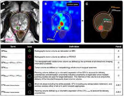
Figure 1. Tumor and target volume definitions used in prostate cancer radiation therapy. (A) Information from a magnetic resonance imaging (MRI) of this patient and (B) information from positron emission tomography (PET)/ computed tomography (CT). (C) A schematic view of the prostate with the combined information from MRI and PET.
Abbreviations: CTV = clinical target volume; GTV = gross tumor volume; GTVBoost = Boost gross tumor volume; GTVHisto = Histopathological gross tumor volume; PTV = planning target volume; PTVBoost = Boost planning target volume.

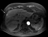
Figure 2. The value of MRI in detection of early HCC in high-risk patients
59-year-old man with Child-Pugh A cirrhosis. A) Compared to baseline MRI 9 months earlier, a new small (5 mm) arterial enhancing subcapsular lesion is seen (arrow, classified as LIRADS-3). B) 14- months follow up MRI with the lesion (arrow) unchanged, C) and D) Nine months later (23 months (about 2 years) after ad. A) the lesion has grown to 13 mm (arrow) including classical signs of an early HCC (BCLC stage 0) with wash-out and capsular enhancement (LIRADS-5). Successful percutaneous microwave ablative therapy was performed. Three years later there were no signs of recurrent or new HCC. MRI is instrumental in time diagnosis of early HCC in high-risk patients with consequently proper treatment and cure.
(Ref. Fiduzi FIF, Willemssen F, de Braak CV, de Lussanet de la Sablonière QG, JNM IJ, Bos D, de Man RA, Dwarkasing RS. Evaluation of Hepatocellular Carcinoma Surveillance with Contrastenhanced MRI in a High-Risk Western European Cohort. Curr Probl Diagn Radiol. 2024;53(6):709-16.)
Rectal cancer imaging
Colorectal cancer is the third most common malignancy and third cause of cancer-related deaths in men and women. With the Erasmus MC a few centers in the Netherlands are specialized in treatment of local recurrent rectal cancer. In our institution many of these patients will undergo surgery with curative intention. Dedicated MRI is an integral part of the work-up and important for clinical decision making. Our research aims to develop and validate modern MR techniques for better selection and follow up of complete responders after preoperative chemo-radiotherapy of (recurrent) rectal cancer. Furthermore, morphological and functional MRI characteristics based on multiparametric MR imaging and post-processing tumor texture analysis may help predict surgical outcome and long-term prognosis.
Cervical cancer imaging
Figure 3. MRI of malignancy complicating perianal fistulizing disease in a patient with Crohn’s disease.
A 42-year-old male patient had a history of chronic persistent fistula (A) and refrained from treatment for 5 years. On follow-up MRI a mass lesion is visible, arising within the fistula tract and ending into the supra-levator space with evident invasion in the surrounding tissue (arrow). (Ref. Arkenbosch JHC, van Ruler O, de Vries AC, van der Woude CJ, Dwarkasing RS. The role of MRI in perianal fistulizing disease: diagnostic imaging and classification systems to monitor disease activity. Abdom Radiol (NY). 2024)

Cervical cancer is the fourth most common cancer among females, and the fifth cause of cancer-related death in women. In daily practice clinical examination with or without anesthesia is until recently the principal investigation for assessment and therapy planning in cervical cancer. However, since our published systematic review comparing clinical examination vs MRI the new national guideline has given the latter a more prominent role in primary staging. Based on our findings the international guidelines of ESTRO, together with ESGO and ESP has shifted completely to MRI as the hallmark for diagnostic staging. In an Erasmus MC lead collaborative prospective multicenter study with UZ Leuven and AMC we investigated the value of optimized MR imaging, including diffusion weighted imaging (DWI) in the detection of early tumor residue in the cervix after radiotherapy. The main finding was that DWI forms an irreplaceable part of the protocol since it increases significantly the detection of residual tumor.
Expectations & Directions

Prostate cancer imaging: Means to detect prostate tumors at an early stage and to robustly characterize the aggressiveness are badly needed. MR imaging in prostate cancer is an important research line within the abdominal imaging section. Image-guided biopsy based on multiparametric MR examinations has been established. MRI/US-fusion guided biopsies have been implemented in clinical trials and in patient care. Association of imaging traits with gene expression profiles of prostate cancer
will certainly be an area of interest, in close correlation with the Erasmus MC departments of Urology, Pathology and Biomedical Imaging Group Rotterdam ( BIGR) . Developments on nuclear imaging techniques (o.a. PSMA PET CT) will be explored to correlate morphological and functional prostate MR images to PET CT images of prostate metabolism, most preferably with the new PET MRI scanner. We intend to explore strategies to improve diagnostic accuracy (Fig 4), reduce the number of biopsy cores needed, and improve therapeutic decisionmaking, thereby lowering morbidity, increasing quality of life, and reducing costs.
Hepatic cellular cancer imaging: MR imaging in hepatic cellular cancer (HCC) is expected to become a growing research line within the department, in close collaboration with the Erasmus MC departments of Hepatology/ Gastroenterology and hepatobiliairy Surgery. HCC screening in patients with hepatitis and cirrhosis is becoming centralized in academic centers. Collaborating networks such as the Dutch Hepatic Cancer Group (DHCG) are evolving, in which the Erasmus MC plays a dedicated and central role. Furthermore, interventional therapy plays an important role in minimal invasive liver cancer treatment. Critical for its success is an accurate target of the lesion. Research is focused on the development of real time image fusion techniques to guide interventional treatment and navigation surgery.
Radiomics: MR imaging biomarkers are being explored and validated for personalized decision-making and for prediction of treatment efficacy. A prerequisite for implementation of the multiparametric MR imaging modality is an accurate and fast post-processing method that can translate complex quantitative information into clinically useful directions. The development and validation
of such methods is research in joint cooperation with industrial partners. ‘Radiomics’ entails the extraction of predictive features for diagnostic decision-making and treatment response from standard CT or MR image using mathematical models. We will further expand our imaging expertise and apply novel imaging methods to improve our understanding of the genetic and disease development in abdominal oncology. We will initiate new projects in the field of oncological imaging analysis, with machine learning and deep learning techniques (Figure 5). We will continue to increase our research efforts into investigating clinically related oncological research questions.
Funding
Niessen, Wiro, Ivo Schoots, Jifke Veenland , and Chris Bangma (Urology) Erasmus MC-TKI-LSH: ‘Personalized Prostate Cancer Management using Multi-parametric MRI and Machine Learning (PPCM4)’. 2020 - 2024
Pensky, Bagdi Ulas, Wallace Bolan, Gonda Hecht, Marco Bruno, and Ivo Schoots NIH grant: ‘Deep learning methods for characterization of pancreatic cysts (Cyst-X project)’. 2021 - 2025
Schoots, Ivo , and Uulke Van der Heide (Radiation Oncology) KWF clinical implementation grant: ‘PROCESS study: PROstate Cancer - Expansion of Surveillance Selection criteria with MR imaging’. 2021 – 2025
Van den Bergh, Roderick, Rik Somford (Urology), and Ivo Schoots SKMS project/ZonMW: ‘Evaluatie en optimalisatie diagnostisch traject prostaatkanker middels MRI’ 2022 – 2026
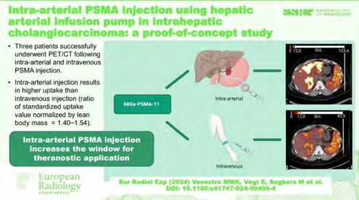
Figure 4. Proof-of-concept study from Mara Veenstra et al showed that it is feasible to inject 68GA- PSMA-11 in the hepatic artery in patients cholangiocarcinoma and that the uptake was higher compared to venous injection, paving the way for eventual intraarterial theranostics.
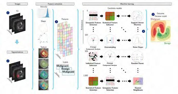
Figure 5. Schematic overview of the radiomics approach. Input to the algorithm are the T2-weighted magnetic resonance imaging scans (1) and the lesion segmentations (2). Processing steps include feature extraction (3) and the creation of a machine
Dwarkasing, Roy, Francois Willemssen , Rob de Man, Bart Takkenberg, and Carine Uyl-De Groot KWF implementation grant: ‘Validation of a Short and effective MRI Surveillance protocol for hepatocellular carcinoma screening in practice’ 2022 – 2026
Lambregts, Doenja, Ritse Mann, and Ivo Schoots KWF implementation grant: ‘Towards clinical implementation of novel diagnostic tools in oncologic imaging: an opensource web-based reviewing infrastructure for validation studies’. 2023 – 2027
Aben, Katja, Berdine Heesterman, Daniela Oprea, Pim van Leeuwen, and Ivo Schoots KWF implementation grant: ‘ADMINISTRATE (Advanced Diagnostic Modalities in ImagiNg Impacting on diagnosiS, TReatment And paTient outcomE) on prostate cancer’. 2023 – 2026
Thomeer, Maarten, et al. KWF clinical research Grant: effect of FAPI PET-CT on management in patients with potentially resectable biliary tract cancers: prospective multicenter study and cost-effectiveness analysis. 2023- 2027
Klein, Stefan, Maarten Thomeer NWO TTW open technology: 'The Liver Artificial Intelligence (LAI) consortium: a benchmark dataset and optimized machine learning methods for MRI-based diagnosis of solid appearing liver lesions'. 2023
learning decision model (5) using an ensemble of the best 100 workflows from 1000 candidate workflows (4), where the workflows are different combinations of the different analysis steps (e.g., the classifier used). (Starmans MPA, et al. Academic radiology. 2023).
Highlights
Ivo Schoots was Co-Chair PI-RADS steering committee, on prostate MR imaging.
François Willemssen contributed to two Dutch guidelines for diagnostic abdominal imaging for HCC and Cholangiocarcinoma.
Maarten Thomeer contirbuted to the Dutch guidelines for ovarian, endometriual and cervical carcinoma.
Additional Personnel
Demi Huijgen – PhD student Petriatic Surgery Erasmus MC Angela Amirabile – Humanitas Research Hospital Italy

Roy Dwarkasing MD, PhD
Project Funding Validation of a Short and effective MRI Surveillance (SMS) protocol for hepatocellular carcinoma screening in practice (KWF 2021-2, project number 13803)
Email r.s.dwarkasing@erasmusmc.nl
Improved detection of early hepatocellular carcinoma in high risk patients with abbreviated (non-contrast MRI).
Hepatocellular carcinoma (HCC) is the sixth most common cancer worldwide and the third most common cause of cancer related death. The incidence of HCC has been rapidly rising in Western countries and is expected to continue to rise in the next decades. In general, prognosis of patients with HCC is poor, except for patients with early-stage HCC who are eligible for curative treatments. According to the Dutch HCC guideline, biannual US surveillance should be offered to high rsik patients (with cirrhosis and chronic hepatitis B or -C). However, the reliability of US is limited in these pa-
tients. Better surveillance tools are urgently needed. We have developed and validated a short MRI surveillance (SMS) protocol for HCC in high-risk patients. The SMS protocol consists of the following three sequences: a) Diffusion weighted imaging (DWI) with a minimum of 2 b-values (50 and 800 s/mm2); b) T2weighted (T2W) fast spin-echo (FSE) with fat saturation; and c) T1-weighted (T1W) in- and out-of-phase imaging. Evaluation of performance in daily practice and assessment of cost-effectiveness of the SMS are the next steps.
Maarten Thomeer MD, PhD
Project Funding KWF clinical research: Effect of FAPI PET-CT on management in patients with potentially resectable biliary tract cancers: prospective multicenter study and cost-effectiveness analysis.
NWO-TTW Grant: A benchmark dataset and optimized machine learning methods for MRI-based diagnosis of solid appearing liver lesions
Email m.thomeer@erasmusmc.nl
Advances in abdominal imaging
One of the forefront topics in abdominal imaging is the integration of a novel tracer called FAPI for PET-CT/MRI imaging. In a recent systematic review, we highlighted the benefits of utilizing FAPI PET-CT/MRI in the pretreatment staging of pancreatic, gastric, and cholangiocarcinomas. Collaborating with Amsterdam University Medical Center and University Medical Center Utrecht, we are poised to embark on a prospective imaging study investigating the utility of FAPI PET-CT in pretreatment staging specifically for cholangiocarcinoma. This study, funded by the KWF funding agency, is spearheaded by Mara Veenstra, MD, who is undertaking her PhD within this research scope. Liver cancer stands as one of the most prevalent cancers globally. While magnetic resonance imaging (MRI) is pivotal in its diagnosis, the diverse nature of both malignant and benign liver lesions poses a significant challenge, often leading to highly subjective interpretations. Rec-
ognizing this, the Liver Artificial Intelligence (LAI) consortium, recently awarded by the NWO, is dedicated to developing innovative MRI analysis techniques. These advancements aim to provide robust support in diagnosing liver lesions, ultimately enhancing treatment decisions tailored to each patient's needs.
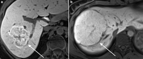

Figure: Our newly described “white-bordered flower sign” on gadoxetate disodium MRI helps distinguish focal nodular hyperplasia from hepatocellular adenoma. (Furumaya et al, 2024)
PhD Students

Mara Veenstra, MSc

Advisors Maarten Thomeer, Erik Verburg & Rutger-Jan Swijnenburg
Project Funding KWF Kankerbestrijding
Email m.m.k.veenstra@erasmusmc.nl
FAPI PET/CT for cholangiocarcinoma
We believe that additional preoperative [18F]F-FAPI PET/CT for patients suffering from proximal cholangiocarcinoma can detect metastases that cannot be found using CT and MRI, and that futile surgeries can be avoided using this new work-up.
Francois Willemssen, MD, MSc
Advisors Roy Dwarkasing & Daniel Bos
Email f.willemssen@erasmusmc.nl
Dr. Willemssen is responsible for the radiological contribution in the Multidisciplinary Liver Tumor Board, participates in the research committee of the Dutch Hepatocellular and Cholangiocellular Group (DHCG), is involved in the revision of two national guidelines, Cholangiocarcinoma and Hepatocellular carcinoma, and contributes to several research projects on liver imaging.
One of the projects is the use of Contrast-Enhanced US in cystic liver lesions.

Neslisah Seyrek, MD, MSc

Advisors Ivo Schoots & Arno van Leenders
Project Funding Jaap Schouten Foundation: “Significance of cribriform growth in prostate cancer risk stratification”
Email n.seyrek@erasmusmc.nl
The value of mpMRI on prediction of cribriform growth in prostate cancer
Assesment of the presence of cribriform growth plays a crucial role in risk stratification and active survellience eligibilty of prostate cancer patients, especially in intermediate risk group. Adverse imaging parameters derived from mpMRI may contribute in prediction of cribriform presence and enhance the presicion of risk assesment of men with intermediate risk prostate cancer.

Céline van de Braak, MSc

Advisors Aad van der Lugt & Rob de Man, Roy Dwarkasing & Daniel Bos
Project Funding KWF Grant
Email c.vandebraak@erasmusmc.nl
Validation of Short MRI Surveillance (SMS) protocol for hepatocellular carcinoma (HCC) screening in practice
An abbreviated MRI protocol for the screening of HCC will be validated in a high-risk patient population and compared to bi-annual ultrasound screening (current standard). To our knowledge, this will be the first study to perform this head-to-head comparison. This may provide improved detection of early stage HCC and consequently improved survival of HCC patients in a surveillance cohort.

Edwin Oei is a Full Professor of Musculoskeletal Imaging, and Section Chief of musculoskeletal radiology in Erasmus MC’s Department of Radiology & Nuclear Medicine. He obtained his medical degree in 2004 and his PhD on MRI for traumatic knee injury in 2009, both from Erasmus University Rotterdam. He also holds an MSc in Clinical Epidemiology from the Netherlands Institute for Health Sciences. Dr Oei is the principal investigator of musculoskeletal imaging research and engages in many academic activities including supervising 14 PhD students, lecturing, board and committee memberships, and refereeing for various journals.
Until October 2024 he was the President of the European Society for Magnetic Resonance in Medicine and Biology (ESMRMB). In 2013, Edwin Oei spent a one year research sabbatical as a Visiting Assistant Professor in the Joint and Osteoarthritis Imaging with Novel Techniques (JOINT) lab of the Department of Radiology of Stanford University, CA/USA. Dr. Oei is also the Clinical Research Focus Group leader in the Department of Radiology & Nuclear Medicine, and the principal coordinator of the Academic Center for Molecular and Cellular Imaging at Erasmus MC. e.oei@erasmusmc.nl
ADVANCED MUSCULOSKELETAL IMAGING RESEARCH ERASMUS MC (ADMIRE)
Edwin HG Oei, MD, PhD full professor
Context
Imaging is key in the study of musculoskeletal diseases. More sensitive and accurate imaging techniques are needed to advance our understanding of the development, mechanisms, and subtypes of musculoskeletal disorders. This is especially important for disorders that are common and have large impact on patients and the society, such as osteoarthritis, osteoporosis, sports injuries, and chronic musculoskeletal pain. New imaging biomarkers based on CT, MRI, ultrasound and nuclear imaging can also facilitate early diagnosis and development of targeted therapies. Before they can be applied routinely, new imaging techniques need to be optimized and validated. When applied in clinical research studies, novel, quantitative, imaging tools can provide additional imaging biomarkers. In large population-based studies, imaging is essential to characterize the development and aging of the musculoskeletal system. To analyze large imaging datasets, efficient image analysis methods, incorporating radiomics and artificial intelligence, are crucial.
Top Publications 2024
Van Zadelhoff TA, PK Bos, A Moelker, SMA Bierma-Zeinstra, RA van der Heijden, EHG Oei. Genicular artery embolisation versus sham embolisation for symptomatic osteoarthritis of the knee: a randomised controlled trial. BMJ Open 2024; 14:087047
Wu T, S Estrada, R van Gils, R Su, VWV Jaddoe, EHG Oei, S Klein. Automated Deep Learning-Based Segmentation of Abdominal Adipose Tissue on Dixon MRI in Adolescents: A Prospective Population-Based Study. AJR Am J Roentgenol. 2024; 222:2329570.
Kok J, MSAM Bevers, B van Rietbergen, EHG Oei, R Booij. Quantification of bone microarchitecture using photon-counting CT at different radiation doses: A comparison with µCT. Eur J Radiol. 2024; 181:111717.
Research Projects: Objectives & Achievements
Optimization and validation of advanced imaging techniques for common musculoskeletal diseases
This research line addresses the optimization and validation of novel imaging techniques based on CT, MRI, ultrasound and nuclear imaging. We successfully validated quantitative MRI techniques to assess altered processes or composition in musculoskeletal tissues such as cartilage, meniscus and tendon, against tissue references and clinical outcomes. We also validated advanced, contrastenhanced ultrasound and shear-wave elastography in osteoarthritis and tendinopathy. More recently, photon counting CT has become of great interest as it has shown promise to characterize bone disorders such as fractures and osteoporosis better than current methods because of its ultra-high resolution (Figure 1). Validation for routine clinical use also includes assessment of additional value for patients and clinicians. We are currently conducting a randomized controlled diagnostic trial to study the value of PET-MRI for chronic musculoskeletal pain. In this research line, we collaborate closely with the Physics In MR and CT Technology Groups, Biomedical Imaging Group Rotterdam, Departments of Orthopedics and Sports Medicine and Pain Medicine (Frank Huygen), and external partners (University of Wisconsin, Stanford University, Mayo Clinic, GE Healthcare, and Siemens Healthineers).
Current projects:
– Quantitative cartilage MRI and SPECT in knee osteoarthritis ( Joost Verschueren )
– Photon counting CT (Ronald Booij, Post-doc Physics in CT group)
– PET-MRI for chronic musculoskeletal pain ( Marijn Mostert )

Application of advanced imaging techniques in clinical studies
A strength of our research is the multidisciplinary embedding and collaboration with many clinical research groups, which creates the possibility to apply novel imaging techniques in clinical studies, providing additional imaging biomarkers. Previously, we successfully applied advanced quantitative MRI techniques for cartilage composition (dGEMRIC, T1rho-, and T2-mapping) in several clinical studies on knee osteoarthritis. In some studies we also applied perfusion imaging with dynamic contrast-enhanced MRI (DCE-MRI) (Figure 2). We successfully implemented ultrashort echo time (UTE) MRI and shearwave elastography (SWE) ultrasound in the largest randomized controlled trial of patients with patellar tendinopathy (study funded by General Electric and National Basketball Association). We recently also introduced a short MRI scan of both hands without the need for contrast agents in a cohort of patients with early clinically suspected arthralgia. These projects are conducted in collaboration with the departments of Orthopedics and Sports Medicine (Duncan Meuffels, Robert-Jan de Vos, Koen Bos, Max Reijman, Denise Eygendaal, Sita BiermaZeinstra), Rheumatology (Annette van der Helm-van Mil, Pascal de Jong).
Current projects:
– Advanced MRI and ultrasound in a randomized controlled trial of patellar tendinopathy ( Stephan Breda , Jie Deng )
– Short Dixon MRI for hand arthralgia ( Sanne Boeren, Daniek van der Kaaij )

Figure 2. Dynamic contrast-enhanced MRI (DCE-MRI) based perfusion map of the synovium in the knee.
Figure 1. Ultra-high resolution photon counting CT image of the elbow displaying an osteochondral defect of the capitellum of the humerus.
Figure 3. 3D shape analysis of the femur based on automated segmentation of MRI scans in the Generation R cohort, showing different shape modes and the variations they explain.

Musculoskeletal imaging in population studies
We are actively involved in the two large population studies in Erasmus MC – the Rotterdam Study and Generation R. In the Rotterdam Study among middle-aged and elderly, our research focus is on studying determinants and imaging aspects of osteoarthritis and osteoporosis, apart from our support in phenotyping these diseases on radiography (multiple joints and spine), MRI (knee) and, in the future, EOS imaging. In the Generation R cohort of children and adolescents we play a leading role in studying development, growth and abnormalities of the hip, knee, and spine on focused rapid MRI scans as well as body composition on whole-body MRI (Figure 3). For such large datasets, it is necessary to make use of automated image analysis tools, which we develop together with the Biomedical Imaging Group Rotterdam (Dr. Stefan Klein and Dr. Jukka Hirvasniemi). In both projects, we collaborate closely with researchers from the departments of General Practice (Sita Bierma-Zeinstra, Marienke van Middelkoop), Internal Medicine (Joyce van Meurs), Pediatrics (Vincent Jaddoe, Liesbeth Duijts), and Public Health (Hein Raat).
Current projects:
– Hip shape in children ( Desirée de Vreede, Mirthe Kamphuis )
– Spine abnormalities in adolescents ( Marleen van den Heuvel )
– Knee shape in adolescents ( Rosemarijn van Paassen )
– Body composition in adolescents ( Tong Wu )
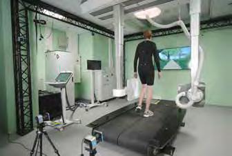
Figure 4. The MOBI lab setup.
Biomechanics and imaging
An important new direction in our research is the integration of (PET-)MR imaging with biomechanical measurements in the new Motion Biomechanics & Imaging (MOBI) lab, which was opened in October 2024. This is one of the first clinical laboratories in Europe that combines traditional motion capture equipment with fluoroscopy imaging during movement (Figure 4). This work will enable precision diagnosis of joint load in the context of osteoarthritis and other musculoskeletal disorders affecting biomechanics, by combining biomechanical data from the MOBI lab with advanced imaging using (PET-) MRI. This joint initiative with reseachers from TU Delft (Ajay Seth, Jaap Harlaar) marks one of several projects, largely conducted within the Flagship “Healthy Joints” as part of the “Health & Technology by Convergence” initiative between Erasmus MC Rotterdam, TU Delft and Erasmus University Rotterdam, in which our group plays a prominent role.
Current projects:
– Biomechanical precision diagnostics in osteoarthritis ( Niels Dur )
– Healthy Loading to combat osteoarthritis (LoaD project) ( Wouter Schallig )
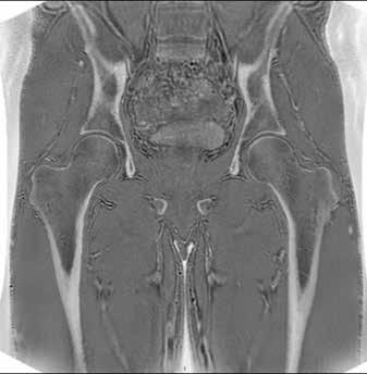
Miscellaneous projects
We study various other topics in the field of musculoskeletal imaging. We are involved in many clinical studies on osteoarthritis using "conventional" radiographic or MR imaging with semi-quantitative osteoarthritis grading. Examples include studies on specific risk factors for osteoarthritis such as overweight, anterior cruciate ligament rupture, or menopause, in collaboration with the departments of Orthopedics and General Practice. We also completed a randomized sham-controlled clinical trial on the efficacy of genicular artery embolization with follow-up using advanced (DCE-)MRI. In another project, we assess hip and groin disorders in athletes, utilizing generated CT-like images from MRI (Figure 5). Together with the Value-based Imaging group (Jacob Visser) we conduct several clinical validation studies of artificial intelligence algorithms, e.g. for fracture detection or osteoarthritis grading on radiographs, collaborating with industrial partners, and with the BIGR group we work on more fundamental AI and radiomics projects, e.g. on musculoskeletal tumors. We also fulfil a consulting or supportive role in many other studies primarily conducted by other groups at Erasmus MC and internationally (e.g. University of Queenland, LaTrobe, Lund, and Stanford Universities).
Current projects:
– Role of the meniscus in knee osteoarthritis ( Jan van der Voet )
– Genicular artery embolization for knee osteoarthritis ( Tijmen van Zadelhoff, with Adriaan Moelke r)
– Imaging of hip and groin disorders in athletes ( David Hanff )
– Artificial intelligence for musculoskeletal radiology ( Huib Ruitenbeek )
– Trustworthy artificial intelligence for musculoskeletal tumors ( Xinyi Wan )
Expectations & Directions
We have planned an expanding research activity in PETMRI, both for assessment of knee osteoarthritis (integrated with the MOBI lab) and musculoskeletal pain, which will be directed mainly by Rianne van der Heijden. There will also be exciting new developments in photon counting CT for musculoskeletal applications which will need to be validated. In image analysis there will obviously be an increased activity in artificial intelligence, both in terms of validating and determining additional value of clinical AI algorithms as in the development and application of research oriented algorithms. Finally, we expect a growing role in Generation R with the availability of more MR imaging biomarkers across different joints and with regard to body composition.
Funding
Oei, Edwin General Electric Healthcare: ‘Pinpointing the source of chronic pain and therapy response with wholebody 18F FDG-PET/MRI’. 2021-2025
Van der Heijden, Rianne Erasmus MC Starting Grant. 2024-2030
Oei, Edwin General Electric Healthcare: ‘RSNA QIBA MSK Profile Stage 3 and 4 Conformance Testing’. 2023-2025
Van der Heijden, Rianne NWO/ZonMw Veni talentprogram: ‘Towards better care for patients with chronic low back pain using advanced imaging’. 2024-2029
Oei, Edwin NWO Research along routes by Consortia (NWA-ORC): ‘Healthy Loading to combat osteoarthritis: Leveraging molecular variations in load bearing capacity for individualized movement aDvice: The LoaD project’. Main applicant: G.J.V.M. van Osch (Orthopedics). 2022-2030
Van der Heijden, Rianne Stichting Erasmus MC Pijnfonds: ‘Richting betere zorg voor patienten met chronische lage rugpijn met geavanceerde beeldvorming’. 2024-2027
Oei, Edwin NWO ROBUST program and General Electric Healthcare: ‘Innovation Center for Artificial Intelligence (ICAI) lab Trustworthy AI for Magnetic Resonance Imaging’. 2023-2026
Van der Heijden, Rianne AASPT New Investigator Grant, Freedom of movement Grant, and General Electric (GE) sponsored project : 'Identifying Factors Associated with Early Signs of Osteoarthritis in Former Collegiate Athletes after Anterior Cruciate Ligament Reconstruction'. 2022-2026
Figure 5. CT-like images of the hips and pelvic skeleton generated from MRI using a zero echo time (ZTE) technique.
Oei, Edwin Horizon 2020 EIC Accelerator: ‘AI algorithms in musculoskeletal radiography’. Main applicant: (Radiobotics, Copenhagen, Denmark). 2020-2025
Oei, Edwin Independent Research Fund Denmark: ‘Investigating pathology and tissue biomarkers of Osgood Schlatter to enhance treatment of children with growth-related pain’. Main applicant: M.Rathleff (University of Aalborg, Denmark). 2019-2024
Oei, Edwin NWO Zon-MW Open Competition: ‘Biomechanical precision diagnostics in osteoarthritis’. Main applicant: S. Bierma-Zeinstra (General Practice/Orthopedics). 2020-2025
Oei, Edwin NWO Zon-MW Goed Gebruik Geneesmiddelen: ‘Efficacy of antibiotic treatment for patients with chronic low back pain and Modic type I changes – randomised placebo controlled trial’. Main applicant: B.W. Koes (General Practice). 2023-2027
Oei, Edwin, Rianne van der Heijden, and Jukka Hirvasniemi TU Delft-Erasmus MC Convergence Flagship: ‘Healthy Joints’. Main applicant: S.M.A. Bierma-Zeinstra (General Practice), J. Harlaar (TU Delft). 2022-2027
Oei, Edwin ZonMw Gender en Gezondheid – Algemene onderzoeksronde: ‘The FOCUM human disease model for development of OA’. Main applicant: S.M.A. Bierma-Zeinstra (General Practice). 2019-2024
Oei, Edwin ZonMw Gender en Gezondheid – Algemene onderzoeksronde: ‘Diagnosis, prevalence and associated factors of osteoarthritis in adults with intellectual disabilities’. Main applicant: D.A.M. Maes-Festen (AVG/General Practice). 2020-2026
Oei, Edwin National Health and Medical Research Council, Australia: ‘SUPER rehabilitation RCT for young people with old knees’. Main applicant: K. Crossley (La Trobe, Melbourne, Australia). 2018-2024
Oei, Edwin European Research Council (ERC) Advanced grant: ‘Biomechanical precision diagnostics in osteo-arthritis Modelling trajectories and mechanisms of childhood hip dysplasia’. Main applicant: S.M.A. Bierma-Zeinstra (General Practice/Orthopedics). 2023-2028
Oei, Edwin FOREUM Foundation for Research in Rheumatology, Preclinical Phases of Rheumatic and Musculoskeletal Diseases: ‘Novel Treatment Targets in Early-stage Osteoarthritis’. Main applicant: M. Englund (Lund University, Lund, Sweden). 2018-2024
Invited Lectures
Edwin Oei. ‘ Oedema in bone and soft tissues of the knee’. ECR, Vienna, Austria. March 2024.
David Hanff . 'Ultrasound of the elbow'. Esser Masterclass, Rotterdam, the Netherlands. March 2024.
Edwin Oei. 'Is there a role for MRI in acute body & MSK Trauma? Pro: There is a role for MRI in acute trauma: M.D. perspective'. ISMRM, Singapore, Singapore. May 2024.
Edwin Oei. 'Slipping & sliding: Imaging of articular cartilage: Introduction'. ISMRM, Singapore, Singapore. May 2024.
Rianne van der Heijden . 'Whole body MRI in marrow evaluation'. ISMRM, Singapore, Singapore. May 2024.
David Hanff . 'MSK ultrasound'. Radiologist days, Den Bosch, the Netherlands. May 2024.
Edwin Oei. 'Photon counting CT for musculoskeletal applications'. UK Imaging and Oncology Congress, Liverpool, UK. June 2024.
Edwin Oei. 'Hot topics in MSK radiology: Photon-counting CT'. NVvR Joint Dutch-Belgian MSK section day, Rotterdam, the Netherlands. June 2024.
Edwin Oei. 'Genicular artery embolization for pain treatment in patients with knee OA: Results from clinical trials'. International Workshop on Osteoarthritis Imaging, Marrakech, Morocco. June 2024.
David Hanff . 'Ultrasound of the shoulder'. Association of Surgery for Medical Students, Orthopedics, Rotterdam, the Netherlands. June 2024.
Rianne van der Heijden . 'PET/MRI for the identification of the pain generator in chronic pain'. The Future of Medical Imaging and Radiotherapy, Almere, the Netherlands. June 2024.
David Hanff. 'Hip MRI-findings could send you down a rabbit hole – 5 lessons to athletes and their medical teams'. Young Athlete’s Hip Conference, Oxford, UK. Sept 2024.
Rianne van der Heijden . 'The potential of FDG PET/MRI for the identification of the pain generator in chronic pain'. GE Healthcare PET/MRI Summit, Hamburg, Germany. Oct 2024.
Edwin Oei, Ronald Booij, and Denise van Beekveld. 'Photon-counting CT for musculoskeletal applications'. European Society of Musculoskeletal Radiology webinar, online. Nov 2024.
David Hanff . 'Ultrasound of the hip and groin'. Sports Medical Scientific Annual Congress, Vianen, the Netherlands. Nov 2024.
Highlights
Edwin Oei was the President of the European Society for Magnetic Resonance in Medicine and Biology (ESMRMB) until October 2024.
Rianne van der Heijden was appointed assistant professor in the Department or Radiology & Nuclear Medicine.
David Hanff became the president of the Musculoskeletal Radiology section of the Dutch Society of Radiology (NVvR).
Stephan Breda was awarded the best PhD thesis award during the annual scientific meeting of the Dutch Society for Sports Medicine (VSG) from 28 to 29 November 2024 in Vianen, the Netherlands.
Niels Dur received a Young Investigator Award during the International Workshop on Osteoarthritis Imaging (IWOAI) held in Marrakech, Morrocco, from 25 to 28 June 2024.
The opening of the Motion Biomechanics & Imaging (MOBI) lab attracted a lot of attention in the national public media. Interviews were given for TV (Omroep Max, WNL), radio (BNR, Radio 1), newspapers (Algemeen Dagblad).
Rianne van der Heijden completed her 2-year visiting assistant professorship in translational Body/MSK MRI at the University of Wisconsin-Madison, funded by Bracco Diagnostics.
Marijn Mostert won the first prize in the clinical trainee abstract competition of the Musculoskeletal MR imaging Study Group during the ISMRM annual meeting in Singapore from 4-9 May 2024.
The Amphibi trial on the added value of FDG PET/MRI for chronic pain was highlighted by AuntMinnie after Marijn Mostert ’s presentation at the ISMRM.
Rianne van der Heijden co-organized the Junior Fellow symposium ‘Innovations & Future Perspectives in MRI Technology’ during the ISMRM annual meeting in Singapore from 4-9 May 2024.
Edwin Oei hosted Dr. Francis Baffour from the Mayo Clinic, Rochester, USA, as the inaugural Gabriel P. Krestin Visiting Professor in our department, from 26 August to 6 September 2024.
David Hanff organized the Sandwich Course Musculoskeletal Radiology for the Dutch Society of Radiology (NVvR) in Ede, the Netherlands, on 6-7 November 2024. He also organized and chaired the Dutch and Belgian MSK meeting of the NVvR in Rotterdam, the Netherlands on 22 June 2024.
Mariëlle Olsthoorn represented the Dutch Society of Radiology (NVvR) in developing the new national guideline on acute muscle injury of the lower extremity in athletes.
Edwin Oei served on the MSK Scientific Program Committee of the RSNA, the MSK Scientific Subcommittee for the ECR, and the Research Taskforce of the ESSR.
Additional Personnel
Galied Muradin, MD, PhD – Musculoskeletal radiologist.
Mariëlle Olsthoorn, MD – Musculoskeletal radiologist.
Koen Willemsen, MD, PhD – Affiliated post-doc focusing on 3D imaging and printing.
Erin Macri, PhD – Affiliated assistant professor from Dept. of Orthopedics focusing on biomechanics.
Guillaume Renaud, PhD – Affiliated associate professor from TU Delft focusing on ultrasound of bone.
Eveline Molendijk, MD – Affiliated researcher from Dept. of General Practice.
Huib Ruitenbeek, MSc – Affiliated PhD student, see p. 275
Xinyi Wan, MSc – Affiliated PhD student, see p. 112
Thom van der Laan, MSc – Affiliated PhD student, see p. 53
Mirthe Kamphuis, MSc – Affiliated PhD student, see p. 104
Núria Jansen, MSc – Collaborating PhD student from Dept. of General Practice.
Visiting researchers and interns
Max van Zijderveld, BSc – Student Technical Medicine, TU Delft. Dec 2023-Feb 2024. Daily supervisor Marijn Mostert.
Josefien van den Berg, BSc – Student Technical Medicine, TU Delft. June-Aug 2024. Daily supervisor Marijn Mostert.
Tim Kortman, BSc – Student Technical Medicine, TU Delft. Sept-Nov 2024. Daily supervisors Marijn Mostert, Rianne van der Heijden.
Willemijn Scholtes, BSc – Student Technical Medicine, TU Delft. Sept-Nov 2024. Daily supervisors Marijn Mostert, Rianne van der Heijden.
Josefien van den Berg, BSc – Student Technical Medicine, TU Delft. Nov 2024-June 2025. Daily supervisors Marijn Mostert, Rianne van der Heijden.
Netanja Harlianto, MSc – Student Medicine, University of Utrecht. Throughout 2024. Daily supervisor Jukka Hirvasniemi.
Lucas Bronder, MSc – Student NIHES Research Master Health Sciences, Nov 2023-Sept 2025.
Tobias Haueise, MSc – Visiting PhD student, Tübingen University Hospital, Germany. Sept 2024.
Yijie Fang, MD – Visiting radiologist/researcher, The Fifth Affiliated Hospital, Sun Yat-Sen University, China. Sept 2024-Sept 2025.
Assistant Professor Rianne van der Heijden, MD, PhD
Email r.a.vanderheijden@erasmusmc.nl

ADVANCED PHYSIOLOGICAL MUSCULOSKELETAL IMAGING
Rianne van der Heijden completed her medical degree at Erasmus University Medical Center, followed by a PhD at the Departments of Radiology and Nuclear Medicine and General Practice. Her doctoral research focused on applying advanced physiological imaging techniques, such as perfusion MRI, in patients with chronic knee pain. She subsequently entered the Radiology and Nuclear Medicine residency program, specializing in musculoskeletal imaging including Nuclear Medicine techniques. After completing her residency, she was awarded a Bracco research fellowship through the University of Wisconsin-Madison, where she deepened her technical expertise. She currently holds the position of Adjunct Clinical Assistant Professor at UW-Madison. Rianne co-leads translational studies that implement cutting-edge (PET)/MRI techniques, with a particular emphasis on imaging chronic pain at both Erasmus MC-Rotterdam and UW-Madison. In 2024, she was honored with a ZonMW VENI grant to further develop her research line in pain imaging.
Musculoskeletal pain
Chronic musculoskeletal pain places a significant burden on both patients and society. Many patients receive suboptimal treatment or no treatment at all, largely because current anatomy-based imaging techniques cannot accurately identify the underlying causes of pain. Innovative hybrid molecular imaging approaches, such as 18F-fluorodeoxyglucose (FDG) and Manganese (Mn) PET-MRI, have emerged to address this gap. These methods are designed to target the underlying pathophysiological changes associated with pain, such as low-grade inflammation and heightened neuronal excitation. Additionally, low-grade inflammation can also be detected using perfusion imaging techniques, such as dynamic contrast-enhanced MRI. These advanced diagnostic imaging methods have demonstrated promise in identifying pain generators in an ongoing randomized controlled trial (RCT) involving patients with chronic low back and hip pain. Future research aims to explore other PET tracers that may offer greater specificity for distinct pain pathways, further advancing diagnostic capabilities in this field.
Technical advances
My work bridges technological innovation and clinical application. I am actively involved in advancing PET/MRI acquisition and postprocessing techniques as member of the PET/MRI Center of Competence (CoC). Our work includes improving PET/MRI attenuation correction near metal, which is essential for accurate imaging in patients with implants and developing automated tools for PET lesion detection in patients with

Figure 1. FDG PET/MRI in a patient with chronic low back pain. MRI shows morphologic abnormalities at both the left and right facet joint, while the combination with FDG PET shows only the right is metabolic active as sign of low-grade inflammation. Facet denervation followed, leading to pain relief.
chronic pain to shorten reporting time. Another key area of focus is optimizing quantitative DCE-MRI analysis to facilitate clinical translation. As part of this effort, I have been a longstanding contributor to QIBA (Quantitative Imaging Biomarker Alliance of the RSNA) and OSIPI (Open Science Initiative for Perfusion Imaging
Additionally, I am a member of the Advanced Musculoskeletal Imaging Research group (ADMIRE), where I co-supervise two PhD students conducting research on the application of advanced (PET/)MRI in knee osteoarthritis.
Post-docs

Wouter Schallig, PhD
Project Funding NWA-ORC: the LoaD project
Email w.schallig@erasmusmc.nl

Combining imaging and biomechanics to unravel knee osteoarthritis
In 2040, osteoarthritis is the most common disease in the Netherlands affecting 1.8 million people according to the RIVM. Forces affecting the knee play an important role in the development of osteoarthritis. This so-called joint loading can be quantified with biomechanical measurements. However, up until now these measures have not been sufficiently accurate to quantify the cartilage loading. My research focuses on developing protocols in the recently established MOBI lab in our department, and subsequently applying these to answer clinically relevant questions. The MOBI lab is the first lab in the Benelux that combines fluoroscopy imaging with the traditional marker-based motion capture technology.
PhD Students

Sanne Boeren, MD, MSc

Advisors Annette van der Helm - van Mil, Edwin Oei & Pascal de Jong
Email a.boeren@erasmusmc.nl
Short MRI scan for clinically suspect arthralgia
Patients with Clinically Suspect Arthralgia are at risk for Rheumatoid Arthritis. MRI detected subclinical joint inflammation is an important predictor. A short modified Dixon sequence has been developed (in the LUMC) to reduce scantime and lower MRI-costs. We study its accuracy in the Rotterdam CSA cohort, the relation with ultrasound and we determine the cut-off for positivity in an "ATLAS-study". This project takes places in the Medical Delta. Ultimately, we envision that scans will be processed with AI and that a rapid scan is followed by a rapid answer.
With this combination of techniques we can accurately quantify bone movements, joint loading and cartilage strain and stresses. In the LoaD project we apply these measurements to patients with knee osteoarthritis during walking, running and cycling. We will also relate those loading parameters to the outcomes of PET/MRI scans to better understand the relation between loading and biology within osteoarthritis and eventually improve personalized treatment and prevention strategies. This project is embedded in the Convergence initiative between Erasmus MC and TU Delft.
Joost Verschueren, MD, MSc

Advisors Edwin Oei, Max Reijman & Sita Bierma-Zeinstra
Project Funding Dutch Arthritis Association (Reumafonds) and Netherlands
Orthopaedic Association (NOV): “Optimal timing for orthopaedic surgery in osteoarthritis”
Email j.verschueren@erasmusmc.nl
PhD Obtained 20-06-2024
Brace versus Osteotomy trial
Multicenter RCT investigating clinical and structural effects of a surgical (high tibial osteotomy) and non-surgical (valgus unloader brace) treatment in patients with medial knee osteoarthritis and a varus knee malalignment. Changes in articular cartilage and subchondral bone are assessed using quantitative MRI and SPECT/CT.

Jie Deng, MD, MSc

Advisors Edwin Oei, Denise Eygendaal & Robert-Jan de Vos
Project Funding Chinese Council Scholarship
Email j.deng@erasmusmcmnl
The 5-year course of patellar tendinopathy after performing exercise therapy
This project is the continuation of the JUMPER study, a randomized controlled clinical trial evaluating two exercise therapies for patellar tendinopathy, evaluated with advanced ultrasound (including shearwave elastography) and MRI (including ultrashort echo time techniques). We evaluate the prognostic value of physical measurements and imaging on clinical outcomes, and conduct a 5-years follow-up of the trial that includes repeat ultrasound scans.

Tong Wu, MD, MSc

Advisors Edwin Oei, Stefan Klein & Liesbeth Duijts
Project Funding China Scholarship Council
Email w.tong.1@erasmusmc.nl
PhD Obtained 21-o6-2024
Body composition imaging in adolescents
Our project (embedded in the Generation R Study, a population-based prospective cohort study) aimed to implement a deep learning-based method for the automated quantification of abdominal adipose tissue on whole-body Dixon MRI scans in adolescents. Subsequently, the associations of physical activity, body composition, and adolescent respiratory health were examined

David Hanff, MD, MSK radiologist

Advisors Edwin Oei, Adam Weir, Rintje Agricola & Joshua Heerey
Email d.hanff@eramsusmc.nl
Hip and groin pain in athletes
In collaboration with the orthopedics department and the La Trobe University Melbourne we investigate clinical and imaging findings in athletes with hip and groin pain. The main focus is on the pubic symphysis and the hip concerning FAI (femoro-acetabular impingement). We aim to assess the utility of a zero echo time (ZTE) MR imaging technique providing CT-like images.

Marijn Mostert, MSc

Advisors Edwin Oei & Rianne van der Heijden Project Funding General Electric Healthcare: “Pinpointing the source of chronic pain and therapy response with whole-body 18F FDG-PET/MRI”. Email m.mostert@erasmusmc.nl
18F-FDG PET/MRI for chronic pain
Chronic lower back pain and hip pain have a very large burden on patients and the healthcare system. Accurate identification of sources of pain in these patients is not straightforward, and conventional imaging techniques are insufficient. The AMPHiBI Trial aims to evaluate the value of 18F-FDG PET/ MRI for identifying chronic pain generators in a randomized controlled clinical trial. In addition, I work on technically oriented projects in the field of PET/MRI and DCE-MRI.

Niels Dur, MD, MSc

Advisors Jaap Harlaar, Edwin Oei, Erin Macri & Rianne van der Heijden
Project Funding ZonMw Open Competition 'Biomechanical precision diagnostics in osteoarthritis'
Email n.dur@erasmusmc.nl
The role of biomechanics in OA
By combining high-fidelity biomechanical measurements with multi-scale modeling, we aim to clarify the role of disturbances in joint biomechanics in early stages of osteoarthritis (OA). By correlating these measurements with findings on advanced PET-MR imaging, we hope to identify potential imaging-biomarkers for early-OA, which will aid in developing preventative or disease-modifying treatment strategies In future research.

Daniek van der Kaaij, MSc

Advisors Annette van der Helm - van Mil, Edwin Oei & Pascal de Jong
Email d.vanderkaaij@erasmusmc.nl
Dixon MRI for clinically suspect arthralgia
Imaging-detected subclinical joint inflammation is a promising predictor of progression from clinically suspect arthralgia to rheumatoid arthritis. A short Dixon MRI sequence without contrast enhancement has been developed to improve the feasibility of MRI. Our project in collaboration with Leiden University Medical Center aims to implement Dixon MRI in risk stratification. In the future we envision to automatically evaluate the scans using AI.

Jan van der Voet, MD, MSc
Advisors Sita Bierma-Zeinstra, Edwin Oei, Jos Runhaar & Dammis Vroegindeweij
Project Funding ZonMw, ReumaNederland
Email javandervoet@gmail.com
PhD Obtained 07-03-2024
The role of the meniscus in knee OA, studied with MRI
To evaluate the association between meniscus extrusion, (change in) meniscus volume and the interplay between meniscus volume and extrusion with incident knee osteoarthritis. Factors associated with extrusion might be potential targets to prevent or delay the degenerative process. MRI data from the PROOF study and population-based Rotterdam Study are used.

Tijmen van Zadelhoff, MD, MSc

Advisors Edwin Oei, Koen Bos & Rianne van der Heijden
Project Funding Stichting Coolsingel, COOK Medical, Erasmus MRace
Email t.vanzadelhoff@erasmusmc.nl
Genicular artery embolization for knee osteoarthritis
Genicular artery embolization (GAE) is a minimally invasive treatment for knee osteoarthritis (KOA) with promising results from multiple cohort studies. This project is a 1 year RCT to determine the efficacy of GAE in symptomatic KOA patients resistant to conservative therapy. We showed that GAE does not offer more pain reduction than sham treatment. Advanced MRI including dynamic contrast-enhance MRI is applied to assess tissue changes following embolization.

Rosemarijn van Paassen, MSc

Advisors Edwin Oei, Marienke van Middelkoop & Sita BiermaZeinstra
Project Funding ReumaNederland: “Identifying risk factors for knee osteoarthritis by understanding adolescent knee joint development”
Email r.vanpaassen@erasmusmc.nl
Semi-automatic segmentation of knee MRI
The developed semi-automatic segmentation algorithm, consisting of a spatial and an appearance component, is accurate and can be applied to large MRI datasets. This method enables us to explore the association between shape variation, development of the knee and the presence of knee pain in children in the population-based Generation R Study
Desirée de Vreede, MSc, MD
Advisors Edwin Oei & Aad van der Lugt
Email d.devreede@erasmusmc.nl
MRI of the hips in the generation R study
This research Is part of a population-based prospective cohort study following children from fetal life until adulthood (Generation R). A total of 2863 MRI scans of the pelvic and hip area were collected at 10.2 years (mean, range 8.6-12.9 years) We aim to identify genetic and environmental causes of normal and abnormal growth and development of the lumbar spine and hip.

Marleen van den Heuvel, MD

Advisors Marienke van Middelkoop, Edwin Oei & Sita Bierma-Zeinstra
Project Funding EUR Fellowship 2017
Email m.m.vandenheuvel@erasmusmc.nl
Growing Up: lifestyle and jointh health
Structural spinal abnormalities and shape variations might play a role in the development of back pain in children and later in life, and have been shown in children on MRI. For this project data of children aged 9 years old from the Generation R Study is used to assess the prevalence of structural spinal abnormalities and shape variations, and to investigate associations with weight status and physical activity level of the children.


INTERVENTIONAL RADIOLOGY
Aad van der Lugt, MD, PhD
full professor

Context
Interventional radiology is a rapidly expanding field of image-guided procedures allowing to obtain tissue samples or treat various diseases through a small hole in the skin.
In the Erasmus MC, many interventional radiology procedures are being performed yearly with a focus on advanced oncological interventions, hepatobiliary interventions, vascular (arterial and venous) interventions, urological and neuro-interventions.
The Erasmus MC is a center of expertise for complex neuro-interventions, vascular malformations, hepatobiliary diseases, and oncological interventions, accredited by the European society of interventional radiology (CIRSE IASIOS certification).
The section of interventional radiology is actively participating in scientific research, aiming at improving current treatment strategies and development novel therapies in the fields mentioned above.
Top Publications 2024
Terlouw LG, L van Dijk, D van Noord, OJ Bakker, DC Bijdevaate, NS Erler, B Fioole, J Harki, DAF van den Heuvel, JW Hinnen, JJ Kolkman, S Nikkessen, AS van Petersen, HFM Smits, HJM Verhagen, AC de Vries, JPM de Vries, D Vroegindeweij, RH Geelkerken, MJ Bruno, A Moelker. Covered versus bare-metal stenting of the mesenteric arteries in patients with chronic mesenteric ischaemia (CoBaGI): a multicentre, patientblinded and investigator-blinded, randomised controlled trial. Lancet Gastroenterology and hepatology 2024; 9:299-309.
Van Rijn MJ, MAF de Wolf. Go With the Flow. Eur Journal of Endovascular surgery 2024; 69.
Research Projects: Objectives & Achievements
The HJ trial – biodegradable stent placement versus standard-of-care for treatment of post-transplant anastomotic biliary strictures
Biliary complications are among the most frequently observed complications of liver transplant surgery and include postischemic strictures of the bile ducts and anastomotic bile duct strictures at the level over the hepaticojejunostomy. For anastomotic strictures, treatment approaches differ per center. Standardized treatment approaches are lacking with no comparative trials between the different treatment methods conducted thus far. Treatment trajectories often take several months, with multiple treatment sessions necessary to resolve the obstruction.
In the proposed randomized controlled trial, we aim to evaluate whether biodegradable stent have the potential to reduce the number of interventions necessary and how they impact on days of hospitalization, patient satisfaction and healthcare costs. We will do so by comparing percutaneous biodegradable stent placement to current standard of care at the Erasmus MC (stricture dilatation using internal-external biliary drainage tubes). We expect to submit the study proposal to the METC in the first months of 2025. The project is conducted by dr. Kay Pieterman (study PI, Interventional radiology) in close collaboration with the department of gastro-enterology and hepatology (dr. Raoel Maan, co-principal investigator).
Flush trial – effects of catheter flushing on cholangitis and ho spitalization in patients with biliary drainage tubes
Patients with biliary disease are frequently treated using percutaneous biliary drainage tubes to restore flow of bile to from diseased bile ducts to the bowel. In this ongoing RCT we investigate whether patients treated with biliary drainage tubes have better outcomes when biliary drainage tubes are flushed 3 times a day using a syringe of standard physiological saltwater solution. Patients are randomized between either flushing or no flushing of the drain. We are currently halfway with inclusion of patients towards the desired sample size. The study is conducted by Kay Pieterman (study PIin close collaboration with the department of gastro-enterology and hepatology and department of hepatobiliary surgery (physician asssistant Chulja Pek and Prof. Bas Groot Koerkamp).
Percutaneous direct portal pressure measurement using pressure wires – a pilot study
The gold standard for assessing portal hypertension is currently hepatic portal vein pressure gradient measurement, in which the portal pressure is derived indirectly by measuring the pressure increase when inflating a balloon occlusion catheter in the hepatic vein. Although this method is elegant, it is known that derived pressures do sometimes over- or underestimate actual portal pressure. In the proposed pilot by Dr. Raoel Maan (hepatologist, EMC) and dr. Kay Pieterman (Interventional radiologist), we will evaluate the feasibility, patient satisfaction and procedural time when performing a direct portal pressure measurement. For this method we will introduce a pressure wire - a microwire with a pressure sensor at the tip - in the portal system using ultrasound guidance. We aim to show that this method is easy, safe and reliable for invasive direct portal pressure measurement and will also explore potential applications of this approach during portal interventions such as the TIPS procedure. An NVGE gastrostart grant of 10.000 euro was awarded for the proposed pilot study. Dr. Kay Pieterman and Dr. Maan plan to commence with the pilot in 2025.
Identification of imaging biomarkers of tre atment success in patients treated with radioembolization for hepatocellular carcinoma
Dr. Kay Pieterman together with dr. Dwarkasing (abdominal radiologist, EMC) are currently building an imaging database of all the patients treated in the last 15 years with radioembolization or chemo-embolization for primary liver tumors. We aim to identify imaging biomarkers on baseline imaging that allow to predict treatment success of these procedure. Dr. Hong Liu Hong from China (radiologist) is currently working on the project for a 1 year research fellowship.
Fine needle aspiration versus core biopsy of suspected met astatic liver lesions.
Percutaneous core biopsies of suspected metastatic disease are frequently performed to provide histological proof of metastatic disease. At the Erasmus MC, approximately 300 core biopsies are performed each year for this indication. In the proposed trial currently in preparation, we aim to study whether fine needle aspiration is able to provide enough tissue for to prove metastatic disease. We will do so by obtaining additional fine needle aspiration tissue samples during standard-of-care core biopsy procedures. These samples will be separately analyzed after
finalization of the inclusion. If fine needle aspiration can provide enough tissue to confirm metastatic disease, this would result in less invasive procedure with less material costs with less hours of postprocedural monitoring necessary due to much smaller needle size. This potentially allows us to perform tissue sampling without the need of postprocedural hospital stay with monitoring. A 7000 euros Vaillant grant was honored for the proposed project. The study is conducted by Kay Pieterman (study PI) in close collaboration with the department of pathology and Nico Jansen (coordinating investor, radiology resident).

Figure 1. Biodegradable biliary stent (Ella CS, s.r.o., Czech Republic) made of bioresorbable polymer (Polydioxanone), specifically designed for treatment of non-malignant biliary strictures.
Intraductal imaging to assess infiltration depth of bile duct tumors – a pilot study
In the pilot study currently under preparation we hope to evaluate to what extent intraductal ultrasound can contribute to assessing infiltration depth and tumor length of irresectable malignant bile duct tumors. Endobiliary imaging, for example using intraductal ultrasound probes has the potential to provide more detail on tumor location and extent. This might help facilitate more precise stent placement and tissue sampling as well as potential future therapies endobiliary tumor ablation. We strive to perform a pilot study in 2025 as a collaborative project between Kevin Wiese (Interventional radiologist), Dr. Pieterman (Interventional radiology) and Prof. Bas Groot Koerkamp (HPB surgery).
Dragon
PLC trial
–
portal vs. double vein
embolization for future remnant growth in oncological liver resections
We are in preparation of collaborating in the multicenter Dragon Primary Liver Cancer trial initiated by Maastricht Medical Center (MUMC). In this randomized controlled trial, we aim to study whether double vein embolization (portal and hepatic vein) results in more liver volume increase of normal liver tissue than portal embolization alone for patients with a unilateral primary liver tumor
with insufficient liver remnant for resection. The study is funded by ZonMW and will run in different centers across Europe. The study is led by Maastricht Medical Center (PI Dr. Ronald van Dam) and CHU Liege, Belgium (Prof. Dr. Olivier Detry), with Sinead James PhD student and coordinating investor of the trial. Dr. Kay Pieterman is the local PI of the study.
Evaluation
of a standardized treatment protocol for patients with acute mesenteric
and portal vein thrombosis
Patients in the region presenting with an acute mesenteric vein or portal vein thrombosis are preferentially being referred to the Erasmus Medical center as a treatment center for vascular hepatology. Together with hepatologists and surgeons, a protocol was introduced in the clinic in order to achieve a more standardized treatment approach for this patient group. With this standardized approach we hope to improve patient outcome. We are currently evaluating how introduction of this protocol altered treatment outcome by comparing patients treated according to the protocol with a historical cohort of patients treated before the protocol was introduced. The study Is conducted by Sophie Ciere, master student and future PhD HPB surgery under supervision of dr. Kay Pieterman (Interventional radiology), dr. Sarwa Darwish Murat (gastro-enterology and hepatology) and dr. Jeroen de Jonge (HPB surgery).
Pressure study – intra-arterial pressure measurements during mesenteric artery stenting in chronic mesenteric ischemia.
In this prospective trial conducted as a collaborative project between the departments of radiology, gastroenterology and vascular surgery, we aim to perform intraarterial pressure measurements during mesenteric artery stenting. The study will be coordinated by Eva Bocharewicz, PhD chronic mesenteric Ischemia (department of radiology and gastroenterology), and supervised by Dr. Kay Pieterman (study PI), Dr. Leemreis (gastro-enterologist) and prof. Marco Bruno (gastro-enterologist).
The aim of this study is to evaluate whether pressure measurements can predict patient symptom relief of mesenteric artery stenting in chronic mesenteric ischemia. We hypothesize that mesenteric artery pressure measurements across a stenosis can provide additional information about its hemodynamical significancy, because it is known that extensive collateral networks between the different mesenteric arteries exist, with possible compensation of flow through collateral circulation pathways. We hypothesize that pressure measurements
across the stenosis can provide information on the sufficiency of the collateral circulation in maintaining mesenteric artery flow, thereby allowing us to predict which patient will benefit from stent placement. This is relevant, hence approximately 30% of the patients treated for suspected chronic mesenteric ischemia do not experience symptom relief after mesenteric artery stenting.
Silent Stroke trial
Ongoing trial assessing whether silent embolic events occur during endovascular interventions performed through the radial artery. 75% of the patients required for the study sample size have been included. The study Is coordinated by Kay Pieterman as current PI on the study.
Expectations & Directions
During 2025, novel techniques will be tested and introduced in the clinic and more collaborative research projects will be organized with a focus on hepatobiliary and vascular mesenteric interventions.
Funding
Maan, Raoel, and Kay Pieterman NVGE gastrostart grant: 'Continuous periprocedural portal pressure measurements using pressure microwires to study effect of sedation on portal pressure and evolution of portal pressure in the hours following TIPS – a pilot study'. 2024
Pieterman, Kay, and Nico Jansen Vaillant grant: 'Fine needle aspiration versus core biopsy of suspected metastatic liver lesions'. 2024
Van Walsum , Theo, Sabrina Siregar, Tessa van Ginhoven, Kay Pieterman. TKI grant: 'X-ray vision for surgeons: tumor localization and visualization using magnetic seed tracking and augmented reality – The Inside Project'. 2024
Leemreis-van Noord, Desirée, Eva Deerenberg, Pum le Haen, Eva Bocharewicz , Marco Bruno, Peter Siersema, Kay Pieterman , Jorg de Bruin, Bob Geelkerken, Floor Metz, and Koen Vree Egberts. Stichting bevordering onderzoek Fransiscus ALPrES2MA Study: 'Anastomotic Leakage Prevention by Endovasculair Stenting of the Superior Mesenteric Artery'. 2024
Highlights
A gastrostart grant was awarded for a proposed pilot project exploring the usefulness of pressure wires to study portal pressure in patients with portal hypertension planned for intervention.
A TKI award was granted for the proposed research project ‘X-ray vision for surgeons: tumor localization and visualization using magnetic seed tracking and augmented reality (the inside project)’.
A Vaillant grant was awarded for the project ‘Fine needle aspiration versus core biopsy of suspected metastatic liver lesions’.
Additional Personnel
Duygu Harmankaya – former PhD candidate chronic mesenteric ischemia
Lui Hong – research fellow assessing imaging biomarkers of treatment success in radiological oncology interventions
Emmelieve van Breejen – MSc student Technical Medicine, TU delft
Nico Jansen – Radiology resident Maasstad hospital & Erasmus MC
Post-docs
Sandra Cornelissen, PhD
Project Funding Qmaestro project
Email s.cornelissen@erasmusmc.nl
Quantitative evaluation of cerebral DSA images after cerebral thrombectomy
During endovascular treatment of ischemic stroke, the interventional radiologist uses fluoroscopic and digital subtraction angiography (DSA) images for navigation and for qualitative evaluation of treatment result. Algorithms for quantitative evaluation of DSA images are developed by the BIGR group under supervision of dr. Theo van Walsum in collaboration with Philips medical systems and dr. Cornelissen as clinical advisor. Dr. Cornelissen is co-promotor of PhD students Matthijs van der Sluijs and Frank te Nijenhuis. Recent projects involve automatic calculation of a more detailed TICI score than currently used in
the clinic (autoTICI) and automatic perforation detection on DSA images. Dr. Cornelissen works as an interventional radiologist and did an MSc in computer science in the past. She aims to build a bridge between technical research and clinical applications.
Mark de Wolf, MD, PhD
Email m.dewolf@erasmusmc.nl
Mechanical Characterization of Venous Thrombi Retrieved with Endovascular Thrombectomy
Deep venous thrombosis is classically treated with oral anticoagulants. Thrombus removal strategies have been pioneered since the conception of interventional radiology. Current therapy consists of mechanical removal of the clot in these patients through endovascular catheter deployment, which removes the thrombus either by suction or by trapping the clot in a stent-like apparatus and "pulling" the thrombus out of the blood vessel. We know from studies on arterial and cerebral clots that the morphology and mechanical properties of the thrombus are associated with the success rate of treatment.

Knowing these properties pre-interventionally might therefore be key in predicting treatment success and, more importantly, in choosing which kind of endovascular mechanical thrombus removal device to use in a particular case. We will try to use MRI and CT to predict the mechanical properties of clots to make this choice.
Kay Pieterman, MD, PhD
Email k.pieterman@erasmusmc.nl.nl
Hepatobiliary and mesenteric vascular interventions
Kay Pieterman is an interventional radiologist at the department of radiology and nuclear medicine of the Erasmus MC. He has obtained his PhD during his bachelor's and master's degree in medicine on diffusion MRI imaging of the preterm brain, with different projects in the Erasmus MC and St. Thomas' hospital London. After finalizing his PhD, Kay worked as a radiology resident in EMC and ETZ Tilburg, with subspecialty interventional radiology. After finalizing his residency, a clinical fellowship in interventional radiology was obtained at the EMC.
PhD Students
Eva Bocharewicz, MD, MSc

Advisors Marco Bruno, Peter Siersema, Desirée Leemreis - van Noord & Kay Pieterman Email e.bocharewicz@erasmusmc.nl
Diagnosis and treatment of chronic mesenteric ischemia
My PhD focuses on diagnosing and treating patients with chronic mesenteric ischemia, through studies on intra-arterial pressure measurements, 13C-butyrate breath tests, CT-based stenosis segmentation, sex-based differences in presentation and outcomes, and cost-effectiveness of stent types – aiming to improve diagnostic accuracy, patient selection, and treatment value.
Working as an interventional radiologist for 2 years now, Kay is currently conducting several research projects with a primary focus on advanced hepatobiliary and mesenteric Interventions. He is also co-promotor of Eva Bocharewicz, PhD candidate on chronic mesenteric Ischemia.
Kay Is committee member of the Dutch Mesenteric Ischemia research group (DMIS) and committee member of the scientific board of the Dutch Society of Interventional radiology.


JOINT APPOINTMENT AT UNIVERSITY OF CAGLIARI (ITALY)
Pierluigi Ciet completed his radiology residency at University of Padova (Italy) in 2011. He obtained a PhD in chest magnetic resonance imaging (MRI) at Erasmus MC in 2016. During his PhD, he trained as thoracic radiologist at Beth Israel Deaconess Medical Center (BIDMC) and Boston Children’s Hospital (Harvard Medical School). After finalizing his PhD, he trained as pediatric radiologist at Erasmus MC Sophia Children’s hospital where since 2020 he works as staff pediatric and thoracic radiologist
Since 2020, he is also Associate professor of pediatrics and thoracic radiology at the University of Cagliari (Italy), which collaboration includes research and education activities. Since 2021, he is chair of the thoracic section of the cardiothoracic imaging taskforce of the European Society of Pediatric Radiology (ESPR). Since 2022, he is also member of the research committee of ESPR and of the Artificial Intelligence Taskforce. His main research line focuses on the development of chest MRI as new imaging tool for pediatric and adult pulmonary diseases. In collaboration with Dr. Daan Caudri, he is also involved in chest CT studies with advance post-processing tools for objective quantification of pulmonary disease in both pediatric and adult lung diseases. Since 2022, he coordinates the standardization of Photon Counting CT imaging for pediatric patient at Erasmus MC and within the ESPR. p.ciet@erasmusmc.nl
JOINT APPOINTMENT IN PEDIATRIC PULMONOLOGY
Daan Caudri received a MSc in Clinical Epidemiology in 2006, trained at Erasmus University Rotterdam and Harvard Medical School Boston. In 2007, he obtained his MD at the Erasmus MC. He received an NWO-Top talent grant for his PhD research on childhood asthma. He completed his training in Pediatrics in 2015, followed by a fellowship in Pediatric Respiratory and Sleep Medicine at the Perth Children's Hospital Western Australia from 2015-2018. He then joined the Pediatric Pulmonology and Allergology staff at the Sophia Children’s Hospital, and a joint appointment in Radiology since 2023. He was member of the European Respiratory Society Guideline Working Group and is an active member of the European Board for Accreditation in Pneumology (EBAP).
Since 2020, he is treasurer and secretary of the Netherlands Respiratory Society (NRS) and the Dutch Lung Congress (DLC). Since 2022, he is Director of LungAnalysis image analysis core laboratory for lung images. In a collaboration with Dr. Pierluigi Ciet (Vice Director of LungAnalysis), he is working on continuously improving, standardizing, and validating lung imaging in a wide range of pediatric and adult lung diseases. d.caudri@erasmusmc.nl
ADVANCED THORACIC IMAGING
Daan Caudri, MD, PhD assistent professor & Pierluigi Ciet, MD, PhD assistent professor

Context
Chest CT has superior sensitivity to detect early structural changes compared to lung function tests and can be performed from infancy. Previously the role of chest CT in the clinic was limited, mainly due to the lack of quantitative outcome measures and sensitive image analysis tools. LungAnalysis group of the Erasmus MC has been working on these issues in the past decade. Sensitive low-dose chest CT protocols have been developed and implemented in most of the European CF centers. PRAGMA-CF has become the most accepted CT outcome in several International randomized clinical trials. However, the main drawback of using CT is exposure to ionizing radiation, which is especially concerning for children. While chest MRI can overcome this limitation by avoiding ionizing radiation, it is more complex than chest CT. Also, it typically does not match the image quality of CT, in particularly that of the latest Photon Counting Detector (PCD-CT) scanners, which also holds promises in further reducing the dose. On the other hand, chest MRI remains the optimal technique to obtain functional information related to ventilation, inflammation, perfusion, and structure (VIPS–MRI), all combined in a single examination.
Top Publications 2024
Pieters ALP, T van der Veer, JJ Meerburg, ER Andrinopoulou, MM van der Eerden, P Ciet, S Aliberti, PR Burgel, ML Crichton, A Shoemark, PC Goeminne, M Shteinberg, MR Loebinger, CS Haworth, F Blasi, HAWN Tiddens, JD Chalmers, D Caudri. Structural Lung Disease and Clinical Phenotype in Bronchiectasis Patients: The EMBARC CT Study. Am J Respir Crit Care Med 2024; 210:87-96.
Ciet P, C Eade, ML Ho, LB Laborie, N Mahomed, J Naidoo, E Pace, B Segal, S Toso, S Tschauner, DK Vamyanmane, MW Wagner, SC Shelmerdine. The unintended consequences of artificial intelligence in paediatric radiology. Pediatr Radiol. 2024; 54:585593.
Horati H, C Margaroli, JD Chandler, MB Kilgore, B Manai, ER Andrinopoulou, L Peng, L Guglani, HAWM Tiddens, D Caudri, BJ Scholte, R Tirouvanziam, HM Janssens. Key inflammatory markers in bronchoalveolar lavage predict bronchiectasis progression in young children with CF. J Cyst Fibros 2024; 23:450456.
Aliukonyte I, D Caudri, R Booij, M van Straten, ML Dijkshoorn, RPJ Budde, EHG Oei, L Saba, HAWM Tiddens, P Ciet. Unlocking the potential of photon counting detector CT for paediatric imaging: a pictorial essay. BJR Open 2024; 6:tzae015.
Research Projects: Objectives & Achievements
Chest CT outcome measures
Bronchiectasis and trapped air have been well validated as outcome measures in cystic fibrosis (CF), but traditional scoring systems were time-consuming and semiquantitative. In close collaboration with scientists in Perth (PI: Prof. Stephen Stick), LungAnalysis has developed and validated sensitive alternative image analysis method (PRAGMA-CF) to score chest CT in children and adults. PRAGMA-CF has now been successfully used as primary outcome measure in two large studies (RCT's in young children) and has proven to be substantially more sensitive to detect early CF lung changes than previously semi-quantitative scoring methods. In the last years, we have been working towards automated tools to quantify structural abnormalities. Therefore we have ongoing collaborations with Thirona B.V. (Nijmegen), a Dutch company developing cutting-edge artificial intelligence strategies for quantitative lung imaging. LungAnalysis and Thirona developed a fully automated sensitive system to measure bronchus-artery (BA)-dimensions of all visible BA-pairs as well as automated mucus plug detection on chest CT. The algorithm has been integrated in Thirona certified software platform LungQTM which is installed at the virtual platform DRE at LungAnalysis Erasmus MC. Current work on the validation of the manual and automated tools are ongoing in a wide variety of lung diseases such as CF (PhD students: Pranali Raut , Yuxin Chen , and Qianting Lv ), severe asthma (PhD students: Wytse van den Bosch and Ieva Aliukonyte ), Non-CF bronchiectasis (PhD student: Federico Mollica , Angelina Pieters ), COPD (PhD student: Tjeerd van der Veer ), BPD (PhD student: Ieva Aliukonyte), PCD (PhD student: Federico Mollica ), and immunodeficiencies (PhD student: Astrid van Stigt).
Standardized chest CT
To make optimal use chest CT in clinical care & research and enable the use of automated scoring algorithm, standardized and optimized image acquisition is essential. Since 2020, LungAnalysis on behalf of the European CF Society (ECFS) has standardized 58 CF sites participating in the ECFS clinical trial network (CTN). Standardization has been carried out through an interactive website developed by LungAnalysis staff (https://lunganalysis. erasmusmc.nl/). LungAnalysis remains the official the CT-expert center of the ECFS-CTN, and still trains centers all over the world as part of new clinical studies using lung imaging outcome parameters.

Figure 1. Example of chest CT structural abnormalities scoring using both manual and automated image analysis tools. The top left inspiratory scan highlights: bronchiectasis in red, airway wall thickening in yellow, pink indicates atelectasis, and healthy lung tissue in green. The bottom left expiratory scan highlights: hypoattenuation and/or air trapping in blue. The right image from inspiratory scan illustrates airway segmentation with mucus plugging shown in red.
Chest CTs in modeling studies and patient registries
In rare chest diseases, accurate imaging-based outcome measures are critical not only for clinical studies but also for patient registries. In 2022, a large grant was awarded by the US CF Foundation (CFF) to analyze up to 10,000 CTs and incorporate imagingbased outcome measures in the European CF Society Patient Registry (ECFS-PR) (PI: Daan Caudri and PhD student: Pranali Raut ). A similar project is ongoing for the Bronchiectasis Registry (EMBARC) (PI: Prof James Chalmers and PhD students: Angelina Pieters and Yuxin Chen ).
MR Imaging of pediatric pulmonary disease
LungAnalysis group has been actively involved in chest MRI research since 2006, initially focusing on CF. Currently, we are expanding our studies to include other respiratory conditions such as BPD, asthma, and interstitial lung disease (ILD). Through a collaboration with the department of pediatric surgery (Dr. Marco Schnater), we have successfully validated the chest MRI protocol for assessing congenital lung malformations. This protocol has been implemented as a clinical follow-up procedure for children at the age of 8 years. We also prepared a chest CT protocol for the collaborative neonatal network involved in the first European Congenital Pulmonary Adenomatoid Malformation (CPAM) trial, known as the CONNECT trial. This trial aims to define the advantages and disadvantages of early interventions in patients with CPAM. In 2024, the first four patients have been included in the CONNECT trial.
Previous studies conducted by PhD students Bernadette Elders and Laurike Harlaar have allowed us to implement chest MRI protocols for evaluating upper airway conditions in children and assessing diaphragmatic function in adults, respectively.
MRI for interstitial lung disease
In 2024, considerable efforts were devoted to finalizing the novel chest MRI protocol for adult ILD within the framework of the M-ILD study. This comprehensive study integrates chest MRI, PET-MRI, and photon counting Detector CT (PCD-CT) technologies to achieve precise differentiation between fibrotic and inflammatory ILD. The study is currently underway, and to date, 8 patients have been included. The newly tested MRI sequence enables the acquisition of images with enhanced quality through the integration of deep learning (DL) denoising filters, developed in collaboration with General Electric (GE) Healthcare, as demonstrated in the following figure

Figure 2. Sagittal reformats of A) 1.5 mm original Proton Density weighted (PD-w) Zero-echo time (ZTE) 4D and B) 1.5 mm PD-w ZTE free-breathing MRI with deep learning (DL) filter. Note enhancement of image quality and denoising with sharper definition of interstitial abnormalities (white box).
During 2024, we succeed to obtain approval of the ADVANCER study covered by the Horizon Pathfinder grant awarded as part of the 3D Spiro MRI consortium in 2023. The consortium's primary objective is the development of innovative MRI methodologies for assessing pulmonary function and structure with low-field MRI. The newly devised 3D Spiro-MRI technique will undergo validation against PCD-CT. Additionally, it will be applied in pediatric pulmonary imaging, encompassing a substantial cohort of children diagnosed with CF, BPD, and asthma. Advanced post-processing tools will be utilized to compare spatial resolution and dosage enhancements with standard energy-integrating detector CT.
Collaborations European Society of Pediatric Radiology (ESPR) and European Respiratory Society (ERS)
2024 has been a milestone year for collaborations between the ESPR and ERS. The inaugural Academy of Pediatric Chest Imaging, a joint course organized by the two societies, was held in Rotterdam in October. This course provided clinicians and radiologists working in pediatric pulmonary disease with a comprehensive update on both basic and advanced imaging techniques. Additionally, the collaboration includes an ongoing multicenter project aimed at establishing new imaging recommendations for managing complicated pneumonia in children.
LungAnalysis group is dedicated to the ongoing improvement and standardization of imaging protocols for both CT and MRI in pediatric and adult thoracic/lung diseases, as well as the development and validation of quantitative image analysis techniques. The current LungAnalysis portfolio encompasses quantitative outcomes for various airways disease, such as cystic fibrosis (CF), bronchopulmonary dysplasia (BPD), primary ciliary dyskinesia (PCD), non-CF bronchiectasis, congenital lung abnormalities (CLD), congenital diaphragmatic hernia (CDH), and neuromuscular diseases.
Expectations & Directions
LungAnalysis will continue to develop and validate automated systems to quantify image-related biomarkers in close collaboration with image analysis companies such as Thirona (Nijmegen). Professor emeritus Harm Tiddens has taken on a 0.4 FTE position as chief medical officer at Thirona as of April 1, 2022. This will facilitate the collaboration between LungAnalysis and Thirona. LungAnalysis continues to work closely with the Radiology Department of the Erasmus MC. In 2024, we will work towards automation of manual scores currently performed in clinical care: PRAGMA-CF and PRAGMA-BPD. The roadmap and experience gained in the past decade with CF is currently applied to other lung diseases in both children and adults and may be applied to even more specific diseases in 2025, such as, Bronchiolitis Obliterans, Interstitial Lung Disease.
In the first quarter of 2025, we will complete the baseline scans of the M-ILD study and enroll the first patients of the ADVANCER studies. Additionally, our goal is to create a fully automated scoring system for assessing conditions such as CF, BPD, and asthma using MRI technology, which we hope to achieve through an ongoing collaboration with a research group in Australia. Throughout 2025, we plan to collaborate on multicenter studies that specifically investigate the use of chest MRI in immunocom-
promised children, as well as pediatric and adult patients with diaphragm dysfunction.
Invited Lectures
Daan Caudri. ' APEX CF course'. Online. March 2024.
Daan Caudri. 'AI possibilities and challenges in respiratory field: AI from bench to bedside: are we there yet?', Plenary Session at Dutch Lung Congress, Utrecht, the Netherlands. June 2024.
Daan Caudri. 'Changing landscape in CF'. Bijscholing kinder MDL, Rotterdam, the Netherlands. Nov 2024.
Daan Caudri. 'Lung Image Analysis to Measure CF Lung Disease'. The North American CF Conference, Boston, USA. Sept 2024.
Daan Caudri. 'Quantifying structural lung abnormalities'. Dutch-Italian pediatric respiratory meeting, Rome, Italy. Nov 2024.
Pierluigi Ciet. 'Pediatric chest Imaging and Novel techniques in pediatric pulmonary imaging'. ECR, Vienna, Austria. March 2024.
Funding
Caudri, Daan, and Harm Tiddens American Cystic Fibrosis Foundation (CFF): ' ENRICH the ECFS Patient Registry with Structural Lung data from Chest CT scans'. 2022-2026
Caudri, Daan , Suzanne Terhegge and Pierluigi Ciet Stichting Astma Bestrijding: Developing and validating an AIsupported chest CT score to diagnose Post-infectious Bronchiolitis Obliterans (PiBO) in children. 2024-2025
McNally, Paul, Harm Tiddens, and Daan Caudri CFF grant: 'Real world outcomes with novel modifier therapy combinations in children with CF (ENHANCE study)'. 2023-2026
Ciet, Pierluigi Research Dutch council VENI Grant: Magnetic Resonance Imaging in Interstitial Lung Diseases (MILD study). 2022-2026
Ciet, Pierluigi Horizon AIC Pathfinder: 3D Spiro-MRI Consortium. 2023-2027
Pierluigi Ciet. 'Photon Counting CT imaging, Lung MRI, Cardiothoracic session. ESPR, Sevilla, Spain. June 2024.
Pierluigi Ciet. 'The incidental pulmonary nodule in children'. Italian Society of Radiology, Milan, Italy. June 2024.
Pierluigi Ciet. 'Dynamic Imaging of the Pediatric Airway and Diaphragm'. International Society of Pediatric Respiratory disease, Porto, Portugal. July 2024.
Pierluigi Ciet. 'Congenital Lung Abnormalities and Novel pulmonary MRI and artificial intelligence of neonatal lung disease'. European Respiratory Society Annual Meeting, Vienna, Austria. Sept 2024.
Pierluigi Ciet. 'Chest Radiograph and Imaging of pulmonary infection'. European Course of Pediatric Radiology Neck, cardiovascular and chest, Digital Course, online. Sept 2024.
Pierluigi Ciet. 'How best to image young children according to patient age and clinical indication: the CT versus MRI dilemma' and Moderator at multiple sessions. Academy of Pediatric Radiology, Rotterdam, the Netherlands. Oct 2024.
Pierluigi Ciet. ' Adult applications of chest MRI and Artificial Intelligence in Chest CT'. Beth Israel Deaconess Medical Center Grand Rounds, Boston, USA. Nov 2024.
Pierluigi Ciet. ' Photon Counting Imaging in children' and 'Hot topic in Radio safety'. Italian Society of Radiology, webinar, online. Dec 2024.
Highlights
Daan Caudri was awarded a CFF Out-of-Cycle Research Grant for the project entitled ENRICH the ECFS Patient Registry with Structural Lung data from Chest CT scans.
Pierluigi Ciet together with Professor Emeritus Harm Tiddens organized the first Academy of Pediatric Chest Imaging course in Rotterdam.
Additional Personnel
Punitkumar Makani – Lead Data Scientist
Jorien van de Puttelaar – Project Manager
Merlijn Bonte – Lead Image Analyst
Ahmad Taleb – Intern
Beyza Yagmur Ikiz – Intern
Muhsen Al Sharad – Intern
PhD Students

leva Aliukonyte, MSc

Advisors Ricardo Budde, Harm Tiddens, Pierluigi Ciet & Daan Caudri Project Funding Horizon EIC Pathfinder 2023 Email I.aliukonyte@erasmusmc.nl
Advanced Imaging in Chronic Pediatric Pulmonary Disease (CPPD)
In ADVANCER study, we aim to validate the new 3D Spiro MRI technique, which enables lung function measurements with MRI.The MR Images of pediatric patients with CPPD from the 3D Spiro MRI will be compared with the state-of-the-art Photon Counting detector CTs. 3D SPiro MRI could enable functional and structural imaging in a single examination.

Qianting Lv, MD, MSc

Advisors Harm Tiddens & Pierluigi Ciet Project Funding Nederlandse Cystic Fibrosis Stichting (NCFS) - Health Holland (PPS) Email l.qianting@erasmusmc.nl
PhD Obtained 28-11-2024
Computer-Aided Diagnosis for Monitoring CF Airway Disease: The CAD-CAD method
An AI-based algorithm is used to automatically measure the bronchus and accompanying artery dimensions of bronchus-artery (BA) pairs on chest CT scans of CF patients. This method allows us to objectively assess bronchial wall thickening and bronchiectasis in CF, severe asthma, and bronchiectasis disease. In addition, this method is now used to obtain the reference value of BA-ratios from the Normal Chest CT Group study.

Federico Mollica, MD, MSc

Advisors Harm Tiddens, Pierluigi Ciet & Daan Caudri
Project Funding Research depot Harm Tiddens
Email f.mollica@erasmusmc.nl
Airway Phenotyping of Chronic Lung Diseases with Chest Computed Tomography (CT): an Objective Quantification of Disease Progression
Chronic lung diseases are characterized by progressive airway wall thickening and widening. Within this PhD project we use different Bronchiectasis (BE) manual scoring methods and Artificial Intelligence (AI) algorithms for the analysis of chest CTs. The PhD project includes large studies in BE, Cystic Fibrosis (CF), Primary Ciliary Dyskenisia (PCD) and Non-Tuberculous Mycobacteria (NTM).

Advisors Daan Caudri & Harm Tiddens
Project Funding Cystic Fibrosis Foundation (CFF), United States of America
Email p.raut@erasmusmc.nl
ENRICH: The ECFS Patient Registry with Structural Lung data from Chest CT scans
A retrospective multi-center cohort study making use of real-world evidence from European Cystic Fibrosis Society Patient Registry (ECFSPR). The aim of the project is to enrich the ECFSPR with longitudinal structural lung data by the collection and automated scoring of 5.000 to 10.000 chest CT scans and investigate real-life impacts of the newly developed CFTR modulator drug.

Cristina Crețu

Advisors Juan A. Tamames, Pierluigi Ciet, Thomas Koudstaal & Marlies Wijsenbeek
Project Funding Erasmus MC
Email c.cretu@erasmusmc.nl
Magnetic Resonance Imaging for Phenotyping and Accurate Monitoring of Interstitial Lung Disease: M-ILD Study
M-ILD is a prospective, longitudinal study that aims to develop an innovative MRI protocol for effective phenotyping, patient-tailored treatment and monitoring of therapy response in interstitial lung disease patients through the quantification of both fibrosis and inflammation.


MSc
Advisors Harm Tiddens, Pierluigi Ciet & Daan Caudri
Project Funding Cystic Fibrosis Foundation Therapeutics
Email y.chen.1@erasmusmc.nl
PhD Obtained 12-11-2024
Image Analysis of Chest Computed Tomography: Sensitive Outcomes Measures of Cystic Fibrosis Structural Lung Disease
Early, effective treatments and sensitive outcome measures are important for monitoring and evaluating therapeutic efficacy of cystic fibrosis (CF) structural lung disease. We focus on assessing treatment strategies using sensitive structural outcome measures on chest CT in children with CF to improve longterm disease management.
Pranali Raut, MSc
Yuxin Chen, MD,
Wytse van den Bosch, MSc

Advisors Harm Tiddens, Hettie Janssens & Pierluigi Ciet
Project Funding Vectura Group PLC
Email w.b.vandenbosch@erasmusmc.nl
PhD Obtained 19-11-2024
Imaging of small airways disease (SAD) in children with severe asthma
Structural and functional changes are present in the small airways of patients with severe asthma. These changes determine loss of asthma control and exacerbations. The ability to detect and monitor SAD is therefore highly important and relevant. My PhD focuses on the use of chest CT and MRI combined with advanced post-processing techniques to monitor SAD in children with severe asthma.

Tjeerd van der Veer, MD, MSc

Advisors Joachim Aerts, Gert-Jan Braunstahl & Harm Tiddens
Project Funding Erasmus MC
Email t.vanderveer@erasmusmc.nl
t.vanderveer@lumc.nl
Clinical and Radiological Studies in Bronchiectasis
My thesis explores various aspects of non-CF bronchiectasis, obstructive lung disease and their combinations. Studies include the impact of beclomethasone-formoterol on chronic cough, the efficacy of dupilumab in reducing exacerbations in ABPA, the relationship between structural lung disease and clinical phenotype in bronchiectasis, and automated analyses of bronchus dimensions, mucus plugs, and CT airway dimensions in different populations. My most recent work focuses on automated imaging biomarkers in bronchiectasis and COPD for risk assessment and personalized therapy.

Angelina van Beukering-Pieters, MD
Advisors Harm Tiddens, Pierluigi Ciet, Menno van der Eerden & Daan Caudri
Project Funding Innovative Medicines Initiative, grant agreement n° 115721, FP7/2007-2013 and EFPI
Email a.l.p.pieters@erasmusmc.nl
Bronchiectasis: Quantification, characterization and clinical consequences
Bronchiectasis (BE) is characterized by permanently dilated airways. Chest CT scans are crucial for detecting and quantifying BE. Nonetheless, limited knowledge exists about the spectrum of structural lung changes (SLC) in BE patients. My PhD research seeks to explore SLC, correlating quantitative CT findings with etiology and severity in the European Bronchiectasis registry (EMBARC).

Focus Area
IMAGING IN HEALTH SCIENCES
The ultimate aims are to better understand typical and atypical development over the life course, from childhood to old age; to identify health-related factors that can improve public health and inform healthcare policy; to unravel the etiology of illnesses; and to improve disease prediction and decision-making in clinical practice.
JOINT APPOINTMENT IN EPIDEMIOLOGY
Meike Vernooij was trained in Radiology at Erasmus MC, followed by a clinical fellowship in Neuroradiology and Head & Neck Radiology. Meike combined her clinical work with a PhD in Neuroimaging/ Neuroepidemiology — completed cum laude in 2009 — and has been working as a clinician-researcher in the internationally renowned Rotterdam Study since 2005. Her main interest is the study of brain changes on imaging in aging, and in particular imaging markers for stroke and dementia. Ultimate aim is to improve insight into etiology and prediction of these diseases. She was awarded the Lourens

Penning Prize by the Radiological Society of the Netherlands and the Lucien Appel Prize in Neuroradiology by the European Society of Neuroradiology. She is principal investigator of Population Imaging of the Rotterdam Study and as such leading the Rotterdam Scan Study. In 2017, Meike was appointed as professor of Population Imaging, and in 2023 she was appointed as Medical Delta professor with an honorary appointment at TU Delft (Faculty of Applied Sciences). She has authored over 450 peer-reviewed publications and is currently leading a research group of 7 PhD students, 1 postdoc and 1 assistant professor. m.vernooij@erasmusmc.nl
Meike W Vernooij, MD, PhD full professor POPULATION IMAGING
Context
Once patients present with symptoms, in many cases irreversible damage is already present. This is true for many diseases that are common in the population, like cardiovascular disease and dementia. If we want to understand how disease develops and which factors drive its development, we need to study disease in its earliest forms, when symptoms are not yet present. This is the area in which population-based studies operate. Studies that start out with presumed healthy individuals, assess potential disease determinants and follow participants for occurrence of disease. Imaging plays an important role in such epidemiological studies, by allowing us to non-invasively directly study the tissue at risk. Population Imaging, the large-scale acquisition of medical images in population-based cohorts, allows to investigate structural and functional changes that may indicate early disease, can be used to identify persons at risk of developing disease, or may aid in disease prediction.
Our Population Imaging studies at Erasmus MC primarily take place within The Rotterdam Study, a prospective, population-based study aimed at investigating determinants of chronic and disabling diseases among nearly 15,000 persons aged 45 years and over. Since 2005, we are conducting brain MRI in all study participants (with repeat exams every 3-5 years). In selected subsamples, we have furthermore acquired NECT for vascular calcification, amyloid PET CT and 7T MRI for small vessel function.
Top Publications 2024
Rosbergen MT, FJ Wolters, EJ Vinke, FUS Mattace-Raso, GV Roshchupkin, MA Ikram, MW Vernooij. Cluster-Based White Matter Signatures and the Risk of Dementia, Stroke, and Mortality in Community-Dwelling Adults. Neurology 2024; 103:e209864.
Kilinc D, MW Vernooij, EE Bron, GJ Biessels, EJ Vinke. Normative Population–Derived Data for MRI Manifestations of Cerebral Small Vessel Disease. Stroke 2024; 55:2863-2871.
Nguyen Ho PT, SJW Hoepel, M Rodriguez-Ayllon, AI Luik, MW Vernooij, J Neitzel. Sleep, 24-Hour Activity Rhythms, and Subsequent Amyloid-β Pathology. JAMA Neurol. 2024; 81:824-834.
Research Projects: Objectives & Achievements
Modelling normative brain aging
This research line is focused on modelling normative brain aging using medical imaging and machine-learning technologies. Such normative models can help us understand where, when and how deviations from normal brain aging take place, with the ultimate aim to 1) better understand disease etiology, informing preventive strategies and 2) in a clinical setting, support earlier and more accurate diagnosis and monitoring of neurodegenerative or neurological disease.
A current project in this research line, part of the TAP-dementia consortium, is focused on creating normative data for MRI manifestations of cerebral small vessel disease. These percentile and probability curves with age, stratified by sex, can potentially aid clinicians in quantifying disease burden in individual patients and reveal specific patterns of abnormality (Figure 1).
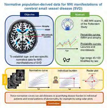
Figure 1. Graphical abstract of normative SVD modelling.
Another project within this research line focuses on identifying patterns of structural brain changes in normal aging, by applying disease progression modelling on the neuroimaging data from the population-based Rotterdam Study. After identification of such patterns within this aging population, we will investigate whether we can use these patterns or the stage of progression within these patterns for prediction of cognitive decline in memory clinic patients with subjective memory complaints.
Current projects (project lead: Eline Vinke ): – Normative modelling of small vessel disease ( Duygu Kilinc )
– Disease progression modelling in normal ageing (Sterre de Jonge)
Etiology and prediction of Alzheimer’s disease
We have acquired one of the largest datasets of amyloid PET scans within a longitudinal European cohort, featuring over 600 participants from the Rotterdam Study. What sets this dataset apart is the extensive collection of genetic, health, behavioral, lifestyle, and brain pathology data. Our research focuses on two complementary goals: (1) investigating risk factors and underlying causes of Alzheimer’s disease and (2) developing prediction models to identify individuals at risk of developing Alzheimer’s disease. In terms of Alzheimer’s etiology, it is still not clear which role vascular brain pathology plays. One pathology may accelerate the other, or they may occur independently. So far, our evidence points towards the latter (Fig 2). We did not find an association between CT-based arteriosclerosis and amyloid-beta plaques on PET. Preliminary findings also indicate no connection with white matter hyperintensities. A next step is to investigate the relation with small vessel functions measured on 7T MRI in collaboration with prof Biessels (UMC Utrecht). We are also investigating how modifiable risk factors, such as diabetes, relate to Alzheimer's and/or vascular disease, providing insights into how these risk factors mechanistically increase dementia risk. Finally, we are developing prediction models for Alzheimer’s disease, which could become very important as anti-amyloid treatments enter the clinic.

Figure 2. Association between Alzheimer’s disease on PET and large and small vessel disease on CT and MRI.
Current projects (project lead: Julia Neitzel ):
– Interplay between Alzheimer’s disease and arteriosclerosis and Alzheimer’s disease (Anna Streiber)
– Interplay between Alzheimer’s and cerebral small vessel disease ( Joyce van Arendonk )
– Prediction of Alzheimer’s disease and uncovering modifiable risk factors ( Thu ỷ Nguy ễ n )
Timely diagnosis and prevention of neurological disease
We strive to translate findings from population-based research to the clinic, and vice versa, in order to develop diagnostic tools and treatments that are broadly applicable in medical practice. In this research line, we use imaging information for a timely diagnosis and prevention of neurological disease. Examples include improved detection and management of covert brain infarction and asymptomatic carotid artery stenosis. Furthermore, we study the interplay of different causes of cognitive decline (e.g., vascular disease and Alzheimer’s disease) in affecting brain health, cerebral hemodynamics and the heart-brain axis. We develop prediction tools to facilitate personalized management of persons with cognitive complaints in primary and secondary care.
Current projects (Projectlead: Frank Wolters):
– Prognosis and Clinical Management of Covert Brain Infarcts (Camiel Box)
– Prediction tools for dementia (Jacqueline Claus, Mathijs Rosbergen )
Neurodevelopment in childhood and brain changes in pregnancy
Within the population-based Generation R, we use population imaging methodologies to study brain development in children, from age 6 to young adulthood. We apply normative modelling to create reference data and apply these techniques for clinical translation (e.g. within the BRAVE study, to detect deviation from normality in girls with anorexia nervosa).
Furthermore, within the Generation Next study, we collect brain imaging data in women who are trying to become pregnant. We scan our study subjects before pregnancy and after pregnancy (3 months and 1 year postpartum). The aim of this study is to better understand the remarkable impact that pregnancy has on the human brain, with a secondary focus on maternal mental health.
Current projects:
– Brain development, imaging trajectories and deviations in brain morphology in the pediatric population ( Marjolein Dremmen )
– Unravelling the neurobiology of anorexia nervosa
( Katrien Bracke )
– Decoding the maternal brain ( Merel de Vries )
Expectations & Directions
In the coming years, we expect that the amyloid PET data that were recently acquired will enable us to study the vascular and amyloid pathways towards dementia in depth, gaining more understanding in how these pathologies alone or in conjunction may lead to neurodegeneration and cognitive decline. Studies like the 7T small vessel disease study in collaboration with University Medical Centre Utrecht, will greatly add to this by allowing to directly image and measure microangiopathy, one of the most important contributors to both dementia and stroke. Furthermore, data-driven artificial intelligence research techniques (machine learning, deep learning) will bring the field of population imaging forward and improve our understanding of normal brain aging as well as neurodegenerative and neurological disease. Also, combining imaging with other high-dimensional data such as genomics, proteomics and metabolomics, is highly promising in unravelling pathways of disease and better understand disease pathophysiology.
Funding
Vinke, Eline Dekker postdoc grant by the Dutch Heart Foundation: ‘Personalized MRI-based Cerebral Small Vessel Disease (SVD) burden quantification, for more accurate diagnosis and prognosis’. 2024-2026
Vinke, Eline Alzheimer Netherlands Early Career grant: ‘Unraveling brain aging patterns predictive of AD or ADRD’. 2024-2025
Neitzel, Julia Erasmus MC Fellowship grant: ’Impact of Modifiable Factors on Dementia Risk across the Adult Lifespan’. 2024-2028
Vernooij, Meike, Esther Bron, and Harro Seelaar ZonMW: ‘TAP-Dementia: timely, accurate and personalized dementia diagnosis’. 2023-2027
Vernooij, Meike and Julia Neitzel Alzheimer’s Association Research Grant: ‘Amyloid pathology and vascular disease: a tale of two pathways’. 2022-2025
Vernooij, Meike, Frank Wolters, and Arfan Ikram ZonMW and Health Holland: ‘A Personalized Medicine Approach for Alzheimer’s Disease (ABOARD)’. 2021-2025
Neitzel, Julia Global Marie Curie Fellowship: ‘DIVERT-AD’. 2021-2025
Invited Lectures
Meike Vernooij. ‘Imaging in neurodegeneration: towards an etiologic diagnosis’. ECR, Vienna, Austria. March 2024.
Meike Vernooij. ‘Imaging in Dementia’. ESNR Basic Course in neuroradiology, online. March 2024.
Meike Vernooij. ‘Vascular cognitive impairment: role for radiologists’. ESNR Advanced course in neurovascular imaging, Annecy, France. June 2024.
Meike Vernooij. ‘ARIA for radiologists: from interpretation to logistics’. ESNR annual meeting, Paris, France. Sept 2024.
Meike Vernooij. ‘Imaging in dementia: the past, the present, the future’. Korean Congress in Radiology, Seoul, South Korea. Oct 2024.
Meike Vernooij. ‘Dementia prediction’. Webinar for the ABOARD consortium, online. Dec 2024.
Julia Neitzel. ‘Data sharing strategies in the Rotterdam Study’. European Meeting on Imaging Neurodegenerative Diseases (EU-MIND), Caen, France. Sept 2024.
Marjolein Dremmen. ‘Prenatal MR imaging of posterior fossa anomalies’. National Dutch Gynecology Course, Rotterdam, The Netherlands. Jan 2024.
Marjolein Dremmen. ‘Imaging in major pediatric trauma’. National Dutch Radiology Course, Ede, The Netherlands. Feb 2024.
Eline Vinke. ‘Modelling brain aging to disentangle the health-disease spectrum’. Erasmus MC Dementia Day, Rotterdam, the Netherlands. June 2024.
Highlights
Meike Vernooij was appointed as program planning chair for neuroradiology for the ECR 2026 conference.
Meike Vernooij coordinated an ESR-RSNA transatlantic course at ECR 2024.
Frank Wolters, Eline Vinke and Meike Vernooij were awarded a Medical Delta grant for implementation of dementia prediction models.
Frank Wolters, Daniel Bos, and Anna Streiber presented imaging data to participants of the Rotterdam Study at a knowledge festival that was organized by the Department of Epidemiology in collaboration with We Care to DisGover.
Mathijs Rosbergen participated in the Hackathon of Alzheimer Nederland.
Jacqueline Claus and Frank Wolters were invited for lectures and a panel discussion at the NCDC consortium meeting.
Jacqueline Claus and Eline Vinke were selected to attend the European Alzheimer Academy Workshop.
Joyce van Arendonk successfully defended her PhD thesis in December 2024.
Esther Bron and Myrthe van Haaften co-organized a session on imaging-based diagnostics in the memory clinic at the National Dementia Conference (organized by the Ministry of Health), at which Myrthe gave a presentation about brain MRI.
Julia Neitzel became PI of the ORACLE Study, which includes extensive data on brain health, including neuroimaging, cognitive testing and motor functions, from 2,000 parents of the Generation R Study.
Julia Neitzel was elected as the first non-Dutch board member of VENA (Vrouwen binnen Erasmus MC Netwerk voor Academici).
The BIRD-NL-consortium on dementia risk reduction, led by Frank Wolters and Arfan Ikram, had its first research output, just as the TAP-Dementia consortium on dementia diagnosis, in which Meike Vernooij and Esther Bron are leading a work package.
Marjolein Dremmen was a co-chair and member of several committees for development of national guidelines (e.g. imaging in trauma, epilepsy, craniosynostosis).
Additional Personnel
Anna Streiber, PhD student (also in group of Daniel Bos, see page 268)
Camiel Box, PhD student (also in group of Frank Wolters, see page 261)
Jacqueline Claus, PhD student (also in group of Frank Wolters, see page 261)
Myrthe van Haaften, PhD student (also in group of Esther Bron, see page 99)
Sterre de Jonge, PhD student (also in group of Esther Bron, see page 99)
Rachida Hadouch – Radiology Assistant MRI Ommoord
Anne-Sterre Schutter – Student Assistant MRI Ommoord
Björn Schrijver – Student Assistant MRI Ommoord
Celine Tuik – Student Assistant MRI Ommoord
Esra Hemmelder – Student Assistant MRI Ommoord
Fengli Bottema – Student Assistant MRI Ommoord
Fenne Bosman – Student Assistant MRI Ommoord
Gaia Hermans – Student Assistant MRI Ommoord
Gunnar Zwart – Student Assistant MRI Ommoord
Hajar El Moussati – Student Assistant MRI Ommoord
Jill Liu – Student Assistant MRI Ommoord
Issrae Affani – Student Assistant MRI Ommoord
Lara Uitdewilligen – Student Assistant MRI Ommoord
Lieke Bouvy – Student Assistant MRI Ommoord
Levy Schimmel – Student Assistant MRI Ommoord – Team Leader
Lucas de Groot – Student Assistant MRI Ommoord
Marije Timmermans – Student Assistant MRI Ommoord
Martijn van der Meer – Student Assistant MRI Ommoord
Mehdi Badaoui – Student Assistant MRI Ommoord
Michiel van den Akker – Student Assistant MRI Ommoord
Ouidad Oujjit – Student Assistant MRI Ommoord
Paula Rijs Alonso – Student Assistant MRI Ommoord –Team Leader
Suheda Yuce – Student Assistant MRI Ommoord
Assistant Professor Julia Neitzel, PhD

Email j.neitzel@erasmusmc.nl

POPULATION BRAIN IMAGING
Julia Neitzel’s research interest is to understand why some people are more resilient against age-related cognitive decline and dementia – important, yet missing knowledge which could inform better prevention programs. For addressing this question, she combines her knowledge on cognition and brain imaging gained during her underand postgraduate studies (MSc in Neuro-cognitive Psychology from Ludwig-Maximilians-Universität (LMU) Munich; PhD at the Neuroradiology of the Technical University Munich, Germany), with her postdoctoral work on genetic and modifiable risk factors of dementia (Institute for Stroke and Dementia Research at LMU; Depts of Radiology and Epidemiology at Erasmus MC). After her research visit at the Harvard School of Public Health in Boston, USA, she is excited to start her independent research group as an Erasmus Fellowship recipient.
Individualized dementia prevention
Prevention of chronic diseases is an ever-increasing forefront of medicine. Especially for dementia, once brain damage has occurred, treatment has been shown to be too late. My lab’s mission is to provide the knowledge for precision prevention strategies that promote brain health tailored to the demographic, genetic and clinical characteristics of each person. I support individualized dementia prevention through three key actions: (1) improving our understanding of the natural history of dementia and associated risk factors, (2) discovering new protective factors to pinpoint potential prevention targets, and (3) creating prediction algorithms that can identify high-risk individuals, enabling us to prioritize them in prevention approaches. Population brain imaging is the central component in my research, functioning as a powerful tool for early disease detection in asymptomatic individuals.
Identifying modifiable risk factors for preventing Alzheimer’s disease pathology
The e4 allele of the Apolipoprotein E (APOE) gene is the major genetic risk factor for late-onset Alzheimer's disease. About 25% of the Dutch population carries one copy, which increases their risk of Alzheimer’s by 2-3 times. To foster personalized prevention, we need to identify modifiable factors that accelerate Alzheimer’s pathology in high genetic risk groups. We have measured eight risk factors for dementia in participant from the Rotterdam Study. All participants underwent 18Fflorbetaben PET about seven years later, on which we quantified their Alzheimer’s disease (amyloid-beta [Aβ] plaque) burden. In the high genetic risk group, we have found associations between more Aβ plaques and weak circadian rhythmicity, low cholesterol, and hypertension.

Diabetes was associated with more Aβ plaques at followup in both the low and high genetic risk group (Fig. 1). We did not find a relationship between Aβ plaques and hearing loss, physical inactivity, obesity and smoking.
Blood-based biomarkers boost detection of Alzheimer’s disease pathology
Biomarkers that detect Aβ plaques in the blood could become cost-efficient tools to identify individuals at risk of developing dementia. We have previously developed a Aβ prediction model based on demographics, genetic and lifestlye factors, that reached medium-to-high accuracy (AUC=0.85). Adding blood-based biomarkers (p-tau217, Aβ42/40, GFAP, NfL) significantly improved accuracy (AUC=0.92). Participants with abnormal blood biomarkers also showed a steeper longitudinal decline of cognition, hippocampal volume and cortical thickness. I have received an Alzheimer Nederland crossborder grant together with the director of the Rhineland Study in Germany to investigate longitudinal trajectories of blood biomarkers and their brain imaging correlates.
Figure 1. Interplay between genetic and modifiable risk factors of dementia and Alzheimer’s disease pathology.
LinkedIn
In 2023 I received the Early Career Grant from Alzheimer Nederland and the Postdoc Dekker Beurs from the Hartstichting. With these grants I am starting my own research line focussing on modelling (healthy) brain Post-docs
Eline Vinke, PhD
Project Funding Early Career Grant – Alzheimer Nederland; Postdoc Dekker Beurs – Hartstichting
Email e.vinke@erasmusmc.nl
Modelling (healthy) brain aging
My research is on the intersection of neuroimaging, aging, epidemiology and machine-learning. In my research I develop and apply machine-learning based technologies that help us understand where, when and how deviations from normal or healthy brain aging take place based on neuroimaging in combination with other biomarkers. I am convinced that we should leverage knowledge on brain aging to ultimately improve our understanding of neurological disease.
PhD Students

Joyce van Arendonk, MSc

Advisors Meike Vernooij, Arfan Ikram & Julia Nietzel
Project Funding ZonMW Memorabel grant
Email j.vanarendonk.1@erasmusmc.nl
PhD Obtained 17-12-2024
Amyloid pathology and vascular disease in focus
To elucidate the interaction between the A β and vascular pathways in dementia etiology, participants of the Rotterdam Study aged 60 years and older underwent amyloid PET imaging. We studied the association between vascular risk factors and A β pathology. We observed that diabetes was associated with a higher prevalence and severity of A β pathology in both APOE4 carriers and noncarriers.

aging based on neuroimaging. Such normative brain models can help us understand where, when and how deviations from normal brain aging take place, with the ultimate aim to 1) better understand disease etiology, informing preventive strategies and 2) in a clinical setting, support earlier and more accurate diagnosis and monitoring of neurodegenerative or neurological disease. The projects that I started this year are focussed on applying disease progression modelling in the context of brain aging, to identify different brain aging patterns within the population-based Rotterdam Study. I will assess the clinical value of these normative brain models in a memory clinic setting.

Katrien Bracké, MD, MSc

Advisors Meike Vernooij, Gwen Dieleman & Tonya White
Project Funding SSWO ( S15-13, S22-65) Mrace Erasmus MC Grant
Email k.bracke@erasmusmc.nl
Unravelling the neurobiology of anorexia nervosa
We aim to unravel the neurobiological underpinnings of anorexia nervosa (AN), using non-Invasive Imaging techniques. We use data of the BRAVE study, a clinical case-control sample of adolescents with first-onset AN compared to gender-, age- and education-matched controls. A deeper understanding of the neurobiology may be a key element In developing novel therapeutic interventions.

Merel de Vries, BSc

Marjolein Dremmen, MD, MSc
Advisors Meike Vernooij & Steven Kushner
Project Funding Donation funds psychiatry department
Email m.w.devries@erasmusmc.nl
Decoding the maternal brain
My research focuses on how pregnancy changes the brain. In collaboration with the population-based Generation R Next study, we collect brain imaging data in women who are trying to become pregnant. We scan our study subjects before pregnancy and after pregnancy. The aim of this study is to better understand the remarkable impact that pregnancy has on the human brain, with a secondary focus on maternal mental health.
Advisors Meike Vernooij & Tonya White
Project Funding Mrace Erasmus MC Grant
Email m.dremmen@erasmusmc.nl
BRain development, Imaging trajectories and Deviations in brain morpholoGy in the pEdiatric population; BRIDGing thE gap
The goal is to construct ‘neuroimaging growth reference curves’ for total and regional brain volumes in children and adolescents. To be able to detect early deviations in brain development in patients groups; crucial for better understanding the neurobiology of these clinical conditions. To gain knowledge on variations in brain development and evaluate the consequences of incidental findings on brain imaging

Duygu Kilinc, MD, MSc

Advisors Meike Vernooij & Geert Jan Biessels
Project Funding TAP-dementia, project TAP-VaMP:
“Timely Accurate and Personalized diagnosis of dementia: Validation of Multimodal diagnostic biomarkers for application in clinical Practice”
Email d.kilinc@erasmusmc.nl
Improving diagnosis of SVD based on MRI scans: what is considered normal?
To bridge the translational gap between small vessel disease (SVD) imaging research and clinical practice , I will focus on creating age- and genderspecific normative data of the SVD markers using the Rotterdam Study data. Ultimately, I want to develop a tool that estimates the potential cognitive impact of SVD , based on lesion burden and distribution, in individual patients.

Mathijs Rosbergen, MSc

Advisors Frank Wolters, Meike Vernooij & Arfan Ikram
Project Funding ZonMW/TKI Health Holland: ABOARD consortium
Email m.rosbergen@erasmusmc.nl
Unraveling MRI markers for dementia prediction
When an individual is diagnosed with Alzheimer's disease, severe brain damage has already occurred. Treatment in an early stage could prevent brain damage and dementia. Therefore, my research focuses on unraveling new MRI predictors to improve existing dementia prediction models, so that we are able to identify individuals with high risk of dementia.

Phuʼoʼng Thuy Nguyên H‘Ô, BSc
Advisors Julie Neitzel & Meike Vernooij
Project Funding Erasmus MC Fellowship Email h.nguyen@erasmusmc.nl
Modifiable risk factors for dementia prevention
My research focuses on identifying which combination of modifiable factors are most effective for different age groups in relation to dementia. By examining how these factors influence imaging and cognitive outcomes, the goal is to better understand their role in dementia prevention alongside genetic and other unmodifiable risk factors.
JOINT APPOINTMENT IN EPIDEMIOLOGY
Frank Wolters obtained his MD at Utrecht University and practiced for a while in clinical neurology, before specialising in neuro-epidemiology with an MSc degree and PhD (cum laude) from Erasmus University Rotterdam. He completed research fellowships at the University of Oxford and the Harvard School of Public Health, applying expertise in research methodology notably to the prevention of cerebrovascular disease and dementia. His main interest lies with the understanding of dementia and stroke etiology, and improving the prognosis and management of subclinical, often incidental findings like covert brain infarction and

asymptomatic carotid artery stenosis. Frank applies various modalities of brain imaging to further prevention and timely diagnosis of cognitive disorders, in clinical research as well as population-based studies. He leads the nationwide BIRD-NL consortium on dementia prevention in the Netherlands, and is a co-investigator on various national and international initiatives including the Cross-Cohort Collaboration, Dementia Risk Prediction Project, and the Heart-Brain Connection collaborative research group. Frank co-founded the working group for vascular cognitive impairment at the Alzheimer Centre, and since 2024, he is the principal investigator of Neuroepidemiology for the Rotterdam Study. f.j.wolters@erasmusmc.nl
CLINICAL EPIDEMIOLOGY OF NEUROLOGICAL DISEASE
Frank J Wolters, MD, PhD assistant professor

Context
Timely diagnosis and prevention of neurological disease are key to reducing the burden of stroke, dementia, and other common neurological diseases in the population. Optimal prevention of neurological disease can only be achieved when clinical medicine and public health join forces. Through the study of population health in representative samples of the general population, often years before onset of disease symptoms, we learn about determinants of disease and subclinical changes that increase for example stroke or dementia risk. Similarly, risk stratification in these early stages allows for tailored diagnostic work-up and treatment, in order to optimize the benefit-harm ratio of preventative interventions in primary care and hospital settings. Generating evidence from population-representative data ensures applicability to the benefit of all patients in the community. Through the application of novel insights in research methodology, ensures thorough and efficient acquisition and analyses of data best suited for answering research questions relevant to clinical practice and public health policy. As such, the strong bridge between clinical neuroradiology and population brain health is a reliable foundation for strengthening clinical guidelines as well as informing healthcare strategies.
Top Publications 2024
Wolters FJ, JA Labrecque. Potential impact of unblinding on observed treatment effects in Alzheimer's disease trials. Alzheimers Dement. 2024; 20:3119-3125.
Hussainali RF, IK Schuurmans, JL Zijlmans, CAM Cecil, MW Vernooij, AI Luik, RL Muetzel, MA Ikram, FJ Wolters. Family history of dementia and brain health in childhood and middle age: a prospective community-based study. Eur J Epidemiol. 2024; 39:1151-1160.
Brück CC, SS Mooldijk, LM Kuiper, ML Sambou, S Licher, F Mattace-Raso, FJ Wolters. Time to nursing home admission and death in people with dementia: systematic review and meta-analysis. BMJ. 2025; 388:e080636.
Research Projects: Objectives & Achievements
Bridging clinical and population science
The Clinical Neuroepidemiology research group aims at improving the prevention of neurological disease, with a particular focus on stroke and dementia. We strive to translate findings from population-based research to the clinic, and vice versa, in order to develop prognostic tools and treatments that are broadly applicable in medical practice. Key objectives are:
1. Identification of causes of neurodegenerative and neurovascular disease, disentangle underlying pathways, and quantify the contribution of these various causes to the clinical phenotype.
2. Determine the relevance and optimal management of asymptomatic and subclinical pathology with respect to the onset and progression of neurovascular and neurodegenerative disease. Examples include asymptomatic carotid artery stenosis, covert brain infarction, and markers of cerebral amyloid angiopathy.
3. Develop and implement tools for timely diagnosis and prediction of neurological disease, with the aims of personalised prognosis, tailored treatment and followup, and targeted recruitment in clinical trials.
Primary collaborators within the Clinical Neuroepidemiology research line are the department of Epidemiology (chair: professor Arfan Ikram), the Population Imaging group (lead: professor Meike Vernooij), the Biomedical Imaging Group Rotterdam, the Cardiovascular Imaging group (lead: dr. Daniel Bos), the Alzheimer Centre (departments of Neurology [lead: dr. Janne Papma] and Geriatric Medicine [chair: professor Francesco Mattace Raso), the department of Public Health (chair: professor Hester Lingsma), and the Generation R study (dr. Ryan Muetzel).
Projects:
– Prediction tools for dementia ( Jacqueline Claus , Mathijs Rosbergen)
The Rotterdam Study
Epidemiological research on neurological disease within the Rotterdam Study currently entails the study of stroke, dementia, parkinsonism, migraine, and polyneuropathy. The entire cohort of close to 18,000 individuals is monitored for the occurrence of these diseases through repeated in-person examinations and linkage to electronic health records. Owing to high lifetime risk of various neurological illnesses, along the 35-year course of the study, these observations have led for example to a diagnosis of dementia in close to 2000 individuals,
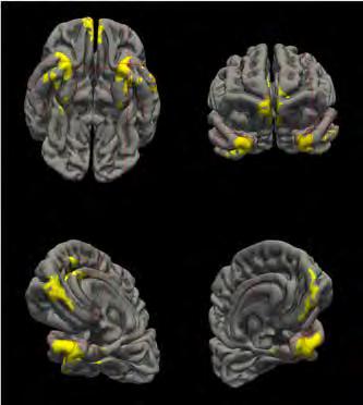
Figure 1. Visualization of a novel imaging signature of Alzheimer’s disease. A high-quality study recently reported promising prognostic value of a novel imaging signature, for risk of developing Alzheimer’s dementia. In order to incorporate this tool in the Rotterdam Dementia Prediction Model, we performed a rapid external validation within months after publication. However, in the Rotterdam Study data, the new signature did not outperform mere hippocampal volume, underlining the importance of replication research, as well as the advantages of phenotyped cohorts with good follow-up data to provide rapid replication (Rosbergen et al. J Alzheimers Dis 2024).
with equal numbers for stroke and a 1000 cases of lifetime migraine. The combination of detailed follow-up and population imaging creates a powerful combination for studying disease etiology and prognosis (Figure 1), Through incorporation of novel examinations in the core study protocol, we continuously look to enhance the phenotyping of participants with innovative techniques for a closer inspection of pathophysiological mechanisms in the early stages of these illnesses. Advances in research methods, like causal mediation analyses and target trial emulation, optimise data use for causal inference. We further apply population-based data to novel multistate models for determining the nationwide burden of disease and anticipated costs and effects of individual, often clinic-based, versus population-wide interventions.
Alzheimer Centre Imaging Core
In addition to the longstanding research cohort on frontotemporal dementia at Erasmus MC, 2024 has seen the birth of the Imaging Core of our Alzheimer Centre re-
search at large. In close collaboration with BIGR and the Imaging Office, imaging data of over 800 patients have been processed, along with extraction of data from neuroradiology reports for research purposes. Requests for use of this data can be directed to the Alzheimer Centre executive board. The dataset will be expanded with a further 300 scans from the 2022-2024 period in the coming year, adding extra value to the research side of the Alzheimer Centre: research to improve care.
Exemplary findings I: Arteriosclerosis acro ss the heart-brain axis in vascular cognitive impairment and dementia
Heart disease is an acknowledged contributor to stroke, and emerging evidence indicates heart failure and atrial fibrillation also predispose to dementia. Combining data from population-based and clinic-based studies like the Heart-Brain Connection Study, we unravel how cardiac dysfunction relates to brain pathology on MRI, and how this translates into clinical sequelae. An important area of interest is the overlap with Alzheimer's disease pathology, which we can now measure in vivo using blood biomarkers. This has taught us for instance that biomarkers traditionally deemed important only to the central nervous system, appear to have systemic effects too that can clarify their role in for example vascular pathology (Figure 2). Moreover, we have shown that intracranial arteriosclerosis predisposes to dementia in the population-based Rotterdam Study. By currently taking these findings to clinical studies like the Alzheimer Centre Imaging Cohort, we aim to translate these findings into clinically applicable tools for diagnosis and prediction that facilitate patient information and personalized care.
Exemplary findings II: Covert brain ischemia and stroke
Covert brain infarcts are defined as ischemic lesions on imaging that were not preceded by (recognized) stroke symptoms. They are seen on routine brain MRI in one in five older individuals, and increase the risk of subsequent cardiovascular disease and cognitive decline. Despite the large potential for secondary prevention, patients often go untreated, as optimal management is undetermined. Our group aggregated data from various cohorts to determine absolute risks for prognosis and trial design, and find determinants of high risk, which include infarct size and number, and concurrent unrecognized myocardial ischemia (Box et al. ESOC 2024). Ultimately, this can improve diagnostic and therapeutic management in clinical patients, like those visiting the Alzheimer Centre outpatient clinic or emergency department after head trauma.
Projects: – Prognosis and Clinical Management of Covert Brain Infarcts ( Camiel Box )
Strengthening research methodology
A reliable, open, and excellent academia is founded on rigorous standards for scientific quality. Through interaction with disciplines across Erasmus MC, the Clinical Neuroepidemiology group supports research on neurological disease and beyond, by offering support with expertise on study design, analyses, and causal inference. We consider methodological development an important part of PhD training, and are keen to support this whenever possible.

Figure 2. Plasma amyloid-beta40 is associated with atherosclerosis. Among nearly 4000 participants of the Rotterdam Study, we found no evidence that amyloid-beta40 was linked to intracranial arteriosclerosis. These observations translated into slightly higher risk of myocardial infarction, but this was in part attributable to traditional cardiovascular risk factors (Wolters et al. Atherosclerosis 2024).
Expectations & Directions
Population attributable fractions indicate that we can explain roughly 90% of the incidence of cardiovascular disease in the population, versus 45% for dementia. With our ongoing research, we aim to better understand the risk of dementia and move the percentage to the realm of cardiovascular disease. Similarly, on an individual patient level, we work to better understand which pathophysiological mechanisms are the principal contributors to the clinical phenotype (i.e. the etiologic fraction). For multifactorial diseases like cognitive impairment, this implies that we need to disentangle for example vascular contributions from those of Alzheimer’s disease, when there are multiple contributing causes present. The same
applies to the identification of the most prominent risk factors in patients with stroke who have undetermined stroke etiology due to multiple ‘TOAST factors’ present.
In terms of risk stratification, we aim to further clinical management of covert brain infarcts by setting up a dedicated clinical service aimed at secondary prevention of vascular events and cognitive impairment. The applicability of new treatments against Alzheimer’s disease also requires better risk stratification, both to guide care referrals and identify individuals who may benefit most from intervention. This importantly includes understanding of the risk and impact of common adverse effects (i.e. microhaemorrhages and cerebral edema).
To promote inclusive and generalisable science, we strive to maintain research that is applicable beyond tertiary patient populations. The Rotterdam Study is a key resource to this aim. We continuously look to incorporate innovative tools like spatial analysis of vascular pathology, functional vascular imaging, blood biomarkers, and digital lifestyle and cognitive assessments, to enhance our understanding of biological processes. Moreover, we explore the strengths and challenges of new data linkages, for example with Statistics Netherlands, Vektis, and IPCI, and look to strengthening ties with regional primary care, referral centres, and community old-age psychiatry services (e.g., Antes).
Funding
Vernooij, Meike, and Frank Wolters , along with investigators from LUMC and TU Delft Medical Delta: 'Applying advanced brain imaging for efficient dementia diagnosis and prediction'. 2024-2026
Wolters, Frank Alzheimer Nederland Biomedical Research Grant: 'The APOE - ε 2 paradox: role of APOE in lipid metabolism, vascular injury and amyloid deposition'. 2024-2026
Wolters, Frank , and Arfan Ikram, along with co-investigators from 12 parties nationwide ZonMw National Dementia Strategy: 'Modifiable factors for dementia prevention (BIRD-NL)'. 2023-2026
Wolters, Frank . Alzheimer's Association Research Fellowship: 'On generalizability and selection in dementia research: bridging academia and routine practice'. 20222025
Wolters, Frank . NWO Veni: 'Prevention of stroke and dementia after silent brain infarction (SHINOBI)'. 20212025
Vernooij, Meike, Frank Wolters , and Arfan Ikram ZonMW and Health Holland: ‘A Personalized Medicine Approach for Alzheimer’s Disease (ABOARD)’. 2021-2025
Invited Lectures
Frank Wolters. ‘External validity of Alzheimer’s disease trials of monoclonal antibodies against amyloid- β ’. Dutch Dementia Researchers Conference, Utrecht, the Netherlands. Nov 2024.
Frank Wolters. ‘Sex and gender differences in dementia: fact or artefact?’. Guest lecture Erasmus Summer Programme, Rotterdam, the Netherlands. Aug 2024.
Frank Wolters. ‘Unravelling the heart-brain connection: from population science to clinical practice’. Cardiovascular Institute Seminar, Rotterdam, the Netherlands. May 2024.
Frank Wolters. ‘Op weg naar preventieve interventies tegen dementie: een geval van Sisyphusarbeid?’. Radboud UMC, Nijmegen, the Netherlands. April 2024.
Frank Wolters. ‘Amyloid and cardiovascular disease’. International symposium 'Future of Heart-Brain research', Rotterdam, the Netherlands. Oct 2024.
Jacqueline Claus. ‘Disease modifying treatments against Alzheimer’s disease’. Erasmus MC Dementia Day, Rotterdam, the Netherlands. June 2024.
Highlights
Frank Wolters was appointed principal investigator of Neuroepidemiology for the Rotterdam Study.
Frank Wolters organized a public session on dementia prevention at the National Dementia Conference (organized by the Ministry of Health).
Frank Wolters was elected on the executive committee of VasCog (the International Society of Vascular Behavioural and Cognitive Disorders).
Frank Wolters received a research grant from Alzheimer Netherlands to study the role of APOE in cognitive decline.
The BIRD-NL consortium, led by Frank Wolters and Arfan Ikram, celebrated its first anniversary.
Frank Wolters , Eline Vinke and Meike Vernooij were awarded a Medical Delta grant for implementation of dementia prediction models.
Frank Wolters, Daniel Bos, and Anna Streiber presented imaging data to participants of the Rotterdam Study at a knowledge festival that was organized by the Department of Epidemiology in collaboration with We Care to DisGover.
Jacqueline Claus and Frank Wolters were invited for lectures and a panel discussion at the NCDC consortium meeting.
The first research projects using newly processed MRI and CT data from >500 patients in the Alzheimer Centre Imaging Core were completed, led by Camiel Box , Geke van Dijk, Ilse vom Hofe, Farog Faghir, Oliyad Merdassa, and Frank Wolters .
Naomi Starmans successfully defended her PhD thesis ‘Hemodynamic disturbances and dementia; On the crossroads between vascular brain injury and Alzheimer’s disease pathology’ on November 26, 2024.
Jacqueline Claus, MD, MSc PhD Students


Advisors Arfan Ikram, Meike Vernooij & Frank Wolters
Project Funding Trustfonds and ABOARD project (ZonMw/HealthHolland) Email j.claus@erasmusmc.nl
Risk prediction of dementia: from theory to clinical practice
Identification of modifiable risk factors for dementia, with a focus on early recognition and treatment of stroke and estimation of post-stroke dementia risk. Identifying those eligible for disease-modifying treatments against b -amyloid. Improvement and implementation of the dementia prediction tool in a clinical setting.
Additional personnel
Amber Yaqub – project coordinator BIRD-NL
Ilse vom Hofe – PhD student
Muhammed Sambou – PhD Student
Amos Pomp – PhD Student
Charlotte Mernart – PhD Student

Camiel Box, MD, MSc

Advisors Frank Wolters, Meike Vernooij & Kamran Ikram
Project Funding NWO Veni
Email c.box@erasmusmc.nl
Prognosis and Clinical Management of Covert Brain Infarcts
One in four elderly individuals are at high risk of stroke and dementia due to prior brain infarction that occurred unnoticed. In the Rotterdam Study we gather evidence on their prognosis and best clinical management in order to create clear guidelines to help clinicians deal with these covert brain infarcts.
JOINT APPOINTMENT IN EPIDEMIOLOGY
Daniel Bos was trained as a medical doctor and epidemiologist at Erasmus MC. He obtained his PhD in the field of vascular imaging and vascular epidemiology in 2013. The main focus of his work is on the elucidation of the etiology and pathophysiology of arteriosclerosis, and the clarification of the contribution of arteriosclerosis to clinical neurovascular and neurodegenerative diseases. For his research he leverages the strengths of population-based studies and combines this with clinical patientstudies in order to be able to make a direct clinical
impact with his work. Over the years, he has been awarded the Best Scientific Paper Award by the Radiological Society of the Netherlands and the Lourens Penning Prize by the Radiological Society of the Netherlands. He is currently the deputy head of department of the department of Epidemiology, and holds a position as Adjunct Associate Professor in Clinical Epidemiology at the Harvard School of Public Health and a position as Professor in Neurosciences at KU Leuven. Daniel has authored over 150 peer-reviewed publications. d.bos@erasmusmc.nl
IMAGING OF ARTERIOSCLEROSIS: FROM POPULATION TO CLINICAL PRACTICE
Daniel Bos, MD, PHD associate professor

Context
Arteriosclerosis is a highly frequent vascular disease causing stiffening of arteries throughout the whole arterial system. Major clinical consequences of arteriosclerosis include myocardial infarction and stroke, which are top causes of morbidity and mortality in middle-aged and elderly persons worldwide. Due to the aging of the population, the global burden of arteriosclerosis, and thereby of these clinical conditions, will continue to rise in the coming decades. The research in “Imaging of Arteriosclerosis: from Population to Clinical Practice” is focused on the elucidation of the etiology and pathophysiology of arteriosclerosis, and on the clarification of the contribution of arteriosclerosis to clinical neurovascular and neurodegenerative diseases, with a strong emphasis on the use of state-of-the-art medical imaging. Within this unique research-field on the interface of arteriosclerosis, neurovascular, and neurodegenerative diseases, the research group specifically aims at obtaining relevant imaging-based biomarkers for improved understanding of arteriosclerosis and improved prediction of abovementioned diseases, and at identifying potential targets for intervention strategies.
Top Publications 2024
Berghout B, R Camarasa, H van Dam-Nolen, A van der Lugt, M de Bruijne, P Koudstaal, M Ikram, D Bos. Burden of intracranial artery calcification in white patients with ischemic stroke. European Stroke Journal 2024; 743-750.
Streiber A, J Neitzel, P Nguyen Ho, M Vernooij, D Bos. Intracranial arteriosclerosis is not associated with cerebral amyloid deposition. Alzheimer's and Dementia: Diagnosis, Assessment and Disease Monitoring 2024; 16:70005.
Van der Toorn J, M Vernooij, M Ikram, M Kavousi, D Bos. Progression of arterial calcification: what, where and in whom? European Radiology 2024; 34:5142-5152.
Research Projects: Objectives & Achievements
Intracranial Arteriosclerosis
This is the major line of research within the research group, and is specifically focused on the causes and consequences of intracranial arteriosclerosis. In this field, important pioneering-work on the identification and quantification of calcification in the intracranial carotid arteries and the vertebrobasilar system has been performed by our research group. By applying non-enhanced computed tomography to participants of the populationbased Rotterdam Study, we demonstrated that the presence and amount of intracranial carotid artery calcification is the strongest risk factor for developing a first-ever stroke, and that the amount of calcification at this location even contributes to the development of dementia, including Alzheimer’s Disease. This work still has a considerable global impact and has also led to an extension of the research horizon towards clinical studies on the influence of intracranial arteriosclerosis in stroke patients. In these patients, the amount of intracranial arteriosclerosis at time of admission to the hospital directly influences the prognosis after stroke treatment. It has also led to valuable global collaborations on the influence of Intracranial arteriosclerosis on endovascular treatment and the Importance of Intracranial arteriosclerosis Is being recognized In International guidelines of the European Stroke Organization.
Previous work in the field of pathology and imaging uncovered the existence of two morphological patterns of intracranial arteriosclerotic calcification. These include intimal, atherosclerotic calcification and circular calcification of the internal elastic lamina (IEL). Work from this research group showed that these two patterns contribute differentially to stroke, and influence stroke treatment, given the divergent effects of calcification on the arterial wall structure and the accompanying structural stresses within the artery. In this light, the research horizon on the consequences of these calcifications has been broadened towards neurodegenerative brain disease, including dementia. The first work into this direction indeed showed that primarily the circular IEL calcification seems to contribute to neurodegenerative changes. In the near future, we will delve into the underlying mechanisms of this association by combining information from different imaging modalities, including CT, PET, and MRI.
Projects:
– Association of mechanical wall stress and wall shear stress with the development of atherosclerosis ( Aikaterini Tziotziou )
– Gaining insight into the outcome variability of endovascular thrombectomy in ischemic stroke patients ( Ching Khan )
– Identification and assessment of imaging markers affecting endovascular thrombectomy prognosis and efficacy in ischemic stroke patients ( Sven Luijten ) – Surrogate modeling techniques for enhancement of the decision-making process for endovascular thrombectomy in acute ischemic stroke patients ( Luca Bontempi )
Advanced Imaging of Arteriosclerosis
This line of research is devoted to more advanced imaging of arteriosclerosis, with a specific focus on its most common subtype, namely atherosclerosis. Atherosclerosis is characterized by thickening of the intimal layer of the arterial vessel wall due to lipid-accumulation, neo-vascularization, and calcification. Especially when located in the carotid artery bifurcation, atherosclerosis is known to substantially increase the risk of ischemic stroke. The work in this research line is targeted at the optimal identification and evaluation of atherosclerosis in the carotid artery bifurcation. Using advanced, stateof-the-art imaging modalities (CT, MRI, etc.), the aim of the research group is to further understand the development of this disease and the value of assessing plaque composition and plaque morphology for the prediction of neurovascular and neurodegenerative diseases. Recent accomplishments of the research group in this field have shown that the presence of intraplaque hemorrhage in carotid artery atherosclerosis is the single most important risk factor for a first-ever stroke and stroke recurrence. Moreover, our work showed that the composition of these plaques substantially differ between men and women, which is one of the topic we aim to further elucidate in coming years.
Within this line of research, an important novel focus is on photon-counting CT (PCCT) for imaging of arteriosclerosis. Specifically in the cerebral vessels, the resolution of conventional CT is too limited and PCCT may represent an important novel modality in this field. Current studies that are being performed within the research group revolve around comparisons of PCCT with conventional CT on calcification detection, calcification quantification and image quality, mainly using autopsy specimen.
Projects:
– Unraveling the relationship between arteriosclerosis and biomarkers for Alzheimer's Disease (Anna Streiber)
– Identifying high-risk carotid plaques using imaging modalities ( Dianne van Dam-Nolen )
Optimization of Cardiovascular Image Analysis
This research line revolves around the early detection of vascular disease, by maximally leveraging existing medical imaging examinations. By using novel methods for image-analysis (e.g. machine-learning algorithms), we aim to obtain more sensitive and more accurate imaging markers of vascular disease. My group performed important pioneering-work on the quantification of epicardial fat as emerging markers of vascular risk. Over the last years, this novel marker of vascular risk has gained rapid attention, which has led to a strong collaboration on its role in heart-transplantation patients with the Dept. of Cardiology and the Dept. of Pulmonology. This collaboration was further extended with additional measurements of imaging-based markers of vascular risk (liver density) and overall-health status (bone density), which can all be readily assessed using conventional CT.
Projects:
– Early life identification and treatment of cardiovascular risk factors ( Maarten Leening )
– Imaging of subclinical atherosclerosis in presymptomatic individuals ( Maarten Leening )
Expectations & Directions
The field of research into arteriosclerosis is rapidly evolving and substantial developments at many levels will occur in which the research of this group will play an important role. First, through the contributions of research of this group, intracranial arteriosclerosis is increasingly being recognized as an important risk factor for stroke, but may also play a key role in the development of dementia. In order to advance these insights, detailed visualization of arteriosclerotic disease in the cerebral vessels (not only the large afferent arteries such as the carotids and the vertebral arteries, but particularly in the smaller arteries) using combinations of different advanced imaging modalities is the way forward. An important focus should be on the difference between ‘pure’ atherosclerotic changes in the arteries (intimal thickening, plaque formation) and hardening/stiffening of the arteries through calcifications in the medial layer of the artery. Subsequently, using population-based data, etiological differences between these types of arteriosclerosis can be determined. Similarly, these features of arteriosclerosis may be added to existing risk-prediction models for stroke and even dementia, further contributing to their early identification and to the development of therapeutic and preventative strategies. One field in which rapid developments are made is that of imagepost-processing (using machine learning algorithms),
which now allows for the possibility to investigate arteriosclerotic disease in much more detail. In addition to ‘conventional’ measurements such as the Agatston score, or volumetric assessments of calcification or plaque-size, it is becoming possible to also assess shape characteristics of the disease, make vessel-stress calculations, link specific configurations of arteriosclerotic disease (including combinations of shape, volume and other properties) with outcomes. Third, an important achievement of this group was the completion of a large-scale follow-up imaging study (10-15 years of follow-up) targeted at the visualization of change in the amount of vascular calcification across multiple arteries. These data are unique in the world and have provided valuable insight into the natural course of arteriosclerosis.
Funding
Leening, Maarten, and Daniel Bos Sanofi Research Grant: 'Unlocking the Preventive Potential of Routine Clinical Imaging: Implementation of the KALK Project'. 2024
Van Dam-Nolen, Dianne, and Daniel Bos Erasmus MC Research Innovation Grant: 'Photon-counting CT for the detection of intraplaque hemorrhage in carotid atherosclerosis? An innovative pilot-study for optimizing stroke work-up'. 2024
Leening, Maarten, and Daniel Bos Erasmus MC Research Innovation Grant: 'LoDoCo-Plaque'. 2024
Roeters-van Lennep, Jeanine, Ricardo Budde, Daniel Bos and consortium partners Dutch Heart Foundation: 'AtheroNETH - consortium'. 2024
Invited Lectures
Daniel Bos. ‘The value of AI in clinical research: current status’. Nederlandse Vereniging voor Internisten Acute Geneeskunde (NVIAG) conference, Eindhoven, the Netherlands. June 2024.
Daniel Bos. ‘Intracranial Arteriosclerosis: A Major Risk Factor for Stroke?’. Dutch Cardiovascular Alliance / NHLI conference, Utrecht, the Netherlands. June 2024.
Daniel Bos. ‘Intracranial Arteriosclerosis, Stroke, and Dementia’. International Conference on Intracranial Atherosclerosis (ICAS), Manilla, Philippines. Aug 2024.
Daniel Bos. ‘Intracranial Arteriosclerosis: A Major Risk Factor for Stroke and Dementia’. Cardiovascular Research Institute, Rotterdam, the Netherlands, Sept 2024.
Daniel Bos. ‘Intracranial Arterial Calcification: A Major Risk Factor for Stroke’. International Symposium on Cardiovascular Calcification (ISCCA), Nice, France. Oct 2024.
Daniel Bos. ‘Causes and Consequences of Intracranial Arteriosclerosis’. The LBI Cerebrovascular Research Center Seminar, Leuven, Belgium. Dec 2024.
Highlights
Sven Luijten, Dianne van Dam-Nolen, and Robin Camarasa successfully defended their theses entitled “Imaging in ischemic stroke: Leveraging imaging beyond diagnosis to predict functional outcome and benefit of endovascular thrombectomy”, "Plaques and Patients: the Role of carotid Imaging In the prevention of Stroke", and "The carotid artery In bits and pieces" (co-promotor: Daniel Bos ).
Luoshiyuan Zuo was awarded an European Society of Cardiology Best Abstract Award at the ESC Congress 2024 for his abstract entitled "Evolution of subclinical carotid atherosclerotic plaque composition: the Rotterdam Study" (senior author: Daniel Bos ).
Daniel Bos was appointed as Associate Programme Director for the MSc-programme in Clinical Epidemiology for the Netherlands Institute of Health Sciences (NIHES).
Daniel Bos delivered a seminar on Intracranial Arteriosclerosis at the Spanish National Centre for Cardiovascular Research (CNIC) (head: Prof. Valentin Fuster) as the kick-off of a novel collaboration.
Additional Personnel
Robin Camarasa – PhD student, Department of Radiology & Nuclear Medicine, graduated in October 2024 (co-promotor: Daniel Bos) (PI: Marleen de Bruijne)
Céline van der Braak – PhD student, Department of Radiology & Nuclear Medicine (co-promotor: Daniel Bos) (PI: Ivo Schoots)
Cevdet Acarsoy – PhD student, Department of Epidemiology (co-promotor: Daniel Bos)
Brian Berghout – PhD student, Department of Epidemiology (co-promotor: Daniel Bos)
Judith van der Bie – PhD student, Department of Radiology & Nuclear Medicine (co-promotor: Daniel Bos) (PI: Ricardo Budde)
Luoshiyuan Zuo – PhD student, Department of Epidemiology (co-promotor: Daniel Bos)
Mitra Nekouei – PhD student, Department of Epidemiology (co-promotor: Daniel Bos)
Peter van Hulst – PhD student, Department of Neurology (co-promotor: Daniel Bos)
Xi Li – PhD student, Department of Public Health (co-promotor: Daniel Bos)
Sanne van Kuijk – PhD student, Department of Epidemiology (co-promotor: Daniel Bos)
Partha Haldar – PhD student, Department of Epidemiology (co-promotor: Daniel Bos)
Kostas Stoitsas – Postdoc, Department of Epidemiology
Hyunho Mo – Postdoc, Department of Radiology & Nuclear Medicine (PI: Esther Bron)
Assistant Professor

Maarten Leening, MD, PhD
m.leening@erasmusmc.nl

JOINT APPOINTMENT IN CARDIOLOGY AND EPIDEMIOLOGY
Maarten Leening obtained his medical degree (cum laude) and MSc in clinical epidemiology at the Erasmus MC. He received his PhD (cum laude) at the Erasmus MC (Depts of Cardiology and Epidemiology). In 2014-2015, he worked as a post-doc at the Division of Preventive Medicine at the Harvard Brigham and Women's Hospital (Nancy Cook and Paul Ridker). Subsequently, he trained as a cardiologist with a focus on non-invasive imaging (EACVI certified in cardiac CT and echocardiography), CVD prevention (EAPC certified), and lipids/atherosclerosis (EAS certified). By the end of 2023, Maarten Leening joined the vascular imaging research group (Daniel Bos).
Preventive Cardiology
Atherosclerotic cardiovascular disease (ASCVD) events are preventable in the vast majority of individuals. My research aims at early life identification and treatment of cardiovascular risk factors and imaging of subclinical atherosclerosis in presymptomatic individuals.
PhD Students

Ching Khan, MD, MSc
Advisors Aad van der Lugt, Daniel Bos & Bob Roozenbeek
Project Funding The COllaboration for New TReatments of Acute Stroke (CONTRAST)
Email j.vanderbie@erasmusmc.nl
LinkedIn c.khan@erasmusmc.nl
The use of imaging to unravel the variability in outcomes after endovascular thrombectomy for ischemic stroke
The aim of my research is to find out through different imaging modalities why some ischemic stroke patients have worse functional outcomes than others when treated with endovascular thrombectomy. Ultimately, my goal is to identify ischemic stroke patients that may or may not benefit from thrombectomy treatment in clinical practice.
Specific interests Includes the biology and treatment of atherogenic lipids and chronic inflammation.
On the side of imaging, areas of interest include noninvasive assessment of coronary atherosclerosis and aortic valve disease, and the role of automated software and AI in cardiovascular imaging.

Judith van der Bie, MSc

Advisors Ricardo Budde, Daniel Bos & Marcel van Straten
Project Funding Siemens Healthineers
Email j.vanderbie@erasmusmc.nl
LinkedIn www.linkedin.com/in/judithvdbie
Clinical applications of photon-counting computed tomography
Photon-counting CT (PCCT) is a novel imaging technique which enables higher spatial resolution and more advance spectral imaging compared to conventional CT. During my PhD I will investigate the clinical impact of PCCT on cardiovascular- and neuro-imaging. For example, the assessment of coronary in-stent restenosis with PCCT in patients with recurrent chest pain with invasive angiography as reference, to potentially minimalize number of patients undergoing this intervention.

Anna Streiber, MSc Aikaterini Tziotziou, MSc

Advisors Meike Vernooij, Julia Neitzel & Daniel Bos
Project Funding Cure Alzheimer’s Fund
Email a.streiber@erasmusmc.nl
Disentangling the Role of Arteriosclerosis in Alzheimer’s Disease
The etiology underlying Alzheimer’s disease is multifactorial and may be associated with cerebrovascular changes. Within the Roterdam study, we investigate the relationship between arteriosclerosis and various markers for Alzheimer’s diseas. These markers are measured in blood plasma, on (f)MRI, or PET-CT.


Advisors Ali Cagdas Akyildiz, Daniel Bos, Ton van der Steen & Aad van der Lugt
Project Funding PhD-project Erasmus MC-Grants (MRace) 2019
Email a.tziotziou@erasmusmc.nl
Atherosclerotic plaque onset and progression are known to be affected by local biomechanical factors.
We investigated the association of mechanical wall stress (MWS) and wall shear stress (WSS) to atherosclerosis development in human coronary (IMPACT Study) and carotid (PARISK Study) arteries.

Sven Luijten, MD, MSc

Advisors Aad van der Lugt, Diederik Dippel, Daniel Bos & Bob Roozenbeek
Project Funding Collaboration for New treatments of Acute Stroke (CONTRAST)
Email s.luijten@erasmusmc.nl
PhD Obtained 01-05-2024
Imaging-based prediction of outcome in ischemic stroke
The overarching aim of my research is to assess and identify imaging markers that affect patient prognosis and efficacy of endovascular thrombectomy (EVT) for ischemic stroke, with the ultimate goal of imaging-based prediction of functional outcome and benefit of EVT in individual patients.
Dianne van DamNolen, MD, MSc

Advisors Aad van der Lugt, Peter J Koudstaal & Daniel Bos
Project Funding The PARISK study is supported by the Center for Translational Molecular Medicine (CTMM) and the Dutch Heart Foundation
Email h.nolen@erasmusmc.nl
PhD Obtained 08-05-2024
Imaging of high-risk carotid plaques
Carotid atherosclerosis is a main cause of ischemic stroke. The Plaque At RISK study shows that imaging of carotid atherosclerosis by CTA and MRI improves the prediction of recurrent ischemic strokes. Carotid imaging can identify high-risk carotid plaques based on specific plaque characteristics such as intraplaque hemorrhage.
Luca Bontempi, MSc

Advisors Frank Gijsen, Behrooz Fereidoonnezhad & Daniel Bos
Project Funding GEMINI – A Generation of Multiscale Digital Twins of Ischaemic and Haemorragic Stroke Patients
Email l.bontempi@erasmusmc.nl
Enhancing Decision-Making in Endovascular Thrombectomy for Acute Ischemic Stroke
The overarching aim of my research is to support clinicians in the decision-making process for endovascular thrombectomy (EVT) in ischemic stroke patients by optimizing choices related to device selection, positioning, and treatment methods. Through the development and application of surrogate modeling techniques, the research seeks to enhance the efficacy of EVT, ultimately improving treatment outcomes for patients with acute ischemic stroke.
Jacob Visser is a musculoskeletal radiologist, health economist and epidemiologist. As of July 2020, he is appointed as assistant professor in value-based imaging and working on issues such as artificial intelligence, structured reporting, decision support software, and integrated diagnostics. Furthermore, he is involved in setting up further collaboration with radiologists at the Radiology department of the Massachusetts General Hospi-

tal (MGH) in Boston, MA, USA. In addition, he is a member of the Value-based Radiology Committee of the ESR and the RSNA Working Group for Common Data Elements. He has served in the ESR eHealth and Informatics Subcommittee. As of January 1, 2020, he was appointed as Chief Medical Information Officer at the Erasmus MC.
j.j.visser@erasmusmc.nl
VALUE-BASED IMAGING
Jacob J Visser, MD, PhD, MSc assistant
professor

Context
Ahealth care rapidly changes from volume to value-based, there is an urgent need for radiologists to position themselves from a value-based perspective.
Therefore, the Radiology Department at the Erasmus MC has started the value-based imaging program. This means that all activities in the Radiology Department are evaluated in the light of value-based imaging.
The value-based imaging program is closely related to the IT department. Most topics that are being covered by this program require expertise in the field of information technology.
Top Publications 2024
Boverhof BJ, WK Redekop, D Bos, MPA Starmans, J Birch, A Rockall, JJ Visser. Radiology AI Deployment and Assessment Rubric (RADAR) to bring value-based AI into radiological practice. Insights Imaging 2024; 15:34.
Kemper EHM, H Erenstein, BJ Boverhof, K Redekop, AE Andreychenko, M Dietzel, KBW Groot Lipman, M Huisman, ME Klontzas, F Vos, M IJzerman, MPA Starmans, JJ Visser. ESR Essentials: how to get to valuable radiology AI: the role of early health technology assessment-practice recommendations by the European Society of Medical Imaging Informatics. Eur Radiol. 2024; 39636421.
Hillis JM, JJ Visser, ERS Cliff, K van der Geest-Aspers, BC Bizzo, KJ Dreyer, J Adams-Prassl, KP Andriole. The lucent yet opaque challenge of regulating artificial intelligence in radiology. NPJ Digit Med. 2024; 7:69.
Research Projects: Objectives & Achievements
Artificial intelligence
Currently, several projects are under investigation in the value-based imaging program. Firstly, as good quality of radiology reports is a core element in the era of valuebased imaging, strategies are developed to investigate the quality of these reports. As manual review to identify sufficient cases for retrospective analysis is often infeasible, a stepwise natural language processing (NLP)method for case identification was developed and applied for the identification of critical finding cases.
Quantitative and radiogenomics imaging is becoming more important. It turned out that visual assessment alone was not able to generate all available information. Instead, state-of-the-art computer techniques like machine and deep learning are able to extract quantitative features that are able to play an important role in the diagnostics of musculoskeletal and other diseases. A model was developed and is to be validated that is able to predict the MDM2-amplification in lipoid tumors.
Implementation of artificial intelligence algorithms in the radiology workflow is becoming a hot topic. Although several algorithms have been developed of which some are CE-marked / FDA approved and only a few validated in clinical practice, it is important to make the right decisions about implementation of those algorithms. Depending on the type of the algorithm, automatic measurements, case prioritization, pathology detection, a certain amount of research is needed before usage in the clinic can start.

Current projects:
– Understanding the 3D morphology of the calcaneus (Alexander Wakker)
– Improving incidental pulmonary nodule detection and follow-up through the implementation of AI-based software (Asabi Leliveld, Arlette Odink, Ties Mulders)
– Radiology AI Deployment and Assessment Rubric (RADAR) to bring value-based AI into radiological practice (Bart-Jan Boverhof)
– Value impact assessment of AI in radiology (Bina Tariq)
– Implementation, cost-effectiveness and carbon footprint of AI when aiming to find lung cancer early (Bo Willems, Arlette Odink, Ties Mulders)
– Value driven AI model for detection of incidental pulmonary embolism in CTs (Erik Kemper, Arlette Odink)
– An ethnographic exploration of the digital transformation of healthcare (Gigi Vissers)
– Clinical implementation of Artificial Intelligence solutions for Musculoskeletal Radiology (Huib Ruitenbeek)
– User-acceptance of AI for radiology in a clinical setting (Jamie Verwey)
– Clinical Implementation of AI Technologies for Improved Lung Nodule Detection and Follow-up (Jasika Paramasamy, Arlette Odink, Ties Mulders, Jan-Willem Groen)
– PREDICT_LN: a diagnostic tool with deep-learning model to evaluate thoracic CT scans of colorectal cancer patients for detection and classification of lung nodules (Julie Hamm)
– Implementation of artificial intelligence in radiology practice (Laurens Topff)
Data-driven workflow
More than 200,000 exams are being performed in our department annually. This requires good logistics and insights in the workflow. In order to get the information about the processes, it is needed to identify relevant data sources. Modern software packages allow for visual representation of process and quality indicators. The research focuses on how to involve people and use dash boarding tools in order to improve efficiency and quality metrics.
Integrated diagnostics
Integrated diagnostics will be one of the key steps forward in the coming decade. In this respect, the work-up for patients with adrenal incidentaloma was evaluated. Furthermore, the diagnostic work-up for cervix carcinoma, carcinoma of unknown primary, and chest pain was evaluated. Currently, a workflow integration of radiology and pathology findings is investigated in lung cancer patients. Also for liver and brain tumors, further integration of radiology and pathology can increase efficiency and improve diagnosis. In addition, it is to foreseen that image oriented specialties like dermatology can be involved in the integrated diagnostics approach.
Figure 1. Artificial intelligence algorithm automatically detects fractures.
Expectations & Directions
In the coming years, we aim to further expand our research in the domains of artificial intelligence, datadriven workflow, and integrated diagnostics. An important challenge will be to bring these together and establish reference architectures and pipelines how to structurally evaluate new workflows and technologies.
Funding
Visser, Jacob Qure AI: ‘Validation of AI in stroke patients’. 2024-2025
Visser, Jacob Qure AI: ‘AI to improve nodule detection on chest X-ray’. 2024-2026
Aerts, Joachim, and Jacob Visser AstraZeneca: ‘Pulmonary Incidental Nodules: Improve Detection and Follow-up by integrating Artificial Intelligence (PINPOINT)’. 2024-2028
Visser, Jacob , Martijn Starmans , Ken Redekop, and Frans Vos TKI: ‘Assessing the effect on the clinical workflow and a cost-effectiveness analysis of a comprehensive AI tool for chest CT reading – on the use case of incidental pulmonary embolism’. 2023-2027
Post-docs

Visser, Jacob Radiology Research Fund: ‘Enabling Value impact assessment for Artificial Intelligence Tools in radiology (eVAIT)’. 2022-2027
Klein, Stefan, Martijn Starmans, Jacob Visser, Kees Verhoef, and Dirk Grunhagen Hanarth Fonds: ‘Automatic grading and phenotyping of soft-tissue tumors through machine learning to guide personalized cancer treatment’. 2021-2025
Invited Lectures
Jacob Visser. ‘Early HTA in radiology AI’. EuSoMII Annual Meeting, Pisa, Italy. Oct 2024.
Jacob Visser. ‘Legal considerations in Europe regarding radiology AI’. RSNA, Chicago, USA. Dec 2024.
Highlights
Jacob Visser was appointed as member of the board of the section Techniek of the Dutch Society of Radiology.
Additional Personnel
Mart Rentmeester – PhD, ICT
Jan-Willem Groen, MD, PhD
j.groen@erasmusmc.nl
Artificial Intelligence in Systematic Review formation; Efficiency and time reduction, in search, screening, and data extraction through Natural Language Processing.
A systematic review (SR) is considered one of the most esteemed forms of scientific research. A Living Systematic Review (LSR) is an evolving SR that continually integrates new evidence as it emerges. The process of conducting an (L)SR has become increasingly demanding due to the expanding body of published research. Artificial Intelligence (AI), particularly Natural Language Processing (NLP) tools, can assist researchers in identifying crucial terms and sentences during title/ abstract screening and in extracting pertinent data during full-text appraisal.
Our objective is to assess the current utilization of these tools, employing an online survey. We aim to highlight user demographics and explore the barriers and facilitators influencing their usage. Additionally, we seek to gain hands-on experience by conducting our own SR on AI in Lung Nodule Detection. Subsequently, we plan to transform this SR into a ‘Living’ review by leveraging AI for monthly updates based on newly available literature post-publication.
Ties Mulders, MD, PhD
Project Funding PINPOINT
Email t.mulders@erasmusmc.nl
PINPOINT
(Pulmonary Incidental Nodules: Improve Detection and Follow-up by integrating Artificial Intelligence)
PINPOINT is a one-of-a-kind programme implementing Artificial Intelligence (AI) in clinical practice at scale. It aims to develop insights about incidental pulmonary nodules (IPNs) detection and management in participating hospitals. The project will also identify best practices for IPN management and early lung cancer diagnosis. With the help of AI, pulmonary nodules will be automatically detected, measured, classified and tracked the growth of pulmonary nodules.

Pulmonary
Arlette
Odink,
Project Funding PINPOINT 2
In nodule clinics, a multidisciplinary team evaluates the pulmonary nodules detected in routine practice.They are a practical approach to organising a streamlined follow-up for patients, so no early-stage lung cancer is missed. PINPOINT aims to implement nodule clinics in participating hospitals and establish them as the standard of care. PINPOINT aims to analyse 300.000 to 600.000 scans for early detection within two years. After a pilot stage, the programme will expand to a planned 20 European hospitals across up to ten countries. The project is expected to develop new best practices and change the standard of care for IPN detecting and follow-up.
MD, PhD
Email a.odink@erasmusmc.nl
Incidental
Nodules:
Improve Detection and Follow-up by integrating Artificial Intelligence in a prospective real-world cohort.
To assess the impact of the implementation of CAD combined with dedicated incidental nodule clinics with patient tracking, a prospective observational study will be performed in multiple European medical centres. The rate of stage I and II lung cancer diagnosis with curative intent treatment, health care usage for nodules as well as perform a cost and benefit analysis will be determined. This is de project of a PhD student, and I will be one of the post docs involved.
PhD Students

Bart-Jan Boverhof, MSc

Advisors Ken Redekop, Maureen Ruttem, Carin Uyl & Jan-Jaap Visser
Email boverhof@eshpm.eur.nl
Radiology AI Deployment and Assessment
Rubric (RADAR) to bring value-based AI into radiological practice
Radiology artificial intelligence lacks a comprehensive approach to value assessment. To this end, we proposed a comprehensive framework for value assessment of artificial intelligence (AI) in radiology.

Jasika Paramasamy, MSc

Advisors Jan-Jaap Visser, Aad van der Lugt & Joachim Aerts
Project Funding Kansen voor West
Email j.paramasamy@erasmusmc.nl
Clinical Implementation of AI Technologies for Improved Lung Nodule Detection and Follow-up
Artificial Intelligence (AI) is becoming an increasingly important tool in radiology, particularly in lung nodule detection. AI algorithms can assist in identifying nodules on chest radiographs and CT scans, potentially improving detection rates and clinical decision-making. We aim to explore how AI can enhance radiological workflows, improve patient outcomes, and contribute to healthcare efficiency.

Huib Ruitenbeek, MSc

Advisors Jan-Jaap Visser & Edwin Oei
Project Funding Radiobotics ApS Qure.ai
Email h.ruitenbeek@erasmusmc.nl
Clinical implementation of Artificial Intelligence solutions for Muskuloskeletal Radiology
Artificial Intelligence (AI) solutions are increasingly vital in radiology, aiding or automating tasks to lessen workload. However, clinical evaluation of these algorithms in radiology is scarce. Collaboration between AI vendors and ErasmusMC's scientific expertise can yield new insights.

Julie Hamm, MSc

Advisors Pieter Tanis, Kees Verhoef, Eva Madsen & Jan-Jaap Visser
Project Funding KWF: PREDICT_LN
Email j.hamm@erasmusmc.nl
PREDICT_LN: a diagnostic tool with deeplearning model to evaluate thoracic CT scans of colorectal cancer patients for detection and classification of lung nodules
Pulmonary nodules in patients with CRC are common and often create diagnostic and therapeutic dilemmas. Improvement of diagnostic tools and classification of pulmonary nodules are necessary. Deep-learning models will be used to evaluate thoracic CT scans.

Erik Kemper, MSc
Advisors Jan-Jaap Visser, Martijn Starmans, Ken Redekop & Frans Vos Project Funding Erasmus MC TKI-LSH: ‘An artificial intelligence (AI)-based model for detection of incidental pulmonary embolism in chest CTs’ Email e . h.m.kemper@erasmusmc.nl
Value driven AI model for detection of incidental pulmonary embolism in CTs
The incidental finding of pulmonary embolisms (IPE) on CTs are quite often missed. The goal is to use a value driven approach to develop, deploy, and evaluate an IPE detection AI model for the radiologic workflow. The deployed model will be part of a comprehensive tool to optimize the clinical workflow for the radiologists, treating physicians and patients.

Laurens Topff, MD, MSc

Advisors Jan-Jaap Visser, Erik Ranschaert & Regina Beets-Tan
Email l.topff@nki.nl
Implementation of artificial intelligence in radiology practice
A data-centric approach to the development and validation of deep learning-based applications for detection and segmentation in chest imaging and neuroradiology.
Asabi Leliveld, MD, MSc

Advisors Jan-Jaap Visser & Joachim Aerts Project Funding AstraZeneca Email a.leliveld@erasmusmc.nl
Improving incidental pulmonary nodule detection and follow-up through the implementation of AI-based software
We aim to assess the clinical benefit of implementing computer assisted detection software for nodule detection, combined with a dedicated nodule clinic and patient tracking software. The assessment includes the effect on nodule detection rate, lung cancer stage distribution at diagnosis, healthcare resource use, cost-benefit ratio and psychological burden of follow-up.

Jamie Verwey, MSc

Advisors Sandra Sülz, Jan-Jaap Visser & Maarten Ijzerman
Project Funding ROBUST consortium Email Verwey@eshpm.eur.nl / j.r.verwey@erasmusmc.nl
User-acceptance of AI for radiology in a clinical setting
Understand how factors, values, and actors shape user-acceptance within radiology, and to evaluate the interactions between users and diagnostic AI technology to gain insights into the users’ needs and preferences to further the acceptance and implementation of AI in radiology.

Gigi Vissers, MSc

Advisors Iris Wallenburg, Jan-Jaap Visser, Petra Porte & Rik Wehrens
Project Funding Convergence Health & Technology Flagship ’Consultation
Room 2030’
Email g.c.w.p.vissers@eshpm.eur.nl
An ethnographic exploration of the digital transformation of healthcare: case studies on digital innovations in healthcare and the evolving sociotechnical infrastructure that comprises the social, organisational, technical and ethical aspects of the digitization of care.
Alexander Wakker, MSc

Advisors Jan-Jaap Visser, Theo van Walsum, Mark van Vledder & Michiel Verhofstad
Project Funding Radiology and Trauma Department
Email a.wakker@erasmusmc.nl
Understanding the 3D morphology of the calcaneus
Understanding the 3D morphology of the calcaneus is achieved through an automated pipeline that performs robust 3D measurements. These measurements are being developed and compared between healthy and fractured calcanei, exploring their potential clinical applications to enhance diagnostics and improve decision-making in the treatment of calcaneus fractures.

Bina Tariq, MD, MSc
Advisors Meike Vernooij, Jan-Jaap visser & Ken Redekop
Project Funding Radiology Research Grant from NVvR (Nederlandse Vereniging van Radiologie)
Enlitic
Email b tariq@erasmusmc.nl
Value impact assessment of AI in radiology
Evaluation of the added value of artificial intelligence software in clinical practice within the field of radiology and build a value impact assessment framework to facilitate informed decision making on implementation of AI algorithms in clinical practice.

Bo Willems, MSc

Advisors Jan-Jaap Visser & Joachim Aerts
Project Funding PINPOINT, funded by AstraZeneca
Email b.willems@erasmusmc.nl
Implementation, cost-effectiveness and carbon footprint of AI when aiming to find lung cancer early
Within the PINPOINT project, AI tools will aid in the detection and follow-up of Incidental Pulmonary Nodules (IPN). Besides the clinical benefit, it is important to determine the costeffectiveness and carbon footprint when implemenating AI tools in clinical practice.
JOINT APPOINTMENTS IN EPIDEMIOLOGY AND HARVARD T.H. CHAN SCHOOL OF PUBLIC HEALTH
Dr. Hunink completed a BSc in applied mathematics, an MD degree, and a PhD in health decision sciences. She trained as a radiologist in Amsterdam (VUMC), did sub-specialty training in interventional and cardiovascular radiology in Boston (BWH/Harvard), and did a research fellowship at Harvard (HSPH/BWH/HMS).
She currently directs the Assessment of Radiological Technology (ART) program and the division of Clinical Epidemiology at the Erasmus MC and she is adjunct

professor of Health Decision Science at the Harvard TH Chan School of Public Health, Boston. In addition, she is the Program Director of the Master in Health Sciences at Erasmus MC. Her research interests include technology assessment of imaging biomarkers and image-guided therapies, computerized decision support for evidence-based use of imaging tests, optimizing the design of RCTs, and evaluating the effects of lifestyle interventions that foster resilience and wellbeing among healthcare professionals and students.
m.hunink@erasmusmc.nl
ASSESSMENT OF RADIOLOGICAL TECHNOLOGY (ART)
MG Myriam Hunink, MD, PhD
full professor

Context
Medical imaging tests and imaging biomarkers play an important role in diagnostic and prognostic prediction, which in turn are crucial in making wise therapeutic decisions. Whereas precision medicine aims to provide such predictions at the subgroup level, personalized medicine combines these predictions with individual patient-centered outcomes and values, as well as setting-specific considerations, all with the goal of making smart choices for individual patients. The continual development of novel imaging technologies for diagnosis, prognosis, and treatment requires comparative effectiveness research and health technology assessment to justify its use. The results from such research lead to value-based imaging.
Top Publications 2024
Dijk SW, MGM Hunink. Nurturing health, resilience, and well-being among medical imaging professionals: creating resilient organizations for sustainable healthcare. Eur Radiol. 2024; 34:2168-2170.
Dijk SW, E Krijkamp, N Kunst, JA Labrecque, CP Gross, A Pandit, CP Lu, LE Visser, JB Wong, MGM Hunink. Making Drug Approval Decisions in the Face of Uncertainty: Cumulative Evidence versus Value of Information. Med Decis Making 2024; 44:512-528.
Lu CP, SW Dijk, A Pandit, L Kranenburg, AI Luik, MGM Hunink. The effect of mindfulness-based interventions on reducing stress in future health professionals: A systematic review and meta-analysis of randomized controlled trials. Appl Psychol Health Well Being 2024; 16:765-792.
The main objectives of this research program are to (1) assess the computerized decision support systems that guide imaging referrals for the appropriate and justified use of imaging tests; (2) develop methods to optimize study design for the evaluation of pharmaceutical and non-pharmaceutical interventions; and (3) evaluate the effects of lifestyle interventions that foster resilience and wellbeing. The methods we use include systematic reviews, meta-analyses, prediction modeling, randomized controlled trials, computer simulation of randomized trials, comparative effectiveness research, and health technology assessments. Our research focuses on optimizing health care decisions by combining the best-available quantitative evidence on risks and benefits from diverse sources and integrating patient-centered outcome measures, preferences, quality of life, and costs. Ultimately, we intend to incorporate the results in clinical guidelines and computerized decision support systems to facilitate patient-physician shared decision making and guide the optimal use of imaging technology.
Our work included the design, performance, and supervision of randomized controlled trials (RCTs) and computer simulation models to evaluate diagnostic imaging tests and image-guided therapies. Some of our studies have led to practical web-based prediction models and smartphone tools that can help guide evidence-based and justified use of imaging tests and therapeutic interventions.
Evidence-based and Value-Based Radiology
Imaging is used for screening, early diagnosis, clinical diagnosis, treatment planning, image-guided therapy, and follow-up after treatment. This research line encompasses diverse projects in radiology that aim to determine what the optimal diagnostic and therapeutic algorithm is for patients based on the added value to the patient and society. The projects in this line of research cover a wide range of topics, including diagnostic imaging, image-guided therapy, and imaging surveillance strategies in cardiovascular, neurological, musculoskeletal, and oncological disease.
Symptomatic cardiovascular disease
We support the FUSION study (Ricardo Budde and Alexander Hirsch of the Cardiac Imaging group), an RCT which evaluates the value of CT Fractional Flow Reserve in the workup and treatment planning of patients with chest pain suspected of having coronary artery disease (see contribution Simran Sharma , Cardiac Imaging) . Furthermore, we evaluate the cost-effectiveness of triage strategies for chest pain patients that include pre-test pro-
bability prediction models, CTCA, CT FFR, and PET ( Thom Korthals , master students).
Asymptomatic cardiovascular disease
In another cardiovascular project, we use decision modeling and computer simulation studies to integrate the bestavailable evidence to assess imaging markers for screening and prevention of CVD. With a modeling approach we evaluated the role of CT coronary artery calcium to guide preventive therapy in diabetes mellitus type II patients and to compare novel therapeutic drugs for such patients. This is a collaborative effort with researchers at UCSF in San Francisco (Kirsten Fleischmann, Umesh Masharani, Wendy Max) and at Mt Sinai in New York (Bart Ferket).
Neuroimaging and interventional neurology
We contribute to the PERISCOPE study which analyzes the value of perfusion MRI in the surveillance after treatment of a brain tumor (Jeremy Labrecque). The PERISCOPE project is a collaborative effort with the Physiological Neuroimaging group, led by Marion Smits, and we work with Wouter Teunissen on this project. Currently, we are collaborating on the cost-effectiveness analysis of this project. With Peter van Hulst , Bob Roozenbeek, and Diederik Dippel from the department of Neurology we are working on a cost-effectiveness analysis of the Mobile Stroke Unit –an ambulance with a CT that facilitates early imaging and treatment of stroke patients.
Musculoskeletal imaging
We support the musculoskeletal imaging group, led by Edwin Oei , with their AMPHiBI randomized controlled trial evaluating the value of PET-MRI in patients with chronic hip pain and patients with lower back pain.
Decision support for imaging referrals
To ensure evidence-based choices for the use of imaging technology and evidence-based management of imaging findings in day-to-day clinical practice requires implementation of a computerized decision support system which is available at the point of care and is adaptable to the individual patient. Our vision is to provide the most recent evidence on test performance together with prediction models that are dynamically updated and revised in an easy-to-use format to support decision-making with respect to diagnostic imaging at the point of care.
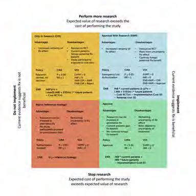
Figure 1. Decision matrix. This 2x2 figure shows the 4 potential combined research-treatment strategies, their advantages and disadvantages, the decision rule when this strategy would be selected as the optimal strategy, and how the expected net monetary benefit (ENB) would be calculated for the value-ofinformation (VOI) analyses to determine the optimal strategy according to the VOI. Difference retrospective/prospective VOI: the number of patients is based on the projections on the date of evaluation (prospective) or on the last data set on the timeline (retrospective). iNB, incremental net (monetary) benefit (NB treat – NB control); p, proportion of patients n in a new randomized controlled trial (RCT) randomly assigned to treatment; 1 2 p, proportion of patients n in a new RCT randomly assigned to control; EVSI, expected value of sample information, calculated in comparison with the optimal treatment (i.e., over and above the iNB gained if iNB . 0); EVSI(n), EVSI dependent on sample size n of the new RCT; cost RCT(n), fixed cost + variable cost of performing a new RCT dependent on sample size n. The optimal sample size is determined by maximizing the function in the quadrant with respect to n. Implementation and reversal costs are assumed to be 0 given that the intervention concerns a guideline change.
A step to achieve this goal is to provide imaging referral guidelines in the form of a computerized decision support system (CDSS). In this line of research, we have initiated a clinical trial to evaluate the effect of introducing a CDSS in the hospital setting that guides imaging requests, the ESR iGuide. The Medical Imaging Decision and Support (MIDAS) study was performed as a multicenter cluster randomized trial with departments acting as clusters combined with a before-after-revert design. Three hospitals with a total of 26 departments (clusters) were recruited for the study and collected data. Departments were randomly assigned to the active intervention or the control condition. In the revert condition, decision support was removed to evaluate the sustainable educa-
tional effect of temporary system use. PhD student Stijntje Dijk worked on this project. This was a collaborative effort with Thomas Kröncke in Augsburg, Jörg Barkhausen in Lübeck, Olav Janssen in Kiel, Peter Mildenberger in Mainz, and Florian Demuth and Katherine Anikina of the ESR iGuide.
Methods to optimize study design
The randomized controlled trial (RCT) has been the standard study design for evaluating health-care interventions for decades. Such trials involve millions of patients and cost billions of Euro’s. Furthermore, the trials are timeconsuming which leads to delayed implementation of potentially beneficial interventions. In this research line we aim to reduce the cost, time, number of patients needed, and burden to patients participating in trials evaluating health-care interventions by optimizing the design of such trials.
Rather than designing trials with the focus on minimizing statistical error, we advocate using a value-driven approach. Using decision modeling of health benefits, patient values, and costs, considering the uncertainty around the input parameters, we can subsequently determine the uncertainty around the outcomes and calculate the value of reducing this uncertainty by doing further research. These analyses help prioritize research and guide study design. In addition, we are utilizing causal inference methods to analyze observational data using target trial designs, that is, emulating a randomized trial design within the data considering confounding factors (Jeremy Labrecque). In addition, we are describing the potential role of causal inference methods in performing systematic reviews, meta-analyses, and decision modeling.
In this research line we focus on non-pharmacological interventions including diagnostic and prognostic imaging biomarkers, medical devices for image-guided therapy, lifestyle interventions, and organizational interventions. Nevertheless, during the pandemic we expanded this line of research to also guide trial design for emerging pharmacological therapies for COVID-19 ( Stijntje Dijk , Eline Krijkamp, Aradhana Pandit). Prioritization of research is of particular interest during a pandemic because performing more trials of an unproven therapy that appears to be beneficial has an opportunity cost: by delaying implementation of the drug many lives could potentially be lost. Since we had the expertise to tackle this policy issue, we felt it was our responsibility to contribute to research efforts related to the pandemic with this analysis and we are currently expanding on this line of research to help prepare for future pandemics.
The effect of lifestyle interventions
Chronic stress and burnout have become an epidemic among health care professionals and form a threat to the sustainability of health care. Radiologists (in-training) and radiological technologists are among those at the highest risk due to the push for productivity and efficiency, the increased interaction with computers rather than human beings, the administrative burden, medico-legal issues, 24/7 connectivity, and long hours in dark rooms. To maintain our professionalism, health, and happiness, preventive measures at both the individual and organizational level are necessary.
In this research line we evaluate lifestyle interventions such as physical exercise, yoga, mindfulness-based stress reduction, martial arts, and music therapy to reduce chronic stress, prevent burnout, and increase resilience and wellbeing among health care professionals and those in training for healthcare professions. We perform systematic reviews, observational studies, RCTs, and modeling studies of interventions. In a clinical trial among medical students, research master students and PhD students, we evaluated the effects of lifestyle interventions. The study had a hybrid design combining a longitudinal cohort, a nested RCT, a preference design, sequential multiple assignment, and adaptive design ( Stijntje Dijk , Larissa Setzer, Kirsty Huininga).
In a commentary published in European Radiology this year we wrote about the challenges faced by medical imaging professionals and how these affect health and well-being. Key points that we make are:
• Prioritizing health, resilience, and well-being among healthcare professionals is not a luxury but a necessity for a sustainable health care system;
• Numerous studies have shown mindfulness practice to be beneficial and safe in enhancing mental health and well-being among healthcare professionals;
• Advocating self-care strategies among healthcare professionals is beneficial but insufficient: organizational interventions are necessary and have a larger effect.
Expectations & Directions
Our future projects will integrate various study designs to maximize the return on research investment. A typical project would integrate simulation studies, a before-after study, a randomized controlled trial (RCT), a prospective longitudinal observational study and value of information analyses. Ideally, we would simulate the study before performing it. The results of our research will inform patients, physicians, insurers, industry, and health-
care policy makers and will guide future research. In the research line concerning professional well-being and resilience we intend to evaluate interventions that reduce experienced stress among radiologists (in-training) and radiological technologists. Our intention is to ensure that Radiology remains an attractive specialty to pursue, and that professionalism will be maintained by ensuring that our employees are happy, healthy, resilient, and engaged.
Funding
Kroencke, Thomas, Myriam Hunink, and Stijntje Dijk German Innovation Fund: 'Medical Imaging Decision and Support (MIDAS)'. 2019-2024
Budde, Ricardo, Alexander Hirsch, and Myriam Hunink ZonMW Health Care Efficiency Research: 'Addition of CT Fractional flow reserve in the diagnostic pathway of patients with stable chest pain to reduce unnecessary invasive coronary angiography (FUSION Study)'. 2021-2024
Ferket, Bart, Kirsten Fleischmann, Umesh Masharani, Wendy Max, and Myriam Hunink RO1, National Institutes of Health, USA: 'Novel antidiabetic medications to reduce cardiovascular events in patients with diabetes mellitus type 2 – a modelling study'. 2021-2024
Invited Lectures
Myriam Hunink. 'Brief teaching residency'. The Center for Health Decision Sciences,' Harvard Chan School of Public Health, Boston, USA. Oct 2024.
Highlights
Stijntje Dijk received the Young Scholar Award in Health Policy in Honor of Sandy Schwartz at the Society for Medical Decision Making meeting, Boston, Oct 2024.
Additional personnel
Thom Korthals – MSc student Clinical Epidemiology, Health Sciences. Supervisor Myriam Hunink.
Jeremy Labrecque – department of Epidemiology, Erasmus MC
Dimitris Rizopoulos – department of Biostatistics, Erasmus MC
Peter van Hulst – department of Neurology, Erasmus MC
Bob Roozenbeek – department of Neurology, Erasmus MC
Diederik Dippel – department of Neurology, Erasmus MC
Kirsten Fleischmann – UCSF, San Francisco, USA
Umesh Masharani – UCSF, San Francisco, USA
Wendy Max – UCSF, San Francisco, USA
Bart Ferket – Mt Sinai, New York, USA
Thomas Kröncke – University Hospital Augsburg, Germany
Jörg Barkhausen – UKSH, Lübeck, Germany
Olav Janssen – UKSH, Kiel, Germany
Peter Mildenberger – University of Mainz, Mainz, Germany
John Wong – New England Medical Center, Boston, USA
Natalia Kunst – University of York, UK
PhD Students

Stijntje Dijk, MD, MSc

Advisors Myriam Hunink, Jeremy Labrecque & John Wong
Project Funding German Innovation Fund: MIDAS
Gordon and Betty Moore Foundation: COVID-19 research
Studievoorschotmiddelen: DESTRESS
Email s.dijk@erasmusmc.nl
Medical Imaging Decision And Support
In a Multi-Center Cluster Randomized Trial we evaluate the effect of implementing active decision support (ESR iGuide) versus control. The decision support is implemented in a physician order entry system. Outcomes are the appropriate use of imaging tests, radiation, and costs. The results will be published early 2025.
JOINT APPOINTMENT IN CHILD AND ADOLESCENT PSYCHIATRY/ PSYCHOLOGY
Ryan Muetzel obtained his BA in Psychology and Biology from the University of Minnesota. After learning neuroimaging at the University of Minnesota’s Center for Magnetic Resonance Research, he joined the Erasmus MC in 2012 for an Msc in Epidemiology and a PhD in population neuroscience, where he also helped build the Generation R Neuroimaging program.

After completing his PhD in 2016, he conducted a postdoctoral fellowship, continued to develop the Generation R Neuroimaging program, and in 2018 became an assistant professor. Ryan now leads the Child and Adolescent Psychiatry department’s pediatric population neuroimaging line and the Generation R Neuroimaging program. Together with his team, the Integrative and Precision Neuroimaging lab, they continue to collect large-scale neuroimaging data through the Generation R Study. r.muetzel@erasmusmc.nl
PEDIATRIC POPULATION NEUROIMAGING
Ryan
Muetzel, PhD assistant professor

Context
The importance and utility of large-scale pediatric neuroimaging initiatives has become increasingly clear over the last decade. Operating at the intersection of epidemiology child psychiatry, and neuroimaging, the pediatric population neuroimaging program at Erasmus MC has been helping to shine light on the importance of this field, representing one of the first and largest neuroimaging initiatives of its kind in the world. Rich, repeated-measures neuroimaging data collected in tandem with prospective deep-phenotyping data during the prenatal, postnatal, and childhood periods provides unprecedented opportunities to understand neurodevelopment across a multitude of disciplines including neuroscience, psychology, psychiatry, pediatrics, public health, nutrition, and more. My team focuses on understanding typical and atypical neurodevelopment, primarily in the context of emergent psychopathology.
Top Publications 2024
Xu B, L Dall'Aglio, J Flournoy, G Bortsova, B TervoClemmens, P Collins, M de Bruijne, M Luciana, A Marquand, H Wang, H Tiemeier, RL Muetzel. Limited generalizability of multivariate brain-based dimensions of child psychiatric symptoms. Commun Psychol. 2024; 2:16.
Gaiser C, R van der Vliet, AAA de Boer, O Donchin, P Berthet, GA Devenyi, M Mallar Chakravarty, J Diedrichsen, AF Marquand, MA Frens, RL Muetzel. Population-wide cerebellar growth models of children and adolescents. Nat Commun. 2024; 15:2351.
Boer OD, H El Marroun, RL Muetzel. Adolescent substance use initiation and long-term neurobiological outcomes: insights, challenges and opportunities. Mol Psychiatry. 2024; 29:2211-2222.
Research Projects: Objectives & Achievements
My team focuses on 4 key themes. First, typical and atypical neurodevelopment. We use repeated neuroimaging assessments to characterize structural and functional brain changes over time, and examine how this relates various exposures (e.g., early life stress, pollution) as well as outcomes (e.g., psychopathology, substance use initiation). Second, we employ multivariate methods (e.g., machine learning) to identify complex patterns in neuroimaging data, primarily focusing on those which are related to mental health outcomes. These methods allow for the use of high dimensional neuroimaging data in prediction, as well as etiological (explainable) work. Third, we focus on etiological work which examines how various risk and resilience factors relate to the brain and mental health. For example, factors such as physical activity and how it is related to (a)typical brain features. Fourth, my team focuses on developing novel techniques which integrate epidemiological concepts into neuroimaging, or population neuroimaging methods. For example, taking into consideration confounding factors in high dimensional data analysis, how selection bias can be accounted for in large-scale neuroimaging studies, and also applying novel methods such as target trial emulation for research questions which would be challenging to address within an RCT framework.
Understanding Typical and Atypical neurodevelopment
Current projects:
– Normative Models of the Human Cerebellum (Bing Xu, Carolin Gaiser)
– Cerebellar-cortical patterns of neurodevelopment (Carolin Gaiser)
Neurodevelopment is a protracted process which begins prenatally and extends into young adulthood. While the last three decades of research have provided an immense amount of information about neurodevelopment from childhood, adolescence and young adulthood, much of the work is based on relatively modest sample sizes and substantial investigation remains a high priority. We have been using the Generation R Study data to better understand typical development in several ways. First, we explored how brain structural connectivity develops over time, and particularly how childhood and pre-adolescent psychiatric problems were related (Dall’Aglio et al.). Using data from both Generation R and the ABCD cohort (Nscans=11,400), we did not demonstrate any robust links between white matter and psychopathology. Given we have identified associations at younger ages, this suggests that earlier periods of development may be more
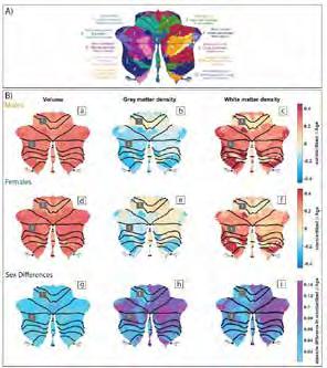
1. A.) The Multidomain Task Battery Functional Parcellation of the cerebellum (adapted with permission from J. Diedrichsen). B.) Standardized beta coefficients for age for each functional parcel for males and females separately, and for sex differences. Volume represents the Jacobian determinant, gray/white densities are average tissue class probabilities within a parcel. Data are from 3 time points, over 7,500 scan sessions from 5,000 individuals.
sensitive, or that delayed neurodevelopment can ‘catch up’ with time. Lastly, we developed a normative model of both the anatomical and functional development of the cerebellum (Gaiser et al.). We demonstrated that the cerebellum follows a gradient of development similar to what is observed in the cortex (e.g., sensorimotor areas develop first). Further, we demonstrated that the normative estimates of the cerebellum show robust links with neurodevelopmental disorders (i.e., Autism) at the individual level (i.e., rather than group level).
Risk and Resilience factors of brain and mental health
Current Projects:
– Air pollution and neurodevelopment (Michelle Kusters
– Sex Differences in Autism (Dylan van der Waal, Caro Gaiser)
– Maternal Immune Activation and Resilience in Neurodevelopment (Frederieke, Gigase, Milan Zarchev)
– Sikkel Cell Disease and Brain Health (Melanie Bruinooge)
– Intergeneration Transmission of Mental Health Problems (Boglarka Kovacs)
Figure

Figure 2. Canonical correlation analysis of psychiatric symptoms and brain structure in 10 year-olds. A.) Psychiatric Canonical variate with high Attention Problems loading, B.) Brain-Based Canonical variate, colors depicting brain areas highly correlated to attenion problems.
While several forms of psychopathology have demonstrated considerable heritability, individual-level genomic risk profiles explain only a modest amount of variability in mental health problems. Thus, it remains crucial to explore how various environmental factors related to risk and resilience for mental health. We have been focusing on several key areas, including physical activity, early life adversity, and various prenatal factors such as maternal immune activation. For example, we found that the number of hours of physical activity per week was related to both white matter microstructure cross-sectionally, but also predicted larger neurodevelopmental changes over time in limbic structures known to be both involved in psychopathology and show considerable plasticity. In terms of risk, recent work from our team has shown that early life adversity, both prenatal and during childhood, is related to intracortical myelin. Importantly, contextual risk factors, such as financial difficulties, were a prominent factor in these associations.
Multivariate techniques for neuroimaging
Current Project:
– Multivariate Prediction of Autism (Carolin Gaiser, Dylan van der Waal, Bing Xu)
Neuroimaging in child psychiatric has elucidated several key features of emerging mental illness over the years. However, we still lack robust ‘biomarkers’ which aid in providing families with better information on for example treatment response prediction and longer-term prognosis. Multivariate techniques, such as machine learning and deep learning, may help to uncover more robust, clinically-relevant patterns in neuroimaging data. My group has been applying several “AI” methods in order to better understand whether such features do meaningfully predict clinical information in children and adolescents with, or at risk for, mental illness. Using nearly 13,000 MRI scans from Generation R and ABCD, we found that with both functional MRI data and with structural MRI data, we could detect patterns which suggested a multivariate ‘biotype’ for attention problems in children. However, while functional MRI data from both cohorts produced similar results when run separately, results trained in one cohort did not translate well to the other. Conversely, results from structural MRI data was generalizable across cohorts, suggesting either more robust signal in sMRI or less issues with harmonization of this modality across cohorts. Figure 2 shows canonical variate (psychiatric symptoms with structural brain data) which was robust across cohorts, and model estimates could be transferred from one cohort to the other.
Population neuroimaging methods development
Current Projects:
– Target Trial Emulation: ADHD Medication and Brain Development (Annet Dijkzeul)
– High-dimensional, whole-brain analysis of repeated measures brain data (Serena Defina)
In addition to a broad range of descriptive, etiological, and prediction work, my team also develops open-source software to help facilitate population neuroscience research. The clearest example is our implementation of a vertex-wise analysis framework for structural MRI data. We developed the QDECR package (github.com/slamballais/QDECR) in order to address substantial gaps in current techniques which are particularly relevant for the field of population neuroscience. For example, many current tools have been implemented for much smaller datasets, and do not run properly when the sample size is too large. Further, most packages do not allow for missing data, which can lead to substantial selection bias in
statistical analysis if ignored. Finally, and perhaps most importantly, we ensured the tool was developed in the R statistical software using familiar R code in order to ensure it was user friendly and approachable to neuroimagers without strong backgrounds in statistics and programming. This work has recently led to funding which will allow us to expand the software package to additional statistical methods, and implement new features such as flexibility with data type inputs (e.g., voxels or connectivity matrices instead of vertices)
Expectations & Directions
One of the most important developments for 2025 is the upgrade of the 3T system used by Generation R. This upgrade brings a host of new opportunities for the Pediatric Population Imaging line. Importantly, we designed and integrated an upgrade sub-study, where nearly 200 individuals will be scanned before and after the upgrade in order to facilitate statistical adjustments for any impact the upgrade will have on our measurements. This new hardware also allows us to collect substantially more data, for example higher spatial, angular, and temporal resolution. The next round of Generation R is scheduled to begin in the first quarter of 2025, where we will continue our standard protocol, but will also add a specialized functional MRI task designed to realize individual-participant functional mapping of the brain. The main research priorities for the team remain understanding the neurobiological causes and consequences of mental health problems, with a particular focus on substance use and affective disorders. Lastly, we will be expanding societal outreach via Healthy Start and Convergence with a recent Sprint Award. The project aims to streamline 3D brain printing of individual MRI scans, as well as developing customizable 3Dcolor-printable brains for education and societal outreach activities.
Funding
Muetzel, Ryan Erasmus MC Fellowship: ‘Disentangling brain-behavior temporality, and multivariate brain-based dimensions of psychopathology’. 2020-2024
Muetzel, Ryan NIH / NIMH - IMPACT Study: ‘Understanding Maternal Immune Activation and child neurodevelopment’. 2021-2026
Muetzel, Ryan , Maarten Frens, Alexandra Badura, Rick van der Vliet , Jeremy Labrecque , and Monique Bernsen Erasmus MC Synergy: ‘Understanding Cortico-cerebellar maturation trajectories’. 2023-2026
Muetzel, Ryan, Jeremey Labreqcue, and Henning Tiemeier Sophia Foundations: 'ADHD, medication use and brain development: Using target trial emulation to infer the effects of stimulant medication'. 2021-2025
Badura, Aleksandra, Ryan Muetzel et al. NWO NWA ORC: 'Why are there more men than women with autism? Sex differences in Autism: Genes, Brain, and Healthcare'. 2024-2029
Diedrichsen, Jorn, Andre Marquand , and Ryan Muetzel Raynor Cerebellum Project: 'Normative growth models of the human Cerebellum'. 2024-2027
Muetzel, Ryan, Sander Lamballais, Pauline Jansen et al. FLAG-ERA-JTC: 'infant2Adult'. [duration of grant missing]
Muetzel, Ryan and Henning Tiemeier EU KA171: ‘Population neuroimaging mobility program - Erasmus MC, Harvard, University of Minnesota'. 2023-2025
Highlights
Lorenza Dall'Aglio defended her PhD dissertation Cum Laude.
Anna Suleri received a Ter Meulen award to visit Mount Sinai School of Medicine in New York to explore further functional connectivity in the context of prenatal maternal immune activation.
Ryan Muetzel hosted the Raynor Cerebellum Project kickoff meeting in Rotterdam in April, with more than 15 invited speakers and 40 attendees, in order to foster collaboration, brainstorm future directions, and initiate a funded project to further our understanding of cerebellum development.
The Sophia Children's Hospital 3T MRI system was upgraded from an MR750W to a Premier system in Fall 2024. This opens a new chapter in the longitudinal MRI scanning for Generation R, as well as a new collaborative endeavor with the EUR and TU-Delft.
Additional Personnel
Jessica Wijngaarden – Data manager
Serena Defina – Data Scientist
Jana Hermans – PhD student, Department of Child and Adolescent Psychiatry/Psychology
Dogukan Koc – PhD student, Department of Child and Adolescent Psychiatry/Psychology
Tessa Mulder – PhD student, Department of Internal Medicine
Laura Monteiro – PhD student, Department of Child and Adolescent Psychiatry/Psychology
Merel de Vries – PhD student, Department of Adult Psychiatry
Shuaiqun Pan – PhD student, Computer Science, Leiden University
Amina Burgess – PhD student, Spinoza Center for Neuroimaging
Sharon Ololo – Researcher, Department of Adult Psychiatry
Jin Ouyang – MSc Student, TU Eindhoven
Remmy Duijsens – MSc Student, TU Delft
Hannah van Heusden – MSc Student, Medicine, Erasmus MC
Suzaan Linnsen – MSc student, Department of Adult Psychiatry
Post-docs
Milan Zarchev
Project funding NIH R01 Understanding Early-life Adversity, Maternal Immune Activation and Neurodevelopmental Outcomes Email m.zarchev@erasmusmc.nl
Early Life Adversity, Maternal Immune Activation, and Neurodevelopmental Outcomes
Early-life adversity has been consistently shown to impact neurodevelopment, including pubertal timing, behavioural and emotional problems such as ADHD, and also brain morphology and function. However, inconsistencies exist in the literature, suggesting mostly very severe forms of adversity affect neurodevelopment. This project aims to a.) utilize novel causal inference methods to understand how ELA links with neurodevelopment, and b.) particularly test whether maternal immune activation profiles are linked to adverse neurodevelopment.


Frederieke Gigase
Project Funding NIH IMPACT: Understanding Maternal Immune Activation and Neurodevelopmental Outcomes
Email f.gigase@erasmusmc.nl
Maternal Immune Activation, and Neurodevelopmental Outcomes
Maternal immune activation has been shown to be considerably distinct from both pre- and post-pregnancy immune profiles. Decades of work has pointed to adverse neurodevelopmental outcomes linked to maternal immune profiles. This has been particularly linked to maternal infections during pregnancy. However, various factors not linked to infections also have been shown to impact immune profiles, for example obesity, smoking, and early life stress. This project aims to examine in detail whether there is a link between the maternal immune profile and the risk for offspring to develop autism. This project will also ex-
PhD Students

Bing Xu, MSc

Advisors Ryan Muetzel & Henning Tiemeier
Project Funding ZonMw Vici: Adolescent Depression (Tiemeier)
Email b.xu@erasmusmc.nl
Bing is a PhD student with a background in developmental and computational psychology. She is interested in applying advanced statistical techniques in neuroimaging to address the heterogeneity in child psychopathology. She has been working on identifying ‘biotypes’ of children’s ADHD via brain connectivity and morphology using machine learning.

plore how the maternal immune profile is linked to other neurodevelopmental outcomes, such as brain structure and function.

Lorenza Dall'Aglio, MSc

Advisors Ryan Muetzel & Henning Tiemeier
Project Funding ZonMw Vici: Adolescent Depression (Tiemeier)

Email l.dallaglio@erasmusmc.nl
PhD obtained 14-05-2024
Lorenza investigates the neurobiology of anxiety and depression across adolescence, in the Generation R and ABCD Studies. In particular, she focuses on bi-directionality of psychiatric neuroimaging and risk factors in brain maturation. She also founded the Rotterdam site of the R.I.O.T. Science Club (riotsciencenl.com), an open research training initiative.

Michelle Kusters, MD, MSc

Advisors Ryan Muetzel, Henning Tiemeier & Monica Guxens
Project Funding ISGlobal
Email m.kusters@erasmusmc.nl
Michelle works on a collaborative PhD project between ISGlobal in Barcelona and the Erasmus MC, which focusses on the influence of environmental pollutants on neurodevelopment in children. Her project has focused specifically on understanding the potential consequences of air pollution on behavior, cognition, and brain development.

Eva Borkhuis, MSc

Advisors Ryan Muetzel, Danielle Remmerswaal, Ruth van der Hallen & Pauline Jansen
Project Funding Convergence: Healthy Start. Email borkhuis@essb.eur.nl
Eva is a PhD student working within the convergence initiative, namely within Healthy Start. Her project is situated within the mental well-being ambition of healthy start, and focuses on understanding risk and resilience factors of mental wellbeing using a novel, youth-centered approach.

Caro Gaiser, MSc

Advisors Ryan Muetzel, Rick van der Vliet & Maarten Frens
Project Funding Deptartment of Neuroscience: Cerebellar Development
Email c.gaiser@erasmusmc.nl
Caro is a PhD student investigating the cerebellum, motor control and motor learning at the Department of Neuroscience and in the Generation R study. Her research focuses on describing typical cerebellar development and finding factors that can explain individual differences in motor learning using behavioral, electrophysiological, neurostimulation, and neuroimaging methods.

Annet Dijkzeul, MSc

Advisors Ryan Muetzel, Jeremy Labrecque & Henning Tiemeier
Project Funding Sophia Foundation: Target Trial Emulation in ADHD and Neurodevelopment.
Email j.dijkzeul@erasmusmc.nl
Annet is a PhD student with a background in Neuropsychology and Neurosciences. Her research focuses on combining neuroimaging and epidemiological techniques to better understand neurodevelopment and ADHD. Specifically, she will utilize target trial emulation to make causal inferences on the impact of ADHD medication on brain development.
Anna Suleri, MSc

Advisors Charlotte Cecil, Tonya White & Veerle Bergink
Project Funding NIH Impact Study
Email a.suleri@erasmusmc.nl
Anna is a PhD student working on an NIH-funded R01 grant to better understand how prenatal exposure to maternal infection and immune activation are related to neurodevelopment. She is focusing on self-reported measures of infection status during pregnancy and a unique cytokine assay of samples collected during early pregnancy.

Melanie Bruinooge, MSc

Advisors Marjon Cnossen, Ryan Muetzel & Sabine Mous
Project Funding Erasmus MC Department of Pediatrics
Email m.bruinooge@erasmusmc.nl
The BRICK Study
Sickle cell disease (SCD) is a recessive red blood cell blood disorder. Individuals with SCD have an increased risk of both overt cerebral infarctions and silent infarctions. This project maps whether brain tissue damage precedes, parallels, or lags behind non-brain tissue damage in youth with SCD.

Dylan van der Waal, MSc

Advisors Ryan Muetzel, Pauline Jansen & Alexandra Badura
Project Funding Erasmus MC Department of Child and Adolescent Psychiatry/Psychology
Email d.vanderwaal.2@erasmusmc.nl
The SCANNER project
The aim within SCANNER is to identify brain features that co-segregate with sex in autism. Large, population-based data (Generation R) will allow us investigate the correlation between structural and functional brain changes over development, autistic traits and sensorimotor difficulties, particularly in the context of sex differences.

Boglarka Kovacs, MSc

Advisors Neeltje van Haren, Alex Neumann, Lisanne van Houtum & Ryan Muetzel
Project Funding Erasmus MC Department of Child and Adolescent Psychiatry/Psychology
Email b.kovacs@erasmusmc.nl
The FAMILY Project
This project aims to characterize if variations in brain metrics constitute plausible pathways from intergenerational risk of mental illness from parent to offspring. Further, Boglarka will elucidate whether identified neuroimaging biomarkers improve the prediction of mental health outcomes in persons at familial high-risk for mental illness.
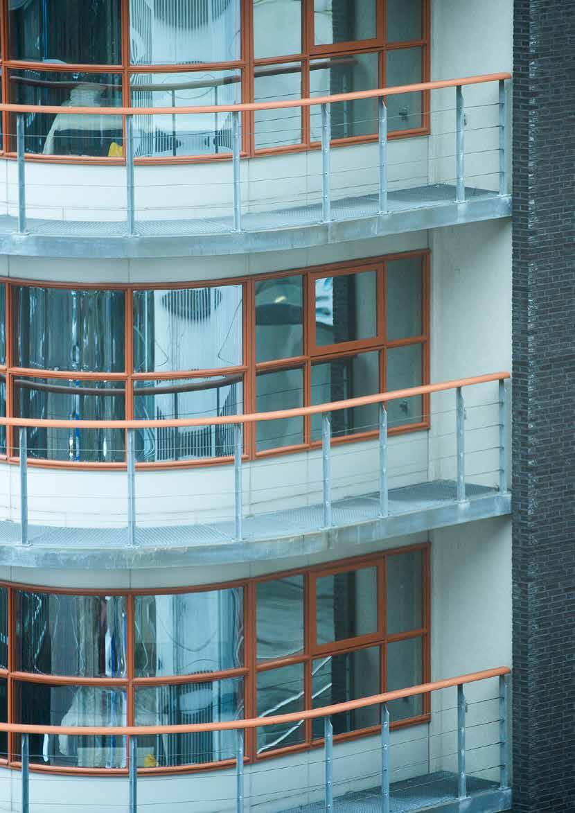

INPUT & OUTPUT
CONFERENCE CONTRIBUTIONS 2024 (SELECTION)
This section provides a selection of the 2024 conference contributions from the Erasmus MC Department of Radiology & Nuclear Medicine. Given the large number of conferences to which we contribute, this overview is necessarily incomplete, but it gives some idea of the magnitude of our conference participation.
The Netherlands
Rotterdam 11
Utrecht 9
Den Bosch 3
Amsterdam 3
Zeist 2
Nijmegen 1
Groningen 1
Vianen 1
Enschede 1
Delft 1
Almere 1
Amersfoort 1
Maastricht 1
Oss 1
Petten 1
Delft 1
United Kingdom
Glasgow 2
Cardiff 1
Cambridge 1
Liverpool 1
Oxford 1
Edinburgh 1
London 1
Austria
Vienna 11
Bad Ischl 1
Germany
Hamburg 5
Stuttgart 1
Spain
Barcelona 3
France
Paris 1
Nantes 1
Morrocco
Marrakesh 7
Switzerland
Lugano 1
Hungary
Budapest 1
Portugal
Coimbra 1
Porto 1
Switzerland
Basel 2
Zweden
Uppsala 1
Japan
Yokohama 1
Philippines
Manila 1
Singapore
Singapore 3
PUBLICATIONS 2024
External publication partners for the Department of Radiology & Nuclear Medicine, 2019-2024
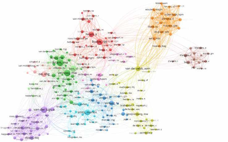
Publicationlist 2024
To see complete publicationlist of all our staffmembers and external publication partners, please scan QR code below.
PhD dissertations
1. Theresa Feddersen. 2024, January 23. MR Thermometry for Hyperthermia in the Head and Neck. Promotor/co-promotor: Prof. dr. Van Rhoon, Prof. dr. Hernandez Tamames, Prof. dr. ir. Paulides, Dr. ir. Poot.

2. Danny Feijtel. 2024, January 31. Improving therapeutic outcome of patients with neuroendocrine cancer: Understanding both the target and the bullet. Promotor/ co-promotor: Prof. dr. Kanaar, Prof. dr. Verburg, Dr. Nonnekens.

3. Jan van der Voet. 2024, March 7. The position of the meniscus in knee osteoarthritis. Promotor/co-promotor: Prof .dr. Bierma-Zeinstra, Prof. dr. Oei, Dr. Runhaar, Dr. Vroegindeweij.

4. Karin van Garderen. 2024, March 13. Signs of Progression. MR image analysis for the management of adult low-grade glioma. Promotor/co-promotor: Prof. dr. Smits, Dr. ir. Klein.
5. Ruisheng Su. 2024, April 2. Quantitative Cerebral Angiography in Ischemic Stroke. Promotor/co-promotor: Prof. dr. Niessen, Prof. dr. Van Der Lugt, Prof dr ir. Ruijters, Dr. Ir. Van Walsum.
6. Luke Terlouw. 2024, April 17. Towards early detection and durable treatment of chronic mesenteric ischemia. Promotor/ co-promotor: Prof. dr. Bruno, Dr. Leemreis – Van Noord, Dr. Moelker.

7. Nadinda van der Ende. 2024, April 23. Analysis of clinical research methods and outcomes in patients treated with reperfusion therapy for ischemic stroke. Promotor/co-promotor: Prof. dr. Dippel, Prof. dr. Van Der Lugt, Dr. Roozenbeek.

8. Sven Luijten. 2024, May 1. Imaging in Ischemic Stroke: Leveraging imaging beyond diagnosis to predict functional outcome and benefit of endovascular thrombectomy. Promotor/co-promotor: Prof. dr. Van Der Lugt, Prof dr. Dippel, Dr. Bos, Dr. Roozenbeek.

9. Dianne van Dam – Nolen. 2024, May 8. Plaques en patiënten: De rol van beeldvorming van de arteria carotis in het voorkomen van herseninfarcten Promotor/ co-promotor: Prof. dr. Van Der Lugt, Prof. dr. Koudstaal, Dr. Bos.
10. Marjolein Verhoeven. 2024, May 14. GRPR-gerichte theranostiek voor de behandeling van kanker: nieuwe benaderingen voor een maximaal therapeutisch resultaat Promotor/co-promotor: Prof. dr. Verburg, Prof. dr. ir. De Jong, Dr. Dalm.

11. Fatemehsadat Arzanforoosh. 2024, May 14. Glioma Oxygenation and Vasculature in the Spotlight of MRI Promotor/co-promotor: Prof. dr. Smits, Dr. ing. Warnert.

12. Nienke Sijtsema. 2024, May 21. Development of imaging-based response predictors for personalized radiotherapy in head and neck cancer Promotor/co-promotor: Prof. dr. Hoogeman, Prof dr. Hernandez Tamames, Dr. ir. Petit, Dr. ir. Poot.

13. Joost Verschueren. 2024, June 20. Valgiserende therapie voor mediale knieartrose: klinische resultaten en overwegingen omtrent kwantitatieve beeldvorming
Promotor/co-promotor: Prof. dr. BiermaZeinstra, Prof. dr. Oei, Dr. Reijman.

14. Tong Wu. 2024, June 21. Body composition, physical activity and adolescent respiratory health: The Generation R Study
Promotor/co-promotor: Prof. dr. Oei, Dr. ir. Klein, Dr. Duijts.

15. 2024, June 21. Beeldvorming en biomarkers in prostaatkanker
Promotor/co-promotor: Prof. dr. De Wit, Dr. Van Der Veldt, Dr. Lolkema.

16. Mohamed Benmahdjoub. 2024, June 26. I See Through... You Promotor/co-promotor: Prof. dr. Wolvius, Prof. dr. Niessen, Dr. ir. Van Walsum.

17. Wietske Bastiaansen. 2024, July 2. Modeling Human Embryonic Brain Development Promotor/co-promotor: Dr. ir. Klein, Prof. dr. Steegers-Theunissen, Prof. dr. Niessen, Dr. Rousian.

18. Ilanah Pruis. 2024, July 5. Theranostic PET-MRI voor hersentumoren Promotor/ co-promotor: Prof. dr. Smits, Dr. Veldhuijzen van Zanten.
19. Sonja Katz. 2024, September 20. From Bytes to Bedside and Back: Enhancing Clinical Decision Support with Explainable AI Promotor/co-promotor: Prof. dr. ir. Martins dos Santos, Dr. Roshchupkin, Dr. Saccenti.
21. Robin Camarasa. 2024, October 18. The Carotid Artery in Bits & Pieces Promotor/ co-promotor: Prof. dr. De Bruijne, Dr. Bos.

22. Tareq Abdel Alim. 2024, October 22. From Mesh to Meaning: Computational analysis of craniofacial dysmorphologies and surgical interventions Promotor/ co-promotor: Prof. dr. Dirven, Prof dr. Niessen, Dr Van Veelen – Vincent, Dr. Roshchupkin.

23. Erika Murce Silva. 2024, October 29. Towards the Improvement of Diagnosis and Treatment of Prostate Cancer: Optimization of PSMA-Targeted Radiopharmaceuticals Promotor/co-promotor: Prof. dr. Verburg, Dr. Seimbille.

20. Riwaj Byanju. 2024 September 26. Accelerated Quantitative MRI by Joint Reconstruction and Quantification Promotor/ co-promotor: Dr. Klein, Dr. ir. Poot.

24. Nikki van der Velde. 2024, October 31. De rol van niet-invasieve beeldvorming bij cardiomyopathieën Promotor/co-promotor: Prof. dr. Budde, Prof. dr. Zijlstra, Dr. Hirsch.

SONJA KATZ
25. Yuxin Chen. 2024, November 12. Chest Computed Tomography: sensitive outcome measures of early cystic fibrosis structural lung disease Promotor/co-promotor: Prof. dr. Tiddens, Dr. Ciet, Dr. Caudri.
26. Wytse van den Bosch. 2024, November 19. Structure and function of small airways in children with asthma Promotor/ co-promotor: Prof. dr. Tiddens, Dr. Janssen, Dr. Ciet.

27. Thom Reuvers. 2024, November 26. Verbetering van de uitkomst van peptide receptor radionuclide therapie: een benadering vanuit de stralingsbiologie Promotor/co-promotor: Dr. Nonnekens, Prof. dr. Kanaar, Prof. dr. Verburg, Dr. Bos.
28. Qianting Lv. 2024, November 28. Clinical Validation of a Fully Automatic Bronchus-Artery Analysis Algorithm: A sensitive and accurate method to detect and monitor bronchial widening and wall thickening on computed tomography Promotor/co-promotor: Prof. dr. Tiddens, Prof. dr. De Bruijne, Dr. Ciet.

29. Yulun Wu. 2024, December 10. Making the invisible visible via advanced post-processing of chemical exchange saturation transfer (CEST) MRI Promotor/co-promotor: Prof. dr. Smits, Dr. ir. Warnert.

30. Joyce van Arendonk. 2024, December 17. Vascular and amyloid pathology in neurodegeneration and cognition, a population imaging study Promotor/ co-promotor: Prof. dr. Vernooij, Prof. dr Ikram, Dr. Neitzel.

INDEX