“Therefore, thus says the Lord, Behold, I am bringing disaster upon them that they cannot escape. Though they cry to me, I will not listen to them.” (Jeremiah11:11). This is a quote from Bible regarding the dooms day. Are we then facing the nemesis? Well the dire, bizarre and horrendous statistics pouring in from all corners of our country makes us almost convinced that the apocalypse is round the corner. But is Lord responsible for all miseries? Certainly not, it is man, callous, unpragmatic creature who has turned this pristine world into a catacomb. Let us zero in on India. Even after having the learned law makers and the policy makers and the political heads of the country who have rightfully enforced their power and onus to control the infection, India, now tops the chart of the highest COVID infected population of the world and the consequent death that follows. “Hindsight” is a word that means taking lessons from an event that has happened previously but blissfully we preferred to be the “Dhritarashtras” and predicted that the tsunami might come but we, the Indians will be floating in the Noah’s ark. Sadly however our ark is almost on the verge of capsizing. Let us discuss in detail. We are fighting with these resources. Namely, in India only 2.3 critical care beds for every 100,000 populations1,2 and less than one doctor for every 1,000 citizens3,4
“As a doctor told me: “when someone dies because you could not provide him oxygen, that is not a natural death; this is murder.”5. What can be more appropriate a sentence to begin our discussion than a noted journalist from India , by quoting a doctor , reflects how we have paved our way to our dire destiny. NO OXYGEN, NO MEDICINES, NO BEDS , NO AMBULENCE, NO TESTS...” plethora of ‘NOS’ are being showered on us because and only because we preferred to live in a fool’s paradise. In a democracy, people get the government they deserve and they deserve what they get – goes a saying. But do we deserve this? No, but only holding the government responsible for the disaster is like shaking off the responsibility off my own shoulder and simply holding the other responsible for the nemesis. Truly this year’s election was no exception. But are we a pack of gullible crowds who can easily forget the disaster that we have just faced and still walking through the fire? Blissfully opting to ignore the pandemic preventive norms we responded to the calls of the “Pied pipers of Hamlin” and flocked in huge numbers at the assemblies almost without masks and social distancing as if like throwing a brazen insolence and defiance against the invisible gargantuan devil. The result - As of 8th May Coronavirus Cases : 21,892,676, Deaths : 238,270, Recovered : 17,930,9606. The
country of 1.4 billion is sinking beneath the weight of infections7
Super spreading took place when thousands filled cricket stadiums and millions of people took a dip in the Ganges during the Kumbh Mela festival. India went ahead with five state elections in April, and an unmasked politicians held huge rallies. Cherished brand of Indian exceptionalism bred complacency. A presumption of national greatness has led to a lack of preparedness, most notably in vaccine production. The west had encouraged India to become a linchpin in global drug-making. China and the US are now manufacturing more COVID-19 vaccines than India8. What an irony? The country being the highest manufacturer of vaccine falls short of vaccinating their own population.
In the first wave, Covid struck India’s cities, but it is now moving to rural areas also. As with many of the countries hit hardest, India’s death toll was largely avoidable. India is a big, complex and diverse country that is difficult to govern during a national emergency. It is now suffering from parallel epidemics of coronavirus and fear. To contain biological and social contagions requires credible reassurance, to quell panic, and for people to wear masks and obey rules of physical distancing. The end result is that the political hubris met pandemic reality in India. A new “double mutant” variant, named B.1.617, has emerged in a devastating
corona virus second wave which has seen hospitals run out of beds and oxygen. Mortuaries are so full that bodies are left to decompose at home. Charities warn that the dead risk of being left on the streets. The hecatomb reminds us of those famous lines from Julius Caesar –
“With Ate by his side come hot from hell, Shall in these confines with a monarch’s voice Cry “Havoc!” and let slip the dogs of war, That this foul deed shall smell above the earth Withcarrionmen,groaningforburial.”
According to an article published in Indian Express, “Our vaccine policy reflects an adhoc social Darwinism, where the strong do what they can and the weak suffer because they must”9. There is an urgent need to fix several policies. But what do policy proposals mean, when all policies are mere publicities without executions. There is a need to fix accountability. But how does accountability get fixed? This is not the time of mudslinging against each other it is time to reconsolidate and empower and fight with all force against the monster.
The only empowering thought in this context has been the splendid work that professionals, frontline government workers, health professionals and
volunteers are doing.. The extraordinary mobilization of civic action whether in supplying oxygen or cremating the dead has been exemplary. Undeniably the depth of commitment of these civic workers is the only healing force against the looming pessimism. This civic commitment has helped mitigate suffering to some extent. To sum up, India has been attacked by alien forces many times before and She has shown gumption and solidarity against such forces to mitigate their onslaught. Let us all put our hands together and
with the help of the other countries of the world , willing to stand by us as allied forces, combat the invisible Satanic virus. Let us build a strong wall of resistance and resilience to counter the attack and work together to establish a brighter future.
REFERENCES
1Bharath Kumar Tirupakuzhi Vijayaraghavan et al. Challenges in the delivery of critical care in India during the COVID-19 pandemic. Journal of the Intensive Care Society 0(0) 1–7. The Intensive Care Society 2020 Article reuse guidelines: sagepub.com/journals-permissions DOI: 10.1177/ 1751143720952590 journals.sagepub.com/home/jics.
2Phua J, Faruq OM, Kulkarni AP, et al. Critical care bed capacity in Asian countries and regions. Crit Care Med 2020; 48: 65462.
4https://www.business-standard.com/article/pti-stories/ indiahas-one-doctor-for-every-1-457-citizensgovt119070401127_1.html (accessed on 25 July 2020).
Effect of Deranged Thyroid Profile on Glycated Hemoglobin : Pre and Post Treatment
Anubha Srivastava1, Sandeep Rai2
Introduction : Glycated haemoglobin (A1C) levels depend on factors other than glycemic status and may have altered levels in different conditions. It has been postulated that A1C levels may vary due to altered thyroid status.
Methods : Non-Diabetic patients of overt hypo- and hyperthyroidism were selected. Age and sex-matched controls were recruited. Baseline values of A1C and reticulocyte count (for RBC turnover) was measured. These values were re-evaluated in randomly selected subgroups after achievement of euthyroid status.
Results : A1C values in patients initially selected, was found to be significantly higher in hypothyroid group as compared to controls though values did not differ significantly in hyperthyroid group. Posttreatment after achieving euthyroid status, A1C levels reduced significantly in hypothyroid group and no such significant effects were observed in hyperthyroid group.
Conclusion : There is the need for evaluation of A1C in patients of hypothyroidism with more caution and prevent the patients from irrelevant investigations and work up for diabetes.
Key words :A1C, Hypothyroid, Hyperthyroid.
Thyroid disorders are perhaps the most common medical conditions throughout the world1. Thyroid hormones are seen to have an intimate relationship with insulin during cellular metabolism. Thyroid disorders can have a significant effect on blood glucose levels and, if left untreated, can affect glycemic control2 Hyperthyroidism has long been recognized to promote hyperglycemia 3 . A relationship between insulin resistance and oxidative stress has also been traced4. The interrelationship between thyroid dysfunction and insulin resistance has also been established by some studies that have shown normalization of long-term indicators of glycemic controls (HbA1c) among nondiabetic thyroid disorder patients following thyroxine replacement therapy5-7. Such findings in turn indicate that inflated HbA 1c values in these patients are unrelated with diabetes and could be normalized only by managing the thyroid disorders, thus reducing an impending diabetic burden to a great extent.
AIMS AND OBJECTIVES
This study was designed to observe an effect of deranged thyroid profile on A1C levels in non-diabetic individuals, with overt hyper- and hypo-thyroidism and later see the effect of treatment on A1C levels.
MATERIALS AND METHODS
A prospective cohort study was conducted in SRN
Department of Internal Medicine, MLN Medical College, Prayagraj
211002
1MD (Medicine), Associate Professor
2MBBS, Junior Resident and Corresponding Author
Received on : 22/07/2020
Accepted on : 27/07/2020
[J Indian Med Assoc 2021; 119(5): 13-5]
Editor's Comment :
Caution is to be excercised for interpretation of HbA1c levels in thyroid dysfunction.
HbA1c levels may be falsely high in unmeated hypothyroid patients which disminish with levothyroxine therapy.
HbA1c values show no correlation in Hyperthyroid patients.
Hospital, Prayagraj from April 2018 to August 2019 with patients more than 18 years of age and either sex with newly diagnosed overt hyper- and hypothyroidism were enrolled and recruited as cases with euthyroid and euglycemic age and gender based control.
Patients with known diabetes or pre-diabetes such as those having deranged fasting and postprandial plasma glucose were excluded from the study as per American Diabetes Association (ADA) 2019. Patients with anemia (Hb<10g/dl), hemoglobinopathies, renal insufficiency, liver dysfunction and pregnant females were also excluded from the study.
A baseline A1C was measured and were started on Thyroid Hormone Replacement Therapy (THRT) with levothyroxine in hypothyroidism and Methimazole in hyperthyroidism. The cases were followed after three months and six months from the date of start of therapy and were reinvestigated for Thyroid Stimulating hormone (TSH) and A1C at follow up.
The statistical analysis was done using SPSS (Statistical Package for Social Sciences) Version 21.0 statistical Analysis Software. The values were represented in Number (%) and Mean±SD. To test the significance of two means the student ‘t’ test was
119, No 5, May 2021Journal
used. The ANOVA test was used to compare the within group and between group variances amongst the study groups. P value of <0.05 is taken as significant.
RESULTS
Table 1A demonstrates comparison of baseline characteristics between hyperthyroid group and controls and it was inferred that no significant difference between two groups was observed for any of the parameters except thyroid hormones (fT3, fT4 and TSH).
Table 1B demonstrates comparison between hypothyroid group and controls. It was observed that in hypothyroidism cases values of A1C and reticulocyte count were significantly higher as compared to that in controls (p<0.001).
In Table 2, hyperthyroid group, mean TSH levels at baseline were 0.02±0.02 µIU/ml which were found to be 3.69±0.93µIU/ml at follow-up, thus showing a significant increase of 3.67±0.93 µIU/ml (p<0.001). On the other hand, mean HbA1c levels were 5.22±0.22% at baseline which dropped to 5.12±0.26% at follow-up, showing a decline of 0.10±0.20%, though was not significant statistically (p=0.090).
In hypothyroid group, mean TSH levels were 27.33±19.76 µIU/ml at baseline which declined to 4.06±0.56 µIU/ml at follow-up, thus showing a significant decline of 23.26±19.69 µIU/ml (p<0.001). Mean HbA1c levels were 5.78±0.19% at baseline which was 5.46±0.11% at follow-up, thus showing a decline of 0.38±0.14% and was significant (p<0.001).
DISCUSSION
Out of 133 patients enrolled 83 patients (62.4%) had deranged thyroid profile and the other 50 Euthyroid patients were taken as controls. Hypothyroidism was more common in our study, 60 patients (45.1%) were hypothyroid and 23(17.3%) patients were hyperthyroid. Our study showed higher prevalence of hypothyroidism in comparison to hyperthyroidism similar to the study done by Ambika Gopalakrishnan et al8 . In our study mean age (in years) of patients in hypothyroid group was 40.58±9.58 and hyperthyroid group was 37.61±7.60. This result was similar to study done by Nagarkar et al9 who showed that the prevalence of
thyroid disorders was significantly higher in higher aged (>31years) patients as compared to lower aged (<30 years) patients (14.1% versus 85.9%, P<0.001). In our study mean baseline A1C values in hypothyroid patients were compared with age and sex matched controls and it was found that values were significantly higher in hypothyroid group(5.77±0.155) in comparison to healthy controls(5.17±0.30) .
Table 1A — Comparison of Baseline general and clinical profile between hyperthyroid group and controls
Table 1B — Comparison of Baseline general and clinical profile between hypothyroid cases and controls
CharacteristicHyperthyroid ControlsSignificance of (n=60)(n=50) difference (Independent samples ‘t’-test) MeanSDMeanSD‘t’‘p’
Age40.589.2839.548.410.6120.542
Gender :
(25.0%)21 (42.0%)
Table 2— Comparison of in TSH levels and HbA1c Levels before and after TRT among cases completing follow-up
Parameter
Hyperthyroidism :
Anantarapu et al6 did a similar study in context of A1C values in hypothyroid patients and made a demonstration that HbA1c values are falsely elevated in hypothyroid patients. Similar observations were demonstrated by Kim et al7(5.54 ± 0.43% versus 5.34±0.31% in hypothyroid patients and controls respectively; p<0.001), despite the lower level of plasma fasting glucose in the hypothyroid individuals. Contrary to the findings demonstrated in hypothyroid group, there was no significant difference observed between A1C and hyperthyroid group in comparison to healthy controls (cases 5.23±0.20, controls 5.17±0.30, p value 0.149) which was statistically insignificant. Similar observations were obtained in a study conducted by Rana Bhattacharjee et al5
In 40 hypothyroid patients were given levothyroxine treatment and were followed up with Thyroid profile at 3 months and 6 months. After attainment of euthyroid status mean A1C was measured and it was found to be 5.46±0.11 which was statically significant as compared to pretreatment group ( 5.78±0.16,p<0.001). Similar observations were obtained by Rana Bhattacharjee et al5( 5.7 ± 0.75 [pretreatment] versus 5.4 ± 0.75 [posttreatment]; P < 0.001). Kim et al7 did a similar follow up study enrolling 30 hypothyroid patients who were given thyroid hormone replacement and concluded that A1C values returns to normal post treatment (pre-treatment 5.57±0.26, posttreatment 5.37±0.32,p value <0.001).
In hyperthyroid group 13 out of 23 patients were enrolled for follow up and were put on anti-thyroid medication. A1C values were measured at follow up once these patients were rendered euthyroid which was sustained. It was observed that there was no statistically significant difference in A1C values post treatment (pre-therapy 5.12±0.20 posttherapy 5.17±0.30 p=0.583).These results were consistent with the results obtained by Rana Bhattacharjee et al5 (5.35±0.45 [pretreatment] versus 5.35 ± 0.3 [posttreatment]; P = 0.323).
CONCLUSION
A1C is an integral part of diagnosis of Diabetes. As we are aware about fallacies of A1C in Anemia, Hemoglobinopathies, Cardiovascular Research Foundation (CRF), we propose by this study that A1C levels are falsely elevated in patients of hypothyroidism. A1C alone is less reliable marker for assessment of dysglycemia in hypothyroid patients. A1C levels in hypothyroid patients leads to false diagnosis of prediabetes and diabetes Following levothyroxine replacement, A1C levels reduced significantly in patients with hypothyroidism. Patients with hyperthyroidism do not show such correlation between glycated hemoglobin levels and thyroid hormone levels both pre and posttreatment. So, caution should to be taken while interpreting A1C values in patients with thyroid dysfunction. So tests like serum fructosamine assay and glyclated albumin have been proposed to overcome this fallacy .
Funding : None
Conflict of Interest : None
REFERENCES
1Vanderpump MPJ — The epidemiology of thyroid diseases. In: Braverman LE, Utiger RD, eds. Werner and Ingbar’s The Thyroid: A Fundamental and Clinical Text. 2005, 9th edn, pp 398-406. JB Lippincott-Raven, Philadelphia.
2Mukherjee S, Datta S, Datta P, Mukherjee AK, Maisnam I — A study of prevalence of primary hypothyroidism in recently diagnosed type 2 diabetes mellitus in a tertiary care hospital. Int J Sci Rep 2015; 1(2): 105-12. doi: 10.4103/22308210.196017.
3Maxon HR, Kreines KW, Goldsmith RE, Knowles HC Jr — Long-term observations of glucose tolerance in thyrotoxic patients. Arch Intern Med 1975; 135(11): 1477-80.
4Ullah A, Khan A, Khan I — Diabetes mellitus and oxidative stress—A concise review. Saudi Pharm J. 2016
5Bhattacharjee R, Thukral A, Chakraborty PP, Roy A, Goswami S, Ghosh S, et al — Effects of thyroid status on glycated hemoglobin. Indian J Endocrinol Metab 2017; 21(1): 26-30. doi: 10.4103/2230-8210.196017.
6Anantarapu S, Vaikkakara S, Sachan A, Phaneendra BV, Suchitra MM, Reddy AP, etal—Effects of thyroid hormone replacement on glycated hemoglobin levels in non diabetic subjects with overt hypothyroidism. Arch Endocrinol Metab 2015; 59(6): 495-500. doi: 10.1590/23593997000000065.Epub 2015 Sep 25.
7Kim MK, Kwon HS, Baek K-H, Lee JH, Park WC, Sohn HS, et al—Effects of Thyroid Hormone on A1C and Glycated Albumin Levels in Nondiabetic Subjects With Overt Hypothyroidism. Diabetes Care 2010; 33(12): 2546-8. doi: 10.2337/dc100988.Epub 2010 Sep 7.
8Unnikrishnan AG, Menon UV — Thyroid disorders in India: An epidemiological perspective. Indian J Endocrinol Metab 2011 Jul; 15(Suppl2): S78-S81. doi: 10.4103/2230-8210.83329
9Nagarkar R, Roy S, Akheel M, Palwe V, Kulkarni N, Pandit P — Incidence of Thyroid Disorders in India: An Institutional Retrospective Analysis. International Journal of Dental and Medical Speciality 2015; 19-23.
Original Article
Plasmapheresis; Reasons & Results, An Epidemiological Study on the Indications and Outcome of Therapeutic Plasma Exchanges in a Tertiary Care Hospital — One
Year Single Center Experience
Soumava Gupta1, Anand Prasad2, Pratik Das3, Deepak S Roy4, Ravi Ranjan Saw Mondal5, Manoj Gupta6, Himadri Sekhar5
Background : Therapeutic Plasma Exchange (TPE) is a commonly accepted procedure used to remove circulating antibodies against specific antigens which are causative to a variety of diseases mediated by the reaction of these Antigen and Antibodies and activation of the complement cascade thereafter. It is also used as a part of the desensitization protocol in case of ABO incompatible renal transplant where circulating donor specific auto antibodies against the donor’s blood group is removed.
Methods : This is a longitudinal observational study which assess the indication, complication and outcome of TPE in the hemodialysis unit of Rabindranath Tagore International Institute of Cardiac Sciences, a Tertiary Care Center for Nephrology and a Renal Transplant Unit. Total 91 patients underwent TPE during study period. The number of TPE sessions varied from 19 to 2 with a mean of 5.28(2.92) per person. Combination of FFP+5% Human Albumin was uses as replacement fluid in majority of patients (86.36%). Fresh Frozen Plasma (FFP) and 5% Human Albumin (HA) was used as isolated replacement solution in (11.36% patients) and (2.37% patients) respectively. 38 patients underwent TPE for Antibody mediated rejection (AMR), 4 patients for CAN/ATN ,4 patients for chronic AMR(CAMR) and 1 patient for Hyper Acute Rejection (HAR) post live donor renal transplantation.TPE was used pre operatively in 23 patients undergoing ABO incompatible renal transplantation. 5 patients underwent plasma exchanges for Hemolytic Uremic Syndrome and Anti Glomerular Basement Membrane Disease.
Results : Hypotension was the commonest complication seen in 62.63% patients followed by anaphyllactoid reaction (0.06%) and parasthesia (0.02%). There was no significant association between anaphyllactoid reaction and the replacement fluid, (OR=0.2; CI=0.5-1.2). TPE was successful in 28 patients having AMR. Partial success was achieved in 6 patients who could be discharged with a mean Creatinine of 2.1(0.4) mg/dl.TPE failed in 8 patients. 23 patients underwent TPE for ABOi KT and all the 23 cases could be successfully transplanted with an uneventful post operative period. Partial success was achieved in all the 5 cases of HUS and Anti-Glomerular Basement Membrane disease (anti-GBM disease).
[J Indian Med Assoc 2021; 119(5): 16-20]
Key words :Therapeutic Plasma Exchange, Fresh Frozen Plasma, Desensitization.
ADepartment of Nephrology and and Renal Transplantation, Rabindranath Tagore International Institute of Cardiac Sciences, Kolkata 700099
1MBBS, Diploma in Geriatric Medicine & Advanced Dialysis Management, Junior Consultant and Corresponding Author
2MBBS, MD, DNB (Nephrology), Post Doctoral Trainee
4MD, DM (Nephrology), Consultant Nephrologist & Head
5MD, DNB (Nephrology), Post Doctoral Trainee
6DNB (Medicine), MNAMS, DNB (Nephrology), Post Doctoral Trainee
Editor's Comment :
Therapeutic plasma exchange(TPE) is a safe and effective way to treat antibody mediated rejection in renal allograft Transplantation.
The usage of TPE is not only restricted to the field of Nephrology but it has a wider spectrum of usage. The complications of TPE are usually limited and can be managed easily.
part from regular hemodialysis, one major role of the dialysis machine is therapeutic plasma exchange (TPE) commonly known as plasmapheresis (PE). In this process the plasma component of the whole blood is separated from the cellular components and is either discarded or re infused following appropriate procedure to desensitize the necessary components (endotoxin, Antibodies etc).This job is usually performed by a dialysis machine using specific exchange filters which separates the necessary factor according to their size and hence the job befalls the nephrologists.
Received on : 29/11/2018
Accepted on : 15/06/2020
TPE is indicated in a wide range of disorders viz
119, No 5, May 2021Journal
Good Pasteur’s Syndrome, Thrombotic Thrombocytopenic Purpura, Chronic Inflamatory Demyelinating Poly neuropathy, Desensitization for ABO Incompatible renal transplantation, Rejection of Antibody mediated rejection in solid organ transplantation1.
The replacement fluid may be colloids like Fresh frozen plasma, Albumin, Hydroxy ethyl starch or crystalloids like Ringer’s Lactate or a combination of two2. From beginning the choice of replacement fluid depends upon the nature of disease and the final aim of plasmapheresis3. Factor Concentrates such as K Centra containing Vitamin K dependant factors, Protein C and Protein S; Ria Stap,a fibrinogen concentrate; Humate P containing Factor 8 and Von Willibrand Factor and Thrombate containing Anti thrombin 3 are being used to treat specific deficiencies avoiding dilution related complications4.
Usually in each cycle 1-1.5 liters of plasma is removed5. The efficacy of TPE depends on the PV removed in relation to the patient’s PV, the distribution of the pathogenic substance to be removed (intra versus extra-vascular spaces), and the synthesis and equilibrium rate of that substance between the compartments6. Immunoglobulin M(Ig M) is mostly distributed in intravascular compartment where as Immunoglobulin G(IgG) is mobile and there is a constant replenishment into the intravascular compartment from the extra vascular spaces7. The treatment volumes and regimens depend on the disease8
Plasmapheresis is associated with its own complications apart from the complications related to the vascular access and citrate related toxicity8. The separated plasma is replaced by replacement fluid. The volume removed is such that if it were not replaced, significant hypovolemia resulting in vasomotor collapse would occur 2 . Other complications include hypocalcemia and anaphyllactoid reactions. However it is found that serious complications like adverse cardiac event, death hemorrhage, severe hypotension do not commonly occur7. Derangement of coagulation factors, prolonged neuromuscular blockade due to removal of acetyl choline esterase, urticaria, parasthesia, hypotension is seen in varying incidences8-10.
AIMS AND OBJECTIVES
Primary Outcome :
To determine
(1) The total number of patients undergoing TPE during the study period.
(2) The indication of TPE
(3) Outcome of TPE
(4) Complication of TPE
(5) Replacement fluid used.
Secondary Outcome :
To determine the total number of TPE sessions performed during study period.
MATERIALS AND METHODS
Study Design : It is a retrospective, observational study carried out at the Department of Hemodialysis of Rabindranath Tagore International Institute of Cardiac Sciences, Kolkata, India.
Study Period : The study was carried out between 01.01.2017 to 31.12.2017.
Inclusion Criteria :
All patients who consented to the study.
Exclusion Criteria :
(1) In situation where the session had to be pre terminated or transferred out to the unit for completion of the sessions.
(2) Carried over patients.
(3) Patients with venous hematocrit less than 60%.
Formal consent was taken from the participant. Data were collected from the patients in a predesigned questionnaire, the centralized software of the Medical Records Department using the unique hospital Identity number (MRN Number) and from the duplicate hard copy of dialysis sheet.
Outcome was defined as -
Success : Target achieved at the end of treatment.
Partial Success : The aimed target could not be achieved; however there was significant clinical improvement
Treatment Failure : Target not achieved.
No clinical or biochemical improvement
Mortality at the end of the treatment due to the cause for which TPE was initiated.
The data obtained were tabulated and analyzed. Frezenius 4008S dialysis machine was used for the purpose.
Plasmapheresis was performed using Plasma filter PF 2000N (Gambro), maintaining a Trans membrane Pressure of 100(20) mm Hg at a blood flow rate of 200(20)ml/min.
3000 IU of Low molecular weight heparin was used to prevent extracorporeal coagulation. The procedure (PE) was performed with a blood flow rate of 100 ml/ min (20) and a TMP of 80(10) mm of Hg.
Colloids like Fresh Frozen Plasma (FFP); 5% Human Albumin (HA) and crystalloids like Ringer’s Lactate Solution (RL), Normal Saline(NS) were used
as substitution fluid
Since Antibody Mediated Rejection and ABOi Renal Transplantation were the commonest etiology, in light of pre determined parameter the Outcome was defined as -
KT
respectively (Fig 1).
Success Creatinine value reducedRenal Transplantation was done to base linewith normal graft function
Partial Creatinine reduction Renal transplantation done but Success achievedi) Partial recovery of graft function but <30% of NADIRii) Post operative AMR needing further PE
Treatment Creatinine reduction notTransplantation aborted Failure achieved Graft NephrectomyHyper acute rejection Re initiation of Hemodialysis
For other clinical condition the following definition was used :
Success Returned to normal level. Became non oliguric
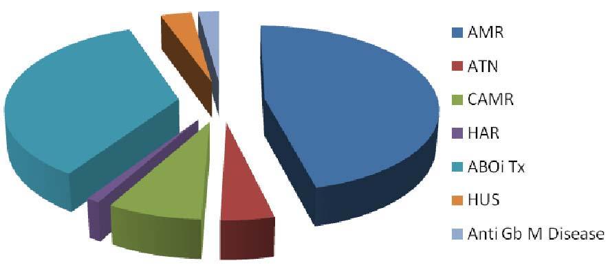
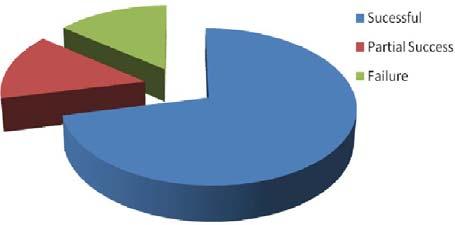
In 42 (46.15%) patients underwent TPE for Antibody mediated rejection (AMR), 4(4.3%) patients for CAN/ ATN, 7(7.6%) patients for chronic AMR (CAMR) and 1(1.09%) patient for Hyper Acute Rejection(HAR) post live donor renal transplantation. Standard triple drug immune suppression protocol consisting of Tacrolimus, Mycophenolate and Prednisolone was continued. TPE was used pre operatively in 32(35.16%) patients undergoing ABO incompatible renal transplantation. All patients undergoing ABO Incompatible renal transplantation got Rituximab 100 mg(single dose) and Intravenous Immunoglobulin @1 gm/kg body weight as adjuvant therapy.3(3.2%) and 2(2.18%) patients underwent plasma exchanges for Hemolytic Uremic Syndrome (HUS) and Anti Glomerular Basement Membrane Disease (Anti Gb M disease) respectively (Fig 2).
Partial Did not return to normalUrine output started/ Success level but >30% reductionincreased but <400 ml/day of NADIR was achieved
Failure1. Creatinine reduction1. Urine output persistently <30% of NADIR <100 ml/day 2. Rise in Serum2. Further reduction Creatinine level in urine output
AMR = Antibody Mediated Rejection; ABOi KT = ABO incompatible kidney transplantation (ABOi-KT) Statistical Tools : Descriptive statistics was used with the help of SPSS software version 21.
RESULTS
Total 91 patients underwent TPE during study period.
52 patients were males and 39 patients were female. (p=0.17)
The mean age of the study population was 43.3 years (13.76)
AVF was used in 76 patients (83.51%). CVC was used in 10 patients (10.98%). Double lummened uncuffed catheter was used in 5(5.49%) patients.
There were 482 sessions of TPE at the study center during study period.
The number of TPE sessions varied from 19 to 2 with a mean of 5.28(2.92) per person.
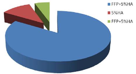
Combination of FFP+5% Human Albumin was uses as replacement fluid in 76(83.51%) patients.
Fresh Frozen Plasma(FFP) and 5% Human Albumin (HA) was used as the only replacement solution in 10 patients (10.9%) and 5 patients (5.45%)
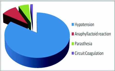
Total 68 episodes of complications (14.1%)were seen during study period. Hypotension was the commonest complication seen in 57(83.8%) with a mean trough MAP of 70(5) mm Hg, followed by anaphyllactoid reaction in 6(8.82%) and parasthesia in 4(0.02%) patients. Coagulation of extracorporeal circuit happened in 1(1.4%) session. There was no significant association between anaphyllactoid reaction and the replacement fluid.(OR=0.2;CI=0.5-1.2).
Anaphyllactoid reaction responded to IV Hydrocortizone therapy (Fig 3).
TPE was successful in 30(71.4%) patients having AMR. Partial success was achieved in 6(14.28%)
FFP+5%HA = Combination of Fresh Frozen Plasma + 5% Human Albumin (83.51%)
5%HA = Only 5% Human Albumin (10.9%)
FFP = Fresh Frozen Plasma (5.45%) Fig 1 — Types of Replacement Fluid Used
Fig 3 — Complication during Therapeutic Plasma Exchange
patients who could be discharged with a mean Creatinine of 2.1(0.4)mg/dl.TPE failed in 6(14.28%) patients of whom 1 patient died, graft nephrectomy was performed in 2 patients and hemodialysis was re initiated in 3 patients (Fig 4).
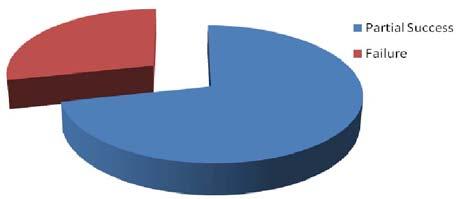
Partial success was seen in 5(71.42%) cases of CAMR. TPE failed in 2(28.57%) cases of CAMR with re initiation of Hemodialysis (Fig 5).
TPE was successful in 1 case of ATN. Partial success was achieved in 3 other cases.
32 patients underwent TPE for ABOi KT and all the 32 cases could be successfully transplanted with an uneventful post operative period.
Partial success was achieved in all the 5 cases of HUS and Anti GbM disease. The mean reduction in Serum Creatinine was 75.2(3.2)% post procedure.
Fig 4 — Outcome of Therapeutic Plasma Exchange in Antibody mediated Rejection
PartialSuccess=71.42%, Failure=28.57%
Fig 5 — Outcome of TPE in CAMR
DISCUSSION
Compared to other study fresh frozen plasma was associated with lesser anaphyllactoid reactions. 20% vs 0.6 %(p>0.05)11
However hypotensive episodes were much higher compared to other trial 62.63% vs 1.5%12,13
Outcome of treatment of HUS/TTP was not yielding in our study compared to others14
Outcome of TPE (in conjunction with IvIg and Rituximab) in reducing antibody titer,thereby leading to successful renal transplantation was comparable to and at time better than international standards15,16
CONCLUSION
TPE is a safe mode of treatment and successfully removes the offending antibodies when used in conjecture with Rituximab and/or IV Ig. TPE is not a successful treatment of CAMR, however, more data and prolonged study is needed. Lack of complete success in case of Hemolytic Uremic Syndrome and Anti Glomerular Basement Membrane Disease could possibly be attributed to the time lag between onset of symptom and initiation of therapy. Hence more rapid detection and high index of suspicion is warranted. Complications associated with TPE could be easily manageable. FFP can be used as a safe alternative to Human Albumin which is more expensive in view of no significant association with the use of replacement fluid
Vol 119, No 5, May 2021Journal of the Indian Medical Association
and anaphyllactoid reaction.
Sessions should be closely monitored with more frequent measurement of Blood Pressure to detect and manage hypotension.
2Conte P Le — Replacement fluids in plasmapheresis: crossover comparative study. Intensive Care Medicine 1997; 23(3): 342-4.
3Nydegger Urs E — Choice of the replacement fluid during large volume plasma-exchange. Ricerca in clinica e in laboratorio 1983; 13(1): 103-9.
4Otterness C — 39 Towards an Ideal Plasma Exchange Fluid. American Journal of Clinical Pathology 2018; 149 (suppl_1, 11): S183. doi.org/10.1093/ajcp/aqx149.408.
5Winters JL -— Plasma exchange: concepts, mechanisms, and an overview of the American Society for Apheresis guidelines. ASH Education Book 2012; 8(10): 17-22.
6Pham Huy P — Therapeutic Plasma Exchange, Chapter 73.
7Pham JP — Therapeutic plasma exchange. Transfusion Medicine and Homeostasis, Second Edition, 2013.
8Chirnside A — Coagulation Abnormalities following Intensive Plasma Exchange on the Cell Separator II. Effects on Factors I, II, V, VII, VIII, IX, X AND Antithrombin III. British Journal of Hematology 1981; 48(4): 627-34. doi: 10.1111/j.13652141.1981.00627.x.
9Naik B — Prolonged neuromuscular block due to cholinesterase depletion by plasmapheresis. Journal of Clinical Anesthesia 2012; 14(5): 281-4. doi: 10.1016/s09528180(02)00382-3.
10Shemin D — Complications of therapeutic plasma exchange: a prospective study of 1,727 procedures. Journal of Clinical Apheresis 2007; 22(5): 270-6. doi: 10.1002/jca.20143.
11Michele H — Therapeutic Plasma Exchange: Complications and Management. American Journal of Kidney Diseases 1994; 23(6): 817-27. doi: 10.1016/s0272-6386(12)80135-1.
13Busund R — Plasmapheresis in severe sepsis and septic shock: a prospective, randomized, controlled trial. Medicine 2002; 28(10): 1434-9. doi: 10.1007/s00134-002-1410-7.Epub 2002 Jul 23.
14Forzley BR — Treating TTP/HUS with plasma exchange: a single centre’s 25 year experience. British Journal of Hematology 2008; 5(9): https://doi.org/10.1111/j.13652141.2008.07317. doi: 10.1111/j.1365-2141.2008.07317.x. Epub 2008 Aug 4.
15Sonnenday CJ — Plasmapheresis, CMV Hyperimmune Globulin, and Anti CD20 Allow ABO Incompatible Renal Transplantation Without Splenectomy. American Journal of Transplantation 2004; 4(8): 1315-22. doi: 10.1111/j.16006143.2004.00507.x.
16Segev DL — ABO Incompatible High Titer Renal Transplantation without Splenectomy or Anti CD20 Treatment. American JournalofTransplantation 2005; 5(10): 2570-5. doi: 10.1111/ j.1600-6143.2005.01031.x.
Disclaimer
The information and opinions presented in the Journal reflect the views of the authors and not of the Journal or its Editorial Board or the Publisher. Publication does not constitute endorsement by the journal.
JIMA assumes no responsibility for the authenticity or reliability of any product, equipment, gadget or any claim by medical establishments/institutions/manufacturers or any training programme in the form of advertisements appearing in JIMA and also does not endorse or give any guarantee to such products or training programme or promote any such thing or claims made so after. — HonyEditor
Original Article
Prevalence of COVID-19 Infection and Identification of Risk Factors among Asymptomatic Healthcare Workers : A Serosurvey Involving Multiple Hospitals in West Bengal
Background : The declining trend of COVID-19 infection in India has made healthcare personnel (HCP) and general public lenient about personal-protective-measures. Serosurveys to estimate the prevalence of SARS-CoV2 IgG antibodies, particularly in high-risk-zones like hospitals can give the real scenario and risk-factors can help prioritise the target population for urgent, effective vacccination.
Methods : 1470 consecutive HCP from 4 tertiary-care-hospitals in Kolkata filled a questionnaire and were tested for serum SARS-CoV2-IgG by Enzyme-linked Immunosorbent Assay (ELISA). The prevalence of SARS-CoV2-IgG among asymptomatic HCPs was studied and the work environment, clinical comorbidities, personal habits and protective measures and pharmacologic prophylaxes were compared between those with and without SARS-CoV2IgG. Parameters of asymptomatic seroconverters were also compared to those with personal history of COVID-19Infection. Logistic regression was done to identify independent risk-factors.
Results : Prevalence of asymptomatic seroconversion was 15.8%. Asymptomatic seroconverters (n=208) were mostly working in mixed hospitals (having both COVID-19 and non-COVID-19 wards, 57.7%), were non-doctors by profession (nurses-25.1%, others–51.4%). Among asymptomatic HCP, indepedendent positive risk factors for SARSCoV2 IgG-positivity were Diabetes Mellitus (DM) and multiple comorbidities (pboth<0.001) and prophylactic use of Hydroxychloroquine and Famotidine (pboth< 0.03). However, for symptomatic COVID-19 infection, working in COVID19 dedicated hospitals, and personal h/o COPD were positive risk-factors and Ivermectin prophylaxis a negative riskfactor (pall< 0.03).
Conclusion: In our study conducted in the immediate pre-immunisation period, rate of asymptomatic seroconversion among HCPs is too low to presume herd immunity. Those working in mixed hospitals and DM, multiple comorbidities are at particularly high risk.
[J Indian Med Assoc 2021; 119(5): 21-7]
Key words :COVID-19, SARS-CoV2 IgG, ELISA, Seroprevalence, Healthcare personnel.
More than a hundred million people worldwide have been infected with severe acute respiratory syndrome coronavirus 2 (SARS CoV 2) over the past fourteen months. With the vaccination campaign targeting healthcare workers and the sudden decline in the daily number of cases in the country, the
Department of Endocrinology and Metabolism, IPGME&R, Kolkata
7MBBS, DTM&H, MD (Medicine), DM (Endocrinology), MRCP, Professor and Head
Received on : 08/03/2021
Accepted on : 23/03/2021
Editor's Comment :
Among 1470 consecutive HCP from 4 tertiary-care-hospitals in Kolkata, prevalence of asymptomatic seroconversion was 15.8%.
Rate of asymptomatic seroconversion among HCPs in immediate pre-vaccination period is too low to presume herd immunity.
Risk factors for asymptomatic SARS-CoV2-IgG-positivity were Diabetes Mellitus (DM), multiple comorbidities, and even prophylactic use of Hydroxychloroquine, Famotidine For symptomatic COVID-19 infection, working in COVID19-dedicated hospitals, and COPD were positive risk-factors and Ivermectin prophylaxis a negative risk-factor.
possibility of a high prevalence of asymptomatic seroconversion in the community is being proposed. Healthcare personnel (HCP) are known to be at an elevated risk of contracting COVID-19. Several IgM/IgG ELISA for COVID-19 have been certified for commercial purpose or research by different agencies which can be used for easy and cost-effective tools
No 5, May
for the detection and surveillance of COVID-19 infection in communities and high risk areas like hospitals1 Initial studies from China reveal a 1% COVID-19 infection rate in HCPs, with higher rates in HCP who reported no exposure to COVID-19 patients and upto 7.0% greater absolute risk among HCP than community in the United States2,3 Majority of infected HCP (62.5%) were nurses3 Studies have reported higher risk of transmission in frontline healthcare workers in the United Kingdom4. From a recent metaanalysis of COVID-19 prevalence among health care workers, the estimated prevalence was 11%, the most frequently affected personnel were nurses (48%) and HCP working in Hospital Non-emergency Wards (43%)5. From different parts of India, the reported seroprevalence was around 0 to 12%, depending on the time of study, type of hospital and IgG antibody assay method used, though majority used chemiluminescence assays and many were not validated assay methods by the Indian Council of Medical Research6-8.
Serological testing to assess the extent of seroconversion among HCP just prior to the vaccination campaign among healthcare workers in India could give a glimpse of the real picture in the community and also help identify high risk groups for prioritising vaccination campaign.
MATERIALS AND METHODS
The primary objective was to evaluate the prevalence of previously undiagnosed SARS-CoV-2 Infection among HCP from four tertiary-level hospitals in Kolkata, one of which was dedicated solely to the care of COVID19 infected patients, one was a purely non-COVID-19 hospital and the other two had mixed proportion of COVID-19 positive or non-COVID-19 patients, these two will be referred to as mixed hospitals henceforth. The study was approved by the Institutional Ethics Committee of IPGMER, Kolkata vide memo no. IPGMER/IEC/2020/482. The secondary objectives were to identify the risk factors associated with COVID19 infection and also risk factors for symptomatic COVID-19 infection among HCPs. For these, we analysed factors including age, Body Mass Index (BMI), profession (whether doctor, nurse or other HCP) , area of work (whether in a COVID hospital, mixed hospital or non-COVID hospital; whether working in critical care settings), history of addiction, personal h/o comorbidities including diabetes mellitus (DM), hypertension, dyslipidemia, established atherosclerotic cardiovascular diseases (ASCVD), hypothyroidism, other autoimmune diseases, chronic kidney or liver disease, malignancies, diseases
causing immunosuppression like HIV/AIDS, history of prolonged use of steroids and/or other immunosuppressants and the use of drugs for COVID-19 prophylaxis including hydroxychloroquine, ivermectin, multivitamins, vitamin-D , famotidine, zinc and personal protective measures adopted including masks ,PPE kits and sanitisers.
Participants with a history of SARS-CoV-2 infection diagnosed by rt-PCR or suggestive symptoms of COVID-19 infection were excluded from analysis for the primary and first secondary objective.However, for the other secondary objective, we compared those without personal h/o COVID-19 infection but having SARS-CoV-2-IgG with a subset of participants with personal h/o symptomatic COVID-19 infection and having SARS-CoV-2-IgG.
Participants were recruited between 1 st of September 2020 to 10th of January 2021. Following informed consent, data were collected through questionnaire filled by the participants, under the supervision of study coordinators, prior to drawing blood samples for SARS-CoV2-IgG antibody estimation. Blood samples were tested for IgG antibodies against the spike proteins of SARS-CoV-2 using SARS-CoV2 enzyme-linked immunosorbent assays (ELISAs) (# CORONA KAVACH IgG, Zydus), validated by ICMR and reported to have a sensitivity and specificity of 98.7% and 100% respectively. All ELISAs were performed at IPGME&R, Kolkata following manufacturer’s instructions3. Negative and positive control samples were run every day and the threshold for a positive ELISA was determined at a value greater than the mean + 3 SD of negative controls, consistent with standard methodology4
Statistical Evaluation :
Statistical analysis was performed using GraphPad Prism v.9 for Mac. Comparison was done between those with SARS-CoV2 IgG-positivity versus those without. Additionally, comparison was also done between those with SARS-CoV2-IgG positivity but without personal h/o COVID -19 infection with a subset with known h/o COVID-19 Infection. Categorical variables were analysed using Chi-square or Fisher’s exact test and quantitative variables using unpaired t-test. Binarylogistic regression analysis was performed to determine independent risk factors.
OBSERVATIONS
Out of total 1470 participants screened, n = 154 were having a personal h/o COVID-19 infection or suggestive symptoms. They were excluded from statistical analysis for the primary objective and n = 22
119, No 5, May 2021Journal
1316 were finally enrolled for this. Out of this, 208 (15.8%) had positive SARS-CoV2-IgG and were classified as asymptomatic seroconverters, while n= 1100 did not have SARS-CoV2-IgG,and 8 participants had indeterminate response. Those with indeterminate results were excluded from further statistical analysis. The baseline characteristics of participants are given
Table 1 — Demographic and clinical characteristics of study participants
ParameterTotal no of participants without personal h/o COVID-19 infection (N= 1316)
Mean age in years (SD)38.6 ( 11.8)
Mean BMI in kg/m2 (SD)
in Table 1. Majority were HCP other than doctors/ nurses (40.5%) and majority worked in non-COVID19-dedicated hospitals (41.3%). Among comorbidites; systemic hypertension was the commonest (10.9 %) followed by DM (8.4%) and hypothyroidism (4.8%). A total of 46.1% of participants were using some form of pharmacologic agents for prophylaxis, multivitamins (31.7%) being the most commonly used agent followed by hydroxychloroquine (31.3%), while 9.7%used Ivermectin.
Working in COVID dedicated hospital (CDH)CDH : 254 (19.3%) or non COVID dedicated hospital (NCDH)NCDH : 543 (41.3 %) or Mixed hospital (having both COVIDMixed hospital : 519 (39.4%) dedicated and non-COVID wards)
Working in critical care settings (ICU/CCU/ITU/337 (25.6%) dialysis units/RTU)
Performing intubation, tracheostomy,bone drilling236 (17.9%) surgeries and other aerosol generating procedures
Receiving steroids / other immunosuppressants45 (3.4%)
Receiving monoclonal antibodies0
Addiction :
Smoking170 (12.9%)
Alcohol119 (9%)
Other addiction25 (1.9%)
Agents used for COVID-19 prophylaxis :
Hydroxychloroquine
412 (31.3)
Ivermectin128 ( 9.7%)
Multivitamin
417 ( 31.7%)
Vitamin D249 ( 18.9%)
Famotidine/ Other H2 antihistaminics60 (4.6%)
Zinc
316 (24%)
Among the 208 asymptomatic seroconverters, majority were HCP other than doctors/ nurses (20.2%), majority worked in mixed hospitals (57.7 %).Compared to those who didn’t have SARS-CoV2-IgG antibodies, a significantly higher number of asymptomatic seroconverters were HCP other than doctors/ nurses (51.4% versus 38.5%,p = 0.0001), worked in mixed hospitals ( 57.7% versus 36%, p<0.0001), had higher prevalence of DM (16.8 % versus 6.3%, p <0.0001), had autoimmune diseases other than Hashimoto’s thyroiditis (4.3% versus 1.3%, p = 0.002), presence of >2 comorbidities ( 11.5% versus 6.5%, p =0.011) and history of prolonged intake of steroids/other immunosuppressants (7.7% versus 2.6%, p=0.0002) (Table 2). A total of 25 patients in our cohort had autoimmune disorders other than Hashimoto’s thyroiditis, including Rheumatoid arthritis (n=8), Psoriasis (n=3), Vitiligo (n=3), Systemic Lupus Erythrematosus (n=3), Graves’ disease (n=3), Myasthenia Gravis (n=2), Immune thrombocytopenic purpura (n=1), Inflammatory bowel disease (n=1) and T1DM (n=1).
There was a slightly higher prevalence of use of some form of pharmacologic prophylaxis in those having SARS-CoV2-IgG than those without (46.4% versus 44.7%, p =0.56). Use of hydroxychloroquine and Famotidine were significantly more common in those having SARSCoV2-IgG (37.5% versus 30.1%,
No 5, May 2021Journal
p=0.03 and 9.1% versus 3.4%, p = 0.0002). Ivermectin use was slightly higher in the group not having SARSCoV2 IgG antibodies ( 8.6% versus 9.8%, p = 0.6).
Upon multiple regression analysis, working in mixed hospitals, being a HCP other than doctor/nurse, presence of Diabetes Mellitus, presence of >2 comorbidities and the use of Hydroxychloroquine and Famotidine for pharmacologic prophylaxis were found as significant independent predictors for the development of SARS-CoV2-IgG among HCP (Table 4).
Personal protective behavior including the use of Personal Protective Equipment (PPE) gears, sanitisers or hand washing were similar in both groups. 95% of the participants used N95 masks, 3% used triple layered masks and 1% used cloth masks while at hospital. Masks were worn all the time at hospital in 80% of the participants, and doffed off at home ( 68%), in the car ( 22%) or disposed in the hospital (10%). Reuse of N95 masks was quite common (56%) mostly at an interval of 4-5 days. PPE kits were used by most during duty hours in COVID dedicated wards/ hospitals. Head shields were used by 66%, surgical caps in 70%, gloves in 50% of the participants. 90% used alcohol based hand sanitisers while hand washing with soap and water was practised by around 56%, mostly before meals.
Out of the 154 participants with a personal h/o rt-PCR proven COVID-19 infection, n=35(22.73%) participants couldn’t mount a IgG antibody response while n = 110 ( 71.42%) were positive for SARS-CoV2-IgG. Remaining nine had an indeterminate response. The median time from symptoms of COVID-19 infection to IgG antibody testing were not different in the two groups with and without SARS-CoV2-IgG ( 7.3 versus 8.8
weeks, p=0.12). Neither were there any differences in median age, severity of infection or the number of symptoms between the two groups.
We also compared the group withSARS-CoV2-IgG seropositivity but without personal h/o COVID-19 infection (n=208) with a group having personal history of COVID-19 infection and positive SARS-CoV2-IgG antibodies (n=110) in order to identify risk factors or
Table 2 — Comparison of parameters between asymptomatic healthcare workers with SARS-CoV-2 IgG antibodies versus those who did not have SARS-CoV2 IgG antibodies
Parameter SARS CoV-2SARS CoV-2p value IgG + N = 208IgG –ve N = 1100
Mean age in years (SD) 38.5(0.82) 38.6 (0.36) 0.88
SD = Standard Deviation, D = Doctors, N = Nurses , O = Other healthcare staff, CDH = COVID dedicated hospital, NCDH = Non COVID dedicated hospital, C = COVID wards/OPDs, NC = Non COVID wards/OPDs, COPD = Chronic Obstructive Pulmonary Disease, CKD = Chronic Kidney Disease, CLD = Chronic Liver Disease
5,
protective factors that might contribute to manifestations or severity of COVID-19 infections. Compared to asymptomatic seroconverters, those with symptomatic COVID-19 infections and SARS-CoV2IgG were older in age (43.5 versus 38.5 years, p= 0.004), majority were doctors (34.5% versus 13.5%, p =0.019) , working in COVID-19 dedicated hospitals (24.6 % versus 12.5% , p<0.0001), worked in critical care settings/ emergencies (34.5% versus 22.1%, p =0.017) and/or performing aerosol generating procedures (27.3% versus 16.3%, p=0.027). Participants with symptomatic COVID-19 infections and SARS-CoV2IgGwere also more likely to have >2 co-morbidities (20% versus 11.5%, p=0.041), established atherosclerotic cardiovascular disease (ASCVD) (8.2% 1.9%, p=0.014) and COPD (10% versus 3.4%, p=0.021) (Table 3). There were no significant differences in the use of pharmacologic prophylaxis with most agents in the groups except ivermectin use which was higher in the group of asymptomatic seroconverters (8.7% versus 1.8%, p=0.017). Upon binary logistic regression, working in a mixed hospital, having COPD as a co-morbidity were found to be independent risk factors for development of symptomatic COVID-19 infection whereas ivermectin prophylaxis was found to be a significant negative predictor for the same (Table 4) .
DISCUSSION
In our study involving multiple hospitals in Eastern India, the prevalence of IgG
Parameter
seroconversion among HCP without a known history of COVID-19 infection was 15.8%. This was slightly higher than a study from Mumbai in whom the reported
symptomatic
Participants withParticipantsp value h/o COVID -19 without h/o infection and COVID -19 SARS CoV-2infection SARS IgG + (n = 110)CoV2 Ig G + N = 208 Mean
Working in critical care settings / emergencies38 (34.5%)46 (22.1%) 0.017
Performing intubation, tracheostomy, other aerosol generating procedures30
Atherosclerotic cardiovascular disease 9(8.2%)4(1.9%)0.014 COPD/Bronchial asthma11(10%)7(3.4%)0.021 CKD/CLD 5(4.5%)3 (1.4%)1 HIV or other immunosuppression disorders02 Known malignancy01 Hypothyroidism3 (2.7%)10 (4.8%) 0.554 Autoimmune diseases (excluding 5(4.5%)9(4.3%)1 Hashimoto’s thyroiditis) >2 comorbidities22 (20%)24 (11.5 %) 0.041 On Glucocorticoids/ other 6(5.5%)16(7.7%)0.454 immunosuppressants Smoking14 (12.7%)21
SD = Standard Deviation, D = Doctors, N = Nurses , O = Other healthcare staff, CDH = COVID dedicated hospital, NCDH = Non COVID dedicated hospital, C = COVID wards/OPDs, NC = Non COVID wards/OPDs, COPD = Chronic Obstructive Pulmonary Disease, CKD = Chronic Kidney Disease, CLD = Chronic Liver Disease
Table 3 — Comparison of parameters between asymptomatic and
healthcareworkers with SARS-CoV2 IgG antibodies
prevalence was 11.1%, at a time when the city was one of the worst affected in the nation9.
We found a higher risk of COVID-19 infection in HCP working in mixed hospitals rather than COVID19-dedicated hospitals. However, those working in COVID-19-dedicated hospitals had a higher likelihood of developing symptomatic COVID-19 infection. HCP other than doctors/nurses were at a higher likelihood of having contracted asymptomatic COVID-19 infection; whereas doctors had higher chances of developing symptomatic COVID-19 infection. Our findings were similar to the study from Mumbai where seroprevalence was significantly higher in ancillary workers (18.5%) and also higher in non–COVID-19 Hospitals9. Published data on community seroprevalence of COVID-19 in India is around 6.6%10. The overall modest prevalence of infection among the HCPs, especially non-doctors and nurses and rather lower prevalence in COVID-19 dedicated hospitals may indicate community source of infection and also hint at the need for universal vaccination of all HCP as well as community dwellers.
We didn’t find an increased risk of contracting COVID-19 infection in those working in critical care settings. However, those working in COVID wards/ OPDS, in critical care settings and/or performing aerosol generating procedures were at higher risk of developing symptomatic COVID-19 infection rather than being asymptomatic seroconverters. This is likely due
to exposure to higher viral load and reinforce the need for strict implementation of infection control measures in all areas of the hospital, and particularly in all critical care units of the hospitaland wherever aerosol generating procedures or surgeries are being performed. Results from different prior trials and metaanalyses have revealed that DM, hypertension, ASCVD, COPD, dyslipidemia could be independent predictors for severity and mortality in COVID-19 infection 11-13 In the current study, on multiple logistic regression analysis, Diabetes mellitus and the presence of multiple comorbisities were found to be independent risk factors for asymptomatic COVID-19 infection whereas the presence of COPD and multiple comorbidities were independent risk factors for symptomatic COVID-19 infection.
The presence of autoimmune diseases was significantly higher in those with COVID-19 seroconversion, although it was not an independent risk factor. Around one third of these participants were receiving steroids or immunosuppressants for their underlying autoimmune diseases. While the prevalence of middle aged female nurses could have a bearing on these results, these diseases could also lead to altered immune response to COVID-19 infection. Few studies have reported autoimmune systemic conditions to be an independent risk factor predicting hospitalisation in COVID-19 patients14 . The role of steroids or other immunosuppressants in enhancing the risk or severity of COVID-19 is unclear. There is a suggestion that low dose prednisolone and tacrolimus might have some beneficial effects on COVID-19 infection whereas with other agents, there is no definite evidence to suggest an enhanced risk14. However, we don’t have data on the multitude of chronic immunosuppressant therapy being used.
Though there are some evidence favoring ivermectin, hydroxychloroquine, famotidine and Vitamin D in preventing or controlling the severity of COVID-19 infection, there is very low certainty of evidence15-20. In our study, use of Hydroxychloroquine and Famotidine were higher in those having contracted COVID-19 infection. Ivermectin use was an independent negative predictor of symptomatic COVID-19 infection.
Our study also revealed that out of those with a personal h/o COVID-19 infection, around one-fourths didn’t develop detectable IgG antibodies, even though testing was conducted within a median time of 8.1 Table 4 — Independent
Vol 119, No 5, May 2021Journal
weeks from the time to first symptom. We could not identify any risk factors that could predict seronegativity despite having had symptomatic COVID19 infection in our cohort. This again re-emphasises the need to vaccinate all, irrespective of a prior history of COVID-19 infection.
This study had few limitations. Questionnaires were not anonymous and therefore subject to volition of the participants. The ELISA kit used was not quantitative. Our results could be biased due to greater representation of middle aged nurses and “other” healthcare personnel.
Acknowledgement :
We earnestly thank Mr Pradip … and Mr Chandan Sarkar for their contributions towards performing all the ELISA reactions and all the doctors, nurses and other healthcare personnel for their participation in the study.
Disclosures : None
Conflicts of interest : None
Funding : None
REFERENCES
1Available from : https://www.icmr.gov.in/ckitevaluation.html
2Lai X, Wang M, Qin C, Tan L, Ran L, Chen D, et al — Coronavirus disease 2019 (COVID-2019) infection among health care workers and implications for prevention measures in a tertiary hospital in Wuhan, China. JAMA network open 2020; 3(5): e209666-.
3Barrett ES, Horton DB, Roy J, Gennaro ML, Brooks A, Tischfield J, et al — Prevalence of SARS-CoV-2 infection in previously undiagnosed health care workers at the onset of the US COVID-19 epidemic. MedRxiv. 2020 Jan 1.
4Nguyen LH, Drew DA, Joshi AD, Guo CG, Ma W, Mehta RS, et al — Risk of COVID-19 among frontline healthcare workers. MedRxiv. 2020 Jan 1.
5Gómez-Ochoa SA, Franco OH, Rojas LZ, Raguindin PF, RoaDíaz ZM, Wyssmann BM, et al — COVID-19 in health-care workers: a living systematic review and meta-analysis of prevalence, risk factors, clinical characteristics, and outcomes. American Journal of Epidemiology 2021; 190(1): 161-75.
6Kumar A, Sathyapalan D, Ramachandran A, Subhash K, Biswas L, Beena KV — SARS-CoV-2 antibodies in healthcare workers in a large university hospital, Kerala, India. Clinical Microbiology and Infection 2020 Sep 16.
7Hawaldar R, Sodani S, Sodani V, Sodani RK — Seroprevalence of COVID-19 infection among healthcare professionals in Central India using SARS-CoV-2 antibody test. The Journal of Community Health Management 2020; 7(4): 146-51.
8Kumar N, Bhartiya S, Desai S, Mutha A, Beldar A, Singh T — Seroprevalence of Antibodies Against SARS-CoV-2 Among Health Care Workers in Mumbai, India. Asia Pacific Journal of Public Health 2020; 1(1): 1010539520977307.
9Kumar N, Bhartiya S, Desai S, Mutha A, Beldar A, Singh T — Seroprevalence of Antibodies Against SARS-CoV-2 Among Health Care Workers in Mumbai, India. Asia Pacific Journal of Public Health 2020; 1(1): 1010539520977307.
10Murhekar MV, Bhatnagar T, Selvaraju S, Saravanakumar V, Thangaraj JW, Shah N, et al — SARS-CoV-2 antibody seroprevalence in India, August–September, 2020: findings from the second nationwide household serosurvey. The Lancet Global Health 2021; 27(1)
11Meng M, Zhao Q, Kumar R, Bai C, Deng Y, Wan B — Impact of cardiovascular and metabolic diseases on the severity of COVID-19: a systematic review and meta-analysis. Aging (Albany NY) 2020; 12(22): 23409.
12Grasselli G, Greco M, Zanella A, Albano G, Antonelli M, Bellani G, et al — Risk factors associated with mortality among patients with COVID-19 in intensive care units in Lombardy, Italy. JAMA Internal Medicine 2020; 180(10): 1345-55.
13Fang X, Li S, Yu H, Wang P, Zhang Y, Chen Z, et al — Epidemiological, comorbidity factors with severity and prognosis of COVID-19: a systematic review and metaanalysis. Aging (Albany NY) 2020; 12(13): 12493.
14Nuñez DD, Leon L, Mucientes A, Rodriguez-Rodriguez L, Urgelles JF, García AM, et al — Risk factors for hospital admissions related to COVID-19 in patients with autoimmune inflammatory rheumatic diseases. Annals of the Rheumatic Diseases 2020; 79(11): 1393-9.
15Hernandez AV, Roman YM, Pasupuleti V, Barboza JJ, White CM — Hydroxychloroquine or chloroquine for treatment or prophylaxis of COVID-19: a living systematic review. Annals of Internal Medicine 2020; 173(4): 287-96.
16Behera P, Patro BK, Singh AK, Chandanshive PD, Ravikumar SR, Pradhan SK, et al — Role of ivermectin in the prevention of COVID-19 infection among healthcare workers in India: A matched case-control study. medRxiv. 2020 Jan 1.
17Grant WB, Baggerly CA, Lahore H — Reply: ”Vitamin D Supplementation in Influenza and COVID-19 Infections. Comment on: Evidence That Vitamin D Supplementation Could Reduce Risk of Influenza and COVID-19 Infections and Deaths Nutrients 2020, 12 (4), 988". Nutrients 2020; 12(6): 1620.
18Jayawardena R, Sooriyaarachchi P, Chourdakis M, Jeewandara C, Ranasinghe P — Enhancing immunity in viral infections, with special emphasis on COVID-19: A review. Diabetes & Metabolic Syndrome: Clinical Research & Reviews. 2020; 14(4): 367-82.
19Pal A, Squitti R, Picozza M, Pawar A, Rongioletti M, Dutta AK, et al — Zinc and COVID-19: basis of current clinical trials. Biological Trace Element Research 2020; Oct 22:1-1.
20Malone RW, Tisdall P, Fremont-Smith P, Liu Y, Huang XP, White KM, et al — COVID-19: Famotidine, histamine, mast cells, and mechanisms.
Review Article
The Resumption and Management of Bariatric Surgical Procedures and Postoperative Care during COVID-19 — A Single Surgeon Experience from India
Hetal Patolia1, Sanjay Patolia2
The Novel Coronavirus Disease 2019 (COVID-19) pandemic has hampered the bariatric surgery, the only effective option for patients with obesity with or without comorbidities, worldwide. Obesity and diabetes are two major risk factors for severe forms of COVID-19 and candidates for bariatric surgery are exposed to a high risk of mortality linked to the pandemic. Thus it is crucial to resume such surgery to attenuate the impact of its interruption on the population. The current article delineates the effective strategies that are devised to resume elective bariatric surgery and provide the continuity of care in the backdrop of COVID-19 crisis.
Since its outbreak in Wuhan, China in December 2019, the novel coronavirus 2 (SARS-COV-2) responsible for the pandemic status of coronavirus disease 2019 (COVID-19) has caused massive stress on healthcare systems globally. The emergence of this disease also disrupted the bariatric surgery program globally and has led to changes in elective surgical care. To oblige the unprecedented pressure to free up inpatient capacity and because of intraoperative risks for viral contagion among patients and staff, the International Federation for the Surgery of Obesity and Metabolic Disorders (IFSO) has recommended that all elective metabolic and bariatric procedures, both surgical and endoscopic, should be postponed until the end of the pandemic1 Similar measures have been recommended by the “Guidance for Triage of Non-Emergent Surgical Procedures,” by the Centers for Medicare and Medicaid Service (CMS) and the American College of Surgeons (ACS) in March 2020, to postpone all elective cases2
The paradigm shift of COVID-19 highlighted the role of obesity and related comorbidities, given the detrimental effects of COVID-19 in these individual’si.e. higher rates of hospitalization and poorer clinical outcomes3. Due to the progressive and relapsing nature of obesity, delaying its treatment would further lead to metabolic derangements especially in times of lockdown where lifestyle modifications are difficult to
1MBBS, MD, Consultant and Chief-Clinical Research, Department of Gynaecology, Asian Bariatrics, Ahmedabad 360015 and Corresponding Author
2 MBBS, MS, Director and Founder of Asian Bariatrics, Ahmedabad 360015
Received on : 17/09/2020
Accepted on : 07/03/2021
Editor's Comment :
In this unprecedented COVID-19 crisis, it is imperative to ensure that pre- and postbariatric surgery patients continue to receive adequate care given the harmful effects of COVID19 in these subset of patients.
This article delineate specific strategies for the safe resumption of bariatric surgery program as well as the following key practice patterns and precautions to mitigate interruptions in patient care.
follow. The delay of bariatric surgery which is a mainstay treatment of morbid obesity and significantly improves comorbidities like diabetes, heart disease and hypertension may affect patients’ health in different ways. However, on the contrary to above recommendations, the statement of American Society for Metabolic & Bariatric Surgery (ASMBS) on metabolic and bariatric surgery during COVID pandemic, dated 23 June 2020, strongly rejects classifying bariatric and metabolic surgery as elective and prefers to use the term “medically necessary time- sensitive surgery” or ‘medically necessary non-emergent surgery”, in order to clarify the effectiveness of this procedure in treating a number of diseases besides obesity. Given this unprecedented scenario and to mitigate interruptions in patient care, we used robust telehealth modalities including video and phone call systems to manage the continuity of patient care and resumed the bariatric surgery program at one of our four centers by unlocking the following practice patterns, considerations and precautions.
Ensuring Continuity of Patient Care during COVID-19 Lockdown : Respecting the national and international guidelines,
all elective cases have been stopped at all our operating centers during the lockdown period and certain strategies were implemented for continuous and consistent management of patient care. During this period of uncertainty, maintaining a line of communication with bariatric patients is essential for continuity of care and also keep those whose surgeries were postponed engaged in their health. It was well established that telemedicine and remote consultations in bariatric patients are proven to be effective in fighting distress and reducing the level of psychological disorders4. Thus, a telehealth system was established via video and audio calls to ensure that pre-operative bariatric patients remain on course. To minimize the risk of nutrition-related complications in patients who have had bariatric surgery, patients were engaged with same time intervals of follow up for postoperative nutritional care. Clinical signs (skin rash, weakness, oedema, and visual changes), and symptoms (nausea, tingling, numbness, fatigue, irritability, and changes in bowel habits) of nutritional deficiency were assessed during virtual clinic sessions.
Patients in the pipeline for surgery during the months of lockdown were also followed closely, but remotely, to ensure continued weight loss or weight maintenance. To support individuals in healthy eating during self-quarantine and isolation, dietary interventions (high protein content and lower glycemic index) for weight control were recommended. Special emphasis was given to modalities like yoga, and online home workout videos as an alternative to reduce the risk of contracting COVID-19 during outdoor exercises. Routine lab tests (eg, albumin, thiamine, B12, vitamin A, vitamin D, iron, and calcium) were carried out by sending trained lab personnel for home collection of samples with all due precautions when indicated. Furthermore, to prevent the overall cross-contamination of COVID-19 infections between healthcare facilities and transmission risk to others, patients were allowed for hospital visit only when parenteral medication or emergency surgical intervention was required.
Resumption of Bariatric Surgeries and Postoperative Care in Times of COVID-19 :
All the four bariatric centers were located in the Indian state of Gujarat, Ahmedabad, Baroda, Surat and Rajkot respectively. Bariatric surgeries were resumed at the Rajkot center due to its green zone status and least prevalence of COVID-19.
Triage and COVID-19 screening :
The prioritization of elective surgery was established according to the clinical need, equity of access, and potential harm caused by delayed access. As per the previous categorizations of elective surgery, the
patients were prioritized for the bariatric surgery in whom it would provide maximum benefits such as patients with cardio-metabolic comorbidities5
Given the risks of severe complications from COVID19 in patients with obesity and type 2 diabetes, the practice guidelines from the international societies strongly recommend performing the COVID-19 screening test before any elective surgery1,6. Following the same, detailed information on travel history and COVID prevalence among family, friends, neighbors and locality was collected to check for the history of suspicious contact. In addition, all surgical patients were screened for clinical signs and symptoms of infection like fever, cough, cold, sinusitis, diarrhea etc. and an RTPCR test for COVID-19 was performed among all patients being admitted for surgery.
Laparoscopic approach :
Despite the potential for higher risk of contagion, the laparoscopic approach in bariatric and metabolic surgery is associated with substantial benefits including lower rates of mortality and complications (including pulmonary and procedural), and shorter hospital stays compared with traditional open surgery, especially in patients with severe obesity7, 8. For these reasons, the laparoscopic approach was accessed and followed with all due precautions according to prescribed guidelines specified in Fig 1.
Anesthetic precautions included avoiding high pressure ventilation use of video laryngoscope and viral filters in the circuit.
Air tight trocars to prevent leaks.
Minimizing the use of electrocautery.
Use of instruments like endostitch and power gun or I drive to expedite the surgery.
Close suction circuit with sodium hypochlorite for fumes and gases from ports.
Removing the Specimen only after decompression.
Decompression thru the lower most port in closed circuit suction.
Restricting the entry of staff with maximum four people in the OR.
Adequate and appropriate PPE for all the staff involved in patient care.
Postoperative patient care was modified.
Patients were allowed only one personal attendant who too was thoroughly screened and tested if required.
Visitors were strictly prohibited.
Food and beverages were provided in the patient’s room with no touch technique.
Sanitizing the room surfaces was done twice a day. Attending staff were trained in maintaining social distancing and wearing adequate PPE.
1 — Guidelines for laparoscopic approach in bariatric and metabolic surgery
Fig
119, No 5, May 2021Journal
Table 1 — Variables of total elective cases performed
MonthTotalElective Surgery EmergencyVenti Hospital Complications Cases SLEEVE RYGBP surgery Usage stay Post-op Pre-op
April11 internal- 2-days-Hernia
May413--2-3 DaysJun16313-13-4 Days-1
In the month of April, emergency surgery was performed in one patient with internal hernia. In May, the bariatric surgery program was unlocked with 4 elective surgery out of which 3 were Roux-en-Y gastric bypass (RYGBP) and 1-SLEEVE gastrectomy. All 3 patients of RYGBP had cardio-metabolic comorbidities along with obesity. In June, 16 patients were operated of which 13 were RYGBP and 3 -SLEEVE gastrectomy. One patient for grade sleep apnea required ventilator support for 12 hrs postoperation. 1 patient developed palpitation & showed elevated D dimer suggestive of an early pulmonary embolism on the second postoperative day and was heparinized with higher dose for 3 days (Table 1). The median length of hospital stay was 3 days and patients were discharged on the third postoperative day.
To inspect the postoperative wound dehiscence following the surgery, the patients were advised to remove the dressing of the surgical site and were requested to send a picture of the same. To facilitate systematic interventions to improve communications and achieve patient-centered solutions, tele follow-ups were recommended at regular intervals as per the bariatric protocol. After discharge, none of the patients have developed any complications and no significant complaints have been reported. The wound healing was found to be satisfactory in all the patients. Further, the patients were contacted every week for 1 month and every 15 days for the next two months and were advised with dietary options and exercises considering their local environment individually. Overall, the weight loss in all the patients has been shown to be satisfactory which is associated with resolution of related comorbidities and stoppage of medicines.
CONCLUSION
In summary, given the uncertainty regarding the progressive nature of obesity, diabetes, and related conditions, combined with the effects and duration of
the COVID-19 outbreak, delaying bariatric and metabolic surgery could increase the risks for morbidity and mortality in these candidates which would make them vulnerable to a severe form of COVID-19. During these challenging times, it is paramount that online consultations and telehealth must be embraced for delivering care to overcome the negative effects of the pandemic on the patient’s health. Bariatric surgery results in a postoperative betterment of those comorbidities reducing the risk while waiting for the anti COVID-19 vaccination.
Funding : None
Conflict of Interest : None
REFERENCES
1Yang W, Wang C, Shikora S, Kow L — Recommendations for Metabolic and Bariatric Surgery During the COVID-19 Pandemic from IFSO. Obes Surg [Internet]. April, 2020 [cited 2020 Aug 30] :1-3. Available from https://link.springer.com/ article/10.1007/s11695-020-04578-1.
2American College of Surgeons. Guidance for triage of NonEmergent Procedures [Internet]. [cited 2020 Aug 30] Available from http s://www.facs.org/covid-19/clinical-guidance/ elective-surgery.
3Simonnet A, Chetboun M, Poissy J, et al. High Prevalence of Obesity in Severe Acute Respiratory Syndrome Coronavirus2 (SARS-CoV-2) Requiring Invasive Mechanical Ventilation. Obesity 2020; 28(7): 1195-9. doi: 10.1002/oby.22831. Epub 2020 Jun 10.
4Sockalingam S, Leung SE, Cassin SE — The Impact of Coronavirus Disease 2019 on Bariatric Surgery: Redefining Psychosocial Care. Obesity 2020; 28(6): 1010-2. https:// doi.org/10.1002/oby.22836
5Bureau of Health Information. Elective surgery hospital quarterly: performance of NSW public hospitals April to June 2013 [Internet]. [cited 2020 Aug 30] Available from http:// www.bhi.nsw.gov.au/__data/assets/pdf_file/0003/196446/ HQ13_ES_Apr-Jun2013.pdf.
6Francis N, Dort J, Cho E — SAGES and EAES recommendations for minimally invasive surgery during COVID-19 pandemic. Surg Endosc 2020; 34(6): 2327-31. doi: 10.1007/s00464-020-07565-w.Epub 2020 Apr 22.
7Nguyen NT, Hinojosa M, Fayad C, Varela E, Wilson SE — Use and Outcomes of Laparoscopic Versus Open Gastric Bypass at Academic Medical Centers. J Am Coll Surg 2007; 205(2): 248-55. doi: 10.1016/j.jamcollsurg.2007.03.011. Epub 2007 Jun 27.
8Weller WE, Rosati C — Comparing Outcomes of Laparoscopic Versus Open Bariatric Surgery. Ann Surg 2008; 248(1): 105. doi: 10.1097/SLA.0b013e31816d953a.
Review Article
Chemoprophylaxis in COVID-19 — Where Do We Stand Today?
Chiranjib Bagchi1, Biswajit Majumder2
Till December 16 th , 2020, World has witnessed 72,196,732 confirmed cases of COVID-19 including 1,630,521 deaths, reported to WHO. This huge toll to human civilization has been created an unprecedented fear among world population complicated with loss of job, economic shutdown as well. Naturally, with the passage of time, people need to move out to earn their daily bread in the face of opening the economy. This increases the risk of acquiring the infection and put an extra load over the hard-pressed health care system. Many frontline workers including healthcare personnel have succumbed in the battle against Covid -19. Before a safe and effective vaccine becomes available to all, an useful chemoprophylaxis along with adequate personal protection may come as a rescue. In this article we are only trying to explore this issue.
[J Indian Med Assoc 2021; 119(5): 31-5]
Key words :Chemoprophylaxis, COVID-19, PrEP, PEP, HCQ, Ivermectin.
Prevention is better than cure’ the age - old proverb is universally true even in case of infectious diseases like COVID-19. Chemoprophylaxis or Chemoprevention refers to the administration of a medication for the purpose of preventing a disease or infection. Presently during the Covid -19 pandemic, where medical and scientific fraternity is desperately trying for a remedy, it is not surprising to search for an effective chemoprophylaxis against SARS-Cov-2 infection too. While talking about chemoprophylaxis, medicines can be administered well before or immediately after the actual exposure of the infection among high risk population and contacts, are wellknown as preexposure (PrEP) and postexposure prophylaxis respectively (PEP).
In fact, several drugs in modern medicine have been repurposed for both treatment and chemoprophylaxis of COVID-19, similar trend is being observed in other branches of medicine as well. Noteworthy, healthcare and other frontline workers are exposed to infection repeatedly and carry more risk of acquiring disease and are appropriate candidates for chemoprophylaxis. There are various measures of prevention against SARS-Cov-2. One is universal masking and physical distancing. But, during long work schedule and in crowded environment these measures are often not followed. Vaccine is undoubtedly the most important prophylactic measure against viral infections but a
1DGO, MD, DM, Associate Professor, Department of Clinical and Experimental Pharmacology, School of Tropical Medicine, Kolkata 700073 and Corresponding Author 2MD, DM, Associate Professor, Department of Cardiology, RG Kar Medical College and Hospital, Kolkata 700004
Received on : 11/02/2021
Accepted on : 25/02/2021
Editor's Comment :
Chemoprophylaxis in COVID-19 has been a highly discussed topic since the emergence of the disease. Many studies and guidelines in favour or against of prophylactic use of hydroxychloroquine and ivermectin are on record.
Adequate personal protection and covid appropriate behaviour should always be followed on the top of any prophylaxis
highly effective and safe vaccine is yet to come in near future. Here lies the role of chemoprophylaxis and we can buy time for developing a safe and effective vaccine. Several agents are being proposed, few of them are recommended by different national and state health authorities in India and abroad.
Hydroxychloroquine : The ‘Queen’ in disguise !
The mostly discussed and controversial molecule is hydroxychloroquine (HCQ). The advantage of this molecule is it is cheap and relatively safe though many arguments, counterarguments and concerns are still in the air about it’s possible adverse effects1.
A handful of basic science and clinical research studies speak for the usefulness of HCQ both in the prophylaxis and treatment of COVID-19 infection. HCQ has a good safety profile and a long half-life and have demonstrated in vitro efficacy against SARS-CoV. There are several proposed mechanisms as follow : HCQ raises the pH level of endosomes preventing viral entry, increases the intracellular concentration of zinc blocking viral replication or as immunomodulator limiting the cytokine storm.
We know that the Indian Council of Medical Research (ICMR) acted promptly to recommend hydroxychloroquine for prophylaxis among
119, No 5, May 2021Journal
asymptomatic health care workers and household contacts of confirmed patients (published bulletin dated March 21, 2020). In the revised advisory they again retained the original recommendation of 400 mg twice a day on Day 1, followed by 400 mg once weekly to be taken with meals for next 7 weeks in case of frontline workers and for 3 weeks in case of household contacts. Before issuing the recommendation, ICMR scientists have performed in-vitro testing of HCQ for antiviral efficacy with log reduction in viral RNA copy of SARsCoV-2. They further recommended for its use beyond 8 weeks on weekly dosage with strict monitoring of clinical and ECG parameters under supervision 2 .
Accordingly the revised guideline has been issued following some beneficial study reports eg, a retrospective case-control analysis at ICMR has found that there is a significant dose-response relationship between the number of prophylactic doses taken( four and more) and reduction of frequency of SARS-CoV2 infection in symptomatic healthcare workers 3 Similarly few observational studies - one from AIIMS and other from three diferrent hospitals, New Delhi have shown a beneficial effect of HCQ prophylaxis among healthcare workers (HCW)2
Based on the available evidence and long track record of it’s daily use in some chronic ailments, it has been opined that HCQ is relatively safe and barring few contraindications may have some beneficial effects as a prophylactic option.
But many people are not confident with these reports and demand for a randomised control study (RCT) of adequate sample size.
As per the published Randomized Controlled Trial (RCT) in JAMA, on efficacy and safety of daily HCQ at a dose of 600mg versus placebo as preexposure prophylaxis, did not find any significant difference in acquiring infection among hospital based HCW at high risk, but the trial was limited by small sample size and all of the SARS Cov2 positive HCW developed mild disease, did not require hospitalisation and all recovered. Adverse events, although more among HCQ users (45% versus 26%, p=0.03), were mostly gastrointestinal, without any cardiovascular toxicity (syncope or arrhythmia) and no significant difference of QTc prolongation was observed4.
In another PrPEP RCT (Rajasingham R et al), HCQ was given at a loading dose of 400mg twice at an interval of 6-8 hours followed by (i) 400 mg once weekly or (ii) 400 mg twice weekly for 12 weeks, in a matched fashion with a placebo, randomly among healthcare workers, showed no significant reduction in COVID19 incidence among them.
But studies with evidence of beneficial effect are also on record. An encouraging result of PrEP with HCQ has also been reported from West Bengal (preprint version medRXiv).The executed cohort study demonstrated that voluntary consumption of HCQ as prophylaxis(ICMR schedule) by HCW at high risk, significantly reduced the rate of RTPCR positivity as compared nonusers (p<0.001)5 Another retrospective study of HCQ as PrEP (ICMR schedule) involving HCW also significantly (relative risk of 0.1046, 95% confidence interval: 0.0510–0.2147, P<0.0001), reduced SARS-Cov-2 infection among compliant versus noncompliant users.6
Few published studies also deny the role of hydroxychloroquine as postexposure prophylaxis (PEP) against SARS Cov 2 infection, although a small but potentially valuable benefit still exists7. Here, a long delay between perceived exposure to SARS-CoV2 and the initiation of hydroxychloroquine (>3 days in most participants) (Boulware etal), might be the reason of failure of prevention of infection rather that reduced clinical symptoms and severity. Even in this study there was a 2% risk reduction of infection in HCQ arm (statistically not significant) but upon extrapolation in a larger population, that may have a significant impact in preventing further spread of the pandemic8. According to another study from PGI, Chandigarh, the absolute risk reduction of COVID-19 infection by HCQ as PEP was 8.9%, which was statistically significant9.
In a preprint study by Mitja etalshowing an infection rate of 17.8% in control compared to 18.7% in treatment (HCQ prophylaxis) group (no significant difference), thus disfavouring PEP with HCQ. However, on serology test, there are significantly more participants with IgM/IgG positivity on day 14 in HCQ arm compared to control (14.4% versus 8.7%, p<0.0006), thus proposing HCQ can increase activation of adaptive immunity. It is also suggested that using HCQ on day 1, day 0 and pre-exposure can have better protection against symptomatic COVID19.There was no serious side effect reported in those studies. So, it can be concluded that if used early HCQ can provide some benefit as PEP8
On the otherhand, in a population-based cohort study among rheumatology patients where prior hydroxychloroquine use before SARS-CoV-2 infection found no significant difference in standardised cumulative COVID-19 mortality (0·23% among hydroxychloroquine users and 0·22% among nonusers). with an adjusted hazard ratio of 1·03 (95% CI 0·80–1·33)10
Zhong et al analyzed 42 families and concluded
119, No 5, May 2021Journal
that rheumatic diseases increased risk of infection (OR 2.68, p<0.023).Adjusted for all other factors, rheumatology patients who were taking HCQ had a lower risk of COVID-19 infection than patients taking other disease-modifying anti-rheumatic drugs (OR 0.09, p=0.044).
Overall, the retrospective studies about HCQ as a prophylaxis give conflicting results. Patients with rheumatic diseases may be more susceptible to the infection thus may negate the potential prophylactic benefit of HCQ as shown in some studies. However, no firm conclusion can be made using these observational studies.
A substantial uncertainity still prevails among medical fraternity about the role of HCQ as a prophylaxis against SARS-Cov-2 infection, demanding more data from large, well designed studies. Still, many countries continue to recommend HCQ for treatment and prophylaxis.
In fact many arguments for and against the effectiveness of HCQ as prophylactic and therapeutic agent against SARS-Cov-2 are in the air and a consencious guideline is yet to be released. Many studies are ongoing and hopefully in the coming days we will get the evidence based scientific verdict on this issue.
Ivermectin : The ‘Nobel’ drug for a ‘Noble’ cause !
Another drug which attained much enthusiacism is ivermectin, well known for the treatment of helminthic diseases in humans, now being tried for SARS-CoV-2 infection. Usual oral anthelmintic dose is 0.15 mg/kg–0.2 mg/kg body weight for most infestations and is well tolerated. The half-life of ivermectin is 12–36 hours in human, and its metabolites may persist for up to 12 days due to high liposolubility. In-vitro studies are on record promoting it’s antiviral effects against several DNA and RNA viruses.
The anti-SARS-CoV-2 action of ivermectin is likely occuring through multiple mechanisms – (i) Viral replication inhibition, (ii) Viral entry blockage into the host cell, (iii) Ionophore Molecule like activity, (iv) Microvascular thrombosis prevention, (v) Affinity and deposition to the pulmonary tissue11
Caly et al found that a single dose of ivermectin (5 ìM),almost completely eliminated (5000-fold reduction in viral RNA) SARS-Cov2 infection in Vero-hSLAM cells at 48 h, which formed the basis for it’s consideration in the treatment and prophylaxis of COVID-19 disease11,12
While extrapolating this in-vitro study dose into equivalent human dose, it comes to a very large one which is far beyond the usual anthelmintic human dose
of ivermectin and effective human dose for SARSCoV-2 infection is yet to be established13. Other anti Covid actions may be important here and cumulative effect of those actions need further exploration.
A handful of clinical trials, observational studies and case reports covering the entire severity spectrum of COVID-19 are available, and many are yet to be published. Taking reference from these study reports and local experience gained from pilot studies different state governments in India have already incorporated ivermectin in the treatment and chemoprophylaxis of COVID-1914
In West Bengal ivermectin has been incorporated in the guideline as pre exposure prophylaxis at a dose of 12mg once daily preferably a fatty meal in day1 and day7 followed by day 30 and to be repeated monthly15 As post exposure prophylaxis it is recommended as 12 mg at day 1 and to be repeated at Day 7 in some other states in India14,16
Some studies are reported regarding prophylactic use of ivermectin. One randomized controlled trial was done in Zagazig university, Egypt which used ivermectin at a dose of 300mcg/kg as postexposure prophylaxis at Day 1 and Day 3 showed statistically significant benefit in preventing COVID-19 disease among asymptomatic close contacts of confirmed cases.17Similarly taking clue from this, a recently completed study published as a preprint version from AIIMS, Bhubaneswar showed a 73% reduction in contacting COVID-19 infection in the following one month among HCW taking ivermectin at 300mcg/kg once a day with a gap of 72 hours18 One recently published study using monthly single dose of oral ivermectin (12mg) tablet and another study of four hourly oral ivermectin drop along with carrageenan nasal spray appeared as highly effective PrEP modalities among HCW respectively19,20. According to another study, as PrEP weekly ivermectin (at a dose of 0.2mg/kgbw) is superior to monthly intake which is even better than no prophylaxis21
All of those studies documented no mortality and any serious adverse events due to ivermectin. So ivermectin could play a major role in prevention of COVID-19 both as PrEP and PEP.
Considering the role of ivermectin as immunopotentiating agent when administered along with few vaccines as revealed through some animal studies22, a thorough research is urgently required on usefulness of ivermectin chemoprophylaxis in covid vaccinated people as breakthrough infections and even deaths are on record after full dose vaccination.
119, No 5, May 2021Journal
HCQ and Ivermectin Combination Prophylaxis — Are They Better ?
Another point needs to be discussed is combination chemoprophylaxis. Although no COVID-19 guidelines recommend this approach, many healthcare workers are consuming both hydroxychloroquine and ivermectin chemoprophylaxis concomitantly. No study is available till date in this issue but few authors have opined towards better therapeutic and prophylactic response by way of different and synergistic actions. But a well designed RCT can only be decisive in this regard23
Quadruple Therapy — Is It Promising?
As a better and successful prophylactic and therapeutic measure, a quadruple therapy regimen has been proposed involving four medicines namely ivermectin, doxycycline, zinc fortnightly with weekly vitamin D3 claiming high efficacy. Ivermectindoxycycline-zinc triple drug therapy may possess a synergistic action but emergence of resistance to doxycycline among other pathogens is also a matter of concern.
Other Drugs and Agents :
Among other drugs Mefloquine and Tafenoquine are proposed to have effect against SARS-Cov-2 infection as weekly PrEP but are not recommended in any guideline in India or abroad.
Moreover, many people are taking preparations from other branches of medicines along with the drugs from modern medicine. This is not recommended as there is every chance of unknown drug drug interactions and thus can lead to serious adverse events.
Regular habit of consumption of multivitamins, Vitamin D, Zinc, some readily available spices as prophylactic measure for COVID -19 needs to be explored through a well designed study.
Strict adherence to the public health measures such as physical distancing and wearing protective equipment is difficult to follow than simply taking a drug. Thus proper use of personal protective equipments like mask, face shield, maintaining physical distancing and hand hygiene is to be stressed even after the use of any chemoprophylactic agent. There should be no complacency among HCW and they never should loosen their guard in their work place or other risk zones.
CONCLUSION
Active chemoprophylaxis is necessary till an effective and safe vaccine is available to all eligible people in the face of opening economy, school, colleges and work places. The successful PrEP and PEP for
diseases like malaria, /AIDS encourage to adopt this strategy among larger population to prevent the spread and halt the COVID-19 pandemic. On the hindsight, people, may desperately venture for a ‘cure’ or ‘prevention’ will often believe ‘something is better than nothing’ in the hour of crisis. This could also lead to widespread self-medication and undue adverse events.
Thus, it is important to conduct and analyze the gold standard randomized controlled trials using different chemoprophylactic agents in question to prove or refute their efficacy conclusively. Till then we need to abide by the National and State guidelines strictly to confront this deadly enemy of human civilization.
REFERENCES
1Pareek A, Bhandari S, Mehta RT— Hydroxychloroqine for prophylaxis in COVID-19: Need to revisit dosing regimen. Indian J Med Res 2020; 152: 130-2.
2ICMR recommendation on hudroxychloroquine prophylaxis. https://www.mohfw.gov.in/pdf/ Advisory on the use of Hydroxychloroquin as prophylaxis for SARSCoV2 infection.pdf ,https://www.i cmr.gov.in/pdf/covid/techdoc/ V5_Revised_advisory_on_the_use_of_HCQ_SARS_CoV2_ infection.pdf Accessed on 14.06.20
3Chatterjee P, , Anand T, Singh K, Rasaily R, Singh R, Das S, et al — Healthcare workers & SARS-CoV-2 infection in India: A case-control investigation in the time of COVID-19.I 2020; 151: 459-67
4Abella BS, Jolkovsky EL, Biney BT, et al — Efficacy and Safety of Hydroxychloroquine vs Placebo for Pre-exposure SARS-CoV-2 Prophylaxis Among Health Care Workers: A Randomized Clinical Trial. JAMAInternMed. Published online September 30, 2020.
5Raja Bhattacharya et al. Pre exposure Hydroxychloroquine use is associated with reduced COVID-19 risk in healthcare workers. http s://www.medrxiv.org/content/10.1101/ 2020.06.09.20116806v1.full.pdf accessed on 14/12/20
6Mathai SS, Behera V, Hande V — Hydroxychloroquine as pre exposure prophylaxis against COVID-19 in health care workers: A single center experience. J Mar Med Soc 2020; 22: S98 104
7Boulware DR, Pullen MF, Bangdiwala AS, etal—A randomized trial of hydroxychloroquine as postexposure prophylaxis for COVID-19. N Engl J Med 2020; 383(6): 517-25.
8Yang AC, Liu Y, Shao Y, Yang C, Xu J, Yang B — Hydroxychloroquine and Interferons for the Prophylaxis and Early Treatment of Covid-19-Current Clinical Advances. J Clin Cell Immunol 2020; 11: 596. DOI: 10.35248/21559899.20.11:596.
9Dhibar DP — Post-exposure prophylaxis with hydroxychloroquine for the prevention of COVID-19, a myth or a reality? The PEP-CQ Study, International Journal of Antimicrobial Agents 2020; 56(6):
10Rentsch CT — Effect of pre-exposure use of hydroxychloroquine on COVID-19 mortality: a populationbased cohort study in patients with rheumatoid arthritis or
Vol 119, No 5, May 2021Journal of the Indian Medical Association
systemic lupus erythematosus using the OpenSAFELY platform. November 05, 2020. DOI:https://doi.org/10.1016/ S2665-9913(20)30378-7
11Kand S — Ivermectin – A Potent Weapon in the AntiCOVID-19 Armamentarium. Indian Journal of Clinical Practice 2020; 31(5): October 2020.
12Caly L, Druce JD, Catton MG, Jans DA, Wagstaff KM — The FDA-approved drug ivermectin inhibits the replication of SARSCoV-2 in vitro. Antivir Res B 2020;178: 1047-87.
13Momekov G, Momekova D — Ivermectin as a potential COVID19 treatment from the pharmacokinetic point of view: antiviral levels are not likely attainable with known dosing regimens. Biotechnol Biotechnol Equip 2020; 34: 469e74.
14Northern Indian State of Uttar Pradesh Drops Hydroxychloroquine & Replaces with Ivermectin for COVID19 Patients [Cited 19 July 2020]. Available from: https:// www.trialsitenews.com/northern-indian-state-ofuttarpradesh-drops-hydroxychloroquine-replaceswithivermectin-for-COVID-19-patients
15Management protocol for Covid 19. Department of Health and Family Welfare. Goverment of West Bengal. p16-9.
16Vora A, Aror VK, Behera D, Tripathy SK — White paper on Ivermectin as a potential therapy for COVID-19. Indian J Tuberc. 2020; 67(3): 448-51.
17Shoumann WM, Hegazy AA, Nafae RM — Use of Ivermectin as a Potential Chemoprophylaxis for COVID-19 in Egypt : A Randomized Clinical Trial. Journal of Clinical and Diagnostic Research 2021; 15(2): OC27-32.
18AIIMS find ivermectin prophylaxis reduces infection in COVID19 patients https://www.pharmaceutical-technology.com/
news/aiims-study-COVID-19/ accessed on 13/12/20 and Priyamadhaba Behera et al.Role of ivermectin in the prevention of COVID-19 infection among healthcare workers in India: A matched case-control study. http s://www.medrxiv.org/ content/10.1101/2020.10.29.20222661v1.full.pdf
19Alam MT — Ivermectin as Pre-exposure Prophylaxis for COVID-19 among Healthcare Providers in a Selected Tertiary Hospital in Dhaka – An Observational Study. EuropeanJournal of Medical and Health Sciences 2020; 2(6): https://doi.org/ 10.24018/ejmed.2020.2.6.599
20Héctor C, Roberto H — Study of the Efficacy and Safety of Topical Ivermectin + Iota Carrageenan in the Prophylaxis against COVID-19 in Health Personnel. Journal of Biomedical Research and Clinical Investigation Volume 2 Issue 1.1007. https://media.marinomed.com/8b/7a/c7/nota-journal-ofbiomedical-research-safety-adn-efficacy-iota-carrageenanand-ivermectin.pdf(Accessed on 16/12/20.
21Morgenstern J, Redondo JN, Olavarría A, Rondon I, Roca S, León AD, et al — Retrospective cohort study of Ivermectin as a SARS-CoV-2 pre-exposure prophylaxis method in Healthcare Workers. medRxiv 2021.doi: https://doi.org/ 10.1101/2021.04.10.21255248
22Sajid MS, Iqbal Z, Muhammad G, Sandhu MA, Khan MN, Saqib M, et al — Effect of ivermectin on the cellular and humoral immune responses of rabbits. Life Sci 2007; 80(21): 196670. doi: 10.1016/j.lfs.2007.02.025. Epub 2007 Feb 27. PMID: 17379254
23Patrì A, Fabbrocini G — Hydroxychloroquine and ivermectin: A synergistic combination for COVID-19 chemoprophylaxis and treatment? J Am Acad Dermatol 2020; 82(6): e221.
Review Article
Potential Health Benefits of Fruits and Vegetables :
EPIC Inspite Glycemia
Lovely Gupta1, Priti Rishi Lal2, S V Suma3, Gagan Goel4, Anjalee Sharma5, Deepak Khandelwal6
Fruits and vegetables are richest source of micronutrients among all the food groups. Many persons with diabetes assume that most fruits and vegetables should be restricted for them. However, most of the fruits and vegetables have low to moderate glycemic index and other health benefits. Adequate amount of their servings every day provides many nutritional advantages beyond micronutrients and fiber. The aim of this review is to discuss potential beneficial properties of fruits and vegetables in persons with diabetes.
[J Indian Med Assoc 2021; 119(5): 36-40]
Key words :Diabetes, Fruits, Vegetables, Nutrition, Recommendations, Health benefits.
With prevailing dual burden, the prevalence of chronic energy deficiency ranges from 33.2%49% and the prevalence of chronic diseases due to over nutrition ranges from 4.8-40.0% in India1,2. As a triple burden, it is reported that almost one third population is suffering from vitamin and micronutrient deficiency 2 There is no mention regarding fruit availability or consumption (per caput/g/day), in India, as per Indian Council of Medical Research (ICMR). Most people with diabetes eat fruits and vegetables selectively assuming it may elevate blood glucose levels, specifically fruits. Their restricted consumption may affect diet quality and inadequacy in nutrients requirement especially micronutrients.
Nutritional Composition :
Fruits and vegetables are generally low in energy density with varying nutritional contribution. They are relatively low in calories and fat; have no cholesterol and are rich in carbohydrates3. The consumption of different fruits play basic roles in human health care and exhibit beneficial effects owing to the presence of
1MSc, Department of Food and Nutrition, PhD Research Scholar, University of Delhi, New Delhi 110001 and All India Institute of Medical Sciences, New Delhi 110029 and Corresponding Author
2PhD, Department of Food and Nutrition, Assistant Professor, University of Delhi, New Delhi 110001
3MSc, Department of Dietetics, Dietician, All India Institute of Medical Sciences, New Delhi 110029
4MBBS, Department of Trauma Centre Emergency, Chief Medical Officer, Postgraduate Institute of Medical Sciences, Rohtak
5Postgraduate Diploma, Department of Dietetics, Dietician, Maharaja Agrasen Hospital, New Delhi 110026
6MBBS, MD, DM, Department of Diabetes & Endocrinology, Consultant Endocrinologist, Maharaja Agrasen Hospital 110026, and Khandelwal Diabetes Thyroid and Endocrinology Clinic, New Delhi 110063
Received on : 19/11/2020
Accepted on : 23/03/2021
Editor's Comment :
Fruits and vegetables are the rich source of micronutrients. Their restricted consumption among people with diabetes may affect diet quality.
Most of the fruits and vegetables have low to moderate glycemic index and other health benefits.
Owing to the potent effectors of biological systems, fruits and vegetables are glycemic-friendly hub of beneficial healthful nutrients.
vitamins, trace minerals, fructose, dietary fibre and chemical compounds called phytochemicals4,5. They have biologically active compounds that can have complementary and overlapping mechanisms of action, including detoxification, enzyme modulation and antioxidant effect4. Hence, they may prove to be a promising tool for the prevention and/or amelioration of a wide range of diseases.
Recommended Dietary Allowances (RDA) :
World Health Organization (WHO) recommends an intake of five to eight portions (400–600 g) daily of fruits and vegetables to reduce risk of cardiovascular disease, cancer, poor cognitive performance, other dietrelated diseases as well as for the prevention of micronutrient deficiencies3.
Aeronautical Development Agency (ADA), 2019 recommends carbohydrates and fibre-containing foods, such as whole grains, legumes, fruits, and vegetables etc instead of refined carbohydrates and added sugars among both children and adults with diabetes6
The National Institute of Nutrition (NIN) (ICMR, 2017) formulated the dietary goals and guidelines for the consumption of plenty of vegetables and fruits1. The Expert Committee of the ICMR, taking into consideration the nutrient requirements, has recommended that every individual, with or without diabetes, should consume at least 300 g of vegetables
119, No 5, May 2021Journal
(Green leafy vegetables: 50 g; Other vegetables: 200g; Roots & Tubers: 50g) in a day with a minimum consumption of 100g fresh fruits per day for Indian adult population. These guidelines recommend increase consumption for the elderly and increase RDA of 100g of leafy vegetables to meet additional requirements of iron and folic acid among pregnant women1,5,7
Such recommendations and nutritional requirements are same for both people with or without diabetes considering potential health benefits. Furthermore, the insulin-to-carbohydrate ratios for meal planning can be effectively used to modify insulin dosing from meal composition per day and improve glycemic control 6 Fructose consumed as “free fructose” from naturally occurring fruits may result in better glycemic control compared with isocaloric intake of sucrose without detrimental effects on triglycerides as long as intake is not excessive (>12% energy) among the people with diabetes.
However, the people with poor glycemic control (fasting blood glucose >300mg/dl) must be mindful of total caloric intake, portion size, frequency and glycemic load while consuming fruits and root vegetables. Table 1 discusses apt ways for consuming fruits and vegetables among people with diabetes4-7.
Beneficial Effects :
(1) Health Benefits :
Fruits and vegetables, being rich sources of vitamins such as vitamins C and E, fibre, minerals, folic acid,
a-tocopherol, vitamin A precursor b-carotene, zinc and selenium, strongly reduce the risk for cancers of the mouth and pharynx, esophagus, lung, stomach and colon while moderately reduce cancers of the breast, pancreas, and bladder8. They function to modify the metabolic activation and detoxification/ disposition of carcinogens, or even influence processes that alter the course of the tumor cell9-15 β -Carotene is an excellent dietary antioxidant which may prevent lung cancer8. Substantial intake may cause subsequent favorable changes in anthropometry, insulin levels and improved HDL cholesterol in combination with improved nutritional markers and health benefits in the long term16-20
(2)Alkaline Properties :
The metabolic, enzymatic, immunologic, and repair mechanisms function at their best when an alkaline environment is maintained in the body. Most vegetables and fruits act as mineral-buffering reserves containing higher proportions of alkaline-forming elements that are essential to the maintenance of sustained health. Increased fruits and vegetables may improve the K/Na ratio, help prevent plaque formation in blood vessels, stop calcium from accumulating in urine, prevent kidney stones, build stronger bones, and boost the immune system. It helps to balance body pH, reduce morbidity and mortality from numerous chronic diseases or ailments such as hypertension, diabetes, arthritis, vitamin D deficiency and low bone density. It
Table 1 — Mindful ways for eating fruits and vegetables in persons with diabetes[4-7]
Food Examples How muchWays to make all fruits and vegetables categoriesto eatglycemic-friendly
FruitsApple, Apricot, Berries, Orange, Peach, Fresh fruits and vegetables are better than juice, Guava, Pear, Cherry, Plum, Strawberry high fructose syrup and sucrose. Whole is better than small cuts/puree.
Vegetables Peas, Carrot, Avocado, Broccoli,Eat plenty Raw is better than cooked. Cabbage, Cauliflower, Cucumber, Unprocessed is better than processed. Green leafy vegetables, Lettuce, With peel is better than without peel. Mushrooms, Tomatoes, Lady finger, Onions, Turnips.
FruitsBanana, Grapes, Mango, Pineapple,Eat in Shall be preferred as healthy Lichi, Kiwi, Fig, Raisin moderationsnacks to achieve normoglycemia.
Vegetables Beets, Corn
FruitsDried dates, Watermelon, over-ripe BananaEat moderately Shall be consumed with low glycemic food items. and use ways Shall be consumed with high fibre food items.
Vegetables Potato, canned or frozen Corn, Sweet potatoto make it Additionally, shall be restricted with glycemic peaks. glycemic-friendly
119, No 5, May 2021Journal of the Indian Medical Association
causes more alkaline urine pH level which helps protect healthy cells and balance essential mineral levels. It may lead to increase in intracellular magnesium, which is required for the function of many enzyme systems, may reduce acid load leading to preservation of muscle mass in older men and women and may further activate vitamin D to provide its beneficial effects in the vitamin D apocrine/exocrine systems. Additionally, it may provide added benefit for some chemotherapeutic agents requiring a higher pH21,22
(3)Other Protective Components :
As per ICMR, fruits and vegetables are hub of protective components such as flavonoids, dithiolthiones, glucosinolates, indoles, isothiocyanates, coumarins, protease inhibitors, plant sterols, isoflavones/ lignans, saponins, inositol hexaphosphate, allium compounds, limonene1,4. They also provide nitrogen-containing compounds, organosulfur compounds and carotenoids 8 Its protective mechanisms include alkaline properties and electrolyte properties also besides functional properties, such as low glycemic load and energy density9
Common Fruit Exemplars :
Grapefruit (Citrus paradisi) is used in traditional medicine as an antimicrobial, antifungal, antiinflammatory, antioxidant, and antiviral, as an astringent solution and as a preservative agent. As an excellent source of many phytochemicals and nutrients like vitamin C, folic acid, phenolic acid, potassium, calcium, iron, limonoides, terpenes, monoterpenes, and D-glucaric acid, it also contain varieties also contain beta-carotene and lycopene, antioxidants that the body can convert into vitamin A. The flavonoid present has the greatest concentration of naringin, which humans metabolize into naringenin. Evidence support its beneficial role in cellular regeneration, cholesterol reduction, the detoxifying process, in the maintenance of heart health, in rheumatoid arthritis, for the control of body weight, and in cancer prevention23
Blueberries/cranberries (Vacciniumspp) has shown to be beneficial in cardiovascular diseases, neurodegenerative diseases and other diseases associated with aging, in obesity, and in some human cancers (mainly esophageal and gastrointestinal with the possible agents responsible being diverse phenolictype phytochemicals such as flavonoids (anthocyanines, flavonols, and flavanols); tannins [condensed tannins (proanthocyanidins) and hydrolyzable tannins (ellagitannins and gallotannins)]; stilbenoids and phenolic acids. Oxidative stress (OS) and dysfunction of cellular immunity are important
indicators in the pathogenesis of hepatic diseases caused by diverse xenobiotics. The anthocyanins present show antioxidant, anticarcinogenic, and antiinflammatory biological activity23
Grape (Vitisvinifera L) - The leaves, as well as the fruit, are a stupendous source of vitamins and minerals and other active ingredients responsible for exhibiting laxative, astringent, diuretic, cicatrisant, immunological stimulant, anti-inflammatory, hypocholesterolemic activities, as well as preventive activity against cardiovascular disease and some cancers (mainly prostate and colon)23
Mango (Mangifera Indica): It is a fruit with good nutritive value, low glycemic load as well as with acceptable glycemic index. It provides all the significant Vitamins A, B, C, and K along with calcium, iron, copper, and potassium, respectively. Mangiferin, a bioactive substance found in mango seeds (0.42 mg/ kg), peel (1690.4 mg/kg), and pulp (4.4 mg/kg), is thought to have hypoglycemic properties. Mangiferin is a xanthone with high antioxidative activity. It inhibits sucrase, isomaltase, maltase and thus decreases in glucose intestinal absorption. Mango also contains dietary fiber, which can reduce digestion of carbohydrate and lower glucose absorption. It may improve postprandial glucose, hemoglobin A1C, markers of atherosclerosis and decrease systolic blood pressure23,24
Some other potential disease-preventive mechanisms of action of different fruits and vegetables evidenced in human dietary studies have been summarized in Table 225-30
Clinical Highlights :
An RCT published by Christensen et al, 2013 has shown that recommendation to reduce fruit intake as part of standard medical nutrition therapy in overweight patients with newly diagnosed type 2 diabetes is ineffective on HbA1c, weight loss and waist circumference12
A systematic review and meta-analysis conducted by Carter et al, 2010 had shown that greater intake of green leafy vegetables was associated with a 14% (hazard ratio 0.86, 95% confidence interval 0.77 to 0.97) reduction in risk of type 2 diabetes (P=0.01)18. The intake of whole fruits and vegetables instead of fruit juices may reduce long-term risk of obesity and weight gain among middle-aged women19,20
Another systematic review conducted by Amiot et al, 2016 has shown that dietary polyphenols have beneficial effects on features of metabolic syndrome (MetS) (obesity, dyslipidemia, blood pressure and glycemia), and associated complications (oxidative
Vol 119, No 5, May 2021Journal of the Indian Medical Association
stress and inflammation) hence popularising Mediterranean diets among such patients. Such diets include functional foods such as fruits, vegetables, oily fish, olive oil, and nuts etc providing wholesome natural contents of nutraceuticals, including polyphenols, terpenoids, flavonoids, alkaloids, sterols, pigments and unsaturated fatty acids10,11
A review published by McMacken et al, 2017 has highlighted various mechanisms with respect to the benefits of a plant-based diet in ameliorating insulin resistance, including promotion of a healthy body weight, increases in fibre and phytonutrients, food-microbiome interactions, and decreases in saturated fat, advanced glycation end products, nitrosamines and heme iron25.
Since vegetables and fruit include roots, leaves, stems, fruit and seeds from >40 botanical families, they have the potential to contribute significant variety and complexity to the human diet. They slow or prevent the onset of cardiovascular disease, several common cancers and other chronic diseases. The phytochemicals in cell-culture systems and animal models provide a wealth of information on the mechanisms by which a diet high in fruit and vegetables may lower the risk of chronic disease in humans13-15.
Conclusion :
Vegetables and fruits, owing to the potent effectors of biological systems among both people with or without diabetes, are shown to modify antioxidant pathways, detoxification enzyme profiles and the immune system, as well as alter cholesterol and steroid hormone concentrations and metabolism. Hence, fruits and vegetables can be essentially regarded as glycemicfriendly hub of beneficial healthful nutrients.
Prior publication : Nil
Support : Nil
Conflicts of interest : Nil
Permissions : Nil
REFERENCES
Table 2 — Potential disease-preventive mechanisms of fruits and vegetables evidenced in human dietary studies5,13,16,25-30
Function
Action/Mechanism
Antioxidant Fruits and vegetables are signiûcant sources of the activity antioxidant enzymes (metalloenzymes)
Antioxidant vitamins C, E, β-carotene and flavonoids decrease plasma lipid peroxide concentration. Inhibit N-nitroso compound formation by destroying nitrosating agents
Modulation of Flavonoids, isothiocyanates, and allyl sulphides may detoxiûcation modulate cytochrome P450 (CYP) enzymes monooxygenases (which catalyze oxidation, hydroxylation, and reduction reactions—convert hydrophobic compounds to reactive electrophiles in preparation for their reaction with water-soluble moieties to improve excretion)
Stimulation of Affect Natural killer (NK) cells activity without the immune infuencing cell number which is a component systemof the antitumor host defenses during tumour growth and metastasis
Supplementation with vitamins C and E results in a transient increase in cytokine production
Alteration in Isolated dietary ûbers from vegetable and fruit cholesterol sources (like pectin) show hypocholesterolemic metabolism action in humans
Addition of pectin- and ber-containing foods to experimental diets also lowers plasma cholesterol to varying degrees
Increase excretion of fecal bile acids and neutral steroids, altere ratios of primary to secondary bile acids, increase fecal cholesterol and fatty acid excretion
Modulation of Alters circulating concentrations of sex steroid steroid hormonehormones, increase fecal excretion of estrogens concentrations and change hormonal profiles and metabolism May influence metabolism of endogenous steroid hormones
Alter the potency of testosterone, estrogen, and their derivatives via oxidation and hydroxylation reactions
Blood pressure
Higher intakes of dietary fiber and minerals from reduction fruits and vegetables help to reduce blood pressure
Antibacterial Cranberry juice is believed to treat urinary and antiviraltract infections in women activity
1Manual A — Dietary guidelines for Indians. Nat Inst Nutrition 2011; 2: 89-117.
2Prakash V Kotecha. Micronutrient Malnutrition in India: Let Us Say “No” to it Now. Indian J Community Med 2008; 33(1): 910.
3Rodriguez-Casado A — The Health Potential of Fruits and Vegetables Phytochemicals: Notable Examples. CritRevFood SciNutr 2016; 56(7): 1097-107.
4Slavin JL, Lloyd B — Health benefits of fruits and vegetables. Adv Nutr 2012; 3(4): 506-16.
5Gupta L, Khandelwal D, Singla R, Gupta P, Kalra S — Pragmatic dietary advice for diabetes during Navratris. Indian J
Vol 119, No 5, May 2021Journal of the Indian Medical Association
6American Diabetes Association. 5. Lifestyle management: standards of medical care in diabetes—2019. Diabetes Care 2019; 42(Supplement 1): S46-60.
7Evert AB, Dennison M, Gardner CD, Garvey WT, Lau KH, MacLeod J, et al — Nutrition therapy for adults with diabetes or prediabetes: a consensus report. Diabetes Care 2019; 42(5): 731-54.
8Liu RH — Health-promoting components of fruits and vegetables in the diet. Adv Nutr 2013; 4(3): 384S-92S.
9Alissa EM, Ferns GA — Dietary fruits and vegetables and cardiovascular diseases risk. Crit Rev Food Sci Nutr 2017; 57(9): 1950-62. doi: 10.1080/10408398.2015.1040487.
10Amiot MJ, Riva C, Vinet A — Effects of dietary polyphenols on metabolic syndrome features in humans: a systematic review. Obesity Reviews 2016; 17(7): 573-86. doi: 10.1111/ obr.12409. Epub 2016 Apr 15.
11Alkhatib A, Tsang C, Tiss A, Bahorun T, Arefanian H, Barake R, et al — Functional Foods and Lifestyle Approaches for Diabetes Prevention and Management. Nutrients 2017; 9(12): 1310. doi: 10.3390/nu9121310.
12Christensen AS, Viggers L, Hasselström K, Gregersen S — Effect of fruit restriction on glycemic control in patients with type 2 diabetes—a randomized trial. Nutr J 2013; 12: 29. doi: 10.1186/1475-2891-12-29.
13Lampe JW — Health effects of vegetables and fruit: assessing mechanisms of action in human experimental studies. Am J Clin Nutr 1999; 70(3Suppl): 475S-490S. doi: 10.1093/ajcn/ 70.3.475s.
14Heber D — Vegetables, fruits and phytoestrogens in the prevention of diseases. J Postgrad Med 2004; 50(2): 145-9.
15Wargovich MJ — Anticancer Properties of Fruits and Vegetables. Hort Science 2000; 35(4): 573-5.
16Pereira AC, Dionísio AP, Wurlitzer NJ, Alves RE, de Brito ES, e Silva AM, et al — Effect of antioxidant potential of tropical fruit juices on antioxidant enzyme profiles and lipid peroxidation in rats. Food Chem 2014; 157: 179-85.
17Järvi A, Karlström B, Vessby B, Becker W — Increased intake of fruits and vegetables in overweight subjects: effects on body weight, body composition, metabolic risk factors and dietary intake. Br J Nutr 2016; 115(10): 1760-8. doi: 10.1017/ S0007114516000970. Epub 2016 Mar 21.
18Carter P, Gray LJ, Troughton J, Khunti K, Davies MJ — Fruit and vegetable intake and incidence of type 2 diabetes mellitus: systematic review and meta-analysis. BMJ 2010; 341: c4229.
19He K, Hu FB, Colditz GA, Manson JE, Willett WC, Liu S — Changes in intake of fruits and vegetables in relation to risk of obesity and weight gain among middle-aged women. International Journal of Obesity 2004; 28(12): 1569-74. doi: 10.1038/sj.ijo.0802795.
20Bazzano LA, Li TY, Joshipura KJ, Hu FB — Intake of fruit, vegetables, and fruit juices and risk of diabetes in women. Diabetes Care 2008 Apr 3. doi: 10.1155/2012/727630
21Schwalfenberg GK — The alkaline diet: is there evidence that an alkaline pH diet benefits health? J Environ Public Health 2012; 727630.
22Hanley DA, Whiting SJ — Does a high dietary acid content cause bone loss, and can bone loss be prevented with an alkaline diet? JClinDensitom 2013; 16(4): 420-5. doi: 10.1016/ j.jocd.2013.08.014.Epub 2013 Oct 2.
23Madrigal-Santillán E, Madrigal-Bujaidar E, Álvarez-González I, Sumaya-Martínez MT, Gutiérrez-Salinas J, Bautista M, et al Review of natural products with hepatoprotective effects. World J Gastroenterol2014; 20(40): 14787-804. doi: 10.3748/ wjg.v20.i40.14787.
24Kalra B, Gupta L, Khandelwal D, Choubey N — Mango and diabetes. Journal of Social Health and Diabetes 2018; 6(1): 56.
25McMacken M, Shah S — A plant-based diet for the prevention and treatment of type 2 diabetes. Journal of Geriatric Cardiology 2017; 14(5): 342. doi: 10.11909/j.issn.16715411.2017.05.009.
26Watson RR, Preedy VR, editors. Bioactive food as dietary interventions for diabetes. Academic Press; 2019 Feb 2.
27Sidhu JS, Zafar TA — Fruits of Indian Subcontinent and Their Health Benefits. InHerbal Medicine in India 2020 (pp. 451478). Springer, Singapore.
28Gupta L, Khandelwal D, Kalra S — Increasing fibre in South Asian Diets. J Pak Med Assoc 2018; 68(7): 1135-6.
29Ho KKHY, Ferruzzi MG, Wightman JD — Potential health benefits of (poly)phenols derived from fruit and 100% fruit juice. NutrRev2020; 78(2): 145-74. doi: 10.1093/nutrit/nuz041.
30Román GC, Jackson RE, Gadhia R, Román AN, Reis J — Mediterranean diet: The role of long-chain ù-3 fatty acids in fish; polyphenols in fruits, vegetables, cereals, coffee, tea, cacao and wine; probiotics and vitamins in prevention of stroke, age-related cognitive decline, and Alzheimer disease. Rev Neurol (Paris) 2019; 175(10): 724-41.
Review Article
The Abdominal Wall Closure - What Do We Know So Far?
Anirudh M1, Senthil Raj S2, Bitan Kumar Chattopadhyay3
Gone are the days when the abdominal wall was just a barrier beyond which the surgeon worked; the intimate understanding of the abdominal wall is a pre-requisite for every surgeon, both in closure of laparotomy wounds and in minimal access surgery of the abdominal wall unit itself. This article reviews the basic concepts of the abdominal wall and the various methods of closure available to the current surgeon; a ready-reckoner of what we know so far.
Key words :Abdominal wall anatomy, Closure, Laparotomy
The abdominal wall was once considered to be just a structure that lies in between a surgeon and his/ her livelihood. Initially considered to be a static myoaponeurotic structure, has now been found to be a dynamic unit with multiple components, each of which contribute to the functioning of this unit. In this chapter, we will try to understand the anatomy and dynamics of the abdominal wall, the different scenarios presented to the surgeon and finally the various techniques of closure available at the surgeon’s disposal in every circumstance.
Anatomy of the abdominal wall1 :
The abdominal wall proper can be divided into medial and the postero-lateral components - the medial component is made up of the rectus sheath containing the rectus abdominis and the pyramidalis fused at the midline, postero-laterally by three sheet like muscles and their aponeuroses – the external oblique, internal oblique, and the transversus abdominis. The aponeuroses of the sheet muscles participate in the rectus sheath formation.
The rectus sheath is formed by the aponeuroses of all lateral muscles – external oblique (EO), internal oblique (IO) and transversus abdominis (TA). The posterior sheath, formed by the posterior slip of the IO and the TA aponeuroses is deficient below the semicircular line of Douglas, below which the rectus muscle rests on the fascia transversalis, pre-peritoneal tissues and the peritoneum itself.
All the muscles and aponeuroses attach to a fibrous
Department of General Surgery, Institute of Postgraduate Medical Education and Research and SSKM Hospital, Kolkata 700020
1MBBS, Final year Postgraduate Resident
2MBBS, MS(Cal), DNB (General Surgery), Assistant Professor, Dhanalakshimi Srinivasan Medical College and Hospital, Tamil Nadu 621113
3MBBS, MS(Cal), Professor and Corresponding Author
Received on : 27/01/2021
Accepted on : 05/03/2021
[J Indian Med Assoc 2021; 119(5): 41-6]
Editor's Comment :
The abdominal wall anatomy and the closure techniques are rapidly evolving.
A review of where we are now will help us advance the field ahead and thus improve our understanding and ultimately, patient outcomes.
structure in the midline – the linea alba.
The potential sites of weaknesses of the anterior abdominal wall are reinforced by the transversalis fascia which forms the deepest part, just above the visceral peritoneum itself (Fig 1).
Wound Healing under the Microscope:
A discontinuity/ disruption in a tissue is termed a “wound”. The surgeon is intimately involved in the controlled creation and attempt at treating wounds. A wound is usually a result of trauma to the local tissue and the healing process (in an uncomplicated wound) occurs in three stages –
i.Inflammation
ii.Proliferation
iii. Maturation
The inflammation phase lasts for a few days, mediated mainly by neutrophils, is responsible for clearing the wound of non-viable and necrotic tissue –microscopicdebridement.
The proliferation phase is seen over the next few days to weeks, where an influx of macrophages lead to fibroblast proliferation and collagen deposition.
The maturation phase lasts from months to years, during which remodelling of the wound occurs leading to deposition of regular collagen fibres which add to the strength of the wound2.
Even the well healed wound does not reach the pre-injury strength [50% strength at 150 days postinjury3].
Though such distinct stages are known, it must be borne in mind that there is some degree of overlap between the stages and wound failure can occur at
any time following injury / surgery due to inadequacy in any of the phases of healing, either due to host factors or due to improper supportive therapy.
The Biochemical Parameters of Wound Healing :
Collagen :
Collagen is a triple-helix molecule (300nm in length) which is synthesised by the tissue fibroblasts and deposited in a matrix of proteoglycans and this molecule is crucial to the strength of any wound. The synthesis of collagen needs adequate levels of Vitamin C which acts as a co-factor in the maturation of these collagen fibrils.
Total collagen content of the wound reaches normal levels within a few weeks – but these are unorganised and do not add any strength to the wound4
A high level of collagen deposition and lysis (collagenolysis) occurs over the period of weeks to months, which ensures that there is a replacement of type III collagen by type I collagen with remodelling making sure that there is adequate wound strength with collagen cross-linking and wound contraction. Any aberration in the steps of healing can lead to immediate or remote wound complications.
Wound Types :
Wounds can be classified based on the type of surgery done – clean, clean-contaminated, contaminated, dirty (www.cdc.gov), whereas wound healing is classified based on amount of tissue lost – primary, secondary, delayed primary/ tertiary intention.
At one end of the spectrum Clean wounds are where there is no expectation of wound contamination,
whereas there is almost a sure chance of surgical site incident / infection (Fig 2).
The classical example of a wound that heals by Primary intention is the surgical wound, which is closed by sutures – heals with no tissue loss. Secondary intention is when there is devitalised tissue in the area which means that the wound cannot be closed and it has to be allowed to heal by granulation and subsequent scarring and fibrosis, whereas Tertiaryintentionis when a dirty wound which has been sufficiently surgically treated which now has healthy tissue which can be closed surgically.
There is always loss of strength with any wound, surgical or non surgical. Providing support to wounds is paramount to wound healing as the tissues never reach the pre-injury tensile strength.
Wound Support :
The health of the wound essentially depends two things – the patient and the surgeon.
Patient factors that adversely impact wound healing are advancing age, poor nutrition, reduced oxygen tension, irradiation, malignant diseases all of which have been discussed time and again.
The surgeon factors, pertinent to the abdominal closure, can be discussed in 2 broad categories –typeofsuturematerial and techniquesofclosure used.
The Suture Material :
Suture materials have come a long way since the Ancient Egyptians and a surgeon now has many an option to choose from. The ideal suture, as described by Lord Moynihan, should have high tensile strength that is reproducible, evoke minimum tissue reaction, be a traumatic and avoid chance of infection, while ensuring that they do not break down until sufficient tissue strength has been achieved. Suture materials used in current practice have most, if not all, of the above characteristics.
Broadly speaking, suture materials can be divided into:
i.Absorbable materials
ii.Non – absorbable materials
Catgut, polyglactin (vicryl), polyglecaprone (monocryl) and polydioxanone (PDS) are absorbable materials; braided silk, polypropelene (prolene), nylon and surgical stainless steel are commonly used nonabsorbable materials. Since the midline abdominal
Fig 1 — Cross section of the abdominal wall unit. (1) External Oblique Aponeurosis, (2) Internal oblique aponeurosis (3) Abdominis aponeurosis (4) Transversalis fascia (5) Peritoneum
Fig 2 — Relationship between probability of wound contamination and the type of wound
No 5, May 2021Journal
incision is a relatively common and versatile incision used for abdominal exploration, the closure of the midline wound and its problems will be discussed in the following sections.
The Surgeon :
The surgeon is by far the more important factor involved in any procedure – patient factors are different for each patient, but improper technique and nonscientific decisions should evoke the expectation of a problem.
Minimal tissue handling (dissection, resection or repair) , identifying intra-operative problems (in anatomy and pathology), and avoiding unintentional movements (tissue/ instrument handling) are fundamental in avoiding intra-operative and eventually postoperative complications.
When faced with closing the abdominal wall itself, identification of the components of the abdominal wall unit, prevention of damage to the unit and underlying structures by improper handling or forceps application and minimal handling and least possible trauma are integral to ensuring a dependable and reproducible closure in every situation.
Midline Closure – Elective Laparotomies :
The cornerstone of an elective surgery is that the procedure is undertaken with a well prepared, surgically fit (barring the disease process itself) patient in whom closure of the abdominal wall is a given – unless some drastic intra-abdominal catastrophe occurs, and certain norms are established when it comes to the actual closure itself.
Closure of the peritoneum is not recommended; mass closure of the rectus sheath is acceptable in the abdominal wound. Considering the collagenase activity that occurs at tissue 5-7mm on either side of the midline(5), closure of the tissues taking a minimum of 10mm width and 10mm away from each other has been found to be adequate closure. Such a technique should ensure that subcutaneous tissue, muscle, omentum or underlying viscera are not included in the bite (Fig 3).
Challenging this concept of “wide” bites, a group of surgeons and scientists from the Netherlands undertook the landmark “ STITCH trial ”. They hypothesised that smaller and closer placed bites would result in a lower incidence of postoperative incisional hernias. (Bite thickness and distance between two bites were 5mm each)
They randomised patients into two groups – one group receiving the small bites and the other group receiving the conventional closure; cases were matched in all variables and all types of cases (including aortic
and gynaecological surgical cases) were included in the study.
At 1 year of follow-up, they found that the group that received the smaller bites had a much lower incidence of incisional hernia as compared to the conventional group (13% versus 21%), while requiring more stitches, more time of closure (14min vs 10min) and higher suture : wound ratio (5.0 versus 4.3).
The inference they postulated was that use of more stitches resulted in a more even distribution of forces which avoided necrosis and led to deposition of the ideal ratio of collagen type 1: collagen type 36
Emergency Laparotomy :
Emergency laparotomies pose specific but different challenges to abdominal closure – the index operation might be a damage control surgery undertaken to stabilise the patient’s physiology, such as massive bleeding due to trauma, intra-abdominal sepsis, peritonitis or bowel ischemia and abdominal closure of such cases might not be feasible.
The major challenge in emergency cases is the possibility of development of postoperative abdominal compartment syndrome. Such cases might benefit from temporary abdomen closure (TAC) techniques7
In cases where closure can be safely performed, a continuous suture using a delayed absorbable material is usually used. But in cases where rise in intraabdominal pressure is encountered/ anticipated, this continuous suturing produces a “Hack-Saw ” effect, which cuts through the abdominal wall unit.
The sutures are fixed at two places and a running stitch is given throughout the wound, which distributes the forces equally across the wound. During any activity that increases the intra-abdominal pressure, the suture bite gets distracted. This leads to a sawing motion at that site and eventually cuts-through, leading to abdominal wound dehiscence (or burst abdomen).
Meta-analytic studies have now recommend that in cases where the chance of burst abdomen is high, interrupted closure of the abdominal wall unit should
be preferred over continuous closure, but the incidence of incisional hernia is similar in either case.
Interrupted Closure of Emergency Laparotomies :
Various techniques have been described which can be adopted in cases where there is a significant risk of postoperative abdominal wound dehiscence.
1.The Smead-Jones technique
2.Modified Hughes’ Stitch
3.Figure of “8” or the “X” stitch.
4.The retention suture.
The Smead-Jonestechnique involves taking an outside-in bite 2cm away from the wound, crossing to the opposite side, taking an inside-out bite close to the wound edge, emerging from the skin, crossing to the opposite side, taking a near bite superior to the previous bite (outside-in) and taking a far bite (2cm away inside-out) on the opposite side and tying a knot (Fig 4).
The Modified Hughes’ Stitch, which is a modification of the Smead-Jones’ technique involves a double far-near near-far bites, done in a horizontal and vertical mattress fashion8 (Fig 5).
The Figure of 8 or the “X” stitch involves an outside-in bite 2cm from the wound edge, crossing the wound and emerging inside-out 2cm away from the wound edge 4cm away from the initial bite; crossing of sutures, and similar bites at right angles to the precious bites and knotting (4 throws). The free end of the suture is passed deep to the sutures using a rightangle forceps, 4 more knots given and the knots are cut and then buried9
The Retention suture is a type of suturing that uses full thickness bites of all abdominal layers (external retention sutures) or all layers except the skin (Internal retention sutures). The external type is then threaded through a rubber/ PVC/ latex tube (red rubber catheter / infant feeding tube / per-urethral catheter) and tied. This technique has been largely abandoned due to lack of evidence in reduction of burst abdomen10 (Fig 6).
Temporary Abdominal Closure (TAC) Techniques :
The Temporary Abdominal Closure (TAC) or Open Abdomen (OA) techniques, originated as a method of Damage Control Surgery for trauma but now has been expanded to non-traumatic cases that require a laparostoma creation almost always as a life-saving measure. This is particularly useful when cases where there was inadequate source control in intra-abdominal sepsis, bleeding or in cases of doubtful gut viability when a relook laparotomy is required.
In cases where closure is not prudent, temporary
abdomen closure provides protection to the intraabdominal viscera, while ensuring decompression and avoiding the potential complications of abdominal compartment syndrome.
The aim of any TAC should be closure of the abdominal wall unit as early as possible without incurring the risk of intra-abdominal hypertension / Abdominal Compartment Syndrome (ACS), latest by post-operative day 8-10. Delay of closure will lead to subsequent loss of abdominal wall domain due to retraction and fibrosis, which will cause difficulty in fascial closure11
1.Simple Packing – Covering of the exposed viscera with non-absorbent moist dressings and covering; repeated change of dressings with peritoneal lavage every 24 hours is required12
2.Skin-only closure – Closure of skin using towel clips or stapling devices with plan to reexplore frequently. This method avoids the fluid and heat loss associated with the open abdomen (Fig 7).
3.Bogota Bag –Originally used at Bogota, Columbia, it involves
Fig 4 — Smead-Jones’ Far-near-near-far technique
Fig 5 — the Modified Hughes’ Stitch
Fig 6 — Retention suturing
suturing a clear, sterile IV bag / urobag to the skin. Enables daily inspection and lavage, while preventing the fluid and heat loss13 (Fig 8).
4.Mesh closure –Use of mesh to bridge the fascial gap (absorbable polyglactin mesh or nonabsorbable polypropylene mesh). This method avoids exposure of the viscera to the atmosphere, while promoting granulation, over which a skin graft can be done. The eventual incisional hernia can be then repaired in an elective setting after 6-12 months. (Fabian protocol) (Fig 9).
5.Vacuum packs / Vacuum Assisted Closure (VAC) dressings – Vacuum pack is the use of moist dressings and attaching the wound to a wall-suction to ensure that the wound effluents are removed from the wound. VAC dressing uses negative pressure applied to a polyurethane sponge which along with removal of wound effluent, helps in wound contraction and eventual wound closure.
6.The Witmann Patch closure – If closure is not feasible by the first week of the laparostoma creation, serial closure using the Witmann patch can be used; this avoids the loss of abdominal wall domain which may complicate future incisional hernia repair.(14)
7.Combination methods – Use of vacuum assistance and fascial bridging using meshes, allowed for more rates of abdominal wall closures. Studies done by Acosta et al., and Rasilainen et al., have used vacuum assisted closure of the open abdomen, with non-absorbable mesh sutured to the fascia which resulted in a 78-89% delayed fascial closure rates, with 7-12% of cases developing entero-atmospheric
fistulae15.
a.The technique involves thorough abdominal toileting, covering of the viscera with a sterile perforated plastic sheet, moist laparotomy pads, dry gauzes followed by suturing of an oval-shaped polypropelene mesh to the fascia using a monofilament running suture material. Between the moist dressings and the mesh, two silicone drain tubes were placed and brought out through the skin, connected to a suction apparatus (100-150mmHg negative pressure). This entire setup was covered with and transparent occlusive dressing.
b.This wound is to be explored every 2-3 days; the mesh being cut open in the midline, peritoneal lavage done, packs changed and the mesh re-sutured tighter than previously, thus bridging the fascial defect.
c.The abdominal closure is attempted when the defect is 3-5cm with weak tension between the edges. The mesh is removed, the abdominal fascia closed and the skin closed (Fig 10).
8.Fascial bridge techniques – At the index operation or during subsequent re-look surgeries, fascial closure can be attempted using the component separation technique, as tight abdominal closure can pre-dispose to development of Abdominal Compartment Syndrome16. Separation of the abdominal wall unit to
Fig 7 — Skin only Closure (using towel clips)
Fig 8 — Bogota Bag closure
Fig 9 — Approximation of the sheath using bio-absorbable mesh - Fabian Protocol
Fig 10 — Combination methods
119, No 5, May 2021Journal
its specific components, along with Transversus Abdominis release bilaterally yields up to 10cm above umbilicus, 20cm at the umbilicus, and 8cm in the supra-pubic region. This can be combined with mesh closure as well17 (Fig 11).
The primary aim of any surgery in emergency is source control. If closure at the index surgery is feasible, it should be done – continuous / intermittent suturing, the choice being case specific.
The option of an Open Abdomen with Temporary Abdomen closure techniques with re-look laparotomies must be remembered, though local complications of intestinal fistula formation, fascial retraction, intestinal ischemia and systemic complications of fluid and heat loss with subsequent hemodynamic collapse must always be borne in mind.
The eventual outcome of these patients is to achieve abdominal closure at the same admission, while planning for an elective incisional hernia repair at a later date with abdominal wall reconstruction techniques.
CONCLUSION
The abdominal wall is a complex, dynamic structure to which surgery acts to disrupt the mechanics of its functioning. The aim of closure is to ensure the return of function of this abdominal wall unit. Understanding the mechanics of this structure and knowledge of the various closure methods are essential in the arsenal of any surgeon.
Acknowledgments :
Thanks to Dr. Abinav Venket Sivarajan for the timely help with the illustrations.
REFERENCES
1“Chapter: 9 - Anatomy of Abdominal Incisions” – Lee McGregor’s Synopsis of Surgical Anatomy (pp. 113-118)
2Chin G, Diegelman R, Schultz G — Cellular and molecular regulation of wound healing. Wound Healing. FL: Taylor, Francis Group, 2005: 17-37.
3Forrester JC — Tape closed and sutured wounds: a comparison by tensiometry and scanning electron microscopy. British J Surg 1970; 10: 729-37.
4Madden and Peacock, Studies on the biology of collagen during wound healing. 3. Dynamic metabolism of scar collagen and remodelling of dermal wounds. Ann Surg 1971; 174: 511-20.
5Adamson RJ, Musco F, Enquist JF — The chemical dimensions of healing incision. Surg Gynecol Obstet 1966; 123: 515-21.
6Deerenberg —, Small bites versus large bites for closure of abdominal midline incisions (STITCH): a double-blind, multicentre, randomised controlled trial The Lancet 2015; 386: 10000: 1254-260.
7Diaz Jr JJ, Cullinane DC, Dutton WD — The management of the open abdomen in trauma and emergency general surgery: part 1-damage control,” Journal of Trauma: Injury, Infection and Critical Care 2010; 68(6): 1425-38.
8Harries RL, Cornish J, Bosanquet D — Hughes Abdominal Repair Trial (HART)—abdominal wall closure techniques to reduce the incidence of incisional hernias: feasibility trial for a multicentre, pragmatic, randomised controlled trial. BMJ Open 2017; 7:e017235. doi:10.1136/ bmjopen-2017-017235
9Srivastava — Prevention of burst abdominal wound by new technique: A randomised trial comparing continuous versus interrupted X-suture. Ind J Surg 2004; 66(1): 19.
10Wasiljew BK — Experience with continuous absorbable suture in the closure of abdominal incisions. Surg Gynecol Obstet 1982; 154: 378
11Steinberg D — On leaving the peritoneal cavity open in acute generalized suppurative peritonitis. The American Journal of Surgery 1979; 137(2): 216-20.
12Smith PC — Alternative approaches to abdominal closure in severely injured patients with massive visceral edema J trauma: Injury, Infection and Critical Care 1992; 32(1): 16.
13Fernandez L — Temporary intravenous bag silo closure in severe abdominal trauma. J trauma: Injury, Infection and Critical Care 1996; 40(2): 258-60.
14Diaz Jr JJ, Dutton WD, Ott MM — Eastern Association for the Surgery of Trauma: a review of the management of the open abdomen. Part 2 ‘Management of the open abdomen’. JTrauma 2011; 71(2): 502-12.
15Acosta S, Bjarnason T, Petersson U — Multicentre prospective study of fascial closure rate after open abdomen with vacuum and mesh-mediated fascial traction. British J Surg, 2011; 98(5): 735-43.
16Ramirez OM, Ruas E, Dellon AL — Components separation’ method for closure of abdominal-wall defects: anatomic and clinical study. Plast Recon Surg 1990; 86(3): 519-26.
17Novitsky YW — Transversus abdominis muscle release: a novel approach to posterior component separation during abdominal wall reconstruction. Am J Surg 2012; 204(5): 70916.
Emergency Surgery on Patients Receiving Dual Antiplatelet Therapy — Review of a Challenging Surgical Problem
Samik Kumar Bandyopadhyay1, Sherif Fawzy2, Muhammad Adil2
Patients on Dual Antiplatelet therapy often suffer from surgical problems that necessitate urgent surgery. Platelet dysfunction induced by the medications exposes them to the risk of major perioperative haemorrhage. Cessation of antiplatelet agents on the other hand increases the risk of adverse outcome due to their concomitant medical illnesses.
We report our experience of performing major surgeries in two patients receiving dual antiplatelet therapy whose medications had to be continued perioperatively. The multidisciplinary care involved in optimisation and monitoring of the patients ensured a successful immediate recovery. We lost one of the patients who contracted COVID-19 later and succumbed from his medical problems but could discharge the other patient successfully.
[J Indian Med Assoc 2021; 119(5): 47-9]
Key words :Dual Antiplatelet Therapy, Emergency Surgery, Colectomy.
Dual antiplatelet therapy (DAPT) is currently prescribed for a wide array of indications starting from selected patients with Acute Coronary Syndrome, Myocardial Infarction, Strokes or Transient Ischaemic Attack (TIA)1-3 It is also prescribed for patients who have undergone Coronary Artery Bypass Grafting4
With the advancements made in medical knowledge and technology over the years, the current society has seen an increase in the lifespan as well as quality of life. Therefore, a significant number of patients on Dual Antiplatelet therapy manage to return to an active lifestyle and importantly get stepped down to Aspirin Monotherapy after a mandatory period of DAPT usage. Unfortunately, quite a few of these patients are found to need a surgical procedure to treat their non-coronary problems.
The decision-making algorithm is simple when an elective surgical procedure e.g. a hernia repair of a hernia, is concerned – the period of mandatory DAPT may be tided over before offering surgery. The situation turns complex if such a patient needs emergency, life-saving surgery. The problem of graft occlusion must be titrated against the possibility of exsanguinating perioperative haemorrhage and multidisciplinary care becomes necessary.
1MS, FNB (MAS), MNAMS, MRCS, FICS, Speciality Trainee Registrar, Royal Blackburn Hospital, UK, Ex Associate Professor of Surgery, RG Kar Medical College & Hospital, Kolkata 700004 and Corresponding Author
2MD, MRCS, Specialty Doctor in General Surgery, NMGH, Manchester University NHS Foundation Trust, Lecturer of General Surgery, Ain Shams University, Egypt
3FRCS, Ex Consultant North Manchester General Hospital, Manchester University NHS Foundation Trust
Received on : 20/04/2021
Accepted on : 25/04/2021
Editor's Comment :
Emergency surgery on patients receiving DAPT is challenging but needs to be performed occasionally. More surgeons are expected to face this situation with widening scope of DAPT. A thorough teamwork and peri operative care is essential for a satisfactory outcome.
We bring forward our experience of treating two patients who underwent major abdominal surgeries while on DAPT.
CASE REPORT
Case 1 :
A fifty nine year old female patient was admitted under the care of the Gastroenterologists with the complaints of abdominal pain, bloating and worsening diarrhoea for the last four years. She was now forced to go at least four times on a better day and the frequency could go much higher on a bad day. The motions were often mixed with blood - Her situation had worsened over the last four months. Unfortunately, she had suffered a Myocardial Infarction a month back and was on ticagrelor and Aspirin dual therapy at admission.
She had been treated with Radiotherapy ten years back for anal cancer. As a sequel, she developed rectovaginal fistula that had healed spontaneously. A colonoscopy performed as part of follow up care had detected diverticulosis. A smoker who drank sparingly, she had undergone previous cholecystectomy. She had no personal or family history of Inflammatory Bowel Disease.
At admission she was found to be tachycardiac with raised inflammatory markers. She was admitted under the Gastroenterologists and after an initial assessment was started on intravenous steroids. Her inflammatory
markers improved but she continued to be suffering from loose motions and episodic crampy abdominal pain. She was started on Infliximab as rescue therapy and a CT scan was requested. The initial scan detected mural thickening of the transverse and descending colon with pericolic fat stranding. Terminal ileum was reported to be intact. She was continued on medical therapy and remained stable. Her symptoms did not abate completely. A repeat CT scan performed two weeks later revealed increased periadventitial fat stranding and a surgical review was requested. She had no evidence of rigidity or peritonism – a decision was made to continue medical treatment but monitor her with regular abdominal x rays (AXR) to detect any evidence toxic dilatation. Her situation was discussed with the Haematology, Cardiology, Gastroenterology and Critical Care teams in anticipation. Marked colonic dilatation was noted on AXR the following day and confirmed with an urgent CT scan which also detected intrahepatic portal venous air. She had, by this time, turned tachycardiac with diffuse abdominal tenderness.
In view of the worsening features a decision to offer surgery without cessation of DAPT was made – the high risk of mortality was explained to the patient and the family. She received 2 units of packed cells and cryoprecipitate prior to induction. Toxic Megacolon with ischaemic patches showing signs of impending perforation was detected at laparotomy – she underwent a total colectomy with ileostomy. A thorough washout was performed after meticulous haemostasis and drains were left in hepatorenal pouch and pelvis. Diffuse ooze from abdominal cavity was noted, unsurprisingly, throughout the operation.
Postoperatively she received Level 2 care and DAPT was continued as advised by cardiologists. 950 ml of drain output with falling haematocrit was noted on serial assessment in the first four hours and she continued to have volume responsive hypotension needing inotropic support. In view of a high risk of mortality, any relaparotomy was ruled out. She received 4 units of packed cells, 2 units of Platelet Rich Concentrate and Cryoprecipitate immediately and significant reduction in drain output was noted by six hours of surgery.
She could be extubated the following day and her ileostomy started functioning by the third day. Though she developed a wound infection that needed vacuum assisted dressing, she continued to improve and after a protracted recovery was discharged by seven weeks.
The histopathology showed features of indeterminate colitis with focal perforations.
Case 2 :
A seventy two year old gentleman presented to the emergency with painful, irreducible and complete right sided inguinoscrotal hernia. He was a known diabetic patient dependent on haemodialysis who could walk only a short distance. He had suffered from a myocardial Infarction five months ago and was on Clopidogrel and Aspirin dual therapy. Coronary Artery Bypass Grafting or
stenting had not been performed as he was suffering from Chronic Heart Failure. He had developed the hernia around six months ago and had been reviewed at the Emergency Department with the same problem two weeks earlier – the hernia had been reduced and the patient discharged.
At assessment, the patient appeared toxic and unwell with a tense, tender and irreducible right inguinoscrotal herniation. The clinical situation was discussed with the Cardiologists, Critical Care team and Renal Physicians before deciding to offer surgery and a review was performed by the Anaesthesiology team. The patient was consented after explaining the high risk of mortality, more so because of the possibility of bowel resection. DAPT was continued on the advice of the Cardiology team.
At exploration, Maydl’s Type hernia through a posterior wall defect was noted. The trapped bowel was dusky and stapled resection anastomosis sacrificing 15 cm of small bowel was necessary before a mesh hernioplasty could be performed. A scrotal drain was placed, and the patient received platelet Rich Concentrate during wound closure. After successful extubation, he was offered bedside Haemofiltration and received Level 2 care. He was able to eat and drink by the second postoperative day and was back on regular haemodialysis from the following day. Antiplatelet therapy was temporarily withheld from the second day in view of fresh bleeding from around drain site but could be reinitiated after three days. He opened his bowels on the tenth day but started developing shortness of breath from thirteenth day onwards. Unfortunately, he tested positive for Covid-19 and had to be transferred to a tertiary care institute offering Haemodialysis to similar patients. Though he recovered well from the surgery apart from wound infection, he succumbed to his medical problems later.
DISCUSSION
Operating on patients receiving DAPT is always a challenge. It is known that an increased bleeding risk is associated in these patients even without any surgery 5 .Cessation of antiplatelet agents in post myocardial infarct patients is fraught with the dangers of graft thrombosis or occlusion which may have a negative impact on the outcome of an otherwise successful surgical procedures. For elective surgery, the option to defer the surgery to a safer later period exists. No such advantage exists when a surgeon needs to decide for an urgent lifesaving surgery.
Common surgical ailments and emergencies including trauma are not entirely avoidable in any patient subset and this hold true for patients on DAPT as well. The decision to offer surgery needs to be made in consultation with the Cardiologists, Haematologists and the Critical Care team. Such patients often have other organ dysfunctions as well and are best cared postoperatively in a Level 2 or a Level 3 facility. Our first patient did not have any other medical problems, but the second patient had renal compromise and was dialysis
119, No 5, May 2021Journal
dependent. Meticulous haemostasis must be considered when undertaking such high-risk surgeries. It is not completely possible to prevent diffuse ooze from dissected surfaces in such patients because of the platelet dysfunction induced by the DAPT but it is to the surgical team to ensure cessation of preventable blood loss to avoid post-operative exsanguination – any attempt at re-exploration would definitely add to the ensuing morbidity and mortality profile.
There are definitive recommendations about perioperative DAPT management when the prospect of elective surgery is assessed. It is advisable to delay an elective procedure till the period of re-endothelialization is crossed. The recommended delay is around 2 weeks after a balloon procedure, 30 days after a bare metal stent placement and around three months (preferably 6 months) if a drug eluting stent has been placed6. For an elective procedure Clopidogrel, Prasugrel or Ticagrelor may be stopped 5-7 days, 7-10 days and 3-5 days prior to surgery respectively. Aspirin is usually continued except for intracranial operations. However, such planning is not possible when it comes to urgent, life saving procedures. The European College of Cardiologists 7 provided some directive suggests delaying the surgery if possible, for at least 30 days but preferably 3-6 months after the cardiac event but continue DAPT perioperatively if the bleeding risk is acceptable. If the risk of bleeding is higher, Aspirin Monotherapy may be used perioperatively with initiation of DAPT as soon as possible. If the operation involves loss of a significant amount of blood, stopping DAPT may be attempted but bridging with intravenous Cangrelor or Epitifibatide may be protective. We had discussed the profile of our patients with the Cardiology team at multiple occasions in the perioperative period and continued with DAPT based on their recommendations which was based on their assessment of the risk of graft thrombosis versus the risk of perioperative bleeding.
CONCLUSION
Emergency surgery on patients receiving DAPT is a major surgical challenge that calls for delicate tissue handling and meticulous haemostasis. A multidisciplinary approach is necessary perioperatively to ensure a favourable outcome. Though challenging, in view of the widening use of DAPT, it is foreseeable that surgeons will be operating on similar situations more frequently in the coming days and will need to be aware of the strategic decisions in peri-operative care.
REFERENCES
1Wilson SJ, Newby DE, Dawson D — Heart 2017; 103: 57380.
2Degrauwe S, Pilgrim T, Aminian A — Dual antiplatelet therapy for secondary prevention of coronary artery disease. Open Heart 2017; 4: e000651. doi: 10.1136/openhrt-2017000651.eCollection 2017.
3Johnston SC, Amaranco P, Denison H — Ticagrelor and Aspirin or aspirin Alone in Acute Ischaemic stroke or TIA. NEJM 2020; 383: 207-17. doi: 10.1056/NEJMoa1916870.
4Antithrombotic Treatment after Coronary Artery Bypass Surgery: Systematic Review and Network Meta-Analysis. BMJ 2019; 367: 15476. doi: 10.1136/bmj.l5476.
5Doyle BJ, Rihal CS, Gastineau DA — Bleeding, Blood Transfusion and Increased Mortality after Percutaneous Coronary Intervention: Implications for Contemporary Practice. J Am Coll Cardiol 2009; 53(22): 2019-27. doi: 10.1016/ j.jacc.2008.12.073.
6Song JW, Soh S, Shim JK — Dual Antiplatelet Therapy and Non-Cardiac Surgery: Evolving Issues and Anaesthetic Implications. KoreanJournalofAnaesthesiology 2017; 70(1): 13-21. doi: 10.4097/kjae.2017.70.1.13.Epub 2017 Jan 26.
7Valgimigli M, Bueno H, Byrne RA — 2017 ESC Focussed Update on Dual Antiplatelet Therapy in Coronary Artery Disease Developed in Collaboration with EACTS: The Task Force for Dual Antiplatelet Therapy in Coronary Artery Disease of the European Society of Cardiology (ESC) and of the European Association for Cardio-Thoracic Surgery (EACTS).
Case Report
Lower Abdominal Parietal Tumours in the Puerperal Period — A
Rare Etiology
Shamita Chatterjee1, Anirban Chatterjee2
Parietal tumours developing in the lower abdominal wall in the puerperal period in post lower segment caesarean section women are rare and can be confused with organized hematoma, nodular fasciitis and scar endometriosis. Desmoid fibromatosis,though rare, should be considered in the differential. The high estrogen physiological state during pregnancy, abdominal wall stretching due to foetal growth and previous scar due to lower segment caesarian section, may trigger its development. These tumors are locally aggressive and highly prone to recurrence but rarely metastasize to distant sites.Some physicians choose a wait and watch policy for this tumour. But, wide surgical excision remains the mainstay of treatment, in the absence of any standardized guidelines for non-operative and adjuvant management. A high level of clinical suspicion supported by imaging and core needle tissue biopsy helps in clinching the diagnosis. We report two cases of abdominal wall DF that presented to our institution within a year of childbirth.
[J Indian Med Assoc 2021; 119(5): 50-1]
Key words :Desmoid tumour, Fibromatosis, Abdominal wall, Puerperal period.
Lower abdominal wall tumours in the puerperal period in post-lower segment caesarean section (LSCS) patients are extremely rare and a cause of great concern to the treating physician. The first differential seems to be a rectus sheath haematoma while a close second is a scar endometriosis. Desmoid fibromatosis (DF) is a rare entity, representing <0.03% of all soft tissue neoplasms, which may affect any musculoaponeurotic area in the abdominal cavity, trunk or extremities 1 . DF as a possible etiology for lower abdominal wall tumour in the puerperal period is rare but has to be kept in consideration. We report two cases of abdominal wall DF that presented to our institution within a year of childbirth.
CASE REPORTS
Case 1 :
A 23-year-old primipara lady presented with a lump in the right iliac fossa, 3 months after undergoing LSCS. The parietal lesion was 8cm x 5cm in size, well circumscribed, elliptical, non-tender and free from overlying skin (Fig 1a). A diagnosis of organized hematoma as a complication of LSCS was considered in view of its proximity to the operative scar and short duration of symptoms. Ultrasonography showed anisoechoic, homogeneous mass from which a core needle biopsy was done. This reported a DF. A MRI scan done subsequently, revealed a solid, homogenous mass
Fig 1 — (a) Case 1 showing lesion in lower abdominal wall close to LUCS scar. (b) MRI showing lesion arising from anterior abdominal wall with no intra-abdominal extension. (c) Intra operative image showing spindle shaped tumor mass arising from anterior abdominal wall. (d) Reconstruction of post excision defect with polypropylene mesh
arising from the anterior abdominal wall.
Case 2 :
1MS (General Surgery), FMAS, FAIS, Professor, Department of General Surgery, NRS Medical College, Kolkata 700014
A 28-year-old gravida 2 lady presented with a painless, slow growing parietal lump in her left iliac fossa 1 year after her second LSCS. The lesion measured 10cm x 6 cm in size. Endometriosis was initially considered. Ultrasonography and MRI confirmed a homogenous fibrotic mass arising from the abdominal wall musculature without any intra-abdominal extension (Fig 1b). Core needle biopsy confirmed DF.
Both patients underwent wide local excision of the lesion with polypropylene mesh reconstruction of the
Fig 2 — (a) Specimen of wide local excision of tumour. (b) Cut section of excised tumour. (c) & (d) Haematoxylin &Eosin stained low and high power microscopic view
abdominal wall defect (Fig 1c,d) and had an uneventful recovery. Histopathology showed bland fibroblasts arranged in broad sweeping fascicles, spindle cells with small slender nuclei and dense collagen consistent with DF (Fig 2). Surgical margins were clear. Immunohistochemistry for CD117, DOG1, CD34, ER, PR were negative. They were followed up for two years and did not show any recurrence.
DISCUSSION
DF is a rare group of slow growing deep fibromatoses characterized by monoclonal fibroblastic proliferation with a variable, unpredictable clinical course2. They are benign, locally aggressive and rarely metastasize. Both sexes can be equally affected. Various intra and extra abdominal sites can be affected since it affects musculoaponeurotic structures, though there is a predilection for the abdominal wall 3,4. Most arise sporadically, though an association with FAP is reported in 5-10% of total cases5 Scar endometriosis is a possible differential diagnosis. However, most endometriotic tumours of the abdominal wall are subcutaneous in location, are likely to be painful and fluctuate in size in relation to menstruation3. Other differentials include gastrointestinal stromal tumour of abdominal wall, schwannoma, nodular fasciitis2. A CT / MRI and tissue diagnosis is mandatory to plan further management.
DF can range in size from small lesions to massive ones3,6. Within the abdominal wall there is a predilection towards the infra umbilical rectus sheath.
There is no clear consensus as to why DF may occur in the puerperal period. A relation between endogenous estrogen levels and DF has been reported, with 33% exhibiting estrogen receptors7. The initial growth of the tumour may be triggered during pregnancy, with subsequent growth occurring in the puerperal period. Fetal growth leading to stretching of the abdominal wall musculature may be another triggering factor. Presence of a surgical scar in the abdomen also further
predisposes to DF development.
Surgical excision is the most effective treatment. A disease free margin of >1 cm has been reported to effectively reduce recurrences. DF is locally aggressive and incidence of local recurrence may range from 2060%. Age <37years, tumour more than 5-7cm,extra abdominal location has been considered as other predictors of recurrence3
Some authors have recommended a conservative, non-operative approach since arrest of growth of the lesion or even regression has been described in postpregnancy, postoophorectomy and postmenopausal period. These authors described tumor regression in patients treated with antiestrogendrugs like Tamoxifen and Toremifene3
Unresectable tumours and those at inaccessible sites may be treated with radiotherapy or hormonal therapy1 Chemotherapy and tyrosine kinase inhibitors have also been tried in large, aggressive DF, not responding to hormonal therapy8. But, there are no definitive guidelines regarding non-surgical management of DF.
CONCLUSION
DF of the abdominal wall, though rare, should be considered in the differential diagnosis of parietal wall swellings during the puerperal period. As its definitive treatment remains an enigma, surgical excision remains the mainstay of treatment. Being a locally aggressive neoplasm, early diagnosis is essential to be able to achieve clear margins.
Source of support - Nil
Conflict of interest - Nil
REFERENCES
1Nuyttens J, Rust P, Thomas CR Jr, Turrisi AT III — Surgery versus radiation ther-apy for patients with aggressive fibromatosis or desmoid tumours. A comparative review of 22 articles. Cancer 2000; 88(7): 1517-23.
2Kasper B, Strobel P, Hohenberger P — Desmoid Tumors: clinical features and treatment options for advanced disease. The Oncologist 2011; 16: 682-93.
3Krentel H, Tchartchian G, DeWilde RL — Desmoid tumor of the anterior abdominal wall in female patients: comparison with endometriosis. Case Rep Med 2012; 725498. Doi:10.1155/2012/725498.
4Gurluler E, Gures N, Citil I, Kemik O, Beber I, Sumer A, Gurkan A — Desmoid tumor in puerperium period: a case report. Clin Med Insights : case reports 2014; 7: 29-32.
5Mulik V, Griffiths AN, Beattie RB — Desmoid tumours with familial adenomatous polyposis in pregnancy. Journal of Obstetrics and Gynaecology 2003; 23(3): 307-8.
6Koshariya M, Shukla S, Khan Z, Vaibhav V, Singh AP, Baghel P — Giant desmoid tumor of the anterior abdominal wall in a young female: a case report. Case Rep Surg 2013; 780862: 1-4. https://doi.org/10.1155/2013/780862
7Sportiello DJ, Hoogerland DL— A recurrent pelvic desmoid tumour successfully treated with tamoxifen. Cancer 1991; 67: 1443-6.
8Constantinidou A, Jones RL,Scurr M, Al-Muderis O, Judson I -Advanced aggressive fibromatosis: effective palliation with chemotherapy. Acta Oncologica 2011; 50(3): 455-61.
Voice of the Expert
The Crisis — Concerns And Solutions
Rajesh Upadhyay
The Crisis in Perspective :
The second wave of COVID-19 infection has caught India on its backfoot and has caused a national turmoil. As the nation was settling down from the previous COVID onslaught, and getting back towards normalcy, it was struck by the second wave in the midst of re-opening of previous restrictions, democratic elections, multireligious festivities and large gatherings.
The epidemic started initially in Maharashtra and has rapidly spread over the entire nation. There was indeed a difference from the previous experience. This time around it was the mutated virus with high infectivity, largely affecting the younger population, spreading like wild fire andwith a higher mortality. The rapidity of spread caught the nation unawares. The immediate need was to continue the COVID appropriate behavior and we as a society needed to comply and achieve the objective of reducing the spread. However, people had just come out of fatigue ofprolonged period of restricted living and economic distress and were out to earn their livelihood. The re-opening of malls, cinema halls, eateries etc was an attraction driving people into complacent behavior. People put down their masks and ignored physical distancing. The governments, too, were not adopting strict containment measures and acceleratetesting, tracking, isolation and treatment strategy. The experience of previous national lockdown whereby we were able to reduce infection and save lives but at the expense of immensepain, suffering and loss of livelihood to millions, is still rife in our minds.Limited lockdown in some cities and major hotspot areas has been imposed with some effect.
A complete national lockdown has been considered as a last resort. However, there are lessons to be learnt from the second COVID spike in UK. Despite implementing a robust testing, tracking, isolation and treatment policy, they were reluctantly forced into a second lockdown. Certainly, it may not be necessary in India if we are able to break the chain, contain the infection and prevent loss of life with all other measures. The tools at hand are limited/national lockdown and simultaneous aggressive vaccination. But where are we as of now?
There is an exponential rise in infectiontoday and India has become the global epicenter. The story of sufferings, loss of life due to deficient health care facilities, shortages of oxygen andcertain drugsare becoming rampant.News hype about the efficacy of drugs like Remdesvir, Baricitinib, Tocilizumab etc.caused further confusion leading to reduced supply and black-marketingof the drug which in any case may not be lifesaving and with no definite evidence of mortality benefit.Oxygen supply hasdwindled
and long queues of people at the wholesale suppliers of oxygen cylinders has become a common sight. Hospitals have started running out of oxygen in many cities severely jeopardizing patient care and deaths due to lack of oxygen are being reported. Bed availability has become scarce and people are dying at home for want of hospital beds. Those in hospitalare facing nightmares due to severe shortage of healthcare workers, who themselves are becoming COVID positive and getting isolated or admitted. Dignity in death is also jeopardized with long queues of dead bodies awaiting cremation.
Dr Rajesh Upadhyay
MBBS, MRCP (UK), FRCP, FICP, FACP (US), FIAMS (Gastro). Senior Director and Head, Department of Gastroenterology and Hepatology, Max Superspeciality Hospital, Delhi 110085, Dean, Indian College of Physicians, Past President, API
We are clearly in a grim situation overall. The nation which received accolades globally for most efficient handling of COVID 1 and helping the rest of the world to fight the infection (including supplying vaccines), was now being looked upon as thenation needing foreign help to fight our battle.
The very robust and world acclaimed vaccine strategy of India has come under criticismwith a paradigm shift in thinking about universal vaccination irrespective of ageor co-morbidities, instead of phase wise vaccination. Needless to say, the more we can vaccinate, the more lives we would be able to save. However,even great strategies need time to work. Vaccinewould not be effective overnight and contain the current problem but would besuccessful in saving lives subsequently. The raging debates have centered around vaccination strategy, vaccine shortages and vaccination hesitancy while the problem israpidly spreading infection and needs urgent control now. The government is acting on a war footingto increase vaccine availability and build capacity and successfully conducting the world’s largest vaccination programmewith 150 million shots being given till date. Despite the concerted effort the demand far outstrips the supply and needs to be addressed.
This is a timewe all need to work together to overcome the crisis. Strictly the tools for prevention of infection or severe disease and deaths are I. COVID appropriate behaviori.e.,masking, hand hygiene measuresand
physical distancing which is the key to prevent spread and 2. Vaccination.
The Problem and Solutions :
1.Contain the rapidly spreading infection : Everyday about 4 lac people are testing COVID positive and it is estimated that for every positive, there are 4-5 others who are infected but have not been tested.It is therefore likely that about 12-15 lac people are getting infected every day. This is mind boggling and unless the spread is controlled, it willstretch any amount of healthcare infrastructure to its limits no matter how much capacity building we do.The entire world witnessed how the excellent American and European healthcare system crumpled during the first phase of COVID pandemic.
The immediate need, therefore,to contain the infection by is by physically distancing people. It is unlikely to be done voluntarily by people themselves unless statutorily mandated. Hence, the need for lockdownis the need of the hour. However, we’ve already experienced the huge economic fallout of this exercise last year and hence should be used only asa last resort. On the other hand, the night curfews and weekend lockdownshave proven to be ineffective. Hence, a strict regional lockdown for at least 2 weeks in areas with 10% or more positivity rate should be imposed. If implemented strictly this will be able to break the infection transmission chain. Depending on the outcome of pushing down the positivity rate, a phase wise relaxation of restrictions and re-opening can be implemented.
2.Increasing healthcare infrastructure : is of paramount importance and an essential requirement for massive magnitude of COVID care. The increase cannot happen unless all large institutions (government or private) with bed capacity of more than 200-300 are mandated to have 70-80% COVID beds and the rest2030% for non-COVID patients.20-30% percent of total COVID beds should have ICU/HDUfacilities.
Dedicated COVID hospitals may be set up by the armed forces in major hotspot areas.
Beds with oxygen supply facility should be created in appropriate places like Banquet Halls /Hotels etc.(under supervision of hospital where patient could be transferred if required.)
There is an urgent need to curtail industrial oxygen and increase medical oxygen supply.
Urgent provision for increasing capacity for cremation should be created by district. Authority with single agency allocating cremation site and time.
Home isolation should be encouraged with proviso of home support if require(provided by government/ NGO’s/private public collaboration models.)
The bed availability and allocation ideally should be done by a single agency.
Health is primarily a state subject and each state must use all resources at hand to control infection. There is greater need to collaborate and cooperate between centre and state without any blamegame.Thecenter should concentrate its efforts on procurement and provide seamless supply of vaccine, oxygen and drugs to states wherever required.
3.Urgent need to increase healthcare workers : Increasing healthcare infrastructure is only beneficial if manned by trained nurses, doctors and paramedics. There is currently a significant shortage of medical,nursing and paramedical staff which can make any healthcare facility a dangerous place to be in. There is therefore an urgent need to increase skilled personnel.
I would entirely endorse the views of some doctors in this regard.It needs flexibility by the Regulatory and Licensing Bodies. If vaccines and drugs can be given emergency use authorization in pandemic times, then a large number of trained healthcare personnel who are awaiting to appear and pass exams or awaiting registration should also be considered for exemption on defined criteria and employed in massive scale COVID care required today.
A large number of medical interns (about 80,000) who have completed their MBBS should be deployed in COVID care and given marks advantage for PG exam and preference in government jobsas incentive.
More than 2 lac nursing students who have completed training and awaiting exams should be offered license if they work for at least 1 year in COVID patient care and also preference for future government jobs.
Recognition by NMC of Diploma in Emergency Medicine/ Critical Care which was notpreviously recognized by MCI, would add thousands of trained medical doctors for managing COVID ICU’s.
About 20,000 doctors who have graduated from foreign medical schools and have not cleared the Indian Entrance Exam may also be offered registration in exchange for 1-year work in COVID care under supervision.
These are some practicalsuggestions which should be considered during this catastrophe.
We all need to work together in this war- like situation to overcome the crisis.We have overcome many challenges facing this nation – this too shall pass.
The nation is reeling under the 2nd wave of COVID19 with a higher infection rate and inclusion of all age groups.
The need of the hour is to contain the infection, strengthen health infrastructure and recruit new healhcare workers.
Dr Rajesh Upadhyay thank you for the valuable insight into ‘COVID-19 Infection’.
Pictorial CME
Inferior Vena Cava Syndrome in Hepatic Trauma and Pyogenic Liver Abscess
Ashwani K Dalal1, Usha Rani Dalal2, Hari Kishan Rathi3, Ravinder Kaur4
Vena cava syndrome presents with features of central venous obstruction due to occlusion of superior or inferior vena cava. Clinically, SVC syndrome is well described whereas there are only few reports on IVC syndrome. IVC occlusion may occur either by direct physical invasion or compression by various pathological processes of the organs in its vicinity and primary congenital or acquired lesions of IVC. Ignorance about this entity due to rarity and overlapping of its clinical features with the clinical course of the underlying aetiology causes significant diagnostic and management delays. Early recognition and prompt management is crucial for a favourable outcome. CECT abdomen is an important tool for timely diagnosis. Only few cases of IVC syndrome due to IVC thrombosis (IVCT) in liver trauma and IVC compression by a pyogenic liver abscess have been reported in literature. We hereby report two cases of IVC syndrome where the clinical course of hepatic trauma and pyogenic liver abscess was complicated by the IVC occlusion due to IVCT and external compression and managed successfully with anticoagulant therapy and by the drainage of the liver abscess respectively.
Superior and inferior vena cava syndrome is a rare group of symptoms produced by central venous obstruction. Superior vena cava syndrome is characterised by facial plethora, jugular venous distension and arm swelling. The manifestations of IVC syndrome are not well described resulting in poor recognition and hence under-reporting of this entity. Early recognition and management of IVC occlusion remains a challenge for clinicians. Occlusion due to IVC thrombosis (IVCT) is an uncommon complication of trauma1-4. In general the Virchow’s triad of “stasis, vessel injury and hypercoagulability” remain the main underlying pathophysiology in the development of IVCT. Other causes are external compression due to lymphadenopathy, hypertrophy and regenerating nodule, trauma, abscess, hydatid disease and malignancy of liver and adjacent structures, aortic aneurysm, pregnant uterus, raised intra-abdominal pressure, pneumoperitoneum during laparoscopic procedures5-9. Internal luminal obstruction may occur due to congenital anomaly, primary tumours of vena cava, direct tumour extension of renal or uterine tumour in its lumen, hydatid cyst and emboli from DVT in of lower limbs 10-12 . Clinical manifestations depend on the speed and degree of occlusion, status of collateral veins, stasis in the venous
Department of Surgery, Government Medical College, Chandigarh 160030
1MBBS, MS, MRCS (Glasgow), Professor
2MBBS, MS, Professor and Corresponding Author
3MBBS MS, Ex Senior Resident
4 MBBS, MD, Professor and Head, Department of Radiodiagnosis, Government Medical College, Chandigarh 160030
Received on : 15/02/2021
Accepted on : 22/02/2021
Editor's Comment :
Clinicians should be watchful when faced with complicated liver abscesses, or liver trauma especially with uncommon presentations like IVC syndrome resulting in swelling of both lower limbs, ascites and scrotal oedema.
High index of suspicion, repeated radiological examinations, prompt early aggressive management with timely surgical intervention, antibiotics and drainage results in life threatening complications of IVCT due to trauma or liver abscess.
flow. IVC occlusion usually complicates the clinical course of the underlying primary aetiology and the diagnosis is missed initially due to its diverse nonspecific presentation and ignorance about this syndrome. Sonography, color-doppler, CECT and MR imaging studies are important diagnostic tools. Treatment of IVCT is individualised according to the cause and severity of the occlusion; anticoagulants alone or a multimodality management by combination of anticoagulants and intervention or surgical procedures. Management of external compression by underlying primary pathology include (1) Excision or surgical debulking of the tumour alone or in combination with chemotherapy or radiation (2) drainage of abscess (3) extraction of tumour emboli. We report two cases of IVC syndrome where IVC occlusion complicated the clinical course of hepatic trauma 3 weeks after the injury and pyogenic liver abscess due to thrombosis and external compression respectively. CECT abdomen revealed the thrombosis of vena cava due to trauma and compression of vena cava by liver abscess. Obstruction was relieved by anticoagulants in thrombotic occlusion and by surgical drainage in case of liver abscess.
Case Report 1 (Case of IVC Thrombosis Following BTA) :
A 28-year-old male patient presented to surgery emergency with blunt trauma abdomen sustained when his motorcycle was hit by a car. On arrival to surgery emergency, his GCS was 15/15, blood pressure was 128/88 mm of Hg, pulse rate was 92 / min and respiratory rate was 22/min. CECT abdomen showed laceration of the caudate of liver, perihepatic haematoma and minimal haemoperitoneum (Fig 1 CECT abdomen showing injury of the caudate lobe of liver). Patient was treated by nonoperative management protocols by I/V fluids along with parentral antibiotics and analgesics. He was discharged in satisfactory condition after 7 days.
After a period of 2 weeks patient again reported in surgery emergency ward with complaint of back ache and abdominal pain and distension. There was no history of vomiting and constipation. His blood pressure was 132/90 mm of Hg, pulse 98/ min. On general physical examination bilateral pedal oedema was present. Abdomen was distended with tenderness all over with evidence of free fluid and sluggish bowl sounds. Investigations like haemogram, serum electrolytes, renal function tests and X-ray of chest and abdomen were normal. Serum bilirubin and proteins level were 1.9 (0.1-1.2 mg/dl) and 7.7 g/dl (6-8 g/dl) respectively. USG abdomen showed significant free fluid in abdomen. With a suspicion of peritonitis, emergency laparotomy was carried out. The intra-operative finding were; significant ascites, gut
oedema and liver contusion. After surgery worsening of clinical condition continued with added development of significant abdominal wall oedema and hypotension. A repeat CECT abdomen was done on postoperative day 10 which revealed a large thrombus in the supra-renal part of IVC along with liver injury and perihepatic fluid collection (Figs 2,3 CECT abdomen showing thrombus in the supra-renal part of IVC).
Patient was managed successfully with low molecular weight heparin (LMWH) and warfarin for 6 months. Thrombus disappeared completely and patient recovered fully without any further complication.
Case 2 (Pyogenic Liver Abscess Causing IVC Compression) :
A 3-year-old male presented to paediatric emergency with 4 days history of pain right upper abdomen, high grade fever and non-bilious vomiting. On general physical examination heart rate, blood pressure respiratory rate were 114/min, 112/72mm of Hg, 41/min respectively. Abdominal examination revealed tender hepatomegaly. Ultrasound, CECT and MRI studies of abdomen showed 8.7* 6.4 cm size lesion involving VI, VII, VIII segments of liver with partial liquefaction causing significant compression of IVC and ascites (Figs 4,5 CECT abdomen showing IVC compression due to liver abscess). Peritoneal fluid showed lymphocytes. In serology tests ANA was negative and amoebic IgG level was 0.51 U.
Around 30 ml of thick, creamy pus was aspirated under USG guidance and culture & sensitivity revealed growth of klebsiella. Antibiotics according to culture sensitivity were started. Even after aspiration and appropriate broad spectrum antibiotics treatment, there was no resolution of symptoms. His clinical condition worsened with development of hypotension refractory to vasopressors, oedema of extremities, abdominal distension, ascites and pleural effusion. There was no decrease in size of the abscess as revealed by repeated ultrasound examination. So under general anaesthesia, open exploration of the abscess cavity via right sub-costal
Fig 1 — CECT abdomen showing injury of the caudate lobe of liver (arrow)
Caudate lobe contusion
Figs 2 & 3 — CECT abdomen showing thrombus in the supra-renal part of IVC (arrow)
SUPRA-RENAL IVC THROMBUS
Fig 2Fig 3
119, No 5, May 2021Journal
incision was carried out and around 200 ml of frank thick, creamy pus was drained along with placement of corrugated drain in the abscess cavity. Antibiotics were administered as per culture sensitivity and vasopressors were weaned off on the second PO day. Drain was removed on 4 th postoperative day and he was discharged in satisfactory condition after 10 days of surgery.
DISCUSSION
Syndrome due to obstruction of superior vena cava is a well known, however, the syndrome related to occlusion of inferior vena cava is not commonly described. IVC thrombosis (IVCT) in abdominal trauma is uncommon and potentially life threatening complication 13,14 . The risk factors include; congenital anomaly of IVC, advanced age, obesity atheroscerotic patch, prolonged bed rest, hypercoagulable states, prior history of DVT, prolonged bed rest and malignancy1,5,10,13,14 Following mechanisms have been attributed in the pathophysiology of post traumatic IVCT; (a) Endothelial injury of the caval wall with secondary thrombus formation, (b) Caval stasis secondary to compression by a pericaval or retroperitoneal hematoma, (c) Hepatic vein thrombosis after liver laceration extending into the IVC, (d) Hypercoagulable state after major trauma.2,3 In our case external compression on IVC by pericaval haematoma and liver trauma resulted in stasis of venous flow which was compounded further by bed rest, gut oedema and laparotomy leading on to IVCT. The vicious cycle of IVCT development in the present case is explained in the flow chart.
The IVCT may occur within few hours, few days, few weeks or 4 years after trauma15,16 IVCT developed in 3rd week in our case. Serial imaging studies should be done in abdominal trauma if there is ongoing nonspecific clinical course. The clinical presentations depend on the speed and degree of occlusion, presence of collaterals and flow of the venous system. Patients with slow and partial occlusion with intact collaterals may be completely asymptomatic, however, cases with sudden occlusion usually present with acute abdominal or back pain, sudden development of lower limb and abdominal wall oedema, hypotension due to decreased venous return, dilated superficial veins, brownish discoloration and ulceration of skin due to venous stasis. Extension of thrombus in hepatic veins may results in Budd-Chiari syndrome. Extension of thrombus in right atrium and pulmonary embolism are the potentially life threatening complications. Among various radiological studies USG, CECT, Doppler and MR imaging with angiography are important diagnostic tools.
Treatment of IVCT is decided carefully based on the underlying aetiology, degree of occlusion and severity of the clinical condition. Once identified, prompt treatment must be initiated for the dissolution of the thrombus, to avoid clot migration and chronic complications. Treatment approach with anticoagulants only or combination of anticoagulants with intervention procedures is titrated carefully as per individual patient and underlying aetiology. Anticoagulation with low molecular weight heparin with close monitoring in ICU setting followed by warfarin is an effective treatment, however, It should be avoided in case of active bleeding & uncontrolled hypertension. Known side effects of anticoagulant therapy are bleeding and heparin induced thrombocytopenia. Prophylactic insertion of a Greenfield caval filters with anticoagulants may be undertaken as a precautionary step to trap any large emboli that might have been produced during dissolution, to avoid fatal pulmonary embolism 4,14,15 . Other thrombectomy modalities are;
Figs 4 & 5 —
CECT abdomen showing IVC compression due to liver abscess
Vol 119, No 5, May 2021Journal
catheter-directed thrombolysis, AngioJet rheolytic thrombectomy, the Trellis peripheral infusion system, amplatz thrombectomy device and the angiovac for aspiration thrombectomy15,16
The first CECT abdomen in our case of BTA showed only injury of caudate lobe of liver and mild haemoperitoneum without any evidence of IVC lesion. After two weeks of discharge he was again admitted with abdominal tenderness, distension, free fluid, sluggish bowel sounds and oedema of extremities. USG abdomen showed gross free fluid. Laparotomy undertaken in view of peritonitis revealed; gut oedema, liver contusion and ascites. Repeat CECT abdomen after 10 days of surgery revealed a large thrombus in IVC. The case was managed with low molecular weight heparin (LMWH) and warfarin.
Case 2 (Liver Abscess Causing Ivc Compression) :
Amoebic or pyogenic liver abscesses or rarely hydatid cyst in endemic areas may result in occlusion of portal vein, IVC or hepatic venous outflow by thrombosis or external compression.7,13-20 The vascular involvement may result in high morbidity and mortality due to the potentially life threatening complication. Children with large liver abscess are more prone to develop complete IVC obstruction as they have relatively small liver tissue mass. The location of the abscess rather than its volume or size is more important than its volume or size. Abscess in vicinity of IVC or portal vein is more likely to compress these vessels than the one away from it. Doppler study (sensitivity of 85-95%) may demonstrate reduced calibre and flow of IVC, portal vein or hepatic veins. In our case CECT and MRI abdomen revealed a large pyogenic abscess in VI, VII and VIII segments of liver causing complete IVC compression. The patient did not improve with medical management by antibiotics. Due to thick pus and multiple septa in the abscess cavity, complete aspiration under USG guidance failed and hence open surgical drainage was done in combination with appropriate antibiotic therapy. The patient recovered completely after the drainage.
Conclusion :
Development of post traumatic IVCT is a challenging clinical scenario from diagnostic and management point of view. In abdominal trauma, combination of a high index of suspicion and repeated radiological examination may help in early recognition and prompt aggressive management of IVCT to prevent potentially life threatening complications of IVCT. In IVC compression due to liver abscess, decompression by early drainage along with appropriate medical treatment results in excellent outcome.
Acknoledgments : None
Funding : Nil
Conflict of interest : None
REFERENCES
1Coon WW — Epidemiology of venous thromboembolism. Ann Surg 1997; 186: 149-64.
3Campbell DN, Liechty RD, Rutherford RB — Traumatic thrombosis of the inferior vena cava. J Trauma 1981; 21(5): 413-5.
4Nagy KK, Duarte B — Post-traumatic inferior vena caval thrombosis: case report. J Trauma 1990; 30(2): 218-21.
5Glen PM — Thrombosis of the iliac artery from the seat belt injury. (letter). West J Med 1985; 142: 400-1.
6Yun M, Kim GH, Cho H, Kim BU — Hypotension due to compression of inferior vena cava by intrathoracic herniation of peritoneal fat during laparoscopic surgery. Anesth Pain Med 2018; 13: 72-6.
7M Soriano Hervas, D Robles-Hernandez, L Herraro Bernuz, MAlmena Marjalizo, I Laros Beltran IL, et al — Inferior vena cava compression by an hydatid cyst. EC Anaesthesia 5.6(2019): 169-72.
8Sharma MP, Sarin SK — Inferior vena caval obstruction due to amoebic liver abscess. J Assoc Physicians India 1982; 30: 243-4.
9Sodhi KS, Ojili V, Sakhuja V, Khandelwal N. Hepatic and inferior vena caval thrombosis: vascular complication of amebic liver abscess. J Emerg Med 2008; 34: 155-7.
10Kallafallah AA, Renu S, Sharp C, Hannan T — Extensive venous thrombosis in a healthy young man with a short inferior vena cava syndrome treated successfully with revaroxaban. BMJ Case Rep 2012; doi:10.1136/bcr-2012-007313.
11Guiu XJ, Villegas AR, Batalla SR, Marti-Mestre FJ, Mestre AR, et al — Inferior vena cava syndrome of uncommon aetiology. Surg Rehabil 2018; 2(2): 1-3.
12O’Connor NTJ, Fletchner EW, Sharp AA — Significance of idiopathic deep venous thrombosis. Postgrad Med J1984; 60: 275-7.
13Roehm EF, Twiest MW, Willaims RC — Abdominal aortic thrombosis in association with an attempted Heimlich maneuver. JAMA 1983; 249: 1186-7.
14Frank MS, Stern EJ, Hoy HM — Occult complication of nonoperative treatment of blunt liver injury: detection by CT, AJR 1994; 163: 333-4.
15Castelli P, Caronno R, Piffaretti G, Tozzi M — Emergency endovascular repair for traumatic injury of the inferior vena cava. Eur J Cardiothorac Surg 2005; 28(6): 906-8.
16Salloum C, Lim C, Hillion ML, Azoulay D — Reconstruction of the inferior vena cava due to blunt hepatic trauma. J Visc Surg 2016; 153(1): 75-6.
17Huddle KR — Amoebic liver abscess, inferior vena-caval compression and the nephrotic syndrome. A case report. S Afr Med J 1982; 61: 758-60.
18Hodkinson J, Couper-Smith J, Kew MC — Inferior vena caval and right atrial thrombosis complicating an amebic hepatic abscess. Am J Gastroenterol 1988; 83: 786-8.
20Yang DM, Kim HN, Kang JH, Seo TS, Park CH, Kim HS — Complications of pyogenic hepatic abscess: computed tomography and clinical features. J Comput Assist Tomogr 2004; 28: 311-7.
Image in Medicine
Bhoomi Angirish1, Bhavin Jankharia2
Quiz 1
CT scan of chest of 56-year-old man presenting with cough and weight loss since 3 months.
Questions :
(1)What is the diagnosis?
(2)What is the mechanism of lymphangitis carcinomatosis?
(3)What are common differentials of thickened interlobular septae?
Answers :
(1)Mass with spiculated margins (green arrows) is seen in right upper lobe. There is thickening of interlobular septae (red arrow) in right upper lobe, suggestive of lymphangitis carcinomatosis.
(2)Lymphangitic carcinomatosis is spread of tumour through lymphatics and is most commonly seen secondary to adenocarcinoma. In most cases (except in bronchogenic adenocarcinoma), spread into lymphatics occurs following haematogenous seeding of the lungs,
Quiz 2
A 25-year-old female presented with swelling over lateral aspect of lower leg since 2 months.
Questions:
(1)What is the diagnosis?
(2)What are the common locations of this lesion?
(3)How to differentiate ewing’s sarcoma from osteosarcoma?
Answers :
with subsequent lymphatic involvement. It may also occur by retrograde spread into the lymphatics from mediastinal and hilar lymph nodes.
(3)The other differentials of thickened interlobular septae are sarcoidosis- which usually shows nodular septal thickening and pulmonary edema- which shows bilateral involvement in central distribution.
(1)An osteolytic lesion showing permeative bone destruction and associated soft tissue component is seen involving metadiaphysis of lower end of fibula. These imaging findings favour diagnosis of ewing’s sarcoma, which was confirmed on biopsy.
Department of Radiology, Picture This by Jankharia, Mumbai, Maharashtra 400004
1MD, DNB (Radiology)
2MD, DMRD (Radiology)
(2)Ewing’s sarcoma is commonly seen in metadiaphyseal or diaphyseal location of bone. The commonly involved bones are long bones like femur, tibia, humerus and flat bones like pelvis, scapula, ribs.
(3)Osteosarcomas are commonly seen in metaphyseal location and the soft tissue component associated with the lesion has amophous calcified matrix. Whereas ewing’s sarcoma is diaphyseal or metadiaphyseal lesion and the associated soft tissue component rarely shows matrix calcification.
119, No 5, May 2021Journal
Student's Corner
Become a Sherlock Holmes in ECG
M Chenniappan1
Series 5 :
“What catches the eye, distracts you”
This is the ECG of 68 years old patient with DM who presents with palpitations and giddiness.
Questions :
1 mv STD; 25 mm/sec
(1) Describe all ECG changes.
(2) Why is this clue?
(3) What are practical implications?
ECG Findings:
ECG shows frequent ventricular depolarisations (VPDs). The VPDs occurring in couplets. The first VPD is R on T to the previous sinus beat and the second VPD is R on T for the previous VPD. These couplets have LBBB pattern with inferior axis indicating the site of origin is from Right Ventricular Outflow Tract (RVOT). The basic sinus beats show 1mm ST elevation in inferior leads. The ST elevation in L II is more than L III suggesting Left Circumflex Artery occlusion (LCX) causing acute inferior wall infarction. The LCX as the culprit artery is confirmed by the absence of reciprocal ST depression in LI and avL. There is also prolonged PR interval indicating first degree Atrio Ventricular Block. The PR intervals are constant. There is no ST depression in V1 to V3 which excludes associated posterior MI.
The Clue :
Although the most prominent finding of frequent couplets draws our attention, the subtle ECG finding of Inferior wall infarction may often be missed. In
1Adjunct Professor, Dr MGR Medical University, Tamilnadu; Senior consultant cardiologist, Tamilnadu; Ramakrishna Medical Centre, Apollo Speciality Hospital, Trichy
addition, the careful study of sinus beats reveals that the culprit artery for this inferior wall MI is LCX and AV node is probably supplied by Left Circumflex (10%LCX inferior wall MI with prolonged and constant PR interval). If one is diagnosing only malignant VPDs because of the prominent finding and misses subtle but most important finding of acute IWMI in sinus beat, the management will be in a wrong direction for treating the VPDs alone and not for Inferior wall MI. That is why the clue of “What catches the eye, distracts you” is given.
Practical Implications :
In addition to the treatment for this malignant couplet by IV lignocaine one should immediately plan for revascularisation of LCX either by primary PCI or thrombolysis depending upon 5As – A vailability, Accessibility, Affordability, Arrival time, and About the institution.
Special Article
[ Simplified Wound Care/Management - Excerpts from 7th National Wound Care Workshop 2021 ]
Wound Care and Nutrition
Manmohan Kamat1,
Sabuj Bala2, Sanjoy Kumar Dutta3, Susant Mishra4, B S Nagaprakash5, Krishna Chaitanya Veligandla6
Optimal nutritional status helps achieve better wound healing outcomes. Proteins, amino acids, carbohydrates, vitamins and minerals are beneficial in wound recovery. Adequate water intake is necessary for perfusion and oxygenation of healthy and healing tissues. Nutritional assessment and enhancing nutrition with apt immunomodulating diets may eventually result in better clinical outcomes in wound healing. Certain non-essential immunonutrients may become conditionally essential for specific situation in wound healing Micronutrient supplements appear to reduce infections in patients with type 2 diabetes mellitus. Enteral nutrition helps to prevent degradation of lean body mass for gluconeogenesis and prevent malnutrition which is a risk factor for infectious complications.
Nutrition therapy needs to be considered by every surgeon for management of wound. Good nutrition will aid in speeding up the wound healing process. However, a non-healing wound places the patient at an increased risk of death. Nutritional deficiencies or malnutrition can have repercussions on wound healing by obstructing normal healing process through various mechanisms (Fig 1). Goals of providing a good nutrition in wound healing are summarized in Fig 2.
Calorie Requirement in Wound Healing : Energy is required for several organ functions such as anabolism, nitrogen synthesis, collagen formation, and wound healing1,2. Moderator suggested to follow guidelines from American Society for Parenteral and Enteral Nutrition and the Wound Healing Society for management of patients with chronic wounds and required calories for optimal wound healing is approximately 30 to 35 kcal/kg/d1,3,4. According to National Pressure Ulcer Advisory Panel (NPUAP), to optimize wound healing in malnourished patients (who are underweight or are losing weight), it is
1MS, FICS, FAIS, FAIGES, Professor & Head, Department of General Surgery and Minimal Invasive Surgery, Nanavati Max Superspeciality Hospital, Mumbai 400060
2MS, Consultant Surgeon, Diamond Harbour Medical College & Hospital, Diamond Harbour 743331, West Bengal
4MS, MCh (Plastic), Professor & Head, Department of Plastic Surgey, IMS & SUM Hospital, Khorda 751003, Odisha
5MS, Consultant Surgen, Unity Health Centre, Davangere 577002, Karnataka
6MBBS, PGDM, Medical Affairs, Dr Reddy’s Laboratories
Received on : 05/05/2021
Accepted on : 08/05/2021
Editor's Comment :
Nutritional assessment and enhancing nutrition with apt immuno-nutrients may eventually result in better clinical outcomes in terms of associated wound complications, duration of wound healing and treatment expenditure. In special populations such as patients with trauma or with upper gastrointestinal malignancies, who are highly susceptible to develop malnutrition or sepsis, IMDs can be most efficacious in healing process.
recommended to increase their energy goals to 35 to 40 kcal/kg/d1,3-5
Nutritional Requirements during Wound Healing : Generally, patients with trauma or chronic wound or who have undergone surgery experience an increase in the metabolic or energy demands. Current standard care includes providing enteral nutrition (EN) that will help to prevent degradation of lean body mass for gluconeogenesis and to prevent malnutrition, a risk factor for infectious complications. Table 1 summarizes the role of nutrients in wound healing.
Proteins : Severe protein depletion results in decreased skin and fascial wound breaking strength and increased wound infection rates. Increased protein intake is associated with enhanced wound healing rates. The recommended range of protein associated with healing is between 1.25 and 1.5 g/kg/d for individuals with chronic wounds. If the patients are severely catabolic, with more than one wound, or with a stage III or IV pressure ulcer, they may require 1.5 to 2 g/kg/d protein.
Amino Acids : Arginine and glutamine are the two vital amino acids that have shown to be beneficial in wound recovery. In case of acute wounds, arginine
supplementation has been shown to improve wound tensile strength. In cases of trauma if you suspect depletion of body stores, the nutritional supplementation with arginine is recommended for wound healing and the arginine-supplemented enteral formulas contain 12.5 to 18.7 g/L arginine. The oral arginine supplements used for benefit in wound healing contained between 17 and 30 g/d. Glutamine plays a role in lymphocyte proliferation and is important in stimulating the inflammatory response during the inflammatory phase of wound healing. It is recommended to provide supplemental glutamine at a dose of 0.57 g/kg/d for wound healing in adults. Moderator recommended amino acid supplementation for diabetic foot wounds. Supplementation with arginine, glutamine, and beta-hydroxy-betamethylbutyrate (HMB) may aid in increased collagen production and thus, improve the wound healing process6
Vitamins and Minerals : These supplements appear to reduce infections in people with type 2 diabetes mellitus that are more prone to micronutrient deficiency.
VitaminA7 The recommended dose of vitamin A is 10,000-50,000 IU/d orally or 10,000 IU intramuscularly for 10 days in patients with injury. Moderator highlighted the importance of topical or systemic administration of vitamin A that will correct delay in wound healing in patients on long-term corticosteroid treatment. Oral administration of 10,000 to 15,000 IU/d is recommended to enhance wound healing in these patients.
Vitamin C : Recommended Vitamin C supplementation for:
Stage I or II pressure ulcers: 100 to 200 mg/d orally until healing occurs
Stage III or stage IV pressure ulcers or severe
trauma: 1,000 to 2,000 mg/d orally until healing occurs
Role of Water in Wound Healing :
Adequate water intake is necessary for perfusion and oxygenation of healthy and healing tissues, prevention and treatment of skin breakdown, and improving efficacy in the treatment of chronic wounds. Recommendations for daily fluid intake are 30 mL/kg or 1 to 1.5 mL/kcal. Increased fluid demands exist in patients who are receiving a high protein intake, experiencing major fluid loss from wounds with high exudate or undergoing treatment with suction or Negative-pressure Wound Therapy (NPWT) devices, use of air-fluidized beds, and fluid losses from other causes.
Nutritional key aspects of Peri-operative Care (ESPEN guideline)8
Integration of nutrition into the overall management of the patient
Avoidance of long periods of preoperative fasting
Re-establishment of oral feeding as early as possible after surgery
Early start of nutritional therapy, as soon as a nutritional risk becomes apparent
Metabolic control of blood glucose
Reduction of factors which exacerbate stressrelated catabolism or impair gastrointestinal function
Minimize time on paralytic agents for ventilator management in the postoperative period
Early mobilization to facilitate protein synthesis and muscle function
Panel Discussion :
Q. What nutritional precautions should be taken in patient with liver diseases in wound healing?
Do not eat foods high in fat, sugar and salt.
Fig 1 — Negative effects of nutritional deficiencies or malnutritionFig 2 — Goals of providing a good nutrition in wound healing
Vol 119, No 5, May 2021Journal of
Table 1 — Nutritional requirements for wound healing
Nutrient Recommended Role
Dietary Allowance (RDA)
Proteins0.8 g/kg body weight Cell mitosis and migration; immune system responses; synthesis and secretion of intracellular and extracellular proteins, especially collagen; synthesis and secretion of growth factors; formation of connective tissue
Carbohydrates 225-325 g/dProvision of ATP for all cellular activity
Vitamin C2.0 mg/d
Collagen synthesis; cell mitosis and migration; immune system function (fibroblast proliferation, capillary formation, and neutrophil activity)
Vitamin A and3.33 IU (vitamin A)Strengthening and maturation of collagen vitamin B
Nicotinamide 16.0 mg NE for men Possess anti-inflammatory, antioxidative properties 14.0 mg NE for women Enhances the healing by induction of collagen bundle synthesis, fibroblast proliferation, and revascularization
Pantothenic 5.0 mg
Enhances both collagen synthesis and collagen cross-linking Acid Promotes cellular multiplication during initial wound healing
Zinc11.0 mg/d for menCell mitosis; protein synthesis; strengthening and maturation 8.0 mg/d for womenof collagen
Copper 1.7 mg
Manganese 4.0 mg
Grape seed-
Cofactor for connective tissue production Collagen cross-linking
Collagen and ground substance synthesis
Potentiated oxidant-induced VEGF expression in human keratinocytes extractand support repair process.
Protein-
Protein hydrolysates provide a rich source of protein which is hydrolysates useful during repair of tissue damage.
The consumption of protein hydrolysates has been shown to result in more rapid uptake of amino acids compared with whole proteins or free-form amino acid mixtures and some peptides in hydrolysates exhibit biological activity.
Stay away from fried foods including fast food restaurant meals. Raw or undercooked shellfish such as oysters and clams are a definite no-no. Prefer eating a balanced diet including foods from all food groups
Consult with your doctor about alcohol and your liver health. Depending on the state of your liver, you should avoid alcohol. If you are allowed alcohol, limit it to no more than one drink a day if you are a woman and two drinks a day if you are a man.
Fiber-rich food (Fruits, vegetables, whole grain breads, rice and cereals) should be included in your diet that will help your liver work at an optimal level.
Adequate water intake prevents dehydration and it helps your liver to function better.
Q. What should be the nutritional perspective to achieve wound healing in obese patients? Choose vegetables and fruits rich in vitamin C, such as strawberries or spinach.
Some wounds may require a higher intake of certain vitamins (vitamin A and C) and minerals (zinc, manganese, copper) to support healing.
Carbohydrates rich foods are also suggested which provides energy for all cellular activities during different phases of wound healing.
119, No 5, May 2021Journal
Q. What dietary precautions should be taken for wound healing in patients on dialysis?
Chronic kidney disease diet involves general principles: Each patient requires an individualized diet prescription based on the stage of the disease and the patient’s weight, symptoms, activity level, other medical problems and goals.
The general diet focuses on: limiting fluids; eating a low-protein diet; restricting sodium, potassium, phosphorous and other electrolytes; and getting enough calories if unintended weight loss is a problem.
The expertise of a registered dietician specializing in renal disease is necessary to individualize the diet to each patient’s unique medical situation.
Q. What is the role of Vitamin D / Vitamin E in wound healing?
When the skin is injured, a higher amount of vitamin D intake will enhance healing and better outcomes. Additionally, vit amin D promotes the formation of cathelicidin, an antimicrobial peptide that is used by our immune system to fight off wound infections.
Vitamin E is recommended in cases of wounds infected with methicillin-resistant Staphylococcus aureus (MRSA), as it modulates cellular signaling, gene expression and acts against MRSA infection.
Q. Which nutritional supplements can be given for wound healing in pregnancy?
Nutrients that have been shown to enhance the healing process include vitamin C, vitamin E, zinc, and two important amino acids, arginine and glutamine. Research has shown that arginine, vitamin C, and zinc may be among the most important nutrients to promote wound healing.
Q. What is the role of immune nutrition (immunomodulating diets [IMD])?
Immunomodulating diets help the immune system in several ways: working as an antioxidant to protect healthy cells, supporting growth and activity of immune cells, and producing antibodies.
Diet rich in vitamins and minerals will help to achieve better wound healing outcomes. Without a good nutritional support, wound will never heal.
Essential role of Arginine has been recently discussed for trauma patient or patients at high risk for malnutrition
Supplementation with arginine, glutamine, omega-3 fatty acids, vitamins, and trace minerals has been associated with decreased infection complications and improved healing
Immune nutrition’s are playing pivotal role as per emerging literatures on clinical outcomes such as ventilator times, hospital stays, rates of infection, and mortality9
REDOXS & GLINT trial are aimed at showing specific clinical outcome differences in the manipulation of levels of specific immunonutrients rather than an entire formula
Pharmaconutrition is a new focus on the effects of high doses of key nutrients as opposed to providing broader and higher volumes of supplementation
The precise mode of action and/or efficacy is evolving amongst the key components of commonly used immunomodulating formulas.
REFERENCES
1Stechmiller JK, Cowan L, Logan K — Nutrition support for wound healing. Support Line 2009; 31: 2-8.
2Langemo D, Anderson J, Hanson D, Hunter S, Thompson P, Posthauer M — Nutritional considerations in wound care. Adv Skin Wound Care 2006; 19(6): 297-298, 300, 303. doi: 10.1097/00129334-200607000-00007.
3Stechmiller JK, Cowan L, Johns P — Nutrition and wound healing. In: Gottschlich M, DeLegge MH, Mattox T, Mueller C, Worthington P, eds. The ASPEN Nutrition Support Science Core Curriculum: A Case-based Approach—The Adult Patient. Dubuque, IA: Kendall Hunt; 2007: 405-23.
4Stechmiller JK, Cowan L, Whitney JD, Phillips L, Aslam R, Barbul A, et al – Guidelines for the prevention of pressure ulcers. Wound Repair Regen 2008; 16(2): 151-68. doi: 10.1111/j.1524-475X.2008.00356.x.
5Dorner B, Posthauer ME, Thomas D — National Pressure Ulcer Advisory Panel. The role of nutrition in pressure ulcer prevention and treatment: National Pressure Ulcer Advisory Panel white paper. Adv Skin Wound Care 2009; 22(5): 21221. doi: 10.1097/01.ASW.0000350838.11854.0a.
6Jones MS, Rivera M, Puccinelli CL, Wang MY, Williams SJ, Barber AE — Targeted amino acid supplementation in diabetic foot wounds: pilot data and a review of the literature. Surg Infect (Larchmt) 2014; 15(6): 708-12. doi: 10.1089/ sur.2013.158.
7Stechmiller JK — Understanding the role of nutrition and wound healing. NutrClinPract 2010; 25(1): 61-8. doi: 10.1177/ 0884533609358997.
8Weimann A, Braga M, Carli F, Higashiguchi T, Hübner M, Klek S, et al — ESPEN guideline: Clinical nutrition in surgery. Clin Nutr 2017; 36(3): 623-50. doi: 10.1016/ j.clnu.2017.02.013. Epub 2017 Mar 7.
9Chow O, Barbul A — Immunonutrition: Role in Wound Healing and Tissue Regeneration. Adv Wound Care (New Rochelle) 2014; 3(1): 46-53. doi:10.1089/wound.2012.0415
Special Article
New Modalities and Challenges in Diabetic Foot Management
Management of diabetic foot ulcer is critical and a multidisciplinary approach. New investigative modalities for diagnosis of diabetic foot ulcer include biothesiometer, hand held Doppler, calculative AVI for vascular studies, podoscan for fore foot pressure and footwear. TIME concept comprising of evaluation of pathology, comorbidities involved and offering wound careis frequently used to manage diabetic wounds. Debridement, effective antiseptics, hydration, biomechanics, offloading and biofilm management are important aspects of rapid wound healing. Negative pressure wound therapy is used frequently for diabetic wound healing.
Key words :Biofilm, Debridement, Diabetic wounds, Offloading.
Several diabetes-related infections occur frequently such as laparoscopic port infections, mesh infection due to polymicrobial infections and diabetic foot ulcer (DFU) leading to amputation. A few years ago, amputation was just cut and done, leading to contralateral foot amputation, increased load on decompensated heart in diabetes resulting in myocardial infarction and death. Anesthetists were not aware about the role of regional blocks in DFU that would decrease the complications and physicians were adapting to newer drugs of diabetes and insulin. Incidence of retinopathy and DFU are much greater than other diabetic complications among patients with longer duration of diabetes.
It is understood by clinicians that management of DFU is a multidisciplinary approach. Insulin infusion is recommended for glucose control during emergency surgical management of DFU. In case of hyperglycemia, if insulin is not provided the patient
1MS, DHA, FICS, FIAGES, FDFM (UTMB), CWS (USA), PGCDFM (AIMS),Consultant General Surgeon, Diabetic Foot & Limb Salvage, Associate Professor, Government Grant Medical College, Mumbai 400008
3MBBS, MS, Consultant Surgeon, NC Das Nursing Home, North Lakhimpur 787001, Assam
4MS, MCh, Professor, Department of Plastic Surgery, Hansaraj Memorial Hospital, Gwalior 474008 Madhya Pradesh
5MBBS, MS, Consultant Surgeon, Paras Spring Meadows Hospital, New Delhi 110065
6MS, Associate Professor, Department of Surgery, JNMC KLE University Campus, Nehru Nagar, Belgaum, Karnataka 590010
7 MD, MBA, Cluster, Head, Medical Affairs, Dr Reddy’s Laboratories Ltd, Ameerpet, Hyderabad 500016
8MBBS, PG(Surgery) FMAS, Consultant Surgeon, Seetharam Hospital, Coimbatore 641007, Tamil Nadu
Received on : 05/05/2021
Accepted on : 08/05/2021
Editor's Comment :
Negative pressure wound therapy is used frequently and is very effective. If surgery fails, hyperbaric oxygen therapy is implemented which improves circulation and granulation ensuring faster wound healing. [ Simplified Wound Care/Management - Excerpts from 7th National Wound
[J Indian Med Assoc 2021; 119(5): 64-9]
Difficult to treat diabetic wounds occur in patients with osteomyelitis, vascular insufficiency, renal foot, biofilms, immunocompromised, and Charcot neuropathic ulcers. Debridement, effective antiseptics, hydration, offloading and biofilm management are important aspects of rapid wound healing.
undergoing surgery develops a state of hormonal catabolism with increased levels of hormones and chemical mediators such as growth hormone, cortisol, glucagon, adrenaline, cytokines, fatty acids and increase glucose. It is observed that with insulin inhospital mortality, bloodstream infection and acute renal failures are reduced. Consensus statement on inpatient glycemic control suggest insulin therapy for treating persistent hyperglycemia and intravenous insulin infusions are ideal for achieving and maintaining glycemic control. However, frequent glucose monitoring is crucial to prevent hypoglycemic events and achieve optimal glucose control1
Understanding wound healing in DFU is critical. Normal wound healing occurs in four stages hemostasis, inflammation, proliferation, remodeling. Wound arrest occurs between inflammation and proliferation. Accumulation of metalloproteins occurs during wound closure. Wound arrest occurs in first week after the onset of wound. Wound debridement should occur with lot of inflammation and should progress to proliferation without any hindrance of vascular supply, infection, foreign body or osteomyelitis.
A 15% reduction in wound area on a weekly basis
Vol 119, No 5, May 2021Journal of the Indian Medical Association
is considered as a normal healing rate. The wounds that respond below the normal healing rate should be considered for alternative therapies or further investigations. A 12-week prospective multicenter clinical trial in patients with diabetes determined that the percent change in foot ulcer area after 4 weeks is a robust predictor of healing at 12 weeks. Prevention of DFU involves detecting high risk foot in diabetes mellitus. New investigative modalities for diagnosis of DFU include biothesiometer, hand held Doppler, calculating ABI for vascular studies, podoscan for fore foot pressure and footwear. TIME concept comprising of evaluation of pathology, comorbidities involved and offering wound care frequently is used as a part of the comprehensive approach to manage wounds (Fig 1)3
Diabetic foot infection and biofilm micro diversityIndian perspective :
Biofilm comprises of mixed strains of microbes, and other cellular debris, formed when these microorganisms adhere themselves to the wound surface. It is difficult to get rid of them with traditional antimicrobial agents as they have several potential antimicrobial resistance mechanisms. Biofilm process occurs in five phases wherein phase 1 Formation within 1-2 hrs after contamination, with biofilm adherence in 6-12 h, Growth & multiplication 24 hrs, maturation occurs in 2-4 days. In phase 5 (dissemination), biofilm splits creating new colonies4. About 77.1% patients with DFU are infected with biofilm producing organisms, particularly the gram-negative organisms with mixed infection. Most commonly biofilm causing gramnegative organisms include Escherichia coli (42.2%), Pseudomonasaeruginosa (23.7%), Klebsiellaoxytoca (11.3%) and Klebsiella pneumonia (9.2%) while few gram-positive bacteria species include staphylococcus
and streptococci5.
World Health Organization has identified the need for antibiotic resistant bacteria and provided a priority list of these organisms, driving the research for developing new antibiotics with novel mechanism of action, absence of cross-resistance, new chemical scaffolds and novel multi-molecular targets. The moderator also highlighted the presence of resistance to all the organisms in the culture list during his routine clinical practice making it difficult to salvage them6
Diabetic Foot Osteomyelitis :
It is usually a consequence of soft tissue infection that spread to bone and any bone from the foot may be involved. Clinical manifestations may be acute in the form of fever, swelling, pain, or chronic in the form of DFU. Diabetic foot ulcer is more common in osteomyelitis when they are >2 cm in size or depth is >3 mm. Bone testing is performed clinically, radiologically and using imaging techniques such as magnetic resonance imaging (MRI). X-ray can detect cortical abnormalities, periosteum or bony destructions. Additional evaluations including microbiological investigations, swab culture, bone biopsy, bone culture, inflammatory markers in the form of increased complete blood count (CBC), erythrocyte sedimentation rate (ESR), CRP, and procalcitonin measurement may support diagnosis. Panel suggested conservative or surgical management for osteomyelitis. Medical management with appropriate antibiotics for 4-6 weeks is effective to salvage patient. After 2-3 weeks, antibiotic review should be taken. Some patients may not respond, so they can undergo conservative management such as meta-tarsal head resection and supportive antibiotics. Major resections like amputation may be carried out.
Current gapsin wound care management
Saline is the most economical option for wound irrigation; however, it is not sufficient for cleaning large and complex wounds complicated with extensive necrotic tissue, exudate, biofilm, and high microbial load.
Currently one wound irrigation solution (squeeze bottle 350 ml/L) and gel containing Polyhexamethylene biguanide (PHMB) surfactant is available but is very expensive (INR 750 for 350 mL bottle). Hence, cheaper alternative is required.
Other shortfalls include unavailability of economical and safe antimicrobial formulation that requires fewer dressing changes.
Fig 1 — Time concept for wound healing3
119, No 5, May 2021Journal of
Most antimicrobial formulations are less or ineffective against biofilms.
Overall cost of irrigation vacuum-assisted closure (VAC) therapy per week is INR 25,000 per person. Hence, in post-surgical debridement, a topical product which has maintenance debridement property would be beneficial and economical.
Following surgical debridement, a product having attributes such as cleansing action, maintenance debridement, antibiofilm with sustained antimicrobial action and efficacy in early wound closure with faster epithelization are desired.
Biofilm Management :
A non-healing wound is indicative of the presence of biofilm. The participating Panel suggested meticulous debridement in the form of surgical, mechanical, biological, and using newer modalities. Offloading, managing infection with adequate antibiotic and finally using ideal antiseptics and dressings are the key strategies in biofilm management (Fig 2).
Silver Solution/colloid :
Colloidal silver solution disrupts and penetrates bacterial biofilm. It kills 91% of MRSA within 10 minutes of application 7 . It provides comprehensive infection control in lesser time with 50% faster wound repair. It provides an effective scar reduction. Owing to its anti-adherent properties, it assists in easy dressing removal and has no cytotoxicity.
Smart Bead Technology
:
Bead technology is capable of acting for 72 h that prevents the formation and breaking of biofilm. Iodine is physically bound to beads (matrix) and released only when required. Controlled release of iodine prevents accumulation thereby minimizing complications. It contains poloxamer (surfactant) that cleans wound and breaks the biofilm, cadexomer absorbs exudate and prevents biofilm formation and iodine is a broad-spectrum antimicrobial agent.
CaseStudy:
Treatment Modalities :
Surgerytypes—
The different types of surgeries include Class1 elective, Class 2 prophylactic, Class 3 Curative, and Class 4 emergency surgery. Preventive surgeries lengthening of tendon, excision of metatarsal heads, charcoat foot surgery, ilizarov external fixator, surgery for claw foot, vascular stents, and bypass surgery decrease the probability of wound formation in patients with diabetes. Several preventive foot care clinics are coming up in India. Hammer toe correction, bunion and claw feet, charcoat foot, pre-operative stenosis is some of the deformity corrective surgeries performed in patients with DFU.
Negative Pressure Wound Therapy (NPWT) :
Negative pressure wound therapy is a process of extraction of fluid and infection from a wound using a special dressing (bandage) with a vacuum pump attached. It is used in myriad of wound types such as open abdominal wounds, traumatic wounds, venous ulcers, skin grafts, pressure ulcers, DFU, and chronic ulcers. This therapy is safe and gives excellent results in healing wounds. Several randomized clinical trials have investigated the safety and efficacy of this therapy in diabetic foot wounds8,9. A comparison of indigenous NPWT system with commercially available NPWT has shown equivalent results at a fraction of expenses in the treatment of DFU for limb salvage (Table 1)10
A 47-year-old female presented with DFU and also had hypertension, IHD, and peripheral neuropathy.
Cadexomer iodine was applied for weeks, would granulated well. VAC therapy continued for three sittings at three-day interval with continuous pressure of 125 mmHg. A STSG was performed for would closure. As the wounds healed the patient was mobilized four weeks after surgery with orthotic shoes. Modified footwear was given to walk.
Fig 2 — Impaired healing with biofilm formation
Steps involved in NPWT method of application :
1.Debride and clean the wound with isotonic solution
2.Cut the foam dressing in shape of wound and place it on the wound bed
3.Seal the wound with semi-permeable drape and make a small hole in the center
4.Keep the multipotent adult progenitor cells (MAPC) to bring distal end in symmetry with the hole in centre and connect with negative pressure source.
Reconstructive Ladder : It is the spectrum of options available for wound closure. It helps to match the complexity of wound with appropriate level of treatment. The more complex and challenging the wound the higher up you climb (Fig 4)11
Extracellular Wound Matrix (ECM)12 :
In chronic wounds, body’s naturally occurring ECM fails. Hence, these exogenous ECM helps generation of key components of basement membrane and epidermal cells, stimulates cell proliferation and migration to assist in modulation of cellular response. Although costly, they play a vital role in wound care and management.
Availability Only metropolitan areasAll rural areas
Foam pore size131 µ330 µ
Thickness >400 µ130-200 µ
Suction pressure 125-150 mmHg150-200 mmHg
Epicel-cultured epidermis
Integra-2 layered, bovine collagen and outer silicone
AlloDerm-human cadaver
Biobrane-porcine collagen and semipermeable silicone membrane
Dermagraft
Apligraph and OrCell
Acell matrix
Fig 3 — Wound care in Diabetes
Table 1 — Characteristics of commercial vs indigenous group treatment8
119, No 5, May 2021Journal
Biomechanics and Offloading :
Diabetic foot ulcers frequently result from abnormal mechanical loading of the foot, hence early identification of the foot at risk can help mitigate DFU. Understanding biomechanics is crucial in the management of DFU and accordingly mechanical or surgical offloading is recommended. Neuropathy, structural deformity, gait abnormalities, and limited joint mobility are contributing factors to abnormal biomechanics of diabetic foot, high plantar pressures and neuropathic ulceration (Table 2). Offloading to counter altered biomechanics include procedures such as appropriate foot wear modification, deformity correction surgeries, tonsillectomy and adenoidectomy (TA), gastric release, tendon transfers and prosthesis.
Stem Cell Therapy :
Table 2 — Factors contributing to abnormal biomechanics of diabetic foot
Stem cell therapy is a promising therapeutic approach for treating diabetic wounds and a favorable option to amputation for some patients having no other substitute for revascularization. These products are recommended only if primary wound healing is unsatisfactory even after standard wound care procedure after 12 weeks.This therapy is implemented after debridement, when wound becomes vascular and healthy. Morbidity period is less with this therapy and patients are satisfied in terms of cost and recovery. It improves healing without surgery.
SensorySecondary Equinus reduced mobility
Motor - Muscle atrophyIntrinsic muscleReduced shock
Hyperbaric oxygen has multi-factorial benefits in managing DFU asthe chief cause of diabetic wound is hypoxic tissue. It has a direct and indirect antimicrobial activity, so potency of antibiotics can be increased. It helps in post-operative wounds in case of thicker skin grafts and can reduce tissue edema, improve vascular angiogenesis thus making healthier grafts. This therapy does not depend on haemoglobin values andhence, it can be use in patients with anemia and renal failure.
Transcutaneous oximetry measurement of tissue oxygen levels can determine whether this therapy will work and if patient requires this therapy or not. There is modified Wagners grade where patients with grade III are observed to benefit from hyperbaric oxygen. In necrotizing wounds and vascular tissues, if transcutaneous oximetry is done and if oxygen level is around 30-40 mmHg then this therapy will work. If 100% oxygen is given and if oxygen level improves by 10 mmHg then it will work. Hyperbaric oxygen therapy is contraindicated in patients with chronic obstructive pulmonary disease (COPD), upper respiratory tract infection, and pregnant particularly in COVID-era in patients with COPD and respiratory infection.
Rehabilitation :
The participating Panel discussed about biomechanical correction for fore foot ulcer healing. Limited joint mobility commonly causes hallux valgus and depending on the degree of rigidity, foot wear modification or callus procedure is beneficial in patients. Footwear like frontal orthowedge shoes can be used but again depends on balancing of patients. If the patient is unable to balance, shoes with two insoles are recommended so that ulcer sits in an opening and wound starts healing without much pressure on it. In addition, podoscan can be done to determine the types of pressures to give different kinds of inserts.
Newer adjunctive technologies for DFU management :
Negative pressure wound therapy (NPWT) with instillation
Stem cell
Maggots
Growth factors
Gene therapy
Lasers
Fig 4 — Reconstructive ladder
Vol 119, No 5, May 2021Journal of
REFERENCES
1Moghissi ES, Korytkowski MT, DiNardo M, Einhorn D, Hellman R, Hirsch IB, et al — American Association of Clinical Endocrinologists; American Diabetes Association. American Association of Clinical Endocrinologists and American Diabetes Association consensus statement on inpatient glycemic control. Diabetes Care 2009; 32(6): 1119-31.
2Sheehan P, Jones P, Caselli A, Giurini JM, Veves A — Percent change in wound area of diabetic foot ulcers over a 4-week period is a robust predictor of complete healing in a 12-week prospective trial. Diabetes Care 2003; 26(6): 1879-82.
3Harries RL, Bosanquet DC, Harding KG — Wound bed preparation: TIME for an update. Int Wound J 2016; 13 (Suppl 3): 8-14.
4Galanakos SP, Papadakis SA, Kateros K, Papakostas I, Macheras G — Biofilm and orthopaedic practice: the world of microbes in a world of implants. Orthopaedics and Trauma 2009; 23(3): 175-9.
5Zubair M, Malik A, Ahmad J, Rizvi M, Farooqui KJ, Rizvi MW — A study of biofilm production by gram-negative organisms isolated from diabetic foot ulcer patients. BiologyandMedicine 2011; 3(2) Special Issue: 147-57.
6Tacconelli E, Carrara E, Savoldi A, Harbarth S, Mendelson M, Monnet DL, et al — WHO Pathogens Priority List Working Group. Discovery, research, and development of new
antibiotics: the WHO priority list of antibiotic-resistant bacteria and tuberculosis. Lancet Infect Dis 2018; 18(3): 318-27.
7Colloidal silver composition having antimicrobial properties. Available at https://patents.google.com/patent/ WO2005000324A2/en. Accessed on 15 April 2021.
8Eginton MT, Brown KR, Seabrook GR, Towne JB, Cambria RA — A prospective randomized evaluation of negative-pressure wound dressings for diabetic foot wounds. Ann Vasc Surg 2003; 17(6): 645-9.
9Armstrong DG, Lavery LA — Diabetic Foot Study Consortium. Negative pressure wound therapy after partial diabetic foot amputation: a multicentre, randomised controlled trial. Lancet 2005; 366(9498): 1704-10. doi: 10.1016/S01406736(05)67695-7.
10Ruke M, Puranik S, Pawar A — Indigenous Negative Pressure Wound Therapy for poor patients in India-an observational case series. The Journal of Diabetic Foot Complications 2013; 5(3): 73-7.
11Simman R — Wound Closure and the Reconstructive Ladder in Plastic Surgery. J Am Col Certif Wound Spec 2009; 1(1): 6-11. Published online 2009 May 1.doi: 10.1016/ j.jcws.2008.10.003
12Mac Neil S — What role does the extracellular matrix serve in skin grafting and wound healing? Burns 1994; 20: S67-70. doi: 10.1016/0305-4179(94)90094-9.
[ Simplified Wound Care/Management - Excerpts from 7th National Wound Care Workshop 2021 ]
Recent Advances in Wound Management
Sudipta Chatterjee1, Aditya Parakh2, G Mahesh Babu3, M Lopamudra4, Pritin Kumar Bera5, Rajkamal Jenav6, Sudipta Mallick7, Umesha C8, Anup Petare9
Modern dressings and adjunctive therapies have been developed to provide a healthy wound environment for prompt recovery of wounds and decrease patient morbidity. Several advanced therapies such as electroporation, bio-electroporation, biofilm disruptors, antibiotic enhancers, bioacoustic and microscopic techniques are available for management of biofilms. Smart bandaging and dressing systems are emerging that are convenient, compatible, highly sensitive and durable. Moreover, 3D-wound care management enables a 3D picture provision for better wound management.
[J Indian Med Assoc 2021; 119(5): 70-3]
Key words :Bioacoustics, Electroporation, Modern wound care dressings, Wound care, Recent advance, 3D wound care.
Wound management and care have tremendously evolved over the years. Modern dressings and adjunctive therapies have been developed to provide a healthy wound environment for prompt recovery of wounds and decrease patient morbidity. Depending on nature of wound the ideal dressing material should be chosen (Table 1).
In the present era, conventional antibiotics are not preferable due to compromised efficacy against biofilm. The moderator suggested that liberal debridement followed by interactive dressings (topical silver) together with systemic antibiotics and adjunctive therapies result in successful management of biofilm in wounds. Further, moderator opined that continuous use of silver colloidal wash removes biofilm effectively (Tables 2 & 3, Fig 1)1
Editor's Comment :
Modern dressing materials, adjunctive therapies and smart techniques should be implemented by the clinicians for superior wound management.
Multidisciplinary approach encompassing early intervention with multiple therapies along with nutritional assessment and its treatment is vital for best clinical outcomes.
Table 1 — Dressing material depending on the nature of wound
Intact skinThin hydrocolloids
Dry wound Hydrogel, Nanosilver Colloidal gel
Less exudateThin foam and hydrocolloid
Moderate exudateCalcium alginate with hydrocolloid, Heavy exudate Foam, Hydrofibre, calcium alginate with hydrocolloid
4 MS, MCh, Associate Professor, Department of Plastic Reconstructive Surgery, IMS SUM Hospital, Khorda 302004, Odisha 5MS, Consultant, Milan Pally Bagnan, North 24 Parganas 700079, WB
6MS, Senior Professor, Department of Surgery, SMS Medical College, Khatipura Jaipur 302012, Rajasthan
9MBBS, MD Medical Advisor, Dr Reddy’s Laboratories, Hyderabad 500034
Received on : 05/05/2021
Accepted on : 08/05/2021
Negative Pressure Wound Therapy Is An Economical And Universal Wound Care Device3,4
Negative pressure wound therapy is considered as a cost-effective approach because of advantages such as minimal probabilities of emergency hospitalization, decreases need for re-admission or hospital stay, low incidence of septicaemia, fewer dressing changes, no drug overdose and decreased need for OPD. NPWT therapy is indicated for Chronic wounds, Acute wounds, Traumatic wounds, Subacute wounds, Dehisced wounds, Partial thickness burns, Ulcers, Flaps and grafts. Although the machine is costly, it is covered under government schemes to make it affordable to the clinicians and patients as well.
Vol 119, No 5, May 2021Journal of the Indian
Table 2 — Advanced techniques for management of biofilms2
Advanced techniques
Mechanism
ElectroporationApplication of electrical field ruptures bacterial membrane.
Bio-electroporation This technique uses antibiotic plus electroporation to act against microbes.
Biofilm disruptors (NanosilverHalts the signalling pathways of bacteria, solution, Silicone elastomer, inhibit initial adhesion and cell division trypsin, sulphathiazole, Carboxamide,)
Antibiotic enhancers
Enhances antibiotic transportation to prevent (tyrosine) and treat biofilms.
Bioacoustic technique
Ultrasonication (500 KHz) in combination with antibiotics increases antibiotic transportation across biofilms.
Antimicrobial photodynamic Produces reactive oxygen species when therapies photosensitizers react with light and oxygen.
Microscopic techniques
Enables to determine biofilm formation (scanning laser microscopy and early diagnosis. and atomic force microscopy)
Table 3 — Adjunctive therapies for wound management
Adjunctive Properties therapies
Monochromatic and infra-red energy
Ultrasound therapy
Electrical stimulation
Hyperberic oxygen therapy
Acts as a local vasodilator by increasing level of NO, promotes blood flow and helps to carry systemic antibiotic in more effective way
Increases collagen deposition
Enhances capillary density and perfusion, wound oxygenation, and speeds granulation and fibroblast activity.
Used for hypoxic wounds, helps wound heal inside out and causes faster healing. It is contraindicated in patients with cardiac failures and thrombosis.
Moreover, indigenous methods including Romovac or syringes used in NPWT makes it an economical treatment strategy. Additionally, it can be used in a wide variety of ulcers, has clear mechanism of action and has universal usage. Concomitant rehabilitation provides better patient tolerability, cost effective therapy and improves patient’s quality of life.
Smart bandaging and dressing systems are emerging that are convenient, compatible, highly sensitive and durable. These smart techniques help to monitor temperature, determine infection and release antibiotics as per the requirement. However, aspects such as cost, availability and usability are yet to be worked upon. 3D-wound care management enables a 3D picture provision for better wound management. Contraindications for NPWT are Osteomyelitis, Ischemic wound, Malignancy of wound, fistulas (nonenteric or unexplored) etc.
The addition of fluids including antiseptics and antibiotics has been proposed and may be a way to extend the usage of NPWT. Microfabricated silicone device that can be precisely made and could allow suction to be transmitted to the wound. The addition of a perfusion circuit could also deliver cells, cytokines and fluids such as antibiotics. During NPWT variety of stem cells including those of the epidermis and dermis as well as adipose derived stem cells that have a large degree of plasticity will evolve. Even the combination of a mechanical device such as NPWT, a dermal scaffold and specific cells is being evaluated
Debridement5 :
The surgical debridement of wounds is evolving since many years. Life expectancy of patients with chronic conditions such as diabetes and others improved, so does the newer therapies evolved to meet the requirements of patients. Growing
Fig 1 — Adjunctive therapies
Monochromatic and infra-red energy
Ultrasound therapy
Electric stimulation
No 5, May 2021Journal
understanding of biofilms came with the realization that with anaerobic species particularly developing deeper within the wound. Debridement became a pivotal fact of surgical wound care. Recent surgical instrument for wound debridement such as the direct contact low frequency ultrasound device (DCLFU), has been introduced. A vacuum sheath is added to the active tip to contain spray dispersion. It can remove all of the wound tissue including biofilm down to a healthy base. Optimal preparation of the wound prior to deployment of an advanced therapy, graft, or flap is possible. Currently it is well-established that debridement enhances wound-healing and improves the efficacy of unconventional therapies and surgical closure.
Offloading6 :
This is crucial component of wound care if neglected successful results are difficult. Standard dressings and topical therapies can never replace the need for offloading. Total contact cast is the gold standard along with modalities such as braces, removable cast walkers, irremovable cast walkers, half shoes, modified surgical shoes, foot casts, and various felt or foam dressings. In neuropathic DFU effective offloading should be considered for surgical internal decompression.
Antimicrobial approach7 :
Wound dressings with antimicrobial activity shall help with contaminated wounds with pathogens found in the surrounding environment, endogenous microbes. Antimicrobial agents incorporation (antibiotics, nanoparticles and natural products), have been utilized to deliberate bactericidal activity to dressings, Further development of co-administration of antibacterial agents shall improve the therapeutic outcome loading of antimicrobial agents into nanodevices into infected wounds shall have bright prospects in wound management.
Revascularisation8 :
Surgical or endovascular revascularisation is look upon as the standard of care in mobile independently living patients. Clinical, biochemical, and noninvasive methods as predictors of wound healing time and wound-free period after surgical and endovascular revascularisation. Wound, Ischemia, and foot Infection (WIfI) classification introduced and recommended to be used in patients with foot ulcer. The WIfI does not only grade wound depth but also the severity of
ischemia and infection and thus predicts better the risk of amputation and need for revascularisation. Allinclusive assessment of nutrition and nutritional support and replacement therapy may be necessary after a successful revascularisation to promote wound healing
A strong connotation between change in ankle–brachial index (ABI) and toe–brachial index and outcomes following revascularisation has been shown. The vascular surgeon should always assess the diabetic foot first with the possibility for revascularisation, before amputation is considered. There is currently no agreement on which of these biomarkers may be used to reliably assess microcirculation after revascularisation. More evaluation is required before the implementation in daily practice the devices such as transcutaneous measurement of oxygen partial pressure (TcpO2), radioisotope clearance, photoplethysmography, indocyanine green fluorescence imaging (ICG-FI) etc.
Growth Factor Therapy :
In chronic wounds there is dysregulation of various growth factors, hence need to provide exogenous growth factors to improve clean outcomes. This therapy helps promote healing in refractory chronic wound. Many types of growth factors are available such as colony stimulating factors, platelet derived factor, fibroblast growth factor, epi-dermal growth factor, vascular endothelial-stimulating factor. These growth factors promote granulation and epithelialization in DFU9 Growth factor therapy in the form of spray and gels are further helping wound healing.
Cell Therapy/biological Dressing :
Biological dressings using cell therapy with fibroblast, keratinocyte, Adipose-derived Stromal Vascular Fraction (ADSVF) cells and platelet concentrate are applied in both acute and chronic wounds.In biological dressing or cell therapy use of processed collagen or hyaluronic acid derived from skin/ amnion, which act like scaffold into which fibroblast keratinocyte cellular in growth takes place and heals effectively in diabetic foot ulcer. It is particularly used in DFU, venous and pressure ulcers followed by skin grafting. However, the risk of infection transmission in biological dressing is high, hence should be used carefully. High cost and availability in rural areas are the major associated challenges (Table 4)10.
Vol 119, No 5, May 2021Journal of the Indian Medical Association
Table 4 — Smart techniques for wound management
NPWT
Moleculight i:x
Spincare
Snap WCS and PICO NPWT
Dermaclip
Monochromatic infrared energy
Foot massage device
Provides a sterile environment, reduces edema, enhances perfusion, increase granulations, decreases bacterial load which in turn result in increased wound healing rate
Locates harmful bacteria in wound site and guide therapy
Portable forms a nano-fibrous layer without touching and promotes wound healing by promoting proliferation
It is disposable, ultraportable, can be used in ambulatory sitting, can combine dressing with application of NPWT
It can be used in emergency, casualty and battle field as it is suture free technique as it is a needle free suturing
Useful for pressure ulcers, arterial ulcers, DFU and venous ulcers,
As increase vasodilation by increasing local NO thereby increasing vascular perfusion
A sitting of 30 minutes per day gives good results
Used in neuropathic wounds, It has shoes plus vibratory motor with remote control
Patient benefits from numbness and pain
REFERENCES
1Phillips PL, Yang Q, Davis S, Sampson EM, Azeke JI, Hamad A, et al — Antimicrobial dressing efficacy against mature Pseudomonas aeruginosa biofilm on porcine skin explants. Int Wound J 2015; 12(4): 469-83. doi: 10.1111/iwj.12142.Epub 2013 Sep 13.
2Khatoon Z, McTiernan CD, Suuronen EJ, Mah TF, Alarcon E — Bacterial biofilm formation on implantable devices and approaches to its treatment and prevention. Heliyon 2018; 4(12): e01067.
3Dowsett C, Davis L, Henderson V, Searle R — The economic benefits of negative pressure wound therapy in communitybased wound care in the NHS. Int Wound J 2012; 9(5): 54452. doi: 10.1111/j.1742-481X.2011.00913.x.Epub 2012 Feb 9.
4Orgill DP, Bayer LR — Negative pressure wound therapy: past, present and future. Int Wound J 2013; 10(Suppl 1): 15-9. doi: 10.1111/iwj.12170.
5Granick MS, Tran BNN, Alvarez OM — Latest Advances in Wound Debridement Techniques. Surg Technol Int 2020; 36: 37-40
6Frykberg RG, Banks J — Challenges in the Treatment of Chronic Wounds. Adv Wound Care (New Rochelle) 2015; 4(9): 560-582. doi:10.1089/wound.2015.0635
7Simões D, Miguel SP, Ribeiro MP, Coutinho P, Mendonça AG, Correia IJ — Recent advances on antimicrobial wound dressing: A review. Eur J Pharm Biopharm 2018; 127: 13041. doi: 10.1016/j.ejpb.2018.02.022.Epub 2018 Feb 17.
8Antonopoulos CN, Lazaris A, Venermo M, Geroulakos G— Predictors of Wound Healing Following Revascularization for Chronic Limb-Threatening Ischemia. Vascular and Endovascular Surgery 2019; 53(8): 649-57. doi: 10.1177/ 1538574419868863.Epub 2019 Aug 12.
9Grazul-Bilska AT, Johnson ML, Bilski JJ, Redmer DA, Reynolds LP, Abdullah A, et al— Wound healing: the role of growth factors. Drugs Today (Barc) 2003; 39(10): 787-800. doi: 10.1358/dot.2003.39.10.799472.
10Lopes L, Setia O, Aurshina A, Liu S, Hu H, Isaji T, et al — Stem cell therapy for diabetic foot ulcers: a review of preclinical and clinical research. Stem Cell Res Ther 2018; 9(1): 188. doi: 10.1186/s13287-018-0938-6.
Special Article
[ Simplified Wound Care/Management - Excerpts from 7th National Wound Care Workshop 2021 ]
Pressure Ulcers Simple Way of Management
Shibani Kumar Sen 1 , Binayak Mishra 2, G Raja Manohar Reddy 3 , Harsh Vardhan Rai 4 , Krishnamurthy G5, Nilesh Tulaskar6, Rajesh S7, Krishna Chaitanya Veligandla8
Pressure ulcer is a localized injury to the skin and/or underlying tissue usually over a bony prominence as a result of pressure, or pressure in combination with shear and/or friction. Prevention in high-risk patients is the key step in the management of pressure ulcer. The management strategy encompasses different treatment modalities, pressure off-loading, wound management by debridement, cleansing, dressing, wound surrounding skin care and wound closure. The treatment strategy differs according to the stage of ulcer. Surgical debridement is the gold standard method of debriding pressure ulcers.
[J Indian Med Assoc 2021; 119(5): 74-6]
Key words :Offloading, Friction, Shear, Surgical debridement , Wound closure.
According to National Pressure Ulcer Advisory Panel (NPUAP) and European Pressure Ulcer Advisory Panel (EPUAP), pressure ulcer is defined as localized injury to the skin and/or underlying tissue usually over a bony prominence as a result of pressure, or pressure in combination with shear and/or friction1. These ulcers are of different types and can occur in several parts of the body. These ulcers are common in bedridden patients, patients with demyelinating disease, human immunodeficiency virus (HIV), syphilis, leprosy, diabetic ulcer on fore foot or heel and head of metatarsal, after prosthesis, ageing skin, poor nutrition and iatrogenic conditions2
Management of Pressure Ulcer3 :
•Prevention in high-risk patients is the key step in the management of pressure ulcer. Treatment of pressure ulcers is necessary for patient comfort and to decrease the risk of systemic infection. Greater force over a short period or less force over a larger period
1MS, FCGU, Diabetic Foot Surgeon, Kanungo Institute of Diabetic Specialty, Bhubaneswar 751019, Odisha and Corresponding Author
2MS, MCh (Plastic), Associate Professor, Department of Burns Plastic Reconstructive Surgery, IMS SUM Hospital, Khorda 751003, Odisha
8MBBS, PGDM, Medical Affairs, Dr Reddy’s Laboratories
Received on : 05/05/2021
Accepted on : 08/05/2021
Editor's Comment :
The management strategy for pressure ulcers encompasses different treatment modalities, pressure offloading, debridement, cleansing, dressing, and wound closure.
Surgical debridement is the gold standard method of debriding pressure ulcers.
Growth factors are not recommended in presence of infection or slough.
Rotation flap, partial calcanectomy and for minor ulcers only dressing is used for wound closure.
disrupt blood supply to the capillary network resulting into ischemia of the skin. The management strategy encompasses different treatment modalities, pressure off-loading, wound management by debridement, cleansing, dressing, wound surrounding skin care and wound closure. The treatment strategy differs according to the stage of ulcer (Table 1).
Offloading :
Measures to be taken while offloading include use of foam, water, gel, air mattresses or mattress overlays, air fluidized mattress or ripple bed, reposition every 2 h, cushioning of bonny prominences and care of heels. Ripple bed is preferred over water bed since the rippling action helps circulation under the skin and adjacent skin tissues. It is helpful in patients who cannot reposition themselves independently. Chair cushions and pillows, foam wedges can also be used to reduce pressure. Ring cushion is not used much since it causes pressure points. Customized foot wear for foot ulcers are available to offer relieve from pressure ulcers (Table 2).
Types of Wound Debridement :
Surgical debridement is the gold standard method of debriding pressure ulcers. Debridement needs to be repeated since biofilm regrows in 48-72 hours. It is
•Offloading the pressure source
•Debridement of devitalized tissue, a very important step
•Control of remaining infection with antibiotics
•Medical and nutritional support with patient optimization, although it is an empirical treatment
•Before initiating antibiotic therapy check for microbial infection, since it is polymicrobial and need more than one antibiotic
•Correct anemia and malnutrition
•Never send superficial swab since there is biofilm formation
•Appropriate dressing selection is critical
•Frequent monitoring of progression of wound
required to be done frequently so surgical method is not feasible; hence, other debridement methods are available including mechanical, ultrasonic, enzymatic, autolytic and water-jet. Bio-debridement is recently explored method wherein larva of a green butterfly is used that eats slough and cleans wound. However, this method is not accepted and less compliant2.
Ideal Dressings for Pressure Ulcers :
Dressings are applied for wounds to remove excess exudates and toxic components. It helps maintain a high humidity at wound/dressing interface. Silicone heel cap is also used for dressing and DuoDERM dressing is required only if ulceration occurs. Honey
•Reduces shear force and pressure
•Prevents damage to surrounding tissue
•Allows free drainage of exudates
•Improves ischemia and healing.
is also effective for dressing and helps in granulation but should be used carefully due to possibility of contamination with Staphylococcus and Streptococcus. Growth factors should not be used in presence of infection or slough (Table 3).
A panelist mentioned about a customized portable mini-VAC system used by him for chronic wounds such as charcot foot, non-healing venous ulcer, trauma wound. After debridement, one episode of VAC system is used to reduce exudation. Further, sponge is placed on the wound and connected to drain and cover with wireband to create a negative pressure. Subsequently, platelet rich fibrin treatment is given, dressing is done for four days and granulation occurs; however, if granulation is not satisfactory then platelet rich plasma is injected followed by skin graft and then customized VAC is applied to go into neovascularization phase. This modified treatment approach helps recovery of wound within 12-15 days in a very economical way. The moderator also suggested to use close wound drainage device under negative pressure (Fig 2). Electrotherapy, ultrasound, VAC, topical oxygen therapy, ozone therapy, hyperbaric oxygen therapy,
Table 1 — The mainstays of pressure ulcer treatment
Table 2 — Advantages of offloading
Fig 1 — Pressure ulcer stages3
Advanced dressing materials
•High level of exudate
Cutimetsorbact, Serapex, Alginate
•Medium to low level of exudate
Foam dressing, Collagen, Seasorb
•Anti-microbial dressing
Nanocrystalline silver containing dressing
•Hydrogel
•Hydrocolloid
•Healing factor
Platelet growth factor
Epidermal growth factor
•Bio-engineered skin
Apligraf
and laser therapy are different adjuvant therapies available for management of pressure ulcers4-6 (Fig 3). Methods for wound closure include rotation flap, partial calcanectomy, for minor ulcers only dressing is adequate.
REFERENCES
1Cosensus development conference statement. West Dundee, III: S N publications; 1989. [Last accessed on 2012 Jul 30]. National Pressure Ulcer Advisory Panel. Pressure Ulcers: Incidence, economics, risk assessment. Available from: http://www.npuap.org/pr2/html.
2Bhattacharya S, Mishra RK — Pressure ulcers: Current understanding and newer modalities of treatment. Indian J Plast Surg 2015; 48(1): 4-16.
3Boyko TV, Longaker MT, Yang GP — Review of the current management of pressure ulcers. Adv Wound Care (New Rochelle) 2018; 7(2): 57-67.
4Kavros SJ, Liedl DA, Boon AJ, Miller JL, Hobbs JA, Andrews KL — Expedited wound healing with noncontact, lowfrequency ultrasound therapy in chronic wounds: a retrospective analysis. Adv Skin Wound Care 2008; 21(9): 416-23
5Bhutani S, Vishwanath G — Hyperbaric oxygen and wound healing. Indian J Plast Surg 2012; 45(2): 316-24
6Al Fadhli A, Alexander G, Kanjoor JR — Versatile use of vacuum-assisted healing in fifty patients. Indian J PlastSurg 2009; 42(2): 161-8.
Newer interactive dressings used for healthy granulation
•Growth factors
•Hydrocolloid dressing
•NPWT
•NPWT with veraflow (Infection)
•Artificial dermis
•Cadexomer Iodine
•Amniotic membrane/ Placental membrane
•Hyaluronic acid dressing
Table 3 — Advanced dressing materials for wound care
Fig 2 — NPWT with customized portable mini-VAC system
Fig 3 — Adjunctive therapy used in diabetic foot pressure ulcer for wound healing
119, No 5, May 2021Journal
[ Simplified Wound Care/Management - Excerpts from 7th National Wound Care Workshop 2021 ]
Management of Non-healing Wounds : A Simple Practical Approach
Non-healing ulcers do not heal or improve after 3-4 weeks of treatment. Ulcers may not heal on account of local, focal or systemic factors. Detailed assessment and record keeping with serial photographs is required to identify the cause of non-healing so that it can be corrected. Debridement, management of Biofilms using appropriate dressing material like anti-biofilm agents, nano-crystalline silver, and adjunctive techniques like negative pressure wound therapy, hyperbaric oxygen therapy application of topical growth factorshelp wound healing. Correction of arterial & / or venous insufficiency, offloading for neuropathic feet ensures faster healing with no relapses. Identification and correction of systemic factors such as anemia, uncontrolled sugars, poor nutrition, immune-suppressed state, cytotoxic drugs further ensures faster results.
Non-healing wounds present with chronic pain, morbidity, stress, financial burden badly impacting the health and quality of life of patients1. These wounds take unusually more time to heal or fail to restore anatomic and functional results.The occurrence of nonhealing wounds increases with age (Fig 1).
Identification of Non-healing Ulcers :
•Ulcers that do not heal after 3-4 weeks of treatment
•Ulcers that do not improve after treatment at three consecutives visits
•Ulcers in high-risk patients including patients with venous insufficiency, diabetes (with/without neuropathy and/or arterial insufficiency, peripheral vascular disease)
•Wound Healing Society classifies chronic
1MS, Vascular & Endovascular Surgeon, The Vascular Clinic, Mumbai 400007
8MBBS, MD, Medical Advisor, Dr Reddy’s laboratories, Hyderabad 500034
Received on : 10/05/2021
Accepted on : 11/05/2021
Editor's Comment :
Most importantly, author suggested to first confirm diagnosis/ presence of malignancy, arterial insufficiency, venous insufficiency, vasculitis, immuno-compromised state before focusing on local treatment of ulcer, debridement, management of biofilm and choice of dressing.
Treatment of systemic conditions and neuro-vascular deficiencies should not bemissed, ignored or neglected. Debridement depends on the type of wound, comorbidities, patient’s comfort and requirements.
Systemic causes of non-healing ulcers should be corrected. Nanocrystalline silver dressings offer a barrier to MRSA and prevent cross contamination.
Hyperbaric oxygen therapy, NPWT and topical growth factors helps faster wound closure and healing.
wounds into four major categories: pressure ulcers, DFU, venous ulcers, and arterial insufficiency ulcers2
Examination of Non-healing Ulcers :
•Recording detailed history and local evaluation.
•Record of wound: site, size, features such as discharge, floor, edge, margins, surrounding skin and base.
•Documentation with photographs helps to monitor the progress of wound healing.
•While performing local examination it is important to look for presence of infection, underlying bone destruction or osteomyelitis, exposed cartilage, presence of malignancy.
•Identification of focal problems comprises of evaluation of arterial supply, venous and lymphatic drainage and neurological assessment.
•Evaluation of general conditions and factors also plays a major role.
Late presentation
Infection: Pyogenic,
Mycobacterial, Fungal
Bone Destruction
Osteomyelitis
Over joints - movement
Joint involvement – exposed cartilage, synovium
Underlying scar tissue
Edema
Malignancy
Arterial insufficiency
Venous insufficiency
Lymphatic blockage
Neuropathic ulcers
Combination of above
Management of Local Causes :
Anemia
Poor nutrition low albumin
Uncontrolled diabetes
Hepatic dysfunction
Chronic Renal failure
Auto-immune- vasculitis
Immuno-compromise
Drugs – hydroxyurea, steroids, etc
Detailed local examination can indicate the possibility of local causes and includes clinical evaluation to check for heavy discharge, gradual increase in pain and tenderness, unhealthy granulation or necrotic tissue or foreign body, malodor, directs towards the development of non-healing wound. Local rise of temperature, edema, inflammatory changes surrounding the wound or fever are other signs and symptoms. Other diagnostic modalities include X-ray of local part, wound swab, tissue culture and biopsy.
Debridement :
This procedure helps to heal wound using multiple methods depending on the type of wound, comorbidities, patient’s comfort and requirements3 It ensures vascularity, removes all dead and infected tissue, stimulates growth factors and angiogenesis4 (Fig 2).
Management of Biofilm :
Biofilm is present in 80-90% non-healing ulcers and cannot always be identified with naked eye. Since its thickness is only 100µ, it is often confused with slough. There are visual and indirect indicators to suggest presence of biofilm5 (Fig 3).
The panel recommended frequent and liberal debridement, determining host factors, presence of diabetes, venous ulcer or arterial diseases for effective management of biofilm. In presence of infections, systemic antibiotics are endorsed. After debridement, local antibiofilm agents like Betain and Polyhexamethylenebiguanide (PHMB), colloidal or nano-crystalline or ionic silver, cadexomeriodene combination available in market should be applied. Furthermore, wound needs to be re-assessed frequently and compared with previous photos to monitor the improvement. After few days, therapy to be optimized according to the patient needs and depending on the culture, antibiotic should be prescribed. After granulation, wound closure with flap or skin graft should be carried out.
As per the consensus statement, debridement is one of the most important treatment strategies against biofilms but they can reform rapidly; hence repeated debridement and appropriate topical antiseptic therapies are required to suppress its reformation6 (Fig 4).
Choice of Dressing
:
After wound improves, different dressing types can be used such as gauze dressing, impregnated gauze dressing, transparent films, foam dressings, hydrocolloid dressings, hydrogels, or topical antimicrobial agents.
An ideal dressing material should be bacteria proof, enable gaseous exchange, be non-adherent, fibre and toxin free, maintain tissue moisture and optimum temperature, be hypoallergenic, patient acceptable and cost-effective.
In recent times, nanocrystalline silver has been evaluated offering beneficial results in wound
Fig 1 — Causes of non-healing or recurrence of ulcers
Fig 2 — Principles of debridement of ulcers
Vol 119, No 5, May 2021Journal
management. Nanocrystalline silver dressings offer a barrier to MRSA, prevent cross contamination and encourage optimal patterns of healing through apoptosis and reduced inflammation.
Role of Adjunctive Therapies :
Negative pressure wound therapy with/without infusion accelerates the development of granulation tissue, improves healing time in DFU and salvages synthetic grafts.7
Hyperbaric oxygen therapy improves endothelial progenitor cell mobilization leading to neovasculari-zation that helps wound healing.
Topical growth factors include platelet derived growth factor (PDGF), fibroblast growth factor (FGF) and granulocytemacrophages colony stimulating factor (GM-CSF). Platelet derived growth factor is the only pharmacologic agent approved for the treatment of DFU. Once granulation is achieved using PDGF, it certainly helps epithelialization and faster wound closure.
Management of Focal Causes :
Principles of Treatment of Ischemic Ulcers :
Patients with arterial insufficiency present with painful ulcers with nocturnal rest pain. This ulcer shows necrotic floor with pale surrounding tissue and absence of inflammation. Ischemic ulcers are diagnosed on the basis of location, rest pain, ischemic rugor with cool limb, absent pulsation and ischemic changes in nails. The panel suggested measuring the ankle-brachial index
(ABI), treadmill test to check for maximum walking distance and pain free walk when possible, duplex ultrasound, digital subtraction angiogram, CTangiography (normal renal function and non-calcified
Fig 3 — Algorithm for detection of Biofilm5
Fig 4 — Summary of step wise treatment of Biofilms6
119, No 5, May 2021Journal
arteries) or MR angiography(calcified arteries in diabetics) with or without contrast (if Creatinine is raised) as important tests to assess the extent of arterial disease.
Improve Vascularity First :
Once compromised vascularity is suspected, medications such as antiplatelet drugs, vasoactive drugs should be given to improve vascularity. Panelemphasized the need to immediately drain the abscess without performing debridementthat may injure healthy ischemic tissue that should be done only after the vascularity is restored by using Endovascular and / or Open Surgical techniques. Medical management to sustain improved vascularity through control of risk factors such as smoking, cholesterol, hypertension along with lifestyle modification in the form of regular exercise, good pedal hygiene and use of appropriate footwearis also very important. The target BP is recommended as <150 mmHg and HbA1c<7 %. Surgical debridement and amputation may be used to manage arterial ulcer.
Principles of Treatment of Venous Ulcers :
Panel emphasized that if both arterial and venous insufficiency are present, arterial ischemia needs to be corrected first before tackling the Venous insufficiency since principles of treatment for both etiologies is diametrically opposite. Initial treatment of Venous insufficiency can worsen the arterial insufficiency compromising the ulcer and increasing the risk of limb loss (Fig 5).
Heal the Ulcer First :
Diagnosis of venous ulcer is based on the location, leg swelling, pigmentation, and presence of venous insufficiency. Both the legs need to be examined for difference in size and if present investigated further with MR venography and or IVUS (intra-vascular ultrasound) along with Venous Doppler to rule out presence of proximal venous obstruction with or without reflux.
When venous ulcers are present, it is difficult to do a proper Venous Doppler examination and an important culprit perforator located just above the site of the ulcer may be missed
and treatment if offered at this stage can become incomplete and ineffective in healing the ulcer. Additionally, Panel suggested that venous ulcers should be examined in standing position that is often not done due to inconvenience and therefore, early reflux may be invariablymissed.
Medication is initiated to control infection, swelling and venous tonics to improve venous tone and function. Co-existing Arterial insufficiency must be ruled out first before offering multilayer compression bandages applied once a week to heal the ulcer.
Multilayer Compression Bandaging :
Wound healing is supported by multilayer bandaging system consisting of four layers. First layer is dressing with cover of cotton wool, then cotton bandage followed by elasticated crepe and short stretch cohesive bandage. Other options include using medical compression ulcer stocking kit with daily topical dressing. It is always advisable to perform dressing without an adhesive bandage as it can damage the skin at the time of removal. The inner layer of stockings that exerts a pressure of 18 mmHg is used for 24 hours and outer layer that exerts pressure of 20-30 mmHg is used only during the day.
It is vital to correct venous insufficiency after the ulcer heals to prevent recurrence of the ulcer.
The moderator discussed whether after using fourlayer bandage in rural people would they come back to vascular surgeon for correction of venous insufficiency with either surgery or endovenous laser treatment (EVLT) or should correction be offered first (Fig 6). The panel recommended a comprehensive
Fig 5 — Venous ulcers with Kollagen and NPWT
treatment as bandages need more time to heal these chronic ulcers. Therefore, patients should be explained about this and make them wear stockings until ready to come for definitive treatment and follow-up.
Principles of Treatment of Lymphatic Obstruction :
Lymphatic obstruction should be suspected when the swelling is unilateral, more in the foot with skin folds and skin changes of peud’orange. It is confirmed by performing Lymphoscintigraphy and corrected by using Lymphapress therapy, Multilayer bandaging and prolonged use of antibiotics. Surgical correction with lymphatic drainage catheters, lymphnodal-venous bypass is reserved for recalcitrant cases and may offer long term relief or until fibrosis occurs and blocks the drainage.
Principles of Treatment of Neuropathic Ulcers: Diagnosis, Detection of Neuropathy is important to get the ulcer to heal and prevent recurrence by focusing on offloading the site of the ulcer and protecting the area. Regular periodic care is important to get rid of callus, correction of any deformity if present, and prescribing 24 x 7 use of protective footwear.
Management of Systemic Causes :
Treating systemic causes of non-healing ulcers include the correction of anemia, poor nutrition, uncontrolled diabetes, compromised hepatic and renal function, immuno-suppressed state, and halting use of cytotoxic drug therapy for some time.Vasculitis can be diagnosed by measuring levels of ESR, C-reactive protein, antinuclear antibody,performing biopsy at appropriate site and doing a PET Scan. It can be treated using immuno-modulators or steroids.
REFERENCES
1Tapiwa Chamanga E — Clinical management of non-healing wounds. Nurs Stand 2018; 32(29): 48-63.
2Non-healing wounds. Available at https://www.facs.org/-/ media/files/education/core-curriculum/ nonhealing_wounds.ashx. Accessed on 19 April 2021.
3Manna B, Nahirniak P, Morrison CA — Wound Debridement. 2021 Feb 20. In: StatPearls [Internet]. Treasure Island (FL): Stat Pearls Publishing; 2021 Jan. PMID: 29939659.
4Frykberg RG, Banks J — Challenges in the Treatment of Chronic Wounds. Adv Wound Care (New Rochelle) 2015; 4(9): 560-82.
5Metcalf DG, Bowler PG, Hurlow J A — Clinical algorithm for wound biofilm identification. Journal of Wound Care 2014; 23(3).
6Schultz G, Bjarnsholt T, James GA, Leaper DJ, McBain AJ, Malone M, et al — Consensus guidelines for the identification and treatment of biofilms in chronic nonhealing wounds Wound Repair and Regeneration 2017; 25(5): 744-57.
7Ubbink DT, Westerbos SJ, Nelson EA, Vermeulen H — A systematic review of topical negative pressure therapy for acute and chronic wounds. Br J Surg 2008; 95(6): 685-92.
Fig 6 — Endovenous laser treatment (EVLT) in rural patients
Advanced trauma life supportprotocol is recommended for evaluating the injury in lower limb. Injury severity scoring determines the need of primary amputation.Debridement is needed for excision of the skin margin, generous extension of the wound, exploration through all layers, and excision of damaged muscle. In case of nerve injury, debridement is not recommended. The decision of replantation in lower limb depends on the hemodynamic stability and functionality.If the tibial nerve is completely transected and beyond repair, then immediate amputation is suggested.
Key words :Amputation, Replantation, Severity scoring, Tibial nerve.
Trauma to the lower limbs occur frequently and commonly seen in emergency practice. Civilian lower extremity injuries are most commonly due to blunt mechanisms such as road traffic accidents (RTA), whereas combat injuries are predominantly due to penetrating or mixed mechanisms. In combat, extremity injuries are present in one half of all casualties.
Evaluation of Lower Extremity :
To evaluate the injury in lower limb, doctor suggested to follow advanced trauma life support (ATLS) protocol. As per the protocol, first evaluate person as a whole and check the vitals. Maintain the ABC protocol and then focus on particular limb. A brief lower extremity exam should be performed during the initial trauma assessment (primary survey); but should be repeated once life-threatening injuries have been
10MBBS, MD, Medical Advisor, Dr Reddy’s laboratories, Hyderabad 500034
Received on : 10/05/2021
Accepted on : 11/05/2021
[J Indian Med Assoc 2021; 119(5): 82-5]
Editor's Comment :
Pre-hospital care, compressive pad, elevation and sterile dressings are important in the management of lower limb wound.
Debridement of non-bleeding muscle can be done; while healthy bleeding muscles should be conserved.
Debridement and revascularization should be done within 6 h. However, if nerve injury exists do not debride.
Replantation can be done if the injury is at a distal level while taking into consideration, the hemodynamic stability and functionality.
If the tibial nerve is completely transected and beyond repair, amputate immediately. [ Simplified Wound Care/Management - Excerpts from 7th National Wound Care Workshop 2021 ]
addressed. The lower extremity evaluation should be structured to assess nerves, vessels, bones, soft tissues. The presence of injury should be evaluated in any one of the four sites or in all sites. Injury to three of these four elements constitutes a “mangled extremity.” This is the situation wherein most of the times patient undergoes amputation. Patients with extremity deformity, point tenderness, ecchymosis, laceration deep to the muscle fascia, joint laxity on primary trauma survey should undergo plain radiographs to rule out bone injury.
Factors that increase the risk of limb loss or amputation
•Lower extremity vascular injury
•Delayed revascularization
•Blunt or high-velocity mechanism
•Multiple additional injuries
•Advanced age and multiple comorbidities
•Shock and obvious limb ischemia
•Severe extremity injury sustained in a resource-limited environment or during a mass casualty event
Vol 119, No 5, May 2021Journal of the Indian Medical Association
Injury severity scoring determines the need of primary amputation1
•Mangled Extremity Severity Score (MESS)
•Limb Salvage Index (LSI)
•Predictive Salvage Index (PSI)
•Nerve Injury, Ischemia, Soft-Tissue Injury, Skeletal Injury, Shock, and Age of Patient Score (NISSSA)
•Hannover Fracture Scale-97 (HFS-97)
•Gustilo-Anderson open fracture grading system
Wound Management (Fig 1):
Debridement :
Debridement is needed for excision of the skin margin, generous extension of the wound, exploration through all layers, and excision of damaged muscle.
•The subcutaneous fat tissue in the lower limb, if undamaged, can be conserved; whereas if damaged, it should be removed.
•Debridement of non-bleeding muscle can be done; while healthy bleeding muscles should be conserved.
•A pneumatic tourniquet and magnification in the form of loupes enables limited blood loss.
•Wound cleaning using plentiful of saline before and after the procedure and post hemorrhage control is essential and later soft-tissue coverage by suturing, skin grafts, or flaps is recommended.2
Generally, dissection is done with sharp instruments, such as scissors, scalpels. In case of an acute injury, normal wound care such as larval therapy or enzymatic debridement can be used. Moreover, larva solution can be used with minimum pressure to gently irrigate and remove/ wash all the microscopic/dirt particles with copious amount of saline. All bleeding wounds can be stopped; but if there is major artery then it is difficult to repair due to lack of expertise or lack of facility to carry out vascular surgeries. In such circumstances tourniquet can be used to control bleeding and then send the patient to other center with desired facilities. If facilities including intraoperative ultrasound doppler and angiogram are available, vascular surgery such as direct arterial or autogenous or synthetic grafts can be conducted.
Panel Recommendations
•In case of contaminated tissue with muscular loss or dead, extend the skin incision generously above the wound to remove all dead necrotic tissues.
•Soft tissue like fascia can be removed while major structures, such as artery, should be preserved.
•Skin does not require to excise and access at the beginning and after 24-48 h, but skin can go for debridement.
•Wound care should follow a methodical order from superficial to deep layers (Osal fascia, muscles and then bone).
•Concerning about bone, remove only contaminated periosteum while preserving maximum bone.
•Be cautious while treating the children, because as children grow, there is a long limblength discrepancy.
•In case of nerve injury, do not debride as patient may have injury in sciatic nerve or femoral nerves that can be repaired later and should only cover it. Tendons can be shortly debrided.
Role of Nanocrystalline Silver in Wound Care :
Nanocrystalline silver provides excellent antimicrobial barrier performance as graft/dermal replacement resulting in outstanding re-epithelialization in a healthier way. These are definitely beneficial for acute injuries, probably if used within 6 h of injury. Particularly, in cases of injuries with MRSA contamination, nanocrystalline silver prevents MRSA
Fig 1 — Precautions during handling of lower limb injury
119, No 5, May 2021Journal
infection and heals wound faster as compared to betadine.3
Salvage the Lower Extremity :
In circumstances where there is no strong indication of the need of amputation, the clinician should aspire to salvage the lower extremity. If patients with severe infection or polytrauma, multiple other injuries, actively bleedingelderly patient, then the principal responsibility of treating physician is to save patient’s life and then the limb. In majority ofinstancessalvaging a mangled extremity is practical; nevertheless, patients with severe multisystem laceration and a mangled extremity, a primary amputation might be essential to save the patient’s life.Clinical scoring systems can indicate the chances of successful limb salvage, but these do not accurately determine the need for emergent primary amputation. It is the primary surgeon’s decision whether patient will undergo amputation or not.Following every initial limb salvage attempt, the extremity should be reevaluated in the short term for signs of sensorimotor function and tissue viability.4
Replantation :
Distal reimplantation have better results of bulk survival than proximal one and might require subsequent surgeries and amputation in case of major infection and necrosis. Results of replantation is better in children compared to adults because of their growth, nerve regeneration, joint mobility and healing is better.Non-functional replanted limb vs. good prosthesis needs to be discussed with patients and decision should be taken by surgeons.
Panel Recommendations
•Replantation can be done if the injury is at a distal level while taking into consideration, the functional risks.
•For the lower limb, decision of replantation depends on the hemodynamic stability and functionality.
•Another important factor is the condition in which the injured distal part is brought to the hospital. If distal lower limb is brought covered in ice pack within 1-2 h of injury, it is possible to achieve successful replantation.
•A team of multispecialty doctors, including microvascular surgeon, orthopedic surgeon, plastic surgeons and general surgeon, is very important and together can make a difference in the field of replantation surgeries.
Vascular Repair :
If facilities for vascular repair are not available, assessment of distal and proximal pulses is very helpful. In case of compound bone fracture, it is difficult to decide whether to proceed with vascular repair or not. As in most of the cases, amputation need to be done, but limb salvage should be the priority, if proper facilities are available. If appropriate facilities are not available, temporary stenting is recommended (Fig 2).
Managing Lower Limb injuries in Remote Areas :
The basic principles of wound care management are same whether it is primary, secondary or tertiary care. He/she should understand and learn the ways in which they need to manage with limited resources in terms of infrastructure, low man-power, etc (Fig 3).
Fig 2 — Lower limb injury management
Need for Amputation :
Immediate amputation is required if the tibial nerve is completely transected and beyond repair.If warm ischemia time is more than 6 h, primary amputation is recommended. However, panel recommended to consider age and hemodynamic stability of the patients before proceeding with amputation.
Skin Grafting in Degloving Injuries :
Degloving injuries are severe and mishandling or delayed management leads to necrosis of avulsed skin or loss of limb. In absence of contamination and presence of actually degloving injury, particularly with large flap of skin, Panel prefer to proceed with primary skin grafting. However, in presence of contamination, it is recommended to avoid skin grafting.
Management of Compartment Syndrome :
Increase in pressure due to internal bleeding or tissue swelling in lower leg results in a painful condition calledcompartment syndrome.
Management
•Usually in lower extremity trauma during revascularization, if ischemia time is higher, primary fasciotomy followed by revascularization is the best strategy for managing compartment syndrome. However, if ischemia time is <3 h, then revascularization is recommended.
•If fasciotomy is not performed, close the wound and frequently open the wound and monitor the patient for edema, toe movements and sensation. This will help in early identification of compartment syndrome and fasciotomy can be done.
•Non-surgical therapy encompassing physical therapy, orthotics (inserts for shoes), and antiinflammatory medications are implemented in few cases.5
REFERENCES
1Schirò GR, Sessa S, Piccioli A, Maccauro G — Primary amputation vs limb salvage in mangled extremity: a systematic review of the current scoring system. BMC Musculoskelet Disord 2015; 16: 372.
2Langer V — Management of major limb injuries. Scientific World Journal 2014; 640430.
3Gunasekaran T, Nigusse T, Dhanaraju MD — Silver nanoparticles as real topical bullets for wound healing. J Am Coll Clin Wound Spec 2012; 3(4): 82-96.
4Fodor L, Sobec R, Sita-Alb L, Fodor M, Ciuce C — Mangled lower extremity: can we trust the amputation scores? Int J Burns Trauma 2012; 2(1): 51-8.
5Compartment Syndrome. Available at https:// orthoinfo.aaos.org/en/diseases—conditions/compartmentsyndrome/. Accessed on 8 April 2021.
Fig 3 — Management of lower limb injury in remote settings
Special Article
[ Simplified Wound Care/Management - Excerpts from 7th National Wound
Wound Care is very important in this COVID-19 pandemic. The factors associated with poor prognosis from COVID-19 which increase the risk for chronic wounds are older age, hypertension, chronic lung disease, diabetes and obesity. Patient prioritization is a key aspect while treating wounds in patients with COVID-19 infection. Telemedicine is a supportive alternative for clinic visits and need to create awareness about use of telemedicine among the patients. The patients should be encouraged and educated about the basics of hygiene and wound care prevention.
[J Indian Med Assoc 2021; 119(5): 86-8]
Key words :COVID-19, Hygiene, Patient prioritization, Telemedicine, Wound.
Wound Care is an essential service in this griming situation of the COVID-19 pandemic. Patients with wounds represent an extremely vulnerable population including the elderly, patients with hypertension, diabetes, chronic obstructive pulmonary disease, kidney disease, urinary tract infections, skin diseases, cancer, human immunodeficiency virus etc1. The factors associated with poor prognosis from COVID-19 which increase the risk for chronic wounds are older age, hypertension, chronic lung disease, diabetes and obesity. If patients with wounds are not treated, it is likely that their condition will worsen requiring hospitalization. Ignoring wounds will not make them better.
Patient Prioritization :
Patients who must be seen —
•Patients who are at high risk for infection include
9MBBS, MD, Medical Advisor, Dr Reddy’s laboratories, Hyderabad 500034, Telangana
10MS, MCh (Vascular surgery), FEVS, Senior Consultant, Subham Speciality Hospital, Nagercoil 629001, Tamil Nadu
Received on : 10/05/2021
Accepted on : 11/05/2021
Editor's Comment :
Patient prioritization is important and those with wounds having COVID-19 infection should be handled with utmost care with a support of tele-medicine whenever pragmatic. The clinicians should follow all COVID-19 protocols, use barrier examination approach and wear two masks. Day surgeries with limited number of people is preferable. The patients should be encouraged and educated about the basics of hygiene and wound care prevention. The wounds that respond below the normal healing rate should be considered for alternative therapies or further investigations.
patients with diabetes, compromised immunity, having necrotic tissue, and are actively undergoing serial debridement
•Patients with exposed vessels and vessel injury, diabetic and peripheral vascular disease and liver disease are the acute patients that need to be treated immediately without RT-PCR test.
•Patients who do not have reliable wound care at home
•Patients in compression with high drainage Patients who can be deferred for in 2-3 weeks
•Change to wound care strategy is likely to be needed in several weeks as the wound improves.
•Patient and/or family is reliable/ educated can treat at local hospitals and will call if there is a problem.
•Patients in compression with low drainage and stable wraps may consider extending visits to every 2 weeks.
•Patients who could be rescheduled to 2-3 weeks were asked to visit their local healthcare centres.
Patientswhocanberescheduledin4-6weeks—
•Stable wound with stable wound care
•Residents of skilled nursing facilities
•Bed sore without septicemia
119, No 5, May 2021Journal
Patients who do not need to be seen can considertelemedicineoptions—
•No open wounds
•Extended follow up in very stable patients
•Panel have recommended to wash wound with clean water or saline and apply ointment if not emergency.
Although telemedicine is very useful in COVID-19 pandemic, it is still used trivially in wound care2 . Integrating telemedicine enables maintenance of social distance andconserve personal protective equipment and medical resources.3
Consider tele-health visits —
Coordinate with caregiver and ask the following :
•Is there undermining or tunneling ? What does the drainage look like ?
•Has drainage increased? Is drainage blue-green with musty or fruity smell ?
•Malodor after the dressing was removed? Is it firm around the wound ?
•Does the patient have fevers, chills, new pain or increased pain or change in blood sugars ?
•Photograph or live video with supplemental lighting if needed.
The panel recommended to follow all COVID-19 protocols including mask if appropriate, sanitization, and washing hands. The patients should defer clinic visit if appropriate based on upper respiratory symptoms. The panel can use barrier examination approach and should always wear two masks. The young assistants should wear full protection, collect all the details of patients and brief the doctor4
Developing a strategy for follow-up of patients—
The Panel should determine the frequency of dressing, whether required to be seen weekly, in 2-3 weeks in semi emergency patients or in 4-6 weeks.
Reduced volume in clinic —
In view of the reduced volume of patients in clinic, the nurses can be involved in approaching patients with telephonic contacts. All the clinic staff should be educated in a way that they will be competent enough to screen patients before sending to the doctor. The family members of patients should be educated about the wound care emphasizing on the preventive measures and hand hygiene, particularly in presence of COVID-19. The panel recommended to follow hospital guidance regarding use of PPE kits, cleaning all the surfaces thoroughly, especially stainless steel
with antiviral wipes, copious amount of normal saline, cleaning waiting room surfaces on a regular schedule, utilizing alternative waiting rooms and clinic rooms for symptomatic patients or patient under investigation who must be seen.
Patient management : Surgical patients :
Postponeallelectiveprocedures—
Elective procedures could be postponed in potential surgical patients with a vital or functional prognosis that will not adversely affect with a two-month delay in treatment. In addition, the panel suggested to call off or postpone procedures in patients with chronic wounds considering flap reconstruction or patients with ulcerated but stable, such as slow growing skin cancers.
Performdaysurgeries—
Surgeries should be performed during the day at an ambulatory facility with limited number of people, scrubbed to the absolute minimum. Triage system is recommended for elective procedure and if there is an emergency then surgery should be done immediately without knowing RT-PCR status. However, examine all vital parameters during this period and check for hemodynamic stability. If all the parameters are normal, patient should be operated immediately by wearing PPE kits.
Basics of Hygiene and Patient Education :
Development and implementation of hygiene practices are crucial steps to prevent the spread of COVID-19 and other flu like illnesses. It is the prime responsibility of Panel to educate patients and encourage them to maintain personal hygiene and take special precautions for maintaining best wound care practice during the COVID-19 pandemic.
•Encourage and educate patients with basics of hygiene including frequent hand washing with soap or sanitizers, wiping down surfaces with antiviral wipes, wearing masks especially if patient has a cough or cold, and physical distancing.
•Educate patients about the basics of wound care prevention encompassing key aspects of wound management such as edema control, pressure relief, good nutrition, hydration, and posture change depending on the wound5
According to the participating panel, the golden period of wound management is within 6 h of onset of injury in case of acute wounds. In grossly contaminated and runover injury, the wound should be cleaned thoroughly, primary closure of wound not to be done, the patient should be admitted for minor operation, hemorrhage should be stopped or controlled.
Panel discussed patient-related factors such as high risk of disease spread in COVID-19 positive patients and difficulty in carrying out frequent dressing and surgery. Disease factor include timely treatment to prevent development of cardiovascular and respiratory complications and amputation. Panel suggested that in emergency situations use of rapid antigen test can be done and if negative, proceed with the decided treatment strategy. Correspondingly, collection of nasopharyngeal swabs for RT-PCR testing should be done. Patients with COVID-19 should be dealt with all the necessary precautions during surgery. Post-surgery dressing periods can be kept to minimum as well as educate and encourage patients and family members to continue wound dressing at home. A few panellists suggested to perform Grade-3 CT-chest to know about COVID-19 infection and delay surgery if possible and treat conservatively. Difference of opinion was seen between the participating panel for risk of infection spreading due to bone fluid spilling that occurs during laparoscopy and further treatment. Panel agreed that Hydroheal wash and ointment gave excellent results.
CASE STUDIES DISCUSSED
Case 1 A patient with recurrent bed sore who was hypertensive, diabetic as well as COVID-19 positive was operated successfully taking all the precautions for COVID-19.
Case 2 A patient with diabetes having chronic neuropathic ulcer was COVID-19 positive. He is on painkillers and antibiotics and yet to come for surgery.
Case 3 A patient with vascular wound but unknown status of COVID-19 infection was treated well.
Case 4 A patient with diabetic foot having hard to heal wound after amputation was treated using VAC therapy.
patients should be handled with utmost precaution and stringently follow COVID-19 guidelines such as screening, wearing mask, washing hands, using PPE kit, and dressing of wounds in open space without air conditioner.As there is no international protocol, universal precautions should be taken to avert from different infections including COVID-19.
REFERENCES
Case study : For a patient with appendicitis, the operation was needed to be performed immediatelywithoutwaitingfortheCOVID-19test results.Opensurgerywasperformedandavoided laparoscopy.Post-surgery,thepatientswasfound to be COVID-19 positive. However, all the procedures were done in isolation and team who treatedthepatientweretested.
In this pandemic, telemedicine is a supportive alternative for clinic visits and need to create awareness about use of telemedicine among the patients. Panel should get themselves vaccinated and check their antibodies status periodically. The COVID-19 positive
1Wound Care is an Essential – Not Elective – Service that Prevents Hospital Admissions and ED Visits Among a Fragile Cohort of Patients at High-Risk of COVID. Available at https:/ /www.woundcare stake holders .org /images /Final2 _Statement_-_Wound_Care_as_Essential.pdf. Accessed on 19 April 2021.
2Schlager JG, Kendziora B, Patzak L, Kupf S, Rothenberger C, Fiocco Z, et al — Impact of COVID-19 on wound care in Germany. Int Wound J 2021:10.1111/iwj.13553.
3Oropallo A, Lantis J, Martin A, Al Rubaiay A, Wang N — Wound care during the COVID-19 pandemic: improving outcomes through the integration of telemedicine. J Wound Care 2021; 30(Sup2): S12-7.
4Tinelli G, Sica S, Guarnera G, Pitocco D, Tshomba Y — Wound Care during COVID-19 Pandemic. Ann Vasc Surg 2020; 68: 93-4.
5AVOID wound infections – use your 5 moments for hand hygiene in wound management. Available at https:// aawconline.memberclicks.net/assets/docs/COVID-19/ Wound_Care/AvoidWoundInfections_FiveMoments_ 100517.pdf. Accessed on 19 April 2021.
Case 1
Case 2
Case 3
Case 4
Special Article
[ Simplified Wound Care/Management - Excerpts from 7th National Wound Care Workshop 2021 ]
Management of Anorectal Wounds
Kushal Mittal1, Ajit Bora2, Atul Setha3, B. Patel4, D D Chavan5, Prasanna Kumar Pelleti6, Syed Asid Shararooni7, Rahul Rathod8
Several challenges are associated with the wound care in anal region. Diversion, drainage, and washout are important aspects of management of rectal trauma. Primary survey, orderly evaluation and resuscitation are initial steps in the anal injury care. Colostomy is recommended only in severe anal pathological conditions or bad rectal perforation. Sitz bath without additives is safe and efficacious. In case of non-healing wounds, debride, curate and apply antibiotic ointment.
Key words :Antibiotic, Colostomy, Debride, Sitz bath.
Anorectal wound is very common, particularly in males than females. Challenges associated with the wound care in anal region include continuous faecal soiling, large amounts of discharge, difficult to dress as anus is left open, cannot apply VAC, dressing required multiple times a day, and patient cannot see the wounds (Fig 1).
To ensure continuous faecal soiling does not happen, anal canal verge reconstruction is done so that the fecal matter spillage is reduced (Fig 2). Wounds are to be kept flat (Fig 3), and skin left open with or without marsupialization.
If wound is away from anus, VAC can be used. Treatment needs to be individualized in patients according to the wound conditions.
Case study — What to do if Road Traffic Accidentcasecomestoyou
A case of 20-year male who fell from the scooter on roadside gravel had multiple abrasions over the hip with a large perianal avulsion injury. There were no fractures. First manage ABC of trauma patients.
1MS, FACRSI, Medical Director, Medicare Hospital, Thane 400604, Maharashtra
Antiseptic solution, non-irritant to eyes can be used for cleaning anorectal wounds and water is the best irrigant. Multiple dressings are recommended to keep wound free from fecal matter.
Sitz bath safety must be followed such as no additives. In anal trauma, management should be initiated with evaluation of proximal colon.
Colostomy is recommended only in severe anal pathological conditions or bad rectal perforation where suturing is not possible. If healing rate is <15% per week, then re-assess to rule out any complication. Individuals with Crohn’s disease, recurrent anal or rectal cancer, and previous pelvic radiation are at increased risk for wound failure.
Failure of wound healing despite all the measures, then consider a myocutaneous flap.
Then suggest you should check for depth of wound, the status of the anal sphincter muscles and injuries involving mucosal surfaces.If no major injuries are there, proceed to do a thorough debridement, remove all the dirt, gravel, excise the nonviable tissues, and dothe primary sphincteric repair. Take a few subcutaneous suture, leaving the skin open. Postop wound cleaning can be tone with tap water or saline, let it dry, followed by Hydrogel application. The wound could be covered by petroleum gauze, followed by gamzee. The problems occurs when the surface is large with recurrent soakage of dressings. In such cases, sanitary napkin or adult diapers can be used. In 6 weeks’ time the wound healed and patient had full functional control of sphincters.
Causes of Anorectal Wound :
•Iatrogenic injuries, commonest being anal fistula surgery. Open wounds often take 6-8 weeks to heal.
•Accidental blunt and all-pervading injuries to the anorectum
•Obstetric anal sphincter injuries (OASIS) are complications that occur during vaginal delivery. Also referred to as third- and fourth-degree perineal lacerations, these injuries involve the anal sphincter complex and, in more severe cases, anal mucosa. In addition to contributing to short-term morbidity, such as wound breakdown and perineal pain, OASIS is a leading risk factor for subsequent loss of bowel control in women
Case Study — A 33-year-old ANC of 39 weeks of gestation presented in casualty in labour. On examination, she was in active stage of labour with cervical dilatation of 7cm at station |+1|, however patient failed to progress over another 2 hours, and with presence of maternal exhaustion with persistent fetal distress, a liberal episiotomy was given and forceps were applied. A 3.5 kg healthy baby was delivered with a third degree perineal tear. What next? How to manage perineal wound?
A woman who has had a forceps delivery has a 44 % incidence of sphincter injury and 14 % incidence of
significant fecal incontinence later in life. Women who only delivered vaginally have a 22 percent incidence of sphincter injury and 10 percent incidence of fecal incontinence. Open wounds to be managed with Sitz bath, drainage of collection, debridement when necessary. Cleaning by normal saline or water followed by hydrogel on the wound and cover with sanitary napkin preferably, self-adhesive.6
•Sex-related forceful sex, fisting or foreign body insertion injuries.
•Traumatic anal sphincter injury due to penetrating injury (bull horn), blunt trauma, or crush injury.Pelvic gunshot injuries.
Physical Examination :
Examine the wound thoroughly, see for the disruption of skin sphincter muscles, and mucosa (Fig 4) Wound size: from a superficial sinus to a complete dehiscence of the perineal wound.
•Wound discharge: Purulent, serous, or feculent discharge should be noted.
•Foreign body reaction: Foreign material (gravel) and stitch granulomas
•Tissue viability and tissue loss
Fig 1 — Challenges in wound care in perineum
Fig 3 — Flat wounds
Fig 2 — Anal verge reconstruction redirects the flow of feces.
Fig 4 — Important to rule out sphincter injuries
119, No 5, May 2021Journal
•Ensure all fibrinous exudate is debrided and all nonviable tissue is resected upto healthy tissue2
Investigations :
• Rigid proctoscopy or flexible proctosigmoidoscopy to rule out rectal injury and to determine the presence and location of injuries.1
•Biopsy In Chronic, non-healing wounds, or wounds which are hard, ulcerated, or fungating.
Deeper wounds can be evaluated using imaging techniques such as CT and MRI (Fig 5). To evaluate the sphincter status, undrained collections, foreign bodies, and enterocutaneous fistulas can be detected and defined.2
•Examination under Anesthesia is often needed to efficiently explore the wound and debride the necrotic tissue
Management of Anal Injuries — Your Role
•Do a primary survey, orderly evaluation and resuscitation are initial steps in the anal injury care and treat life-threatening conditions first
•Maintenance of the pelvis or treatment of intraabdominal injuries may require surgical intervention
•Debridement of devitalized tissue is critical to prevent sepsis
•Severe superficial injuries should be managed with frequent dressing changes and prevention of infections which is cumbersome as it involves multiple dressings a day.
•OASIS - Sphincter defect can be managed with overlapping sphincteroplasty (Fig 6) or require sphincter replacement in case of loss of nerve function to the sphincter.
•The incidence of fecal incontinence after sphincter disruption is roughly 30%. The general incidence of fecal incontinence in all women after childbirth is between 3% and 9% in the first postpartum year.7
Management of Rectal Injuries :
Traumatic Perineal Injuries lead to complex wounds to perineum may be associated with pelvic fracture. A challenge to the surgeon. Such injuries are the result from of road traffic accidents or bullhorn injuries in India. Prompt management is the need of the hour. Early death may ensue as a result of bleeding and pelvic sepsis. There may be extensive soft tissue injury without bony injury. And colorectal region should be evaluated. A step wise approach to the patient involves Primary Care Giver :
1.Resuscitation with hemorrhage control
2.Identify and treat of associated injuries.
3.Aggressive initial debridement with pressurized irrigation
4.Daily intraoperative debridement
5.Most RSA are superficial injures, access and take action.
Involve Other Specialists :
1.Colorectal Surgeon : Fecal diversion
2.Urologist : Urinary diversion for complex urologic injuries
3.Orthopaedic : Immediate fracture fixation
4.Nutritionist : Early enteral nutrition
5.Plastic Surgeon : Wound coverage with skin graft early as possible
Fig 5 — Assess perineal trauma by MRI. Levator ring assessment
Fig 6 — Overlap Sphincteroplasty
119, No 5, May 2021Journal
6.Deep venous thrombosis prophylaxis in view of pelvic injuries
If Colostomy is a must, then do colostomy with distal irrigation. It greatly reduces the incidence of pelvic sepsis. While debriding non- viable tissue, every effort should be made to preserve the anal sphincter mechanism. Debridement of the muscle should be conservative. Sphincter repair when required should ideally be delayed.
Treatment Strategies for Anorectal Wound
Complications :
•The initial management of non-septic wound encompasses debridement and dressing changes.
•Controlling all the sources of infection and debriding all necrotic tissue results in almost 89% of wounds to heal within 6 months.
•Negative-pressure vacuum-assisted wound closure can be used for contaminated wounds or in patients who cannot undergo simultaneous flap reconstruction.
•Use of flaps for delayed wound healing are determined by the size and quality of the wound and the patient’s ability to medically tolerate additional procedures.
•Kapoor et al. demonstrated that selective use of myocutaneous flaps offers significant improvements in wound complication rates.3
•Successful rotational tissue flaps require a healthy patient with a clean wound bed free of sepsis and good granulation tissue.
•Tissue flaps are recommended in patients with delayed closure or unceasing sinuses.
Panel discussed few situations of anorectal wounds. If multiple fistulas are present clean wound everyday with hydroheal, apply gauge sheet and wear diaper. In case of large fistulotomy wounds are present, ensure absence of fecal matter.Wounds can be cleaned with water during bath by using hand shower or saline with 18 number needle or irrigation bottle.If deep wounds are present, the skin should be kept open after curation for which K90 tube can be used to prevent the premature closure.Wounds away from anus can be treated with VAC therapy which is safe and dry dressing or if patient is ambulatory, curate and close it.4 Panel strictly
suggested not to apply steroids, rule out fungal infection, shave the wound if hairy, perform wound culture and use barrier cream to heal.
Sitz Bath :The Ideal Way To Do It :
Sitz bath comprising only lukewarm water is helpful to relieve congestion and edema by improving venous return from the perianal area. Its major effect is thought to be due to the reductions of spasms by relaxing the anal sphincter pressure, reducing anal pain.
Only warm water, assess temperature by back of hand. Higher temperatures lead to burns and blister formation
There should be no additives like povidone iodine, potassium permanganate, chlorhexidine, salt, eusol, Dettol, they are been found to be detrimental to wound healing and results in chemical peeling of skin.
Sitz bath tubs are commercially available for Western commodes, available on amazon, or a simple tub on the floor (Figs 7&8).
Fig 7 — Chemical peeling of skin povidone iodine (left) and savlon (right)
Fig 8 — Sitz bath tube commercially available
The problem with floor tubs, those with knee problems are unable to take it. Dr B S Singhal from Meerut has indigenously designed a Sitz bath tub for Hospitalized patients (Fig 9).
In case of non-healing wounds, debride, curate, and apply antibiotic ointment. Panel suggested to keep instruments used for anal management separately and label or tape them (Figs 10-12).
Deeper wounds with smaller skin openings tend to close early therefore
we need to curette them and insert a tube drain which is not fixed and can be taken out prior each dressing and gradually shortened as the wound heals from below (Fig 13).
DRESSINGS TO BE DONE AFTER MOTION OR WHEN SOAKED
Flush wound with hand shower, pipe, bottle to keep it fecal matter, slough and discharge free. Sit in warm sitz bath 10 minutes each time
Pad dry the surrounding skin, apply spirit/sterilium solution (alcohol based) And barrier cream such as zinc oxide
Spray colloidal silver solution. Metronidazole ointment 2% can be applied to infected wound
Cover wound with dry sterile guage, Gamgee pad and micropore tape
In initial few weeks, if bleeding/ discharges occur then use guage and gamgee
Sanitary napkin and panty liner can be used and in case of excessive discharge, adult diaper is recommended
Fig 9 — Modified Sitz Bath Indian bathrooms
Fig 10 — Instruments used in dressing of anal wounds must be color tapped to keep it away from other dressing instruments
Fig 12 — Protocol for dressing anorectal wound
Fig 11 — Regular hand shower, saline (100ml or 500ml) or mineral water bottle can be used to clean the wounds
REFERENCES
1Herzig DO — Care of the patient with anorectal trauma. Clin Colon Rectal Surg 2012; 25(4): 210-3.
2Kamrava A, Mahmoud NN — Prevention and management of nonhealing perineal wounds. Clin Colon Rectal Surg 2013; 26(2) : 10611.
13 — Left : Patient is prone and retracting their buttocks. Under local anaesthesia the tract is sharp curetted with jet spray of saline bottle with 18 no needle prick; Right : Tube drain for deep narrow wounds, prevents premature closure
3Kapoor V, Cole J, Isik FF, Sinanan M, Flum D — Does the use of a flap during abdominoperineal resection decrease pelvic wound morbidity? Am Surg 2005; 71(2): 117-22.
4Ozturk E, Ozguc H, Yilmazlar T — The use of vacuum assisted closure therapy in the management of Fournier’s gangrene. Am J Surg 2009; 197(5): 660-5.
5Sharma RK, Parashar A — The management of perineal wounds. Indian J Plast Surg 2012; 45(2): 352-63.
(Answer : Mediquiz 05/2021)
Answer keys :
(1)----------D
(2)----------B
(3)----------D
(4)----------D
(5)----------B
(6)----------E
(7)----------D
(8)----------C
(9)----------B
(10) --------D
(11)--------E
(12) --------D
(13) --------D
(14) --------C
(15) --------D
6Bollard RC, Gardiner A, Duthie GS, Lindow SW — Anal sphincter injury, fecal and urinary incontinence: a 34-year follow-up after forceps delivery. Dis Colon Rectum 2003; 46: 1083-8.
7Goetz LH, Lowry AC — Overlapping Sphincteroplasty: Is It the Standard of Care? Clin Colon Rectal Surg 2005; 18(1): 22-31. doi: 10.1055/s-2005-864072.
Ifyouwanttosendyourqueriesandreceivethe responseonanysubjectfromJIMA,pleaseuse the E-mail or Mobile facility.
Know Your JIMA
Website:https://onlinejima.com
For Reception:Mobile : +919477493033
For Editorial:jima1930@rediffmail.com
Mobile : +919477493027
For Circulation:jimacir@gmail.com
Mobile : +919477493037
For Marketing: jimamkt@gmail.com
Mobile : +919477493036
For Accounts: journalaccts@gmail.com
Mobile : +919432211112
For Guideline:https://onlinejima.com
Fig
Mediquiz - 05 / 2021 Orthopaedics
Arnab Karmakar, Associate Professor, Department of Orthopaedics, IPGME&R, Kolkata 700020
(1)Bennett’s fracture is —
(a) reversed Colles’ fracture
(b) fracture of the scaphoid bone in the wrist
(c) fracture of the radial styloid (chauffeur’s fracture)
(d) fracture dislocation of the first metacarpal
(2)A fracture of the midshaft of the clavicle is best treated by —
(a) clavicle rings
(b) a figure-of-eight bandage
(c) open reduction and plating
(d) an intramedullary nail
(3)Immobilization of fractures of long bones should include —
(a) Fractured bone only
(b) Joint involved in the fracture
(c) Proximal joint
(d) Both proximal and distal joints
(e) Distal joint
(4)An elderly female sustained Colles’ fracture which was properly treated. However, she developed severe pain & stiffness of the wrist with coldness and cyanosis of the hand. X-ray examination revealed diffuse decalcification of the bones. She proved to be suffering from :
(a) Causalgia
(b) Tuberculous arthritis of wrist joint
(c) Traumatic tenosynovitis
(d) Sudek’s atrophy
(e) Osteoarthritis of wrist joint
(5)Following a stumble on stairs, a 70-year-old female felt severe pain in the hip and could not stand up. O/E, there was shortening of the limb, external rotation deformity and tender thickening of the greater trochanter. X-ray examination revealed :
(a) Intracapsular fracture of the neck of the femur
(b) Pertrochanteric fracture of the femur
(c) Dislocation of hip
(d) Fracture of acetabulum
(e) Fracture of greater trochanter
(6)Tears of the meniscus of the knee result from which of the following strain :
(a) Hyperextension
(b) Abduction
(c) Adduction
(d) Rotation
(e) Combined flexion and rotation
(7)Causes of gangrene after fracture in a limb do not include :
(a) Direct crushing of the tissues
(b) Injury to the main vessels
(c) Tight plasters
(d) Septic infection
(e) Clostridial infection
(8)Posterior dislocation of elbow joint is characterizedby the following except :
(a) Gross swelling of the elbow region
(b) Loss of all movements at the elbow joint
(c) Shortening of the upper arm
(d) Absence of crepitus
(e) Loss of the normal relationship of the olecranon withthe two epicondyles
(9)The most vulnerable structure in supracondylar fracture of the humerus is the :
(a) Median cubital vein
(b) Brachial artery
(c) Median nerve
(d) Ulnar nerve
(e) Radial nerve
(10) Non-union in closed fractures may due to any of the following except :
(a) Inadequate immobilization
(b) Interposition of soft parts
(c) Impaired blood supply
(d) Impaction of the fragments
(e) Wide separation of the fragments
(11)Non-union is common in fractures of the following bones except the :
(a) Carpal scaphoid
(b) Neck of the femur
(c) Lower third of the tibia
(d) Talus
(e) Tuberosity of the fifth metatarsal
(12)In the following types of fractures of long bones, crepitus can be elicited only in :
(a) Fissures
(b) Subperiosteal cracks
(c) Greenstick fractures
(d) Spiral and oblique fractures
(e) Impacted fractures
(13) The incorrect statement about anterior dislocation of the shoulder joint is that:
(a) shoulder loses its rounded contour & becomes flattened
(b) The elbow is abducted from the side
(c) All movements of the shoulder are limited and painful
(d) The anterior and posterior folds of the axilla are elevated
(e) The hand cannot be placed on the opposite shoulder (Duga’s test)
(14)A march fracture most frequently results from:
(a) Direct trauma
(b) Jumping from a height
(c) Muscle fatigue from prolonged walking
(d) Use of high-heeled shoes
(e) Osteoporosis
(15)Reimplantation of a traumatically amputated limb requires all of the following except:
(a) Limb preservation.
(b) Shortening of bone.
(c) Immediate arterial and venous repair.
(d) Routine angiograms.
(e) Delayed repair of nerves.
(Answer Page 94)
Book Review
“ Handbook of Pharmacology for the Anesthesiologists” by Lopamudra Chowdhury, 1st Edition, 2019, Published by Jaypee Brothers Medical Publishers Pvt Ltd, 4838/24, Ansari Rd, Daryaganj, New Delhi 110002 , pp 1-247. 24cmx16cm, Rs550.00.
THIS handbook of pharmacology appears unparallel! Anesthesiologist, the perioperative physician and the advocate for the patient in the surgical arena, has to play an important role to maintain the vital parameters within range. For this, the anesthesiologists have to exercise their indepth knowledge in the perioperative period to optimize patient’s condition by continuation, adjustment or even discontinuation of existing drug therapy. Altogether, the anesthesiologist has to make Jugglery with drugs. That being said, the anesthesiologist must have proper knowledge on pharmacology and it has to be acquired and
to be updated while busy with the work schedule.
The book is written by Dr. Lopamudra Chowdhury and published by Jaypee Brothers. Dr. Chowdhury MBBS, DA, MD (Pharmacology) has 14 years of experience in anesthesiology. Being a faculty in pharmacology, she felt the urge to amalgamate her experience in anesthesiology with the knowledge of pharmacology and thus ultimately succeeded to crystalize that knowledge and experience into this book, a piece of diamond!
The book contains 21 chapters in 247 pages. This handbook provides many practical titbits and pearls of information. It also provides comparative pharmacology on commonly used drugs such as induction agents, muscle relaxants, anticholinesterases, analgesics and many others. Almost every chapter has been provided with the comparative pharmacology. This comparative tabular description of different drugs of a particular category is immensely helpful for Post Graduate Trainees to have a glimpse on important facts to face the viva-voce examination. The book provides a detailed coverage on various cardiovascular drugs, taking the Lion’s share with 35 pages, which makes it quite attractive for the trainees and practicing anesthesiologists. The book presents some difficult concepts such as nicotinic acetylcholine receptor, neuromuscular block monitoring etc. in a lucid manner with the help of schematic diagrams. Two chapters demand special mention- namely, chapter 20 on ‘Drug therapy in special cases’ and chapter 21 on ‘Management of emergency situations’ both of which contain many favorite topics for practical examination. Besides, this will help the practicing anesthesiologists to sail through day-to-day clinical scenarios. These chapters may further be expanded with some topics of drug overdose or poisoning. Some difficult topics such as antiarrhythmics, anticoagulantsfibrinolytics etc which are considered as ‘hard nut to crack’ and agenda of nightmares for the Post Graduate trainees, have been cracked effectively. This will provide much comfort to the students. There is a future scope to add another chapter on intravenous fluids which are also considered as drugs needing special prescription.
Last but not the least, this book is fully packed with relevant practical information. Its way of presentation is smooth and lucid. It is a ready reckoner. Thus, it would be quite helpful for the anesthesiologists and residents during their day-to-day practices.
Head, Department of Anaesthesiology PROF (DR) RITA PAL Institute of Postgraduate Medical Education & Research Kolkata 700020
Vol 119, No 5, May 2021Journal
Letter to the Editor
[The Editor is not responsible for the views expressed by the correspondents]
Pandemic Review : A Surgeon Perspective
SIR, — Pandemic of COVID -19 has affected everyone in this world directly or indirectly. Some lost their life, some their loved ones , some their jobs & career opportunity. Even healthcare professionals have suffered a lot due to the pandemic.
Surgeons are no exceptions. This is a difficult time for Surgeons too.
One aspect is financial loss for Surgeons due to less number of cases coming for elective surgeries and even Surgeons are fearful for planning more elective cases. The fear is very much for catching covid infection in hospitals and spreading to their loved ones ie, family members.
Another aspect is loss of professional satisfaction due to operating less number of patients.
Spouses also suggest not operating for a few months. Spouse is right to some extent . Even Surgeon understand that taking care of their own health is most important this time.
Dilemma goes into mind of Surgeon whether stop doing elective Surgery completely or doing very selective Surgeries or ignoring the covid situation and doing whole work without restrictions.
It is very difficult for Surgeon to stop himself /herself from doing Surgery. Only a Surgeon can feel sadness of not operating and not using his years earned skill of Surgery.
Hoping for the present situation in the world to improve soon and previous days of normal life come soon for everyone and Surgeons.
Regards & Thanks
Associate Professor
Department of General Surgery Sharda Hospital, SMS & R Greater Noida, UP