
22 minute read
The Inheritence of Clarity


— Nandini Chatterjee MD, FRCP (Glasgow), FICP Professor, Department of Medicine, IPGME&R and SSKM Hospital, Kolkata 700020 and Hony Editor, JIMA
The theme for this year’s World No Tobacco Day was 'Grow food, not Tobacco'.
Advertisement
The onslaught of wars, natural disasters and pandemics have led to a devastating food crisis and it is the need of the hour to device ways of increasing sustainable nutritious crop production to alleviate this problem. A global initiative to augment awareness about transition to alternative agricultural practices and marketing opportunities for tobacco farmers has been taken up.
How has tobacco affected human lives ?
Tobacco acts as a double edged sword harming the producers and the consumers both in the long run.
Growing tobacco contributes to deforestation, water scarcity , involves substantial use of pesticides and fertilizers, leading to soil degradation and depleted soil fertility. Harmful effects on health of the farmers have been documented adding to social and financial burdens.
Tobacco consumption has been designated as a health hazard for decades with identification of more than 43 carcinogens, such as nicotine, nitrosamines and alphaemitting radionuclides such as polonium in its composition. Tobacco smoke contains carbon monoxide, thiocyanate, herbicide, fungicide and pesticide residues, tars, all of which lead to immunosuppression and potential carcinogenesis.
Smoking has been associated with over 85% deaths of all cancer deaths in men. It is estimated that 40-45% of all cancers and 90-95% of all lung cancers have an association with smoking. Chronic Pulmonary Obstructive Disease (COPD), cardiovascular diseases, strokes and peripheral vascular diseases all contribute to morbidity and mortality of the human race.
In regions where smokeless tobacco habits are endemic, like India, oral cancer can account for more than one-third of all cancers.
However, despite definite proof of its deleterious effects, tobacco production, marketing and consumption has been thriving down the ages.
How did it all begin ?
Tobacco is obtained from a plant from the night-shade family, indigenous to North and South America.
Use of tobacco has been traced back to the first century BC, when the Mayan population of Central America used tobacco leaves for smoking, in sacred and religious ceremonies. Migration of Mayan people led to the spread of tobacco use down south America.
Christopher Columbus arrived on the shores of America and apart from discovering the Red Indians, he also got familiarized with the use of tobacco leaves by the native tribes. Thereafter, Portuguese and Spanish sailors helped to spread different forms of tobacco around the world.
Tobacco cultivation in India was introduced by the Portuguese in 1605. Initially tobacco was grown in Gujarat and later spread to other areas of the country. In 1814, seven species of Nicotiana imported from America were cultivated in botanical gardens of Calcutta.
Tobacco has come a long way from this humble beginnings and now it is a booming billion dollar industry in countries like China, USA, the Former Soviet States, India and Brazil.
What is the way forward ?
The WHO and its global partners aims to diversify income options for tobacco farmers, building awareness for health hazards, preventing child labour,responsible water usage and provision of alternative healthier livelihoods .
The 2023 World No Tobacco Day campaign calls on policy-makers to develop suitable strategies for tobacco farmers to shift to growing food crops that would provide them and their families a secure and healthy life and livelihood.
Someofthewaysoutbeingcontemplatedare— Public awareness campaigns for community education.
Healthcare services and screening programs for tobacco handlers.
Strict regulations for tobacco farming practices. Support of research and innovation on alternative sustainable agriculture.
This is easier said than done as tobacco usage is not only addictive, it is rooted in our social and cultural domains coupled with ignorance about the potential harms.
However where there is a will, there is a way. What kind of a world we wish to leave for our children to live in ? The onus rests on our shoulders ….
Further Reading
1Goodman J — Abingdon: Routledge; 1994. Tobacco in history: The Cultures of dependence.
2Bush J, White M, Kai J, Rankin J, Bhopal R — Understanding influences on smoking in Bangladesh and Pakistani adults: Community based, qualitative study. Br Med J 2003; 326: 962-8.
3Digiacomo SI, Jazayeri MA, Barua RS, Ambrose JA — Environmental tobacco smoke and cardiovascular disease. Int J Environ Res Public Health 2019; 16: 96. doi: 10.3390/ ijerph16010096.
4Perricone C, Versini M, Ben-Ami D, Gertel S, Watad A, Segel MJ, et al — Smoke and autoimmunity: The fire behind the disease. Autoimmun Rev 2016; 15: 354-74. doi: 10.1016/ j.autrev.2016.01.001.
5Williams S, Malik A, Chowdhury S, Chauhan S — Socio-cultural aspects of areca nut use. Addict Biol 2002; 7: 147-54.
Original Article
Effect of Tocotrienol on Liver Enzymes, Fatty Liver and Liver Stiffness in People with Type 2 Diabetes and NAFLD : A Pilot Study Based on Biochemical and Transient Elastography Parameters
Arutselvi Devarajan1, Lalith Kumar K Jayashankar2, Prashanth Arun3, Mitalee H Barman4, Hemanga Barman4, Satyavani Kumpatla5, Vijay Viswanathan6
Background and Aims : Very few studies are available on the effect of Tocotrienol on Non-alcoholic Fatty Liver Disease (NAFLD) in people with diabetes in India. Hence our aim was to study the effect of tocotrienol on elevated liver enzymes, fatty liver and liver stiffness in people with Type 2 Diabetes.
Methods : A pilot randomized interventional study was conducted in a Tertiary Care Centre for Diabetes in Chennai during July, 2018 to March, 2020. A total of 34 individuals were recruited based on the inclusion criteria (age <65 years, elevated liver enzymes and an ultrasound finding of fatty liver) and randomized into two groups, group 1 (n=17) (control group) and group 2 (n=17) treated with Tocotrienol and followed up for 3 months. The data was analysed using SPSS version 28.
Results : There was no difference in any of the parameters among the groups at baseline except the levels of fasting glucose and globulin. The tocotrienol group showed significant reduction in the levels of SGOT (baseline versus follow up; 30.6±10.7 versus 25.1±10.1: p=0.035), SGPT, (46(21,104) versus 26.5(16,84): p=0.020), GGT, (35.5(16,103) versus 31.5(13,106): p=0.012), liver stiffness, 10.1(7.4,17.0) versus 9.1(5.5,17.6): p=0.046) and fatty liver (291.2±25.9 versus 272.6±20.4: p=0.023) after the intervention. The control group did not show improvement in any of the parameters post-intervention. The comparison of effect of intervention between the groups were found to be similar.
Conclusion : There was an improvement in fatty liver, liver stiffness and also the levels of SGOT, SGPT and GGT in group treated with Tocotrienol compared to control group. [J Indian Med Assoc 2023; 121(6): 14-8]
Keywords : Type 2 Diabetes, Tocotrienol, NAFLD, Intervention, India.
The prevalence of Non-alcoholic Fatty Liver Disease (NAFLD) in Asia and the Pacific countries is markedly increasing over the last decades compared to the western countries1-4. The prevalence of NAFLD in India varies from 9-32% in the general population and 44 to 72% in people with diabetes5. Vitamin E is an essential nutrient as the human body cannot produce on its own. It is fat soluble in nature and has an anti-oxidative and anti- inflammatory properties against chronic diseases 6-8 . Tocopherols and Tocotrienols are the major derivatives of the Vitamin E
MV Hospital for Diabetes and Prof M Viswanathan Diabetes Research Centre (IDF centre for Excellence in Diabetes care), Royapuram, Chennai, Tamil Nadu 600013
1MSc, PhD, Research Scientist, Department of Epidemiology
2MSc, Senior Clinical Research Coordinator, Department of Clinical Trials
3MBBS, MD, Consultant Diabetologist
4MBBS, MDRC, Consultant Diabetologist
5MSc, MTech, PhD, Senior Research Scientist
6MD, PhD, FICP, FRCP (London), FRCP (Glasgow) Head and Chief Diabetologist and Corresponding Author
Received on : 14/06/2022
Accepted on : 29/06/2022 family. Tocotrienols has the high potential of neuroprotective, anti-cancer properties and also reduces plasma cholesterol levels9 . Suzuki, et al elucidated that Tocotrienol that contains unsaturated side chain would allow more efficient penetration into the brain and liver tissues compared to tocopherols that have saturated fatty layers 10 . A review that compiled the different functions and properties of tocotrienols in animal and human studies showed that there was an improvement in liver function in NAFLD among people without diabetes8. The literature on effect of administration of Tocotrienols for NAFLD among people with diabetes is limited. Hence, our aim was to study the effect of tocotrienol on liver enzymes, fatty liver and liver stiffness in people with Type 2 Diabetes and NAFLD.
Editor's Comment : Tocotrienol reduced liver enzymes significantly in people with type 2 diabetes and NAFLD.
A marked improvement was also noted in liver stiffness and fatty liver in the participants who were on tocotrienol for 3 months.
The findings can be confirmed for any further effectiveness by conducting a longitudinal study with a larger sample.
Materials And Methods
This was an open-labelled pilot study conducted in a Tertiary Care Centre for Diabetes in Chennai during July, 2018 to March, 2020. This randomized interventional trial was approved by the Institutional Ethics Committee(IEC/N-003/04/2018). A written informed consent was obtained from all the study participants. A total of 34 participants were recruited based on the inclusion criteria and randomized into two groups, group1 (n=17) (control group without Tocotrienol) and group 2 (n=17), treated with Tocotrienol (200mg twice daily) and both the groups were followed up after 3 months. The inclusion criteria were age <65 years, HbA1c<8.5%, presence of elevated liver enzymes and an ultrasound finding of fatty liver. Viral markers were done to rule out the presence of viral hepatitis and those with Hepatitis B and C and those with habit of alcohol consumption were excluded from the study. The baseline treatment was not changed during the follow up period. No medicine known to influence the liver pathology was administered at baseline or during the follow up. The socio-demographic details, duration of diabetes, anthropometric details and blood pressure were collected at baseline. The bio-chemical parameters such as fasting and postprandial glucose, HbA1c, Serum albumin, protein, globulin, SGOT(AST), SGPT(ALT), ALP, GGT were collected at baseline and after intervention. BP and BMI were also recorded at follow-up visit. Glycated haemoglobin (HbA1c%) was estimated by immuno- turbidimetric method and liver enzymes by standard enzymatic procedures using fully automated biochemistry analysers. Fibrotouch (FT-100) (Wuxi Hiski Medical Technologies Co, Ltd, China) a non-invasive fibrosis diagnostic system that works on a Transient Elastography technology was used to measure Liver stiffness and also the degree of liver steatosis11. A sample of the report generated on Liver stiffness and Fatty liver from Fibrotouch is given as supplement 1 The compliance of medication was ensured by making telephonic calls or at their hospital visit during the study period.
During the study period, 1 participant each from the control and Tocotrienol group had dropped out due to personal reasons. At the end of the follow up, a total of 32 patients, 16 patients in each group were included for analysis. The paired t - test/Wilcoxon signed rank test was used respectively for intra-group comparison. The difference was calculated from baseline to follow up for all the study variables. Student 't' test or Mann Whitney 'U' test was performed appropriately to compare the difference between the groups at baseline and also the effect of intervention using the difference from follow up to baseline using SPSS version 28.
Results
Table 1 shows the baseline characteristics of the study groups. At baseline, the study groups were matched for their age, duration of diabetes, BMI, blood pressure, post prandial glucose levels, HbA1c levels of serum proteins, liver enzymes, measures of liver stiffness and fatty liver except for mean fasting glucose level (group 1 versus group 2;138±33.6 versus 164±33.3; p=0.043) and the median (min, max) globulin level (group 1 versus group 2; 2.8(2.4,3.4) versus 3.0(2.6,4.3); p=0.014. Around 94% of the study participants in group 1 were on Oral Hypoglycaemic Agents (OHA) compared to 81% in group 2 and the difference between the groups was not statistically significant.
There were also no changes found in parameters such as weight, BMI, blood pressure, blood glucose levels, HbA1c, total bilirubin, total protein, albumin and globulin levels in both the groups after intervention (Table 2). The tocotrienol group showed significant reduction in the levels of mean SGOT(IU/L) (baseline versus follow up; 30.6±10.7 versus 25.1±10.1: p=0.035), median SGPT(IU/L), 46(21,104) versus 26.5(16,84): p=0.020), median GGT(IU/L), 35.5(16,103) versus 31.5(13,106): p=0.012), median liver stiffness (kPa), 10.1(7.4,17.0) versus 9.1(5.5,17.6): p=0.046) and mean fatty liver(dB/m) (291.2±25.9 versus 272.6±20.4: p=0.023) after the intervention. It was noted that the level of ALP (IU/L) was increased in both the groups after intervention, but the difference was non-significant. The control group did not show improvement in any of the parameters post-intervention
(Fig 1 - Panel A & Panel B). None of the study participants reported any adverse events during the study period.
The difference in the levels of all study parameters from baseline to follow up visits were calculated and compared the effect of intervention between the groups. The difference between the groups was similar although there was a relative improvement found in many of the test parameters in the Tocotrienol group at follow up (data not shown).


Discussion
Our study findings highlighted that there was a significant reduction in liver enzymes and also measures of fatty liver and liver stiffness in patients who received tocotrienol for 3 months of follow up period. A randomized, double-blind, placebo-controlled study conducted in Pakistan showed significant reduction in the levels of SGOT, SGPT, GGT and ALP enzymes in the group that received d-tocotrienol compared to placebo12 but ALP did not show any improvement in our study. A meta-analysis by Eder R, et al in 201913 reported that there was a significant decline in the SGPT level in people with NASH (n=61) who received 12 weeks’ supplementation of Tocotrienol (200mg/twice daily).


In PIVENS trial 14, Vitamin E was found to be superior to placebo for the treatment of Non-alcoholic Steatohepatitis in adults without diabetes. There are very few studies conducted in assessing the effect of Vitamin E on NAFLD among people with diabetes. A three arm parallel-group, double-blind, randomized controlled study15 was conducted among people who were having Type 2 Diabetes with NAFLD and followed them up for a period of 18 months. It showed reductions in SGOT and SGPT in both the intervention groups (that received a combination of pioglitazone(45mg/day) and Vitamin E 400IU b.i.d and only Vitamin E) compared to the placebo group. It also showed a significant improvement in steatosis in the intervention groups. But our findings revealed that there was no significant inter-group difference even though there was a fair reduction in Steatosis/fatty liver found in the tocotrienol group.
A multi-centric randomized double blinded placebo controlled study among children and adolescents having diabetes with NAFLD that observed the improvements occurred in ALT, histological features and resolution of NASH using 800 IU of Vitamin E in 58 patients, 1000mg of metformin in 57 patients and placebo in 58 patients. There was significant reduction in ALT in 24 weeks, 48 weeks in the Vitamin E group compared to placebo and metformin16. Madan, et al17 compared lifestyle interventions, lifestyle interventions + UDCA and lifestyle interventions + UDCA + vitamin E in the management of NAFLD. They reported that the people who were in lifestyle interventions + UDCA + vitamin E group showed normal ALT levels and the difference was highly significant when compared to other two groups. Studies have shown both monotherapy of Vitamin E (tocopherol or tocotrienol) or combination of either or other agents improved the condition of NAFLD18,19. But the difference has been observed across the studies discussed in the method and design includes treatment allocation, randomization and duration of the study.
There are few limitations in the present study. First, it was pilot study with small sample size. Therefore, the findings have to be confirmed by further evaluating the effect of Tocotrienol in a larger sample. Second, the prolonged use of this may have beneficial or probable side effects which was not assessed in this study. Hence a longitudinal study will be planned to study the efficacy of Tocotrienol based on the current study.
Conclusion : In this pilot study, Tocotrienol was found to be effective in reducing liver enzymes and
2023Journal of the Indian Medical Association showed improvement in fatty liver and liver stiffness. However, its’ long term use needs to be evaluated for any further effectiveness.
Conflict of Interest : None
Sources of Funding : We did not receive any external funding for this study.
Acknowledgement : We would like to thank FOURRTS for providing us Fibrotouch, machine to assess liver stiffness and fatty liver. We would also extend our gratitude to all the study participants for their participation and co-operation during the study period.
References
1Chalasani N, Younossi Z, Lavine JE, Diehl AM, Brunt EM, Cusi K, et al — The diagnosis and management of non-alcoholic fatty liver disease: practice Guideline by the American Association for the Study of Liver Diseases, American College of Gastroenterology, and the American Gastroenterological Association. Hepatology 2012; 55(6): 2005-23. doi: 10.1002/ hep.25762. PMID: 22488764.
2Wong MCS, Huang JLW, George J, Huang J, Leung C, Eslam M, et al — The changing epidemiology of liver diseases in the Asia-Pacific region. Nat Rev gastroenterol hepatol 2019; 16(1): 57-73. doi: 10.1038/s41575-018-0055-0. PMID: 30158570.
3Sarin SK, Kumar M, Eslam M, George J, Al Mahtab M, Akbar S, et al — Liver diseases in the Asia-Pacific region: a Lancet Gastroenterology & Hepatology Commission. The lancet. Gastroenterology & Hepatology 2020; 5(2): 167-228. doi: 10.1016/s2468-1253(19)30342-5.
4Fan JG, Kim SU, Wong VW — New trends on obesity and NAFLD in Asia. J Hepatol 2017; 67: 862-73.
5Kalra S, Vithalani M, Gulati G, Kulkarni CM, Kadam Y, Pallivathukkal J, et al — Study of prevalence of nonalcoholic fatty liver disease (NAFLD) in type 2 diabetes patients in India (SPRINT). J Assoc Physicians India 2013; 61(7): 448-53. PMID: 24772746.
6Sen CK, Khanna S, Roy S — Tocotrienols: Vitamin E beyond tocopherols. Life sciences 2006; 78(18): 2088-98. https:// doi.org/10.1016/j.lfs.2005.12.001
7Reiter E, Jiang Q, Christen S — Anti-inflammatory properties of á- and ã-tocopherol. Mol Asp Med 2007; 28: 668-91.
8Ahsan H, Ahad A, Iqbal J— Siddqui WA Pharmacological potential of tocotrienols: a review Nutrmetab (Lond) 2014; 11(52): 1-22. https://doi.org/10.1186/1743-7075-11-52
9Sen CK, Khanna S, Roy S — Tocotrienols: Vitamin E beyond tocopherols. Life Sci 2006; 78(18): 2088-98. Doi:10.1016/ j.lfs.2005.12.001
10Suzuki YJ, Tsuchiya M, Wassall SR, Choo YM, Govil G, Kagan VE, et al — Structural and dynamic membrane properties of alpha-tocopherol and alpha-tocotrienol: implication to the molecular mechanism of their antioxidant potency. Biochemistry 1993; 32(40): 10692-9. Doi: 10.1021/ bi00091a020
11Zeng J, Sun WL, Chen GY, Pan Q, Yan SY, Sun C, et al — Efficiency of fibroscan and fibrotouch in liver stiffness measurement and fat quantification: a comparative analysis. ZhonghuaGanZangBingZaZhi2016; 24(9): 652-8. Chinese journal of hepatology. Doi: 10.3760/cma.j.issn.10073418.2016.09.004.
12Pervez MA, Khan DA, Ijaz A, Khan S — Effects of Deltatocotrienol Supplementation on Liver Enzymes, Inflammation, Oxidative stress and Hepatic Steatosis in Patients with Nonalcoholic Fatty Liver Disease. Turk J Gastroenterol 2018; 29(2): 170-6. doi:10.5152/tjg.2018.17297.
13Eder R, Higinio M — The role of tocotrienols in the treatment of non-alcoholic steatohepatitis- a meta-analysis. Gut 2019; 68(suppl 1): A1–A166. http://dx.doi.org/10.1136/gutjnl-2019IDDFAbstracts.280
14Sanyal AJ, Chalasani N, Kowdley KV, McCullough A, Diehl AM, Bass NM, et al — Pioglitazone, vitamin E, or placebo for nonalcoholic steatohepatitis. N Engl J Med 2010; 362(18): 1675-85. doi: 10.1056/NEJMoa0907929.
15Bril F, Biernacki DM, Kalavalapalli S, Lomonaco R, Subbarayan SK, Lai J, et al — Role of Vitamin E for Non-alcoholic Steatohepatitis in Patients with Type 2 Diabetes: A Randomized Controlled Trial. Diabetes Care 2019; 42(8): 1481-8. Doi: 10.2337/dc19-0167.
16Lavine JE, Schwimmer JB, Van Natta ML, Molleston JP, Murray KF, Rosenthal P, et al — Nonalcoholic Steatohepatitis Clinical Research Network. Effect of vitamin E or metformin for treatment of nonalcoholic fatty liver disease in children and adolescents: the TONIC randomized controlled trial. JAMA 2011; 305(16): 1659-68. doi: 10.1001/jama.2011.520. PMID: 21521847; PMCID: PMC3110082].
17Madan K, Batra Y, Gupta DS, Chander B, Anand Rajan KD, Singh R, et al — Vitamin E-based therapy is effective in ameliorating transaminasemia in nonalcoholic fatty liver disease. Indian J Gastroenterol 2005; 24(6): 251-5. PMID: 16424622
18Sanyal AJ, Mofrad PS, Contos MJ, Sargeant C, Luketic VA, Sterling RK, et al —A Pilot Study of Vitamin E Versus Vitamin E and Pioglitazone for the Treatment of Nonalcoholic Steatohepatitis, Clinical Gastroenterology and Hepatology 2004; 2: 1107-15.
19Xiang Z, Chen YP, Ma KF, Ye YF, Zheng L, Yang YD, et al — The role of ursodeoxycholic acid in non-alcoholic steatohepatitis: a systematic review. BMC Gastroenterol 2013; 13: 140. doi: 10.1186/1471-230X-13-140. PMID: 24053454; PMCID: PMC3848865.
Original Article
Etiological Heterogeneity of Pancytopenia without Organomegaly and Lymphadenopathy from A Tertiary Care Hematology Centre of Eastern India
Shuvra Neel Baul1, Kusumita Mondal2, Sandeep Saha3, Biva Bhakat4, Abhishek Sharma5, Rajib De6, Tuphan Kanti Dolai7
Most common cause of Pancytopenia without organomegaly and lymphadenopathy in pediatric age and adolescent group is Acute Leukemia. Hypoplastic Anemia is the most common cause in adult population. Elderly patients were least affected (6.66%) in this study.The sample size is small because of restricting ourselves to only Pancytopenia and no orgnomegaly, longer duration of follow up is not possible due to varied educational status of our patients and last but not the least, the COVID-19 pandemic was a huge deterrent for many patients to attend specialized health care facilities
[J Indian Med Assoc 2023; 121(6): 19-22]
Key words : Pancytopenia, Acute leukaemia, Hypoplastic anemia.
Pancytopenia is an important clinicohematological entity encountered in our dayto-day clinical practice. Pancytopenia is not a disease entity but a triad of findings in which all blood cell lineages ie, Leukocytes, Erythrocytes and Platelets are reduced in blood1 . Presenting symptoms are usually attributable to anaemia, leucopenia or thrombocytopenia. Anaemia leads to fatigue, dyspnoea and cardiac symptoms. Thrombocytopenia leads to bruising, mucosal bleeding and neutropenia to sharply increased susceptibility to infection2. The common clinical manifestations of Pancytopenia are usually Fever (86.7%), Fatigue (76%), Dizziness (64%), Weight loss (45.3%), Anorexia (37.3%), Night sweats (28%), Pallor (100%), Bleeding (38.7%), Splenomegaly (48%), Hepatomegaly (21.3%) and Lymphadenopathy (14.7%)3. Etiological causes of Pancytopenia often vary by geographical region, age, and gender 4 Nutritional megaloblastic anemia, caused by folate or Vitamin B12 deficiency, is one of the leading causes
Department of Hematology, Nil Ratan Sircar Medical College and Hospital, Kolkata 700014
1DM (Hematology), Associate Professor and Corresponding
Author
2MBBS, Senior Resident
3DM (Hematology), Associate Professor
4MD (Medicine), Senior Resident, Department of Internal Medicine, Nil Ratan Sircar Medical College and Hospital, Kolkata 700014
5DM (Hematopathology), Assistant Professor
6DM (Hematology), Professor
7DM (Hematology), Professor and Head
Received on : 15/01/2023
Accepted on : 23/03/2023
Editor's Comment : of Pancytopenia in developing countries5. The Bone Marrow picture may vary depending on the aetiology, from normocellular with non-specific changes to hypercellular being replaced completely by malignant cells. The marrow is generally hypocellular in cases of Pancytopenia caused by a primary production defect 6 . Cytopenia resulting from ineffective haematopoiesis, increased peripheral utilization or destruction of cells and Bone Marrow invasive processes are usually associated with a normocellular or hypercellular marrow7. It is recommended that Bone Marrow Aspiration (BMA) and Biopsy be done simultaneously in cases of Pancytopenia. Aspiration smears are superior for morphological details while Biopsy provides a more reliable index of cellularity and often reveals Bone Marrow Infiltration, Fibrosis and Granulomas8. Although Pancytopenia is a common clinical finding with extensive differential diagnosis, there is a paucity of data on patients without lymphadenopathy and organomegaly. This study has been undertaken to identify common causes of Pancytopenia in age based groups of such population.
It’s not uncommon to encounter pancytopenia in clinical practices, the next step we do is an in detail clinical examination but when no organomgaly or lymphadenopathy is found, a clinician need to go deeper and in most cases. A bone marrow study needs to be performed and across different age groups acute leukaemia is most common and needs urgent attention.
Materials And Methods
It is a descriptive cross-sectional study estimating the prevalence of the etiological causes of Pancytopenia without organomegaly and lymphadenopathy in four subgroups of patients stratified on the basis of age (pediatric ie, <12 years, adolescents and young ie, 13 -24 years, adults ie, 2565 years and elderly ie, >65 years) over a period of one year from June, 2020-May, 2021. A total of 60 patients attending the OPD in NRS Medical College & Hospital from April 2021 to September 2021 presenting with Pancytopenia were analyzed. All patients were clinically examined in detail for palpable lymph nodes, hepatomegaly and splenomegaly. Peripheral blood samples were run on Sysmex XP 100 three part differential cell counter and pancytopenia was screened. Pancytopenia was defined as Hb <10gm/ dl, WBC <4000/ml, Platelet Count<1, 00,000/ml. Peripheral Blood Smear (PBS) examination and morphological study of Bone Marrow Aspiration and Biopsy were done for all patients of Pancytopenia. The Peripheral Blood Smear (PBS) & Bone Marrow
Aspirate (BMA) smears were stained by Leishman Giemsa (LG) stains and the Biopsy sections were stained with routine Hemotoxylin and Eosin (H&E). Iron status was assessed on the BMA smears with Perls stain where possible. Subsequently etiological causes of Pancytopenia were analyzed,peripheral smear were analyzed for Red Blood Cell (RBC) shape and size, any circulating immature White Blood Cells (WBC) and also exact platelet count. Reticulocyte count was also done with new methylene blue in all patients. Vitamin B12 & folic acid were done in all patients with Chemiluminescent Immunoassay Technology (CLIA) assay; the cut off blast percentage for diagnosis of acute leukaemia was taken as more than equal to 20%.Cytogenetics was sent for all patients and it was outsourced as this facility was unavailable in our lab. An Ultrasonography of abdomen was done to rule out hepatosplenomegaly.
Inclusion Criterion :
•Pancytopenic patients as defined
•Without any associated lymphadenopathy, splenomegaly & hepatomegaly
Exclusion Criterion :
•Less than 1 year of age
•Any associated lymphadenophathy
•Any associated splenomegaly & hepatomegaly
•Unwilling for bone marrow study
•Patients with chronic liver disease
•Pregnancy
Results
In Table 1,baseline parameters of the patients were mentioned, patients with Pancytopenia without organomegaly were divided into four age groups notably, pediatrics with age group less than equal to 12 years, adolescent and young adults, adults and elderly .Mean age in each group are 6,17,44 & 73 years respectively. There is male preponderance in each group. The Complete Blood Count (CBC) comprising of hemoglobin, total leucocyte count, platelet counts, absolute neutrophil counts and reticulocyte count depicts pancytopenia like blood picture at baseline
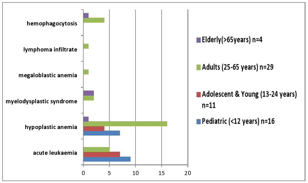
Overall out of 60 patients 21 patients (35%) were diagnosed as acute leukemia, 28 patients (48 %) as hypoplastic anemia, 4(6%) as MDS, 1 (1.5%) as megaloblastic anemia and as lymphoma infiltrate each, lastly hemophagocytosis 5 (8%) (Fig 1).
Age and etiology of pancytopenias were further analyzed, it was noted that acute leukaemis are more
Table 1 — Baseline parameters of patients (N=60) Pediatric years) n=16Mean valuesRange
(25-65 years) n=29
Mean age44 years 25-65 years
Sex (male : female)19:10-
(Hb) 6.2 g/dl3.5-8.6 g/dl
Total Leucocyte Count (TLC)
Elderly (>65 years) n=4
Mean age73 years 66-77 years
Sex (male : female)3:1-
(Hb) 7.8 g/dl4.4 -8.3 g/dl
Total Leucocyte Count (TLC) common in the age group of paediatric and adolescents, compared to adults. Hypoplastic anemias were more common in the adults and other rarer etiologies as shown were more common in adults and elderly (Fig 2).
Discussion
In a study by Pizzo PA, et al from a different geographical region had shown that etiologies of Pancytopenia were quite varied, acute leukemia and Bone Marrow Failure Syndrome were well recognized causes where as infection and Megaloblastic anemia were not common in pediatric population9. Though the above study didn’t excluded lymphadenopathy and organomegaly but their results were similar to ours.
A study from Eastern Mediterranean region conducted among adults by Nafil H, et al had shown that main causes of Pancytopenia were Megaloblastic Anemia (32.2%) and Acute Leukemia (23.7%) followed by Aplastic Anemia (15.2%)10
Another study from Karachi by Farooque R, et al also had shown that main cause of pancytopenia in adult was Megaloblastic Anemia (41.7%)11. But this study revealed main causes to be Hypoplastic Anemia followed by hypersplenism,so organomegaly was not excluded.

A study done among elderly population by Thyagaraj V, et al had shown that most common cause of Pancytopenia was Megaloblastic Anemia(60%) followed by aplastic anemia (7.5%) and Myelodysplastic syndrome (5%)12. Though a few elderly patients were included in the present study, the etiologies of Pancytopenia can be Hypoplastic Anemia and Myelodysplastic syndrome.
As etiology is concerned Megaloblastic anemia seems to be major contributing factor in different studies and it is curable. But in our study shows Hypoplastic anemia and acute Leukemia were the most common causes among younger age group and pediatric population. If Pancytopenia is associated with organomegaly such as lymphadenopathy and hepatospleomegaly as most studies across world has reported, but to report the causes of isolated Pancytopenia is unique in our study. This might be reason for our small sample size.
For any patients regardless of age Pancytopenia is a sinister finding in Complete Blood Count hence Bone Marrow studies are absolutely indicated along with other needful investigations such as immuopheotyping and cytogenetics.
It is also important to evaluate for Vitamin B12, folic acid deficiency as these are curable with replacement therapy. In our study however it was insignificant due to predominance of non- vegetarian population from Eastern India.
Such etiological knowledge is essential for clinicians in remote areas to intervene early and judicious management especially the hygiene for individual patients.
Limitations of the study :
The sample size is small because of restricting ourselves to only Pancytopenia and no orgnomegaly, longer duration of follow up is not possible due to varied educational status of our patients and last but not the least, the Covid-19 pandemic was a huge deterrent for many patients to attend specialized health care facilities.
Conclusion
Most common cause of pancytopenia without organomegaly and lymphadenopathy in pediatric age and adolescent group is acute leukemia. Hypoplastic anemia is the most common cause in adult population. Elderly patients were least affected (6.66%) in this study
Declaration of patient consent : Patient’s consent not required as patients identity is not disclosed or compromised.
Financial support and sponsorship : Nil
Conflicts of interest : There are no conflicts of interest.
References
1Sharma R, Nalepa G — Evaluation and Management of Chronic Pancytopenia. Pediatr Rev 2016; 37: 101-11.
2Khunger JM, Arculselvi S, Sharma U, Ranga S, Talib VH — Pancytopenia: a Clinico-haematological study of 200 cases. Indian J Pathol Microbiol 2002; 45(3): 375-9.
3Imbert M, Scoazec JY, Mary JY, Jouzult H, Rochant H, Sultan C, et al — Adult patients presenting with pancytopenia: a reappraisal of underlying pathology and diagnostic procedures in 213 cases. Hematol Pathol 1989; 3: 159-67.
4Yokus O, Gedik H — Etiological causes of pancytopenia: A report of 137 cases. Avicenna J Med 2016; 6(4): 109-12.
5Gnanaraj J, Parnes A, Francis CW, Go RS, Takemoto CM, Hashmi SK — Approach to pancytopenia: diagnostic algorithm for clinical hematologists. Blood Rev 2018; 32: 361-7.
6Chand R — International Journal of Contemporary Pediatrics. 2018; 5(6): 2173-7.
7Niazi M, Raziq F — The incidence of underlying pathology in pancytopenia. J Postgrad Med Inst 2004; 18: 76-9.
8Jha A, Sayami G, Adhikari RC, Panta AD, Jha R— Bone marrow examination in cases of pancytopenia. J Nepal Med Assoc 2008; 47(169): 12-7.
9Pizzo PA, D’Andrea AD — The Pancytopenias. In: Behrman RE, Kleigman RM, Jenson HB. (eds), Nelson Textbook of Pediatrics.16th edn. W.B. Saunders Co, Philadelphia; 1999; 1495-98.
10Nafil H, Tazi I, Sifsalam M, Bouchtia M, Mahmal L — Etiological profile of pancytopenia in adults in Marrakesh, Morocco. EMHJ - Eastern Mediterranean Health Journal 2012; 18(5): 532-6.
11Farooque R, Iftikhar S, Herekar F — Frequency and Etiology of Pancytopenia in Patients Admitted to a Tertiary Care Hospital in Karachi. Cureus 2020; 12(10): e11057.
12Thyagaraj V, Kulkarni A, Kumar TA — The study of clinicoaetiological profile of pancytopenia in elderly population. J Evid Based Med Healthc 2017; 4(45): 2727-9.
Disclaimer
The information and opinions presented in the Journal reflect the views of the authors and not of the Journal or its Editorial Board or the Publisher. Publication does not constitute endorsement by the journal.
JIMA assumes no responsibility for the authenticity or reliability of any product, equipment, gadget or any claim by medical establishments/institutions/manufacturers or any training programme in the form of advertisements appearing in JIMA and also does not endorse or give any guarantee to such products or training programme or promote any such thing or claims made so after.


