
5 minute read
Case Report A Unique Case of Pulmonary and Hepatic Hydatidosis


Harshit Jain1, Shivmohan Sarraf2, Bhavya Atul Shah3, Arti Julka4
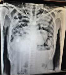
Hydatid disease or Hydatidosis is a rare infectious disease of human being that is caused by Echinococcus and can involves multiple organs. We report a case of multiple pulmonary and hepatic hydatid cysts in a 35-year-old female, admitted in our hospital with complaints of Cough with expectoration, Hemoptysis, Breathlessness, Chest and Abdominal pain with swelling and fever since 7 month. Chest X-ray revealed multiple rounded opacities of varying size in both lung field with some showing air fluid levels. Computed tomography scan of thorax showed multiple cystic lesion in both lungs and liver with air-fluid level in few cyst. Cystic fluid was aspirated that showed scolex of Echinococcus granulosis. Albendazole was started for the patient with relief in symptoms.
Advertisement
[J Indian Med Assoc 2023; 121(6): 62-3]
Key words : Hydatid disease, Hydatid cyst, Echinococcus granulosis, Scolex, Albendazole.
Hydatid disease is caused by ingesting embryonated eggs of cystode tapeworm of the genus Echinococcus. Human beings are incidental hosts and get infected by contamination from infected animals which leads to development of hydatid cyst in various organ, but most common site is liver followed by Lung. Here we present a case of 35 year old female, belonging to rural area who was found to have disseminated hydatid cysts of Lung and Liver.
CASE REPORT
A 35-year-old homemaker was admitted to our hospital with symptoms of Cough with expectoration, loss of appetite and weight since 7 months, Hemoptysis, Breathlessness, Right sided chest pain and also Abdominal pain and swelling since 6 month. Fever which was low grade on and off since 3 months. She also gave history of occasionally having a salty taste in the mouth with grape skin like material in expectoration. She had a history of recurrent hospitalization for similar complaints since past 3 years and had also received a course of Anti Tuberculous treatment empirically. On examination of chest, two visible soft fluctuant cystic swelling were present over 9th and 10th intercostal space in posterior axillary line on the right side of chest. She was to found to have crepitations and rhonchi bilaterally. Chest X-ray revealed multiple rounded opacities of varying size in both lung field with some showing air fluid levels. Ultrasonography of abdomen showed
Department of Respiratory Medicine, RD Gardi Medical College, Ujjain, Madhya Pradesh 456001
1MBBS, Postgraduate Student and Corresponding Author
2MBBS, Postgraduate Student
3MBBS, MD, Assistant Professor
4MBBS, MD, Professor and Head
Received on : 03/06/2022
Accepted on : 26/09/2022
Editor's Comment : multiple well defined cystic lesion of variable size (5.6x5.2x4.9cm) in liver. HRCT thorax revealed multiple cystic lesions in both lung and in liver with air-fluid level in few cyst which appear to be communicating with the bronchi. Her complete blood count showed a low Hemoglobin of 8 gm%, neutrophilic leukocytosis of 15650/cu mm, erythrocyte sedimentation was raised 85mm/hour and newly diagnosed Type 2 Diabetes Mellitus (HBA1c-7.1). The pain over the swelling was relieved by aspiration of a clear fluid which revealed scolex of Echinococcous granulosis. The patient was started on albendazole 400mg (two times a day). She was also given bronchodilators and antibiotics for the secondary infection. The patient improved symptomatically. However, due to disseminated disease, surgery was not considered and patient continued on medical therapy with regular follow up (Figs 1&2).
Hydatid disease is a rare infectious disease of human being that is caused by Echinococcus and can involves multiple organs and have various presentation in the body. As in our case cyst was protruding out from chest wall in the form of a swelling that help in making diagnosis.
Discussion
Hydatid cyst are caused by parasite known as Echinococcus which is particularly endemic in cattle/ sheep-raising rural area. Echinococcus granulosus species is the the most common among human (>95%). In endemic region, human incidence rate can reach more then 50 per 100000 person-year, and prevalence level as high as 5%-10%. The annual incidence in India is varying, 1 to 200 per 100000 population1. The hydatid cyst tend to involve the Liver
(50-70%), Lung (20-30%) but can be found in other organs like Brain, Heart, Bone, Kidney etc. except Hair, Teeth and Nails.
The ingested eggs are ruptured in digestive tract and larvae reached the Liver by portal system and pass through sinusoids and right chamber of the heart and eventually reside in the Lung. They grow in parenchyma and produce hydatid cyst. Cyst are made up of 3 layer (pericyst, ectocyst, germinal/endocyst). Daughter vesicle originate from germinal. Hydatid fluid is clear or pale, neutral pH, Contain Sodiumchloride, Proteins, Glucose, Ions, Lipids and Polysaccharides. The fluid is antigenic and may contain scolices, hooklets and hydatid sand2
Individual may remain asymptomatic for a long time. The pressure symptoms of the enlarging cyst produce pain in chest and abdomen as was seen in our case. Cyst rupture, secondary infection, suppuration and pneumothorax are common complication of pulmonary hydatid cyst. Cyst rupture may cause sudden cHest Or Abdominal Pain, Hemoptysis, Cough, Fever and
Salty taste in mouth as was seen in our case too. This can also lead to anaphylaxis3
Diagnosis is by medical history, physical examination, IgG antibody by ELISA and indirect hemagglutination test. Chest radiograph of the pulmonary cyst show the sharply defined, round to oval homogenous opacities of variable sizes. Ultrasonography of abdomen shows solitary or multiple cyst in Liver as was seen in our case. Computed tomography features shows smooth wall of variable thickness 1-20mm and homogenous internal content of water with various sign like “the crescent” or “the meniscus sign” which are reliable sign but not pathognomonic sign of pulmonary hydatid cyst. Radiologically, ruptured hydatid cyst is called double-arch, combo sign, iceberg sign, sign of rising sun, serpent sign and whirl sign. If the parasitic membrane float on fluid surface, this produce the “water lily sign” or “Camelot sign”. If all parasitic contents are aspirated, only the pericyst remains, known as “empty cyst sign”4
Surgery is the treatment of choice. Drug of choice is albendazole (group-benzoimidazole),10 -15mg/kg/ day in two divided doses given for 3-6 month. Hydatid disease should be treated with either albendazole only or albendazole and surgery like PAIR(Puncture, Aspiration, Injection of scolicidal agent and Reaspiration) to extract cyst without complication. Management of disseminated echincoccal disease is complex, which require a multidisciplinary approach5
In our case, due to extensive dissemination, the cysts were inoperable so medical therapy with albendazole was continued with close medical follow up and serial imaging studies.
References
1Torgerson PR, Devleesschauwer B, Praet N — World Health Organisation estimates of the global and regional disease burden of 11 foodborne parasitic disease, 2010: a data synthesis. PLoS Medicine 2015; 12(12): p. e1001920, 2015.
2Parija SC — A textbook of medical parasitology: 2nd edition. Madras: All India publisher and distributors; 4th Edition(1 January 2013), chapter 11.
3Sarkar M, Pathania R, Jhabta A, Thakur BR, Chopra R — Cystic pulomonary Hyatidosis. Lung India 2016; 33: 179-91. Doi:10.4103/0970-2113.177449.
4Emlik D, Odev K, Poyraz N, Kaya HE — Radiological characteristics of pulomonary hydatid cyst. Available from: https://www.ajroline. Org/doi/ref/10.2214/AJR.18.20928.
Taken from - Gamze Durhan, Aziz Tan, Selin Ardali Duzgun, Selcuk Akkaya and Orhan Macit Ariyrek. Radiological manifestation of thoracic hydatid cyst: pulmonary and extrapulmonary findings. Durhan et al. insight into imaging (2020) 11:116. https://doi.org/10.1186/s13244-020-00916-0.
5Dziri C, Haouet K, Fingerhut A, Zaouche A — Management of cystic echinococcosis complication and dissemination: where is the evidence? World J Surg 2009; 33(6): 1266-73.


