
5 minute read
Case Series


Thoracoabdominal Flap Coverage after Excision of Locally Advanced Breast Carcinoma
Arghya Bera1, Amit Roy2, Gaurab Ranjan Chaudhuri3, Sudip Das4, Ramita Mukherjee5, Risabh Dagra5
Advertisement
Excision of locally advanced breast carcinoma which is not responding to neo-adjuvant chemotherapy, leads to wide anterior chest wall defect with difficult closure of wound also delayed adjuvant radio-chemotherapy. The Thoracoabdominal flap (TA flap), a fascio-cutaneous rotation advancement flap quite better option for local tissue coverage of the wide defect in same stage. Here we present four patients with locally advanced breast carcinoma with no response to neo-adjuvant chemotherapy. The tumors were excised along with the local surrounding tissues and axillary dissection. Those defects were covered with TA flap (laterally based) derived from the same side of thorax and abdomen. One case developed marginal congestion of the flap managed by skin grafting. The anterior chest wall have few options of donor vessel. The TA flap having its blood supply from thoracolumber vessels and it does need a microsurgical procedure. The mean duration of hospital stay, postoperatively, is shorter in the TA flap, no difficulty in shoulder movement. Patients take adjuvant radio-chemotherapy earlier. A TA flap is a better coverage option for a wide defect following excision of locally advanced chemo-radio resistant breast carcinoma.
[J Indian Med Assoc 2023; 121(6): 57-9]
Key words : Thoracoabdominal flap, Locally advanced, Breast carcinoma, Chemotherapy.
Excision of locally advanced chemo-radio resistant breast carcinoma leads to wide chest wall gap. Some times radical resection may need removal of anterior chest wall muscles. Following excision, reconstruction of chest wall needed. Skin grafting is the simplest one. Skin grafting usually fragile and not durable if adjuvant radiation indicated. Various loco-regional myocutaneous, fasciocutaneous, dermofat flaps were previously used for healing and potentially covering the defect after cancer excision1-3. Loco-regional flaps, either medially based (thoracoepigastric) or laterally based (thoracoabdominal) flaps are commonly used fasciocutaneous flap. Thoracoabdominal flap resulted in lesser complications even with larger flap4. It’s a locally rotation advancement flap.
Case 1
A 52-year-old female patient comes with history of ulcerofungating growth on her left breast for a duration of
1MS, MCH, Associate Professor, Department of Plastic and Reconstructive Surgery, Medical College, Kolkata 700073, West Bengal and Corresponding Author
2MS, MCH, Associate Professor, Department of General Surgery, Nil Ratan Sircar Medical College & Hospital, Kolkata 700014
3MS, MCH, Professor, Department of Plastic and Reconstructive Surgery, Calcutta National Medical College and Hospital, Kolkata 700014
4MS, Assistant Professor, Department of General Surgery, Nil Ratan Sircar Medical College & Hospital, Kolkata 700014
5MBBS, Postgraduate Trainee, Department of General Surgery, Nil Ratan Sircar Medical College & Hospital, Kolkata 700014
Received on : 14/02/2022
Accepted on : 26/03/2022
Editor's Comment :
Thoracoabdominal Flap reconstruction is technically easy, can be done by fellows, no need of expertise help. Local tissues used for defect coverage, take less operative time.
Larger defects easily get covered. We can avoid skin grafting related complications, where Radio-therapy almost needed.
8-10 months. On physical examination a hard mass, size around 8x10 cm, immobile which appeared to be attached with the Pectoralis Major (PM) muscle fascia. There was a skin ulceration around 4-5 cm with everted margins. Patient was khown case T2DM and hypothyroidism, taking oral medication.
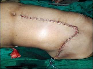
On core-needle biopsy proved it invasive ductal carcinoma of breast with staging of T4bN1M0. She received 3 cycles of NACT, without downstaging or downsizing. Another 3 cycles of taxen based NACT taken with no effect on stage and size. Patient undergone MRM with partial removal of PM muscle, creating a gap of 18x12 cm. the gap was covered with Thoracoabdominal flap. A Thoracoabdominal incision made through midline starting from medial corner of the defect to umbilicus. The skin and subcutaneous tissue from Mastectomy flap’s lower margin is also raised from anterior rectus sheath. Dissection continued laterally on suprafacial layer. As flap is raised, most of perforators from lateral intercostal arteries, subcostal arteries and lumber artereis are preserved for flap circulation, whereas sacrificing most of superior epigastric perforators for maximizing flap’s reach. Closure starts from medial side with flap rotationadvancement, with dog ear management at lateral side. The patient’s shoulder movement was normal. The patient was followed for 2 year postoperative period and remaintumor free.
Case 2
A 45-year-old female presented with a history of large mass on her left breast for a duration of 6-7 months. On physical examination a hard irregular mass, size around 9.5 x11 cm, immobile that appeared fixed to PM fascia. Mass was located at upper and lower inner quadrant. She also had family history of same carcinoma.
On FNAC proved it invasive ductal carcinoma of breast, with staging of T4bN2M0. She received 3 cycles of NACT, without any downstaging or downsizing. Patient undergone MRM with partial removal of PM muscle, leaving gap of 12x15 cm. The gap was covered by the TA flap as in Case 1. After 72 hours there was slight venous congestion at most distal part of flap. Following that distal part of flap get necrosed. Wound was managed by debridement, small skin grafting and opposite breast flap. The patient’s shoulder showed free range of movement. The patient having no recurrence oftumor till date.
lymph node clearance was also done. Resulting defect was around 13x15 cm in the maximum diameter. The huge defect was reconstructed by laterally- based thoracoabdominal flap.

Case 3
A 53-year-old male patient came with a growth at his right breast for few months. On examination, it revealed that a 6x7-cm hard growth at his right breast that seems to be hard to firm on palpation. CT showed a subcutaneous growth around a size of 6x7 cm in.
The tumor excised with approximately 4 cm of surrounding underneath PM fascia and the skin subcutaneous tissue, creating a gap of 11x12 cm. The gap was covered by the TA flap.


Case 4
A 60-year-old male patient presented with a lump on his right breast for 8 months. On physical examination a 8x10-cm mass on his right breast that feel hard on palpation. On true cut biopsy, it proved lobular carcinoma of breast.

MRM was done along with a margin of 5 cm. All level of axillary
Discussion
Advanced and chemo-radio resistant breast CA often needs wide excision, including all breast tissue. Soft tissue reconstruction remain always a problematic following wide-area excision. Before skin grafting era post radical mastectomy wounds were allowed to heal by secondary epithelialization alone5
The TA flap is a fasciocutaneous rotation advancement flap. TA flaps in late 1970s when medial- and lateral-based flaps were both described 2,3 lateral intercostal vessels for several advantages. Medial-based usually did not reach to most distal defect or leads to partial necrosis of the flap. Lateral-based TA flaps have robust circulation even with fewer vessels in a hatchet shape or transversely oriented design.
In our presentation, TA flaps were used for coverage after excision of advanced breast carcinoma in 4 patients. Among them only one patient had partial necrosis of flap that was managed with debridement followed by skin grafting and opposite breast flap.
The TA flap has an axial blood supply, and the procedure is quite simple, safe to dissection and requiring no microsurgical procedure. The mean duration of hospital stay, postoperatively, was less in the TA flap, no difficulty in shoulder movement. Patients take adjuvant radiotherapy earlier.
References
1Charanek AM — A bilobed thoracoabdominal myocutaneous flap for large thoracic defects. Ann Plast Surg 2014; 72: 451-6.


2Davis WM, McCraw JB, Carraway JH — Use of a direct, transverse, thoracoabdominal flap to close difficult wounds of the thorax and upper extremity. Plast Reconstr Surg 1977; 60: 526-33.
3Baroudi R, Pinotti JA, Keppke EM — A transverse thoracoabdominal skin flap for closure after radical mastectomy. Plast Reconstr Surg 1978; 61: 547-54.
4Park JS, Ahn SH, Son BH, Kim EK — Using local flaps in a chest wall reconstruction after mastectomy for locally advanced breast cancer. ArchPlastSurg 2015; 42: 288-94.
5Halstead WS — Resalt of operation for cure of cancer of the breast performed at John Hopkins Hospital from June 1889January 1894. Jhon Hopkins Hosp Rev 1895; 4: 297-350.


