Citation is a standardized method of acknowledging sources of information provided in any publication and helps readers to locate the source for supplemental study.
The importance of citation in scientific publication cannot be overemphasized, though it is perhaps the most neglected section in submitted manuscripts. Awareness about the techniques and utility of citing other authors and their work is quintessential in the process of scientific writing . An accurately constructed bibliography is vital for retrieving information from the ocean of knowledge now available in the virtual realm.
So Why Do We Need to Cite ?
There are many reasons for citing other publications apart from acknowledgement and avoidance of plagiarism. It is a way of comparing ones’ own work or ideas with other related literature,to give a perspective to the reader as to its similarities and deviations from previous studies1
References also direct readers to sources of information for further enrichment. What to and What Not to Cite ?
Choosing the proper sources of information also involves meticulous efforts. It is preferable to use primary sources like original articles published in peer reviewed journals as references rather than secondary sources like review articles.
Well controlled methodologically sound research is an obvious choice and one must refrain from citing outdated information. Care should be taken not to cite retracted articles, conference abstracts or posters. The latest evidences in citation favours the upliftment of ones’ own paper in terms of quality2
How to Cite ?
The author can cite others either by direct quotation or indirectly through paraphrasing or summarizing the key messages. It is distinctive to add ones’ own views either conforming or refuting the external source .However it is vital to put the in-text references in superscript at the end of that piece of information and detail them in the bibliography accurately3.
Compilation of the references at the end of the article should be undertaken with meticulous care. The arrangement may be in the ‘intext ‘ sequence as in the Vancouver style or in alphabetical order as found in the Harvard style. Although there are numerous styles of citation, the above mentioned, are most favoured by medical journals. Having said that, it is the prerogative of individual journals to categorically mention their preferred style of citation in the ‘instructions for authors’4 On the other hand it is the responsibility of the author to follow those instructions to
avoid rejection on account of technical imperfections. Rules have been streamlined regarding every aspect of scientific publication by the International Committee of Medical Journal Editors on the basis of the Uniform Requirements for Manuscripts submitted to Biomedical Journals.
How to be Careful ?
The number of references are an important consideration, as too many references do not necessarily mean that ones’ paper is well researched. Redundant, irrelevant citation undermines the quality of a manuscript and reflects poorly on the validity of the work. The bibliography should be optimum, in accordance to the desired numbers quoted in the guidelines of the individual journal.
Errors in citation result in difficulties in retrieving references. It fails to serve the purpose of accrediting the original author and leads to inaccurate citation indexing.
Moreover mistakes in quotations start a vicious cycle of circulation of false ‘accepted’ facts. Almost 50-70% of cited references have at least one error and the overall prevalence of citation errors is about 1020%. To avoid this, each reference needs to be cross checked with standard electronic citation sources or print copies of original papers and refrain from copying from reference lists of previously published articles5 To err is human, but Journals are not divine and may not forgive.
Also it is very important to check the temptation of over-selfcitation and restrict oneself to relevant
information. Citation management Software programs are available to make life easier for the authors. However, there are chances of duplicate references or errors in journal titles and it is the onus of the author to check reference databases to avoid inaccuracies.
A well - constructed bibliography not only enhances the credibility and prestige of an article, it helps readers to comprehend the research and evaluate the article in context of other manuscripts published previously.
Therefore awareness about the significance of meticulous citation among the budding authors is vital in the preparation of a relevant and unique manuscript.
FURTHER READING
1Masic I — The importance of proper citation of references in biomedical articles. Acta Inform Med 2013; 21(3): 148-55. doi: 10.5455/aim.2013.21.148-155.
2Mc DD — The appropriate use of references in a scientific research paper. Emerg Med (Fremantle) 2002; 14(2): 16670. doi: 10.1046/j.1442-2026.2002.00312.x.
3Penders B — Ten simple rules for responsible referencing. PLoS Computational Biology 2018; 14(4): e1006036. doi: 10.1371/journal.pcbi.1006036.
4Bahadoran Z, Mirmiran P, Kashfi K, Ghasemi A — The Principles of Biomedical Scientific Writing: Citation. Int J Endocrinol Metab2020; 18(2): e102622. doi: 10.5812/ijem.102622. PMID: 32636888; PMCID: PMC7322669.
5Vickers MD — Citation errors—there is still much to be done. Can J Anaesth 1995; 42(11): 1063. doi: 10.1007/ bf03011085.
MD, FRCP (Glasgow), FICP Nandini Chatterjee Professor, Department of Medicine, IPGME&R and SSKM Hospital, Kolkata 700020 and Hony Editor, JIMA
Original Article
Academic Honesty and Dishonesty in Different Disciplines and Degrees at the University of Medical Sciences : A Descriptive CrossSectional Study
Introduction : Dishonesty is considered as a basic challenge in ethics of care, which imposes great burden on the Educational System and the Society. Dishonesty is accompanied with negative impacts on all aspect of academic atmosphere. The aim of this cross-sectional, descriptive-analytical study was to determine dishonesty among 5 majors of study, Undergraduate and Graduate Degrees in School of Nursing and Midwifery.
Materials and Methods : 340 Undergraduate and Graduate students completed a questionnaire about all kinds of academic dishonesty and their causes. Data were analyzed using descriptive and analytic statistics.
Result : Suggested lowest levels of dishonesty among students of Midwifery (7.5%) and Anesthesiology (5.3%). Significant relationship was observed between sex and honesty (P<0.001). Also living place and the major had significant relationship with honesty (P<0.001). No significant relationships were found between dishonesty and education level and other demographic characteristics.
Conclusions : Many types of cheating are preventable through rules, correct training and educational management, which will eventually promote honesty in the educational system. This reveals the necessity of medical students’ familiarity with ethical codes and faculties’ emphasis on importance and role of ethics in Medical Sciences.
[J Indian Med Assoc 2023; 121(1): 15-8]
Key words :Academic dishonesty, Honesty, Undergraduate students, Graduate students.
Professions related to Medical Sciences include various ethical dimensions in such a way that ethics of care and professional ethics comprise the basis of these professions1. Dishonesty is considered as a basic challenge in ethics of care, which imposes great burden on the Educational System and the Society2. Currently the rate of academic fraud has increased worldwide3. Dishonesty is defined as any intentional attempt to distort, counterfeit or manipulate data, information, histories, or any other material related to students’ participation in courses, academic exercises or clinical performance4,5 . Cheating in academic environment is accompanied with incompatibility in clinical environment, which indicates the importance of cultural promotion of honesty and integrity in Universities6. Evidence has shown that some students tend to conduct deceitful educational behaviors. Such students consider these behaviors to be normal and acceptable, which result in consolidation of such behaviors7-11
Dishonesty has a historical background and is a
Department of Nursing, School of Nursing and Midwifery, Shiraz
University of Medical Sciences, Shiraz, Iran 71936-13119
1MSc, PhD in Nursing Education, Community Based Psychiatric Care Research Center
2MSc, MS in Community Health Nursing
3MSc, PhD in Nursing Education and Corresponding Author
Received on : 18/07/2022
Accepted on : 31/07/2022
Editor's Comment :
Dishonesty is considered as a basic challenge in ethics of care, which imposes great burden on the educational system and the society.
The study findings revealed dishonesty among students. There is a necessity of medical students’ familiarity with ethical codes, faculty member emphasis on importance and role of ethics in Medical Sciences, attempt to institutionalize professional ethics in students, and using novel educational methods.
global phenomenon that occurs in both developed and developing countries. Stimmel and colleagues reported the prevalence of cheating in 114 Medical schools in the US and Canada. The results demonstrated the performance of cheating in 70% of the Medical schools in the US and 35% of those in Canada12. Academic dishonesty is shown in different forms and in students of different educational levels and is considered to be misplaced behavior in the academic environment13, which is done by co-operation of a number of students8 In addition, dishonesty is not limited to theoretical courses and may occur in clinical courses, as well10. Dishonesty is a serious issue, which affects the quality of educational systems. It is also unfair for those who do not cheat. Additionally, it causes an incorrect interpretation of students’ knowledge and skills. This can lead to lack of professional quality, eventually harming the society. Lack of professional quality in medical sciences, in turn, affects human life14-16
121, No 1, January 2023Journal
In order to reduce dishonesty, students’ awareness, knowledge and skills should be improved6. In this context, teachers usually warn students rather than punishing them. In their opinion, warning and consultation cause students to re-evaluate their ethical values and to avoid unethical behaviors17. Generally, dishonesty is affected by various cultural, situational, attitudinal and psychological factors18,19. Students with weak English proficiency and those who have limited access to educational sources may tend to copy reference materials. Besides, students who are not educated and supported to plan for documentation of their scientific activities may get involved in plagiarism in informal formats. Therefore, these mostly neglected issues should be taken into account in studies on dishonesty in less developed countries, so that this unethical behavior can be prevented20.
In total, dishonesty is accompanied with negative impacts on students, professors, educational environments and the society. However, few studies have been conducted on the prevalence of this phenomenon and its related factors in students, particularly medical ones. Yet, this is of special importance to attract attentions to the issue and create motivation to find a solution for decreasing its prevalence. Therefore, the aim of the present study was to determine dishonesty among students of Bachelor and Master Programs in School of Nursing and Midwifery.
MATERIALS AND METHODS
This cross-sectional, descriptive-analytical study was approved by the Ethics Committee of Shiraz University of Medical Sciences, Shiraz, Iran (No. IR.sums.REC.1394.S275). All the participants were informed about the study objectives and signed informed consent.
The study participants included Undergraduate students of Nursing, Midwifery, Operating room, medical emergencies and Anesthesiology as well as Graduate students of Nursing and Midwifery who studied in a college in Southwest of Iran. According to the previous studies and considering α=0.5, the sample size was calculated as 340subjects. The participants were selected through stratified random sampling. In doing so, each major of study was considered as a stratum. Then, according to the total number of students in each major, a proper number of students was selected. The inclusion criteria were: being a student at the time of sampling, and willing to take part in the study. Exclusion criteria were unwillingness to respond to the questionnaire. Students were not obligated to participate in the study and informed consent form was obtained from them.
The study data were collected using a questionnaire containing two parts that were completed by self-report. The first section included questions about sex, living place, major of study, education level, transcript’s average, average gained during high school, satisfaction with the University and satisfaction with one’s major of study. The second part of the questionnaire included 16 questions about dishonesty, which were responded using a 5-option Likert scale ranging from always to never. It also contained one open question about three major reasons for dishonesty among students. The total score of this part could range from 0 to 80. This questionnaire was validated by Mokhtari Lake and colleagues in Iran in 2012. Accordingly, the content validity of its items ranged from 0.72 to 0.79 and its reliability was approved by Cronbach’s alpha=0.7221 The questionnaires were completed through self-report. It should be noted that the students signed written informed consents for taking part in the research.
After all, the data were entered into the SPSS statistical software, version 22 and were analyzed using descriptive and analytic (t-test, Chi-square test and Pearson’s correlation coefficient) statistics.
RESULTS
The results indicated that the average of most students in the University and high school were between 14 and 15.99. In addition, 162 students (44.5%) were averagely satisfied with their study majors. Besides, most of the students (n=108, 31.76%) were highly satisfied with their University. Other demographic features have been presented in Table 1.
Based on the results presented in Table 2, most students (57.4% of males and 51.4% of females) were moderately honest in their courses. Accordingly, students of Nursing (55.7%), operating room (66.7%), and Medical emergencies (81.2%) were moderately honest. On the other hand, the lowest levels of dishonesty were detected among the students of Midwifery (7.5%) and Anesthesiology (5.3%). Furthermore, most students who lived in dormitories reported moderate honesty (52.7%).
As Table 3 depicts, the rate of dishonesty was higher among females in comparison with males. Besides, a significant relationship was observed between sex and honesty (P<0.001). The results also indicated that the rate of dishonesty was higher among nursing students compared with those of other majors. A significant relationship was also found between the major of study and honesty (P<0.001). Moreover, the rate of honesty was higher among the students who lived in dormitories. A significant relationship was also observed between living place and dishonesty
Table 1 — Frequency of demographic characteristics in students
VariablesNo%Total
Sex341 (100)
Male9426
Female247 68.2
Major341 (100)
Nursing183 50.6
Midwifery67 18.5
OR5114.1
Anaesthesia195.2
ER215.8
Grade 341 (100)
Bachelor31587
Master267.2
Place341 (100)
Dormitory237 65.5
Home104 27.9
Table 2 — Academic dishonesty frequency between students
Table 3 — Relationship between academic dishonesty and demographic-educational characteristics
Academic High ModerateLow RareP value dishonestyNo (%)No (%)No (%)No (%)
Variable
Sex :<0.001
Male11 (11.7)54 (57.4)28 (29.8)1 (1.1)
Female8 (3.3)127 (51.4)103 (41.7)9 (3.6)
Major :<0.001
Nursing13 (7.1)102 (55.7)65 (35.5)3 (1.6)
Midwifery0 (0)26 (38.8)36 (53.7)5 (7.5)
OR3 (5.9)34 (66.7)13 (25.5)1 (2)
Anaesthesia1 (5.3)2 (10.5)15 (78.9)1 (5.3)
ER2 (9.5)17 (81.2)2 (9.5)0 (0)
Place : <0.05
Dormitory14 (5.9)124 (52.3)92 (38.8)7 (3)
Home3 (3)57 (56.4)38 (37.6)3 (3)
ER2 (9.5)17 (81.2)2 (9.5)0 (0)21
Place :
Dormitory14 (5.9)124 (52.3)92 (38.8)7 (3)237
Home3 (3)57 (56.4)38 (37.6)3 (3)101
Grade :
Bachelor 16 (5.1)166 (52.7)124 (39.4)9 (2.9)315
Master3 (11.5)15 (57.7)7 (26.9)1 (3.8)26
(P<0.001). However, no significant relationships were found between dishonesty and education level and other demographic characteristics (P>0.05).
The major reasons that led the students to commit academic dishonesty were fear of failing 85% (289), anxiety about doing proper performance 81.6% (277), and poor time management 81.7% (278).
DISCUSSION
Dishonesty is not a novel phenomenon and is common in all around the world. The findings of the present study indicated that dishonesty existed among students and was more common among females compared with males. Tardy and colleagues conducted a descriptive study and asked students to complete questionnaires through self-report. According to their results, 97% of the subjects reported some sort of dishonesty, 78% had cooperated in at least one type of cheating, 50% had a mild attitude towards such unethical behaviors, and 2% had helped other students to cheat 22. Bedford and Gregg proposed that personal
and psychosocial factors played a role in the occurrence of dishonesty among students. They also demonstrated that age, sex, rules of the study major, and learning environment were effective in occurrence of dishonesty22. Dogas performed a study in 2014 to investigate who helped students to cheat. According to the results, females cheated more in comparison with males 7. Hafeez and colleagues also carried out a research in Pakistan in 2013 and reported higher dishonesty among females compared with males12 In contrast, some studies have revealed higher rates of dishonesty among males in comparison with females3,22. This difference might be attributed to cultural variations as well as differences in study majors. The results of the current study revealed a higher rate of dishonesty among nursing students in comparison with other majors. Similarly, Kacici and co-workers conducted a study in 2014 to assess the rate of cheating in School of Nursing. The results indicated a higher rate of cheating among nursing students4. Hanning and others showed that great expectations from students and high workload in Medical Sciences resulted in dishonest behaviors among students2. However, no similar studies were found to compare the results. Therefore, the reasons for such behaviors have to be assessed in future studies. The findings of the present study revealed no significant relationships between dishonesty and other variables. In the same line, Hafeez performed a study in Pakistan and showed no significant relationships between dishonesty and other variables, such as education level12. However, David (2014) reported a relationship between dishonesty and self-confidence, skillfulness, valuing honesty and educational success. Accordingly, students who had an optimistic view towards Human nature were deceived less. Nonetheless, no significant correlations were observed in this respect. Overall, considering the high rate of cheating and dishonesty,
Vol 121, No 1, January 2023Journal
the educational system needs to be promoted23. A prior study was conducted in Korea to explore the impact of seven sessions of ethics training on students’ ethical sensitivity. Some studies results indicated the need for planning and higher accuracy in designing the curricula24,25. The findings of the current study also showed that dishonesty among students could not be neglected. Due to the negative effects of this problem on academic education and professionalism, solutions have to be found by educational planners and teachers.
CONCLUSION
The study findings revealed dishonesty among students. Hence, this issue has to be taken into consideration by Researchers, Managers and Facultymember. Many types of cheating are preventable through rules, correct training and educational management, which will eventually promote honesty in the Educational System. This reveals the necessity of medical students’ familiarity with ethical codes, faculty member emphasis on importance and role of ethics in Medical Sciences, attempt to institutionalize professional ethics in students, and using novel educational methods.
LIMITATIONS
This study was conducted on students of different majors in School of Nursing and Midwifery. Comparative investigation of different colleges can provide a more reliable comparison of dishonesty among students of various majors. Moreover, all variables, particularly dishonesty, were evaluated through self-report in this study. Thus, responses might have been affected by different factors. For instance, students might have provided responses welcomed by the Society. In other words, they might have exaggerated or underestimated the cases of dishonesty. Therefore, further interventional studies are recommended to find an appropriate solution to decrease this ethical and professional challenge.
Conflicts of Interest :There are no conflicts of interest for the present study.
Funding : There is no funding to declare.
REFERENCES
1Eskandari H, Heidari M, Nezarat S — Attitude of Nursing Students towards Ethics Codes, Engagement in Ethics in Care and Scientific Uncertainty in the Faculty of Medical Sciences of Abadan. Iranian Journal of Ethics and Medical History 2016; 9(3): 55-64.
2AM H, RS, Malpas P — Reasons for academic honesty and dishonesty with solutions: a study of pharmacy and medical students in New Zealand. Journal of Medical Ethic 2013; 40(10): 1-8.
3Macfarlane B, Jingjing Z — Academic integrity: a review of the Literature. Studies in Higher Education 2014; 39(2): 339-58.
4Kececi A, Bulduk S, Oruc D — Academic dishonesty among
nursing students: A descriptivestudy. Nursing Ethics 2014; 18(5): 727-33.
5Murdock TB, Hinton A — Predictors of cheating and cheating attributions: Does classroom context influence cheating and blame for cheating?. . European Journal of Psychology of Education 2008; 23(4): 477-92.
6Macale L, Ghezzi V, Rocco G — Academic dishonesty among Italian nursing students: A longitudinal study. Nurse Education Today 2017; 50: 57-61.
7Dogas V, Jeroncic A, Marusic M — Who would students ask for help in academic cheating? Cross-sectional study of medical students in Croatia. BMC Medical Education 2014; 30(14): 1048.
8Eric J Ip, Pharm D, Nguyen K — Motivations and Predictors of Cheating in Pharmacy School. American Journal of Pharmaceutical Education 2016; 80(8): 1-7.
9Hrabak M, Vujaklija A, Vodopivec I — Academic misconduct among medical students in a post-communist country. Med Educ 2004; 38(3): 276-85.
10Lynch J, Everett B, Ramjan LM — Plagiarism in nursing education: an integrative review. Journal of Clinical Nursing 2017; 26(19): 2845-64.
11Saana SB, Ablordeppey E, Mensah NJ — Academic dishonesty in higher education: students’ perceptions and involvement in an African institution. BMC Research Notes 2016; 25(9): 234.
12Hafeez K, Laiq-Uz-Zaman Khan M, Jawaid M — Academic misconduct among students in medical colleges of Karachi, Pakistan. Pakistan Journal of Medical Sciences. 2013; 29(3): 699-702.
14Desalegn AA — Cheating on examinations and its predictors among undergraduate students at Hawassa University College of Medicine and Health Science, Hawassa, Ethiopia. BMC Medical Education 2014; 30(14): 89.
15Divall MV — Academic Dishonesty: Whose Fault is it Anyway? American Journal of Pharmaceutical Education 2016; 80(3): 35.
16Korn L — The Proûle of Academic Oûenders: Features of Students Who Admit to Academic Dishonesty. Med Sci Monit 2016; 22: 3043-55.
17Oran NT, Can HO, ªenol S — academic dishonesty among health science school students. Nurs Ethics 2016; 23(8): 919-31.
18Anderman EM — Psychology of Academic Cheating. San Diego: Elsevier; 2007.
19Orosz G, Farkas D — Are competition and extrinsic motivation reliable predictors of academic cheating? Frontiers in Psychology 2013; 4: 87.
20Lim J, Ho PM — Modulation of incentivized dishonesty by disgust facial expressions. Frontiers in Neuroscience 2015; 9: 1-9.
21Mokhtari Lakeh N, Nafar M, Ghanbari Khanghah A — Nursing Students’ views on Code of Ethics, Com mitment to the Ethic of, Academic Dishonesty and Neutralization behaviors. Comprehensive Nursing and Midwifery. 2014; 24(73): 64-71.
22Bedford D W, Gregg RJ, Clinton MS — Preventing Online Cheating with Technology: A Pilot Study of Remote Proctor and an Update of Its Use. JournalofHigherEducationTheory and Practice 2011; 11(2): 41-59.
23David Tl — Academic cheating in college students: relations among personal values, self-esteem and mastery. . Procedia - Social and Behavioral Sciences 2015; 187: 88-92.
24Naghdipour BH, Emeagwali LO — Students’ Justifications for Academic Dishonesty: Call for Action. Procedia - Social and Behavioral Sciences 2013; 83: 261-5.
25Yeom AH — Effects of ethics education on moral sensitivity of nursing student. Nursing Ethics 2017; 24(6): 644-52.
Original Article
Association between Sleep Quality and Different Aspects of Memory along with Assessment of Post Exercise and Post Meditation Effects
Mohita Singh1, Sunil Sachdev2, Monica Manhas3, Dev Raj4
Background and Aims : Sleep is a highly conserved behaviour across animal evolution. The functions of sleep include restoration, memory processing, dreaming etc. Memory is informational processing system with explicit and implicit functioning made up of sensory processor, short term memory and long term memory. The present study was designed to analyse the impact of sleep quality on memory and effect of exercise and meditation on same.
Material and Method : The present study was performed on 110 subjects chosen randomly with no gender bias. In first phase, baseline values were assessed for different sub tests of sleep quality and different aspects of memory. Subjects were divided into two groups with each group including 27 males and 27 females. One group was required to perform moderate intensity exercise and other meditation for one month duration. In the second phase, parameters were again assessed.
Statistical analysis : Paired t-test was used for comparison of memory and sleep components between males and females. Independent t-test was used between baseline and post intervention values of exercise, meditation. Correlation studies were also carried out between sleep quality and different aspects of memory using Pearson correlation coefficient.
Result : Significant and non significant results were obtained on comparison of memory and sleep components in males and females. Total memory score was better in females. Exercise and meditation exhibited statistically significant result on memory and sleep quality.
Conclusion : Good sleep quality is associated with better memory. There is improvement across domains of memory and sleep with meditation and exercise.
[J Indian Med Assoc 2023; 121(1): 19-23]
Key words :Daytime dysfunction, Recall, Recognition, Retention, Sleep latency.
Sleep is a naturally recurring state of body and mind characterized by altered consciousness, relative inhibition of sensory activity, reduced voluntary muscles activity during REM sleep along with reduced interactions with the surroundings1. Memory is the faculty of the brain by which data or information is encoded, stored and retrieved when needed. It is retention of information over time for the purpose of influencing future action2
Meditation is a practice of using mindfulness techniques to train attention, awareness, reach emotionally calm state, enhance peace and for overall well being of an individual3. Physical exercise is any bodily activity that enhances or maintains physical fitness and overall health and wellness4.
Memory is not a perfect processor and is affected
Department of Physiology, Government Medical College and Hospital, Jammu, Jammu and Kashmir 180004
1MBBS, MD (Physiology), Demonstrator and Corresponding
Author
2MBBS, MD (Physiology), Professor and Head
3MBBS, MD (Physiology), Assistant Professor
4Statistician, Lecturer, Department of Community Medicine
Received on : 06/05/2022
Accepted on : 13/07/2022
Editor's Comment :
Appropriate sleep quality is required for proper brain functioning including memory. Exercise and meditation both helps to improve sleep quality and hence memory.
by many factors. The way in which information is encoded, stored and retrieved can be interrupted. Is memory affected by sleep quality? Is there any difference in memory in males and females? Does moderate intensity physical exercise and meditation has any effect on sleep quality and memory? To answer these intriguing questions, the present study was designed to study the effect of sleep quality on different aspects of memory along with post exercise and post meditation effects.
MATERIAL AND METHOD
Ethical clearance was obtained from the Institutional Ethics Committee. The subjects were briefed about the study and informed written consent for participation in the study was taken.
Sampling :
The present study was performed on 110 subjects chosen randomly with no gender bias in the age group
121, No 1, January 2023Journal
of 30-40 years. From the selected subjects, two were non compliant and hence were excluded from the study. From total of 108 subjects, 54 were males and 54 females. A minimum sample size of 108 was calculated taking correlation coefficient between sleep quality and memory r = -0.266 and usual constrains Type 1 error α=0.05 and Type 2 error β=0.2 by using MedCalc software.
The sample was collected via simple random sampling method. The subjects with significant medical history, significant drug/ alcohol history, psychiatric illness, clinical diagnosis of sleep or memory disorder, those already engaged in exercise or meditation practices were excluded.
Study design :
The present prospective study was conducted in two phases. In the first phase, informed written consent was taken and the procedure was explained to subjects. Their baseline value was assessed for different sub tests of sleep quality and different aspects of memory. Based on memory scores, they were divided into five groups of excellent memory, average, above average, below average and low level of memory. In each group, different aspects of sleep quality were assessed. They were divided into two groups (n = 54 each) with each group having 27 males and 27 females subjects (selected equally from each group based on memory scores). One group was required to perform Moderate Intensity Physical Exercise (MIPE) and other meditation for one month duration. In the second phase, parameters were again assessed and comparison was made between two interventions. Complete anonymity was maintained as honest responses are given.Only educated volunteers were chosen because adequate educational background is required for filling the questionnaires and performing memory test.
Study tool :
Sleep quality was assessed via Pittsburgh sleep quality index5 and for memory PGI memory test6 was used. The MIPE consisted of walking briskly at the rate of 4 mph7 for 30 min/day for 5 days/week (ie, 150 min/week)8. The meditation was performed for 15 - 20 min per day9 . Both exercise and meditation were performed for one month duration.
Statistical analysis : Comparison of different components of memory and sleep quality between males and females was done using paired t-test. Correlation studies were carried out between sleep quality and different aspects of memory using Pearson correlation coefficient. Independent t-test was used between baseline and
post intervention values of exercise, meditation in males and females. The results were computed as significant at p<0.05 level (*), more significant at p<0.01 level (**) and highly significant at p<0.001 level (***).
RESULT
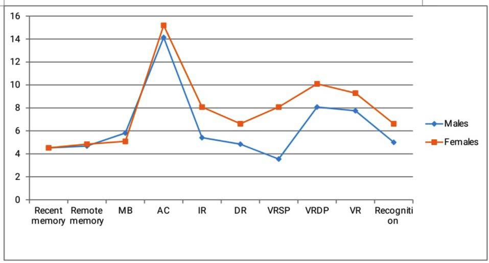
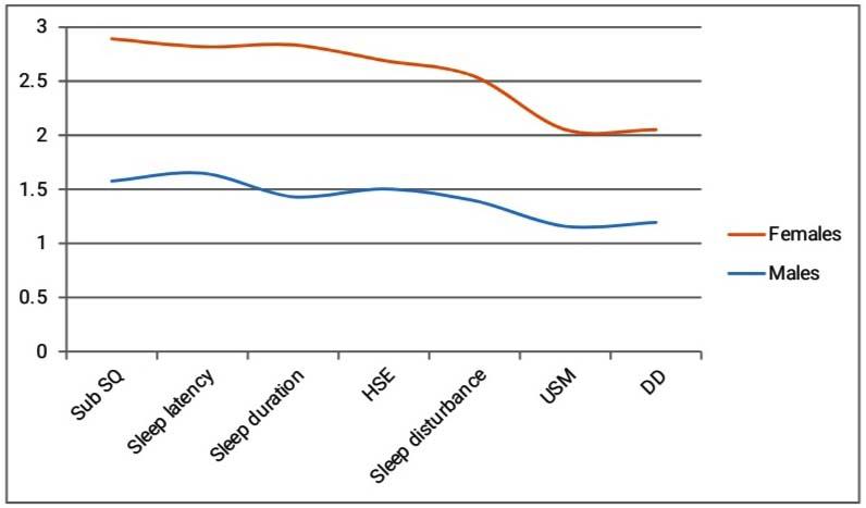
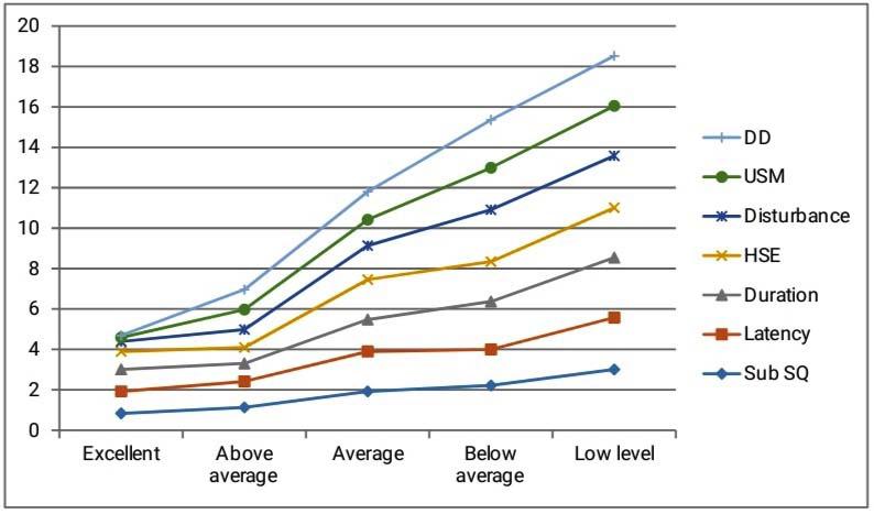
Out of 108 chosen subjects, 54 were males and 54 females. Different components of memory and sleep quality were studied in males and females (Tables 1a & 1b and Figs 1a & 1b).
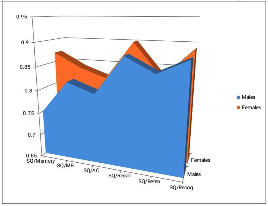
For 108 subjects chosen, results were analysed for correlation between sleep quality and different sub tests of memory as depicted in Table 2 and Fig 2.
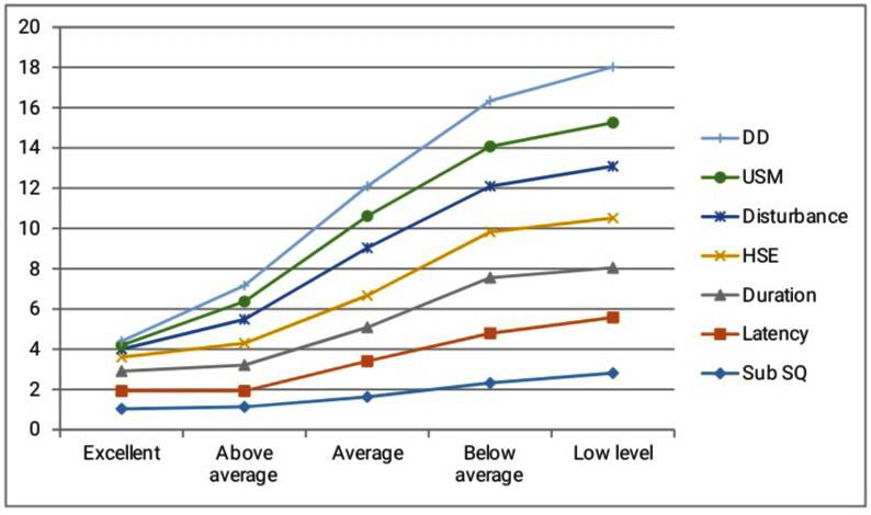
Based on memory scores, subjects were divided into five groups. Sleep quality components (mean ± SD) were analysed for different groups of memory in males and females as depicted in Table 3 and Figs 3a & 3b.
After one month practice of MIPE and meditation, results were compared with baseline value for sleep and memory in males and females (Table 4).
DISCUSSION
Memory is considered as the retention, reactivation and reconstruction of the experience with independent
Table 1(a) — Different sub tests of memory in males and females
ParametersMales FemalesP value (n=54)(n=54) Memory
(a) Similar pairs3.55±0.968.01±1.09<0.001*** (b) Dissimilar pairs8.01±1.09 10.05±2.42 <0.001***
Visual7.70±1.199.27±2.00 <0.001***
Recognition5.00±1.06 6.57±1.75<0.001***
Total 62.83±11.30 73.50±16.67 <0.001***
Table 1 (b) — Different components of sleep quality in males and females
internal representation. During sleep, most profound changes occur in brain. The present study was designed to study the effect of sleep on memory with post exercise and post meditation effects. Present study reported no significant result in memory and attention concentration scores in males and females that is in corroboration with previous studies10. Also, in present study mental health and balance was stronger in males compared to females, the results being similar to previous studies11. In the
Table 2 — Correlation between sleep quality and different sub tests of memory
ParametersMales
SQ/
SQ/
SQ/
SQ/
SQ/
present study, significant results were obtained for recall, retention and recognition in females compared to males. Previous studies have reported that females outperform males on recall of both positive and negative life events that is probably due to differences in the details of encoding12, retention indicating long term memory capacity of females13 and recognition that was directly related to female scanning behaviour at encoding14
Table 3 — Analysis of components of sleep quality in different memory groups
Excellent memory
Average memory :
Below average :
Low level :
M is males and F is females
Fig 2 — Correlation between sleep quality and different sub tests of memory
Fig 1a — Different sub tests of memory in males and females
Fig 1b — Different componenets of sleep quality in males and females
Present study reported that overall sleep quality was better in females than males. Previous studies have reported that women have better sleep quality compared with men, with longer sleep times, shorter sleep onset latency and higher sleep efficiency. Despite this, women have more sleep related complaints compared to men15
Present study reported significant and negative correlation between sleep quality and different aspects of memory in males and females. Similar study concluded that sleep quality was associated with updating in working memory only when working memory demands were relatively high and with recall. [16] Studies have also reported similar and significant association between sleep quality and psychological well being17; between sleep quality and attention concentration18. It was also reported that poor sleep quality was associated with significantly lower recall at the longer retention period (30-46 days) but not at shorter ones (2-15 days)19
In the present study, sleep quality components were studied in different memory groups. Improved memory scores were observed with better sleep quality (as depicted by decrease in score). Previous studies have reported similar result of significant relation
Table 4 — Effect of exercise and meditation on parameters in males and females
Post Spirituality73.03 ± 11.1485.03 ± 17.390.004**
between sleep quality and memory signifying that poor sleep quality and long sleep duration were linked to low memory performance20. Studies have reported that longer sleep latencies and poor sleep depth significantly predicted poorer next day prospective memory reaction time21. Scientists were of the view that habitual sleep quality was directly linked to memory recall of content, time and details of event19 Previous studies have reported that sleep deprivation and disturbances lead to impairment in working memory capacity due to decrease in speed of processing information 22. With the use of sleep medications, memory deficits were reported23. Excessive daytime sleepiness is a predictor of subjective memory impairment and such individuals are potential candidates for interventions related to dementia care24 In the present study, there was improvement in sleep quality and memory scores in both males and females after exercise and meditation interventions. However, the sleep quality scores were not significant on comparison between males and females at baseline and post intervention level. Memory scores were better in females both at baseline and post intervention level. Previous studies have reported similar results of great impact of physical training on working memory and executive attention25; and positive interrelationship between sleep and exercise. [26] Also, increase in memory test scores27 and positive effect on sleep quality28 post meditation have been reported, thus making way for mindfulness based interventions for greater benefits and treating aspects of sleep disturbances.
Limitation of Study : The result of the study maybe specific to the selected age group and educational background of the subjects. However, the study gave us important insight into the fact that sleep and memory are correlated and that exercise and meditation both had positive impact on them.
Fig 3b — Analysis of components of sleep quality in different memory groups in females
Fig 3a — Analysis of components of sleep quality in different memory groups in males
No 1, January 2023Journal
CONCLUSION
Memory shares positive relationship with sleep quality concluding that better sleep quality is associated with more effective memory. Overall memory and sleep quality was better in females. Meditation and exercise both had positive effect on sleep quality and memory in both sexes. Memory scores were better in females at post exercise and post meditation interventions. There was no significant difference in sleep quality scores in males and females post interventions with females having better tendency for good sleep quality than males.
ACKNOWLEDGMENT
We are grateful to all the subjects for their participation in this research work. This work is an original article and no financial grants were obtained from any source. There is no conflict of interest.
REFERENCES
1Ferri R, Manconi M, Plazzi G, Bruni O, Vandi S, Montagna P, et al — A quantitative statistical analysis of the submentalis muscle EMG amplitude during sleep in normal controls and patients with REM sleep behaviour disorder. J Sleep Res 2008; 17(1): 89-100.
2Sherwood L — Human Physiology: From cells to systems. Cengage Lrn 2015: 157-62.
3Campos D, Cebolla A, Quero S, Breton – Lopez L, Botella C, Soler J, et al — Meditation and happiness: Mindfulness and self compassion may mediate the meditation happiness relationship. Pers Individ Differ 2016; 93: 80-5.
4Kylasov A, Gavrov S — Ethno cultural diversity of sport. Sport Sci Magister Press UNESCO 2011: 462-91.
5Bussy DJ, Reynolds CF, Monk TH, Berman SR, Kupfer DJ — The Pittsburgh Sleep Quality Index: A new instrument for psychiatric practise and research. Psychiat Res 1989; 28: 193-213.
6Pershad D, Wig NN — Reliability and validity of a new battery of memory tests (PGI Memory Scale). IndJPsy 1978; 20: 7680.
7Haskell LW, Lee MI, Pate RR, Powell KE, Blair SN, Franklin BA, et al — Physical activity and public health updated recommendation for adults from the American College of Sports Medicine and the American Heart Association. Circu 2007; 116: 1081-93.
8World Health Organization — The global recommendations on physical activity for health. WHO Press, Geneva 2010; pp: 16:26.
9Basso JC, McHale A, Ende V, Oberlin DJ, Suzuki W —. Brief daily meditation enhances attention, memory, mood and emotional regulation in non- experienced meditators. Behav Brain Res 2019; 356: 208-220.
10Solianik R, Brazaitis M, Skurvydas A — Sex related differences in attention and memory. Medicina 2016; 52(6): 372-7.
11Van Droogenbroeck F, Spruyt B, Keppens G — Gender differences in mental health problems among adolescents
and the role of social support: result from the Belgian health interview survey. BMC Psy 2018; 18(6): 1591-4.
12Seidlitz L, Diener E — Sex differences in the recall of affective experiences. J Pers Soc Psychol 1998; 74(1): 262-71.
13Astie AA, Scardamaglia RC, Muzio RN, Reboreda JC — Sex differences in retention after a visual or a spatial discrimination learning task in brood parasitic shiny cowbirds. Behav Process 2015; 119: 99-104.
14Heisz J, Pottruff M, Shore DI — Remales scan more than males: A potential mechanism for sex differences in recognition memory. Psychol Sci 2013; 24(7): 1157-63.
15Krishnan V, Collop NA — Gender differences in sleep disorders. Curr Opin Pulm Med 2006; 12(6): 383-89.
16Rana BK, Panizzon MS, Franz CE, Spoon KM, Jacobson KC, Xian H, et al —Association of sleep quality on memory related executive functions in middle age. J Intl Neuropsychol Soc 2018; 24(1): 67-76.
17Zhai K, Gao X, Wang G — The role of sleep quality in the psychological well being of final year undergraduate students in China. Intl J Environ Res Public Hlth 2018; 15(12): 2881.
18Pacheco AA — Relation between sleep quality and attention in students of business administration. BiolRhythmRes 2014; 45 (1): 131-42.
19Murre J, Kristo G, Janssen SMJ — The effect of self reported habitual sleep quality and length on autobiographical memory. Memory 2014; 22(6): 633-45.
20Tsapanou A, Gu Y, O’Shea DM, Yannakoulia M, Kosmidis M, Dardiotis E, et al — Sleep quality and duration in relation to memory in elderly: Initial result from hellinic longitudinal investigation of aging and diet. Neurobiol Learn Mem 2017; 141: 217-25.
21Goldberg ZL, Thomas KGF, Lilinska G — Bedtime stress increases sleep latency and impair next day prospective memory performance. Front Neurosci 2020; 14: 1-10.
22Peng Z, Dai C, Ba Y, Zhang L, Shao Y, Tian J — Effect of sleep deprivation on the working memory related N2 – P3 components of the event related potential waveform. Front Neurosci 2020; 14: 469.
23Dokkedal SV, Oliveira MGM, Galduroz LCF, Tufik S, Anderson ML — The effect of sleep medications on perspective retrospective memory: A population based study. Prog Neuropsycholpharmacol Biol Psy 2021; 104: 110043.
24Okamura T, Ura C, Miyamae F, Sugiyama M, Niikawa H, Ito K, et al — Excessive daytime sleepiness is related to subjective memory impairment in late life: a cross sectional community based study. Psychogeriatr 2016; 16(3): 196-201.
25De Sousa AFM, Medeiros AR, Del Rosso S, Stults-Kolehmainen M, Boullosa DA — The influence of exercise and physical fitness status on attention: A systematic review. Int Rev Sport Exerc Psychol 2018; 12: 202-234.
26Dolezal BA, Neufeld EV, Boland DM, Martin JL, Cooper CB — Interrelationship between sleep and exercise: A systematic review. Adv Prev Med 2017; 2017: 1364387.
27Fleischmann Rposner M. Meditation for increased mindfulness and memory: An analysis on the impact of meditation on mindfulness and working memory capacity in high school students. J Student Res 2020; 9(2): 1-17.
28Rusch HL, Rosario M, Levison LM, Olivera A, Livingston WS, Wu T, et al — The effect of mindfulness meditation on sleep quality: A systematic review and meta analysis of RCT. Ann N Y Acad Sci 2019; 1445(1): 5-16.
Original Article
Prevalence of Undiagnosed Diabetes and Impaired Glucose Tolerance in a Semi-urban Population from Calicut City
Rajesh K P1
Though the prevalence of Diabetes is increasing worldwide, a thorough knowledge of the prevalence of undiagnosed Diabetes a pre-diabetes is lacking. This study from India is to evaluate the prevalence of asymptomatic diabetes among adults with comorbidities and without any history of Diabetes. Prevalence of asymptomatic individuals with Diabetes and impaired glucose tolerancewas 3% and 15%, respectively. The high prevalence found in the study raises concern over the health care indices and the need for urgent public health action to control the pandemic. Regular screening for Diabetes in adults is required to prevent complications of long-term diabetes.
Early detection of Diabetes is important. Longstanding diabetes may lead to end organ damage including Nephropathy, Neuropathy, Retinopathy, Cardiovascular events. However, in the general population, there is a prevalence of asymptomatic Diabetes that goes unnoticed1 and may eventually increase the prevalence of long-standing disease.
ADA (2022) guidelines recommend that all adults without risk factors should be screened with a test for pre-diabetes and Type 2 Diabetes starting at age 35, instead of the earlier cut-off of 45 years. The new Standards of Care also emphasizes screening with a Fasting Glucose Test for undiagnosed Diabetes in all women who are planning pregnancy, especially if they have risk factors. It advocates that a risk-based approach should be considered in screening for prediabetes and/or Type-2 Diabetes in those with age >10 years/ onset of puberty, whichever is earlier, in youth who are overweight or Obese and who have at least one additional risk factor for Diabetes. If initial screening is normal, it should be repeated at a minimum interval of 3 years or more frequently if BMI is increasing. American Association of Clinical Endocrinology recommends screening for pre-diabetes and Diabetes in individuals about 45 years of age2 and USPSTF recommends screening in adults aged 35 to 70 years who have overweight or Obesity1
In a highly populous country like India with wide diversity, Socio-economic status contributes to the
1MD, DNB, MRCP, FRCP, Consultant, Department of Internal Medicine and Diabetology, KIMS Trust Hospital, Calicut 673571, PVS Hospital, Calicut 673002, Kerala and Corresponding Author
Received on : 28/06/2022
Accepted on : 17/11/2022
Editor's Comment :
There is a significant incidence of undiagnosed diabetes in our population.
Routine screening of adult population is necessary to pick the cases early and to introduce lifestyle changes earlier so as to slow the progression.
The health care system needs to be sensitised on the hidden burden of lifestyle diseases and the need to propagate healthy lifestyle and early screening.
disparities in the diagnosis of illnesses such as Diabetes Mellitus and also in healthcare delivery3. With the prevailing Healthcare Management System and due to a lack of insurance; in the present scenario, routine annual health check-up is not a norm among major part of the population in India3. Therefore, early recognition of an impending future illnesses burden is challenging.
As a Consultant Diabetologist with more than 18 years of clinical practice in Kerala (India), I have come across a large number of diabetic patients who were asymptomatic and were accidentally detected. Considering the risk of the complications of long-term Diabetes, there is a need to screen individuals irrespective of the existence of symptoms. In this study, we plan to expose the burden of undiagnosed Diabetes in our region by screening asymptomatic individuals with no history of Diabetes Mellitus, at camps conducted by the hospital.
With this study we aim to highlight the importance of routine screening of adult population for Diabetes especially in a country like India which is deemed to be the Diabetes capital of the world4 This will help in early detection of Diabetes and pre-diabetes so that lifestyle modification and treatment can be initiated early.
MATERIALS AND METHODS
Study Design :
A pilot observational study was conducted at KIMS Trust Hospital, Calicut over a period of 6 months from October 15th 2021 to April 15th 2022.
The patient population included adults over 18 years of age with existing comorbidities and without any prior history or knowledge of pre-existing Diabetes Mellitus. The study participants had experienced no symptoms of Diabetes Mellitus until the time of enrolment. All patients provided informed written consent prior to participation in the study.
Patient demographic characteristics, history of prevailing comorbid illnesses were recorded. Body weight was assessed in minimum comfortable clothing by trained staff to the nearest 0.1 kg, and height to the nearest 0.5 cm. Waist circumference was taken at the minimum abdominal girth and hip circumference was measured at the maximum protrusion of the hips at the level of the symphysis pubis to the nearest 0.1 cm. Based on the World Health Organisation Asia Pacific guidelines, Obesity was defined as BMI more than or equal to 25 kg/m 2 and abdominal adiposity as waist circumference above the 80th sex-specific centile (men: >90 cm; women: >80 cm)5,6
Blood Pressure (BP) was measured in the right arm in sitting position after a fifteen-minute rest using a validated automatic device (OMRON). Three readings were taken and the mean of the second and third measurement was used for the analysis. Systolic Blood Pressure (SBP) more than 130 mm Hg or Diastolic Blood Pressure (DBP) more than 80 mm Hg was taken as Hypertension in accordance with ACC/AHA guidelines7.
A structured interview was used to elicit the medical history including use of prescription drugs. Subjects were asked regarding the frequency, mean duration and intensity (regular/ moderate/vigorous) of physical activity during leisure. Less than 1 h activity in a week was taken as low physical activity. History of Diabetes in parents was also assessed and documented as either paternal or maternal Diabetes or both.
Glycemic parameters including HbA1c levels, FBS and 2h PPBS were measured in all individuals. Blood Glucose was estimated by Hexokinase method (Roche Diagnostics). HbA1C values were assessed using turbidimetric inhibition immunoassay (Roche Diagnostics).Diagnosis of IGT and Diabetes was made according to the criteria of HbA1c levels 5.6-6.5 and >6.5; PPBS 140-199 and >200, respectively. Individuals with FBS>126 was considered as diabetic.
Data Analysis :
Data was collated and analyzed using Microsoft excel and GraphPad Prism v9.3. All categorical variables were represented as percentage proportions. All descriptive variables are represented as mean ± SD, min and max. Analysis was done separately for males and females. Age-specific prevalence (95% confidence intervals) of Diabetes, IGT, and IFG was calculated. An analysis was accounted for sampling weights and clustering to obtain point estimates, Standard Deviations and 95% confidence intervals. Sample design-based standard deviations were calculated from the standard errors. Trend tests (age) for the different Glucose tolerance categories were performed by including an ordinal variable in a logistic regression model. Crude age-sex-specific prevalence of newly detected diabetes, IFG, and IGT was also directly standardized to the Indian population. For log-normal distributed variables, geometric means and Standard Deviation factors were calculated. The Number Needed To Screen (NNTS) to expose one subject with undetected Diabetes was computed for various risk factor-groups. NNTS (95%CI) were derived from sample design-based logistic regression models as the inverse of the estimated prevalence of undiagnosed Diabetes in the risk-groups. A p value of less than 0.05 was considered as statistically significant.
RESULTS
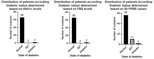
Table 1 provides the demographic characteristics and comorbid conditions of the study population. Majority of the population were female. Prevalence of other comorbidities is high in the study population with 65/68 having Hypertension. The mean HbA1c, FBS and 2h PPBS values are normal within the study population (Table 1). However, distribution of population based on the HbA1c criteria for IGT and diabetes
Duration of Hypertension28y2y One anti-hypertensivesYN
>One anti-hypertensivesNY CADYN
CAD Duration10y0
DyslipidaemiaNN
Duration of dyslipidaemia00
Drugs to treat dyslipidaemiaYN
UACR268 Data not available
Blood Pressure (SBP/DBP)150/80150/90
Total CholesterolData not available227
Low density lipoproteinData not available151
Triglycerides Data not available153
High density lipoproteinData not available46
Hemoglobin11.69.7
ESR3412
HbA1c8.16.8
Serum Creatinine1.10.9
Thyroid stimulating hormone levels1.11Data not available
Serum uric acid levelsData not availableData not available
indicated 2 individuals with HbA1c >6.5 and FBS >140. These 2 individuals (2.9%) were diabetic. Analysis based on the criteria of 2h PPBS helped identified additional 10 individuals in the state of pre-diabetes (Fig 1).
The characteristics and biochemical profile of the two asymptomatic diabetic individuals are provided in Table 2. Individual 1 was a 75-year-old female with HbA1c of 8.1 and Hypertension and CAD for over 28 and 10 years, respectively. Individual 2 is a 44-yearold male with HbA1c of 6.8 and Hypertension for 2 years.
The prevalence estimates using HbA1c identified two asymptomatic diabetic individuals. Based on the criteria for FBS the same two diabetic individuals were identified (Table 2).
With the criteria for 2h PPBS indicated that around 10 individuals with IGT and two having diabetes. A consolidated view based on this study findings indicate that the prevalence of asymptomatic diabetic individuals is about 3% and the prevalence of IGT among the study population was about 15%.
DISCUSSION
India is a large populous country with majority of the population being young adults. Environmental, lifestyle habits, diet and genetic factors have paved way for an alarming increase in Obesity, metabolism Related Disorders. The alarming prevalence of diabetes and its complications remain a threat and early diagnosis is essential to prevent complications including Cardiovascular Disorders and end organ failures at a later stage of life. A recent study conducted among adult population showed that about 45% of the individuals who are diabetic or IGT did not make conscientious effort to adopt lifestyle modifications. This was attributed to the adults mainly being asymptomatic8.
The prevalence of asymptomatic T2DM in children has been on a rise9. Recent recommendations by ADA
Fig 1 — Distribution of study population based on predicted diabetic status
Table 2 — Biochemical characteristics of the asymptomatic diabetic individuals
121, No 1, January 2023Journal
and the Canadian Diabetes Association calls for risk assessment strategies to predict and screen for asymptomatic early T2DM among children based on family history, DM during gestation, signs of insulin resistance or conditions associated with insulin resistance10
Our study indicates that about 2% of the individuals could be asymptomatic and up to 15% could be having IGT. Although the proportions are seemingly small, given the current epidemic of Diabetes, this could translate to a large absolute number in millions among the general population. The DECODE study had corroborated that asymptomatic individual with high PPBS had increased Cardiovascular risk11. The PPBS level is suggested to be much relevant especially with advancing age than the fasting glucose levels. In the long-run, early detection could help build awareness and adopt proper measures to prevent lifetime complications of serious illnesses.
Limitations :
This is a retrospective study with a very small sample size. Therefore, statistical analysis and validation is limited. Family history, Obesity related data are unavailable. Most similar studies used Oral Glucose Tolerance Test for detecting the prevalence of diabetes and pre-diabetes, here we have used FBS, 2hr PPBS and HbA1C due to the practical difficulties involved in conducting the test at the camp sites and the financial constraints. However, the study results can be extrapolated by studying this in a large population.
CONCLUSION
Prevalence of asymptomatic diabetes poses concerns in the healthcare managements of an individual. Therefore, regular screening for Diabetes status in adults is warranted for timely management and to prevent the severe complications of long-term Diabetes. Furthermore, building awareness among these individuals will aid in the adoption of proper lifestyle measures at an early stage itself.
Taking into consideration the high cost involved in various steps of screening, diagnosis, monitoring, and management, it is imperative that cost-effective measures of Diabetes care are necessarily implemented. Public awareness, as well as updating the Medical Fraternity on various developments in the
management of Diabetes, are required to combat the current Diabetes epidemic in India. Regular screening for Diabetes and associated non-communicable diseases in Urban areas should be considered by policy makers.
External Funding : None
Acknowledgements : Nil
Sources of Financial Support : Nil
Conflict of Interest : Nil
REFERENCES
1Davidson KW, Barry MJ, Mangione CM, Cabana M, Caughey AB, Davis EM, et al — Screening for Prediabetes and Type 2 Diabetes. JAMA 2021; 326: 736.
2Handelsman Y, Bloomgarden ZT, Grunberger G, Umpierrez G, Zimmerman RS, Bailey TS, et al — American Association Of Clinical Endocrinologists And American College Of Endocrinology -Clinical Practice Guidelines For Developing A Diabetes Mellitus Comprehensive Care Plan – 2015. Endocrine Practice 2015; 21: 1-87.
3Reshmi B, Unnikrishnan B, Rajwar E, Parsekar SS, Vijayamma R, Venkatesh BT — Impact of public-funded health insurances in India on health care utilisation and financial risk protection: a systematic review. BMJ Open 2021; 11: e050077.
4Pandey S, Sharma V— World diabetes day 2018: Battling the Emerging Epidemic of Diabetic Retinopathy. Indian Journal of Ophthalmology 2018; 66: 1652.
5Lim JU, Lee JH, Kim JS, Hwang YI, Kim TH, Lim SY, et al— Comparison of World Health Organization and Asia-Pacific body mass index classifications in COPD patients. International Journal of Chronic Obstructive Pulmonary Disease 2017; 2: 2465-75.
6WHO — World Health Organization. Appropriate body-mass index for Asian populations and its implications for policy and intervention strategies. Lancet 2004; 363: 157-63.
7Whelton PK, Carey RM, Aronow WS, Casey DE, Collins KJ, Himmelfarb CD, et al — 2017 ACC/AHA/AAPA/ABC/ACPM/ AGS/APhA/ASH/ASPC/NMA/PCNA Guideline for the Prevention, Detection, Evaluation, and Management of High Blood Pressure in Adults: A Report of the American College of Cardiology/American Heart Association Task Force on Clinical Practice Guidelines. Hypertension 2018; 71: e13–e115.
8Nagarathna R, Bali P, Anand A, Srivastava V, Patil S, Sharma G, et al — Prevalence of Diabetes and Its Determinants in the Young Adults Indian Population-Call for Yoga Intervention. Frontiers in Endocrinology 2020; 11: 507064.
9Wu WC, Li HY, Chiang CC, Sung FC, Wei JN, Chuang LM — Screening for diabetes in asymptomatic children: A simple and efficient method. Journal of the Formosan Medical Association 2020; 119: 974-81.
10American Diabetes Association. 2. Classification and diagnosis of diabetes: standards of medical care in diabetes-2018. Diabetes care 2018; 42(Supplement_1): S13–S28.
11Glucose Tolerance and Cardiovascular Mortality. Archives of Internal Medicine 2001; 161: 397.
Original Article
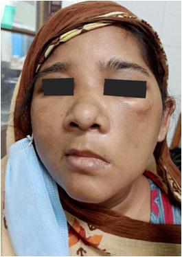
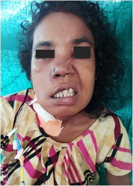
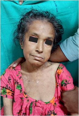
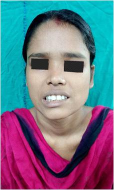
New Onset Facial Nerve Palsy : A Part of Post COVID Mucormycosis Disease Spectrum — A Descriptive Observational Study
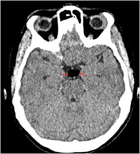
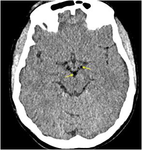
Introduction : COVID Associated Mucor (CAM) is a well known entity with defined symptomatology. Cranial Nerve Palsy involving II, III, IV, V, VI th Nerve is common. Facial Nerve involvement is an out of tract presentation. The study was aimed to find the incidence of Facial Nerve involvement in CAM and document their route of involvement.
Material and Method : Descriptive observational study was done in an Apex Centre for CAM in West Bengal between April, 2021 to January, 2022. CAM having Rhino-orbital-cerebral Mucormycosis (ROCM) and new onset Facial Palsy were considered. Participants were included following stipulated inclusion and exclusion criteria. Collected data was analysed.
Observations : Total 11 patients of new onset Facial Palsy in COVID-19-Associated ROCM were included. 81.8% had coexisting other Cranial Nerve involvement. Facial Palsy was one of the primary presentations in the patients of ROCM.
Discussion : CAM is angioinvasive and can cause concomitant hypoxic neural damage due to involvement of the vasa nervorum. Skull base involvement can be hypothesized to be the predominant route of Facial Nerve involvement. Facial palsy can be an important initial presentation of CAM.
Conclusion : Facial Nerve Palsy may be a part of the spectrum of disease presentation in CAM.
[J Indian Med Assoc 2023; 121(1): 28-32]
Key words :COVID-19, SARS-CoV-2, Mucormycosis, Facial Nerve, Cranial Nerve Paralysis.
COVID Associated Mucor (CAM) is a well-known entity with defined symptomatology; though its pathophysiology and route of spread is still not completely understood. Its symptoms are commonly rhinological, orbital or intracranial. Involvement of the Facial nerve by CAM is an out of the tract presentation. Rhino Orbito Cerebral Mucor (ROCM) develops by inhalation/ inoculation of spores of Mucorales on the inferior/ middle turbinate. From there it spreads to sinuses, pterygopalatine fossa, orbit by contagious spread or by angioinvasion. Cranial nerve palsy involving the II, III, IV, VI, Vth nerve is very common during orbital apex involvement (including superior orbital fissure) and/or cavernous sinus. Involvement of the Facial Nerve in its Lower Motor Neuron (LMN) segment is difficult to explain considering the abovementioned route of spread.
Department of ENT, IORL HNS, IPGME&R and SSKM Hospital, Kolkata 700020
1DLO, MS, Professor
2MS, DNB, Professor, Director & Head
3MS, DNB, Assistant Professor and Corresponding Author
4MS, Consultant ENT Surgeon, Calcutta Heart Clinic and Hospital, Salt Lake, Sector III, Kolkata 700106
5MS, Assistant Professor
6DLO, MS, Assistant Professor
7MS, Associate Professor
Received on : 23/05/2022
Accepted on : 10/06/2022
Editor's Comment :
Be Mucor minded.
Be cautious while dealing with Facial Nerve Palsy in the Post COVID Era.
Elicit past history of COVID-19 infection.
Nasal Endoscopy is a must.
Judiciously use systemic steroid medication.
This study aims to find the incidence of Facial Nerve involvement in CAM. To enlist the clinicoradiological features in patients of CAM with Facial Nerve Palsy. To document the probable route of spread of the disease in this subgroup of patients.
MATERIAL AND METHOD
A descriptive observational study was approved by the Institutional Review Board & carried out in the Institute of Otorhinolaryngology & Head Neck Surgery, Centre of Excellence, IPGME&R and SSKM Hospital, which is the Apex Tertiary Care Hub for treatment of COVID associated Mucormycosis in West Bengal, India between April, 2021 to January, 2022. Patients of all ages admitted in the Institute with CAM having ROCM and new onset Facial Nerve Palsy were considered as the study population. Working definition is as follows: “A case of Mucor was defined as laboratory identification of Mucorales by culture,
histopathology or polymerase chain reaction in a Patient with a clinical diagnosis of Invasive Mucormycosis. Cases were considered COVID-19 associated if the patient received a positive Reverse Transcription Polymerase Chain Reaction or Antigen test result for SARS-CoV-2 during the 60 days preceding the mucor diagnosis1.” Every patient is evaluated at presentation with detailed history, Clinical examination, ENT, Ophthalmic and Neurological examination to assess the extent of disease. Patients’ occupational history, personal habits, Socio-economic status (Modified B G Prasad Scale), prior treatment, vaccination status & COVID status were recorded at admission. Patients with pre-existing Facial Nerve palsy due to other known causes like cerebrovascular accident, non-covid intracranial, temporal bone, parotid pathology were excluded. Facial soft tissue involvement if mimicking Facial Palsy was also excluded. Facial Nerve function assessment was done in detail. It was first subdivided into upper and lower Motor Neuron type of deficit. LMN lesions were further graded by the House Brackmann Scoring System. Topo-diagnostic tests like Schirmer test, Stapedial reflex test and Taste sensation were assessed as far as practicable. Diagnostic Nasal Endoscopy (DNE) and Radiological assessment (Computed Tomography and Magnetic Resonance Scan of Head, Neck and Orbit) was done. Patients undergoing surgical debridement and those considered for conservative management were followed up at three months to check for change in the state of Facial Palsy. All interventions were done maintaining institutional and ICMR COVID protocol.
Collected data was tabulated and analysed using standard statistical software.
OBSERVATION
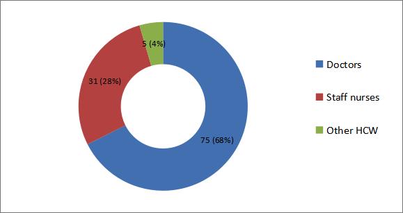
Of all our COVID associated Mucormycosis cases149 in a time frame of 9 months, 11 patients had new onset Facial Nerve Palsy who were included in the study (Figs 1-5). So, the incidence of new onset Facial Nerve palsy in CAM turns out to be 7.38 per 100 cases of ROCM. Maximum and minimum age of the study population was 68 and 25 years respectively with a median age of 50 years. 72.72% of the study population were female and 21.28% were male. As per religious background, 81.81% belonged to Hinduism and the rest were from Islam community. It was found that 28.6% of the people affected were from upper, lower middle and lower Socio-economic status respectively. Only 14.3% were from upper middle class Socioeconomic status.
Certain clinical details of these CAM patients with
Facial Nerve Paresis were as summarized. 54.54% of the study population was diabetic. 81.81% of the population had involvement of Cranial Nerves other than Facial Nerve. Nose and Para nasal sinus presentation were the second most common clinical presentation accounting for 72.72% respectively. Oral presentation and Orbital presentation were found in 54.54% of the population respectively. Disseminated Mucor was seen in 45.45% patients. 27.27% patients had features suggestive of Intracranial involvement. None of the patients had features suggestive of otological involvement.
Initial presenting symptoms in these patients was documented. 4 patients had Facial Palsy as the first symptom. 3 patients had headaches. Cheek numbness, eye congestion, double vision and diminished vision were respectively the first presenting feature amongst the rest 4 patients.
Radiological assessment in the form of Magnetic Resonance Imaging and Computed Tomography of the diseased site was done. Contrary to the clinical presentation, all of the patients (100%) had Nose and Para nasal sinus involvement. Orbit was involved in 72.72% of patients. Pterygopalatine fossa involvement and intracranial disease extension was found in 54.54% of the patients respectively. None of the patients had infra-temporal fossa and temporal bone involved by Mucor.
Route of disease spread was classified (based on clinico radiological findings) as Superior route if there were features of Nose and Skull Base involvement. Patients having Nose and Oral cavity involvement without involvement of the Skull base were considered as Mucor with Inferior Route of spread. It was found that 9 out of 11 patients of CAM with Facial Nerve Palsy had features suggestive of Superior route of Mucor spread.
All the patients had unilateral Facial Nerve involvement on the diseased side. None of the patients had any other coexisting pathology of the Brain, Parotid Gland or the ear. The involvement was classified as Upper and Lower Motor Neuron type of involvement. Only 18.2% (ie, 2 patients) had Upper Motor Neuron lesion of the Facial Nerve. The minimum duration of onset of Facial Palsy from the day of CAM symptomatology in these patients was day one seen in 4 patients. The maximum duration of onset of Facial Palsy was day 14 of the symptomatic disease. Average duration of onset was 5.63 days with Standard Deviation of 4.43 days.
Topo-diagnostic tests (which included test for taste sensation, Schirmer’s test and Stapedial reflex test)
which is usually done to localise the site of involvement of Facial Nerve was carried out. But this battery of tests could not be applied to most of the patients because of poor general health, pre-existing orbital involvement. Taste sensation was reported to be preserved in 5 out of 11 patients. Schirmer’s test was possible in 3 patients. Stapedial reflex was assessed in 4 patients. 3 had inconclusive findings and one had reduced reflex.
All the patients underwent surgical debridement and concomitant medical therapy with Liposomal Amphotericin B. 4 patients died during follow up. 54.5% of the patients had improvement in Facial Nerve function. 9.1% of the patient had no improvement in Facial Nerve motor function.
DISCUSSION
Our generation is still fighting strong with the Coronavirus pandemic caused in the last two years.
COVID-19 is primarily known to cause severe Acute Respiratory Distress in its severe form, but over the time we have found growing evidence of involvement of several other Organ System including Central (encephalitis, hypoxic encephalopathy, toxic encephalopathy, post infectious demyelination etc) and Peripheral (smell, taste, visual disturbances) Nervous Systems which can be due to either direct action of the virus on the Nervous System and/or indirect effect through activation on immune mediated mechanisms. With all waves several evidence-based treatment protocols were updated. At a certain point of time due to unchecked overuse of steroids, antibacterial & antifungal and possible mismanaged supply chain of medical oxygen, we happened to see the first ever fungal epidemic caused by Mucormycosis, mostly the rhino orbito cerebral form. Mucormycosis has a fulminant locally invasive course involving Nose, Palate, Sinuses, Orbital, Brain and Eventual death if not intervened urgently.
Facial Nerve Paralysis in a setting of COVID Associated Mucormycosis (CAM) is not a frequent scenario. At times Facial Nerve Paralysis have been found as the initial or only presentation of this dreaded disease which has been misdiagnosed at initial differential diagnoses. The diagnosis of the same encourages clinicians to dive deep to know the pathophysiology of such presentation.
ROCM according to progression and severity can start from the Nose, then progress to Paranasal Sinuses, Orbits and Brain either by direct local progression or through various foramina. The clinical and radiological assessment helped in assessing the route of disease spread. Disease invading the Nose and paranasal sinus mucosa can gain entry into the pterygopalatine fossa across the sphenopalatine foramen or direct invasion of the post wall of maxilla to further invade the contents of the fossa. Mucor can also cause Skull base Osteomyelitis specially the greater wing of sphenoid. Not all extensive Mucor cases present with Facial Nerve Paralysis. Probable neural and perineural invasion in this region has also to be taken into consideration to explain the disease spread. These are now well-established modes of
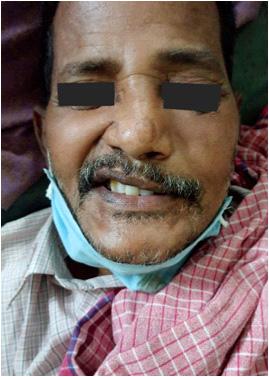
Fig 1Fig 2Fig 3
Fig 4Fig 5
Figs 1-5 — Photographs of patients suffering from Post COVID Mucormycosis with new onset Facial Nerve Palsy
No 1, January 2023Journal
progression but the Facial Nerve still lies miles away from the action area.
Mucormycosis causes inflammatory disease and causes contiguous tissue damage with special predilection to Nerves and blood vessels. Mucor is a known angioinvasive pathogen. Invasion into vasa nervosa can cause Nerve damage due to hypoxia, inflammation and oedema. Oedematous Facial Nerve in a non-yielding bony canal brings into picture a vicious cycle encouraging neural damage. Mucor migrating along Peripheral Nerves and perineural invasion can also account for neural pathology2
Demographic profiles of such patients have been reported in our study. Mean age of such patients was 50.18 years with 72.72% of them being female. In a similar study conducted by Rupa Mehta et al found the mean age to be 48 years. In another study conducted by Rajashri Mane et al with 4 such patients, the mean age was 50.75 years. All of the patients were male3, 4
Our study reports 54.54% of the study population to be affected by Diabetes. The entire study population (100%) was found to be diabetic in a study conducted by Rupa Mehta et al and Rajashri Mane et al3, 4. Dave, et al has reported 76% of patients to be diabetic in CAM5. In a large multicentric study in India evaluating 2826 CAM (ROCM) patients 78% of the patients were found to be diabetic6. Diabetes, more so if uncontrolled, is a major risk factor for this subset of patients.
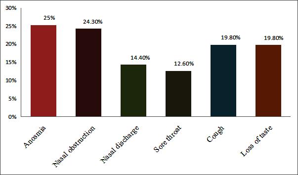
Facial asymmetry was the most common presenting symptom followed by headache. Other initial presentations were cheek numbness, eye congestion, double vision and diminished vision. Facial asymmetry as an initial presentation is difficult to explain. The disease would require a path to traverse before involving the Facial Nerve and in doing so involve various regions of the Skull base. So, a clinical presentation of involvement of the Skull base is likely to proceed Facial Nerve Palsy. On the other hand, an associated SARSCoV-2 induced Lower Motor Neuron Bell’s palsy-like phenomenon would justify such a presentation. Subsequent clinical presentations in this group of patients were mostly in the form of other Cranial nerve Palsy, followed by features of Nose and Paranasal sinus involvement. Orbital, Oral and other intracranial involvement features were also found. Reported commonest presenting feature of ROCM includes periorbital pain., proptosis, nasal discharge, diplopia, headache6,7.
All the patients in our study population had unilateral Facial Palsy. This can be explained by the unilateral onset of CAM. The disease can progress further to
have midline and bilateral invasion. Most common presentation being the lower motor neuron type of facial Palsy in our study which corroborated with other similar studies. Average duration of onset of Facial Palsy from the day of first symptom appearance was 5.63 days in our study with 4 patients even reporting it to be the first presentation. The first presentation can either be explained by concomitant Bell’s Palsy-like phenomenon or absence of overwhelming associated symptoms of ROCM which was ignored by the patients or other respiratory symptoms overshadowing initial ENT evaluation in light of COVID pandemic. Associated Diabetes in these patients further has deleterious effects because Diabetes causes Microangiopathy further enhancing the effect of vascular damage. So, a clinician must be very meticulous in evaluating Facial Palsy and enquire about the history of preceding COVID-19 infection. If there is a positive history, he/ she must try to exclude an underlying Mucor infection by clinical and radiological means. From the available data, it is evident that Facial Palsy in invasive Mucor presents usually in the first week. It can even be the first presentation. So judicious use of steroid medication should be done and it should only be started after excluding ROCM in such patients.
Topo-diagnostic tests like test for taste sensation, Schirmer’s test, impedance audiometry are an important battery of tests for Facial Nerve evaluation. But such tests are not always feasible in this group of patients. Moribund patient, patient with multiple neuropathies, coexisting orbital, cheek and infratemporal fossa involvement makes it difficult to interpret the result of the topo-diagnostic tests. Adverse effects of Amphotericin B further make this interpretation difficult. It mostly deals with localisation of intratemporal sites of constriction which can be decompressed surgically. The higher the lesion, the more dysfunction. Moreover, none of the case demonstrated any temporal bone pathology
Radiological assessment revealed Nose and Paranasal Sinuses to be involved by the invasive disease in all the patients (100%) included in our study. Orbit was involved in 72.72% patients although pterygopalatine fossa was involved in only 54.54%. So, it is evident that in addition to disease spreading from the pterygopalatine fossa to Orbit there is invasion of the medial and inferior orbital wall from the sinuses. In almost half of the study population intracranial extension of the disease was found radiologically. The clinical and radiological assessment helped in assessing the route of disease spread. Disease invading the Nose and Paranasal Sinus Mucosa gains
No 1, January 2023Journal
entry into the pterygopalatine fossa across the sphenopalatine foramen to further angio-invade the contents of the fossa. This can also cause Skull base (greater wing of sphenoid) Osteomyelitis. Probable neural and perineural invasion in this region has also to be taken into consideration to explain the disease spread. Retrograde spread of the disease along Vidian Nerve, Greater superficial Petrosal Nerve explains the Facial Nerve involvement. Involvement of the Infratemporal fossa followed by facial trunk invasion outside the stylomastoid foramen is feasible but cannot be accounted for in our study population. Intracranial disease can directly invade the Nerve along its intra cerebral and intracranial extra cerebral course. This explains the Superior route of disease spread causing Facial as well as other Cranial Nerve Palsy. Disease spread along the maxillary floor without involvement of the skull base is considered the Inferior route of spread. Premaxilla and cheek involvement in this route enables the disease to reach the branching Facial Nerve. This explains the Facial Nerve involvement in the inferior route. From our study it is evident that the Superior route is the predominant route of spread. In the four patients examined by Rajashri Mane, et al Superior route was found to be involved in 3 cases and only one patient had inferior route of disease spread. Rupa Mehta, et al in their series of 17 similar subset of patients had documented pterygopalatine and infra temporal fossa involvement in 10 cases and premaxilla involvement in 4 cases3,4. It has been commented by authors that pterygopalatine fossa acts as a reservoir for Mucor to invade into various spaces and structures. Infraorbital Nerve involvement has been found to be one of the commonest involved structures leading to cheek numbness. This Nerve acts as a channel for the disease to spread from the pterygopalatine fossa to premaxilla8.
Surgical debridement of the hypoxic and necrotic tissue along with Liposomal Amphotericin B therapy as the standard accepted protocol was followed in this subset of patients9. Oral or injectable steroid therapy was refuted as the diagnosis of invasive mucor disease was established and comorbidities like Diabetes Mellitus was considered. None of the patients were tried with Galvanic stimulation pre operatively. Minimal clinical recovery of Facial Palsy was found in all the cases. Complete recovery was not expected owing to the fact that late presentation and mostly pathology of Mucor were neural invasion and tissue necrosis dominated over oedema and inflammation which cannot be well reversed by decompression, debridement and
drugs (steroids, anti-inflammatory, antifungals and antibiotics)
CONCLUSION
Facial Nerve Palsy may be a part of the spectrum of disease presentation in CAM. Middle aged, diabetic population is most vulnerable. Onset of palsy might be found within a week of the Mucor invasion. It might even be the first presentation. Treating Clinicians must be meticulous in obtaining history of preceding COVID19 and exclude associated Mucormycosis while treating a case of Facial Palsy. Administration of steroid medication should be done judiciously. Prompt surgical debridement along with antifungal medication remains the mainstay of treatment.
Financial Support : None
Ethical standards : The author asserts that all the procedures contributing to this work comply with the ethical standards of India and international guidelines as per Helsinki Declaration of 1975, as revised in 2008.
REFERENCES
1www.cdc.gov/mmwr/volumes/70/wr/mm7050a3.htm assessed on 28-12-2021
2Spellberg B, Edwards Jr J, Ibrahim A — Novel perspectives on mucormycosis: pathophysiology, presentation, and management. Clinical Microbiology Reviews 2005; 18(3): 556-69.
3Mehta R, Nagarkar NM, Ksbs KS, Ty SS, Arora RD, Aggarwal A — Facial Nerve Palsy in COVID-19-Associated Mucormycosis Patients: A Case Series. Cureus 2021; 13(11)
4Mane R, Patil B, Mohite A, Mohanty R — Facial Nerve Palsy: An Unusual Presentation in Patients with Rhino Cerebral Mucormycosis. Indian Journal of Otolaryngology and Head & Neck Surgery 2019; 71(3): 2110-3.
5Dave TV, Nair AG, Hegde R, Vithalani N, Desai S, Adulkar N, et al — Clinical presentations, management and outcomes of rhino-orbital-cerebral mucormycosis (ROCM) following COVID-19: a multi-centric study. Ophthalmic Plastic and Reconstructive Surgery 2021; 37(5): 488.
6Sen M, Honavar SG, Bansal R, Sengupta S, Rao R, Kim U, et al — Epidemiology, clinical profile, management, and outcome of COVID-19-associated rhino-orbital-cerebral mucormycosis in 2826 patients in India–Collaborative OPAI-IJO Study on Mucormycosis in COVID-19 (COSMIC), Report 1. Indian Journal of Ophthalmology 2021; 69(7): 1670-92.
7Pakdel F, Ahmadikia K, Salehi M, Tabari A, Jafari R, Mehrparvar G, et al — Mucormycosis in patients with COVID 19: A cross sectional descriptive multicentre study from Iran. Mycoses 2021; 64(10): 1238-52.
8Pal P, Singh B, Singla S, Kaur R — Mucormycosis in COVID19 pandemic and its neurovascular spread. European Archives of Oto-Rhino-Laryngology 2021; 12: 1-8.
9Das S K, Barman D, Shrivastava S R, Adhikari S, Biswas PS — Fungal Epidemic by Mucor, an Unprecedented Threat in Medical History: Our Experience from ENT Perspective. In: Pal J, Chatterjee N, Saha B. Mucormycosis 2022. p. 66-68.
Original Article
A Cross Sectional Study of Prevalence of COVID-19 and ENT Manifestations among Health Care Workers in Our Centre
Smitha S G1, Nikitha Pillai2, Jedhin Kunchu Raveendran3, Bindya Nayak3
Background : Health Care Workers are among the frontline COVID warriors who have been fighting all odds in serving their patients at the cost of their lives. Bangalore has been one among the hot cities in news during this pandemic and our centre, being one of the busiest working hospitals right in the middle of the city, we hereby throw limelight to the Health Care Workers of our centre who have been fighting over COVID-19 with all might and courage. This study was done to know the prevalence of COVID-19 and its ENT manifestations among Health Care Workers in our centre.
Materials and Methods : A cross sectional study was done in KIMS Hospital, Bangalore, targetting all Health Care Workers of our centre, divided into 3 groups. Prevalence of ENT manifestations was studied.
Results : In our study, majority who tested positive for COVID-19 were Doctors (67.6%) followed by staff nurse (27.9%) and auxiliary HCW (4.5%). One fourth of the study subjects had anosmia as the most common ENT manifestation followed by nasal obstruction (24.3%), cough and loss of taste (19.8% each).
Conclusion : With inadequate precautions being taken with the mutating virus in air, causing a surge in cases, the health care workers are the most vulnerable group to acquire the deadly infection, during both the waves of the pandemic. We hereby, stress on this, with the help of our study, done during the first wave, targeting our Health Care Workers.
Key words :COVID-19, ENT presentations, Health Care Workers.
As quoted by Dr Jeckros Adhanom, WHO director General, COVID-19 pandemic has reminded all of us the role Health Care Workers play to relieve suffering and save lives. No country, hospital or clinic can keep its patients safe unless it keeps its Health Care Workers safe”
This pandemic has highlighted the risk of burnout among Health Care Providers and the extent to which protecting health workers is key to ensuring a functioning Health and Economic system.
As of April, 2020, WHO reported a daily monitoring report of about 22,073 Health Care Workerss across 52 countries infected by COVID-19 of which the number was estimated to be probably under represented. Preliminary results have suggested health care workers to have got infected both in their workplace where the risk of exposure is maximum through direct and indirect contacts and also in Community.
Department of ENT, Kempegowda Institute of Medical Sciences (KIMS), Bangalore, Karnataka 560070
1MBBS, MS (ENT), Associate Professor
2MBBS, MS (ENT), Senior Resident and Corresponding Author
3MBBS, Junior Resident
Received on : 02/08/2021
Accepted on : 04/03/2022
[J Indian Med Assoc 2023; 121(1): 33-6]
Editor's Comment :
Health Care Workers, being one of the most vulnerable group in the community, are the ones to get affected by COVID-19 the most.
Commitment and dedication towards the welfare of the patients defines the negligence of the Health Care Workers towards their own health. We here have made an attempt to bring light to this and stress on the importance of screening for early diagnosis and treatment of COVID-19.
MATERIALS AND METHODS
This is a cross sectional study done in Kempegowda Institute of Medical Sciences, Bangalore, by Department of ENT, to study the prevalence of ENT manifestations of COVID-19 in Health Care Workers of our center. The study was started in May 2020 targetting all health care workers of KIMS who were screened and had turned positive during this pandemic and willingly gave consent for our study.
The targeted groups were divided into 3 groups: Group 1 included doctors comprising of House Surgeons, Postgraduates and all teaching staff who turned to positive for COVID, Group 2 had nursing staff and Group 3 included auxilary staff comprising of
No 1, January 2023Journal
Administrative Staff, House Keepers, Porters, Ward Boys and Attenders.
Statistical analysis was done and prevalence of ENT manifestations of COVID-19 among Health Care Workers in each group was assessed at the end of October 2020. Risk of exposure in each group was also noted.
DISCUSSION
The impact caused by the deadly Corona Virus has shook the world and has left many countries and its people and various sectors standstill. Health care workers have been one among the most high risk and vulnerable communities to have acquired this infection. They have been in the frontline of this fight from day 1 of its onset and continues to be, inevitably, during the course of which many have even succumbed to the virus.
The incubation period of COVID-19 virus, since the exposure to SARS-CoV-2 is believed to reach 14 days, but majority of cases develop symptoms after 4-5 days which can range between 2 and 7 days after being infected.
The most common manifestations of COVID-19 include fever, malaise, difficulty in breathing, anorexia, fatigue, diarrhoea. Patients may be diagnosed early with the help of various ENT symptoms like anosmia, aguesia, sore throat, sudden sensorineural hearing loss, tinnitus and headache1.
According to CDC guidelines2, positive patients are divided into 3 categories
•Category A includes all asymptomatic patients who maintain saturation of over 97% in room air,
•Category B includes all patients who maintain saturation above 90% but less than 95%, with mild tachypnoea and hypotension.
•Category C includes all patients who do not maintain saturation above 90% at room air, having severe tachypnoea and hypotension.
In our study, done during a period of 6 months from May, 2020 to October,2020, targetting 1062 HCWS in our centre, 111 HCWS were found to have been detected positive. Among the 111 HCWs, 75 were doctors, 31 were nurse and 5 were the auxiliary staff. The prevalence of COVID-19 during the period among doctors, nurse and auxiliary staff was 16.7%, 5.4%, 13.5% respectively.
The most common age group of infected Health Care Workers in our centre was found to be between 25 to 29 years. This age group had mostly interns and Postgraduates bearing COVID duties.
In our study, most of the HCWS, 59 (53.2%), turned
out to be positive during their non COVID hospital duties in OPDs and OTs. 50 (45%) of them acquired the infection during their COVID duties and remaining 2 (1.8%) were found to have community acquired infection. The risks involved in non COVID hospital duties to Health Care Workers were attributed to inadequate protective measures taken by the health care workers in OPDs, patients coming to OPDs and for emergency OTs with unknown COVID-19 status, maximum contact time with patients while taking history and performing detailed clinical examination and procedures. Our speciality deals with examination of nose and throat which are the most risky systems involved in causing spread of the infection.
Inadequate protective and precautionary measures taken by health care workers were assessed and found to be due to many possible inevitable reasons like
Table 1 — Proportion of HCWs tested positive during study period
Table 2 — Distribution of study subjects according to age (n=111)
Table 3 — Distribution of study subjects according to exposure and type of duty (n=111)
Table 4 — The study showed that, majority of the study population belonged to blood group ‘O positive’ (41.4%) followed by ‘B positive’ (29.7%)
scarcity of and provision of poor quality Personal Protective Equipment (PPE) kits to compensate for the shortage faced in the initial stages of the pandemic and improper use of the same. The sudden surge in cases and many of our Health Care Workers getting affected at the same time led to long working hours and very frequent and tiring COVID duties of the other
HCWs. The saturation and dehydration with improper donning and doffing of the PPE kits also could be the contributing factors to our Health Care Workers getting affected.
2 of our Health Care Workers were tested positive from family members who would have got exposed from their work place or social gatherings or from inevitable sources like shopping areas, pharmacies etc.
In our study, the most common ENT manifestation was anosmia (25.2%) followed by nasal obstruction (24.3%) and cough and loss of taste (19.8% each). Carlos M, et al3 in his study showed 81.9% of affected patients to have developed olfactory dysfunction. Study done by Gane, et al4 concluded sudden anosmia to be highly suspicious of COVID-19. Many asymptomatic patients were found to not develop any further symptoms except isolated sudden anosmia as brought in light by Gane, et al. In a study conducted by Mohammad Waheed El Anwar, et al5 in COVID-19 positive patients, the most common ENT manifestation was sore throat (13.3%) followed by headache (10.7%) and pharyngeal erythema (5.3%) while study done by Priyanka Chaurasia, et al6 showed sore throat to be the most common symptom. Corona virus has great affinity towards ACE 2 receptors which is abundant in human tissues including oral cavity, pharynx and digestive tract. Most of the symptoms of COVID-19 are attributed to this concept.
In our study, most of our affected HCWs were O positive (41.4%) followed by B positive (29.7%) and A positive (22.6%). F Powrali, et al7, in his study on relationship between Blood Group and risk of infection and death in COVID-19 and Blood Group found that patients with 'O' BLood Group were more susceptible for the infection and A blood group was least susceptible. This, he explained to the fact that ACE receptors which is the binding site for the virus was in least concentration in patients with 'O' blood group and in highest concentration in A blood group.
80 of our affected HCWs (72.1%) recovered from ENT symptoms in less than 1 week while 19 of them showed recovery in 1-2weeks. 12 of them took more than 2 weeks to recover.
In our study, 99 of our affected HCWs belonged to category A. The intense fear factor and home isolation not being allowed during the initial phase of the pandemic had almost all our category A patients, including the affected health care workers, admitted in our hospital. 12 of them belonged to Category B wherein oxygen administration was required.
Fig 1 — Healthcare workers detected positive for COVID-19
Fig 2 — Age distribution of health care workers affected ny COVID-19
Fig 3 — ENT manifestations of COVID-19 among affected healthcare workers
Vol 121, No 1, January 2023Journal of the Indian Medical Association
CONCLUSION
COVID-19 pandemic is not over yet. Everyday, new cases are emerging in thousands and lakhs and now new strains of Corona Virus have also emerged causing new outbreaks as well. Adequate protective measures in COVID ward duties, OPDs and during surgical procedures should be followed. Patients being responsible enough to take necessary protective measures and not masking their symptoms with good cooperation plays a major contribution in combating the spread.
Conflict of interest : There were no conflicts of interest
Ethical committee : Clearance was taken
REFERENCES
1Lovato A, de Filippis C — Clinical presentation of COVID- 19: a systematic review focusing on upper airway symptoms. Ear Nose Throat J 2020; 99(9): 569-76.
2Centers for disease control and prevention. 2020 [cited 13 January 2021]. Available from: http s://www.cdc.gov/ coronavirus/2019-ncov/hcp/clinical-guidance-managementpatients.html
3Lechien JR — Eur Arch Otorhinolaryngol. 2020 Apr 6 111 :Olfactoryand gustatory dysfunctions as a clinical presentation of mild-to-moderate forms of the coronavirus disease (COVID-19): a multicenter European study. doi: 10.1007/s00405-020-05965-1 .
4Gane SB, Kelly C, Hopkins C — Isolated sudden onset anosmia in COVID-19 infection, 2020. A novel syndrome? Rhinology.https://doi.org/10.4193/Rhin20.114
5El-Anwar MW, Elzayat S, Fouad YA — ENT manifestation in COVID-19 patients. AurisNasus Larynx 2020; 47(4): 559564.
6Chaurasia P, Kuchhal V, Ahmad S, Rawat P — ENT manifestations in Covid-19 positive patients. Int J Health Clin Res 2020; 3(10): 187-91
7Pourali F, Afshari M, Alizadeh-Navaei R, Javidnia J, Moosazadeh M, Hessami A — New Microbes New Infect. 2020 Sep; 37: 100743. Published online 2020 Aug 11. doi: https://doi.org/10.1016/ j.nmni.2020.100743.PMCID.PMC7418722
Ifyouwanttosendyourqueriesandreceivethe responseonanysubjectfromJIMA,pleaseuse the E-mail or Mobile facility.
Know Your JIMA
Website:https://onlinejima.com
For Reception: Mobile : +919477493033
For Editorial:jima1930@rediffmail.com
Mobile : +919477493027
For Circulation: jimacir@gmail.com
Mobile : +919477493037
For Marketing: jimamkt@gmail.com
Mobile : +919477493036
For Accounts: journalaccts@gmail.com
Mobile : +919432211112 For Guideline:https://onlinejima.com
Original Article
Knowledge about COVID-19 Infection and Stress Levels among Ambulance Drivers during COVID-19 Pandemic — A Cross Sectional Study
Introduction : The medical personnel especially have to deal with both the Biological and Psychological consequences originating from the influence of virus. Emergency Care Units equipped with ambulance service always should be at the fore front to face all emerging untoward incidents. Ambulance Drivers often disregard their physical health and mental stress in order to fulfil the duties entrusted on them.
Methodology : Cross-sectional questionnaire based on-line survey was conducted. Ambulance drivers were North and South India were taken as study subjects. Knowledge based questions and mental stress were assessed using Perceived Stress level Scale (PSS). The questionnaires were translated toTamil, Kannada, Telugu, Malayalam and Hindi languages. Chi-square test was done to find the association between variables.
Results : Among the 101 respondents, 84.2% were having good knowledge about the pandemic. Statistically significant association between number of patients transported per day and the knowledge levels of Ambulance drivers was found (p =0.048)(Chi-square value=15.65). Drivers in Government sector were having more knowledge compared to private sector (p =0.038)(Chi square value=6.53). The perceived mental stress was found to increase with a greater number of patients being transported per day, which was statistically significant (p =0.001)(Chi-square value= 30.42).
Conclusion : Knowledge regarding COVID-19 virus was adequate among the Ambulance Drivers. Knowledge was found to be more among drivers who work in Government medical establishments. Mental stress was more among drivers who transported more than 5 patients per day. [J Indian Med Assoc 2023; 121(1): 37-41]
Key words :COVID-19, Awareness, Stress, Psychological, Ambulance Drivers.
Ever since the outbreak of Corona Virus has made its footprint on the headlines, which has its origin from Wuhan City in China, the world has not been the same. Quarantine and isolation has become the norm across continents. The symptoms of infected with Corona virus mimics that of viral Pneumonia1 The contagious nature of Corona Virus has made nations to enforce lockdown and has made humans to stay
1MD, DNB, Assistant Professor, Department of Emergency Medicine, Amrita School of Medicine, Amrita Vishwa Vidyapeetham, Kochi, Kerala 682 041
2MDS, Additional Professor, Department of Public Health Dentistry. Amrita School of Dentistry, Amrita Vishwa Vidyapeetham, Kochi, Kerala 682 041 and Corresponding Author
3MBBS Student, Amrita School of Medicine, Amrita Vishwa Vidyapeetham, Kochi, Kerala 682 041
4MD, DNB, MNAMS, Principal and Professor, Department of Rheumatology, Amrita School of Medicine, Amrita Vishwa Vidyapeetham, Kochi, Kerala 682 041
5 MSc, Assistant Professor(Biostatistics),Department of Community Medicine, Ramaiah Medical College, Bangalore
6MD, Assistant Professor,Department of Rheumatology, Amrita School of Medicine, Amrita Vishwa Vidyapeetham, Kochi, Kerala 682 041
7BDS student, Amrita School of Dentistry, Amrita Vishwa Vidyapeetham, Kochi, Kerala 682 041
Received on : 20/08/2021
Accepted on : 08/09/2022
Editor's Comment :
People seeking medical care is considered as vulnerable to acquire infections through diverse modes.
Awareness about COVID-19 among ambulance drivers is of utmost importance as they come into close interaction with highly vulnerable patients.
Lapse in adhering to infection control protocols is often linked to lack of awareness about the mode of spread and preventive measures.
Creating health care awareness to ambulance drivers who attend to highly compromised patients can bring down the mortality and morbidity rates among people seeking health care.
indoors all over the World to minimise exposure2 The COVID-19 pandemic has threatened the entire population of the planet raising serious concerns about health with its alarming rate of spread. It has led to Public Health crisis worldwide which was seen never before3 The nations around the world, rely heavily on its medical and support staff to contain the ill-effects of COVID-19 to some extent. The medical personnel especially have to deal with both the Biological and Psychological consequences originating from the influence of virus. Like any other humans there are no guaranteed immunity to medical personnel which adds to their woes4 Emergency Care Units equipped with
121, No 1, January 2023Journal
Ambulance service always should be at the fore front to face all emerging untoward incidents. Ambulance Drivers often disregard their physical health and mental stress in order to fulfil the duties entrusted on them. They often have to circumvent the lack of adequate supply of personal protective equipment, fatigue and psychological stress4
Often, the Ambulance Drivers and personals associated have to deal with vulnerable population and this increases their chance of them being contracted with the Scary Virus. A minor mistake from their part can draw the attention of the authorities and put their reputation at stake. This expectation that the community have for them puts them at high mental stress. The virus had its influence on their financial and affected their psyche5. This puts the Ambulance Drivers and other emergency Health Care Staff at more risk compared to general population to be victims of server mental stress. The fear of getting infected with the virus and the social stigma they might have to encounter can lead to severe stress. This condition is exaggerated by lack of adequate medical supplies, other essentials and fear of being discriminated against6,7
Witnessing loss of lives due to Corona and related morbidities can disturb their mental condition leading to severe stress, which coupled with prolonged duty hours can have an adverse impact on their mental and physical health8. Assessment of awareness regarding the COVID-19 among a targeted Community can be of great use in knowing their perspective towards the Virus infection. Accurate information about the Virus, its mode of transmission and preventive strategies is very much essential among these front line Health Care Workers9.
Knowing the status of their knowledge and the resulting psychological stress caused due to the virus spread can help take adequate measures to quell the anxiety among these personnel and can help them maintain the necessary mental well-being.
Objective : To assess the knowledge, attitude and practice among Ambulance Drivers about COVID-19 infection and their stress levels.
MATERIALS AND METHODS
A cross sectional pilot study was conducted among the Ambulance Drivers on duty during COVID-19 pandemic across India using a pre-validated questionnaire consisting of Socio-demographic questions (Age, location, education, Govt/private hospital, daily patient transfer). The study was conducted using a self-administered questionnaire having 20 questions divided into two sections- first section had 10 question based on health awareness, knowledge and practice of Ambulance Drivers during
ongoing COVID-19 across India and the 2nd section includes 10 questions to assess the mental stress level based on Perceived Stress level Scale (PSS) using score ranging from 0 to 4 per question. The response options for the stress levels were :0-never,1almost never, 2–sometimes, 3- fairly often, 4-very often. Individual total scores on the PSS can range from 040 with higher scores indicating higher perceived stress. Scores ranging from 0-13 were considered low stress, from 14-26 were considered moderate stress and from 27-40 were considered high perceived stress. For health awareness questionnaire, each appropriate response was given score 1. If the score is more than 7 – it was considered very good knowledge, 5-7-good knowledge and less than 5-poor knowledge. An online form containing the questionnaire were made and send via social-media platforms. Data will be tabulated and statistical analysis.
The questionnaire was subjected to content validation by four experts and reliability was checked using Kappa statistics which was found to be 0.81. The questionnaire was also translated to Tamil, Kannada, Telugu, Malayalam and Hindi language. The questions were forward translated, back translated and pre-tested before being administered to the subjects. They were administered to the subjects through online forms. An on-line informed consent form was also attached prior to the questionnaire section. Since no prior evidence of knowledge among Ambulance Drivers in India could be found, this study was conducted as a pilot survey.
RESULTS
A total of 101 responses from all over India were obtained based on convenient sampling. The age group of the study population ranged from 25 to 59 years of age. There were 72 Ambulance Drivers from North India and 29 from south India. Among the subjects, 82.2 % had received formal training on COVID-19 infection control and 53.5% of them were working in private institutions. More than one third of the subjects (34.7%) transported 5 to 10 patients per day. Regarding educational level, 12th standard pass were 59.4% and 40.6% were 10 th standard pass. The knowledge regarding need for sanitizing the Ambulance vehicle after transporting each patient was 81.2%, most of them knew about use of appropriate hand sanitizer (84.2%), 75.2% knew that N95 mask contains the spread of virus and 76.3% knew about source of Corona Virus. The least knowledge among the subjects were about social distancing norms, which was found to be only 25.7% (Table 1).
One of the significant findings of the present study
121, No 1, January 2023Journal
was that with increasing number of patients transported per day, the knowledge levels was found to be increasing among the Ambulance Drivers. Knowledge about COVID Virus was good among the drivers who transported between 5-10 patients per day and those between 1120 patients per day. The association between these categories and other categories of less than 5 patients per day and more than 30 patients per day was found to be statistically significant (Tables 2&3).
Another significant finding of the present study was that the knowledge among Ambulance Drivers who worked in Government Institutions were high and it was found to be statistically significant (Tables 4&5)
Stress levels of Ambulance Drivers was found to increase with the number of patients transported per day per driver. Moderate level of stress was experienced by drivers who transported five to ten patients per day (54.3%) and among 11 to 20 patients per day (70%). The association between stress levels experienced by various categories of Ambulance Drivers according to number of patients transported by day was found to be statistically significant, with 70% of drivers transporting between 11 to 20 patients and 57.2% of drivers in five to ten category experiencing at least moderate level of psychological stress.
Psychological stress levels among Ambulance Drivers.
DISCUSSION
As observed in the study conducted at Saudi, in the present study too age and gender were not significantly associated with awareness about COVID Virus. The awareness of the General Community was satisfactory in the study. However, the sample population in the Saudi study had more than half of the respondents holding a bachelor or higher degree of education10. However, the results from the general population with a sample size of 8591 showed that education is significantly positively associated with knowledge about COVID-19 virus. In the present study, such an association were not found probably due to reason that the subjects did not vary much among their level of education status11 Similar results were
Table 1 — Correct and incorrect responses to knowledge questions*
found in Nigeria, where good knowledge about COVID virus and its mode of transmission was found among the higher Social class of population. Education level was found to be a major factor in determining the overall awareness about COVID. These studies show that in general population, Education level plays a major part in the overall awareness of the population about the COVID virus and therefore higher chances of curtailing the spread of virus in the country12. This could also be the reason why Government employed drivers were having more knowledge than private employees as Government recruited drivers usually have a minimum standard of education in-order to get employed.
Adequate knowledge and a positive attitude towards preventing the spread of COVID virus was found in a study conducted among Health Care Workers in Nepal. The survey also revealed that with proper awareness programmes, the awareness among the Health Care workers could be further enhanced13. This was very much similar to that of present study, where moderate to high knowledge about COVID virus was obtained for various queries regarding COVID virus.
2 — Knowledge about COVID-19 based on patients transported per day by subjects
Patients transported per day < 55 –1011-2021-30 More than 30Total
Health Poor 7(17.5%)3(8.6%)5(25.0%)1(20.0%)016(15.8%)
Knowledge Good 30(75%)18(51.4%)10(50%)4(80%)1(100%)63(62.4%)
Table 3 — Association between knowledge about COVID-19 and number of patients transported by the subjects
Patients transported per dayPearsonP -value < 55-1011-2021-30 More Chi-square than 30 value
Health Poor 7(17.5%)3(8.6%)5(25.0%)1(20.0%)015.650.048
Knowledge Good 30(75%)18(51.4%)10(50%)4(80%)1(100%)
Very Good 3(7.5%)14(40%)5(25%)00
Total40(100%)35(100%)20(100%)5(100%)1(100%)
Table
Table 4 — Place of work and Knowledge about COVID-19 infection Place of work
Health Knowledge :
Government PrivateTotal
Poor3 (6.4%)13 (24.1%)16 (15.8%)
Good31 (66.0%)32 (59.3%)63 (62.4%)
Very Good 13(27.7%)9 (16.7%)22 (21.8%)
Total47 (100%)54 (100%)101 (100%)
Table 5 — Association between place of work and Knowledge about infection Government Private
Health Knowledge :
Poor3 (6.4%)13 (24.1%) 6.530.038
Good31 (66.0%)32 (59.3%)
Very Good 13(27.7%)9 (16.7%)
Total47 (100%)54 (100%)
Ambulance drivers are one of the first to come into contact with a suspected or confirmed case of Covid virus infected patients. The fact that they often have to deal with the patient combined with fear of getting infected can severely affect their mental state. Lack of adequate support, lack of appropriate Personal Protective Equipment (PPE) and long working hours with increasing number of patients or the distance travelled and the expectation that is bestowed upon them by the community at large can all lead to increased mental stress among frontline health care workers like ambulance drivers14-17. Even though mortality rate is low due to Covid virus, high rate of transmission and increasing number of mortalities can cause more mental stress among health care workers18
The fear and anxiety of getting infected with the new infectious virus which the general population considers as fatal and the sudden reversal of role from a health care worker to a patient who needs care can
cause anxiety, fear of being stigmatized and cause mental trauma to the Health Care Workers19. The Ambulance Drivers being at the fore front of any emergency medical situation too are very much prone to such mental stress.
Similar observations were made among Health Care Workers in Nepal where it was found that 38% of the health care workers who were in duty for COVID patient care were suffering from Psychological distress in the form of anxiety or depression. The authors of the particular study were of the opinion that inadequate supply of PPE and probability of getting infected with COVID were the main reasons for the mental stress20.
Similar findings were reported where relationship between Physiological anxiety can lead to depression or even agonistic behaviours could be developed. The perceived stress caused by fear of being infected by an agent whose cure is yet to be found can cause severe stress among frontline Paramedics. Psychological stress resulting from depression, anxiety and emotional distress can lead to agonistic behaviour in individuals. In addition to this, the lack of effective PPEs and its availability can further aggravate mental stress among frontline Health Care Workers21-24. It is expected that as the number of patients transported increases, the stress levels too tend to increase as revealed from the results of this study.
Uncertainty at the work site, lack of autonomy at the work, organizational politics are the other factors that lead to increased mental stress to Health Care Workers. Ambulance Drivers especially has to go to unfamiliar territory or are redeployed to face any emergency situations. Pre-existing mental illness, comorbidities can worsen their morale. Timely interventions form the organisation or counselling can go a long way in reducing or mitigating stress among such employees25.
Table 6 — Stress levels of subjects based on number of patients transported per day
Patients transported per day < 55 – 1011-2021-30 More than 30
Instances of mental stress among Health Care Workers have known to occur during the outbreak of other epidemics previously too like in the case of SARS outbreak in 2003.Online counselling, dedicated team of mental health care personnel and group activities to reduce the mental stress among Health Care Workers are some of the suggestions put
121, No 1, January 2023Journal
forward to assist such workers including Ambulance Drivers to cope with mental stress during COVID-19 pandemic25
The drawback of this study is that since it was conducted as a pilot survey, the results cannot be generalised. More studies among frontline Health Care Workers such as Ambulance Drivers needs to be conducted.
The strength of the present study is that it involved Ambulance Drivers from several North Indian and South Indian states, in which the questions were translated to local languages and an overall first-hand information about the Ambulance Drivers from different states of India could be gathered.
Since the questionnaire was online based, there could be memory or recall bias though the authors have requested for authentic information from the study participants. This study had representative participants from North and South India only and participants from other zones could not be reached. This is another limitation of this study.
CONCLUSION
Knowledge regarding COVID-19 Virus was adequate among the Ambulance Drivers. Knowledge was found to be more among drivers who work in Government medical establishments. Mental stress was more among drivers who transported more than 5 patients per day. Periodic training programmes about prevention and control of COVID-19 infection and Psychological counselling are recommended for Ambulance Drivers to carry out their duties more efficiently and cope up with daily stress in a more positive mental frame.
REFERENCES
1Malik YS, Kumar N, Sircar S, Kaushik R, Bhat S, Dhama K, et al — Coronavirus Disease Pandemic (COVID-19): Challenges and a Global Perspective Pathogens 2020; 9(7): 519.
2Dubey S, Biswas P, Ghosh R, Chatterjee S, Dubey MJ, Chatterjee S, et al — Psychosocial impact of COVID-19. Diabetes Metab Syndr 2020; 14(5): 779-88.
3He Z,Ren L,Yang J,Guo L,Feng L,Ma C, et al — Seroprevalence and humoral immune durability of anti-SARSCoV-2 antibodies in Wuhan, China: a longitudinal, populationlevel, cross-sectional study. Lancet 2021; 397: 1075-84.
4Spoorthy MS, Pratapa SK, Mahant S — Mental health problems faced by healthcare workers due to COVID 19 pandemic- A review. Asian J Psychiatr 2020; 51: 102-19.
5Shaukat N, Ali DM, Razzak J — Physical and mental health impacts of COVID-19 on healthcare workers: a scoping review. Int J Emerg Med 2020; 13(1): 40.
6Si M, Su X, Jiang Y — Psychological impact of COVID-19 on medical care workers in China. Infect Dis Poverty 2020; 9: 113.
7Munawar K, Choudhry FR. Exploring stress coping strategies of frontline emergency health workers dealing COVID 19- A qualitative inquiry. Am J Infect Control 2020; 7: S01966553(20)30638-6.
8Temsah MH, Alhuzaimi AN, Alamro N, Alrabiaah A, Al-Sohime F, Alhasan K, et al — Knowledge attitude and practices of
health care workers during early COVID-19 pandemic in a main academic tertiary care centre in Saudi Arabia. Epidemiol Infect 2020; 148: e203.
9Puspitasari IM, Yusuf L, Sinuraya RK, Abdulah R, Koyama H — Knowledge, attitude and practice during the Covid-19 Pandemic: A review 2020: 13: 727-33.
10Tripathi R, Alqahtani SS, Albarraq AA, Meraya AM, Tripathi P, Banji D, et al — Awareness and Preparedness of COVID-19 Outbreak Among Healthcare Workers and Other Residents of South-West Saudi Arabia: A Cross-Sectional Survey. Front. Public Health 2020; 8: 482.
11Erfani A, Shahriarirad R, Ranjbar K, Mirahmadizadeh A, Moghadami M — Knowledge, attitude and practice toward the novel coronavirus (COVID-19) outbreak: a populationbased survey in Iran. Bull World Health Organ 2020; 30.
12Reuben RC, Danladi MMA, Saleh DA, Ejembi PA — Knowledge, Attitudes and Practices Towards COVID-19: An Epidemiological Survey in North Central Nigeria. Journal of Community Health https://doi.org/10.1007/s10900-02000881-1 accessed 21st November 2020.
13Basnet S, Dahal S, Tamrakar D, Shakya YR, Jacobson C, Shrestha J,etal—Knowledge, Attitude, and Practices Related to COVID-19 among Healthcare Personnel in a Tertiary Care Hospital in Nepal: A Cross-Sectional Survey. Kathmandu Univ Med J 2020; 70(2): 21-8.
14Cai H, Tu B, Ma J, Chen L, Fu L, Jiang Y, Zhuang Q — Psychological impact and coping strategies of frontline medical staff in Hunan between January and March 2020 during the outbreak of coronavirus disease 2019 (COVID19) in Hubei, China. Med Sci Monit 2020; 26: e924171.
15Tam CWC, Pang EPF, Lam LCW, Chiu HFK — Severe acute respiratory syndrome (SARS) in Hong Kong in 2003: Stress and psychological impact among frontline health care workers. Psychol Med 2004; 34: 1197-204.
16Lee SM, Kang WS, Cho AR, Kim T, Park JK — Psychological impact of the 2015 MERS outbreak on hospital workers and quarantined hemodialysis patients. Compr Psychiatry 2018; 87: 123-7.
17Styra R, Hawryluck L, Robinson S, Kaspinovic S, Fones C, Gold WL — Impact on health care workers employed in highrisk areas during the toronto SARS outbreak. J psychosom Res 2008; 64: 177-83.
18Mahase E — Coronavirus COVID-19 has killed more people than SARS and MERS combined, despite lower case fatality rate. BMJ 2020; 18: 368:m641.
19Rana W, Mukhtar S, Mukhtar S — Mental health of medical workers in Pakistan during the pandemic COVID-19 outbreak. Asian J Psychiatr 2020; 51: 102080.
20Gupta AK, Mehra A, Niraula A — Prevalence of anxiety and depression among the healthcare workers in Nepal during the COVID-19 pandemic. AsianJPsychiatr 2020; 54: 102260. doi:10.1016/j.ajp.2020.102260.
21Shahzad F, Du J, Khan I — Perceived Threat of COVID-19 Contagion and Frontline Paramedics’ Agonistic Behaviour: Employing a Stressor-Strain-Outcome Perspective. Int J Environ Res Public Health 2020; 17(14): 5102.
22Satici B, Gocet-Tekin E, Deniz ME, Satici SA — Adaptation of the fear of COVID-19 scale: Its association with psychological distress and life satisfaction in Turkey. Int J Ment Health Addict 2020; 1–9.
23Suleiman A, Bsisu I, Guzu H, Santarisi A, Alsatari M, Abbad A, et al — Preparedness of frontline doctors in Jordan healthcare facilities to COVID-19 outbreak. Int J Environ Res Public Health 2020; 17: 1-13.
24Patnaik NM — The Effects of COVID-19 and Its Psychological Impact on People, Agricultural Extension in South Asia. Academic Press; Cambridge, MA, USA: 2020. pp. 1–5.
25Tomlin J, Dalgleish-Warburton B, Lamph G — Psychosocial Support for Healthcare Workers During the COVID-19 Pandemic. Front Psychol 2020; 11: 1960.
Review Article
Epidemiological Trend of Paediatric COVID-19 in First and Second Wave of Corona Pandemic — An Observational Study
Kalpana Mishra1, Medha Mishra2
Objective : To compare the epidemiological trend of COVID-19 in pediatric patients in First and Second wave of Corona pandemic.
Material and Methods : Data of all RTPCR samples for SARS-CoV-2 collected between June, 2020 and June, 2021, were retrospectively analyzed and compared between pediatric and adults in 1st and 2nd wave of pandemic in Central Hospital, North Western Railway Jaipur Rajasthan.
Results : Total 9766 samples were collected, out of it paediatric samples were 533 which is 5.47% of total. 137 out of 533 paediatric samples reported positive for SARS-CoV-2. Overall paediatric positivity rate is 25.7% and adult positivity rate is 28.4%. Positivity rate, Hospitalization and death rate in First wave in paediatric population is 20.7%, 0% and 0% and for adult 29.5%, 30% and 0.29% respectively. In Second wave positivity rate, hospitalization and death rate for pediatric is 30.5%, 2.4%, 0% and for adult it is 27.7%, 32.8% and 5.16% respectively
Conclusion : Overall adult and paediatric positivity is comparable. Pediatric positivity increased significantly by 47.3% in Second wave specially in age group 6 to 12 years. Positivity is more in male child as compared to female. Though in Second wave more paediatric patients reported positive and 2 cases (2.4 %) required hospitalization also but no severe COVID infection or death was reported in children in this study. [J Indian Med Assoc 2023; 121(1): 42-4]
Key words :Epidemiological, North Western Railway (NWR), SARS-CoV-2, RTPCR, Positivity rate, Pandemic
Novel Corona Virus disease 2019 (COVID-19), caused by severe acute respiratory syndrome Corona Virus 2 (SARS-CoV-2), has spread widely in India. Children account for 1-5% of diagnosed COVID19 cases. Till date children have relatively been spared of serious disease and poor outcomes. In the second wave, there was a sharp increase in the numbers of cases of COVID-19, the peak crossing 4 lakh new cases a day1. During the second wave, large numbers of younger individuals had moderate-severe disease2. Disease burden of paediatric COVID-19 in second wave is not exactly known. Various experts are predicting a third wave with a disproportionately high burden among the paediatric population.
We are presenting an observational comparative study of epidemiological trend of COVID-19 in children as compared to adults in First and Second wave of pandemic.
MATERIALS AND METHODS
This study is Hospital-based Record Review conducted in a Non-teaching Central Government Hospital of North western railway, Jaipur. All patients
1MBBS, DCH, Additional Chief Health Director, Indian Railway, Jaipur 302007 and Corresponding Author
2MBBS, General Practioner, Department of Practicing General Physician, RNT Medical College, Udaipur 313001
Received on : 27/07/2021
Accepted on : 08/12/2021
Editor's Comment :
Susceptibility of infection is comparable in paediatric and adult populations but the disease is mild in children in both the first and second wave.
Cases of the Multisystem Inflammatory Syndrome in Children (MISC) after 4 to 6 weeks of the second wave may increase, hence augmentation of existing health facilities for children, particularly ICU and HDU may be done with the strengthening of community-level awareness.
Reopening of schools may be planned in a phased manner with the encouragement of vaccination of older children.
including both children and adults attending the fever/ flu clinic from June, 2020 to June, 2021 were registered, proper history was taken pertaining to symptoms, contact and travel. They were advised RTPCR Test for COVID-19 as per ICMR guidelines. Both oropharyngeal and nasopharyngeal swabs were collected and sent for analysis3. We retrospectively reviewed records of all (9766) RTPCR samples. The Ethics Committee of our Hospital approved this study. This data was analyzed in terms of sampling, case positivity in age and gender wise hospitalisation and death rate in first and second wave of Corona in all patients All data entry and analysis were done in Microsoft excel spreadsheet7
RESULT
Total 9766 samples were collected from June, 2020
121, No 1, January 2023Journal
Table I — Data of RTPCR samples collected in Fever Clinic from June, 2020 to June, 2021
MonthTotalPositiveNo of No of Positive Cases in Children
samples cases paediatrics0-6 6-1212-18Total
(Total)samples yearsyearsyears out of paediatric MFMFMFsamples
June, 2020651200000000%
July, 20202015500000000%
August, 202038362000000000%
September, 2020 125941960102130712%
October, 2020 1033285541121521222%
November, 2020 13295778333331273137%
December, 20205569134 00000339%16.86%
January, 2021297101500000000%
February, 202132131800000116%
March, 2021533252510000014%
to June, 2021 out of which paediatric samples were 533 which is 5.47% of total. All paediatric samples were taken from symptomatic patients. 137 out of 533 paediatric samples reported positive for SARS-CoV-2. Details of data of month wise sampling, positive samples, paediatric samples, paediatric positive cases in 0 to 06 years, 06 to 12 years and 12 to 18 years age group, pediatric positivity percentage and adult positivity percentage is shown in Table 1. Overall pediatric positivity rate is 25.7% and adult positivity rate is 28.4% which is comparable.
In this study First wave is considered from July, 2020 and 2nd wave from January, 2021 to June, 2021. In first wave total Positivity rate, Hospitalization and death rate in First wave in paediatric is 20.7%, 0% and 0% and for adult it is 29.5%, 30% and 0.29% respectively. In Second wave positivity rate, hospitalization and death rate for paediatric is 30.5%, 2.4%, 0% and for adult it is 27.7%, 32.8% and 5.16% respectively. Comparative study of epidemiological profile of first and second wave is shown in Table 2. Male children were more positive than female children in both waves.3.8% of all positive cases in First w ave and 6.1% in Second wave are children. Paediatric positivity increased significantly by 47.3% in
Second wave specially in age group 6 to 12 years9 (Figs 1&2).
DISCUSSION
Overall adult and paediatric positivity is comparable. It means that children are as susceptible as adults to infection. Positivity percentage remained same but hospitalization and mortality increased many folds in adult population in second wave5. While in pediatric patients positivity increased significantly in Second wave but only 2 cases (2.4%)required hospitalization and no mortality reported. Majority of paediatric COVID-19 cases had mild disease and were managed in home isolation6.
In the view of increasing trend of pediatric COVID-
Fig 1 — Line graph showing adult and paediatric positivity in 1st and 2nd wave. In 1st wave peak paediatric peak is below adult but in 2nd wave peak it is equal to adult
19 cases in Second wave and vaccination of adult population it may be predicted that Third wave is likely to affect more children4. Also, we should expect more cases of Multisystem Inflammatory Syndrome in Children (MISC) in coming months as it occurs following 4-6 weeks of Corona infection.
A limitation of the present study is the small sample size. This is an unicentric study in a medium size hospital and that covers a relatively small geographical area. However,we believe that the results obtained are relevant since they might be representative of many similar centers in the country. Most medium and small sized hospitals do not have adequate paediatric critical care facilities. This study may be used in estimation of burden of paediatric COVID-19 cases in the third wave and preparedness for the same. It can also point to the timeline of reopening of schools. There should be no hurry in opening schools as the incidence of covid increased in the age group 6-12 years in the second wave.
It is important to augment existing health facilities for children, particularly ICU and HDU facilities, while also strengthening community level awareness.
REFERENCES
1Guidelines on Operationalization of COVID Care Services for Children &Adolescents. Government of India Ministry of Health and Family Welfare, June, 2021 https:// cdnbbsr.s3waas.gov.in/s3850af92f8d9903e7 a4e0559a98ecc857/uploads/2021/06/2021061633.pdf (accessed on Jan 05,2023) : 4-7
2Centers for Disease Control and Prevention. Coronavirus disease Guidelines on Operationalization of COVID Care Services for Children &Adolescents 2019 (COVID-19): situation summary.https://www.cdc.gov/coronavirus/2019ncov/hcp/pediatric-hcp.html: 2-4
Fig 2 — Number of positive cases in 1st (July 20 to December 20) and 2nd wave (January 21 to June 21). It is observed that cases increased in 2nd wave more so in 06 to 12 years age group
3Coronavirus disease Guidelines on Operationalization of COVID Care Services for Children & Adolescents 2019 (COVID-19): situation summary: 4-6
4Indian Academy of Pediatrics (IAP) IAP Viewpoint on the third Wave of COVID-19 IN INDIA : 2-3
5National Centre for Disease Control — COVID -19 Outbreak Guidelines for Setting up. New Delhi (IN): Ministry of Health & Family Welfare: 2-15
6Clinical and epidemiological features of 36 children with coronavirus disease 2019 (COVID-19) in Zhejiang, China: an observational cohort study. Lancet Infect Dis 2020; (published online March 25): 2-5
7Comparison of Symptoms and RNA Levels in Children and Adults With SARS-CoV-2 Infectionin the Community settings .jamapediatrics_chung_2021_oi_210036_1622826601.77873.pdf: 3-8
8Gupta ML, Gothwal S, Gupta RK, Sharma RB, Meena JS, Sulaniya PK, etal— Duration of Viral Clearance in Children With SARS-CoV-2 Infection in Rajasthan, India. Indian pediatrics:http s://www.ncbi.nlm.nih.gov/pmc/articles/ PMC7926061/, 2-5
9Jalali SF, Ghassemzadeh M, Mouodi S, Javanian M, Akbari Kani M, Ghadimi R, et al — Epidemiologic comparison of the first and second waves of coronavirus disease in Babol, North of Iran. https://pubmed.ncbi.nlm.nih.gov/33425273/
WHAT THIS STUDY ADDS
This study adds that paediatric COVID cases increased significantly in second wave of the pandemic though majority were mild but we should expect more case of MISC (Multisystem Inflammatory Syndrome in Children). If this trend continues the incidence of COVID infection in pediatric age group is likely to increase in third wave.
Case Report
Mullerian Anomaly with Large Ovarian Mass — Where Two Worlds Collide
When young women diagnosed with Large Ovarian Masses present with associated mullerian abnormality it shakes the world of Gynaecology and requires the joint help of General Gynaecologists and gynae-oncologists as reconstruction of Mullerian anomaly and fertility preservation is as important as management of Malignant Ovarian Masses. We report a case of 21 year, unmarried woman with complaints of primary amenorrhea and large abdominopelvic mass and short, blind vagina. Magnetic Resonance Imaging showed Uterus Didelphis with normal endometrium and upper vaginal agenesis. Computed Tomography images revealed a large ovarian mass. Intraoperatively there was a 30cm large Ovarian Tumour, Didelphyic and hypoplastic (2cm) uterus and 2 cm blind vagina. Management of this case involved extensive discussion among the Gynecologists, Gynae-oncologist and radiologists. In non-oncology setup where the rate of surgeries are high, option between frozen section and Mullerian reconstruction are required especially when the Ovarian mass looks benign as both surgeries require time and expertise. Hence, involvement of patient and family members in decision making form an integral part of management.Intraoperative findings also influence surgical decisions in Mullerian anomaly.
[J Indian Med Assoc 2023; 121(1): 45-7]
Key words :Mullerian anomaly, Malignancy, Reconstruction, Ovarian mass.
When young women with Large Ovarian Masses present with associated mullerian abnormality it shakes the world of Gynaecology and requires the joint help of General Gynaecologists and Gynae-oncologists as reconstruction of mullerian anomaly and fertility preservation is as important as management of malignant ovarian masses.
CASE REPORT
A 21 year, unmarried woman came to Central Referral Hospital (CRH) with complaints of primary amenorrhea and abdominal mass increasing in size for 3 months. She was investigated 2 years back in Kolkata for primary amenorrhea which showed normal hormonal profile while Magnetic Resonance Imaging (MRI) showed didelphic uterus withnormal endometrium and upper vaginal agenesis. No intervention was done then. Her examination revealed normal secondary sexual characters, a midline abdominopelvic mass reaching the xiphi-sternum and a vaginal canal of 2 cm in length. Computed Tomography (CT) revealed 32 cm right ovarian complex mass and a 5 cm left ovarian simple cyst. MRI
Department of Obstetrics and Gynaecology, Sikkim Manipal Institute of Medical Sciences, Gangtok, Sikkim 737102
1MS (Obstat & Gynaecol), Professor and Corresponding Author
2MS, Associate Professor
3MD, Professor, Department of Radiology
4MS, Senior Resident
5MBBS, Postgraduate Trainee
Received on : 11/02/2022
Accepted on : 14/04/2022
Editor's Comment :
In a non-oncology setup where the burden of surgeries are high it is better to opt for frozen section and staging laparotomy first, then plan for mullerian reconstruction as a second surgery later. It is not wise to plan for both at the same time as both surgeries require time and expertise. Involvement of patients and family members in decision making form an integral part of management.
showed didelphis uterus with normal bilateral endometrial thickness and separate cervices but vagina was not distinctly appreciated due to mass effect (Fig 1). Her CA125 was 82.6 U/ml while other tumour markers were normal. Her images were discussed with Radiologists who suggested that ovarian mass looked benign. Patient and family members were counselled regarding plans of surgery and they opted for Mullerian re-construction over frozen section due to the benign nature of the mass. Intra-operatively there was 20ml of hemorrhagic free fluid, a 30cm benign looking large right ovarian cyst and 5cm left ovarian functional cyst. Uterus was didelphyic, hypoplastic (2cm), globular with no cervical tissue (Fig 2). Vaginal length was 2 cm with obliterated upper end. Need of Reconstructive Surgery was discussed among the Gynecologists present in the Operation Theatre. It was decided that patient would not need any because of the following reasons: Firstly, both Uterus looked hypoplastic with no hematometra. Secondly, there was no redundant cervical tissue which would make creation of neo cervix difficult, also vaginal length was adequate for sexual activity. Reconstructive Surgery would be an unnecessary and traumatic
procedure. Right salpingo-ophorectomy, left ovarian Cystectomy and Omental Biopsy was done and the specimens sent for histopathology while peritoneal fluid was sent for cytology which revealed mucinous cystadenoma. Postoperatively patient and the family members were counselled extensively regarding fertility issues and that surgery for reconstruction should be planned only if she develops hematometra or has problem in sexual activity.
DISCUSSION
Mullerian agenesis occurs when paramesonephric ducts fail to differentiate into Fallopian Tubes, Uterus, Cervix and upper part of vagina. Ovaries develop from the coelomic epithelium, hence, they are normal in these women. Primary amenorrhea is due to anatomical abnormality but they have normal thelarche and adrenarche. Their initial evaluation would include Testosterone & FSH levels and Karyotyping (46 XX). Androgen Insensitivity Syndrome (AIS) is an important differential diagnosis where uterus is absent and the gonads are testes. Also there is decreased or absent pubic and axillary hair andthe vagina is shortened. The karyotype in AIS is 46, XY. The anatomical appearance of the mullerian structures in our patient was suggestive of failure of paramesonepheric ducts fusion leading to Didelpic, Hypoplasic Uterus and absent upper part of vagina. We did not advice Karyotyping as the anatomical appearance was highly suggestive of 46XX. Karyotyping is expensive and advising it when clinically evident would means financial burden with no advantage. Concomitant congenital malformations are found in 53% of patients 1 , especially of the urinary tract which varies from 27-29% 2 , however, our patient did not have any. MRI helps detect Mullerian agenesis in 90% of patients with an additional advantage of assessing presence of endometrial activity within the rudimentary structures 3 . Her raised CA125 with Large Ovarian Mass raised a suspicion of either Epithelial Ovarian Malignancy or Endometriosis. We performed CT and MRI as we needed to rule out ovarian malignancy and also plan surgical reconstruction. The presence of normal endometrium was of concern but our patient neither complained of cyclic abdominal pain nor was
Fig 1 — 1A –CT-abdomen coronal image and 1B- T2 SPAIR axial image showing mucinous right ovarian cystadenoma (orange and blue arrows). 1C and 1D- T2 SPAIR axial images showing two separate divergent uterine horns (green arrow) with separate cervices with normal bilateral endometrial thickness representing uterine didelphys
Fig 2 — Intraoperative finding of didelphic uterus and ovarian cysts. Fig. 2a shows a large right ovarian cyst and a small left ovarian cyst and two uterine bodies. Fig. 2b shows hypoplastic didelphic uterus attached to the vaginal vault.
Vol 121, No 1, January 2023Journal of the Indian Medical Association
there any evidence of haematometra. Presence of minimal blood (20ml) in the Pouch of Douglas was indicative of retrograde menstruation,however, there were no signs of endometriosis. Right salpingo-ophorectomy and left cystectomy was done based on the intraoperative findings. Primary dilator therapy is the first-line approach for vaginal elongation and surgery should be reserved for dilatation failures4 in these women.Counseling is an integral part of management of patients with mullerian agenesis as the psychologic effect has immense impact on the life of the patient questioning even her identity. In addition, parents and guardians also benefit from counseling to address their feelings and to allow them to support their child better.
CONCLUSION
While treating young women with Mullerian anomaly and ovarian masses, multidisciplinary discussion is necessary. Intraoperative findings also influence surgical decisions in Mullerian anomaly. In non-oncology setup where the rate of surgeries are high we need to opt between frozen section and Mullerian reconstruction as both surgeries require time and expertise. Hence, involvement of patient and family members in decision making form an integral part of management.
Source(s) of support : There was no financial support for conducting this study
Conflicting Interest : There is no conflit of interest
Acknowledgement : We would like to thank Dr Latha
V Kharka, Associate Professor OBG, Dr Aradhana Sinha, Associate Professor (Anaesthesiology) and Dr Mingma Sherpa, Professor (Pathology) of Central Referral Hospital, for providing additional support in management of the patient.
Human and Animal rights : This report does not involve any research involving Human participants and/ or Animals.
Consent : Informed consent from the patient was taken
REFERENCES
1Oppelt P, Renner SP, Kellermann A, Brucker S, Hauser GA, Ludwig KS, et al — Clinical aspects ofMayer-RokitanskyKuester-Hauser syndrome: recommendations for clinical diagnosis and staging. Hum Reprod 2006; 21: 792-7.
2Kapczuk K, Iwaniec K, Friebe Z, Kedzia W— Congenital malformations and other comorbidities in 125 women with Mayer-Rokitansky-Kuster-Hauser syndrome. Eur J Obstet Gynecol Reprod Biol 2016; 207: 45-9.
3Preibsch H, Rall K, Wietek BM, Brucker SY, Staebler A, Claussen CD, et al — Clinical value of magnetic resonance imaging in patients with Mayer-Rokitansky-Kuster-Hauser (MRKH) syndrome: diagnosis of associated malformations, uterine rudiments and intrauterine endometrium. Eur Radiol 2014; 24: 1621-7.
4Edmonds DK, Rose GL, Lipton MG, Quek J — MayerRokitansky-Kuster-Hauser syndrome: a review of 245 consecutive cases managed by a multidisciplinary approach with vaginal dilators. Fertil Steril 2012; 97: 686-90.
Disclaimer
The information and opinions presented in the Journal reflect the views of the authors and not of the Journal or its Editorial Board or the Publisher. Publication does not constitute endorsement by the journal. JIMA assumes no responsibility for the authenticity or reliability of any product, equipment, gadget or any claim by medical establishments/institutions/manufacturers or any training programme in the form of advertisements appearing in JIMA and also does not endorse or give any guarantee to such products or training programme or promote any such thing or claims made so after.
Case Report
A Case Report of Ischemic Stroke in an Adult Patient of Dengue Fever
Neurological complications in Dengue patients are extremely rare with 0.5-6% incidence including encephalopathy, Guillain Barre Syndrome, Brachial neuritis, Myelitis, Encephalomyelitis. Stroke as a neurological complication is extremely rare with very few cases reported previously. We present the case of a 69-year-old male with Dengue fever where the patient presented with persistent hiccups was found to have pontine infarct.
[J Indian Med Assoc 2023; 121(1): 48-9]
Key words :Dengue fever, Neurological complications, Ischemic stroke, Pontine infarct.
Dengue fever, a Vector-borne disease transmitted by the bite of Aedes aegypti mosquito is endemic in Tropical Countries. Common manifestations include high-grade fever, headache, myalgia and retro-orbital pain. Encephalitis, Myocarditis, Hepatitis and Cholecystitis are atypical manifestations. Although rare, common neurological manifestations include encephalitis, Guillain Barre Syndrome, Myelitis, and Brachial neuritis. Stroke is rarely reported neurological complication with very few reported cases. We report a case of pontine infarct in a 69-year-old patient with Dengue fever.
CASE REPORT
A 69-year-old male, Hypertensive, Non-diabetic patient was admitted with complaints of fever with chills and rigor since 5 days associated with hiccups from the last 2 days. On general examination, he was conscious, cooperative and oriented to time, place, and person. His Pulse rate was 96/min, Blood Pressure 110/70 mmHg, Oxygen Saturation 97% on room air. Systemic examination revealed mild tenderness in right hypochondrium. Neurological examination showed exaggerated deep tendon reflexes with positive Babinski sign on right side. Routine blood investigation revealed Leucopenia (TLC 2.06 x 10⁹/L) and
Max Super Speciality Hospital, Vaishali, Uttar Pradesh 201012
1MBBS, Third year DNB (Medicine) Resident, Department of Internal Medicine and Corresponding Author
2MBBS, DTCD, MD (Medicine), Senior Consultant, Department of Medicine
3MBBS, MD (Medicine), Senior Consultant, Department of Medicine
4MBBS, First year DNB (Medicine) Resident, Department of Internal Medicine
5MBBS, DNB (Neurology), Senior Consultant, Department of Neurology
Received on : 31/01/2022
Accepted on : 17/06/2022
Editor's Comment :
Although rare, we emphasize that Dengue could be an important cause of stroke in epidemic areas when patients present with fever, focal neurological deficit, and encephalopathy.
Early diagnosis and supportive therapy will help in improving the clinical outcome.
thrombocytopenia (90 x 109/L). Liver Function Test revealed Transaminitis (SGOT 98 U/L, SGPT 70 U/L, GGT 109 U/L, ALP 111 U/L). Renal profile was normal. Dengue NS1 was positive. Serial monitoring of hematocrit and Platelet Count was done along with supportive management and IV fluids, however, he continued to have hiccups for which he received symptomatic treatment but without benefit. He gradually became drowsy and was arousable by painful stimuli. In view of worsening sensorium, MRI brain was done, which revealed acute infarct in the left half of Pons with minimal extension into the left brachium pontis (Figs 1&2). Aspirin 75 mg and atorvastatin 40mg were initiated, however, he worsened and had to be intubated and given Ventilator support and was later tracheostomized. Chest and Limb physiotherapy was continued and was discharged in stable condition after 21 days of admission.
DISCUSSION
Dengue fever, an arboviral disease caused by Flavivirus transmitted by the bite of mosquito Aedes aegypti, caused by four serotypes DENV 1-4, 1 is characterized by acute febrile illness with frontal headache, retro-ocular, muscle and joint pain, nausea, vomiting, and rash for 5-7 days2. Dengue virus has been considered a non-neurotropic virus, however, there are cases reporting neurological complications 3
No 1, January 2023Journal of
Neurological complications in Dengue infection include Brachial neuritis, Encephalopathy, Guillain Barre Syndrome, Myositis, Myelitis, Acute Disseminated Encephalomyelitis and opsoclonus myoclonus with 0.5%-6% incidence 4,5 . Hypothesized mechanism of pathogenesis of neurological complication includes (i) Neurotropism leading to Encephalitis, Meningitis, Myositis and Myelitis (ii) systemic complication causing Encephalopathy, Stroke and Hypokalemic paralysis (iii) Immune-mediated mechanism leading to Guillain Barre syndrome, optic neuritis and acute disseminated encephalomyelitis6. Stroke is a rare complication in dengue patients and the incidence of ischemic stroke is even less.Meningovasculitis and transient hypercoagulable state are the possible hypothesized mechanism of Stroke in Dengue patients7,8.
Table 1 — previously reported cases of stroke
YearPatient age Radiological Ischemic site investigation
2006 Wei-His Chen9
2007 Seet and Lim10
2008 Li-Min Liou8
2010Stephen Mathew11
2013 Rajesh verma7
2015 Robin George Manappallil12
Site of infarct along with age and investigation modality used for diagnosis in previously reported casesare enumerated in Table 1.
Apart from old age and Hypertension, our case doesn’t have any other predisposing vascular risk factors for Ischemic Stroke. Patient’s presenting with fever and focal neurological deficits should be evaluated and the common causes are Tuberculous Meningitis, Pyogenic Meningitis and infective Endocarditis and rarely, Ischemic Stroke can present in similar manner.
Although rare, we emphasize that Dengue could be an important cause of Stroke in epidemic areas when patients present with fever, focal neurological deficit, and encephalopathy. Early diagnosis and supportive therapy will help in improving the clinical outcome.
REFERENCES
1Wilder-Smith A, Schwartz E. Dengue in travelers. N Engl J Med 2005; 353(9): 924-32.
2WHO — Dengue: Guidelines for Diagnosis, Treatment, Prevention and Control. Geneva: World Health Organization; 2009 [webpage on the Internet [cited July 10, 2014]; available from: http://whqlibdoc.who.int/publications/2009/ 9789241547871_eng.pdf?ua=1].
3Brinton MA — Flaviviruses. In: McKendall RR, Stroop WG, editors. Hand book of neurovirology. New York’ Marcel Dekker; 1994. 379-89.
5Verma R, Sahu R, Holla V — Neurological manifestations of dengue infection: a review. J Neurol Sci 2014; 346(1-2): 26-34.
6Murthy JM — Neurological complication of dengue infection. Neurol India 2010; 58(4): 581-4.
7Verma R, Sahu R, Singh AS, Atam V — Dengue infection presenting as ischemic stroke: an uncommon neurological manifestation. Neurol India 2013; 61(3): 317-8.
8Liou LM, Lan SH, Lai CL — Dengue fever with ischemic stroke: a case report. Neurologist 2008; 14(1): 40-2.
9Chen WH. An unusual transitory increase of lupus anticoagulant in dengue virus infection complicated with cerebral ischaemia. J Infect 2006; 52(3): e87-91.
10Seet RC, Lim EC — Dysarthria-clumsy hand syndrome associated with dengue type-2 infection. J Neurol 2007; 254(8): 1129-30.
11Mathew S, Pandian JD — Stroke in patients with dengue. J Stroke Cerebrovasc Dis 2010; 19(3): 253-56.
12Manappallil R — Ischemic stroke following Dengue fever: A case report. Asian Journal of Medical Sciences 2015; 7(2): 107-8.
Fig 2 — Axial DW images: Acute infarct in left half of pons
Fig 1 — Acute left pontine infarct
Drug Corner
Coenzyme Q10
in Male and Female Infertility
P M Gopinath1, Abhilasha Chaturvedi2, Anish Desai3
Infertility is a medical condition that can cause psychological, physical, mental, spiritual, and medical detriments to the patient. Infertility can also be a marker of an underlying chronic disease associated with infertility. It is currently affecting one out of six couples worldwide. The pathophysiology of male and female infertility is multifactorial and still not fully elucidated. Both are related to an imbalance between the production of Reactive Oxygen Species (ROS) and antioxidant defences. Antioxidants are biological and chemical compounds that are synthesized endogenously or exogenously, counteract oxidative stress and act as free radical scavengers. Coenzyme Q10 (CoQ10) is a lipidsoluble quinone acting as an effective antioxidant, which prevents lipid peroxidation and DNA oxidation. It empowers the body’s energy production cycle through Adenosine Triphosphate (ATP) synthesis and has long been used to ameliorate infertility outcomes. Evidence suggests that CoQ10 shows beneficial effects on semen quality, quantity, and mobility in male infertility. Moreover, the potential benefits of oral antioxidants on female infertility treatment are being increasingly investigated, including CoQ10. CoQ10 treatment significantly increases fertilization rate, the number of high-quality embryos, and higher clinical pregnancy and live birth rates. Furthermore, CoQ10 administration enhances ovarian response to stimulation and improves oocyte and embryo quality. Hence, available evidence and clinical studies suggest that CoQ10 supplementation could be considered an inexpensive, safe therapy to enhance infertility treatment in men and women of reproductive age.
Key words :Infertility, CoQ10, Reproductive health, Antioxidant.
When a clinical pregnancy cannot be established after 12 months of regular, unprotected sexual intercourse, it is classified as infertility.It is estimated to affect 8 and 12% of reproductive-aged couples worldwide. Males are found to be solely responsible for 20-30% of infertility cases but contribute to 50% of patients overall. The World Health Organization (WHO) has identified infertility as a global public health problem 1 . Infertility is divided into primary and secondary categories based on the presence or absence of a previous pregnancy. Both female and male factors can lead to infertility. Among the most common factors of female infertility include menstrual and ovulation abnormalities and uterine complications. Male infertility factors are known to reduce the production of sperms with normal morphology and progressive motility. The prevalence of infertility has increased significantly in recent years. Infertility in reproductive-aged women is predicted to affect one out of every seven couples in the Western world and one out of every four couples in developing countries2
1Gynecologist, Obstetrician, Infertility Specialist, Director and Senior Consultant – SIMS Hospital, Chennai, Tamil Nadu 600026
2Obstetrician and Gynaecologist and Infertility Specialist, Director, Abhilasha Hospital & Research Centre, Allahabad, Uttar Pradesh 211001
In India, the overall prevalence of primary infertility lies between 3.9% and 16.8%, as per WHO estimates3 Significant factors that may influence the spontaneous fertility of couples are (a) time of unwanted nonconception, (b) age of the female partner and (c) disease-related infertility4
Problems with sperm quality can be treated through lifestyle modifications such as weight loss in obese men and correction of endocrine abnormalities such as thyroid disease and hyperprolactinaemia. Hypogonadotropic hypogonadism due to acquired causes in adults can usually be corrected by Human Chorionic Gonadotropin (HCG) monotherapy. Assisted reproduction involves Intra Uterine Insemination (IUI), In Vitro Fertilization (IVF) andIntracytoplasmic sperm injection treatment (ICSI).However, IUI is not recommended routinely for mild male factor infertility. IVF can be used for mild male factor problems. Still, ICSI is the mainstay of treatment for severe sperm abnormalities like low sperm count, motility, morphology, or a combination of these. Lack of health insurance coverage for the diagnosis and treatment of infertility presents a significant barrier for couples struggling with infertility. Infertility care is hampered by education level, household income, cultural norms, religious views, geographic location, and the availability of speciality-trained reproductive urologists5,6.
Couples are incredibly frustrated by low treatmentrelated pregnancy rates if the female partner suffers from Poor Ovarian Reserve (POR). The management
No 1, January 2023Journal
of POR is well recognized as a significant challenge in assisted reproduction. As a result,infertile couples frequently seek alternative therapies, mainly through food and drugs, despite a lack ofsupporting scientific evidence regarding their effectiveness.The rate-limiting factor for pregnancy success is oocyte developmental competence (or quality), as oocytes provide almost all of the cellular components, including the mitochondria needed for embryogenesis.The most common cause of poor oocyte quality is natural ageing, which is strongly tied to reduced oocyte mitochondrial efficiency and increased oxidative stress 7
Dehydroepiandrosterone (DHEA) is a steroid hormone converted in the ovarian follicle to and rostenedione and estrone, the precursors for testosterone and estradiol. Some reports suggest that DHEA supplementation dramatically increases the probability of implantation and pregnancy; however, other studies did not show significant outcomes among patients. Hence,its role in fertility is still unknown[8].
Reactive Oxygen Species (ROS) and other oxidant radicals play a harmful role in male and female infertility,as proved by a growing body of evidence. Coenzyme Q10 (CoQ10) is found in biological membranes and is part of the mitochondrial respiratory chain.It is crucial in reducing oxidative stress and enhancing mitochondrial function, thus improving infertility-related symptoms9. This review describes the significant advances and clinical aspects of CoQ10 in improving reproductive health.
CoQ10 and its Functions :
CoQ10 is an essential compound found naturally in virtually every cell in the human body. CoQ10 is also referred to as ubiquinone due to its abundant occurrence in nature and quinone structure.It is present in cell membranes, and during aerobic cellular respiration, it plays a crucial role in the electron transport chain in mitochondrial membranes. Adequate amounts of CoQ10 are necessary for cellular respiration and ATP production. CoQ10 also functions as an intercellular antioxidant, and its presence was then demonstrated in all cell membranes and blood. Dietary supplementation affecting CoQ10 levels has been shown to cause multiple phenotypic effects in several organisms. It significantly impacts the expression of many genes mainly involved in cell signalling, intermediary metabolism, transport and transcription control and inflammation, and as a potent gene regulator 10,11 . CoQ10 supplementation is beneficial for various disease processes linked to a deficiency of CoQ10, including primary and secondary CoQ10 deficiencies, mitochondrial diseases,
fibromyalgia, cardiovascular disease, neurodegenerative diseases, cancer, diabetes mellitus, male infertility, and periodontal disease12
CoQ10 is found at higher concentrations in tissues with high metabolic activity, such as the heart, kidney, liver, and muscle. However, different factors such as genetics, ageing, and statin treatment can lower its physiological concentrations. CoQ10 is widely distributed in plant and animal tissues that are part of our diet. Though it can be found in vegetables, fruits and cereals (1 to 10 mg/kg range), the richest dietary sources of CoQ10 are meat, fish, nuts, and some oils, which contain 10 to 50 mg/kg13,14.
CoQ10 and Male
Infertility :
The deep involvement of CoQ10 in male fertility is suggested both by its role in mitochondrial bioenergetics and in the antioxidative mechanisms. Large amounts of mitochondria are present in spermatozoa, and it is known that sperm motility requires high energy. As a result of DNA, protein, and lipid oxidation, ROS impact the quality of sperm and contribute to the pathophysiology of male infertility.The preventive role of CoQ10 against sperm damage caused by ROS is supported by research. CoQ10 inhibits superoxide production, and a strong negative association has been observed between CoQ10 levels and hydrogen peroxide 15 . In patients with idiopathic Oligoasthenoteratozoospermia (OAT), a significant increase in superoxide dismutase, TAC, and catalase activity after CoQ10 treatment has been reported. CoQ10 treatment could reduce or inhibit oxidative stress markers such as ROS, TAC, catalase, superoxide dismutase, and glutathione peroxidase in infertile men with idiopathic OAT and idiopathic Oligoasthenozoospermia (OA)16,17. Studies have shown that CoQ10 concentration in seminal plasma significantly increases after three months of supplementation and CoQ10 levels in seminal plasma correlate with sperm motility and morphology. Interestingly, treating infertile men with CoQ10 has led to a considerable improvement in seminal parameters, including concentration and motility, as well as the concentration of CoQ10 in the seminal plasma. Table 1 describes the clinical trials of CoQ10 in male infertility in detail.
Sperm DNA fragmentation (SDF) is one of the main underlying molecular-level disruptions that may explain idiopathic male infertility. Oxidative stress and excess ROS are considered critical mechanisms causing SDF. DNA fragmentation can lead to infertility by altering sperm function. Males with a high SDF rate are significantly less likely to conceive naturally or through
No 1, January 2023Journal of
Assisted reproductive technology (ART)procedures18. Evidence has shown that antioxidant treatment reduces the prevalence of SDF in semen samples. A significant decrease in SDF levels and a substantial rise in sperm concentration were observed following CoQ10 treatment in infertile patients with low-grade varicocele and high SDF levels17. CoQ10 is a respiratory chain component with potent antioxidant properties that counteract OS by reducing ROS production in mitochondria and protecting spermatozoa membranes from lipid peroxidation (Fig 1). This suggests that CoQ10 not only improves sperm count and motility but also protects sperm from oxidative damage, thereby improving OS markers and SDF (Table 1).
CoQ10 and Female Infertility :
Oxidative stress and mitochondrial dysfunction are among the most investigated possible mechanisms of POR and poor oocyte quality. Mitochondria are the most abundant organelles in oocytes and early embryos that generate approximately 90% of ROS, the end products of oxygen metabolism, and convert ROS into an inactive state via antioxidant defence mechanisms. Higher levels of ROS accumulating in mitochondria during multiple physiological conditions contribute to mitochondrial dysfunction and an increase in oxidative stress. Thus, improving mitochondrial function by supplementing antioxidants has been proposed as one of the essential strategies to enhance reproductive performance.A decrease in CoQ10 level is commonly observed in individuals in their late thirties. It appears with the
age-related decline in fertility, suggesting a contribution of the reduced expression of CoQ10 to ovarian ageing. CoQ10 strengthens cellular endogenous antioxidant mechanisms by preventing DNA oxidation and lipid peroxidation to function as an antioxidant. Animal studies have shown that CoQ10 protects the ovarian reserve, slows physiological ovarian ageing by restoring mitochondrial function, and increases the blastocyst formation rate24-26.
Clinical investigations have shown that CoQ10 therapy may enhance ovarian response in women with poor ovarian reserve. Pre-treatment with CoQ10 results in a significant decrease in the total amount of gonadotrophin needed to achieve ovarian response and a substantial increase in fertilization rate and in the number of high-quality embryos.After receiving CoQ10 treatment, it was discovered that the clinical pregnancy and live birth rates were improved.The apparent effect of CoQ10 on reproductive function is attributed to its impact on the oocyte’s antioxidative capacity and energy production27. CoQ10 supplementation has
Fig 1 — Mechanism of action of
Table 1 — Clinical trials of CoQ10 in male infertility Design Subjects IndicationDosage Duration Outcomes
Improvement in the forward and total motility of sperm cells, increase in seminal CoQ10 and ubiquinol levels19
Improvement in sperm density and motility, Positive correlation between treatment duration and sperm count, sperm motility and sperm morphology. Decrease in FSH and LH levels and increase in inhibin levels and acrosome reaction20
Improvement in sperm density, motility, and Morphology, Positive correlation between treatment duration, seminal plasma antioxidant capacity and semen parameters and Decrease in FSH and LH levels21
Improvement in sperm density, motility, and morphology, Decrease in FSHand LH levels and increase in inhibin levels, Improvements remained significant 12 months after CoQ10 discontinuation22
Increase in sperm density, forward motility and seminal plasma total antioxidant capacity23
CoQ10 – Demonstrates antioxidant properties by counteracting oxidative stress and protects spermatozoa from lipid peroxidation.
No 1, January 2023Journal
been shown to enhance mitochondrial ATP production, mitotic spindle orientation, and mitochondrial membrane potential, suggesting this is a practical approach to counteract the effects of reproductive ageing28. Oral CoQ10 supplementation appears to exert positive results, particularly at the follicular level, by creating a more favourable environment for competent follicle development.One study on women with PCOS demonstrated that adding 180 mg CoQ10 during ovulation induction with clomiphene citrate improved ovarian response in clomiphene-resistant women and resulted in a higher clinical pregnancy rate29.Two months of CoQ10 exposure may increase ovarian energy production. Still, that amount of time may not be sufficient to reverse the long-lasting effects of oxidative damage, according to the evidence. It takes about three months for a primordial follicle to reach the preovulatory stage. Hence, the short duration of CoQ10 administration is likely to influence late events of follicle maturation. Moreover,pre-treatment with CoQ10 improves ovarian response to stimulation and embryological parameters in women with POR in IVFICSI cycles (Fig 2, Table 2)30
DISCUSSION AND CONCLUSION
The current findings indicate that CoQ10 supplementation typically has a positive impact on seminal parameters, POR, and oocyte quality in reproductive health.Regarding CoQ10 monotherapy, most of the research found that sperm motility and concentration significantly increased.Spermatozoa are susceptible to oxidative stress that is generated when seminal fluid scavenging mechanisms are overwhelmed by ROS. CoQ10 supplementation enhances the antioxidant
capacity of seminal fluid and considerably increases seminal CoQ10 levels33,34. The decrease in FSH levels and the rise in inhibin B levels have been used as substantial evidence of the beneficial effects of CoQ10 supplementation on spermatogenesis. Inhibin B strongly correlates with testicular volume and sperm counts and controls FSH secretion, so the levels of these hormones are inversely proportional. Thus, an increase in inhibin B associated with a reduction in FSH appears strongly indicative of an improvement in testicular function35 Additionally, CoQ10 supplementation improves the ovarian response to stimulation in female patients undergoing ovarian stimulation for ART treatment. Further, CoQ10 enhance oocyte and embryological parameters in young, poor responders.It can be administered following various protocols and at different ART treatment time points. Oral CoQ10 may benefit women with POR, poor response to ovarian stimulation, advanced age, or PCOS30. CoQ10 has been shown to be a safe and well-tolerated antioxidant treatment with mild and occasionally occurring side effects such as nausea, diarrhoea, and abdominal pain36. The available data suggests that CoQ10 provides a promising impact
Women with PORCoQ10 6008 weeks double-blind, undergoing IVF-ICSImg/day placebo-controlled
Prospective78
Women with PORCoQ10 60012 weeks randomized undergoing IVF-ICSI mg/twice a day controlled study.
Prospective,169
Women with POCoQ10 6008 weeks randomizedundergoing mg/day controlled study IVF-ICSI
The rate of aneuploidy was 46.5%,and the clinical pregnancy rate was 33% in the CoQ10 group31
No significant differences in the number of MII oocytes retrieved, implantation rate and clinical pregnancy rate32
Significantly lower gonadotrophin requirements, higher peak E2 levels, increased number of retrieved oocytes, higher fertilization rate, and more high-quality embryos. Higher clinical pregnancy and live birth rates per embryo transfer and per one complete stimulation cycle in CoQ10 group30
Table 2 — Clinical trials of CoQ10 in female infertility
Fig 2 — Mechanism of action of CoQ10 – Improves oocyte quality and quantity and improves embryo quality in an assisted reproductive technology setting through mitochondrial function and ROS counteraction.
Vol 121, No 1, January 2023Journal of the Indian Medical Association
on oxidative stress status and mitochondrial function in male and female infertility. There is a possible beneficial effect on clinical pregnancy and live birth rates in female infertility, but this needs to be confirmed in larger randomized controlled studies.
ACKNOWLEDGMENT
We want to acknowledge Hemen Ved from IntelliMed Healthcare solutions for providing all the support for drafting the manuscript.
REFERENCES
1Borght M vander, Wyns C — Fertility and infertility: Definition and epidemiology. Epub ahead of Print 2019. DOI: 10.1016/ j.clinbiochem.2018.03.012.
2Moridi A, Roozbeh N, Yaghoobi H — Etiology and Risk Factors Associated With Infertility. Aras Part Medical international Press 2019; 7: 346-53.
3Patel A, Sharma PSVN, Narayan P — Prevalence and predictors of infertility-specific stress in women diagnosed with primary infertility: A clinic-based study. J Hum Reprod Sci 2016; 9: 28.
4Gnoth C, Godehardt E, Frank-Herrmann P, et al. Definition and prevalence of subfertility and infertility. Human Reproduction 2005; 20: 1144-7.
5Pillai RN, McEleny K — Management of male infertility. ObstetGynaecolReprod Med 2021; 31: 192-8.
6Mehta A, Nangia AK, Dupree JM — Limitations and barriers in access to care for male factor infertility. Fertil Steril 2016; 105: 1128-37.
7Homer HA — The Role of Oocyte Quality in Explaining ‘Unexplained’ Infertility. Semin Reprod Med 2020; 38: 21-8.
8Gat I, Blanco Mejia S, Balakier H — The use of coenzyme Q10 and DHEA during IUI and IVF cycles in patients with decreased ovarian reserve. Gynecol Endocrinol 2016; 32: 534-7.
9Rodríguez-Varela C, Labarta E — Does Coenzyme Q10 Supplementation Improve Human Oocyte Quality? Int J Mol Sci; 22. Epub ahead of print 1 September 2021. DOI: 10.3390/ IJMS22179541.
10Groneberg DA, Kindermann B, Althammer M — Coenzyme Q10 affects expression of genes involved in cell signalling, metabolism and transport in human CaCo-2 cells. IntJBiochem Cell Biol 2005; 37: 1208-18.
11Crane FL — Biochemical functions of coenzyme Q10. J Am Coll Nutr 2001; 20: 591-8.
12Turunen M, Olsson J, Dallner G — Metabolism and function of coenzyme Q. BiochimBiophys Acta Biomembr 2004; 1660: 171-99.
13Eskin M, Tamir S — Dictionary of Nutraceuticals and Functional Foods. Dictionary of Nutraceuticals and Functional Foods Epub ahead of print 19 December 2005. DOI: 10.1201/ 9780203486856.
14Arenas-Jal M, Suñé-Negre JM, García-Montoya E — Coenzyme Q10 supplementation: Efficacy, safety, and formulation challenges. Compr Rev Food Sci Food Saf 2020; 19: 574-94.
15Mancini A, Balercia G — Coenzyme Q 10 in male infertility: Physiopathology and therapy. BioFactors 2011; 37: 374-80.
16Alahmar AT — The effects of oral antioxidants on the semen of men with idiopathic oligoasthenoteratozoospermia. Clin Exp Reprod Med 2018; 45: 57-66.
17Alahmar AT, Calogero AE, Sengupta P —. Coenzyme Q10 Improves Sperm Parameters, Oxidative Stress Markers and Sperm DNA Fragmentation in Infertile Patients with Idiopathic Oligoasthenozoospermia. World J Mens Health 2021; 39: 346.
18Selvam MP, Sengupta P, infertility AA-G of male — Sperm
DNA fragmentation and male infertility. Springer 2020; 26: 370-2.
19Balercia G, Buldreghini E, Vignini A — Coenzyme Q10 treatment in infertile men with idiopathic asthenozoospermia: a placebo-controlled, double-blind randomized trial. FertilSteril 2009; 91: 1785-92.
20Safarinejad MR — Efficacy of Coenzyme Q10 on Semen Parameters, Sperm Function and Reproductive Hormones in Infertile Men. J Urol 2009; 182: 237-48.
21Safarinejad MR, Safarinejad S, Shafiei N — Effects of the reduced form of coenzyme Q10 (ubiquinol) on semen parameters in men with idiopathic infertility: a double-blind, placebo controlled, randomized study. J Urol 2012; 188: 526-31.
22Safarinejad MR — The effect of coenzyme Q• • supplementation on partner pregnancy rate in infertile men with idiopathic oligoasthenoteratozoospermia: an open-label prospective study. Int Urol Nephrol 2012; 44: 689-700.
23Festa R, Giacchi E, Raimondo S — Coenzyme Q10 supplementation in infertile men with low-grade varicocele: an open, uncontrolled pilot study. Andrologia 2014; 46: 805-7.
24Rodríguez-Varela C, Labarta E — Does Coenzyme Q10 Supplementation Improve Human Oocyte Quality? Int J Mol Sci; 22. Epub ahead of print 1 September 2021. DOI: 10.3390/ IJMS22179541.
25Turi A, Giannubilo SR, Brugè F — Coenzyme Q10 content in follicular fluid and its relationship with oocyte fertilization and embryo grading. Arch Gynecol Obstet 2012; 285: 1173-6.
26Florou P, Anagnostis P, Theocharis P — Does coenzyme Q10 supplementation improve fertility outcomes in women undergoing assisted reproductive technology procedures? A systematic review and meta-analysis of randomizedcontrolled trials. J Assist Reprod Genet 2020; 37: 2377-87.
27Torner H, Brüssow KP, Alm H — Mitochondrial aggregation patterns and activity in porcine oocytes and apoptosis in surrounding cumulus cells depends on the stage of preovulatory maturation. Theriogenology 2004; 61: 1675-89.
28Burstein E, Perumalsamy A, Bentov Y — Co-enzyme Q10 supplementation improves ovarian response and mitochondrial function in aged mice. Fertil Steril 2009; 92: S31.
29elRefaeey A, Selem A, Badawy A — Combined coenzyme Q10 and clomiphene citrate for ovulation induction in clomiphene-citrate-resistant polycystic ovary syndrome. Reprod Biomed Online 2014; 29: 119-24.
30Xu Y, Nisenblat V, Lu C — Pretreatment with coenzyme Q10 improves ovarian response and embryo quality in lowprognosis young women with decreased ovarian reserve: A randomized controlled trial. Reproductive Biology and Endocrinology 2018; 16: 1-11.
31Bentov Y, Hannam T, Jurisicova A — Coenzyme Q10 Supplementation and Oocyte Aneuploidy in Women Undergoing IVF–ICSI Treatment. Clin Med Insights Reprod Health 2014; 8: 31.
32Caballero T, Fiameni F, Valcarcel A — Dietary supplementation with coenzyme Q10 in poor responder patients undergoing IVF-ICSI Treatment. Fertil Steril 2016; 106: e58.
33Tirabassi G, Vignini A, Tiano L —. Protective effects of coenzyme Q10 and aspartic acid on oxidative stress and DNA damage in subjects affected by idiopathic asthenozoospermia. Endocrine 2015; 49: 549-52.
34Salvio G, Cutini M, Ciarloni A — Coenzyme Q10 and Male Infertility: A Systematic Review. Epub ahead of print 2021. DOI: 10.3390/antiox10060874.
35Safarinejad MR — Efficacy of Coenzyme Q10 on Semen Parameters, Sperm Function and Reproductive Hormones in Infertile Men. J Urol 2009; 182: 237-48.
36Rodríguez-Varela C, Labarta E — Does Coenzyme Q10 Supplementation Improve Human Oocyte Quality? Int J Mol Sci; 22. Epub ahead of print 1 September 2021. DOI: 10.3390/ IJMS22179541.
Image in Medicine
Bhoomi Angirish1, Bhavin Jankharia2
Quiz 1
CT Scan Image of a 34 year Old Man who Presented with Repeated Episodes of Cough on and off.
Questions :
(1)What is the diagnosis?
(2)What is the pathology?
(3)What are the types?
Answers :
(1)Triangular shaped density is seen in left lower lobe which shows direct arterial supply from descending aorta. There is no communication with tracheobronchial tree. These findings are consistent with intralobar sequestration.
(2) Pulmonary sequestration refers to aberrant formation of segmental lung tissue that has no connection with the bronchial tree or pulmonary arteries. It is a bronchopulmonary foregut malformation.
(3)There are two types:
A)Intralobar sequestration – venous drainage commonly occurs via the pulmonary veins but can occur through azygos-hemiazygos system, portal vein, right atrium or inferior vena cava. It is closely connected to the adjacent normal lung and do not have a separate pleura.
B)Extralobar sequestration –venous drainage most commonly through systemic veins into the right atrium. It is separated from surrounding lung by its own pleura.
Quiz 2
CT Scan Images of a 28 Year Old Male who Presented with Headache.
Questions :
(1)What is the diagnosis?
(2)What are the other locations of this lesion?
Answers :
(1)Well defined fat density lesion is seen in suprasellar cistern (red arrows). Few fat density foci are seen in perimesencephalic cistern and along subarachnoid spaces (yellow arrows). These findings are suggestive of ruptured intra-cranial dermoid cyst.
2)Intracranial dermoid cysts are located in the midline. The common locations are – midline in sellar and suprasellar region, parasellar, frontonasal region and posterior fossa / vermis.
Department of Radiology, Picture This by Jankharia, Mumbai, Maharashtra 400004
1MD, DNB (Radiology)
2MD, DMRD (Radiology)
Letter to the Editor
[The Editor is not responsible for the views expressed by the correspondents]
Say No To Charity ...
SIR, — The title may not sound good enough but doctors have been pushed to wall in this Covid pandemic testing times as people from all walks of life continue to come forward with a scalpel to cut across doctors with their criticism and to demean the touching & healing role of doctors in society. This COVID-19 period has exposed our health delivery & care system as bare as possible while both government, as well as private sector hospitals,s have failed miserably. Government hospitals continued to ill-equipped and illskilled manpower since ages with most economical packages as uncalled-for bare enough facilities continued sarcastically. On the other hand private hospitals took this as an opportunity in adversity as their hundred percent bed Occupancy and a long waiting list for admission seekers, pricing escalated manifold as private hospitals collected and now sitting on a pile of huge cash. This has presented with a sort of exploitation of citizens in this never seen before pandemic adversity. This may be a perception and may be true but if we try ever to understand, Medical- Engineering has costed hell lot of medical professionals and also to end user as well. Simple X Ray with an age-old machine cost around rupees 300 for chest while the same X Ray costs now a days around rupees 600 to the patient while X-ray is done on Digital X Ray machine. CT Scan is a Marvel of Engineering till only around 30 years ago. Now MRI Scan or PET scan or DOTA Scan have been on the top of state of Art of Engineering to pick up smallest possible Space Occupying Lesion (SOL) or FDG hotspot to reach to a Diagnostic conclusion at an early stage of cancer or anything like that. HIGH end Engineering has escalated both diagnosis of regular course and rare course diseases both alike. Same way high end Engineering have asked human resources to get trained amicably for different interventional procedures or laparoscopic surgical procedures or to recently evolved robotic surgical procedures. All these technically advanced skills have reduced the time consumed in hospital stay and have helped in quick diagnosis altogether. Exponential Medical Technology growth and advancement of Medical Science to reach never before molecular level, have costed dearly to one and all. And here is the difference between government hospitals and private hospitals as variety of financial factors play limiting or liberal roles in their setups. Budgetary constraints in government sector have been a common factor possibly because of political or bureaucratic indecisions and likelihood of corruption. Medical community has been considered as a soft target for their deeds by means of Criticism or Consumer Protection Act or legal actions or even Media Trial to get invariably be held responsible scapegoat by neck.
Why and How ?
IT may be answered but political commitment and bureaucratic red tapism will be caught at wrong foot who are at the top of power hierarchy. Whichever political party rules the State, Health sector and medical education always remained on neglected platform with the wheels on loopline, both for budgets and human resources management.
The easy way out to handle such problems in health sector requires to -
Ask medical schools to inculcate training programs in rural areas with attached Primary Health centres Community Health centres and hospitals to them.
Ask government Finance Department to allocate budget to medical schools and district hospitals under different heads of regular expenses head and upgradation head.
Ask government procurement Agencies to seek proper demands from district hospitals, Community Health centres and Primary Health centres & for those demands a Central level purchasing committee has to be established which executes with proper negotiation regarding specification, installation annual maintenance contract and services through portals like government electronic market.
Ask medical colleges and NGOs like Indian Medical Association to impart 1-2 your educational and training programs specifically in some Lifestyle diseases and surgical interventional procedures compulsorily for doctors on credit points basis so that skill development in general and in specific speciality fields can be achieved.
Ask state government to recruit doctors, paramedics on contractual basis for Short Service Commission sort of method.
Ask top bureaucrats to post, doctors and nurses with such preference that district hospital in community health centres can run at least one operation theatre for emergency surgery and xray as well as Pathology Laboratory be run on a minimum working basis. Operation theatre without an aneasthesia doctor is just a room. At the same time surgeons working on pregnancy or on broken bones or on general surgery will boost the action plan at the hospital. Proper human resource management will make a deciding difference.
To best of my knowledge, doctors do Charity to their maximum to extend their favour in their fee, discount in investigation and drugs as well. Despite all these favors doctors are always pushed to wall for all possible non-creative criticism are made and are made scapegoats.
Shall Indian Medical Association ask all is its doctors to stop extending all Charity to patients and start working to stringent rules with no flexibility? As this is being followed, costing and pricing of medical treatment and cure will again be enhanced and humanitarian touch will be lost. Work to stringent rules with keeping no probability and uncertainity may pose a threat to many lives of patients in emergency. In non emergency situations as laid down protocols will attract undue delay in interventions scanning and in surgical as well as OPD patient dealings.
Assistant Director, Anil Kumar Bhadoria ESI Services, Government of MP, Indore President, IMA Indore Branch
ASSOCIATION NOTE SUPPLEMENT
Simplifying Cough Management in India
Jayesh Lele1, Sanjiv Maniar2, Meena Wankhedkar3, R V Asokan4, Agam Vora5,
Cough or tussis, is a voluntary or involuntary act of rapid expulsion of air from lungs for clearance of throat and breathing passage. After fever, cough accounts for the second most common symptom reported in primary care practice in India. Though cough is an essential protective mechanism for the airways and lungs, extensive coughing can be exhausting with minor discomfort to incapacitating symptoms. Cough can be classified based on causes of cough, duration of cough and sputum production. For successful management and treatment of cough, an accurate diagnosis is important. A careful medical history and precise diagnostic tests provides an important clue that leads to targeted treatment without the need for further investigation. In certain circumstances, it is important to refer the patient to a specialist for additional diagnosis and improvement in patient outcomes. This guideline will assist medical professionals to enhance the diagnostic accuracy, promote evidence-based therapy, use of the right fixed dose combination (FDC) in the management of cough and prompt referral to specialist for adult as well as pediatric cough patients.
[J Indian Med Assoc 2023; 121(1): 57-62]
Key words :Cough, diagnosis, Fixed-dose combinations, Management, Pediatric.
Acough, commonly known as tussis, is an involuntary or voluntary act of rapid expulsion of air from the lungs. This clears the breathing passage including throat of irritants, fluids, foreign particles, microbes and mucus1. Cough is one of the most common problem reported in primary care practice in India2. It is an essential protective mechanism for human respiratory system. However, it is an unpleasant experience in respiratory tract diseases and might describe as an exaggerated reflex.
Epidemiology :
As per Cough Feedback and Profiling survey conducted in 5115 Indians, 35% patients aged 21-30 years and 34% aged 31-40 years visited family physicians with cough as the primary complaint3 . Majority of the patients presented with acute cough, 57% had dry cough with no or minimal sputum, 24% patients had productive cough with thicker viscous mucus with difficulty, 16% had productive cough with no difficulty in expectoration and 3% had broncho-
spastic cough3
Pediatric cough is a common complaint in primary health-care center. A study conducted in 2019 including 1998 children with cough (58% males and 42% females), between the age of 1 to 12 years was conducted in 6 cities viz. Delhi (403), Lucknow (397), Bangalore (350), Mumbai (301), Hyderabad (298) and Ahmedabad (249). 25% of the children had nonproductive cough whereas 61% had productive cough with no or scanty expectoration2.
Impact of Cough :
Extensive coughing can be exhausting and can cause a patient to experience anything from minor discomfort to incapacitating symptoms (Fig 1)4
Pathophysiology :
Cough is an essential protective mechanism for the airways and lungs. Cough reflex mediates the removal of aspiration, inhaled irritants, particulates and pathogens and facilitates clearance of the airways and
1MBBS, General Practitioner, Senior Consultant, Dr. Jayesh Lele Clinic, Maharashtra 400064
2MBBS, General Practitioner, Senior Consultant, Dr Sanjiv D Maniar Clinic, Maharashtra 400064
3MBBS, General Practitioner, Senior Consultant, Sitaram Hospital, Dhule, Maharashtra 424001
10MBBS, MD, Consulting Physician, Senior Consultant, Diabetes, Heart & Child Care Center Diagnostics & Polyclinic, Maharashtra 410210
11MBBS, MD, Consulting Physician, Senior Consultant, Health Harmony, Maharashtra 400064
121, No 1, January 2023Journal
lungs. It is initiated with the activation of bronchopulmonary C-fibers and cough receptors that acts centrally to produce a heightened sensitivity5. The cough reflex arc includes: (i) afferent pathway, consist of sensory nerve fibers located in the upper airways, and cardiac and esophageal branches from the diaphragm; (ii) central pathway or cough center, located in the upper brain stem and pons; and (iii) efferent pathway, through which impulses from the cough center travel via the vagus, phrenic, and spinal motor nerves to diaphragm, abdominal wall and muscles6
Cough Classification :
Cough can be classified based on causes, sputum production and duration, as shown in Fig 27-10
Diagnosis of Cough :
For successful management of underlying etiology of cough, an accurate diagnosis is important. Examination of sputum characteristics like sputum color, odor and consistency helps in diagnosis of underlying causes of cough (Table 1)8, 11, 12
A careful medical history and precise diagnostic tests provides an important clue that leads to targeted
treatment without the need for further investigation (Table 2)11, 13-15
Management :
Cough can be treated non-pharmacologically as well as pharmacologically16
Non-Pharmacological treatment
•Cough suppression therapy and lifestyle modification: It may consist of education behavioral/ speech therapy, improving laryngeal hygiene and hydration, breathing exercises and counseling
Pharmacological treatment
•Cough expectorants: Expectorants enhance the clearance of mucus by coughing, by decreasing viscosity of mucus or increasing airway secretion. Types of cough expectorants include:
#Demulcents
#Secretion Enhancers
#Mucolytics
#Bronchodilator or Mucokinetics
•Antitussives (suppressants): These act in central nervous system and suppress cough reflex; may act centrally or peripherally in the respiratory tract.
#Central Acting: Narcotic (Pholcodeine, Ethylmorphine) and Non-Narcotic (Noscaoine, Dextromethorphan, Chlophedianol)
•Adjuvant use along with antitussive: Antihistamines (Chlorpheniramine, Diphenhydramine, Promethazine)
NOTE: Cough treatment must be directed towards underlying etiology. Cough must only be treated if it is bothersome or is associated with cough complications
Fig 1 — Impact of Cough
Fig 2 — Classification of Cough Based on (A) Causes, (B) Sputum production, and (C) Duration
Vol 121, No 1, January 2023Journal of the Indian Medical Association
Questions Findings
Table 1 — Cough Diagnosis Based on History and Symptoms
Suggestive of
Medical HistoryCharacteristics of sputum
Smoking history
Occupational or environmental exposure
Medication history
Timing of cough
Relieving and aggravating factors
Family historyTuberculosis exposure
Course of the cough Constant, worsening, persistent, paroxysmal, diurnalvariation
Duration of coughLess than 3 weeks: acute coughPneumonia, acute bronchitis
Nature of cough
More than 8 weeks: chronic coughPostnasal drip syndrome, GERD, smokers, tuberculosis, bronchiectasis, lung abscess, interstitial lung disease, pertussis
or if it is required to stop spread of disease like COVID19 and tuberculosis.
Combination therapies for cough :
•Fixed dose combinations tend to improve clinical effectiveness in addition to reducing the number of pill and prescriptions, simplified packaging, better patient adherence, and reduced administrative costs17
•Combination therapy for cough management need to be used as per treating physicians’ discretion and judgment based on the clinical assessment of the patient. Judicious use of the formulations is recommended.
•Drug to drug interaction with concomitant medication needs to be checked for Cough therapy. Children irritability to the therapy needs to be handled carefully.
•The combination of an antitussive and an expectorant is not recommended for cough management as these two classes exhibit incompatibility, while an antitussive suppresses a cough, an expectorant promotes coughing18
•Combination medicines containing bronchodilators, expectorants, and decongestants are recommended for use in patients experiencing
Vol 121, No 1, January 2023Journal of
Table 2 — Laboratory and Other Diagnostic Tests
Laboratory tests
Complete blood count
Leukocytes
Erythrocyte Sedimentation Rate
HighPneumonia
High Tuberculosis
CRPHigh
Leukocytes
Eosinophils
High
High
Allergic Cough, Asthma
Leukocytes, NeutrophilsVery high COVID-19
LymphocytesLow
LymphocytesLow Lung Cancer
Neutrophils
Leukocytes, Lymphocytes
Chest X-Ray/RadiographRoutinely recommended for chronic cough
High
High Pertusis
Diagnosis of different pulmonary disorders: Nodules (lung cancer), tuberculosis, pneumothorax, pneumonia, atelectasis, cardiomegaly, pneumothorax, consolidation, emphysema, fibrosis and COVID-19. If an obvious abnormality is observed on plain films, additional investigation is selected based on the characteristics of the lesion
CT scanDone to detect lesions anterior and posterior to the mediastinum; small pulmonary nodules; thickening and calcification of trachea; stenosis of the trachea; and enlargement of mediastinal lymph nodes, broncholithiasis, relapsing polychondritis, and bronchial foreign body. High-resolution CT is helpful for the early diagnosis of interstitial pulmonary diseases and atypical bronchiectasis
Sputum microscopyClinical Indications andculture
Microbiology : Productive cough with sputum
Infective exacerbations of any chronic lung disease
Pneumonia
Cytology : Suspected lung cancer
TuberculosisTrue NAT: Chip-based RT-PCR micro device for the detection of extrapulmonary tuberculosis
CB NAAT: Detects tuberculosis bacilli and screens for rifampicin drug resistance
AFB culture: Smear-positive sputum is usually an initial clue for diagnosis of pulmonary tuberculosis
Diagnostic pleural
aspiration
PET scan
Other Tests
Clinical Indications
Pleural effusion either detected clinically or with imaging, e.g. Chest X-Ray, Ultrasound, CT scan
Straw colored: Pleural fluid is normal
Heavily bloodstained: Can be malignancy, pulmonary infarction or trauma
Creamy opalescent fluid: Chylothorax (lymphoma, trauma to thoracic duct, yellow nail syndrome, lymphangioleiomyomatosis) or pseudochylothorax, eg, in tuberculosis
Especially for diagnosing lung cancer, using a special tracer that marks cancer cells
Pulmonary functiontests Includes pulmonary ventilation tests, spirometry and the bronchial provocation test. Diagnose asthma, chronic obstructive pulmonary disease and other conditions that affect breathing
Cough provocation testPositive findings on the cough provocation test are important in the diagnosis of CVA. Average daily peak expiratory flow variation of >10% suggests
CRPA low CRP test may be helpful in excluding antibiotic use or delayed prescribing of antibiotic
PCTCan be used as a marker of bacterial infection
IgEIt is an important diagnostic test for evaluation of cough associated with suspected allergy
NT-proBNPIn subjects with coronary disease, it acts as a diagnostic measure for ventricular dysfunction
Variant Asthma; PCT: Procalcitonin; CRP: C-reactive protein
concomitant productive cough, nasal congestion, and wheezing.
•A combination of Ambroxol, Levosalbutamol and Guaifenesin relieves productive cough and makes breathing easier. Ambroxol is a mucolytic that aids in thinning and loosening of mucus/phlegm, which makes coughing out easier. Levosalbutamol acts as a bronchodilator that relaxes the airway muscles and widens the airways. Guaifenesin is an expectorant which reduces stickiness of phlegm and helps in its
removal from the airways19
•Guaifenesin was found to reduce cough frequency and intensity by 75%.
•Centrally acting agents, also known as, cough suppressants or antitussives act by decreasing the release of nerve impulses that cause coughing at the coughing centre in the brain.
•It is not recommended to give codeine for the alleviation of cough because it has a very high risk of causing habit forming.
Vol 121, No 1, January 2023Journal of the Indian Medical Association
•Dextromethorphan is preferred to codeine for alleviating a cough,benefits are summarized in Fig 39
First-generation antihistamines can antagonize acetylcholine at neuronal and neuromuscular muscarinic receptors, while second-generation antihistamines cannot. Hence, first-generation antihistamines are found in cough medicines. The sedative effect of first-generation antihistamines is highly beneficial for a cough that disturbs sleep. Few cough formulations which are approved by CDSCO for the symptomatic management of dry and productive cough are shown in Table 3.
REFERRAL TO A SPECIALIST
It is crucial to refer the patient to a doctor for further diagnosis because a cough can be a sign of a serious medical condition like pneumonia. The situations that call for a referral for a patient presenting cough are listed below20:
•Coughing for more than 2 weeks
•Coughing at night
•Underlying chronic disease
•Aspiration of foreign objects
•Sputum with blood (or yellow/green/ brown in color)
•Cough presenting as an adverse effect
Table 3 — List of CDSCO approved fixed dose combination for cough management
121, No 1, January 2023Journal
In pediatric population, referral to a specialist by general practitioners should be considered in following situations:
•Cough causing inability to feed or sleep in an infant
•Infants having apnea or cyanosis during paroxysms of coughing
•Persistent fever and vomiting
•Child who has had an episode of choking indicative of a possible inhaled foreign body
•Recurrent, partially resolved or prolonged (>3 months) protracted bacterial bronchitis
•Chronic wet cough unresponsive to antimicrobial therapy or cough associated with persistent hypoxemia
•Prominent dyspnea, particularly at rest or at night
•Significant contacts with Tuberculosis or pertussis
•Suspicion of congenital/ developmental defect
REFERENCES
1Sharma S, Hashmi MF, Alhajjaj MS — Cough. 2018.
2Paramesh H, Nimain M, Vijay K, Vikram P, Rohit R, Gaurav P, et al, — Cough profile and trends in cough management in children across India: Results of a multi-centric, crosssectional survey. Journal of the Pediatrics Association of India 2019; 8(1): 18.
3Narayanan V, Pawar S, Rege P — Patient profile and prevailing trends of cough management in India: Results of the COFPRO Survey. The Indian Practitioner 2017; 70(10): 17-24.
4Won H-K, Song W-J. Impact and disease burden of chronic cough. Asia Pacific Allergy 2021; 11(2).
5Canning BJ — Afferent nerves regulating the cough reflex: mechanisms and mediators of cough in disease. Otolaryngologic Clinics of North America 2010; 43(1): 1525.
6Polverino M, Polverino F, Fasolino M, Andò F, Alfieri A, De Blasio F — Anatomy and neuro-pathophysiology of the cough reflex arc. Multidisciplinary respiratory medicine 2012; 7(1): 1-5.
7Irwin RS — Complications of cough: ACCP evidence-based clinical practice guidelines. Chest 2006; 129(1): 54S-8S.
7Irwin RS, Baumann MH, Bolser DC, Boulet L-P, Braman SS, Brightling CE, et al — Diagnosis and management of cough executive summary: ACCP evidence-based clinical practice guidelines. Chest 2006; 129(1): 1S-23S.
9Padma L. Current drugs for the treatment of dry cough. The Journal of the Association of Physicians of India 2013; 61(5 Suppl): 9-13.
10Morice AH, Millqvist E, Bieksiene K, Birring SS, Dicpinigaitis P, Ribas CD, et al. ERS guidelines on the diagnosis and treatment of chronic cough in adults and children. EuropeanRespiratory Journal 2020; 55(1).
11Morice A, McGarvey L, Pavord I — Recommendations for the management of cough in adults. Thorax 2006; 61(suppl 1): i1-i24.
12Lai K, Shen H, Zhou X, Qiu Z, Cai S, Huang K, et al — Clinical Practice Guidelines for Diagnosis and Management of Cough—Chinese Thoracic Society (CTS) Asthma Consortium. Journal of Thoracic Disease 2018; 10(11): 6314-51.
13Corteville DC, Bibbins-Domingo K, Wu AH, Ali S, Schiller NB, Whooley MA — N-Terminal Pro–B-Type Natriuretic Peptide as a Diagnostic Test for Ventricular Dysfunction in Patients With Coronary Disease: Data From the Heart and Soul Study. Archives of internal medicine 2007; 167(5): 483-9.
14Jose RA, Gopal K, Samuel Johnson AK, Johnson Samuel JA, Abraham SS, Goswami T, et al — Evaluation of Truenat MTB/ RIF Test in Comparison with Microscopy and Culture for Diagnosis of Extrapulmonary Tuberculosis in a Tertiary Care Centre. JournalofClinical&DiagnosticResearch 2021; 15(1).
15Li Y, Min L, Zhang X. Usefulness of procalcitonin (PCT), Creactive protein (CRP), and white blood cell (WBC) levels in the differential diagnosis of acute bacterial, viral, and mycoplasmal respiratory tract infections in children. BMC Pulmonary Medicine 2021; 21(1): 1-8.
16Malesker MA, Callahan-Lyon P, Ireland B, Irwin RS, Adams TM, Altman KW, et al. Pharmacologic and nonpharmacologic treatment for acute cough associated with the common cold: CHEST Expert Panel Report. Chest 2017; 152(5): 1021-37.
17Jadav SP, Parmar DM — Critical appraisal of irrational drug combinations: A call for awareness in undergraduate medical students. Journal of Pharmacology and Pharmacotherapeutics 2011; 2(1): 45-8.
18Janson C. Cough preparations to give or not. Chron Pharm Sci 2017; 1(5): 262-7.
19Kiran DMD, Pawaskar LJ, Sheikh SN — Efficacy and Safety for the Combination of Levosalbutamol, Ambroxol and Guaifenesin for the Symptomatic Treatment of Productive Cough: Phase IV Clinical Trial. InternationalJournalofInnovative Research in Medical Science 2019; 4(01): 11 to 5.
20Ganga NP — The Therapeutic Applications of Cough Medicines in Respiratory Diseases. International Journal of Pharmaceutical And Phytopharmacological Research 2021; 11(3): 6-10.