Prevention and Care of Non-communicable Diseases among Youth : Call for Action
Non-communicable Diseases (NCDs), aptly described as the modern “invisible epidemic,” are responsible for 71% or 41 million of current annual deaths globally1, of which more than 15 million people die from a NCD between the ages of 30 and 69 years and 85% of these “premature” deaths occur in low- and middleincome countries2. The growing burden of NCDs have threatened poverty reduction initiatives by increasing morbidity and household expenditure on health care. This has slowed down the global objective of meeting the Sustainable Development Goal (SDG) target 3.4 ie, to reduce premature mortality from non-communicable diseases by one third by 20303. WHO member states of which India is signatory, pledged to reduce premature mortality in the age group 30-70 years from cancer, cardiovascular diseases, respiratory diseases and diabetes, the four major groups of diseases accounting for over 80% of all premature NCD deaths by one-fourth within 20252 India’s commitment to tackle NCDs was initiated with launching of the robust National Program for Prevention and Control of Cancer, Diabetes, CVDs and Stroke (NPCDCS) in 2008. This program is further strengthened by the‘National Multisectoral Action Plan for Prevention and Control of Common NCDs’ in 2017-2022 which addresses the need for integrated and coordinated multisectoral approach for effective control of the rapidly increasing burden of NCDs4. Despite these initiatives, challenges are many, amongst which lack of population awareness, shortage of trained human resources, dependence on private health sector, and gaps in referral and follow-up of cases are some of the policy gaps being faced5. It is needless to say that the recent Coronavirus disease 2019 (COVID-19) pandemic resulting in near disruption of the health systems across the world has also negatively impacted the lives of people living with NCDs6
Contrary to common belief, NCDs have also impacted the health of children and adolescents. Each year, globally approximately 1.2 million people aged under 20 years die from treatable NCDs (such as chronic respiratory illness and cancer), accounting for 13% of all NCD mortality1. NCDs cause 24.8% of Disability-affected Life Years (DALYs) and 14.6% of deaths among children and adolescents, and NCD risk factors such as child overweight and obesity have negative impacts not only on their mental and Emotional wellbeing, Peer relations, Learning and Other opportunities, these risk factors also expedite the occurrence of NCDs among them in early adulthood1 India’s NCD scenario is no exception. India is home to the highest number of children and adolescents aged 0-19 years with Type 1 Diabetes
Mellitus (Type 1DM) in the world. Prevalence of Type1DM in India is 10/100,000 population with certain urban pockets reporting over 30/100000 population7,8 In India, the prevalence of hypertension among adolescents aged 10 to 19 years ranges from 2% to 21.5%9 . Combined prevalence of overweight and obesity among adolescents in India wasfound to be 23.9%, where prevalence of obesity and overweight was 6.8% and 17.1% respectively10
However, the National Programme for Prevention and Control of Cancer, Diabetes, Cardiovascular Diseases and Stroke (NPCDCS) focuses mainly on adults and there are no major initiatives for addressing young people living with or at risk of NCDs. Many isolated studies point at the enormity of the disease burden among them but programmatic efforts to screen and detect NCDs at the earliest opportunity is yet to materialize,thus making it difficult to understand the rapidity at which the disease burden among Young People Living with NCDs (YPLWNCDs) is increasing in India. Our health system does not provide the platform where the voices of the YPLWNCDs pertaining to their health needs for a better quality of life can be heard. Nor does culturally specific and acceptable chronic care model at primary care level exist to cater to their health needs. Though NPCDCS has a robust population based NCD screening mechanism through Community Based Assessment Checklist (CBAC) Form, screening initiation is from 30 years onwards, thus creating missed opportunity to detect NCDs among youth at the earliest. Hence,it is increasingly being felt that it is high time for the health system to gear up interventions withtwo-pronged approach ie,integrate care of YPLWNCDs within the existing health programmes and create, sustain and expand health-promoting environments to reduce modifiable risk factors of NCD among children and adolescents.
To reach young people at risk or suffering from NCD, World Health Organization (WHO) promotes integrating prevention and control of NCDs with other health programs such as sexual and reproductive health services, maternal and child health services, HIV/AIDS and communicable diseases. The benefits of integration include reaching more young people with NCD services, pooling scarce resources to gain maximum cost effectiveness, reducing stigma often associated with seeking sexual health services and HIV care 1 . In 2018, an independent High Level
Commission on NCDs recommended health-in-all policies, whole-of-government, whole-of-society, cross sectoral and life course approach to NCDs1. American Diabetes Association (ADA) recommends opportunistic screening for Diabetes Mellitus of at-risk asymptomatic children ie, children >10 years in age, who are overweight (BMI>85th percentile for age and sex, weight for height >85th percentile, or weight >120% of ideal for height) and have any one of the following risk factors ie, family history of type 2 diabetes in first- or second-degree relative, signs of insulin resistance or conditions associated with insulin resistance (acanthosis nigricans, hypertension, dyslipidemia, polycystic ovary syndrome, or small for gestational-age birth weight) and or maternal history of diabetes or Gestational Diabetes Mellitus during the child’s gestation. Similar guidelines for opportunistic screening for Diabetes Mellitus of at-risk asymptomatic children need to be developed in Indian context.
Aligning evidences from above mentioned notable international best practices, integrating NCD preventionand careserviceswith the existing maternal and child health services may be one approach in reaching out to these vulnerable populations in Indian context. Other avenues may be scaling upthe existing NPCDCS program for effective servicedelivery intermsof early detection by lowering thepopulation-based screening ageto 18 years by Community Based Assessment Checklist form. Inclusion of screeningof NCDs at school/college level routinely at specified intervals and screening of modifiable risk factors like obesity, substance abuse, depression etc. using available screening tools during routine schoolhealth checkups through existing School Health Program will help in early detection of NCDs and facilitate better health outcomes. Creation of a national registry of YPLWNCDs in similar line with the existing Young Diabetes Registry would be enormously beneficial to track, treat and provide them with consistent care. Longitudinal database of the registry would also help to understandthe impact of early-onset disease on children as they grow up; this is needed to ascertain specific intervention targets at appropriate time during their life course.
The heath needs of the children and adolescents who are suffering from NCDs like Type1 DM are intensive as they need a complex and time-consuming lifelong daily Type1DM management which is difficult
to sustain. Parents of Type1DM patients often experience psychosocial stressors due to the daily Type1DM responsibilities. Similarly,management protocols, referral criteria, lifestyle modification and counseling strategies for adolescent hypertensive children are also different which needs capacity building of the health care providers including training of grassroot level workers for providing home based supportive care services. The existing NPCDCS framework can be expanded to cater to service delivery for the YPLWNCDs in the form of primary health-care package for their diagnosis and effective management and ensure equitable access to affordable essential medicines (including insulin) and technologies (including diagnostic equipment and supplies).
At the same time, to scale down the modifiable risk factors among children and adolescents, it is equally important to create, sustain and expand health promoting environments by formulating culturally appropriate strategies to promote the intake of healthy locally available sustainable balanced diet and reduce the intake of unhealthy food and sugar-sweetened beverages. Implementation of fiscal measures to raise the price of sugar-sweetened beverages and unhealthy foods and/or lower the price of healthier foods andlaws and regulations that reduce children’s and adolescents’ direct and indirect exposure to tobacco, alcohol, illicit drugs,unhealthy foods through media and at points of sale have now become essential to curb the exposure to risk factors of NCDs. Awareness generation on Front of Package nutrition labeling, promotion of breastfeeding, providing access to safe, affordable opportunities for physical activity and making every school ahealth promoting school as per WHO guidelinesare also some of the time-tested initiatives for reducing burden of modifiable risk factors.
To achieve the overarching goal of reducing the preventable and avoidable burden of morbidity, mortality and disability due to NCDs among children and adolescents, aconcerted, multipronged effort is needed, involving the community, health care
providers, professional medical bodies, teachers and schools, media, programmatic support and political willingness to generate the momentum for better health outcomes of young India.
REFERENCES
1Programme Guidance for Early Life Prevention of NonCommunicable Diseases. UNICEF. 2019. Available from: https:/ /www.unicef.org/media/61431/file. Last accessed: April 1, 2022.
2Noncommunicable diseases.WHO. 2021. Available at: http:// www.who.int/newsroom/fact-sheets/detail/ noncommunicable-diseases. Last accessed on April 1, 2022.
3NCD Countdown 2030 collaborators. NCD Countdown 2030: worldwide trends in non-communicable disease mortality and progress towards Sustainable Development Goal target 3.4. Lancet 2018; 392(10152): 1072-88.
4National multisectoral action plan for prevention and control of noncommunicable diseases. WHO. 2017. Available at: https:// www.who.int/india/news/det ail/12-07-2017-nationalmultisectoral-action-plan-for-prevention-and-control-ofnoncommunicable-diseases. Last accessed April 1, 2022.
5Pati MK, Swaroop N, Kar A, Aggarwal P, Jayanna K, Van Damme W — A narrative review of gaps in the provision of integrated care for noncommunicable diseases in India. Public Health Rev 2020; 41: 8.
6Azarpazhooh MR, Morovatdar N, Avan A, Phan TG, Divani AA, Yassi N, et al — COVID-19 Pandemic and Burden of NonCommunicable Diseases: An Ecological Study on Data of 185 Countries. J Stroke Cerebrovasc Dis 2020; 29(9): 105089.
7International Diabetes Federation. IDF Diabetes Atlas, 10th edn. Brussels, Belgium: 2021. Availableat:https:// www.diabetesatlas.org. Last accessed on April 1, 2022.
8Kalra S, Dhingra M — Childhood diabetes in India. Annals of Pediatric Endocrinology & Metabolism 2018; 23(3): 126-30.
9Daniel RA, Haldar P, Prasad M, Kant S, Krishnan A, Gupta SK, et al —Prevalence of hypertension among adolescents (10-19 years) in India: A systematic review and meta-analysis of cross-sectional studies. PLoS ONE 2020; 15(10): e0239929.
10Seema S, Kusum KR, Vasantha C K, Prerna B. Prevalence and contributing factors for adolescent obesity in present era. Journal of Family Medicine and Primary Care 2021; 10(5): 1890-4.
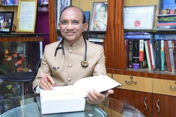
1MD, DCH, Associate Professor, Bobby Paul1 Department of Preventive Masuma Yasmin2 and Social Medicine, All India Institute of Hygiene and Public Health, Kolkata 700073 and Corresponding Author
2Research Fellow, Department of Endocrinology and Metabolism, Institute of Post-Graduate Medical Education and Research, Kolkata 700020
Original Article
Screening for Non-communicable Diseases and Health Education for Lifestyle Modification in Wellness Clinic at a Tertiary Care Hospital
Background : Non-communicable Diseases (NCD) like, Diabetes and Hypertension are highly prevalent and make a substantive contribution to the global burden of morbidity and mortality in both developing and developed Countries. Because lifestyle behaviors have been shown to be effective in preventing and treating several types of diseases that can ultimately lead to a high prevalence of morbidity and mortality, several widely accepted treatment guidelines for specific diseases include lifestyle modification strategies. In our study, we aim to identify the suspected cases of Diabetes Mellitus and Hypertension & the risk factors among screened participants. To give Health Education for lifestyle modifications.
Methodology : It was a cross-sectional study for a period of one year. The participants were patients relatives, caretakers and friends who were admitted to the Hospital. The sample size constitutes 2200 respondents who were screened in wellness Out Patient Department (OPD) for a period of one-year.
Results : In the present study by investigating Random Blood Sugar Tests during screening, we found 5% of them were found to be suspected as Diabetics and recording of the Blood Pressure shows 10% of them were suspected to be Hypertension. We observed statistically significant association with Risk Factors between both the known cases and suspected cases of Diabetes Mellitus (DM) and Hypertension.
Conclusion : Screening programs can strengthen Healthcare System initiatives and reduce the growing burden of both Diabetes and Hypertension in India.
[J Indian Med Assoc 2022; 120(4): 14-8]
Key words :Wellness clinic, Lifestyle modification, Risk factors, Screening.
Non-communicable diseases (NCD) like, Diabetes and Hypertension are highly prevalent and make a substantive contribution to the Global Burden of morbidity and mortality in both developing and developed countries. Preventing and treating Chronic Diseases through lifestyle modifications is becoming an important aspect of patient-care regimens1. In 2003, the Institute Of Medicine (IOM) published a report outlining its recommendations for educating students in the Health professions. The recommendations describe the need for all programs that Educate Health Care Professionals to integrate five core competencies. One of the five core competencies includes delivering patient-centered care, described as a type of care that continuously advocates for disease prevention, wellness and the promotion of healthy lifestyles2
Organizations outside of Higher Education have also stressed the importance of lifestyle modifications for
BLDE (Deemed to be University), Vijayapur, Karnataka 586103
1MD, Professor, Department of Community Medicine and Corresponding Author
2MS, Professor, Department of General Surgery
3MSc, PhD (Statistics), Associate Professor, Department of Community Medicine
Received on : 02/11/2021
Accepted on : 06/12/2021
Editor's Comment :
Screening for Non-communicable Diseases like Diabetes & Hypertension is very essential to identify the risk factors related to NCD
Early diagnosis and treatment will reduce the morbidity & mortality of NCD
Health Education and Counseling is required to change lifestyle modification and promotion of well-being of the community.
improving overall Health. Healthy People 2010 (sponsored by the US Department of Health and Human Services) are a set of Health Objectives for the US to achieve over the first decade of the Century3.
Because lifestyle behaviors have been shown to be effective in preventing and treating several types of diseases that can ultimately lead to a high prevalence of morbidity and mortality, several widely accepted treatment guidelines for specific diseases include lifestyle-modification strategies. The lifestylemodification strategies that are most commonly recommended within treatment guidelines include proper nutrition, physical activity, weight control, tobacco cessation, alcohol moderation and health behavior change strategies1.
Even small improvements across a large portion of the population would have a greater impact than
120, No 4, April 2022Journal
focusing on a small portion of the population that is at the upper end of the risk distribution. In our study, we aim to identify Hypertension and Diabetes among screened participants and also their Risk Factors, so then we can advise them to change their lifestyle modification to reduce the burden of these diseases (Tables 1&2).
OBJECTIVES
•To identify the suspected cases of Diabetes Mellitus and Hypertension by screening
•To identify the Risk Factors among screened participants
•To give Health Education for lifestyle modifications.
MATERIALS AND METHODS
It was a cross-sectional study for a period of one year. The participants were patient relatives or friends who were admitted to the hospital. The sample size constitutes 2200 respondents who were screened in wellness OPD for a period of one year. Institutional
Table 1 — Distribution of the participants according to Demographic profile
Table 2
Family
Habits : Yes51823.5 No163974.5 Occasionally432.0 Use of table salt or pickles : Yes96040.0 No88016.4 Occasionally36043.6 Diet : Mixed 109049.5 Vegetarian111050.5 Practice of
Ethical Committee permission and consent from the patient were taken before the start of the study. Screening for Diabetes and Hypertension was done to identify the suspected case .statistical analysis was done using SPSS VERSION 21.
Tool for measurement4 :
Measurement of Height : The Stadiometer comprises a rigid vertical backboard and a horizontal headboard running free, perpendicular to the backboard and without cross-play. The top of the head must be in contact with the headboard. A 0.5 kg weight is placed on the headboard. It consists of a ruler and sliding horizontal headpiece which can be fixed above the head to measure height. The subject’s shoes and socks are removed. The participants are placed so that their heels, buttocks and shoulders are in contact with the vertical plane of the Stadiometer. The feet must be flat against the floor while either ankles or knees remain in contact.
Measurement of Weight : The weight was measured in kilograms (kg) using a Standardized Weighing Machine with the study subject standing erect on the center of the platform with the body weight evenly distributed between both the feet together and toes apart without footwear with accepted clothing
Vol 120, No 4, April 2022Journal of the Indian Medical Association
and looking straight ahead. The weight was recorded to the nearest 0.5 kg.
Body Mass Index (BMI) : In this study, BMI the Classification proposed by the WHO, Western Pacific Regional Office in collaboration with International Obesity Task Force (IOTF) Steering Committee (2000) for Asian People was used to assess obesity and is computed by
BMI=Weight (in kg) / Height (in meter)2
It is classified as BMI <18.5 (Underweight), 18.522.9 (Normal), 23.0-24.9 (At Risk Obesity), 25.0-29.9 (Obese I) and > 30 (Obese II).
Random Blood Glucose Sugar (RBS) testing was using the Glucometer Method. In the present study a value of 200 mg/dl or above indicates that a person may have Diabetes Mellitus (DM). Less than 140 mg/ dl is normal & 140 to 199 mg/dl indicates Prediabetes5.
Blood Pressure (BP) was measured by using a Sphygmomanometer. Reading the value of Systolic Blood Pressure of 120 mmHg & Diastolic Blood Pressure 80 mmHg classified as normal, Prehypertension as - > 120 -130/ >80 - 85 mmHg and Hypertension as 140/ 90 mm Hg6
RESULTS
In the present study, both males and females were in equal distribution and the maximum numbers of them were Hindus (89%) followed by Muslims. The mean duration of the age group is 45.24±14.402. Majority of their in the age group of 40-49 years followed by >50 years. and >30 years. 35% of them are illiterate & 65% of them were literate. 29% of them were farmers by occupation and 26% of them were homemakers among females.
16% of the participants have a family history of Diabetes Mellitus (DM) and 13% of them have a family History of Hypertension (HTN). It was good to know that 75% of the participants did not have any habits. Multiple answers were found with regard to habits. The majority of them were having the habit of tobacco chewing (14%) followed by smoking and alcohol (3%).
some of them had mixed habits also, but the range is from 3%- 0.9 %. The duration observed of all their habit was in a range of 5-10 years. (37%). followed by 1-5 years.
Respondents said in their routine diet, 44% of them use pickle, table salt or chutney. But no association was observed with an intake of pickle, table salt or chutney with Hypertension. 46% of the participants have the habit of doing regular exercise and only 5% of them do irregular physical exercise .among the exercise majority of them preferred walking (95%) followed by jogging (2%). The maximum number of them practiced for a duration of 1 hour (70%). Family history of Diabetes Mellitus and Hypertension was observed in 16% and 13% of the respondents respectively.
Among the participants, 17% of them were known cases of Diabetes, 13% of them were known cases of Hypertensive and 6% of them were Cardiac Diseases. The duration of diseases both for Diabetes and Hypertension was between 1-5 years. followed by 510 years but for the Cardiac Disease, it was observed reverse pattern. 85% of them were on regular treatment for both Diabetes and Hypertension and but for Cardiac Disease it was observed 92%.
The mean weight of the participant is 63.080±20.4637. The majority of them were healthy (54.6%) and more than 10% were obese.
In our study, 7% of they were known cases of both DM and Hypertension, Diabetes and Cardiovascular Disease was 0.64%, similarly Hypertension and Cardiovascular Disease was 0.55%. All three together was 0.55%.
We found in our study a statistical significance association between all the risk factors like Modifiable and Non-modifiable factors with related to Diabetes, but for Hypertension except for diet and habits, all other risk factors was observed significant association. Similarly for Cardiovascular Disease only age and Body Mass Index was found a significant association (Table 3).
Table 3 — Association between risk factors & known case of diabetes mellitus,hypertension and cardiovascular disease
In the present study by investigating Random Blood Sugar test during screening, we found 5% of them were found to be suspected as Diabetics (>200mg/dl) and recording of Blood Pressure shows 10% (140/ 90mmhg) of them were suspected to be Hypertension. Similarly, we found 21% of them were Pre-diabetic and 25% of them were Pre-hypertensive during screening.
We observed the mean duration of Systolic Pressure is 124.68±17.628 and Diastolic Pressure is 79.83±10.279 of the participants. Similarly, the mean duration of Blood Sugar level is 144.38±69.823.
Our study observed a statistically significant difference was found between Diabetes Mellitus with related Gender (P=0.022), Occupation (P= 0.0001) and Body Mass Index (P=0.012).
Also for Hypertension, we found a significant association with related to Gender (P=0.023), habits (P=0.013), Occupation (P= 0.0001), Physical Exercise (P=0.017) and BMI (P=0.0001).
We found a highly statically significant association between Rural and Urban with related both Hypertension (at P=0.0001) and DM (P=0.0001)
No statistically significant association was observed for other risk factors like diet and family History for both Diabetes Mellitus and Hypertension (Table 4).
DISCUSSION
In the present scenario, Non-communicable Diseases (NCD)are accounts for 71% of death Worldwide and also about 48% of healthy life years lost7 . They are the major cause of mortality and morbidity among adults. In the present study during screening, we observed that many of our respondents were not screened before in their lifetime for diseases like Diabetes Mellitus and Hypertension. The majority of them are not aware of the risk factors for developing these diseases.
We found a majority of them were Hindu by Religion and belongs to the age group of 40-49 years. This could be due to the geographic distribution of the population as the majority belongs to Hindu by Religion in this area. With regards to age, all of them were patient attenders who are matured to take care of patients at the Hospital. The majority of them were farmers by the occupation because the patients who come to this Tertiary Center are usually from surrounding villages and their main occupation is mainly farming.
A finding of the present study has provided a useful screening tool for the detection and prevention of diabetes and Hypertension at our Wellness Clinic. We found 5% of them were suspected as Diabetics (>200mg/dl) in our study. A similar study of a population -based study conducted by Bharthi et al8 observed 47% of study subjects were suspected of DM. This is more than our study. In another study of screening of DM in a Rural area of North India found, 2.9% were Diabetic (RBS > 200 mg/dl), which is lower than our study. These differences could be due to the lifestyle behavior of the different study populations.
Family History of DM is one of the risk factors for Diabetes Mellitus (DM), as though there was no significant association in our study with related to family history & DM. in the present study, 16% of the participants have a family history of DM. A similar finding of Positive Family History of DM (16.9%) was observed in a study conducted by Ram Chandra et al9
A significant association was observed between DM & Body Mass Index (BMI) in the present study. A similar observation was found in the study conducted by Bharthi et al8 and Vasanthakumar et al10
For Hypertension, we found a significant association with related to Gender, Habits, Occupation, Physical Exercise and BMI. The study conducted by Shikha .S et al11 and Vanitha D, et al12 observed similar finding like Gender, occupation, BMI, and tobacco use were significantly associated with Hypertension. This shows that both for DM and Hypertension risk factors are very important and also strengthen the importance of risk factors responsible for the causation of Noncommunicable Diseases.
CONCLUSION AND RECOMMENDATION
From our study, we conclude that screening programs can strengthen Healthcare System initiatives and
Table 4 — Association between risk factors and suspected case of Diabetes Mellitus and Hypertension
Vol 120, No 4, April 2022Journal of the Indian Medical Association
reduce the growing burden of DM and Hypertension in India. The current cross-sectional study was formulated to screen individuals for Diabetes and Hypertension to obtain the trends of distribution of Blood Glucose Level and Blood Pressure Record, also identifying modifiable and Non-modifiable Risk Factors.
Based on the finding of our analysis report, those who were Pre-diabetic and Pre-hypertension for them also, we are advising to adopt lifestyle modification so that they should not suffer from both DM and Hypertension in future days. It is recommended to adopt screening programmes to strengthen the Health System for early detection of both DM and Hypertension at the Community level. Also, awareness programmes to educate them about risk factors and adoption of a Healthy Lifestyle like daily Physical Exercise, Yoga and Meditation to Reduce Body Weight, reduce or quit the habits of Smoking, Tobacco, Alcohol, reduce the Salt intake and Oil consumption. The practice of a Healthy balanced diet and regular intake of treatment and follow up for the known cases of Diabetes and Hypertension
ACKNOWLEDGMENT
I thank our Department Faculties for their support and also Institute for providing Free-of-cost Facilities for Patient Care.
Conflicts of Interest : Nil
REFERENCES
1Counseling Patients about Lifestyle Modification. Accessed on Dec 4.2020. Available at: https://www.uspharmacist.com/ article/counseling-patients-about-lifestyle-modification
2Greiner AC, Knebel E, eds. Health Professions Education: A Bridge to Quality. Executive Summary . Institute of Medicine of the National Academies. Washington, DC: The National Academies Press; 2003: 3-4.
3Healthy People 2010, Office of Disease Prevention and Health Promotion, U.S. Department of Health and Human Services. Accessed on May 10, 2020. Available at: www.healthypeople.gov.
4A training manual for height, weight and BMI assessment. Developed by BMI task force. Accessed on Jan 15th, 2021 Available at: www.achi.net>BMIContent>Documents.Scholar 5American diabetes association position statement standards of medical care in diabetes. Diabetes Care 2013; 13: S4 10.
6The Seventh Report on the Joint National Committee on Prevention, Detection, Evaluation, and Treatment of High Blood Pressure. Bethesda, MD: National Institutes of Health, U.S. Department of Health and Human Services; 2004. Accessed on Jan 15th. 2021 Available at : http://www.nhlbi.nih.gov/ health-pro/guidelines/current/hypertension-jnc-7/completereport.
7Non-communicable disease –WHO. Accessed on Jan 20th, 2021 Available at: http s://www.who.int/news-room/factsheets/detail/noncommunicable-diseases
8Bharati DR, Pal R, Kar S, Rekha R, Yamuna TV, Basu M — Prevalence and determinants of diabetes mellitus in Puducherry, South India. J Pharm Bioallied Sci 2011; 3(4): 513-518.
9Ramachandran A, Snehalatha C, Kapur A — High prevalence of diabetes and impaired glucose tolerance in India: National Urban Diabetes Survey. Diabetologia 2001; 44: 1094-101.
10Vasanthakumar J, Kambar S — Prevalence of obesity among type 2 diabetes mellitus patients in urban areas of Belagavi. Indian J Health Sci Biomed Res [serial online] 2020 accessed on march 20th.2021. Available from: http://www.ijournalhs.org/ text.asp?2020/13/1/21/276421
11Singh S, Shankar R, Singh GP — Prevalence and Associated Risk Factors of Hypertension: A Cross-Sectional Study in Urban Varanasi”, International Journal of Hypertension 2017; 12Vanitha D, Anitha Rani — M Knowledge and Practice on lifestyle modifications among males with hypertension. Indian J Comm Health 2015; 27, 1: 143-9.
Disclaimer
The information and opinions presented in the Journal reflect the views of the authors and not of the Journal or its Editorial Board or the Publisher. Publication does not constitute endorsement by the journal.
JIMA assumes no responsibility for the authenticity or reliability of any product, equipment, gadget or any claim by medical establishments/institutions/manufacturers or any training programme in the form of advertisements appearing in JIMA and also does not endorse or give any guarantee to such products or training programme or promote any such thing or claims made so after.
Original Article
A Study on Prevalence, Clinical Features and Organ Damage in Systemic Lupus Erythematosus (SLE) with Special Reference to Metabolic Profile
Introduction : Systemic Lupus Erythematosus (SLE) is an Autoimmune Disorder with broad spectrum of clinical presentation and is associated with increased prevalence of Atherosclerosis and Cardiovascular events. Metabolic Abnormality, when present in SLE patients increases proinflammatory condition and increased Cardiovascular and Cerebrovascular morbidity and mortality.
Objectives : The objectives of this study were to evaluate the prevalence of Metabolic Abnormality in SLE patients and to analyze the association with clinical and Demographic Factors.
Methods: The study was a single center, hospital based, prospective, observational study for a span of one and a half years over one hundred patients. SLE was diagnosed by revised American Rheumatology Association Criteria for SLE and Metabolic Syndrome by National Cholesterol Education Program Adult Treatment Panel III (NCEP ATP III) Criteria. Data analyzed with SPSS 23.0 software.
Results : The Metabolic Syndrome (MetS) was prevalent in SLE patients (56%). A statistically significant association is detected between MetS and SLE related variables - Serositis, Cutaneous manifestations, Oral Ulcer, Arthralgia, but no significant association found between MetS and QoL (Quality of Life) related variables like Age, Sex. The MetS components, Hypertension, Diabetes and Hypertriglyceridemia were significantly more prevalent in SLE.
Conclusion : MetS contributes to long term Cardiovascular risk in SLE patients and thus identifying MetS can contribute to major benefit towards management of IHD risk.
Key words :Ischemic heart disease, Metabolic syndrome.
SLE is an Autoimmune Inflammatory Disease with multisystem involvement which affects predominantly female in their reproductive age. A bi-modal mortality pattern is observed in patients with SLE. Early mortality is more likely related to disease itself whereas late mortality is mainly associated with comorbidities - Coronary Artery Disease being most common causes of morbidity and mortality at all stages of disease. Five to sixfold increase in incidence of Myocardial Infarction (MI) found in SLE (Manzi S et al1 ) compare to Framingham Offspring Cohort. Subclinical generalized Atherosclerosis has also been demonstrated in few studies. The Toronto Risk Factor Study shown light on the fact that SLE patients more likely to develop Diabetes, Hypertension and Dyslipidemia compared to age matched control.
Department of General Medicine, NRS Medical College and Hospital, Kolkata 700014
1MD (General Medicine), Senior Resident
2MD (General Medicine), DM (Cardiology), Associate Professor and Corresponding Author
3MD (General Medicine), Assistant Professor
4MD (General Medicine), Professor, IPGME&R and SSKM Hospital, Kolkata 700020
Received on : 25/01/2022
Accepted on : 16/02/2022
[J Indian Med Assoc 2022; 120(4): 19-22]
Editor's Comment :
Metabolic Syndrome is prevalent in SLE patients of thirty to fifty years age group and accelerates morbidity and mortality. So, Metabolic Syndrome components should be routinely investigated in SLE patients and if present ,early treatment to be initiated to prevent or to reduce Cardiovascular Risk.
However, it is not clear whether derangement of Metabolic Parameter in SLE is same as in general population and whether Steroid use in SLE is a major contributor for it. Inflammation and Metabolic Factor Interact in SLE but results in this aspect is confusing in different studies.
In our study we put an endeavor to elicit the prevalence of characteristic clinical feature and organ damage in SLE and associated Metabolic Profile with a focus to Metabolic Syndrome and its outcome on health in SLE.
MATERIALS AND METHODS
Our study is a single center, observational, prospective study over one hundred patients (N =100) comprising both female and male diagnosed to have SLE, at Nil Ratan Sircar Medical College & Hospital, Kolkata during the period from February, 2019 to September, 2020. We aimed to study association of
Vol 120, No 4, April 2022Journal of the Indian Medical Association
Metabolic Abnormality especially Metabolic Syndrome in SLE and its influence on Cardiovascular System for a span of one and a half years. After proper explanation about the study, consent was taken from guardian and nearest relatives of the patients. Detailed history, clinical examination and relevant investigations were done. SLE were diagnosed by 1997 Update of the 1992 Revised American College of Rheumatology Classification Criteria for Systemic Lupus Erythematosus. Metabolic Parameters were studied such as Urea, Creatinine, eGFR (Estimated Glomerular Filtration Rate), ACR (Albumin Creatinine Ratio), ESR (Erythrocyte Sedimentation Rate), CRP (C Reactive Protein), HDL (High Density Lipoprotein), TGL (Triglycerides), TC (Total Cholesterol). Metabolic Syndrome cases were diagnosed by the National Cholesterol Education Program (NCEP) Expert Panel on Detection, Evaluation and Treatment of High Blood Cholesterol in Adult (Adult Treatment Panel, ATPIII) of 2001, modified in 2005 by American Heart Association and National Heart Lung and Blood Institute.
Statistical Methods :
The data has been analyzed by Chi-square test and student t test with 95% confidence level (CI 95%), (p<0.05) with the help of SPSS 23.0 software.
Ethical Clearance :
Taken from Institutional Ethical Committee as per memo No/NMC/10091, Dated 09/01/2019.
RESULTS
In this study out of 100 patients 92% patients belonged to age above 25 years. Female patients comprise 69%. Smoking habit found in 31% overall. As per clinical parameter, Serositis found in 70%, Hair
loss in 59%, Cutaneous manifestation in 70%, Oral Ulcer in 46%, Arthralgia in 70%. Hypertension present in 56% cases, Diabetes Mellitus in 70%, h/o of IHD in 65%, Thyroid Disorder in 58%, Renal Disorder 67% cases, h/o Stroke in 42%, h/o intake of other drugs in 42% cases and intake of Prednisolone (>10 mg/ day) in 55% of cases. The prevalence of abnormal Metabolic parameters in these patients were 56%. Serological test positivity for ANA, Anti ds- DNA, Anti-sm Ab are 79%, 69% and 65% respectively. From Tables 1 & 2 it is observed that statistically there is no significant association between components of MetS (Metabolic Syndrome) and the various independent variables (P Value 0.05) but the Odds Ratio with respect to ESR, CRP, HDL having some positive impact on Metabolic Syndrome. From correlation Table 2, eGFR have moderate correlation with Creatinine, ESR and Triglycerides whereas, Table 4 reveals a statistically significant association between Metabolic Syndrome with SLE related variables – Serositis, Cutaneous
Table 1 — Association of Metabolic parameters in SLE with Metabolic Syndrome (n=56)
VariablesBetaStandardWald DegreeP-ValueOdd in thePower Errorof Ratio equation Freedom
Table 2 — Correlation of parameters in SLE among Metabolic Syndrome positive patient population (n=56)
TLC -Total Leukocyte Count, PLT -Platelet Count, e GFR- Estimated Glomerular Filtration Rate, ACR -Albumin Creatinine Ratio, ESR- Erythrocyte Sedimentation Rate, CRP- C Reactive Protein, HDL -High Density Lipoprotein, TGL Triglycerides, TC- Total Cholesterol.
Vol 120, No 4, April 2022Journal of the Indian Medical Association
manifestation, Oral Ulcer, Arthralgia (P Value <0.01) but no significant association found between Metabolic Syndrome with QoL (Quality of Life) - related variables like age, sex. Table 3 reveals statistically significant association of TLC, PLT, Urea, Creatinine, eGFR, ACR, ESR, HDL, Triglycerides with MetS with P Value <0.001 but not with CRP where P Value was 0.389.
DISCUSSION
In our study, the SLE population associated with Metabolic Syndrome belong to age group above 25 years (92% cases) and most are female (69%). Most frequent clinical manifestations found to be Serositis, Cutaneous manifestation and Arthralgia, all in 70% cases. Abnormal Metabolic parameters or Diabetes Mellitus are also found in 70% followed by Renal Disorder, IHD, Thyroid Disorder and Stroke. History of Prednisolone intake (>10 milligram/ day) present in 55 SLE patients out of 100. The prevalence of Metabolic Syndrome among our SLE patients is 56% which is more as compared to few African14 and European15 studies where it was 40% and 45.2% respectively and one study from South Indian which is 32.5% 12 invoking further study in this field. The prevalence of Metabolic Syndrome is much higher over pooled prevalence of MetS among adult general population in India which is 30% and also pooled prevalence of MetS in Eastern India which is 33% (95% CI : 23%-43%)16. This implies Metabolic Abnormality is frequently associated with SLE and may be a cause for Organ Damage but, this high prevalence in compare to other studies may be due to our convenience sample population attending the Tertiary Care Center with long duration of active disease or high disease magnitude. The serological test positivity rate with respect to ANA, Anti - ds DNA, Anti-sm Antibody found to be 79%, 69% & 65%, similar to study by William Maidhof et al11. No statistically significant association found between MetS and various independent variables like Total Leukocyte Count, Platelet Count, Urea,
Table 4 — Analysis of SLE related and Quality of life (QoL) related variables in patients with and without Metabolic Syndrome (N=100)
Factor Patients Patients not ODDS RatioP-Value associatedassociated with 95% CI with MetSwith MetS
Creatinine, eGFR, Albumin creatinine ratio but the odd ratio with respect to ESR, CRP, HDL revealed some positive impact. The eGFR reveals moderate correlation with Creatinine, ESR and Triglycerides.
Our study shows significant association between Metabolic Syndrome with SLE related variables Serositis, Cutaneous manifestation, Oral Ulcer, Arthralgia (P Value <0.01) but not with QoL related variables like Age, Sex. The Parameters TLC, Platelet, Urea, Creatinine, eGFR, ACR, ESR, HDL, TGL with the exception of CRP in SLE patients shown statistically significant relation with Metabolic Syndrome.
Table 3 — Mean and standard deviation of Metabolic & haematological parameters in SLE patients (N=100) with(MetS+) and without(MetS-) Metabolic syndrome and its significance
However, the data from this study cannot be extrapolated to all section of people as this patient population is taken those who attended in the tertiary Care Center in Kolkata, West Bengal, India and the sample size in this study is not representative of general population. Information related to role of Steroids on Metabolic Syndrome in the study have not been evaluated due to constrains in our study design.
CONCLUSION
Metabolic Syndrome is a set of
Vol 120, No 4, April 2022Journal of the Indian Medical Association
cardiovascular risk factors in SLE patients, which may lead to a proinflammatory condition and increased morbidity and mortality. Metabolic Syndrome is prevalent in SLE patients The SLE patients with age between 30 to 50 years are usually affected by Metabolic Syndrome. There is a significant association between Metabolic Syndrome with SLE related variables -Serositis, Cutaneous Manifestation, Oral Ulcer, Arthralgia. So Metabolic Syndrome components should be routinely investigated in patients with SLE to initiate early treatment in order to prevent/ reduce Cardiovascular Risk.
ACKNOWLEDGEMENTS
We acknowledge Prof (Dr) Saibal Kumar Mukherjee, Prof (Dr) Kripamoy Das, Dr Arijit Sinha, Dr Kaustav Chattopadhyay, Dr Mrinmoy Chanda, Mr Kausik Patra and the consented patient population whose participation made the study possible. We acknowledge the immense help received from the scholars whose articles are cited and included in reference of this manuscript. We are also grateful to Authors, Editors and Publishers of all those articles, Journals and Books from where the literature of this article has been reviewed and discussed.
Source of Funding : Nil
Conflict of Interest : Nil
REFERENCES
1Manzi S, Meilahn EN, Rairie JE — Age-specific incidence rates of myocardial infarction and angina in women with systemic lupus erythematosus: comparison with the Framingham Study. Am J Epidemiol 1997; 145(5): 408-15.
2Hochberg MC — Updating the American College of Rheumatology revised criteria for the classification of systemic lupus erythematosus, Arthritis Rheum 1997; 40: 1725.
3Executive summary of the third report of the National Cholesterol Education Program (NCEP) expert panel on detection, evaluation , and treatment of high blood cholesterol in adults(Adult Treatment Panel III ), JAMA 2001; 285(19): 2486-97 .
4Hoffman IE, Lauwerys BR, De Keyser F, Huizinga TW, Isenberg D, Cebecauer l, et al — Juvenile onset systemic lupus erythematosus :Different clinical and serological pattern than adult – onset systemic lupus erythematosus. Ann Rheum Dis 2009; 68: 412-5. 10 .1136/ard.2008. 094813 .
5Ei – Magadmi M, Ahmad Y, Turkie W — Hyperinsulinemia ,insulin resistance, and circulating oxidized low density lipoprotein in women with systemic lupus erythematosus. J Rheumatol 2006; 33(1): 50-6.
6Asanuma Y, Oeser A, Shintani AK — Premature coronary –artery atherosclerosis in systemic lupus erythematosus. N Engl J Med 2003; 349(25): 2407-15.
7El – Magadmi M, Bodill H, Ahmad Y — Systemic lupus erythematosus : an independent risk factor for endothelial dysfunction in women. Circulation 2004; 110(4): 399-404 .
8Bruce IN — Not only but also : factors that contribute to accelerated atherosclerosis and premature coronary heart disease in systemic lupus erythematosus. Rheumatology 2005; 44(12): 1492-502.
9Chung CP, Avalos I, Oeser A — High prevalence of the metabolic symdrome in patients with systemic lupus erythematosus : association with disease characteristics and cardiovascular risk factors. Ann Rheum Dis 2007; 66(2): 208-14.
10Tso TK, Huang WN — Elevation of fasting insulin and its association with cardiovascular risk in women with systemic lupus erythematosus. Rheumatol Int 2009; 29(7): 735-42.
11William Maidhof, Olga Hilus — Lupus ; an overview of the disease and management option : PT 2012 Apr ; 37(4): 240246, 249.
12Balachandra S. Bhat , Molly Mary Thabah , Vir Negi ,Zachariah Bobby — Metabolic syndrome in patients with systemic lupus erythematosus from South India. Journal of Indian Rheumatology Association 2015; 10(4):
13William Maidhof, Olga Hilas — Lupus: An Overview of the Disease And Management Options. PT 2012 Apr; 37(4): 240246, 249.
14Avela Ntombenkosi Nkabane and Bridget Hodkinson :High Prevalence of Metabolic Syndrome in South African Systemic Lupus Erythematosus Patients. Lupus : Open Access 2019; 4(2):
15Marta Maris das Chagas Medeiros , Idila Mont’Alverne Xavier de Oliveira, Adilla Thaysa Mendes Ribeiro. Rheumatol Int 2016; 36(1): 117-24.
16Krishnamoorthy Y, Rajaa S, Murali S, Rehman T, Sahoo J, Kar SS — Prevalence of Metabolic Syndrome among adult population in India : A Systematic review & Meta-analysis . PLOS ONE 15(10); e0240971, October 19, 2020.
17Meher T, Sahoo H — The epidemiological profile of metabolic syndrome in Indian population : A comparative study between men & women. Clinical Epidemiolgy and Global Health 2020; 8(4): 1047-52, December 01, 2020.
Original Article
The Role of Haematological and Biochemical Parameters for Diagnosis and Management of COVID-19 Patients
Vineet Banga1, Stuti Jain2
Introduction : COVID-19 Pandemic has affected the Healthcare System adversely. It should be diagnosed early to prevent mortality and morbidity. Thus various Haematological and Biochemical markers can be used specially in developing countries where clinicians have limited access to Molecular Diagnostic Technique.
Aim and objectives : The study aims to observe the role of haematological and biochemical parameters in diagnosing as well as predicting the prognosis along the course of the disease.
Material and methods : Retrospective study performed in Department of Pathology from April, 2021 to May, 2021 on 200 COVID-19 positive patients. The tests were conducted using the Haematological and Biochemistry Auto analysers.
Results : Out of 200 Reverse Transcription Polymerase Chain Reaction (RT-PCR) positive COVID-19 patients analysis of Haematological Parameters showed Leucocytosis, Neutrophilia, Lymphopenia and Eosinopenia. Neutrophil Lymphocyte Ratio, Platelet Lymphocyte Ratio and Systemic Inflammatory Index were also found to be elevated in comparison to the control cases. Statistically significant difference was observed in Total Leucocyte Count, Absolute Neutrophil Count, Absolute Lymphocyte Count, Kidney Function Tests (KFT) and Liver Function Tests (LFT) between severe and non severe cases. Biochemical parameters were found to be more elevated in severe cases. C-Reactive Protein (CRP) levels >50 mg/dl and Lactate Dehydrogenase (LDH) levels >1000U/L were found only in severe cases.
Conclusion : Haematological and Biochemical Markers being easily available and reliable can be utilised as useful prognosticator for early prediction of disease. Elevated Neutrophil Lymphocyte Ratio, Platelet Lymphocyte Ratio and Systemic Inflammatory Index can be useful in diagnosing COVID-19 especially when clinical suspicion is present despite negative Polymerase Chain Reaction (PCR) reports.
[J Indian Med Assoc 2022; 120(4): 23-7]
Key words :COVID 19, Haematological parameters, Biochemical parameters.
The World is facing a Public Health crisis with the emergence and spread of a new Type of Coronavirus (Severe Acute Respiratory Syndrome Coronavirus 2; SARS-CoV-2)1 Coronavirus Disease was first reported in Wuhan City, Hubei, China, in December, 2019. It was termed as Coronavirus Disease 2019 (COVID-19) by the World Health Organization (WHO) in February, 20202 The disease spread Globally and hence was declared as a Pandemic on 11th March, 20203
Editor's Comment : Leucocytosis and Neutrophilia in Covid 19 patients indicate severe disease and patient needs hospitalisation. High CRP (>50 mg/dl) and/or high LDH (>1000U/L) in COVID19 patients show that there is Severe Lung Damage and patient needs ICU care.
Elevated Neutrophil Lymphocyte ratio (>3), Platelet lymphocyte ratio (>130) and Systemic Inflammatory Index (>7.0) are useful in diagnosis of COVID-19 in clinical suspicion even when RTPCR test for COVID-19 is negative. Calculation of NLR, PLR, SII: Neutrophil Lymphocyte ratio is calculated by Absolute Neutrophil Count/absolute Lymphocyte Count, Platelet Lymphocyte Ratio is calculated by Platelet count in lakhs/cmm ×100/ Absolute Lymphocyte Count, Systemic Inflammatory Index is calculated by multiplying Neutrophil Lymphocyte Ratio and Platelet Count in lakhs/cmm
Department of Pathology, Acharyashree Bhikshu Government Hospital (Government of NCT of Delhi), Moti Nagar, New Delhi 110015
1MBBS, MD (Pathology), Senior Specialist & Head and Corresponding Author
2MBBS, MD (Pathology), Senior Resident
Received on : 08/11/2021
SARS-CoV-2 belongs to the Coronavirus family being part of genus β coronavirus, which has Genetic Homology similar to Severe Acute Respiratory Syndrome (SARS-CoV) and Middle East Respiratory Syndrome-related Coronavirus (MERS-CoV)4. SARSCoV-2 is enveloped, Ribonucleic Acid (RNA) virus which has led to the COVID-19 Pandemic. The main routes of transmission of the virus are through Respiratory Droplets and Contact Transmission. The virus enters the body through Pulmonary Epithelial Cells via ACE2 receptors, leading to Pneumonia, followed by Systemic Inflammatory phase which can advance to respiratory failure or even Multi-organ Dysfunction5. The patient
Accepted on : 25/02/2022
Vol 120, No 4, April 2022Journal of the Indian Medical Association
can be asymptomatic or can have symptoms like Fever, Cough, Headache, Body Ache, Increased Sputum Production, Dyspnoea, Haemoptysis, Diarrhoea, Acute Respiratory Distress Syndrome, Cardiac Failure or any Secondary infection6.
COVID-19 Pandemic has affected the Health Care System Adversely with an increase in severe cases which require intensive care, leading to a huge economic burden on our inadequate Healthcare facilities7. To control the pandemic, diagnostic tools help in detecting cases early and accurately. Various molecular techniques have been developed but in developing Countries the Healthcare Professionals have limited access. The most commonly used Molecular Technique is the Real-time Reverse Transcriptase-PCR but it has the limitations like long turnaround time, limited availability, expensive equipment, need for trained staff and chances of falsenegative results8. Thus, tests for early diagnosis should be developed to rapidly detect the cases and identify severity of diseases to reduce mortality and prevent the spread of this pandemic9
This study aims to observe the role of various Haematological and Biochemical Biomarkers in COVID-19 patients and also assess the role of these markers in the severity of the disease. This will help the Clinicians to Group the Patients and predict the prognosis and mortality, thereby help in better management of the patients.
MATERIALS AND METHODS
The retrospective study comprises of 200 patients hospitalised due to COVID-19 from April, 2021 to May, 2021 at Acharyashree Bhikshu Government Hospital (Government of NCT of Delhi) Delhi. These patients were tested positive by RT-PCR test or Rapid Test as per ICMR Criteria. The data of the patient including Age, Sex, Clinical Condition as well as any associated comorbidity was collected from the Medical Records Department. The Haematological and Biochemical Parameters were assessed during the time of admission. Clinical features were used as a tool to classify the patients into severe and Non-severe categories. Whole Blood Ethylenediamine Tetraacetic Acid (EDTA) samples sent at the time of admission were run on Automated Haematological Analyser SYSMEX XN-1000. Biochemical Parameters were assessed using Biochemistry
Auto Analyser Erba Mannheim XL 640. Statistical significance was calculated based on 't' test using Microsoft excel.
RESULTS
Demographic Data :
A total of 200 COVID-19 positive cases were included in the study. The mean age of the patients
Table 1 — Laboratory Parameters of Patient with COVID-19 (N = 200)
Total Leucocyte Count (X 109/L)4.0-11.012.26±5.4612.13 (8.22-15.80)
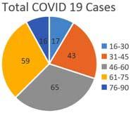
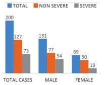
were 54.45±15.51 years with 32.5% of the patients belonging to the age range 46-60 years (Fig 1). The severity of the disease increases as age increases. Of all the cases, 131 (65.5%) were male and 69 (34.5%) were female (Fig 2).
Analysis of Haematological profile in these patients showed Leucocytosis, Neutrophilia, Lymphopenia, Eosinopenia and normal to reduced Platelet Count. Comparative analysis of Severe to Non-severe group showed statistically significant difference in White Cell Count (<0.00001) and Absolute Neutrophil Count (ANC) (<0.00001). 71.2% of all the severe cases showed Thrombocytopenia while most of the patients of the Non-severe group had Platelet Count in the normal range.
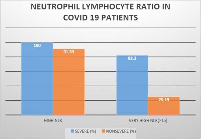
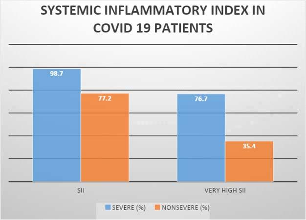
The reference range for novel Hematological parameters were determined using the control subjects, Neutrophil Lymphocyte Ratio (NLR) (Reference range: 1.752.93), PLR (Reference range: 90-130) and Systemic Inflammatory Index (SII) (Reference range: 3.6-6.6). It was observed that these parameters were significantly higher in COVID-19 cases and also Statistically Significant difference was observed between Severe and Non-severe group (p value< 0.0001) (Figs 3-5).
Table 2 — Comparison of Laboratory parameters among severe and non severe COVID-19 patients Cut OffTotal SevereNon(%)(%)severe(%)
Leucocytosis>11 X109/L57.576.746.4
Neutrophilia>8 X10⁹/L7084.961.42
Thrombocytopenia<150 X109/L49.571.237
Urea >45 Mg/Dl5064.3841.73
Creatinine>1.4 MG/DL9.813.697.08
SGOT>35IU/L7391.7862.2
SGPT >35IU/L6375.3455.9
Table 3 — Statistical significance of haematological and biochemical parameters ParametersMean ValueMean Value't' statp-ValueStatistical in Severein Non Severe Significance Covid CasesCovid Cases
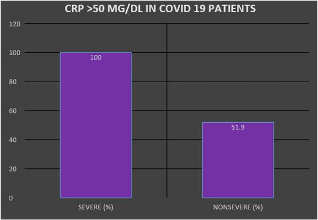
Biochemical Parameters like Kidney Function Tests (KFT) and Liver Function tests (LFT) were found to be abnormal in most of the COVID 19 cases especially as the severity of the disease increases. C Reactive Protein (CRP) was found to be raised in all the cases and it was noted that patient with CRP> 50mg/dl had severe disease (Fig 6). At the time of admission, LDH levels correlated with the condition of the patient which could help Clinicians to stratify the cases based on severity of the disease. It was noted that 90% of the severe cases had LDH levels >1000U/ L (Fig 7). Thus, LDH and CRP can be used as early parameters for ICU admission.
Correlation analysis showed a significant direct relation between LDH and WBC count (r = 0.4328, p < 0.0001), Neutrophil Count (r = 0.46, p < 0.0001), CRP (r = 0.59, p < 0.0001), NLR (r=0.34, p<0.001) and Age (r=0.27, p <0.001). CRP values correlated with WBC count (r = 0.37, p<0.0001), Neutrophils Count (r = 0.41, p < 0.0001) and showed a slight inverse correlation with Lymphocyte Count (r = -0.19, p < 0.05)(Tables 1-3).
DISCUSSION
SARS-CoV-2 has spread Globally with most of the patients having mild to moderate disease while few suffering from life threatening severe disease10. The
Fig 3 — Neutrophil Lymphocyte Ratio among severe and non
present study will help to analyse the accuracy of Haematological, Inflammatory and Biochemical Parameters for diagnosing the patients with COVID19 and thus help during the unavailability of PCR test or false negative PCR test. Thus, we summarised a comparative analysis of laboratory parameters among cases having non-severe disease and cases needing immediate hospital admissions which will be useful in the clinical settings to support clinical management which will lead to improvement in the Survival Rate. The study showed that the most common age range for patients suffering from COVID is between 46-60 years. It also observed that as age increases severity of the disease increases which is in accordance with the studies done by Shen et al11 and Qin et al12. This can be explained with the fact that as age increases it leads to Biological Ageing, Impaired Immune Function and decreased Lung capacity13. We also observed that majority of our patients were male and severity of the disease was also seen more in male cases. Similar observations were done by Li et al14 and Guan et al15
Thus, it can be considered that COVID-19 is more frequently seen in males and in middle-aged patients. In the study, TLC and Neutrophil Counts were increased while Lymphocyte Count was reduced. This was seen more frequently in severe cases which is also supported by studies done by Singh et al16 and Sheng et al11. In COVID-19, Cytotoxic Lymphocytes which help in control of viral infection get exhausted this correlates with progression of disease. After one to two weeks, there is ‘Cytokine Storm’ and Lymphopenia becomes prominent due to Atrophic Lymphoid Organs. Thus, Lymphopenia is considered to be the most important prognostic markers in COVID19 cases 17. NLR, PLR and SII were found to be significantly increased in COVID-19 patients as compared to the control cases in the present study. Our findings show that Thrombocytopenia is associated with severely diseased individuals similar to many previous studies Hypercoagulability state in COVID-19 disease is accompanied with microthrombi formation along with consumption of Platelet which leads to Thrombocytopenia 18 . Therefore, Thrombocytopenia could be used as a useful indicator
Fig 4 — Platelet Lymphocyte Ratio among severe and non severe COVID-19 patients
Fig 5 — Systemic Inflammatory Index (SII) among severe and non severe COVID-19 patients
Fig 7 — LDH>1000 U/L among severe and non severe COVID19 patients
Fig 6 — CRP>50mg/dl among severe and non severe COVID-19 patients
Vol 120, No 4, April 2022Journal of the Indian Medical Association
for disease stratification.
Biochemical parameters help in assessing disease severity. LDH, an enzyme used in production of energy by converting Lactate to Pyruvate, is present in almost all body tissues being a General Indicator of Tissue damage and considered as marker of inflammation19 CRP is considered as a reliable marker of Acute Inflammation. LDH and CRP thus can be markers of Lung damage and reflect respiratory distress due to Abnormal Inflammation Status. Thus the levels of both these parameters is markedly increased as the severity of disease increases which is seen in most of the previous studies15.
Thus, evaluation of laboratory parameters at the time of admission and along the course of the disease can assist Clinicians in working out an Effective Treatment Protocol and promptly providing Intensive Care to Severe Patients.
CONCLUSION
The study concluded that Leucocytosis, Neutrophilia, Lymphopenia and Eosinopenia along with elevated LDH, CRP, higher Liver enzymes and abnormal KFT is seen in patients with severe COVID-19 disease. Hematological, Biochemical and Inflammatory Markers, being easily available and reliable Markers, can be utilized as useful prognosticator for early prediction of disease. Thus, appropriate management can be planned for patients at an early stage. The Abnormal Hematological Parameters can serve as markers for diagnostic and prognostic importance in determining the course, outcome and severity of COVID-19 infection. Thus, mortality and morbidity can be lowered in Critical Patients and those having comorbidities. We suggest that, elevated NLR, PLR and SII can be useful in diagnosis of COVID-19 along with other relevant tests, especially when clinical suspicion is present despite negative RT-PCR Reports.
Limitatations : Main limitation of this study is small sample size.
Funding : None.
Conflict of Interest : None.
REFERENCES
1Singhal T — A Review of Coronavirus Disease-2019 (COVID19). Indian Journal of Paediatrics 2020; 87(4): 281-6.
2Chan JWM, Ng CK, Chan YH, Mok TYW, Lee S, Chu SYY — Short term outcome and risk factors for adverse clinical
outcomes in adults with severe acute respiratory syndrome (SARS). Thorax 2003; 58(8): 686-9.
3World Health Organization (WHO) — WHO Director-General’s opening remarks at the media briefing on COVID-19 - 11 March 2020. Geneva: 11 Mar 2020.
4Zhou P, Yang XL, Wang XG, Hu B, Zhang L, Zhang W, et al — A pneumonia outbreak associated with a new coronavirus of probable bat origin. Nature 2020; 579(7798): 270-3.
5Lippi G, Henry BM — Chronic obstructive pulmonary disease is associated with severe coronavirus disease 2019 (COVID19). Respir Med 2020; 167: 105941.
6Huang C, Wang Y, Li X, Ren L, Zhao J, Hu Y — Clinical features of patients infected with 2019 novel coronavirus in Wuhan, China. Lancet 2020; 395(10223): 497-506.
7Martin A, Markhvida M, Hallegatte S, Walsh B — Socioeconomic impacts of COVID-19 on household consumption and poverty. Economic Disaster Climate Change 2020; 4(3): 453-79.
8Xie X, Zhong Z, Zhao W — Chest CT for typical 2019-nCoV pneumonia: relationship to negative RT-PCR testing. Radiology 2020; 200343.
9Di Gennaro F, Pizzol D, Marotta C — Coronavirus diseases (COVID-19) current status and future perspectives: a narrative review. Int J Environ Res Public Health 2020; 17(8): 2690.
10Simsek Yavuz S, Ünal S — Antiviral treatment of COVID-19. Turk J Med Sci 2020; 50(SI-1): 6119.
11Sheng L, Wang X, Tang N — Clinical characteristics of moderate and severe cases with COVID-19 in Wuhan, China: a retrospective study. Clinical and Experimental Medicine 2020. doi: 10.1007/s10238-020-00662-z.
12Qin C, Zhou L, Hu Z — Dysregulation of immune response in patients with coronavirus 2019 (COVID-19) in wuhan, China. Clin Infect Dis: an official publication of the Infectious. Diseases Society of America 2020; 71: 762-8.
13Shahid Z, Kalayanamitra R, McClafferty B — COVID-19 and older adults: what we know. Journal of the American Geriatrics Society 2020; 68: 926-9.
14Li Q, Guan X, Wu P — Early transmission dynamics in Wuhan, China, of novel coronavirus-infected pneumonia. N Engl J Med 2020; 382: 1199-207.
15Guan WJ, Ni ZY, Hu Y — Clinical characteristics of coronavirus disease 2019 in China. N Engl J Med 2020; 382(18): 1708-20.
16Singh P, Kumar A, Singh S — Utility of Routine Haematological Parameters and Infectious Biomarkers to Assess the Disease Severity in COVID-19 Positive Patients, Analysis and Early Trend from India.
17Yang A-P, Liu J-P, Tao W-Q — The diagnostic and predictive role of NLR, d-NLR and PLR in COVID-19 patients. Int Immunopharmacol 2020; 84: 106504.
18Wool GD, Miller JL — The Impact of COVID-19 Disease on Platelets and CoagulationPathobiology2021881152710.1159/ 000512007.
19Sepulveda J — Challenges in Routine Clinical Chemistry Analysis: Proteins and Enzymes. Editor(s): A. Dasgupta, J. L. Sepulveda, Chapter 9, Accurate Results in the Clinical Laboratory, Elsevier 2013: 131-48.
Original Article
Study to Find Out the Correlation Between Cognitive Defect and Non-alcoholic Fatty Liver Disease
Sriradha Chatterjee1, Saumik Datta2, Sujoy Sarkar3, Mrinal Kanti Ray4
Introduction : Non-alcoholic Fatty Liver Disease (NAFLD), a hepatic manifestation of Metabolic Syndrome, has now become a Global Phenomenon and along with its increasing prevalence various morbidities and mortality are also increasing.
Aims and Objectives : The objective of the present study was to establish whether patients with NAFLD, in the absence of other comorbid conditions suffer from cognitive impairment.
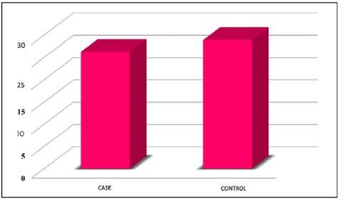
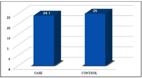
Materials and Methods : This cross sectional study was conducted at the Department of General Medicine, Calcutta National Medical College and Hospital. 90 patients with NAFLD and 90 healthy controls were recruited after matching all the inclusion and exclusion criteria, from the out patient and in patient department over a period of 1 year starting March, 2019. NAFLD was diagnosed by noninvasive methods including Elastography (fibroscan). Cognition was assessed by MoCA (Montreal Cognitive Assessment test) score.
Result : The mean age of cases and control were 49.2 and 48.5 years, respectively. Out of total cases and controls 48.9% was male and 51.1% was female. The mean BMI of the cases and control were 30.21±4.24 and 22.60±1.52 Kg/m2, respectively. The mean Elastography score among the cases was 4.91±0.23 kPa and that among the controls was 3.84±0.31 kPa. The mean Fibroscan Score among male cases and controls were 4.907±0.26 kPa and 3.83±0.35, respectively (p<0.05). In case of females, Fibroscan Score was 4.906±0.21 for cases and 3.85±0.29 for controls. After the groups were matched for age and gender, we found that 33.3% of the cases had a MoCA score < 26, whereas only 6.7% of the control population showed similar results. The mean score among the cases was 26.24±1.58 which was significantly less than that found in the control population (28.89±1.2). The patients with normal BMI with cognitive defect had a mean MoCA score of 23.80±1.5 and those without cognitive defect had a mean MoCA score of 29.13±1.1. The difference between the two groups was statistically significant.
Conclusion : A statistically significant cognitive difference was found between the two groups (NAFLD versus controls), with a higher cognitive deficit recorded among patients with NAFLD. Percentage of people with Cognitive defect appears to be greater among the NAFLD patients even after they were matched for Body Mass Index (BMI). [J Indian Med Assoc 2022; 120(4): 28-31]
Key words :Fibroscan, MoCA, BMI.
The prevalence of Non-alcoholic Fatty Liver Disease (NAFLD) is constantly increasing (15% in 2005 to 25% in 2010) with a Global Prevalence of 25.24% currently1
The stages of NAFLD are Fatty Liver (steatosis), Non-alcoholic Steatohepatitis, Fibrosis and Cirrhosis. NAFLD is now recognized as the Hepatic manifestation of Metabolic Syndrome2. Obesity has the strongest association with NAFLD. While 30% of
1MD (General Medicine), Senior Resident, Department of General Medicine, Calcutta National Medical College, Kolkata 700014
2MD, DM (Endocrinology), FICP, FISH, Associate Professor, Department of General Medicine, Calcutta National Medical College, Kolkata 700014, At present : Professor, Department of General Medicine, Burdwan Medical College, Burdwan 713104 and Corresponding Author
3MD (General Medicine), Associate Professor, Department of General Medicine, Calcutta National Medical College, Kolkata 700014
4MD, DM (Neurology), Professor (Retd), Department of General Medicine, Calcutta National Medical College, Kolkata 700014
Received on : 19/08/2021
Accepted on : 01/12/2021
Editor's Comment :
NAFLD is a fast emerging and vastly prevalent disease. While it's negative implications are far reaching, not only by it's impact on General Health, but also it's ability to cause cognitive defect, awareness regarding the same and early intervention can help stall the disease process.
patients who are obese have Fatty Liver, up to 80% of morbidly obese patients (BMI > 35) have NAFLD3. Noninvasive studies like Ultrasonography, Computed Tomography Scanning and Magnetic Resonance Imaging (MRI) and MR Elastography are useful tools to establish a diagnosis of Steatosis4, complementing blood tests. Studies show that Ultrasonographic diagnosis of Steatosis of any degree was seen to be 60.9% sensitive and 100% specific. Liver stiffness measured by transient Elastography [recorded in kilopascals (kpa)] have demonstrated diagnostic accuracy for assessing Fibrosis5. Transient Elastography is a remarkable alternative to Liver Biopsy, which, being an invasive procedure is garnering
Vol 120, No 4, April 2022Journal of the Indian Medical Association
reluctance with most patients, especially Asymptomatic ones.
Our study aims to correlate cognitive defect with NAFLD. ‘Cognition’ means "thinking and awareness". MoCA (Montreal Cognitive Assessment test) is a test of cognitive function, testing 7 separate domains and is multiple languages accessible. It is a 30-point test administered over 10 minutes. The sensitivity and specificity of MoCA for detection of MCI (Minimum Cognitive Impairment) was found to be 90% and 87% respectively, compared with 18% and 100% respectively for the MMSE6
Various studies have proven that a correlation exists between NAFLD and CNS manifestations like Depression, Dementia, etc. Our study aims to substantiate the same, eliminating other risk factors.
AIMS AND OBJECTIVES
The objective of the present study was to establish whether patients with NAFLD, in the absence of other comorbid conditions suffer from cognitive impairment.
MATERIALS AND METHODS
This cross-sectional study was conducted at the Department of General Medicine, Calcutta National Medical College and Hospital in collaboration with the Ultrasound Unit of the same Department. 90 patients with NAFLD and 90 healthy controls were recruited from the Outpatient and Inpatient Department over a period of 1 year starting March, 2019.
Inclusion Criteria :
Patients aged 18-60 years with NAFLD. Controls- patients of the same age group without any comorbidities.
For defining NAFLD, there must be (1) evidence of Hepatic Steatosis (HS), either by imaging or histology, and (2) lack of secondary causes of Hepatic Fat accumulation7
We have diagnosed our patients based on Ultrasonography and Fibroscan which is sufficient for diagnosis.
Cognitive assessment was done using the MoCA scale. A score <26 out of 30 was taken as evidence of cognitive defect8
Exclusion Criteria :
•Previous history of Hepatitis, Cirrhosis, or other Chronic liver disease, Autoimmune hepatitis, Haemochromatosis
•Presence of severe Cardiopulmonary disease
•Obstructive Sleep Apnoea Syndrome
•Endocrinological disorders: Hypothyroidism, Hypercorticism, Syndrome of the polycystic ovaries
•History or Clinical signs of excessive Alcohol abuse (>20 g/day for males and >10 g/day for females)
•Visible focal or diffuse changes in the grey matter of the brain on MRI.
•Fazekas score more than 0 on MRI scan
•Rheumatological disease
•Psychiatric disease and/or Psychiatric medication history or Hepatotoxic drugs
•Traces of illicit drugs abuse: positive urine multiple drug tests.
•Use of antidiabetic drugs, insulin, antilipemic drugs, uricosuric drugs, steroids and oral contraceptives.
•Advanced Liver Disease with Hepatic Encephalopathy
The data obtained from the above study was analyzed by standard statistical methods using SPSS v.20.
Descriptive statistical analysis was performed to calculate the means with corresponding Standard Deviations (SD). Test of proportion was used to find the Standard Normal Deviate (Z) to compare the difference proportions and Chi-square ( χ2) test was performed to find the associations.
t-test was used to compare the means of the two groups. Fisher Exact test was used where Chi-square (χ 2) test was not applicable. p<0.05 was taken to be statistically significant.
RESULTS
We recruited 90 NAFLD patients (cases) and 90 controls aged between 18 and 60 years. The mean age of cases and control were 49.2 and 48.5 years, respectively. Percentage of male and female patients was 48.9% and 51.1%, respectively in total groups.
The mean weight (kg) in study population was found to be 83.27 ± 10.87 which was more than the control group (60.93 ± 7.65).86.7% of the cases and 6.7% of the controls were found to have a BMI above 24.9. The mean BMI of the cases and control were 30.21 ± 4.24 and 22.60 ± 1.52 Kg/m2, respectively.47.8% of males had a waist :hip ratio > 0.9 among cases and 33.3 % of the females had a ratio > 0.85; in comparison, 3.3% of males and 7.8% of females in the control group had a ratio > 0.9 and 0.85 respectively.The mean waist hip ratio among cases was 0.99±0.16 and among the controls was 0.83±0.04 which is statistically significant.
NAFLD group, 10 out of 44 male patients had an abdominal girth >102 cm (11.1%), while none of the male controls fell in the same category. 28 out of 46 female cases had an abdominal girth >88cm (31.1%) and 1.1% of the controls (1 female) showed similar
120, No 4, April 2022Journal
results. The mean abdominal girth for cases was 94.32 ± 6.92 cm and that of control population was 81.23 ± 4.48 cm, which was significantly different.
In our study 77.8% of cases and 24.4% of controls had a liver span >155mm (normal <155mm). The mean liver size among cases was 156.12 ± 1.65 and 152.94 ± 2.19 among controls, which was significant.96.7% of the cases and 100% of the control patients had a spleen size less than 125 cm (normal average).
95.6% of patients with NAFLD had an AST > 35 IU/L as compared to only 10% of control patients. The mean Aspartate Transaminase (AST) of cases and control were 46.77 ± 6.94 and 28.11 ± 4.88 which was significant. 98.9% of cases had an ALT of 40 IU/L as compared to only 7.8% of the control patients. The mean Alanine Transaminase (ALT) of the cases group was 58.10 ± 8.21 and that of the control population was 32.58 ± 5.01 which was significant.
The Gamma-glutamyl Transferase (GGT) levels of all patients who participated in the study was <60U/L. 81.1% of cases and 98.9% of controls had a Total Bilirubin value within normal range of 0.8 to 1.2 mg/dl.
All the cases and 98.9% of the controls had a Total Protein (TP) value within the normal range. The mean difference was 0.15 and was not significant.
The ideal Fibroscan values for a normal Liver without any insult or scarring are between 2-5.7 kPa. In our study 98.9% of the cases and all controls had a normal score. The mean score among the cases was 4.91 ± 0.23 and that among the controls was 3.84 ± 0.31.
The mean Fibroscan value among the male and female cases were 4.907±0.26 and 4.906±0.21, respectively, signifying no difference.
The mean Fibroscan score among male cases and controls were 4.907±0.26 and 3.83±0.35, respectively (p<0.05). The mean Fibroscan score among female cases and control were 4.906±0.21 and 3.85±0.29, respectively. Mean difference was 1.056 and statistically significant.
Further, the mean Fibroscan values were seen to be higher in patients who were older: 4.50±0.1 among the NAFLD patients in 21-30 age group versus 4.96±0.3 in the 51-60 group.
The NAFLD patients were divided based on gender among the different age groups and Fibroscan values compared. There was no statistically significant difference among Fibroscan Scores between male and female cases when distributed among different age groups.
After the groups were matched for age and gender, we found that 33.3% of the cases had a MoCA score <26, whereas only 6.7% of the control population
showed similar results. The mean score among the cases was 26.24 ± 1.58 which was significantly less than that found in the control population (28.89 ± 1.2).
84 out of total 180 persons had a high BMI, whereas 96 had a normal BMI.
Among whole study population (irrespective of NAFLD),30.9% ofpersons with a BMI =25 Kg/m2 had a MoCA score <26; whereas, 89.6% of the persons with a normal BMI had a normal Cognitive Score (>26)(Fig 1).
The patients with high BMI with cognitive defect had a mean MoCA score of 24.04 ± 1.3, those without cognitive defect among the above group had a mean MoCA score of 27.48 ± 1.2 (Fig 2).
The patients with normal BMI with cognitive defect had a mean MoCA score of 23.80 ± 1.5, and those without cognitive defect had a mean MoCA score of 29.13±1.1. The difference between the two groups was statistically significant.
Further, 25 out of 78 NAFLD (32.2%) cases with high BMI had Cognitive score <26 and only one out of 6 controls with high BMI had a Cognitive Score <26 (16.7%). Five out of 12 NAFLD cases with normal BMI had a Cognitive Score <26 (41.7%). Five out of 84 controls with a normal BMI had a Cognitive score <26 (5.9%).The mean Cognitive score among the NAFLD
Fig 1 — Comparison of mean Cognitive Score of the patients between the two groups
Fig 2 — Comparison of mean Cognitive defect of the patients with BMI>25 kg/m2 of the two groups
120, No 4, April 2022Journal
patients with a normal BMI was 24.0±0.7 and among controls was 23.6±2.1. The difference of the mean cognitive defect between these two groups is however, not statistically significant (P>0.05).
DISCUSSION
The results of our study show that there is a definite correlation between NAFLD and Cognitive Defect (diagnosed using the Montreal Cognitive Assessment scale). The cases showed a greater decline in cognitive function along with higher BMI, Waist Hip Ratio, Triglyceride and Total Cholesterol levels. Thus, various parameters of Metabolic Syndrome appear to be intricately involved in causing NAFLD and further, a decline in cognition as has also been proved in studies by Sang Won Seo, Rebecca F Gottesman, Jeanne M Clark, et al9
The Pathophysiology behind the relation between these risk factors and Cognitive deficit though still elusive, is a much explored and debated prospect and various studies are attempting to prove and explain the same. While the degree of Cognitive defect was significantly higher in the NAFLD patients most of whom had a higher BMI, it was surprisingly enough seen that the percentage of Cognitive defect was greater among the NAFLD patients even after they were matched for BMI.
Most studies correlating NAFLD and Metabolic Syndrome have shown a direct relation to NAFLD and cognitive defect with Diabetes10. Our study is different in that we excluded Diabetes; and NAFLD without Diabetes was independently associated with a decline in Cognitive function. Further studies are required in this field to achieve more clarity.
Finding an association between NAFLD with cognitive defect as shown in this study can help in early intervention and more intensive management to try to stall, if possible, halt, further progression of the disease.
Ascertaining the validity of these non-invasive measures in diagnosing NAFLD and in prediction of further disease course including progression of cognitive deficit can however be achieved only with a longer study duration, a much larger study population, and more stringent follow up of patients. A greater recruitment into both groups with equal representation of patients with high and low BMI can provide unbiased clarity when studying the repercussions of NAFLD on Cognitive defect.
CONCLUSION
Our study, to find out the correlation between NAFLD
and Cognitive defect is possibly the first such study conducted in East India. A statistically significant Cognitive Difference was found between the two groups (NAFLD versus controls), with a higher Cognitive Deficit recorded among patients with NAFLD.
Percentage of people with Cognitive defect appears to be greater among the NAFLD patients even after they were matched for BMI.
BMI, however, appears to be an important confounding factor with respect to cognitive defect as our study was not confidently able to rule out the above to satisfaction.
Funding : None
Conflict of Interest : None
REFERENCES
1Younossi ZM — Global epidemiology of non alcoholic fatty liver disease Metaanalytic assessment of prevalence, incidence, and outcomes. Hepatology 2016; 64: 73-84.
2Luyckx FH, Desaive C,Thiry A — Liver abnormalities in severely obese subjects:effect of drastic weight loss after gastroplasty. Int J Obes Relat Metab Disord 1998; 22: 2226.
4European Association for the Study of the Liver (EASL); European Association for the Study of Diabetes (EASD); European Association for the Study of Obesity (EASO) (June2016).” EASL EASD EASO Clinical Practice Guidelines for the management of non alcoholic fatty liver disease”. Journal of Hepatology (Professional society guidelines). 64(6): 1388-402.
5Srinivasa Babu A, Wells ML, Teytelboym OM, Mackey JE, Miller FH, Yeh BM, et al — Elastography in Chronic Liver Disease: Modalities, Techniques, Limitations ,and Future Directions”.Radiographics (Review) 2015; 36(7): 1987-2006.
6Ziol M, Handra-LucaA, Kettaneh A, Christidis C, Mal F, Kazemi F, et al — Non invasive assessment of liver fibrosis by measurement of stiffness in patients with chronic hepatitis C. Hepatology 2005; 41: 48-54.
7Dong, YanHong; Sharma, Vijay Kumar; Chan, Bernard PoonLap; Venketasubramanian, Narayanaswamy; Teoh, Hock Luen; Seet, Raymond Chee Seong; Tanicala, Sophia; Chan, Yiong Huak; Chen, Christopher. The Montreal Cognitive Assessment (MoCA)is superior to the Mini-Mental State Examination (MMSE) for the detection of vascular cognitive impairment after acute stroke”. Journal of the Neurological Sciences 299(1-2): 15-8.
8Younossi Z, Lavine JE, Charlton M, Cusi K, Rinella M, Harrison, et al — The Diagnosis and Management of Nonalcoholic Fatty Liver Disease: Practice Guidance From the American Association for the Study of Liver Diseases Naga Chalasani, Aktuelnosti: neurologijiipsihijatriji, vol 17, 2009.
9Sang Won Seo, Rebecca F Gottesman, Jeanne M Clar K, RubenHernaez,Yoo soo Chang, Changsoo Kim,, Kyoung Hwa Ha, Eliseo G uallar, Mariana Lazo — Non alcoholic fatty liver disease is associated with cognitive function in adults: Neurology 2016; 86(12): 1136-42.
10Haukeland JW, Konopski Z, Linnestad P — Abnormal glucose tolerance is a predictor of steatohepatitis and fibrosis in patients with non-alcoholic fatty liver disease. Scand J Gastroenterol 2005; 40: 1469-77.
Original Article
Vast Potential for Clinical Trial Opportunities; Adipose Tissue Derived
Introduction : This is a research innovation that aims to provide an additional therapeutic tool. It will open up a vast panorama of regenerative medicine by application of Adipose Derived Mesenchymal Stem Cells (ADMSCs). ADMSCs are selected since a large amount is available for lipoaspiration and a larger percentage (30%) of Mesenchymal Stem Cells (MSCs) obtainable there from. The applications in clinical practice extend across Mesoderm, Endoderm and Ectoderm layers1
Material and Methods : There are three products that can be derived from the lipoaspirate.
They are (1) Stromal Vascular Fraction (SVF), (2) Islet Cell Aggregates (ICAs) Translated from ADMSCs, (3) and ADMSCs with ~95% purity. They are deployed to illustrate the safety and efficacy in clinical trials for (1) Mesoderm Translation as in Osteoarthritis Knee, (2) Endoderm translation to Insulin-producing Cells as applicable to diabetes, and (3) Ectodermal Translation as applicable on Non-healing Indolent Ulcers on the Skin.
Results : All three products are found safe with no adverse side effects. Proof of concept studies along with initial clinical trials for Osteoarthritis, Diabetes Types I and II, and Non-healing ulcer of any aetiology is demonstrated with objective evidence.
Discussion : The evidence based on the results of the clinical trials across all three Germinal Layers is cited along with literature support.
Results are explained based on a plausible scientific hypothesis.
Conclusion : The study enunciates that Autologous SVF and ADMSCs are in futuristic domain for conducting clinical trials across all the three Germinal Layers.
[J Indian Med Assoc 2022; 120(4): 32-7]
Key words :Research innovation, ADMSCs, SVF, Clinical trial, Regenerative medicine.
This is a research innovation utilizing the properties of MSCs. MSCs provide a futuristic therapeutic hope for Regenerative medicine. MSCs have fundamental properties such as honing to the site of injury or inflammation2 and translation to the honed tissue in situ3. They are immunoprotective4 as well. MSCs derived from bone marrow have been the subject of extensive research. However, bone marrow has lower yield of MSCs. Further, there is a limitation to the quantity of bone marrow aspiration. Lipoaspiration of Adipose Tissue contains ~30% MSCs5 and can easily be obtained up to ~1,000 cm3 under local anaesthesia.
This article aims at adding some more tools in the armamentarium of medical doctors and corporate Hospitals in pursuit of Regenerative Medicine. The tools are: Stromal Vascular Fraction (SVF) from lipoaspirate, Islet Cell Aggregates (ICA) obtained by transgerminal
Total Potential Cells (P) Ltd, Raopura, Vadodara, Gujarat 390001
1MS, Stem Cell Scientist, Department of Plastic Surgery
2PhD, Stem Cell Scientist, Department of Stem Cell Research
3MD, DGO, Gynaecologist, Stem Cell Scientist, Department of Stem Cell Research and Corresponding Author
4PhD, Stem Cell Researcher, DY Patil Vidyapeeth, Pune, Maharashtra 411018
Received on : 02/02/2021
Accepted on : 25/02/2021
Editor's Comment :
The research innovation based on transgerminal translation of ADMSC is to provide numerous opportunities for deriving products that will constitute a futuristic regenerative medicine.
Proof of concept is provided.
Opportunities are doable according to ICMR, DCGI approvals. Multi specialty Hospitals/Groups will qualify.
translation of ADMSCs and purified MSCs. An Autologous cell-based product, Platelet-rich Plasma (PRP) is added as an adjunct since several analgesic and Growth-promoting Cytokines are immediately released by this product 6 . PRP does not have regenerative capability. PRP acts for a short duration and the action does not last more than 3 months7 This Stem Cell Research is permissible as per ICMR Guidelines. It also falls in permissible funding research area by Medical Research Council of UK. Stem Cell Research was also recommended by the European Parliament.
MATERIALS AND METHOD
Enunciation of Concept : The concept has a fundamental scientific basis which has been established in our laboratory1. It has
120, No 4, April 2022Journal
Global literature support 8 , the clinical trials were presented to the Institutional Ethics Committee of Total Potential Cells. Further approval was obtained with Institutional Committee for Stem Cells Research, Total Potential Cells.
Proof of Concept Study for Diabetes : (n = 7)
A clinical trial comprising Type I Diabetes patients (n = 2) and Type II (n = 5) was registered with DCGI registered no. REF/2013/02/004619. Partly funded by Small Business Innovation Research Initiative, Government of India.
•• Six patients of Osteoarthritis Knee (Grade III: 3 and Grade IV: 3) were treated. They were followed up with clinical assessment as per Western Ontario and McMaster Universities Osteoarthritis Index (WOMAC) and Knee Society Score (KSS) for one year with successful subjective and objective improvement.
Phase I Clinical Trial10
•• The project was submitted to technical expert Committee, Biotechnology Industrial Research Assistance Council (BIRAC). It was approved. It was funded as Business Ignition Grant (BIG) by BIRAC, Department of Biotechnology and Government of India. It began as Clinical Trial Phase 1 by June, 2015 and was completed by December, 2016. Fifty knees were studied as per well defined Criteria for Clinical Trial.
•• Inclusion and exclusion criteria for treatment were defined.
Inclusion Criteria : Patients of either sex with age 45 to 75 years, near-normal Body Mass Index and Osteoarthritis (OA) diagnosis based on Radiological Evidence were included. Patients with Metabolic Disorders such as Hypothyroid, Diabetes, or Abnormal Blood Pressure level were controlled before the initialization of treatment. Proofs of cartilage damage in the joint(s) were obtained Radiological. Assessment of WOMAC, KSS and VAS Score of each individual enrolled was done.
Exclusion Criteria : Patients with history of taking Corticosteroids, NSAIDs, Glucosamine or suffering from Active cardiac or Respiratory disease; patients positive for markers for Hepatitis B, C, or HIV; and patients with history of allergic reactions were excluded from the study. Patients with loose bodies in the joint were also excluded.
•• Studies of Indolent Chronic Ulcers (N = 3)
Autoimmune process is attributed as a causative factor to Indolent Ulcer of long duration. Since MSCs have immunoprotective property they were deployed as an addition to ongoing local treatment in 3 cases. Two patients had Autoimmune Disease, one of them
had Autoimmune Rheumatoid Disease and other had scleroderma.
METHODOLOGY
Lipoaspiration :
Lipoaspiration of Adipose Tissue was done by plastic surgeon under local anesthesia. 500 to 600 mL lipoaspirate was collected with fine cannulae for treatment of Diabetes and osteoarthritis. The amount of lipoaspiration for application to chronic ulcer varied between 50 mL to 250 mL, depending upon the size of the Ulcer.
Preparation of SVF :
Samples of lipoaspirate were studied. 1 ml of lipoaspirate yielded 0.5 million SVF cells with the following protocol:
Lipoaspirate collected was processed in GMP class V laboratory. Lipoaspirate was washed with Phosphate Buffered Saline (PBS) to remove blood. Later the washed lipoaspirate was subjected to enzymatic digestion with collagenase Type 1 to separate the cells from aspirate. With further centrifugation a concentrate of SVF cells was obtained as a small pellet, in about 2ml. The cell pellet obtained was washed twice and subjected to cell counting. Flow cytometry studies (Fig 1) reveal a mixture of MSCs, Preadipocytes, Hemopoietic cells, Endothelial progenitor cells, T cells, B cells, Mast cells, and Macrophages with 30% of MSCs (CD90 positive) and 10% - 12.5% of Hemopoietic cells (CD34 positive). Culturing of ADMSCs :
Cells obtained from SVF were plated in T25 tissue culture flasks at the density of 2000 cells/cm2. These were cultured in (Dulbecco’s Modified Eagle Medium)
Fig 1 — Flow Cytometry analysis of SVF
120, No 4, April 2022Journal
DMEM with 10% FBS and 0.1% Antibiotic Antimycotic Solution. Media was changed every third day. Cells expanded in the culture were characterized with Immunocytochemistry. Thus, MSCs with ~95% purity was obtained with 2 passages.
Translation of ADMSCs to ICAs (Islets-like Cell Aggregates) :
The ADMSCs obtained were cultured in Serum Free Media - SFM A, SFM B and SFM C as per Chandra et al (2011)8. The ICAs obtained via this process were evaluated for Insulin production, C-peptide presence. These ICAs produced were injected Intrahepatically in patients of Diabetes.
Preparation and Culturing for Obtaining
MSCs
:
Cells were cultured for 7 days to obtain pure ADMSCs. These were cells that showed characteristics of MSCs. They were adherent to plastic surface of flasks, positive for CD29, CD44, CD90 and CD105 cell surface markers (Fig 2) and were negative for CD34, CD31 and CD45 Cell surface markers (Fig 3).
Translation to ICAs :
Approximately 500 ICAs were obtained in each plate. These showed glucose-dependent insulin release in in vitro tests.
Islets were positive for C-peptide marker.
Clinical Data :
Osteoarthritis —
SVF was mixed with 4 mL of PRP and were injected intra-articular in the patients via Supra and Medial portal in the Knee joint by an Orthopaedic surgeon—3 million
cells/kg of body weight were injected. Patient was kept under observation for 24 hours and was advised rest for 8 days. Pre- and post- treatment WOMAC and KSS scores were taken to access efficacy of the SVF.
Diabetes —
Initially, SVF amounting to 3 million cells/kg of Body Weight were injected Intramuscularly divided into 4 injections. After 21 days, the second injection of ICAS was given Intrahepatically via Percutaneous Route with Sonography control. Approximately 500 ICAs were injected. Patient was kept under observation for 24 hours.
Non-healing Ulcers —
Wound site was cleaned and disinfected with Topical Polyvinyl Iodine 5%. The dead tissue was removed and later 15 - 150 Million ADMSCs were injected in the wound periphery with Hypodermal needle; also, 50 million cells were sprayed over the granulating surface of the wound. The wound was lightly covered with tulle-graze dressing.
RESULTS
There was no adverse reaction recorded in any of the treatment procedures. Thus, safety is well established.
Osteoarthritis —
Pain relief was obtained after 2 to 7 hours of injection in each knee. Post WOMAC and KSS scores improved as shown in Figs. 4 & 5. WOMAC score decreased significantly in both left and right knee with Brown Forsythe Statistical Analysis Test (p = 0.0208, 0.0058) in all the subjects after the treatment with SVF.
KSS score showed increase in all categories, that is, as applied to pain relief, walking and climbing (p = 0.0111, 0.0005; 0.0054 < 0.0001; 0.8194, 0.9266). One patient was lost to follow-up during the treatment.
Diabetes —
There was no adverse reactions post injection of SVF. There was mild improvement as seen by decrease in Blood Glucose levels. However, after ICA injection the patient showed remarkable improvement in Blood Glucose levels both in fasting and Post Prandial Blood Sugar States and were even advised to decrease their Insulin dosage or Oral Drug Therapy as shown in the Table 1.
Non-healing Ulcers —
SVF as well as simultaneous application of cultured MSCs have proved highly efficacious in cases of indolent Ulcers that heal within a month or so. We have treated a case of Chronic Indolent Ulcer in Scleroderma. The patient had a Non-healing Ulcer on fingers following the breakdown after manipulation. It healed within a month with injection of MSCs on the peripheral edges of the ulcer. A decubetous Ulcer on the heel in a paraplegic patient, 5 cm in diameter, extending in depth up to Calcenium was treated. Following debridement and gauging out necrotic bone, SVF was locally applied. Approximately, 150 Million ADMSCs were injected on the periphery of the wound. There was no irritation or adverse reaction. The wound came to surface with obliteration of cavity by 15 days. The Ulcer got completely healed with Scar Tissue remaining as visible in 30 days (Fig 6).
A patient with Rheumatoid Disease had Chronic Non-
healing Ulcer on medial side of the lower leg, 10 cm by 3 cm in diameter, extending up to medial maleolus. Following debridement, it was treated with local application of SVF. On the peripheral edges of the Ulcer, injection of MSCs, approximately 200 million were given Subcutaneously. The interval between two injections was 7 days. The healing occurred in about a month.
Coincidental Finding for Tissue
Regeneration :
Grey hair turning to black was observed in 4 aged patients.11 This published research has generated insights into Melanin Metabolism. It is being pursued for further Fundamental Research.
Improvement of vision in a Diabetic patient having retinopathy was observed.
Cure for widespread Peripheral Neuropathy in a diabetic patient was observed.
DISCUSSION
Regenerative medicine has catapulted into the Surgical domain recently with the seminal publications by Nguyen et al (2016) and Guo et al (2016)12,13. Thus a Clinical Trial Platform is created for all Medical Doctors and Corporate Hospitals. The study has combined PRP with SVF. PRP does not have any of the properties of the Stem Cells for Regeneration7 However, it is a cluster of ~150 Cytokines that are known to have analgesic properties as well as Growthpromoting Factors.
The cost of finding a new molecular drug is steadily increasing from $ 1.2 billion in 2001 to ~ $ 3.0 billion
Fig 4 — WOMAC score of osteoarthritis subjects before and after treatment with SVF
Fig 5 — KSS score of the Osteoarthritis subjects before and after treatment with SVF
Table 1 — Lowering of Blood Sugar Levels and Insulin requirement reduced after Stem Cell Therapy
6M (47)249198 130160Mixtard Crystalline Mixtard Crystalline 50 U 40 U 40 U 20 U
7M (78)19526711013022 U18 UTabNo drug
in 2017 14 . The focus has shifted to Biological Resources. In the first decade of the present millennium, Obstetricians discovered a gold mine in the waste product that they routinely throw away; this was by way of Fetal Cord Blood.
Simultaneously, MSCs derived from bone-marrow underwent extensive research and application for regeneration. In the present decade this cell line is being replaced by yet another easily accessed and more abundant source of MSCs - this is Adipose Tissue. Refinement of laboratory methods has yielded an easier to obtain product, that is, SVF. SVF is easier to obtain since it does not require purification to obtain, culture, expand, or for translineage translation. This study reflects on a huge potential for Medical Doctors and corporate hospitals to utilize the skills so as to extend to a super specialty of Regenerative Medicine.
The safety of MSCs by Intra-articular, 15 Intramuscular16 and Intravenous17 route is established widely. Up to 4 million MSCs/kg body weight are safe. There are no side effects as well. The successful results in our Clinical Trial in Osteoarthritis Knee patients can be attributed to two factors: (1) application of unprocessed SVF combined with PRP and (2) application of high dose of SVF, not reported so far in literature. Osteoarthritis anywhere else in the body
has similar Pathogenesis and Morbid Anatomy with only minor variations. Thus, SVF with enrichment of Platelets may also be applied to a Vascular Necrosis of Femur, Frozen Shoulder; delayed healing at fracture sites, etc. Further research possibilities exist for application of SVF in Rheumatoid Joints as well. This is on account of not only its Regenerative Properties but also because of Immunoprotection exhibited by MSCs. It seems plausible that in Insulin Resistance Type II Diabetes patients, injection with 3-4 million SVF cells/kg of body weight may lower the Insulin requirement 8. Chandra et al (2011) have demonstrated the hypoglycemic effect of stromal vascular fraction (Fig 7).
Further, with expansion of MSCs, it will be possible to get larger number of ICAs. This may be utilized to treat Type I Diabetes. Type I Diabetes patients are generally controlled with ~40 Units of Insulin a day. It is likely that there will be individual variations in dosage for Diabetic patients; yet, an entirely safe method can easily be repeated with periodic intervals to arrive at an optimal level without Hypoglycaemia as a side effect. The possibility of obtaining 95% purified MSCs not only opens up the possibility of its application to Retinal blindness 18 but may extend into arena of Neurorestoration elsewhere. Translation to Ectoderm unfolds a wide vista of applications for healing of surface wounds. SVF as well as simultaneous application of cultured MSCs have proved efficacious in cases of indolent Ulcers. A dominant etiological factor in Nonhealing is autoimmune process at the local area of occurrence or at Systemic Autoimmune Disease. Immunomodulatory property of MSCs19 may be pivotal in the healing of these Ulcers. The recovery from Peripheral Neuritis in a diabetic patient is explained on the basis of transgerminal neurotranslation. Combined application of SVF + MSCs/pure MSCs can be judiciously applied to several Degenerative disorders that merit research. A coincidental finding—turning gray hair to black is difficult to explain but would have been possible only with enhancing endogenous melanin production. It remains to be further established whether this could be due to a single translineage translation to Ectoderm (hair) or a Simultaneous Endogenous translation (melanin)11
Fig 6 — Wound healing of full-thickness nonhealing trophic ulcer at different time points after autologous MSCs transplantation
Vol 120, No 4, April 2022Journal
CONCLUSION
The protocol to produce SVF, ICAs and purified ADMSCs is doable by medical doctors and Corporate Hospitals. It opens up vast possibilities for application in Clinical Research.
ACKNOWLEDGEMENTS
We are grateful to Toprani Labs for flow cytometry studies as well as to Manipal Institute of Regenerative Medicine, Bangalore, Karnataka, India. Vatsal Naik provided help with text editing and computer formatting.
Limitations of the study : Requires approvals from Ethics Committee, Institutional Committee for Stem Cell Research and DCGI.
Funding: None
Conflicts of interest : None declared.
Ethical approval : Approved by Institutional Ethics Committee of Total Potential Cells.
REFERENCES
1Vyas B, Shah A, Marathe A, Ansarullah, Vyas R, Bhonde R. Adipose tissue: A natural resource for multipotent mesenchymal stem cells with potential translation to trigerminal layers. IndianJPlastSurg 2018; 51(2): 177-181. doi:10.4103/ ijps.IJPS_150_17
2Kallmeyer K, Pepper MS — Homing properties of mesenchymal stromal cells. Expert Opin Biol Ther 2015; 15(4): 477-9.
3Dimarino AM, Caplan AI, Bonfield TL — Mesenchymal stem cells in tissue repair. Front Immunol 2013; 4: 201 10.3389/ fimmu.2013.00201
4Zhao Q, Ren H, Han Z — Mesenchymal stem cells: Immunomodulatory capability and clinical potential in immune diseases. Journal of Cellular Immunotherapy 2016; 2(1): 320.
5Lee RH, Kim B, Choi I — Characterization and expression analysis of mesenchymal stem cells from human bone marrow and adipose tissue. Cell Physiol Biochem 2004; 14(4-6) : 311-24.
6Qian Y, Han Q, Chen W — Platelet-rich plasma derived growth factors contribute to stem cell differentiation in musculoskeletal regeneration. Front Chem 2017; 5: 89.
7Halpern B, Chaudhury S, Rodeo SA — Clinical and MRI outcomes after platelet-rich plasma treatment for knee osteoarthritis. Clin J Sport Med 2013; 23(3): 238-9.
8Chandra V, Swetha G, Muthyala S — Islet-like cell aggregates generated from human adipose tissue derived stem cells ameliorate experimental diabetes in mice. PLoS One 2011; 6(6): e20615
9Marathe A, Thadani J, Kshatriya P, Vakodikar S, Vyas R — Role of Autologous Adipose derived Mesenchymal Stem Cells and Platelet Rich Plasma in Knee Osteoarthritis. Journal of
Case Reports 2017; 7(4): 378-82.
10Kshatriya P, Marathe A, Thadani J, Vakodikar S, Vyas R, Bhonde R — Innovation of a Novel Therapeutic Modality for Treatment of Osteoarthritis of Knee with Autologous Adipose Derived Stromal Vascular Fraction (AD-SVF). IP Journal of Indian Orthopaedic Rheumatology Association 2017; 3(2): 59-66.
11Thadani J, Kshatriya P, Marathe A, Vyas R, Vyas B, Deb K — Reversal of hair greying following autologous adipose mesenchymal stem cell transplantations: a coincidental finding. Stem Cell Biology and Research 2015; 2(1): 3.
12Nguyen A, Guo J, Banyard DA — Stromal vascular fraction: a regenerative reality? Part 1: current concepts and review of the literature. J Plast Reconstr Aesthet Surg 2016; 69(2): 170-9.
13Guo J, Nguyen A, Banyard DA — Stromal vascular fraction: a regenerative reality? Part 2: mechanisms of regenerative action. J Plast Reconstr Aesthet Surg 2016; 69(2): 180-8.
14DiMasi JA, Hansen RW, Grabowski HG — The price of innovation: new estimates of drug development costs. Journal of health economics 2003; 22(2): 151-85.
15Freitag J, Bates D, Boyd R — Mesenchymal stem cell therapy in the treatment of osteoarthritis: reparative pathways, safety and efficacy—a review. BMC Musculoskelet Disord 2016; 17(1): 230.
16Debin L, Youzhao J, Ziwen L, Xiaoyan L, Zhonghui Z, Bing C — Autologous transplantation of bone marrow mesenchymal stem cells on diabetic patients with lower limb ischemia. J Med Coll PLA 2008; 23(2): 106-15.
17Ra JC, Shin IS, Kim SH — Safety of intravenous infusion of human adipose tissue-derived mesenchymal stem cells in animals and humans. Stem Cells Dev 2011; 20(8): 1297-308.
18Siqueira RC — Stem cell therapy for retinal diseases: update. Stem Cell Res Ther 2011; 2(6): 50.
19Yi T, Song SU— Immunomodulatory properties of mesenchymal stem cells and their therapeutic applications. Arch Pharm Res 2012; 35(2): 213-21.
Fig 7 — Demonstration of hypoglycemic effect of SVF
Original Article
Stress Levels among Indian Doctors – An Online Survey
An Online survey was conducted by sending a ten-point questionnaire to about 200 Doctors in India, using the Survey Monkey App. A total of 146 responses were received, out of which 132 were complete. Majority of the Doctors had more than 20 years of experience in the Medical Profession and 80% of the respondents were males. Surprisingly, majority of the Doctors were happy with their income and though the prevalence of smoking and alcohol intakes was low, almost 60% had High Stress Levels. Professional Stress was almost twice that of Domestic Stress. The causes and solutions for these high levels of stress in the Indian Doctors needs to be explored further.
[J Indian Med Assoc 2022; 120(4): 38-9]
Key words :WhatsApp survey, Survey Monkey App, Indian Doctors, Stress levels.
Stress is a normal physical reaction to an internal or external pressure that is placed on a Person’s system. Stress becomes a problem when one feels overwhelmed by its challenges. Among various population subsets who are affected by such Emotional Stress and Disorders, Doctors comprise an important group because of their workplace uniqueness. Mental Health of a Doctor is a cause of great concern owing to the nature of the profession they serve. Long working hours, sleep deprivation and repeated exposure to emotionally charged situations play an important role in causing stress in them coupled with allied factors like job/income satisfaction, family and personal problems 1 . Added to this existing condition, the unforeseen situation of COVID-19 pandemic has also led to Psychological problems among the Doctors, who have been relentlessly working in the forefront combating the crisis. Stress levels in doctors working in Critical Care and during COVID era has been studied previously. The present study was conducted to assess the stress levels in Indian Doctors working in routine Medical Care. Information was retrieved via WhatsApp by sending a ten-point questionnaire to about 200 Doctors all over India. The present study also tried to look for prevalence of other Allied factors like smoking., alcohol abuse, professional satisfaction and medico-
1MD, DRM, FICP, FIACM, FACP, FDI, Consultant Physician & Cardio-Diabetologist, Virus' Diabetes & Cardiac Care Centre, Jamshedpur, Jharkhand 831011 and Corresponding Author
2MD, FRCP, FICP, WHO Fellow, Professor, Department of Medicine, R G Kar Medical College, Kolkata 700004
3MD, DM (Clinical Pharmacology), Fellow Respiratory and Critical Care, Post Grad Dip Endo and Diabetes, Senior Resident, Department of Clinical Pharmacology, School of Tropical Medicine, Kolkata 700073
4PhD Scholar, Clinical & Experimental Pharmacology, Department of Clinical Pharmacology, School of Tropical Medicine, Kolkata 700073
Received on : 09/11/2021
Accepted on : 14/12/2021
Editor's Comment :
Despite a high level of satisfaction for the medical profession, the survey showed a very significant stress level (> 60%) in Indian doctors, more professionally related. Professional stress can be reduced by a strong peer support and a safe environment to work.
legal issues among the respondents.
MATERIALS AND METHODS
A ten-point questionnaire using the Survey Monkey App was sent via WhatsApp to over 200 Doctors panIndia among known contacts. The questions consisted of several items like respondent’s details (viz gender, smoking status, alcohol intake), total experience in medical practice, type of practice, stress levels, medico-legal issues, professional satisfaction, satisfaction from income, and whether they had ever committed an error of judgement. Responses were tabulated and descriptive data were analysed using statistical software like Microsoft Excel.
RESULTS
A total of 146 responses were received back. Five persons skipped the questions on Stress Levels, professional & income satisfaction and whether they had committed an error of judgement. 79.3 % of the respondents had more than 20 years of professional experience. The type of practice was almost equal for Hospital based and Private clinics. 81.25% were males while rest Female Doctors. Only 6.25% were current smokers, while 75% had never smoked. Only 3.47% had history of regular alcohol intake while 45.83% were teetotallers and 41.87% were social drinkers. Only 20.98% had ever faced a Medico-legal problem. Surprisingly 61.43% were undergoing Emotional stress, out of which 40% had Professional stress and 20.43% had Domestic stress. 92.14% were satisfied with their profession and 75% with their income.
Table 1 — Responses Received for 10-Item Questionnaire
Gender Distribution :
Male
99.31% 118 (81.38%)
Female27 (18.62%)
Smoking :
Non-Smoker 109 (75.17%)
Current Smoker
99.31%9 (6.21%)
Quitted Smoking27 (18.62%)
Alcohol Use :
Never67 (46.21%)
Social drinker
99.31%60 (41.38%)
Only on weekends13 (8.97%)
Regular5 (3.45%)
Experience in Medical Practice :
< 5 years2 (1.37%)
5-10 years
100%6 (4.11%)
10-20 years22 (15.07%)
>20 years116 (79.45%)
Types of Practices :
Hospital based36 (24.83%)
Private clinic99.31%47 (32.41%)
Own nursing home16 (11.03%)
Both46 (31.72%)
Do you have any type of Stress :
No55 (39.01%)
Yes96.57%86 (60.00%)
Professional56 (39.72%)
Domestic30 (21.28%)
Are you happy with your profession?
Yes130 (92.2%)
No96.57%5 (3.55%)
Would be happier in other profession? 6(4.26%)
Income Satisfaction :
Yes106 (75.18%)
No96.57%16 (11.35%)
Want to earn more money19 (13.48%)
Have you ever committed an error of judgement in your career?
Yes95.89%103 (73.57%)
No37 (26.43%)
Ever Faced a Medico Legal Problem?
Yes98.63%30 (20.83%)
No114 (79.17%)
73.38% admitted to having committed an error of judgement in their career (Table 1).
DISCUSSION
The Modern Medical Workplace is a complex environment, and Doctors respond differently to it, some finding it stimulating and exciting, while some being stressed and burnt-out from the excessive workload, thus inviting lower career satisfaction, greater propensity of choosing pre-mature retirement and greater risk of experiencing difficulties in personal relationships. As stress among Doctors is not taken into much consideration and often a neglected topic, so this study was planned with objectives of estimating the prevalence of stress among Doctors in our region and to find the associated Risk Factors for the same.
Rahul Amte et al measured stress levels of Critical Care Doctors in India and found a low prevalence of 40% despite higher workload, more responsibility and managing VIP Patients2 Another study found a gender discrimination with Female Doctors having a high level of perceived stress3 Grover et al4, found a very high level of stress (67.2% had moderate stress whereas 13% had high levels of stress) amongst Residents and Faculty Members.
Though the present study did not use specific Stress questionnaires, this was a preliminary study to assess the overall prevalence of stress levels amongst Doctors in India involved in routine medical care. Surprisingly, 61.43% reported perceived stress levels despite the majority having professional and income satisfaction, with no pressing Medico-legal Issues. Moreover, the prevalence of smoking and alcohol abuse was very low.
The study has some obvious limitations. The results of this study are preliminary and depends largely on the veracity of the respondents. Overall, the causes of stress levels were not addressed and neither was the questionnaire designed to assess the Stress Levels through a quantitative outcome measure. The sample may not be a true representation of the entire Indian Medical Fraternity. The purpose was just to get an idea about the prevalence of stress in Indian Doctors. Whether the stress is related to various other factors like professional and income satisfaction or abuse of tobacco or alcohol, needs in-depth quantitative Assessment using Psychometric Measures.
CONCLUSION
The stress levels as perceived by the Doctors in India, was alarmingly high (61.43%) with two-thirds due to Professional and one-thirds due to Domestic Stress. Majority of the respondents had more than twenty years of professional experience and were satisfied professionally and with their income. Smoking and alcohol abuse was very low. The causes of stress need to be further evaluated and solutions found.
REFERENCES
1Saini NK, Agrawal S, Bhasin SK, Bhatia MS, Sharma AK — Prevalence of stress among resident doctors working in Medical Colleges of Delhi. Indian J Public Health 2010; 54: 219-23.
2Amte R, Munta K, Gopal PB —. Stress levels of critical care doctors in India: A national survey. Indian J Crit Care Med 2015; 19(5): 257-64.
3Passey S, Sandhu JS, Shenoy S — Stress among Indian Doctors: A Gender Variation. The International Journal of Indian Psychology 2015; 3(1).
4Grover S, Sahoo S, Bhalla A, Avasthi A — Psychological problems and burnout among medical professionals of a tertiary care hospital of North India: A cross-sectional study. Indian J Psychiatry 2018; 60: 175-88.
Original Article
A Comparative Study of Clinical Presentation of Scrub Typhus Patients between Pre and Post Monsoon Clusters in a Tertiary Care Hospital in Kolkata
Rimi Som Sengupta1, Sagnic Mondal2, Kaushik Mukhopadhyay3, Upal Sengupta4
Background : Scrub typhus is an endemic zoonoses, caused by Orientia (formerly Rickettsia) tsutsugamushi and transmitted through the chiggers of the genus Leptotrombidium. Humans are accidental hosts and are infected through the vector mites during a blood meal. Scrub typhus is a reemerging cause of Acute Undifferentiated Fever in India as well as Worldwide. Previously considered a seasonal disease with distribution mainly in the rural areas, cases are being increasingly recognized in the Metropolitan Cities and beyond the Monsoon months.
Objective : In this study, we present a comparative clinical data of Scrub Typhus cases presenting during the wet Postmonsoon months (August to November) with those presenting during the dry summer months (April-May) in a Tertiary Care Teaching Hospital in Kolkata.
[J Indian Med Assoc 2022; 120(4): 40-6]
Key words :Scrub typhus, Urban versus Rural, Pre-monsson versus Postmonsoon, Acute undifferentiated fever.
Scrub typhus is an endemic zoonoses caused by Orientia (formerly Rickettsia) tsutsugamushi. The disease is transmitted through Chigger mites of genus Leptotrombidium (family Trombiculidae). The Trombiculid mites serve as vector for the infectious agent. Rodents are the natural hosts as well as reservoir for infection. Humans are the accidental hosts. The disease is transmitted to humans when an infected mite deposits the bacteria into host body during a blood meal1.
Orientia tsutsugamushi is an obligate intracellular gram-negative bacterium. It is maintained by Transovarial Transmission in the Trombiculid Mites. After hatching, infected larval mites known as Chiggers (the only stage that feeds on a host) inoculate the organisms into the Skin2
Scrub typhus was first described by the Chinese in the third century, however in Modern Medical Literature, it was reported from Japan only in 1899. Occurring in periodic outbreaks in the early 1900s, Scrub typhus was classified as a typhus-like Fever in 19173
Scrub typhus Epidemiology :
Department of General Medicine, ESI-PGIMSR & ESIC Medical College, Joka, Kolkata 700104
Editor's Comment :
Scrub typhus infection must always be considered as differential diagnosis of any Acute Febrile Illness
This disease occurs in dry as well as wet Monsoon months
This disease occurs in Urban and Semiurban areas as well, in contrary to the previous concept that it occurs mainly in village and forest areas
Eschar is the specific manifestation but not always present. Like Dengue, Scrub typhus is also a common cause of Fever with Thrombocytopenia.
Our study found a higher incidence of abdominal pain and vomiting in Postmonsoon cluster of patients.
Our study has detected a greater severity of Thrombocytopenia, Hyponatremia, ALT elevation, Creatinine elevation in Postmonsoon cluster of patients in comparison to Pre-monsoon cluster of patients.
Incidence of complication like AKI, ARDS, shock, myocarditis, ICU admission, need of mechanical ventilation was higher in Postmonsoon cluster of patients
More chiggers remain attached to rodents in Monsoon months may have some bearing with greater incidence and severity of infection in Postmonsoon months
A greater degree of inflammation as evidenced by significantly higher extent of CRP level in Postmonsoon cluster may explain the higher incidence of severity and complication in Postmonsoon cluster
Scrub typhus prevails in the East and South Asia, Western Pacific Islands, and northern Australia. The enclosed area is often known as the “Tsutsugamushi Triangle”4
1MD (Medicine), Associate Professor
2MD (Medicine), Assistant Professor
3MD (Pharmacology), Assistant Professor, Department of Pharmacology, AIIMS, Kalyani 741245
4MD (Medicine), DM (Nephrology), Assistant Professor, Department of Nephrology, KPC Medical College, Kolkata 700032 and Corresponding Author
Received on : 19/01/2022
Accepted on : 16/02/2022
In India, the infection was first documented among the field troops in Assam and West Bengal during WWII. Since then, a Pan-India presence of Scrub typhus has been documented including Jammu and Kashmir, Himachal Pradesh, Uttaranchal (now known as Uttarakhand), Bihar, West Bengal, Meghalaya, Rajasthan, Maharashtra, Karnataka, Tamil Nadu and Kerala5.
Incidence of Scrub typhus is common among Rural
120, No 4, April 2022Journal
population because they are more likely to have exposure to rodents at home or at work or to the chiggers sitting on grass blades or bushes during occupational or recreational activities6. The disease is seasonal in many parts of India. Outbreaks generally occur after the Monsoons, because occurrence of Trombiculid mites is influenced by rainfall, with more Chiggers attached to the rodents during the wetter months7
After 1970s, the Scrub typhus saw a decline in prevalence probably due to wide-spread use of pesticides and use of Tetracycline and Chloramphenicol in treating acute febrile episodes. Unfortunately, this disease has shown resurgence in the recent years, with many changes in its epidemiologic patterns. Previously confined to the Himalayan region and Southern States of Tamil Nadu and Kerala, the disease is now reported from majority of States in India. Scrub typhus has also spread from its classical forest and Rural location to the Urban sector. There have been outbreaks of the disease in the Metropolitan cities like Delhi and Kolkata in the recent years. Once thought to be a Seasonal disease, Scrub typhus cases are now being recognized round the year. In the present study we have diagnosed cases both from the Hot and Dry months of April and May and during the Postmonsoon months, August to November.
Clinical Features :
The incubation period of Scrub typhus ranges from 6-21 days (average 10-12 days)9. Common symptoms are Fever, Chills, Headache, Myalgia, Enlarged lymph nodes with or without Nausea, Vomiting, Pain abdomen, Cough, Rash and Mental confusion. Clinical diagnosis is often made by the presence of an Eschar, at the site of the Chigger bite. Identification of eschars in Indian population is difficult due to dark skin with incidence ranging from 4%-46%5
Patients with severe illness may develop complications like Septic or Hypovolemic Shock, ARDS and Respiratory failure, Myocarditis, Encephalitis, Hepatitis, Acute kidney injury, Rhabdomyolysis, DIC, Haemophagocytic syndrome, Transient adrenal insufficiency and Subacute painful thyroiditis, Multiorgan failure, and Death10
Laboratory findings such as Anaemia, Thrombocytopenia, normal or low WBC counts with Predominant lymphocytosis, Electrolyte abnormalities, Mild to moderate elevations in Serum transaminases, and raised inflammation markers (Creactive Protein and ESR) were frequently seen.
Establishing the etiologic diagnosis of rickettsioses is very difficult during the acute stage of illness and definitive diagnosis usually requires the examination of Serum samples for IgM antibody or O.
tsutsugamushi DNA PCR during the acute and convalescent phases of illness11
Doxycycline is the drug of choice in Scrub typhus infection. A good treatment response was defined as recovery of scrub typhus without complications. The case–fatality rate for untreated classic cases is 16% but would probably be lower if all mild cases were diagnosed and treated on time11.
AIMS AND OBJECTIVES
•To compare the clinical features, laboratory characteristics and complication of the scrub typhus cases admitted during the Postmonsoon months (AugNov) with those presenting in the Pre-monsoon (AprilMay) months, in our Tertiary Care facility.
Study Design :
Cross-sectional, Observational study.
MATERIALS AND METHODS
All serologically proven Scrub typhus cases, aged above 16 years, who were admitted in our hospital during the period April to November in the year, 2018 were included in the study and divided into two groups. The Pre-monsoon group included cases diagnosed during April to May, while the Postmonsoon group included cases presenting between August and November.
The diagnosis of Scrub typhus was established by detection of the scrub typhus IgM antibody through Indirect Immunofluorescence Assay.
Any other cause of acute undifferentiated fever was excluded by following the institutional protocol. [Blood for Malarial Parasite, MPDA, Dengue NS1 Ag and IgM, S typhi IgM, and Viral Serology (HIV I, II, HBsAg, Anti HCV,IgM Anti HAV and IgM Anti HEV as and when required].
Following laboratory parameters were checked in all the patients:
(1) Complete haemogram including haematocrit.
(2) Total Leucocyte count, Differential count and Platelet counts.
(3) CRP level
(4) Sodium, Potassium, Urea, Creatinine.
(5) Liver Function Test
(6) Chest X-ray
(7) Ultrasound whole abdomen
Special tests such as Arterial Blood Gas analysis, Neuroimaging, CSF analysis ,EMG were done as and when required.
Inclusion criteria :
• Age group : 16 years and above.
• Positive Scrub typhus IgM serology.
• Absence of other causes of Acute Undifferentiated Fever.
Exclusion criteria :
• Age below 16 years.
120, No 4, April 2022Journal
• Laboratory evidence of any other cause of Acute Undifferentiated Fever.
All patients satisfying the above-mentioned criteria during the time period April to November, 2018 were included in the study.
A predesigned questionnaire was used for systematic data collection.
Statistical Analysis :
Categorical variables were presented as percentage and continuous variables were presented as mean ± standard deviation and IQR. Categorical variables were analysed using Chi-squared test or Fisher’s Exact test, whichever applicable.
For all tests, a two-sided P value of 0.05 or less was considered statistically significant.
All statistical analyses were performed using SPSS software.
RESULTS
Demographic Characteristics :
A total number of 32 cases of Serologically proven Scrub typhus are documented. Among them 12 cases are diagnosed during the Pre-monsoon months, April to May, whereas 20 cases are detected during the Postmonsoon months (August to November).
The oldest patient is 50 years while youngest is of 16 years. The mean age at presentation is 31.67 in the Pre-monsoon group versus 38.25 in the post monsoon group.
The Gender distribution in both the groups were almost similar [Males 58% versus 55%; females 42% versus 45%]. The M to F ratios is almost similar in both the groups (1.4 versus 1.2).
Majority of the patients are from Urban and semiurban region around Kolkata (60%). The Geographic distribution in both groups was not much different (Table 1 and Figs 1&2).
Clinical Characteristics :
Fever was the most common presenting feature and was present in all the cases. The average duration of fever was 9.16 days. There was no significant difference in fever duration between the Postmonsoon and Pre-monsoon disease clusters (9.93d versus 9.02d).
Constitutional symptoms like headache, myalgia, Malaise was present in all the cases.
Lower respiratory symptoms like Cough, Chest pain and Shortness of breath were present in 27 (84.3%), 23(71.88%), 26(81.25%) of total 32 patients respectively and these symptoms were almost equally represented in both the clusters.
Upper respiratory Symptoms like Nasal stuffiness, Sore throat was present in total 17 out of 32 patients (53%).
Table 1 — Demographic characteristics of Scrub typhus patients in the Pre monsoon and Postmonsoon clusters
GI Symptoms like Nausea, Vomiting Diarrhea were reported in total 46% patients mostly in Postmonsoon cluster of patients which is comparable to data from South Indian study (28-40%) but lower than North Indian reports (57-60%)8,12. GI Symptoms like Vomiting and abdominal pain were statistically significantly higher in Postmonsoon cluster. Only 4 patients in the Pre-monsoon cluster reported nausea and none had vomiting or pain abdomen. This difference was statistically significant.
Nearly half of the patient presented with Cognitive impairment which is higher than figures reported in the previous studies (20-30%)11-15. 50% patients in the Pre-monsoon and 55% patients in the Postmonsoon cluster had altered sensorium.
Tachycardia, Tachypnea and Hypotension were the most common signs on physical examination. Tachycardia was present in all the patients. Tachypnoea was present in 83.3% and 80% patients respectively. Hypotension was present in 66.7% and 80% patients respectively, most of them improved with fluid resuscitation, few patients required ionotropic support.
Presence of Eschar was documented in 4 patients in the Pre-monsoon cluster and 10 patients in the Postmonsoon cluster (33% and 50% respectively) and the difference was not statistically significant. Total 43.8% of patients had Eschar which is comparable to the reports from South India(43.5%)but much higher than those reported from North India (9.5-14%)8-15
Maculopapular rash with sparing of face, palms and
Fig 1 — Pie chart showing Geographic location of patients in Pre-monson cluster
Fig 2 — Pie chart showing Geographic location of patients in Postmonson cluster
120, No 4, April 2022Journal
soles was present in only 2 patients in the Premonsoon group as compared to 4 patients in the Postmonsoon group. (16.7% versus 20% respectively).This data was comparable to the data in studies from Vellore and the Himalayan region(2022%)8,11
Spleen was palpable in 59% patients, whereas Liver was palpable in 25% patients and the Incidence of Organomegaly was not much different between two clusters of patients. Incidence of splenomegaly is higher to study from PGI, Chandigarh (45%)15. Though another study from North India reported a similar incidence of splenomegaly (60%)16. Lymph nodes were palpable in 6 patients (18.75%).
None of the clinical characteristics was different in between two clusters except the incidence of GI manifestation in the form of abdominal pain and vomiting which was significantly higher in Postmonsoon cluster of patients.
Laboratory Characteristics :
The most common laboratory finding on Routine b lood Tests are Raised Serum Creatinine and Thrombocytopenia. Serum Creatinine was raised in 5(41.7%) patients and 13(65%) patients in Pre and Postmonsoon cluster respectively. Though the difference of incidence of AKI was not statistically significant in between both the clusters, renal impairment was more severe in Postmonsoon cluster.
Anemia was present in 53% of patients in the study. Incidence of anemia as 33.3% in the Pre-monsoon cluster as compared to 65% in the Postmonsoon group. The difference was almost close to statistical significance.
Leucopenia was present in 33.33% of patients in Pre-monsoon cluster and in 20% in the Postmonsoon cluster.
Leukocytosis was present in 16.7% of patients in the Pre-monsoon cluster as compared to 20% patients in the Postmonsoon cluster. Neither of the difference was statistically significant.
The most common electrolyte abnormality was Hyponatremia. The incidence of Hyponatremia in Premonsoon group was 41.7% as compared to 40% in the Postmonsoon group. Though the difference in incidence of Hyponatremia was not of any statistical significance in between two clusters, hyponatremia was much more severe in Postmonsoon cluster of patients. Mean sodium value in Pre-monsoon cluster of patients was 130±6 mEq/L and Postmonsoon cluster was 122±5 mEq/L and the p value was <0.001. Incidence of hypokalemia is 33.3% in the Premonsoon group as compared to 25% in the Postmonsoon group.
In the Pre-monsoon cluster 33.3% of patient had
Hyperkalemia as compared to 45% in the Postmonsoon group. Difference in incidence as well as Mean Potassium value was not significantly different in between the groups.
Deranged Liver Function Tests (LFT) were found in many of the patients. The most common abnormality in LFT was elevation in AST and ALT. Other parameters were within normal range. None of the patients had Hyperbilirubinemia or elevation in cholestasis markers (ALP). Transaminitis defined in the present study as greater than 3 times elevation in AST, ALT levels were observed in 50% of patients in the Pre monsoon group as compared to 55% patients in the Post monsoon group. Mean value of ALT in Pre monsoon cluster was 100±40 IU and in Postmonsoon cluster was 200±160 IU and the severity of ALT elevation was significantly higher in Postmonsoon cluster (p value 0.04).
CRP is an inflammatory marker. The upper limit of normal for CRP in our laboratory is 6.0 mg/L. CRP greater than the cut off value indicates activation of systemic inflammatory response. Raised CRP was present in all the patients but the extent of elevation was higher in Postmonsoon cluster of patients suggesting more severe degree of inflammation in Postmonsoon cluster of patients. Mean CRP level in Pre-monsoon cluster was 35±27 mg/L and in Post monsoon cluster was 92±80 mg/L and the difference was significant (p value 0.02).
Complications and Clinical Outcomes : AKI was a common complication and was present in 41.7% and 65% patients in Pre- and Postmonsoon group of patients . However, the AKI was non oliguric and none of the patients required Renal Replacement Therapy.
Incidence of ARDS was widely variable from studies all over India (ranging from 12% to 73.3%)11-15.In our study ARDS was present in 3 (25%) and 8 patients (40%) in Pre- and Postmonsoon cluster respectively. 5 patients from Postmonsoon cluster required mechanical ventilation and none of the patients from Pre-monsoon cluster required mechanical ventilation. Myocarditis was another complication which was found in both the clusters. The incidence of Myocarditis was 16.7% in the Pre-monsoon cluster as compared to 30% in the Postmonsoon cluster. In our study overall 75% of patients had Hypotension at presentation but only 18.75% required pressor support. Incidence of shock in report from North India was 16-20%12-15. However two studies from Vellore reported a strikingly higher incidence of shock (60-65%)8,12. One patient in the Postmonsoon group also developed myositis which resolved without any sequelae.
Two patients from Pre-monsoon cluster (16.7%) and 8 patients from Postmonsoon cluster (40%)
120, No 4, April 2022Journal
needed ICU admission and all of them recovered (Tables 2-4).
DISCUSSION
After its re-emergence, Scrub typhus has become an important cause of morbidity and mortality in patients presenting with Acute Febrile Illness during monsoon and Postmonsoon season19
According to WHO, Scrub typhus is probably one of the most underdiagnosed and underreported febrile illnesses requiring hospitalization4
Scrub typhus is an endemic zoonoses caused by O. tsutsugamushi. Trombiculid mites serves as vector as well as reservoir for this Infectious Disease. Humans are the accidental hosts. The disease is transmitted to humans through bite of Trombiculid mites.
Previously considered a Seasonal Disease with distribution mainly in the Rural Areas, cases are being increasingly recognized in the Metropolitan Cities and beyond the Monsoon months1,8. In our study we got a greater number of patients from urban and semiurban are in comparison to Rural areas.
In our study we got clustering of cases with 12 patients were admitted during the dry months of April and May and another cluster of 20 patients during the Postmonsoon months from August to November. In this study we present a comparison of clinical profile and laboratory data of the Pre-monsoon versus Postmonsoon cluster of Scrub typhus patients admitted in our institution.
Fever and constitutional symptoms like Headache, Myalgia, Malaise were the most common presenting features in both these groups.
Upper and Lower Respiratory Symptoms like Cough, Shortness of Breath, Chest Pain and Sore Throat, Stuffy Nose was common manifestation which was present in both the groups without any significant difference in incidence.
Our study found a statistically significant higher number of GI manifestation in the form of vomiting and abdominal pain in Postmonsoon cluster of patients the significance of which is not known.
Tachycardia and Tachypnoea were the most common signs present on physical examination in both the clusters of patients.
Presence of Eschar is more common in the Postmonsoon group (50%) compared to 33.4% in the Pre monsoon group, but the difference was not statistically significant. Though one of the most specific clinical features of Scrub typhus infection, clinician should not give over reliance on presence of eschar as absence of it does not rule out the possibility of Scrub typhus infection.
Table 2 — Clinical characteristics of Scrub typhus patients in the Pre monsoon and Post monsoon clusters
Clinical April-May Aug-Nov p value features (N=12)(N=20) (Fisher’s exact test)
Maculopapular rash was present in 4 patients in the Postmonsoon group (20%) which is comparable to the data in studies from both Vellore and the Himalayan region (20-22%)8,11
However, rash was absent in all patients in the Premonsoon cluster.
Among laboratory findings, most conspicuous finding is Thrombocytopenia (78%). Most studies in
Table 3 — Laboratory characteristics of Scrub typhus patients in the Pre monsoon and Post monsoon clusters
Laboratory abnormalitiesCluster 1Cluster 2 p value (cut-offs) (n=12)(n=20) (Fisher’s exact test)
ALT (IU/L)100±40200±160 0.04 AST (IU/L) 90±30 155±130 0.10 C reactive protein (mg/dl) 35±2792±80 0.02
India have reported almost similar rates of Thrombocytopenia11-15. Severity of Thrombocytopenia was statistically significantly higher in Postmonsoon cluster (p<0.001). So it is to be kept in mind that like Dengue, Scrub typhus is also a cause of fever with Thrombocytopenia. Besides Thrombocytopenia, incidence and severity of Anaemia was much more common in Postmonsoon cluster and it was very close to statistical significance.
Leucocytosis was more common in the Postmonsoon group (20%) whereas leukopenia was more common in Pre monsoon cluster (33.3%).
The most common Electrolyte abnormality in rickettsial illness is Hyponatremia. This is attributable to Endothelial Injury and increased capillary permeability as well as Syndrome of Inappropriate Antidiuretic Hormone (SIADH). In our study 40.6% of patients had Hyponatremia. Although the incidence of hyponatremia is equivalent in both Pre-monsoon and Postmonsoon clusters, severity of Hyponatremia was more in Postmonsoon cluster which was statistically significant (p<0.001).
Previous studies have reported hepatic dysfunction in patients with Scrub typhus with Hepatocellular pattern of abnormality. This was ascertained by our study as well. Transaminitis up to 5 times ULN was noted in 50% and 55% of the patients in Pre- and Post monsoon cluster of patients respectively. Severity of ALT elevation was significantly higher (p value 0.04) in Postmonsoon cluster suggesting more severe level of Hepatocellular Injury. But Hyperbilirubinemia or clinical Jaundice was not seen in any of our patients17
The most common complications of Scrub typhus in various studies are ARDS, AKI, Myocarditis and Circulatory dysfunction, Sepsis, MODS, and DIC11-18. ARDS incidence is widely variable from studies all over India (ranging from 12% to 73.3%)11-15. In our study 41.7% and 65% patient fulfilled the criteria for ARDS. Out of them 5 (15.6%) was put on ventilatory support. Myocarditis is another dreaded complication of
Scrub typhus. In our study 75% of patients had hypotension at presentation, but only 18.75% of patients required pressor support.
AKI was present in 41.7% and 65% patients from Pre- and Postmonsoon cluster respectively. This figure was in line with figures reported elsewhere in India (18-64%)8,11-15
One female patient in the Postmonsoon group presented with bilateral thigh pain and lower limb weakness. Her serum CPK total level was raised and EMG suggestive of myositis. Muscle involvement is a rare complication in Scrub typhus patients, and mostly in the form of Rhabdomyolysis20,21. Our patient had transient myositis which resolved without any sequelae.
Case fatality was nil in our study. Data from other Indian studies have shown that the case fatality rate in Scrub typhus has ranged from 1.2% to as high as 46.3% depending on the complications8,11-15 Higher case fatality rates are attributable to a lack of awareness of this disease and delay in diagnosis and administration of antibiotics. From a study in South India, an observational study has shown a reduction in mortality with an increasing awareness of this infectious disease18
This study has some limitations.
Firstly, this is a single-centre study of a relatively small number of patients.
Secondly, data collection was done retrospectively. So, the generalizability of the clinical and laboratory data remain unclear.
Further studies are needed to find out the correlation of disease severity with Demographic and Clinicolaboratory data with larger number of patients. Further studies are also needed to find out the role of greater numbers of Chiggers being attached to rodents during Monsoon months and role of more severe degree of inflammation in Postmonsoon cluster of patients in explaining its greater severity in Postmonsoon months.
CONCLUSION
With the changing epidemiology of Scrub typhus, it is now among the commonest causes of AFI in India. According to WHO, Scrub typhus is probably one of the most under diagnosed and under-reported febrile illnesses requiring hospitalization. In our study we found significant number of patients from Urban and semiurban areas and in wet Monsoon months as well as in dry months of the year. We must keep this in mind that Scrub typhus infection does not always follow definite Geographical and Seasonal pattern of distribution.
A high index of suspicion should be maintained as the clinical feature cannot be differentiated from other causes of Acute Febrile ILlness. Like Dengue,Scrub
120, No 4, April 2022Journal
typhus is also a cause of fever with thrombocytopenia. Presence of eschar, though one of the most specific manifestations, is less sensitive. Absence of eschar does not exclude scrub typhus infection. Early diagnosis and management are warranted to prevent disease complications and fatality. The disease shows dramatic response to Doxycycline if instituted early.
In this study we compared the clinical and laboratory profile of pre-monsoon and Postmonsoon cluster of patients and the incidence of their complication .This is probably the first study in India that focuses on Scrub typhus cases occurring during wet Monsoon months with those presenting beyond these months.
Our study had some interesting observation. Despite apparent similarities in the clinical presentation of this disease in the Pre-monsoon and Postmonsoon months, our study demonstrated some differences in clinical and laboratory profile as well as incidence of complication in between two clusters of patients.GI manifestation in the form of abdominal pain and vomiting was significantly more common in Postmonsoon clusters. Severity of Thrombocytopenia, severity of Hyponatremia, extent of ALT elevation and severity of CRP elevation was significantly greater in the Postmonsoon group (P value <0.05) .Renal parameters was worse in Postmonsoon cluster of patients. Extent of elevation of Serum Creatinine was much more severe in Postmonsoon cluster of patients (p value <0.05).
We found Scrub typhus patients presenting during classical Monsoon months had greater severity in terms of complications. Incidence of AKI, ARDS, Myocarditis, Shock were common complications in both the groups and were found in larger number of patients in Postmonsoon cluster, but the difference was not statistically significant. More number of patients in Postmonsoon clusters required ICU admission and Mechanical ventilation. Though a significant number of our patients had morbidity and complications, case fatality was nil in our study. We observed that though the Scrub typhus infection occurs in Postmonsoon wet months as well as Pre-monsoon dry months, it poses bigger challenges in Postmonsoon months in terms of its severity. More Chiggers remain attached to the rodents during wetter months may have some implication in relation to the grater incidence and severity of this infection. More severe degree of inflammation as evidenced by clearly greater elevation of CRP in Postmonsoon cluster may explain the occurrence of greater complication in that cluster. Further studies are required in this field. Further studies are also needed to find out the correlation of disease severity with Demographic and Clinico laboratory data with larger number of patients.
REFERENCES
1Parola P, Paddock CD, Raoult D — Tick-borne rickettsioses around the world: emerging diseases challenging old concepts. Clin Microbiol Rev 2005; 18: 719-56.
2Dogra S — Recent advances in understanding pathophysiology of scrub typhus. JK Science 2010: 12: 701.
3Tattersall R — Tsutsugamushi fever on the India-Burma border. Lancet 1945; 2: 392-4.
4Seong SY, Choi MS, Kim IS — Orientia tsutsugamushi infection: overview and immune responses. Microb Infection 2001; 3(1): 11-21.
5DHR — ICMR Guidelines for diagnosis and management of Rickettsial diseases in India. Pub. February 2015; Pg 11.
6Sharma P, Kakkar R, Kaore S, Yadav V, Sharma R— Geographical Distribution, Effect of season & Life 17. Cycle of Scrub Typhus. JK Science 2010; 12(2): 63-4.
7Gurung S, Pradhan J, Bhutia PY — Outbreak of scrub typhus in the North East Himalayan region-Sikkim: An emerging threat. Indian J Med Microbiol 2013; 31: 72-4.
8Varghese GM, Janardhanan J, Trowbridge P, Peter JV, Prakash JAJ, Sathyendra S, et al — Scrub typhus in South India: Clinical and laboratory manifestations, genetic variability and outcome. Int J Infect Dis 2013; 17: e981-7
10Sharma PK, Ramakrishnan R, Hutin YJ, Barui AK, Manickam P, Kakkar M, et al — Srub typhus in Darjeeling India: opportunities for simple, practical preventive measures. Trans R Soc Trop Med Hyg 2009; 103: 1153-8.
11Kumar V, Yadav AK, Iyengar S, Bhalla A, Sharma N, Aggarwal R, et al — Srub typhus is an underrecognised cause of acute febrile illness with acute kidney injury in India. PLoS Negl Trop Dis 2014; 8: e2605.
12Sethi S, Prasad A, Biswal M, Hallur VK, Mewara A, Gupta N et al — Outbreak of scrub typhus in North India: a reemerging epidemic. Trop Doct 2014; 44: 156-9.
13Sinha P, Gupta S, Dawra R, Rijhawan P — Recent outbreak of scrub typhus in Northwestern part of India. Indian J Med Microbiol 2014; 32: 247-50.
14Griffith M, Peter JV, Karthik G, Ramkrishna K, Prakash JA, Kalki RC, et al — Profile of organ dysfuncton and predictors of mortality in severe scrub typhus infection requiring intensive care admission. Indian J Crit Care Med 2014; 18: 497-502.
15Sharma N, Biswal M, Kumar A, Zaman K, Jain S, Bhall A — Srub typhus in a tertiary care hospital in North India. Am J Trop Med Hyg 2016; 95(2): 447-51.
16Kumar Bhat N, Dhar M, Mittal G, Shirazi N, Rawat A, Prakash Kalra B, et al — Scrub typhus in children at a tertiary hospital in north India: clinical profile and complications. Iran J Pediatr 24: 387-92.
17Park MJ, Lee HS, Shim SG, Kim SH — Scrub typhus associated hepatic dysfunction and abdominal CT findings. Pak J Med Sci 2015; 31(2): 295-99.
18Mathai E, Rolain JM, Verghese GM, Abraham OC, Mathai D, Kalra B, et al — Outbreak of scrub typhus in Southern India during cooler months. Ann N Y Acad Sci 2003; 990: 359-64.
19Himral P, Sharma KN, Kudial S, Himral S — Scrub Meningitis Complicated by Multiple Cranial Nerve Palsies and Cerebellitis. J Assoc Physicians India 2019; 67: 88.
20Praveen Kumar BA, Praveen Kumar AS, Shravanan E — Rhabdomyolysis in scrub typhus: an unsual presentation. Int J Prev Med 2013; 4(12): 1472-5.
21Young PC, Hae CC, Lee KH, Hoon CJ— Tsutsugamushi infection associated acute rhabdomyolysis and acute renal failure. Korean J Int Med 2003; 18: 248-50.
Review Article
Glycated Haemoglobin : It’s Diagnostic and Prognostic Efficacy in Various Clinical Scenarios
Ravi Kant1, Shankar Roy2, Mukesh Bairwa3
Glycated Haemoglobin (HbA1c) gives a measure of long term Glycemic control. However, how the values of HbA1c affects the outcome in various comorbidities and its effect on the short term and long term outcome of these comorbidities remains a matter of Grey Zone. Cardiovascular Diseases, Chronic Kidney Disease, Anemia, Chronic Liver Disease etc, can alter the interpretation of HbA1C level, where it may not reflect the appropriate Glycemic control. Hence this review is done to look for the evidence and appropriateness of HbA1c as Diagnostic and Prognostic marker for Glycemic control in various clinical scenario.
[J Indian Med Assoc 2022; 120(4): 47-51]
Key words :Glycated Haemoglobin (HbA1c) , Comorbidities, Diagnostic efficacy.
Glycated Haemoglobin (HbA1c) is a form of haemoglobin which gives a measure of long term Glycemic control. It has been seen that it is produced in a Non-enzymatic Glycationpathway in an interaction between haemoglobin and Plasma Glucose. Biochemically, HbA1c reflects the beta-N1-deoxy fructosyl component of haemoglobin1. In 1976, a study has shown that it can be used as a mean for monitoring the Glycemic control among Diabetic patients 2 . Currently Diabetes Mellitus is diagnosed if a patient has one of the following3
• Fasting Blood Glucose level >126 mg/dl*
• Postprandial Blood Sugar >200 mg/dl*
•HbA1c >6.5%*
•Random Blood sugar value >200 mg/dl with classic symptoms of Hyperglycemia or Hyperglycemic crisis
[*“In the absence of unequivocal Hyperglycemia, diagnosisrequirestwoabnormaltestresultsfromthe same sample or in two separate test samples”.]
As compared to the test of Random Blood Glucose level, Hba1c can give a better assessment of the long term diabetic control. Many trials including the famous prospective studies eg. Diabetes Control and Complications Trial (DCCT), the UK Prospective Diabetes Study Group (UKPDS) and the Epidemiology of Diabetes Interventions and Complications (EDIC) study, have given definite evidence that diabetic
Division of Diabetes and Metabolism, Department of Internal Medicine, All India Institute of Medical Science, Rishikesh 249203
1 MD (Medicine), Additional Professor and Head and Corresponding Author
2MBBS, Junior Resident
3MD (Medicine), Assistant Professor
Received on : 11/03/2020
Accepted on : 25/08/2021
Editor's Comment :
HbA1c gives the measurement of long term Diabetic Control as well as diagnosis of Diabetes. However, in special clinical scenarios such as Chronic Kidney Disease, Chronic Liver Disease, Anemia, we must be cautious while interpretatiing the result of Hba1c. Other molecules such as fructosamine and Glycated Albumin can give a better prediction of Glycemic Control in such condition. Hence before ordering and interpreting HbA1c report one must be aware of the patients comorbid conditions.
complications are directly related to mean glycemic control, as measured by the HbA1c value4-6. However, how the values of HbA1c affects the outcome in various comorbidities and its effect on the short term and long term outcome of these comorbidities was a matter of Gray Zone. Hence, these reviews are aiming to find the association between the levels of Hba1c with various comorbidities.
Methods of HbA1c Measurement :
Hba1c measurement depends on mainly the two methods7 —
(a) Separation methods — HbA1c and Nonglycated Haemoglobin have different chemical properties which allows for the chemical separation of the two and quantification of HbA1c. The methods based on this principle are : Ion Exchange Chromatography (IEC), Capillary Electrophoresis (CE) and Affinity Chromatography (AC).
(b) Chemical methods — Here, HbA1c concentration is measured by specific chemical reaction to the Glycated N-terminal valine of the βchain. Total Haemoglobin concentration is measured with Photometry. Two independent tests, HbA1c and total Haemoglobin tests are required in this method of calculating the HbA1c value. Immunochemical and
Vol 120, No 4, April 2022Journal of
Enzymatic Assays are based on this technique. Standardization- Interpretation of HbA1c value needs appropriate standardization. The normal reference range and clinical decision limits for International Federation of Clinical Chemistry (IFCC) and National Glycohemoglobin Standardization Program (NGSP)-standardized HbA1c values are shown below. The normal reference range is derived from the landmark Diabetes Control and Complications Trial (DCCT) Study4
The major advantage of measuring HbA1c over any Random Blood Sugar value is the lack of influence of Fluctuating Plasma Glucose after meals and with illness. However, there are many conditions where interpretation of the HbA1c value needs caution. Especially in those conditions where the erythrocyte lifespan gets shorter (eg, renal anemia with use of Erythropoietin, Chronic and Hemolytic Anemia, Acute Blood Loss and Recent Transfusion), results will show a false, low level of HbA1c. Similarly, Liver disease, Dialysis and Chronic Malaria may also cause a false, Low-level HbA1c. Iron deficiency anemia may cause a false, high-level HbA1c, cause of which is presumed to be altered glycation rates8-9. These conditions should be kept in mind while interpretation of result of HbA1c concentration while assessing the Diabetic control .
HbA1c In Various Comorbidities : Cardiovascular Morbidity —
Diabetes Mellitus is an independent risk factor for Cardiovascular events and Hba1c value acts as a strong predictive marker10. A Preferred Reporting Items for Systematic Reviews and Meta-analyses Protocols (PRISMA-P)based systemic review and Meta-analysis, which was registered with PROSPERO, the MOOSE (Meta-analysis of observational studies in epidemiology: a proposal for reporting), PRISMA and Cochrane Collaboration Handbook, have provided a clear and structured procedure for analyzing the
relevant information and providing summarized information on the importance of HbA1c levels for controlling the risk of CVD outcomes and mortality11-14 They have shown that high Hba1c is associated with an increased risk of all form of Cardiovascular events. There was a famous study, named “Syntax trial” where the HbA1c of Non-diabetic patients were assessed in terms of Cardiovascular events and it has shown a positive correlation using SynTAx (Synergy between PCI with Taxus and Cardiac Surgery) score. To characterize and objectively quantify the severity and extent of CAD,Syntax Score (SS) was developed as part of theSyntax trial15 (Fig 1).
Cerebrovascular Disease —
Diabetes Mellitus is an important risk factor for atherosclerotic changes in the Cerebral Vasculature with literature showing 20% of Stroke cases associated with Diabetes Mellitus16. A recently concluded cohort study comprising of more than 40,000 diabetic patients has shown that there is an increased risk of stroke among young Diabetic females as evidenced by Hazard ratio of 2.32 among females and 2.08 among male. The increase in risk was found to be highest among young women with Hazard ratio of 8.18 and it decreased with age17. The pattern of stroke is different in Diabetics than Non-diabetics. Measurement of HbA1c in every Ischemic Stroke patient is very important even if the patient is not a known case of Diabetes Mellitus, because the Pre-stroke Glycemic Control is an independent predictor of Stroke severity along with poor long term outcome18. Kamouchi et al in their study on 3627 patients, have shown that neurological improvement is less associated with age or sex as compared to the HbA1c value on admission 19. Another study has showed that the
Fig 1 — Linear regression analysis between HbA1c and SYNTAX score in non diabetic patients15
120, No 4, April 2022Journal
NIHSS value is directly proportional to the HbA1c value, means a better prognosis (ie, lower NIHSS) with lower HbA1c and vice versa20. The correlation of NIHSS scoring and hba1c level from their study is shown below (Fig 2).
Hence, routine monitoring of HbA1c level may be used as a measure of Secondary Prevention of Stroke in Diabetic patients.
Chronic Renal Failure — Patients with Chronic Kidney Disease (CKD) are usually anaemic due to a variety of reasons, including Anaemia of Chronic Disease, iron deficiency due to maintenance Haemodialysis and Erythropoietin insufûciency21.There are several studies that have documented a fall of HbA1C in patients treated with Erythropoietin Stimulating Agent (ESA) and iron therapy. The hypothesis behind this fall in HbA1c value following either treatment has been postulated to be secondary to the formation of new Erythrocytes in the blood stream, causing a change of proportion of young to old cells and also from an alteration in the Red-cell gyration rates and which is independent of glycemic changes in patients, Hence continuous glucose monitoring can be a better option for those patient22 Similarly another study has shown the similar result with a preference for Glycated Albuminas a better indicator than HbA1c in patients with Diabetes who are on Dialysis and Erythropoetin Therapy. Since CKD is a known complication of Diabetes, so interpretation of Hba1c should be done in caution23 (Fig 3).
In Chronic Liver Disease —
The prevalence of Diabetes is higher in patients with Chronic Liver Disease than in those without the disease, especially after discovery of Non Alcoholic Steato Hepatits (NASH) as a cause of it24. Cirrhosis is associated with Porto-systemic Shunts and Shrunken Hepatic Mass, both of which can impair Insulin clearance by the Liver, Leading to peripheral
Insulin resistance by Insulin receptor down regulation25. Moreover, Cirrhosis is also associated with increased levels of advanced glycation end products26. There was a study that has found that the sensitivity of HbA1c for diagnosing Diabetes in hospitalized patient (sensitivity 58.3%) is much lower as compared to the Outpatients (sensitivity 87.0%)and they have given recommendation that OGTT as better diagnostic tool27. In fact, now a days, a term called ‘hepatogenous diabetes is coming into the literature28 Studies have shown that there is a higher value of PPPG:FPG (Postprandial Plasma Glucose to Fasting Plasma Glucose ratio), Fasting plasma Insulin and Insulin Resistance (IR) in cirrhotic patients with Hepatogenous diabetes as compared to those with T2DM29
In ICU and Critical Care —
There are studies that have shown that HbA1c is a useful tool predicting the mortality among critically ill patients. There was a study that showed, Nonsurvivors had significantly higher HbA1c values compared to survivors irrespective of prior history of Diabetes30. The study also revealed that with each increase in HbA1c level, the risk of death also doubled, however, the relationship was not statistically significant. There was a recent study that compared predictive value of HbA1c for hospital mortality and length of stay among diabetic patients with Sepsis. They have found that HbA1c and APACHE II score are independently related to length of hospital stay. HbA1c was found as an independent predictive factor for hospital mortality and longer stay in hospital among the study
Fig 2 — Correlation between levels of serum HbA1c and NIHSS20
Fig 3 — Relationship between mean HbA1c level and mean dose of monthly ErythropoiesisStimulating Agent (ESA) in diabetic patients on dialysis. HD-Hemodialysis; PD-Peritoneal Dialysis23
Vol 120, No 4, April 2022Journal of the Indian Medical Association
population31. It can be said that mortality of critically ill patients are definitely affected by acute Hyperglycemia, and is Influenced by Chronic Hyperglycemia.
In perioperative and postoperative period — Although it is considered that the current blood sugar can only affect the perioperative care, but it has also been suggested that optimizing a patient’s preoperative Glycemic Control. A recently published systemic review comprising of seven studies showed that higher Pre-operative HbA1c levels in patients without prior diagnosis of diabetes acts as a predictor of Postoperative complications and a potentially modifiable factor32. While another study showed that pre-operative HbA1c level is not associated with the risk of Postoperative infection in a statistically significant manner33. Hence further studies are needed to verify this factor more precisely (Table 1).
Table 1 — Statistical analysis of pre-operative predictors of Postoperative infection
[Source-Blankush JM, Leitman IM, Soleiman A, Tran T — Association between elevated pre-operative glycosylated hemoglobin and Postoperative infections after non-emergent surgery.Annals of medicine and surgery 2016 Sep 1; 10: 77-82.]
CONCLUSION
Diabetes Mellitus is one of the most Widely Prevalent Non-communicable Disease in this era and is considered to a silent killer . It is one of those few diseases that affects almost all the system. Similarly diagnostic, therapeutic and prognostic value of Diabates also get affected by different comorbid conditions. HbA1c is one of the most important parameters used for diagnosis and monitoring of patients. However, since it’svalue depends on the function and Pathophysiology of Red Blood Cell (RBC) and Haemoglobin, in some conditions as elaborated above, some caution must be taken before final interpretation. While most of the co-morbidities are associated independently with high HbA1c (eg, Cardiovascular, Neurological, Peripheral Arterial
Disease ) but there are some condition (eg, Renal Disease, Chronic Liver Disease, Anemia), which may underestimate the glycemic control with the level of Hba1c. A clinician must be aware of these conditions before proceeding for the therapeutic intervention. Limitations :
•Relatively small number of studies were included and only published article were taken for review
•Most of the data obtained is from the studies on western population
•All the previous publications considered here for review are not randomized and some observational studies were also included
REFERENCES
1Kobold U, Jeppsson JO, Dülffer T, Finke A, Hoelzel W, Miedema K — Candidate reference methods for hemoglobin A1c based on peptide mapping. Clinical chemistry 1997; 43(10): 194451.
2Koenig RJ, Peterson CM, Jones RL, Saudek C, Lehrman M, Cerami A — Correlation of glucose regulation and hemoglobin AIc in diabetes mellitus. New England Journal of Medicine 1976; 295(8): 417-20.
3American Diabetes Association. 2. Classification and diagnosis of diabetes: standards of medical care in diabetes—2019. Diabetes Care. 2019 Jan 1;42(Supplement 1):S13-28.
4Diabetes Control and Complications Trial Research Group. The effect of intensive treatment of diabetes on the development and progression of long-term complications in insulin-dependent diabetes mellitus. New England journal of medicine 1993; 329(14): 977-86.
5UK Prospective Diabetes Study (UKPDS) Group. Intensive blood-glucose control with sulphonylureas or insulin compared with conventional treatment and risk of complications in patients with type 2 diabetes (UKPDS 33). The Lancet 1998; 352(9131): 837-53.
6White NH, Cleary PA, Tamborlane WV, Sun W — Effect of prior intensive therapy (IT) in type 1 diabetes (T1D) on 10year progression of retinopathy in DCCT/EDIC: Comparison of adults and adolescents. Diabetes 2005; 54: A224.
7CasWeykamp, Ph.D. HbA1c: A Review of Analytical and Clinical Aspects : Ann Lab Med 2013; 33: 393-400.
8Little RR, Rohlfing CL — The long and winding road to optimal HbA1c measurement. Clinica Chimica Acta 2013; 418: 6371.
9Sacks DB, Bruns DE, Goldstein DE, Maclaren NK, McDonald JM, Parrott M — Guidelines and recommendations for laboratory analysis in the diagnosis and management of diabetes mellitus. Clinical Chemistry 2002; 48(3): 436-72.
10Selvin E, Steffes MW, Zhu H — Glycated hemoglobin, diabetes, and cardiovascular risk in nondiabetic adults. N Engl J Med 2010; 362: 800-11.
11Moher D, Shamseer L, Clarke M, Ghersi D, Liberati A, Petticrew M, et al — Preferred reporting items for systematic review and meta-analysis protocols (PRISMA-P) 2015 statement. Systematic reviews 2015; 4(1): 1.
12Stroup DF, Berlin JA, Morton SC, Olkin I, Williamson GD, Rennie D, et al — Meta-analysis of observational studies in epidemiology: a proposal for reporting. JAMA 2000; 283(15): 2008-12.
Vol 120, No 4, April 2022Journal of the Indian Medical Association
13Moher D, Liberati A, Tetzlaff J — Preferred reporting items for systematic reviews and meta-analyses: the PRISMA statement. J Clin Epidemiol 2009; 62: 1006-12.
14Higgins JPT, Green S — Chapter 7: selecting studies and collecting data. Cochrane Handbook of Systematic Reviews of Interventions, Version 5.1.0 [updated March 2011]. Cochrane Collaboration, 2011. http://www.cochranehandbook.org
15Sianos G, Morel MA, Kappetein AP, Morice MC, Colombo A, Dawkins K, et al — The SYNTAX Score: an angiographic tool grading the complexity of coronary artery disease. EuroIntervention 2005; 1(2): 219-27.
16Stegmayr B, Asplund K — Diabetes as a risk factor for stroke. A population perspective. Diabetologia 1995; 38: 1061-8.
17Mulnier HE, Seaman HE, Raleigh VS, Soedamah-Muthu SS, Colhoun HM, Lawrenson RA, et al — Risk of stroke in people with type 2 diabetes in the UK: a study using the General Practice Research Database. Diabetologia 2006; 49(12): 2859-65.
18Hjalmarsson C, Manhem K, Bokemark L, Andersson B — The role of prestroke glycemic control on severity and outcome of acute ischemic stroke. Stroke research and treatment 2014; 2014.
19Kamouchi M, Matsuki T, Hata J, Kuwashiro T, Ago T, Sambongi Y, et al — Prestroke glycemic control is associated with the functional outcome in acute ischemic stroke. Stroke 2011; 42(10): 2788-94.
20Patibandla S, Appikatla T, Jayasingh K — Study of the severity of stroke at the time of presentation in diabetic patients correlating with glycemic control. International Journal of Advances in Medicine 2017; 4(2): 396-400.
21National Collaborating Centre for Chronic Conditions (Great Britain). Anaemia management in chronic kidney disease: national clinical guideline for management in adults and children. Royal College of Physicians.
22Ng JM, Cooke M, Bhandari S, Atkin SL, Kilpatrick ES — The effect of iron and erythropoietin treatment on the A1C of patients with diabetes and chronic kidney disease. Diabetes care 2010; 33(11): 2310-3.
23Sany D, Elshahawy Y, Anwar W — Glycated albumin versus glycated hemoglobin as glycemic indicator in hemodialysis patients with diabetes mellitus: variables that influence. Saudi Journal of Kidney Diseases and Transplantation 2013; 24(2): 260.
24Petrides AS, Stanley T, Matthews DE, Vogt C, Bush AJ, Lambeth H — Insulin resistance in cirrhosis: prolonged reduction of hyperinsulinemia normalizes insulin sensitivity. Hepatology 1998; 28: 141-9.
25Ng JM, Cooke M, Bhandari S, Atkin SL, Kilpatrick ES — The effect of iron and erythropoietin treatment on the A1C of patients with diabetes and chronic kidney disease. Diabetes Care 2010; 33(11): 2310-3.
26Orsi E, Grancini V, Menini S — Hepatogenous diabetes: is it time to separate it from type 2 diabetes? Liver Int 2017; 37: 950-62.
27Kim MG, Choi WC — Differential Diagnosis of Diabetes Mellitus caused by Liver Cirrhosis and Other Type 2. ³\Õ¬YÕŒÖÀÉ“È. 2006; 12(4): 524-9.
28Kumar R — Hepatogenous diabetes: An underestimated problem of liver cirrhosis. Indian journal of endocrinology and metabolism 2018; 22(4): 552.
29Elkrief L, Rautou PE, Sarin S — Diabetes mellitus in patients with cirrhosis: clinical implications and management. Liver Int 2016; 36: 936-48.
30Sehrawat T, Jindal A, Kohli P, Thour A, Kaur J, Sachdev A, Gupta Y— Utility and Limitations of Glycated Hemoglobin (HbA1c) in Patients with Liver Cirrhosis as Compared with Oral Glucose Tolerance Test for Diagnosis of Diabetes. Diabetes Therapy 2018; 9(1): 243-51.
31Mahmoodpoor A, Hamishehkar H, Shadvar K, Beigmohammadi M, Iranpour A, Sanaie S— Relationship between glycated hemoglobin, Intensive Care Unit admission blood sugar and glucose control with ICU mortality in critically ill patients. Indian journal of critical care medicine: peer-reviewed, official publication of Indian Society of Critical Care Medicine 2016; 20(2): 67.
32Gornik I, Gornik O, Gašparoviæ V — HbA1c is outcome predictor in diabetic patients with sepsis. Diabetes research and clinical practice 2007; 77(1): 120-5.
33Dronge AS, Perkal MF, Kancir S, Concato J, Aslan M, Rosenthal RA — Long-term glycemic control and postoperative infectious complications. Archives of Surgery 2006; 141(4): 375-80.
Ifyouwanttosendyourqueriesandreceivethe responseonanysubjectfromJIMA,pleaseuse the E-mail or Mobile facility. Know Your JIMA
Website:https://onlinejima.com
For Reception:Mobile : +919477493033
For Editorial:jima1930@rediffmail.com Mobile : +919477493027
For Circulation:jimacir@gmail.com Mobile : +919477493037
For Marketing: jimamkt@gmail.com Mobile : +919477493036
For Accounts: journalaccts@gmail.com Mobile : +919432211112
For Guideline:https://onlinejima.com
Review Article
Non-alcoholic Fatty Liver Disease (NAFLD) in India : Challenges and the Ways Forward
Non-alcoholic Fatty Liver Disease (NAFLD) is a distinct hepatic condition and one of the most common causes of Chronic Liver Disease globally. In February, 2021, the Government of India had launched and integrated interventions to prevent and control NAFLD in the ongoing National Programme for Prevention & Control of Cancer, Diabetes, Cardiovascular Diseases and Stroke (NPCDCS). This review was conducted to identify challenges and proposes solutions for effective program implementation. The authors identified that since NPCDCS has been implemented as District-based program and NAFLD being new component, the lack of familiarity of various sub-group of staff could be a major challenge in roll-out. The sensitization of Health Workers, Medical Officer in Primary Healthcare System, the specialist doctors at all levels of care as well as private practitioners, on various aspects of NAFLD (including epidemiology, clinical features, treatment approach and other aspects) should be conducted. The Information Education Communication (IEC) material should be developed and campaigns for awareness generation amongst general public in prevention and management of the disease should be conducted.Ongoing activities to set up Health and Wellness Centres under Ayushman Bharat Program, is a good opportunity to integrate of NAFLD in primary care level. This will help India to accelerate progress towards Universal Health Coverage.
[J Indian Med Assoc 2022; 120(4): 52-5]
Key words :Ayushman Bharat Program, Chronic Liver Diseases, Health and Wellness Centres, Non-alcoholic Fatty Liver Disease, NPCDCS, Universal Health Coverage.
Non-alcoholic Fatty Liver Disease (NAFLD) is an umbrella term for a range of Liver conditions and is a distinct hepatic condition and one of the most common causes of Chronic Liver Disease globally1. A healthy Liver should contain little or no fat. The main characteristic of NAFLD is excessive fat stored in liver cells (>5% of the Liver weight). The four stages of NAFLD described in Box 12
In India, the prevalence NAFLD is estimated to be around 9-32% in the general Indian population with a higher incidence rate amongst Obese and Diabetic patient3. A large scale multicentric study from 101 Cities across India, found the overall prevalence of NAFLD at 56.5% among Type 2 Diabetes Mellitus (T2DM) patients aged between 25 to 84 years4. A wide spectrum of disease presentation from asymptomatic to Nonalcoholic Steatohepatitis (NASH) with or without Liver Cirrhosis and later into Hepatocellular Carcinoma (HCC)
1MD, Assistant Professor, Department of Community and Family Medicine, All India Institute of Medical Sciences, Rishikesh, Uttaranchal 249203
2MD, Senior Resident, Department of Community and Family Medicine, All India Institute of Medical Sciences, Raipur, Chhattisgarh 492099
3MD, National Professional Officer, Department of Health Systems Development, World Health Organization India Country Office, New Delhi 110002 and Corresponding Author
Received on : 18/03/2021
Accepted on : 13/04/2021
Editor's Comment :
The Rising prevalence of Non-alcoholic Fatty Liver Diseases (NAFLD) is an identified public health problem for developing Countries such as India.
NAFLD is preventable if identified early. Therefore, the integration of health services related to early identification, screening and community awareness should be prioritized. There is a need for high level commitment from all stakeholders for effectively integrating services for NAFLD into the primary level and up to the community level.
makes the disease a significant public health concern. About 3-15 per cent of the Obese Patients with NASH progress to Cirrhosis and about 4-27 per cent of NASH with Cirrhosis patients transform to HCC.The trends point that NAFLD will become the leading cause of End Stage Liver Disease (ESLD) ie, Cirrhosis and HCC5. In patients with NAFLD, the annual incidence of HCC is 1.8 cases per 1000 person-years (95% CI: 0.8-3.1) with overall mortality rate as 5.3 deaths per 1000 person-years (95% CI: 1.5-11.4)6
Epidemiology of NAFLD :
The NAFLD has similar behavioural risk factors as other Non-communicable Diseases: Tobacco use, physical inactivity, Unhealthy diet including high intake of Salt, Sugar & Fats, HFSS, low intake of fruits & vegetables, harmful use of alcohol. NAFLD are usually the accidental finding of Ultrasound Sonography (USG) in patients presenting with dull aching abdominal pain,
Box 1 : Four stages of Non-alcoholic Fatty Liver Disease2
Simple Fatty Liver (steatosis) – a largely harmless build-up of fat in the Liver cells that may only be diagnosed during tests carried out for another reason
Non-alcoholic Steatohepatitis (NASH) – a more serious form of NAFLD, where the Liver has become inflamed; this is estimated to affect up to 5% of the UK population
Fibrosis – Where persistent inflammation causes scar tissue around the Liver and nearby blood vessels, but the Liver is still able to function normally
Cirrhosis – The most severe stage, occurring after years of inflammation, where the Liver shrinks and becomes scarred and lumpy; this damage is permanent and can lead to Liver Failure (where your Liver stops working properly) and Liver Cancer.
extreme tiredness, weight loss and weakness.
The current recommendations to screen for NAFLD remain poorly defined and inconsistent. The EASL/ European Association for the Study of Diabetes (EASD)/European Association for the Study of Obesity (EASO) guidelines recommend screening in high-risk groups with metabolic risk factors7. The German8 and UK9 guidelines, incorporate clear algorithms for NAFLD routine screening in high-risk populations (eg, those with Type 2 Diabetes and Obesity). The American Association for the Study of Liver Diseases (AASLD) do not recommend routine screening in high-risk groups from primary care though acknowledge that the need for high suspicion of NAFLD and NASH in patients with T2DM.
The lack of consensus regarding the efficacy and/ or cost-effectiveness of systematic NAFLD screening among patients with Metabolic Syndrome conditions, eg, Obesity10 and Diabetes11 reduces the likelihood of recommendations being uniformly included in clinical guidelines or consistently adopted into practice.
Challenges in NAFLD Prevention and Control :
India has the National Programme for Prevention and Control of Cancer, Diabetes, Cardiovascular Diseases and Stroke (NPCDCS) to tackle major NCDs12. The program covers a fairly broad range of diseases, however, the Non-alcoholic Fatty Liver Disease (NAFLD) have been included in the program only in February, 2021. Considering this is a new component to the program, one of the main challenges is likely to be the lack of familiarity of Community Healthcare Workers as well as Medical Officers in Primary Healthcare System about the NAFLD.
The Ways Forward :
On 22 February, 2021, the Ministry of Health and Family Welfare, the Government of India integrated NAFLD in NPCDCS. This is a much awaited and the right step. However, going by the experience in the past, integration is an important step but not sufficient.
More need to be done to ensure that program is effectively rolled-out at the ground13
The sensitization of Health Workers, Medical Officer in Primary Healthcare System, the Specialist Doctors at all levels of care as well as Private Practitioners, on various aspects of NAFLD (including epidemiology, clinical features, treatment approach and other aspects) should be conducted. The Information Education Communication (IEC) material should be developed and campaigns for awareness generation amongst general public in prevention and management of the disease should be conducted. Ongoing activities to set up Health and Wellness Centres under Ayushman Bharat Program, is a good opportunity to integrate of NAFLD in primary care level. With the introduction of Health and Wellness Centre (HWC) under Ayushman Bharat scheme nearly 150,000 Sub-Centres and Primary Health Centres would be transformed as Health & Wellness Centres by 2022 to provide comprehensive and quality primary care close to the Community while ensuring the principles of equity, affordability and universality14
The Government of India has announced the Pradhan Mantri Atmnirbhar Swasth Bharat Yojana (PMANSBY) in the Union Budget 2021-2215. One of the focus areas of this new scheme is to establish new HWCs in Urban Settings and Strengthen the infrastructure of Primary Healthcare facilities to be upgraded as HWCs. More specifically, the Urban India has some of the weak Urban Primary Healthcare infrastructure16. Therefore, the Urban Health will be scaled up, it is a great opportunity to integrate NAFLD in Urban Primary Healthcare Services. This is an opportunity integrate and scale up NAFLD specific interventions from the very beginning through following interventions (Indicative list) :
•Public awareness
•Screening for Fatty Liver Diseases (FLD) (Ultrasound / Fibroscan)
•Utilizing simple Non-invasive Biochemical
120, No 4, April 2022Journal
markers like ALT, AST/ALT Ratio and AST/ Platelet Count Ratio to detect Fibrosis among FLD cases
•Early detections and Weight Management strategies including lifestyle modifications (Dietary and Physical Activity)
•Not only Fat and Calories, but Carbohydrate should also be reduced in diet Interventions to prevent progression of FLD to CLD and Liver Cancer
The operationalization of NAFLD care through PHC System can be done by adding few information in Community Based Assessment Checklist (CBAC). The inclusion of appropriate questions and SOP development should be guided by expert groups. As an example, the ASHA worker to refer the same to Medical Officer for further evaluation. Also, at Auxiliary Nurse Midwife (ANM)/ Community Health Officer (CHO) level dietary history can be taken and per day consumption using 24-hour Dietary Method can be done. If increased intake of fat is noticed then history of raised total cholesterol questions from WHO STEP wise approach to surveillance can be used (Box 2) 17 . The treatment approach is listed in Box 313,18
In cases with NASH with or without Cirrhosis/ HCC, the Tertiary Centre like Medical Colleges, AIIMS and Apex Institute has to play a significant role. Detailed investigations, staging and management plans to decrease the associated mortality or prolonging the survival rate with the Cirrhosis/ HCC should be main focus at Apex Centres. Also, capacity building, training of Medical Officers, General Physicians at the Private Healthcare setting to early detect the NAFLD can be done. A proper two-way referral system (from primary to secondary and back for follow-up care) should be in place and the ounce of responsibility for the same can be shared at different Healthcare settings.
The preventive and promotive interventions, under the NPCDCS are mostly to be delivered through Government system. However, considering a proportion of people seek curative services in Private Healthcare Facilities in India, the appropriate mechanisms for
coordination and collaboration with private sector would be needed for increased access and availability of these services19
CONCLUSION
The NFLAD has been integrated into NPCDCS in India. Early identification and prevention of complication ranges from Liver Cirrhosis to Hepatocellular Carcinoma can be done if NAFLD care is integrated at Primary Healthcare settings. Public Health awareness campaigns specifically including preventive aspects of Liver Disease should be conducted.The broader intervention such as those applicable to other NCDs are also applicable for prevention of NAFLD. Healthy habits in terms of food intake and physical activity are first-line approach to prevention and treatment of NAFLD. The next step is to make this functional integration and effectively rollout. NAFLD is one condition which would require effective coordination at all levels and different stakeholders including private sector. The prevention and control of NAFLD is essential for overall Noncommunicable Disease Prevention and Control in India. This will help increase access to additional services at affordable cost, to all people and help achieve the Universal Health Coverage in India.
REFERENCES
1Abd El-Kader SM, El-Den Ashmawy EM — Non-alcoholic fatty liver disease: The diagnosis and management. World J Hepatol 2015; 7(6): 846-58. doi:10.4254/wjh.v7.i6.846
2Non-alcoholic fatty liver disease (NAFLD) [Internet]. nhs.uk. 2017 [cited 2021 Feb 2]. Available from: https://www.nhs.uk/ conditions/non-alcoholic-fatty-liver-disease/
3Kalra S, Vithalani M, Gulati G, Kulkarni CM, Kadam Y, Pallivathukkal J, et al — Study of prevalence of nonalcoholic fatty liver disease (NAFLD) in type 2 diabetes patients in India (SPRINT). J Assoc Physicians India 2013; 61: 448-53.
4Premnath M — Study of prevalence of nonalcoholic fatty liver disease (NAFLD) in type 2 diabetes patients in India (SPRINT). J Assoc Physicians India 2014; 62: 651-2.
5Dhamija E, Paul SB, Kedia S — Non-alcoholic fatty liver disease associated with hepatocellular carcinoma: An increasing concern. Indian J Med Res 2019; 149: 9.
Have you ever had your Cholesterol (fat levels in your blood) measured by a Doctor or other Health worker?
Have you ever been told by a Doctor or other Health Worker that you have raised Cholesterol?
Were you first told in the past 12 months?
In the past two weeks, have you taken any oral treatment (medication) for raised Total Cholesterol prescribed by a Doctor or other Health Worker?
Have you ever seen a traditional healer for raised Cholesterol?
Are you currently taking any herbal or traditional remedy for your raised Cholesterol? Any response as “yes” then the person should be referred to a Medical Officer
Vol 120, No 4, April 2022Journal
Box 3 : Management of NAFLD
through continuum of care13,18
The disease presentation for NAFLD is nonspecific. Nearly 40% of the NAFLD patients are asymptomatic. Among the symptomatic, two fifth present with obesity & easy fatigability, two fifth with pain in right upper quadrant of abdomen, 30% with acidity/ bloating or heartburn. A few with more than one symptom.
At PHC level, Medical Officers need to be trained in questions to be asked to the person came with above mentioned symptoms along with the history of Fatty Liver informed ever by a Doctor or deranged Liver Function Test. The patient should be examined for hepatomegaly/ splenomegaly, signs of jaundice. For further workup ie, USG abdomen the patient can be referred to Community Health Centre/ District Hospital as per availability.
At Community Health Centre other than USG abdomen, investigations such as the complete Liver Function Test, Lipid Profile, Tests for Chronic Viral Hepatitis can be done along with significant investigations applicable. Among biochemical markers, 70% of patient will present with increased Alanine Aminotransferase (ALT), 40% with increased Gamma-glutamyl Transferase (GTT) & Indirect Bilirubin, 30% with high Triglycerides & Anti-nuclear Antibody (ANA) positivity and in 20% of cases abnormal iron indices can be seen. NAFLD patients have two to five times greater risk of developing Diabetes and hence screening for Type 2 Diabetes and other risk factors like Metabolic Syndrome, Polycystic Ovarian Disease, Hypothyroidism etc, should be done.
is a neglected non communicable disease in World Health Organization global action plan for prevention and control of non communicable diseases 2013–2020: A call for policy action. J Med Evid 2020; 1: 75-7.
7EASL–EASD–EASO Clinical Practice Guidelines for the management of non-alcoholic fatty liver disease - Journal of Hepatology [Internet]. [cited 2021 Feb 2]. Available from: https:/ /www .journal-of-hepatology.eu/article/S01688278(15)00734-5/fulltext
8Roeb E, Steffen HM, Bantel H, Baumann U, Canbay A, Demir M, et al — S2k-Leitlinie nicht alkoholische Fettlebererkrankungen. Z Für Gastroenterol 2015; 53(07): 668-723.
9Non-alcoholic fatty liver disease (NAFLD): summary of NICE guidance | The BMJ [Internet]. [cited 2021 Feb 2]. Available from: https://www.bmj.com/content/354/bmj.i4428
10Blond E, Disse E, Cuerq C, Drai J, Valette P-J, Laville M, et al EASL–EASD–EASO clinical practice guidelines for the management of non-alcoholic fatty liver disease in severely obese people: do they lead to over-referral? Diabetologia. 2017; 60(7): 1218-22.
11Byrne CD, Targher G. EASL–EASD–EASO Clinical Practice Guidelines for the management of non-alcoholic fatty liver disease: is universal screening appropriate? Diabetologia 2016; 59(6): 1141-4.
12Lahariya C, Mishra S, Daniel RA, Bhadoria AS, Mishra DK, Smith RD — How strengthened primary health care can contribute to the prevention and control of cardiovascular diseases in India? A narrative review. J Med Evid 2021.
13Press Information Bureau Government of India. [Internet]. [cited 2021 Jan 25]. Available from https://pib.gov.in/ PressReleseDetailm.aspx?PRID=1699904
14Lahariya C— Ayushman Bharat program and universal health coverage in India. Indian Pediatr 2018; 55: 495-506.
15Press Information Bureau Government of India. [Internet]. [cited 2021 Jan 25]. Available from https://pib.gov.in/ PressReleseDetail.aspx?PRID=1652976
16Lahariya C — Health and wellness centers to strengthen primary health care in India: Concept, progress and ways forward. Indian J Pediatr 2020; 87: 916-29.
17World Health Organziation. NCDs | STEPwise approach to surveillance (STEPS) [Internet]. WHO. World Health Organization; [cited 2021 Mar 9]. Available from: http:// www.who.int/ncds/surveillance/steps/en/
18Operational Guidelines for the integration of Non-alcoholic Fatty Liver Disease (NAFLD) into NPCDCS [Internet]. [cited 2021 Mar 5]. Available from https://main.mohfw.gov.in/sites/ default/files/OG%20print%20ready%20version%20_0.pdf
19Lahariya C. Stronger government health subsystemis the way to advance universal health coverage in India. J Med Evid 2020; 1: 133-7.
Review Article
Challenges in Diagnosis of Extrapulmonary Tuberculosis
Jyotirmoy Pal1, Moni Sankar Bhattacharjee2
Even in the era of twenty-first century tuberculosis is still considered as a major health burden around the globe particularly around the Indian sub-continent. Though Pulmonary system is the principal site for Mycobacterium but extra-pulmonary involvement is not so common. Extra-pulmonary involvement can be seen in isolation or even with pulmonary involvement also. Immunocompromised individuals like HIV-affected individuals carries a greater risk for disseminated tuberculosis with involvement of multiple extra-pulmonary sites but Immunocompetent persons also can develop extra-pulmonary manifestation. Among all sites, Lymph nodes are the commonest in extra-pulmonary involvement.
Though conventional Sputum smear examination and culture sensitivity is still reliable in diagnosis of pulmonary tuberculosis but are less helpful in extra-pulmonary cases due to its paucibacillary nature. Modern molecular methods in background of strong clinical suspicion with or without radiological evidences forms the pathway to confirm the diagnosis. These Diagnostic difficulty makes the delay in response to treatment in these patients.
[J Indian Med Assoc 2022; 120(4): 56-60]
Key words :Tuberculosis, Immunocompromised, Disseminated, Lymph nodes, Paucibacillary, Molecular method.
Tuberculosis is the one of the major health problems in India from ancient times and it covers almost one-fourth of total burden of tuberculosis of the world. RNTCP program and DOTS initiative have decreased the incidence of pulmonary tuberculosis where diagnosis is based on demonstration of Mycobacterium by microbiological, cytological or by histopathological way but not so in case of Extra Pulmonary Tuberculosis (EPTB) where conventional smear and culture have less yielding value.
Extrapulmonary tuberculosis means involvement of any organ other than Lungs.
However, More common site : Lymph node
Less common site : Breast
Pleura
Bone
Pericardium
Pancreas
GI tractEyes
CNS
Genitourinary
Prevalence of Extra-pulmonary tuberculosis is almost 15-20% in immunocompetent persons but more than 50% in immunocompromised individuals like in HIV positive individuals1. In the context of HIV and tuberculosis both presentations, disseminated tuberculosis like TB lymphadenitis, pleural
Department of Internal Medicine, RG Kar Medical College and Hospital, Kolkata 700004
1MD, FRCP, FICP, FACP, WHO Fellow, Professor
2MBBS, Junior Resident and Corresponding Author
Received on : 08/04/2022
Accepted on : 11/04/2022
Editor's Comment :
Extrapulmonary Tuberculosis can occur both in immunocompetent and immunocompromised individuals. Diverse clinical features with absence of definitive diagnostic modality makes the difficulty. Clinical findings are the main clue to diagnosis which must be corroborated with investigative findings.
effusion and TB meningitis are common where Classical sputum examination shows low positivity with atypical body fluid chemistry that makes CBNAAT testing mandatory for allcases.
CHARACTERISTICS
OF EXTRAPULMONARY TUBERCULOSIS
:
Extrapulmonary Tuberculosis is —
• Pauci-bacillary in nature
• Lesser contagious and highly disseminated
•Radiology suggestive but not diagnostic in many cases.
Along with accessibility challenges in many cases with less predictive value of classical diagnostic modalities makes the diagnosis of extra-pulmonary tuberculosis difficult and often missed.
CHALLENGES IN EXTRAPULMONARY TUBERCULOSIS
:
Definitive diagnosis of pulmonary tuberculosis done by demonstration of Mycobacterium directly in sputum smear examination or by culture.
Contrary to that in extra pulmonary tuberculosis,
Vol 120, No 4, April 2022Journal of
as it is pauci-bacillary in nature sensitivity of smear examination is low (<50%), culture takes long time to provide results definitive diagnosis of extrapulmonary tuberculosis remains a great challenge to our physician.
SENSITIVITY OF CULTURE OF SPUTUM IN VARIOUS EXTRAPULMONARY CASES :
ABDOMINAL TB- 28-50%
TB PERICARDITIS-10-11%
TB MENINGITIS -24-29%
TB LYMPHADENTITIS -5-14%
DIFFERENT DIAGNOSTIC MODALITIES :
I.FINE NEEDLE ASPIRATION BIOPSY AND HPE :
Epithelioid cell granuloma with caseous necrosis is highly suggestive of presence of tubercular bacilli. However, in immunocompromised patients, suppuration is found instead of caseation. Z-N staining is helpful for detection but due to paucibacillary nature sensitivity of AFB in microscope less than 50%. Sensitivity and specificity can be increased up to 80% with modern fluorescent microscopy and PCR technology. Excision biopsy also can be done with increased sensitivity where Fine needle aspiration is inconclusive.
This kind of granulomatous presentation also seen in other conditions like Sarcoidosis, Brucellosis, Non-tubercular mycobacteria and Histoplasmosis and tubercular bacilli have a chance to be destroyed in formalin solution so the tissue sample must be kept in saline solution.
Challenges : difficulty in access of tissue particularly from deeper part of Body like Retroperitoneal Lymph Nodes , Mediastinal Lymph nodes etc.
II.CULTURE :
Isolation of mycobacteria from clinical specimen by culture is the gold standard for diagnosis of extra-pulmonary tuberculosis2 .
Advantages are —
•More sensitive method as it requires only 100 bacilli
•Species detection and drug sensitivity assessment can be done in same settings.
Limitations are —
•Yield can vary from 30-80% as they are paucibacillary in nature.
•Culture in solid media takes more time (2-6 weeks) though culture in liquid media takes less time (7-14 days).
III. ANALYSIS OF BODY FLUID :
Mainly Pleural, Pericardial, Peritoneal, Synovial and Cerebrospinal fluid analysis has been done:
(a)CHEMISTRY:
•Usually,these body fluids are Exudative in nature with lymphocytic predominance in extrapulmonary tuberculosis but neutrophilic leukocytosis found in early phase of TB meningitis and tubercular pericardial effusion.
• Cobweb formation seen in CSF of Tb meningitis cases and atypical body fluid chemistry noted in immunocompromised patients.
• More than 5% mesothelial cells in these fluids exclude tuberculosis
(b)ADENOSINE DEAMINASE LEVEL: (ADA)
•This enzyme found in all human tissue particularly in T-lymphocytes of lymphoid tissue where it converts adenosine to inosine.
•Excessive stimulation of Tlymphocytes by mycobacterial antigen causes release of ADA
• High ADA level is not specific to tuberculosis, it can be elevated in lymphoma, rheumatoid arthritis, empyema and parapneumonic effusion also
Vol 120, No 4, April 2022Journal of the Indian Medical Association
• Estimation of Isoenzyme may be helpful in doubtful cases. ADA2 is more elevated in Tubercular pleural effusion than other causes. ADA1 more in parapneumonic Pleural effusion.
ADACut Off SensitivitySpecificity
Pleural Effusion40 IU/L92%89%
CSF10 IU/L79%86%
Cirrhosis 27IU/L100% 97%
Pericarditis40 IU/L88%83%
IV.MOLECULAR METHOD :
Highly Sensitive (can detect even 10 mycobacterium) and specific method and helps in rapid diagnosis also3
(a)PCR Technology :
•Useful in various clinical samples.
•Can detect MPB64 or insertion sequence IS6110
•Highly sensitive but false positivity makes limitation of this method
(b)CBNAAT :
(c)LINE PROBE ASSAY :
•Initially approved for sputum positive cases only but now used in sputum positive, sputum negative and extra-pulmonary tuberculosis cases (culture positive should be).
•Rapidly molecular testing for detection of MDR pathogen from pulmonary specimen
•Can detect Mycobacterium with INH resistance (inhA, KatG gene) and rifampicin resistance (rpoB gene)
CBNAAT SEQUENCING :
•Automated cartridge based nucleic acid amplification test
•Result obtained by 2 hours
•Can detect mycobacterium in both body fluids and tissue samples
•Can detect Rifampicin resistance also
•Sensitivity varies according to sample like in
Lymph node
CSF
Pleural Effusion
•CBNAAT using pleural tissue more sensitive that pleural effusion
•Overall sensitivity- 83.1% and specificity- 98.7%
V.IMMUNOLOGICAL TESTING
METHOD :
These have limited diagnostic value, only used to support diagnosis of extra-pulmonary tuberculosis.
(1)TUBERCULIN SKIN TEST (TST) :
•Can be false positive and negative
• POSITIVE test - Active TB, Past infection, BCG vaccination, Sensitization to mycobacterium antigen
• NEGATIVE test - Immunocompromised and undernourished patients.
• CANNOT DIFFERENTIATE BETWEEN PRESENT AND PAST.
(2)INTERFERON GAMMA RELEASING ASSAY (IGRA) :
•Measurement of INF-gamma by activated mononuclear cells response to different tubercular antigen
•Sensitivity higher in chronic infection rather than acute infection
•Body fluid is more sensitive than blood in diagnosis.
120, No 4, April 2022Journal of
•IGRA positivity indicate Tubercular infection of Body, not disease. Helpful to detect Latent infection. But in India there is very high prevalence of Latent TB infection , that is why in India IGRA not recommended.
VI. ANTIGEN DETECTION :
•Antigen 5, 14kDa, antigen a60 and LAM
•These antigens lack sensitivity and specificity
•Limited value
•And not used in India
VII. IMAGING: (LIKE USG, CT scan, MRI and PET Scan)
•Not diagnostic but suggestive
•Can help in getting tissue for HPE
DESCRIPTION OF VARIOUS EXTRA-PULMONARY TUBERCULOSIS :
A.TB Lymphadenitis :
Most frequent complication of extra-pulmonary tuberculosis in Indian sub-continent.
•35% among extra-pulmonary tuberculosis and 60% among HIV-TB cases.
• Cervical, mediastinal and abdominal lymph nodes are commonly affected.
• Cold abscess and sinus formation are common though can present with obstructive features sometimes.
•Diagnosis mostly depends on FNAC derived sample tissue histopathology examination and Z-N staining for detection of acid-fast bacilli.
• Excision Biopsy recommended where FNAC could not derived enough sample for histopathology.
•Incision biopsy is not recommended as it may create sinus formation.
•As per RNTCP guidelines , gene X-pert technique is recommended in each sample.
B.Tuberculosis of Eye :
Inflamed, swollen, soft and fluctuant
Cervical Lymph Node leading to Abscess formation
choice.
C.Renal Tuberculosis :
•Often starts insidiously with nonspecific symptoms.
• Persistent painless pyuria with negative urine culture.
• Detection of mycobacterium is the key to diagnosis but AFB staining is not reliable due to presence of another acidfast bacilli Mycobacterium smegmatis.
D.Urinary Tract TB :
•Smear and culture sensitivity from urine sample low.
•Lipoarabinomannan (LAM) increases yield of renal TB.
E.CNS Tuberculosis :
•Usually presented as meningitis or spaceoccupying lesions.
• CSF culture is the gold standard for diagnosis of TB meningitis however laboratory data lacks sensitivity as culture is positive only in 25-70% cases.
• CSF ADA Value more than 8 IU/L have sensitivity of 96% but low specificity of 59% only as it can not differentiate between TB meningitis and pyogenic meningitis4.
• Gene X-pert now considered as first line due to rapid results (obtained within 2 hour) and high sensitivity (100%), specificity (60%).
MRI Spectroscopy helps in detection of tuberculoma due to high lipid lactate peak.
F.Abdominal Tuberculosis :
Posterior uveitis is common form of ocular TB. Granuloma formation seen in choroid, iris and ciliary body. There are some challenges present here like-
•Constitutional symptoms are often absent.
•It is difficult to get microbiological evidence
•That makes presumptive diagnosis is only
•Usually have florid presentation like Ascites, hepatosplenomegaly, Intestinal obstruction, doughy abdomen, hepatic abscess
•Diagnosis is done by same way like demonstration of Acid-fast bacilli, culture shows growth of mycobacterium.
• But due to paucibacillary in nature and
absence of easy accessibility of tissue, it is often difficult to diagnosis.
•Ascitic fluid study shows exudative fluid with high ADA level.ADA morethan 39 IU/L suggestive but false-positive results may be seen in other inflammatory condition and falsenegative results may be seen in immunocompromised patients.PCR have low sensitivity and Gene X-pert also have limited value as yield is low.
•Mantoux test, chest X-ray, IGRA, High ALP level are suggestive.
• Abdominal radio-imaging also suggestive but not diagnostic
•Sometimes endoscopy may be helpful in obtaining tissue for histopathology.
G.Tubercular Pleural Effusion :
•Usually Right sided, bi-lateral rarely
•Can present as Acute, subacute or chronic illness
•Instead 0f fever, chest pain and non-productive cough are common.
• Pleural fluid study is the mainstay of diagnosis.
•Pleural fluid Aspiration shows neutrophilic predominance in early stages followed by lymphocytic dominance. High ADA value suggestive of tuberculosis, but false positive results found in lymphoma, empyema and late para-pneumonic pleural effusion also.
• Pleural fluid CBNAAT have sensitivity of 45% but specificity 99%
•Pleural effusion is pauci-bacillary in nature , have smear positivity of 5% only with culture positivity of 20-30%. Culture positivity increases if biopsy included.
•Pleural biopsy has been done only in high suspicious cases. Thoracoscopy or USG guided biopsy have higher yielding value.
•Sputum may be positive in 23% (induced sputum) and culture in 52%
• IN TUBERCULAR PLEURAL EFFUSION NO TEST IS DIAGNOSTIC.
CONCLUSION :
•Diverse pathogenesis responsible for diverse clinical and pathological diversity.
•No single test is adequate.
•Which makes diagnosis of extra-pulmonary tuberculosis challenging
•High index suspicion is required and test results must be corroborated with pathological results.
•Another challenge is DOTs therapy based on Sputum positivity . In Extrapulmonary Tuberculosis yield of getting Sputum is very Low. So tissue diagnosis is very crucial in initiating treatment , as scope of empirical therapy have narrowed down in recent national tuberculosis guideline.
REFERENCES
1Sharma SK, Mohan A — Extrapulmonary tuberculosis. Indian J Med Res 2004; 120(4): 316-53. PMID: 15520485.
2Chakravorty S, Sen MK, Tyagi JS— Diagnosis of extrapulmonary tuberculosis by smear, culture, and PCR using universal sample processing technology. J ClinMicrobiol 2005; 43(9): 4357-62. doi: 10.1128/JCM.43.9.4357-4362.2005. PMID: 16145077; PMCID: PMC1234147.
3Ketata W, Rekik WK, Ayadi H, Kammoun S — Les tuberculosesextrapulmonaires [Extrapulmonary tuberculosis]. Rev Pneumol Clin 2015; 71(2-3): 83-92. French. doi: 10.1016/ j.pneumo.2014.04.001. Epub 2014 Aug 15. PMID: 25131362.
4Gupta BK, Bharat A, Debapriya B, Baruah H — Adenosine Deaminase Levels in CSF of Tuberculous Meningitis Patients. J Clin Med Res 2010; 2(5): 220-4. doi: 10.4021/jocmr429w. PMID: 21629544; PMCID: PMC3104661.
Intussusception is the invagination of a proximal segment of intestine into a distal segment leading to intestinal obstruction. The rarest form of intussusception is the Compound Intussusception. In adults, the typical signs and symptoms may be present in only a small subset of patients. Hence, the diagnosis may be missed clinically. Computerised Tomographic (CT) Scan can prove to be a useful adjunct for the diagnosis. In patients with Compound Intussusception, the Triple Circle Sign may be seen on CT scan, which gives a useful clue to the diagnosis. This clinico-radiological correlation can help in early diagnosis and intervention and lead to decreased morbidity in patients.
[J Indian Med Assoc 2022; 120(4): 61]
Key words :Intussusception, Compound intussusception, Triple circle sign, CT scan.
Intussusception is the invagination of a proximal segment of intestine into a distal segment leading to intestinal obstruction. Rarely, it may invaginate in a retrograde direction. The rarest form of intussusception is the Compound Intussusception in which there is combination of antegrade and retrograde or double antegrade intussusception 1. It has to be differentiated from ‘Multiple Intussusception’ where there are two (or more) intussusceptions in different parts of the intestine and are usually due to congenital or acquired lead points, which may cause a motility problem 2
Clinical features are often not very sensitive in diagnosing adult intussusception and are pathognomonic in only 10% of adult intussusceptions 3 . The diagnosis of a Compound Intussusception is usually made intra-operatively. However, pre-operative CT scan can give a clue by showing the Triple Circle Sign4. This is formed by multiple concentric circles, the outer circle being the distal segment, the middle being the second prolapsed segment and the inner being the most proximal segment.
Editor's Comment :
Clinical features not sensitive in diagnosing adult intussusception.
Pathognomonic in only 10% of adult intussusceptions. Rarest form of intussusception is the Compound Intussusception. Usually diagnosed intra-operatively. Preoperative CT scan showing Triple Circle Sign can give an important clue.
was detected, leading to the diagnosis of idiopathic intussusception, which occurs in only 10% adult intussusceptions 5 . Resection and anastomosis of the involved segment was done. The patient had an uneventful recovery.
We report a case, where the patient presented with recurrent episodes of lower abdominal pain. Patient was managed conservatively and a CT Scan advised which showed the Triple Circle Sign (Fig 1). It provided a clue, as the classical triad of palpable abdominal mass, abdominal pain and bloody stool was absent. Intraoperatively, after reduction of the antegrade Ileo-colic segment, another ileo-ileal antegrade intussusception was found proximally, leading to the diagnosis of Double Compound Intussusception (Fig 2). But, no lead point
1MS, FMAS, FAIS, Professor, Department of General Surgery, IPGME&R and SSKM Hospital, Kolkata 700020 and Corresponding
Author
Received on : 08/06/2020
Accepted on : 22/04/2021
Thus, the Triple Circle Sign on CT Scan helps in early diagnosis of a Compound Intussusception, even when typical clinical signs and symptoms are absent.
REFERENCES
1Arnold M, Sidler D, Moore SW — Compound colonic intussusception: a reason for failure of pneumatic reduction. J Pediatr Surg 2010; 45(10): e25-e28.
2Destro F, Cantone N, Maffi M, Gargano T, Lima M -An Interesting Case of Double Compound Intussusception without Intestinal Occlusion in a 5-Year-Old Boy. EurJPediatrSurg 2014; 2: 20-2.
3Wang N, Cui XY, Liu Y, Long J, Xu HY, Guo RX, Guo KJ — Adult intussusception: a retrospective review of 41 cases. World J Gastroenterol 2009; 15: 3303-8.
4Kazez A, Ozel SK, Kocakoc E, Kiris A — Double intussusception in a child: the triple-circle sign. J Ultrasound Med 2004; 23(12): 1659-661.
5Chatterjee S, Das KK, Gupta S — Chronic Idiopathic Intussusception: An Unusual Cause of Intestinal Obstruction in Adults. Journal of Surgical Academia 2013; 3(2): 74-6.
Fig 1(a,b) — CT scan images showing Triple Circle Sign suggestive of Compound Intussusception
Achu Jacob Philip1, Ashwin Chand2, Anita Ramdas3, Joe J Punnooran4
Human Cysticercosis, a potentially deadly infestation, is the consequence of ingestion of eggs of Taenia solium. Cysticercosis is the most common parasitic infestation of the Central Nervous System, Muscle and Subcutaneous Tissue. About 54% of the patients present with subcutaneous nodules combined with nervous involvement. Cysticercosis is the most common Parasitic Disease of the Central Nervous System in the world but Cysticercosis cutis has been reported much less frequently(less than 2%). Here we are discussing a case of peripheral cysticercosis for which Surgery was done followed by medical treatment. Our patient came with a swelling on the left forearm near cubital fossa duration progressively increasing in size for the past 4 months. Complaints of pain over the swelling. MRI of the swelling was taken which showed a cystic swelling in the subcutaneous plane with a scolex in situ. After ruling out Neurocysticercosis, patient was taken up for excision of the cyst. Histopathology reported positive for cysticercosis with in situ scolex.Postoperative period uneventful and the patient was given a course of albendazole for two weeks. Majority of the patients will be presenting with Neurological Symptoms as Neurocysticercosis. Albendazole is considered as the treatment of choice in most of the cases and is cost effective. An alternate choice is praziquantel which might be given for some patients. Surgical management is preferred for peripheral cyst which is one of the rarest presentation. [J Indian Med Assoc 2022; 120(4): 62-3]
Key words :Cysticercosis, Scolex, Taenia solium, Neurocysticercosis, Tapeworm infection.
All types of Cysticercosis are caused due to adult cestode larva of Taenia solium. Its also called as pork tape worm. In India, the first case of Cutaneous Cysticercosis was recorded by Campbell and Thomson in 1912. The ova of pork tapeworm are spread via the Faeco-oral route1. Most commonly it involves the Nervous System mainly in immunocompromised patients. Cysticercosis is a disease of ancient origin which has been identified in Egyptian Mummies. This disease has been endemic in countries like South America, Africa, China and India2. Acute inflammatory reactions can occur due to toxins released from the dead parasite.
Peripheral Cysticercosis is very rare and most of them can be treated conservatively based on the size. Tapeworm infections are more common in developing Countries where sanitation is poor. In India Cysticercosis is more prevalent in the Northern States of Bihar, Orissa, Uttar Pradesh and Punjab. Humans get infected after consuming raw or undercooked, infected meat or food and water contaminated with tapeworm eggs or through poor hygiene practices.In this paper we are going to discuss about a young girl from South India (Tamilnadu) diagnosed with Peripheral Cysticercosis and the line of treatment she underwent.
Department of General Surgery, Pondicherry Institute of Medical Sciences, Puduchery 605014
1 MS (General Surgery), DNB, Assistant Professor and Corresponding Author
2MS (General Surgery), Associate Professor
3 MD (Pathology), Professor, Department of Pathology, Pondicherry Institute of Medical Sciences, Puduchery 605014
4MBBS, Intern
Received on : 09/11/2021
Accepted on : 04/12/2021
Editor's Comment :
Peripheral Cysticercosis is one of the novel diagnosis for a cutaneous swelling. Proper pre-operative diagnosis should be made and with adequate excision of the lesion there will be least chance for recurrence.
CASE REPORT
A 13-year-old female came with complaints of swelling on the left forearm near cubital fossa for four months duration,which had been progressively increasing in size. She complained of having pain over the swelling for the past two months. No history of fever or trauma. No previous Surgery done. She gave history of consuming pork meat once a month for past two years. Clinically patient was stable with no Neurological Symptoms.
Local Examination — Left forearm two centimetre distal to the cubital fossa there was a swelling of size 4x3 cm, firm in consistency, mobile, surface smooth & skin over swelling normal. There was no restriction of movement at the elbow joint or distal sensory loss. Peripheral pulses were palpable. She was evaluated initially with an Ultrasound of the left forearm over the swelling which showed a cystic swelling of size 4x3 cm with scolex in situ.
Subsequently MRI of the swelling was taken which showed a cystic swelling in the subcutaneous plane with a scolex in situ. After ruling out Neurocysticercosis with an MRI of the Brain, patient was taken up for excision of the cyst under Regional Anaesthesia. She underwent excision of cyst in toto. Histopathology reported positive for Cysticercosis with in situ scolex. Postoperative period was uneventful and sutures were removed on
Vol 120, No 4, April 2022Journal
postoperative day 7.Patient was given a course of albendazole for two weeks. On follow up after one month of Surgery there was no recurrence of lesion (Figs 1-5).
DISCUSSION
Cysticercosis spread mainly occurs through fecal-oral. Human after accidental consumption of contaminated food, water, undercooked meat and poor practice of hand hygiene have high risk of procuring the disease. In Taenia solium lifecycle humans are considered as the definitive host and swine as the intermediate host. Cysticercosis is commonly seen in the brain and eyes, which together constitute 86% of these cases3
Neurocysticercosis is considered as one of the preventable causes of seizures. It is also considered as the most common parasitic infection of the Central Nervous System. As by WHO statistics Neurocysticercosis is the cause for almost 30% of all Epilepsy cases in Endemic Countries and almost 3% seizure cases Globally4. Its more common in places where there is more close contact of animal husbandry with humans. The peripheral involvement of Cysticercosis is in the Muscles, Heart, Lungs, Peritoneum and Breast. Subcutaneous Cysticercosis is a relatively rare form of Cysticercosis but should always be kept in mind as a secondary diagnosis during the evaluation of subcutaneous swellings mainly in patients who consume pork meat or have close contact with animal husbandry.
Most of the cases of Peripheral Cysticercosis will be Asymptomatic and Pain will only arise due to compression or due to breakage of the cyst. Rarely patients can present with hypersensitivity reaction post breakage of the cyst. The clinical features of subcutaneous Cysticercosis depend on the location of the cyst the cyst burden, and the host reaction. It may cause painless or painful subcutaneous nodules. High resolution Ultrasound is a Valuable, Safe, Non-ionizing, Cost-effective, Widely-available and Easily-reproducible imaging tool for diagnosis of subcutaneous Cysticercosis. Magnetic resonance imaging will give detail images of the surrounding tissues and also details of any small nodules which were missed on Ultrasonography. Some centers use non contrast Computed Tomography (CT) scan of Brain for newly symptomatic patients5
Main line of treatment for Neurocysticercosis and Asymptomatic Peripheral Cysticercosis is medical management. Taeniasis can be treated with single doses
of praziquantel (10 mg/kg) or niclosamide (adults and children over 6 years: 2 g, children aged 2-6 years: 1 g). Albendazole at 400 g for 3 consecutive days has also been used. Extended two weeks regimen of albendazole is useful for Peripheral Cysticercosis with multiple nodules. When it comes to management of symptomatic peripheral lesion, excision of cyst without capsule rupture is the proven to be the line of treatment. Postoperative period patients should be started on albendazole atleast for two weeks. In view of prevention, proper awareness have to be given about hygiene and also about the route of spread of Taeniasis.
REFERENCES
1Suchitha S, Vani K, Sunila R, Manjunath GV — Fine needle aspiration cytology of cysticercosis – a case report. Case Rep Infect Dis 2012; 2012: 854704.
2Bruschi F, Masetti M, Locci MT, Ciranni R, Fornaciari G — Short report: cysticercosis in an Egyptian mummy of the late Ptolemaic period. Am J Trop Med Hyg 2006; 74(4): 598-9.
3Laxman R, Gupta V, Jawed A — An unusual hand swelling! A report on cysticercosis of intrinsic muscles of the palm. European Orthopaedics and Traumatology 2014; 5(2): 165-7.
4Taeniasis/cysticercosis | National Health Portal Of India [Internet]. [cited 2021 Jul 30]. Available from: https:// www.nhp.gov.in/disease/communicable-disease/taeniasiscysticercosis
5White AC Jr, Coyle CM, Rajshekhar V, Singh G, Hauser WA, Mohanty A, et al — Diagnosis and Treatment of Neurocysticercosis: 2017 Clinical Practice Guidelines by the Infectious Diseases Society of America (IDSA) and the American Society of Tropical Medicine and Hygiene (ASTMH). Clin Infect Dis 2018; 66(8): e49-75.
Fig 1 — Microscopy of cystFig 2 — MRI showing cyst with scolex
Fig 4 — Open cyst with scolex
Fig 3 — Cystic swelling on the left forearm
Case Report
Primary Leiomyosarcoma of Liver — Case Report of a Rare Cancer
Bijan Kumar Saha1, Maneesh Vijayvargiya2
Primary Hepatic Leiomyosarcoma are very rare Tumours with only 30 cases reported Worldwide 1 . Patient complains of nonspecific symptoms and often presents with Enlarged Liver. Diagnosis is usually delayed and patients have poor prognosis2. 35-year-old woman presented to Outpatient Department (OPD) complaining of dull aching pain and mass in upper part of abdomen for 6 months. On palpation liver was enlarged and lower border extended up to umbilicus. Ultrasonography of abdomen showed Liver enlarged 23 cm with multiple hypoechoic nodules noted in both lobes of liver. Triphasic Contrast-Enhanced Computed Tomography (CECT) abdomen multiple variable sized Peripherally Enhanced Hypodense Nodules in both lobes of Liver suggestive of Multifocal Hepatocellular Carcinoma (HCC)/ Metastasis. Tumour markers S alpha fetoprotein, S. CA 19-9, S. CEA was within normal limits. CECT Chest was normal. Positron Emission Tomography and Computed Tomography (PET-CT) was done, which showed Liver enlarged 24.5cm with FDG avid multiple nodules in both lobes of Liver and multiple enlarged FDG avid periportal, Portocaval and Peripancreatic Lymph Nodes. Rest of body organs were normal. Biopsy from the Liver showed Mesenchymal Tumour composed of spindle cells arranged in fascicular growth pattern, nucleus cigar shaped with atypia and mitotic figures seen. On Immunohistochemistry (IHC) Tumour was positive markers were Smooth Muscle Actin (SMA), VM, Desmin and H-caldestron and negative for CK, S-100, SOX-10, CD-117 and Dog-1. From above work up a diagnosis of primary Leiomyosarcoma of Liver was made. Palliative Chemotherapy was offered to the patient.
Key words :Primary Leiomyosarcoma of Liver.
Sarcomas are tumours which arise from Mesenchymal Tissues. They are very rare and form about 1% of all adult tumours. Leiomyosarcomas constitute 5-10% of all Sarcomas. Primary Hepatic Leiomyosarcomas are very rare tumours. Most of Leiomyosarcomas in Liver present as Metastatic Tumour 3 . Primary Leiomyosarcomas most commonly occurs in Uterus, Retroperitoneum, Lower and Upper Limbs 4 . Hereby, reporting a case of 35-year-old lady presenting as primary Hepatic Leiomyosarcoma with Periportal, Portocaval and Peripancreatic lymph nodes enlargement.
CASE REPORT
A 35-year-old woman presented to Outpatient Department (OPD) complaining of dull aching pain and mass in upper part of abdomen for 6 months. History of decreased appetite and weight loss was present. No history of alcohol abuse and no history of malignancy in family members was noted. On palpation liver was enlarged and lower border extended up to umbilicus
1MBBS, MS (General Surgery), DNB (Surgical Oncology), Assistant Professor, Department of Surgical Oncology, National Institute of Medical Sciences and Hospital, Jaipur 303104; At present: Specialist, Department of Surgical Oncology, TATA Main Hospital, Jamshedpur, Jharhkand 831001 and Corresponding Author
2MBBS, MD (Pathology), Senior Cosultant Pathologist, Department of Pathology, Pathoscope Lab, Jaipur 302017
Received on : 15/09/2021
Accepted on : 07/12/2021
[J Indian Med Assoc 2022; 120(4): 64-6]
Editor's Comment :
Primary hepatic leiomyosarcoma is a rare condition and diagnosis should be made by ruling out other common conditions like liver metastasis from Gastrointestinal Tract (GIT), uterus,extremities, retroperitoneum and common primary tumours of liver.
Tumour markers are usually normal and liver biopsy shows mesenchymal cells on histological examination.
Excision of tumour with negative margins is the treatment of choice.
Chemotherapy can be offered to patients with positive margins, unresectable and metastatic disease.
10cm below costal margin. Liver F unction Test (LFT) was normal with Alanine Aminotransferase (SGPT) 14 IU/L (14-63 IU/L), Aspartate Aminotransferase (AST) 59 IU/L (45-145 IU/L), Alkaline Phosphatase (ALP) 76 IU/L (28–94 IU/L), Serum Albumin 2.3 g/dL (3.5-5.2 g/dL). Serum Bilirubin, Prothrombin Time, Complete Blood Count, Renal Function Test was within normal. HbsAg, HIV and Anti HCV test was non-reactive. Tumour markers was within normal limits. S. alpha-fetoprotein was 5.5 (0-10 ng/mL), S. CA 19-9 3.78 IU/mL and S. CEA 7.5 ng/mL (5-10 ng/ mL). Ultrasonography of abdomen showed Liver enlarged 23 cm with multiple hypoechoic nodules noted in both lobes of Liver. Intrahepatic Biliary radical and hepatic vasculature was normal. Triphasic Contrast-Enhanced Computed Tomography (CECT) abdomen multiple variable sized peripherally enhanced hypodense nodules in both lobes
Vol 120, No 4, April 2022Journal
of Liver suggestive of multifocal Hepatocellular Carcinoma (HCC)/ Metastasis. Rest of abdomen was normal. CECT Chest was normal. Positron Emission Tomography and Computed Tomography (PET-CT) was done, which showed Liver enlarged 24.5cm with Fludeoxyglucose (FDG) avid multiple nodules in both lobes of Liver (maximum SUV 18.6) and multiple enlarged FDG avid periportal, portocaval and Peripancreatic Lymph Nodes (largest lymph node 4.1 x 3.6 cm, maximum SUV 10.9). Rest of body organs was normal. Biopsy from the Liver showed Mesenchymal Tumour. Spindle cells were arranged in fascicular pattern. Tumour cells were merged with blood vessels. Nuclei was cigar shaped with variable Atypia, with Cytoplasmic Vacuoles noted at both ends and mitotic figures seen. On Immunohistochemistry (IHC) Tumour was positive markers Smooth Muscle Actin (SMA), Vimentin, Desmin and H-caldestron and negative for CK, S-100, SOX-10, CD-117 and Dog-1. From above work up a diagnosis of Primary Leiomyosarcoma of Liver was made. Palliative Chemotherapy was offered to the patient (Figs 1-5).
DISCUSSION
Sarcomas occur rarely in Liver and they form only 12% of Malignant Tumours of the Liver. Hepatic Leiomyosarcomas mostly present as secondary with primary being elsewhere like Gastrointestinal Tract, Retroperitoneum, Uterus, Major Blood Vessels and Genitourinary Tract3. Extensive imaging is needed in form of CECT abdomen, Chest, Endoscopy and PET-CT to rule Metastasis from other organs. Therefore, Primary Hepatic Leiomyosarcoma should only be diagnosed after ruling out Metastasis from other organs. Angiosarcoma followed by Leiomyosarcoma forms the most common type of Primary Malignant Mesenchymal Tumour of Liver4 Smooth muscle cells in Liver like Intrahepatic Blood Vessels or Biliary Ducts gives origin to Primary Hepatic Leiomyosarcoma 5,6 . Association between Leiomyosarcoma of Liver and Immunosuppression is well documented in literature. AIDS, Epstein-Barr Virus (EBV) infection and immunosuppression in post transplant patients are risk factors for leiomyosarcoma of Liver 7-9 . Primary Liver Leiomyosarcomas usually present with non-specific symptoms and the diagnosis is often delayed resulting in poor prognosis2. Abdomen tenderness and enlargement are most common signs10. Patient may rarely present with haemorrhage from tumour site 11 . Presentation usually Mimics Hepatocellular
Fig 1 — CECT showing peripherally enhanced hypodense nodules in both lobes of liver
Fig 2 — Histopathological examination showed mesenchymal tumour composed of spindle cells arranged in fascicles (10x)
Fig 3 — Tumour cells were positive for desmin on IHC (10x)
Fig 4 — Tumour cells were positive for H-caldestron on IHC (10x)
Vol 120, No 4, April 2022Journal of the Indian Medical Association
Carcinoma but Serum Alpha-fetoprotein is usually normal as noted this patient. On Biopsy and Histopathology Examination shows tumuor with intersecting bundles of spindle-shaped cells which helps to differentiate from Hepatocellular Carcinoma. On Immunohistochemistry tumour tests positive for Desmin, Vimentin and SMA and tests negative for Keratin, S-100 Protein and Neuronspecific enolase 12. The above-mentioned finding was noted in this patient. Another uncommon finding noted in this patient is enlargement and enhancement periportal lymph nodes which is usually seen in Cholangiocarcinoma 13 but serum CA19-9 and S. Carcinoembryonic Antigen (CEA) was normal. Standard guidelines for treatment of Hepatic Sarcomas are not well defined but it is well documented that resection with negative margins gives patient maximum survival14,15 Evidence for role of Adjuvant Chemotherapy is lacking. Chemotherapy be given in R1 resection, unresectable and Metastatic Disease16. Liver transplantation is hepatic Leiomyosarcoma has shown long term survival17,18
CONCLUSION
Primary Hepatic Leiomyosarcoma is rare Cancer and diagnosis is usually made by ruling out Metastasis to Liver from GIT, common primary tumours of Liver and Metastasis to Liver from common sites of Leiomyosarcoma like Uterus, Retroperitoneum and Extremities. It may mimic Hepatocellular Carcinoma or Intrahepatic Cholangiocarcinoma by presentation but Serum Tumour markers usually will be normal. Extensive imaging, Biopsy, Histopathological Examination and immunohistochemistry helps in diagnosis. More reporting is required for better understanding of this rare variety of tumour.
REFERENCES
1Shivathirthan N, Kita J, Iso Y, Hachiya H, Hwa PK, Sawada T, et al — Primary hepatic leiomyosarcoma: Case report and literature review. World J Gastrointest Oncol 2011; 15: 14852.
2Chelimilla H, Badipatla K, Ihimoyan A, Niazi M — A Rare Occurrence of Primary Hepatic Leiomyosarcoma Associated with Epstein Barr Virus Infection in an AIDs Patient. Case Reports in Gastrointestinal Medicine 2013; 1-5.
4Gustafson P, Willen H, Baldetorp B, Fern´O M, Akerman M, Rydholm A — Soft tissue leiomyosarcoma. A population based epidemiologic and prognostic study of 48 patients, including cellular DNA content. Cancer 1999; 70: 114-49.
5Civardi G, Cavanna L, Iovine E, Buscarini E, Valus DA, Buscarin L — Diagnostic imaging of primary hepatic leiomyosarcoma: a case report. ItalianJournalofGastroenterology 1996; 28(2): 98-101.
6Holloway H, Walsh CB, Thomas R, Fielding J — Primary hepatic leiomyosarcoma. Journal of Clinical Gastroenterology 1996; 23(2): 131-3.
7Ross JS, Del Rosario A, Bui HX, Sonbati H, Solis O — Primary hepatic leiomyosarcoma in a child with the acquired immunodeficiency syndrome. Hum Pathol 1992; 23: 69-72.
8Brichard B, Smets F, Sokal E, Clapuyt P, Vermylen C, Cornu G, et al — Unusual evolution of an Epstein-Barr virus-associated leiomyosarcoma occurring after liver transplantation. Pediatr Transplant 2001; 5: 365-9.
9Fujita H, Kiriyama M, Kawamura T, Takegawa S, Dohba S, Kojima Y, et al — Primary hepatic leiomyosarcoma in a woman after renal transplantation: report of a case. SurgToday 2002; 32: 446-9.
11Jeong TY, Kim YS, Park KJ, Lee JS, Huh JG, Ryu SH, et al — A case of primary leiomyosarcoma of the liver presenting with acute bleeding. Korean J Gastroenterol 2008; 51: 1948.
13Hee lee B, Ra lee A, Hwan Kim K, Soo Do Y, Yil Chin S — Lymph Node Metastasis in the Biliary Tract Carcinoma: CT Evaluation. Journal of the Korean Radiological Society 1994; 31(1): 119-23.
14Weitz J, Klimstra DS, Cymes K, Jarnagin WR, D’Angelica M, La Quaglia MP, etal—Management of primary liver sarcomas. Cancer 2007; 109: 1391-6.
15Matthaei H, Krieg A, Schmelzle M, Boelke E, Poremba C, Rogiers X, et al — Long-term survival after surgery for primary hepatic sarcoma in adults. Arch Surg 2009; 144: 339-44.
16Oosten AW, Seynaeve C, Schmitz PI, den Bakker MA, Verweij J, Sleijfer S — Outcomes of first-line chemotherapy in patients with advanced or metastatic leiomyosarcoma of uterine and non-uterine origin. Sarcoma 2009; 2009: 348910.
17Liang X, Xiao-Min S, Jiang-Ping X, Jie-Yu Y, Xiao-Jun Z, Zhi Ren F, et al — Liver transplantation for primary hepatic leiomyosarcoma: a case report and review of the literatures. Med Oncol 2010; 27: 1269-72.
18Husted TL, Neff G, Thomas MJ, Gross TG, Woodle ES, Buell JF — Liver transplantation for primary or metastatic sarcoma to the liver. Am J Transplant 2006; 6: 392-7.
Commentary
Workplace Violence against Doctors : A Controversial and Contradictory Situation for India
Puja Bansal1, Anirban Das2
Workplace violence is not something new to this world, it even existed in ancient times. Evidence of this menace can be found in Ancient Assyria where it has been found out that any loss of patient life due to a Surgeon lead to brutally cut off its hand by the patient party (Sullivan, 1996). The famous code of Hummarabi mentioned the Surgeon’s role, Rewards and Punishment. Along with this protection of the doctors is also mentioned in this code as long as the Physicians and Surgeons diligently follow the standardized rules and regulations of this code (Ali, Johna, 2015). In ancient times many people avoid the career of a surgeon for the said reason. For example, in Ancient Assyria Surgeons were often seen as more conservative compared to Surgeons in India, Greece and Egypt (Ali, Johna, 2015). Even in the land of Jesus, it failed to save his healers from this evil. Seventy percent of Doctors and Paramedical staff in Israel reported verbal violence (Derazon et al, 1999).
“No Physician, however conscientious or careful, can tell what day or hour he may not be the object of some undeserved attack, malicious accusation, blackmail or suit for damages” — JAMA
The Journal of the American Medical Association, almost 130 years ago quoted this in its journal showing the prevalence and the evidence of this issue in present and future as well.
Review of Literature:
History : Review of the workplace first started when a Scottish General Practitioner was stabbed (Hobbs, 1994). The largest study on violence was conducted in UK and USA shows that 63% of the Practitioners experienced physical or verbal abuse in the last 12 months (Hobbs, 1991). In 1992, the Centers for Disease Control and Prevention considered Workplace Violence a serious Public Health Issue (Kinney et al, 1993).
According to the Occupational Safety and Health Administration (OSHA), annually out of 25,000 workplace attacks 75 percent (approximately) of them are from Hospitals only. This could give an idea about the seriousness of this problem.
Violence against Doctors in China during the Past
1 Research Scholar, Guru Jambheshwar University of Science and Technology, Hisar, Haryana 125001 and Corresponding Author
23rd Professional Part 1 MBBS, IQ City Medical College, Durgapur 713206
Received on : 25/03/2022
Accepted on : 28/03/2022
Editor's Comment :
If we can’t pay gratitude towards a fraternity who have been treating us for ages, then we don’t have the right to even harm them. We should respect every Doctor.
decades has caught everyone’s attention due to the frequency and scale of attacks. The majority 87% didn’t want their children to OPT for this profession and 9% of them clearly stated they were opting for this due to the violence (Wu D, et al, 2014). Neighboring countries like Bangladesh, and Pakistan are also facing emerging cases of violence against Doctors. Studies have shown that 74-76% of Doctors faced violence of varying degrees during practicing (Rasul, 2012).
According to the report by the Bureau of Labor Statistics (BLS), the rate of Non-fatal Occupational Injuries and illnesses involving days away from work was 15.1 per 10,000 full-time workers in 2012. It has also been reported by the Bureau of Labor Statistics, United States (2014) that Healthcare Workers are at high risk for experiencing violence in the Workplace.
Meaning & Definition of Workplace Violence :
The generalized meaning of workplace violence is given by Occupational Safety and Health Administration (OSHA) as “it is getting violent or threat of harming workers”. It may occur at or outside the workplace and can range from threats and verbal abuse to physical assaults and even homicide.
One more definition of workplace violence for healthcare care employees is given by WHO. The World Health Organization (WHO) defined workplace violence as “Incidents where staff is abused, threatened or assaulted in circumstances related to their work, including commuting to and from work, involving an explicit or implicit challenge to their safety, Well-being or Health” (Richards, 2003).
National Institute for Occupational Safety & Health (NIOSH) at the Centers for Disease Control & Prevention (CDC) defined Work Place Violence as “Violent Acts (including physical assaults and threats of assaults) directed toward persons at work or on duty”. It is simply the behavior that involves, “Physical force intended to hurt, damage, or kill someone or something” (OED, 2014).
According to a study conducted in 2002, this has a severe effect on Doctors starting from Fear, Anxiety and Cynicism (Hatch-Maillette & Scalora, 2002) to discomfort at work and ‘feeling bad’ (Anderson, 2002). Erosion of Self-esteem also lowers their confidence level(Bairy et
120, No 4, April 2022Journal
al, 2007). In Tamil Nadu study conducted on 174 Doctors, reported that around 59.55% of doctors stated that verbal violence lowers their Confidence and Self-esteem. Sadness and frustrations are some other emotions given by Anand et al (2016) and ranges up to PTSD.
Cases :
(1)NRS Medical College, West Bengal (2019) : This incident has taken place at the night time of 10th June 2019, after Mohammed Shaheed, a 75-year-antique affected person from Tangra, Calcutta, passed away at NRS Medical College. Eleven spouses and children of the affected person had been upset concerning the Patient’s Death 9 and alleged that he died because of Medical Negligence. They claimed that the body of the deceased was not handed over to them on time. Staff on the health facility said that the relatives additionally misbehaved with Junior Medical Doctors. Shortly after that, a mob reached NRSMCH at around 11 pm (UTC+05:30) and fought with the junior medical doctors. The clashes turned the premises into a “Battleground” at night time and in the morning after, Medical Doctors on the facility alleged that over 200 people arrived on trucks to attack Medical Doctors and smash hospital property. Another stated that the people arrived in cars wearing helmets to assault Medical Doctors at the Hospital, Intern Doctors, Paribaha Mukhopadhyay and Yash Tekwani, who had been dealing with the protests by the deceased affected person’s relatives, had been seriously injured in the ensuing clashes. Yash was admitted at NRSMCH with Paribaha being admitted to an Intensive Care Unit at the Institute of Neurosciences in Kolkata when they each suffered head injuries Paribaha suffered a deep dent in the frontal bone suggested by a CT Scan image uploaded by Medical Doctors on the Hospital (Wikipedia).
(2)14th June, 2018 : A doctor was tied to a tree, and robbed of all his money and belongings; his wife and daughter were gang-raped in the Gaya district of Bihar. (https://www.newindianexpress.com/nation/2018/jun/14/ bihar-goons-tie-doctor-to-a-tree-gangrape-his-wifeteenage-daughter-20-suspects-detained-1828218.html)
(3)Hojai District, India’s North-eastern State of Assam (2021) : Dr Seuj Kumar who had secondary duty at a COVID Care Center in Hojai District, attended an already dead patient when he informs about it his family the reaction was horrible. He said they started hurling chairs around the room, breaking windows and abusing staff. Dr. Senapati ran for cover but soon more people joined the family and they found him.
A horrific video of the attack shows a group of mostly men kicking Dr. Senapati and hitting him on the head with a bedpan. Then they drag him outside and continue to beat him. Dr. Senapati, bloodied and shirtless, can be heard howling in pain and fear.
(4) Indore (2020) : “We have been visiting the locality for the past three days for the Screening of Residents. We had information about a person coming in contact
with a COVID-19 patient…We were talking to the person (the elderly woman) when, all of a sudden, residents got agitated and attacked us,” said one of the Doctors.
She said the police had to intervene and save the members of the team. “Two Female Doctors suffered injuries. They managed to save themselves by hiding behind the jeep of a Tehsildar,” Praveen Jadia, chief Medical and Health Officer, Indore.
That’s how people welcome their Female Doctors in a Country where females are often depicted as a Goddess.
These are just four cases one can write a book of more than 100 pages on only cases of Workplace Violence in India where we call Doctors equivalent to God, but when they failed to save the life we harass them, attack them, curses them and that’s how Country India
120, No 4, April 2022Journal
wants to present itself as a Brand Ambassador of peace so at least either it is peace-promoting or violence suffering Country. Where we as a people attack our people.
Government Intervention in Coping up with Situation :
The Government has failed miserably in coping with the Healthcare Sector to save from getting victimized. According to news published, the Indian Medical Association has reportedly appealed to the Prime Minister and Union Home Ministry demanding a Central Law containing the section of the Indian Penal Code “Against Healthcare Violence” but no result. The Ministry of Health, Government of India proposed the passing of the ‘Health Services Personnel and Clinical Establishment (Prohibition of Violence and Damage of Property) Bill’, which proposed the imprisonment of 10 years and fine up to rupees 10 lakhs to culprit found in cases of violence against healthcare personal. Here Healthcare personnel includes Dentists, Nurses, Paramedical staff, Medical Students, etc, but the Ministry of Home Affairs declined by stating that no separate law can be introduced for a specific sector. However, in Delhi, there is a separate act for institutions operating in Delhi only. So we can conclude here that Government has no Precautionary Role in controlling Workplace Violence against Doctors. There will be not so Special treatment for the cases that have been arising out of Workplace Violence against the Healthcare Sector.
Comments and Suggestion :
Some suggestions for the Physicians :
(1)Better communication skill by the Physicians is considered the most workable strategy to deal with violence. The Physician should also understand the Class, Background, Economic and Educational status of the Patient.
(2)Inclusion of Philosophy of Ethics, Medicine and Empathy training in the Medical curriculum.
(3) Proper written consent in word should be taken from the patient in the patient’s dialect and language. Also witnesses must be obtained before undertaking major investigations or treatments.
(4)Second opinion should be given very cautiously.
(5)A sense of security need to be provided to the patient and relatives.
Some Suggestions for the Hospital Administrations:
(1)Strengthening Security.
(2)All visitors must register at the front desk.
(3)Restrict the entry of attendants to the Clinical workplace.
(4)Well trained Psychologists need to be available to serve the emotional needs of the patients and their relatives
(5) Hospital Administrations must have made an alternative emergency plan including an evacuation plan in case of a major outbreak of violence.
Suggestions for the Government:
(1)Strengthening the Law against Doctors
(2)Proper implementation of the Law and action should be taken fast.
(3)Mass Insurance Schemes for whole population.
(4)Government should Co-operate with the Hospitals and take their suggestions to prevent the violence.
(5)Mass Media shall also be used by the Government to educate the Public about the Violence and their consequences.
REFERENCES
1Sullivan R — The identity and Work of the Ancient Egyptian Surgeon. Journal of the Royal Society of Medicine 1996; 89(8): 467-73.
2Johna SD, Ali A — Crime and Punishment in Ancient Surgery: An Examination of Assyrian and Egyptian Physicians. Journal of Ancient Diseases and Preventive Remedies 2015; 3(1).
3Derazon H, Nissimian S, Yosefy C, Peled R, Hay E. Harefuah 1999; 137(3-4): 95-175.
4Hobbs F — Violence in general practice: a survey of general practitioners’ views. BMJ 1991; 302(6772): 329-32.
5Kinney JA, Johnson DL — Breaking point: The workplace violence epidemic and what to do about it (p. 24). Chicago: National Safe Workplace Institute Press. 1993.
6Wu D, Wang Y, Lam K, Hesketh T — Health system reforms, violence against doctors and job satisfaction in the medical profession: a cross-sectional survey in Zhejiang Province, Eastern China. BMJ Open 2014; 4(12): e006431.
7BLS U — Nonfatal occupational injuries and illnesses requiring days away from work, 2010, Table 16. Washington DC: US Bureau of Labor Statistics, Retrieved July 18, 2013.
8Richards J — Management of workplace violencevictims. Joint Programme on Workplace Violence in the Health Sector, Geneva. 2003.
9Hatch-Maillette M, Scalora M — Gender, sexual harassment, workplace violence, and risk assessment: Aggression and Violent Behavior 2002; 7(3): 271-91.
10Anderson C Workplace Violence: Are Some Nurses More Vulnerable? Issues in Mental Health Nursing 2002; 23(4): 351-66.
11Bairy K, Thirumalaikolundusubramanian P, Sivagnanam G, Saraswathi S, Sachidananda A, Shalini A — Bullying among trainee doctors in Southern India: A questionnaire study. Journal of Postgraduate Medicine 2007; 53(2): 87.
12Anand T, Grover S, Kumar R, Kumar M, Ingle GK — Workplace violence against resident doctors in a tertiary care hospital in Delhi. Natl Med J India 2016; 29(6): 344-8.
Voice of Expert
Telemedicine in Vogue
Mangesh Tiwaskar Harihar1
1. How to define Telemedicine? In this COVID-19 pandemic time virtual CMEs are increasing profusely. What is your view on virtual medical education as an integral part of telemedicine? Technology dependent health has both merits and limitations. Would you please elaborate?
World Health Organization definition of telemedicine which was adopted by Telemedicine Practice Guidelines1 released by MOHFW in association with NITI ayog and under supersession of the Medical Council of India, - Telemedicine is defined as:
‘The delivery of health care services, where distance is a critical factor, by allhealth care professionals using information and communication technologiesfor the exchange of valid information for diagnosis, treatment and preventionof disease and injuries, research and evaluation, and for the continuingeducation of health care providers, all in the interests of advancing the healthof individuals and their communities.’
Also, “Tele” is a Greek word meaning “distance “and “mederi” is a Latin word meaning “to heal”.2
Time magazine called telemedicine “healing by wire”.2
Virtual medical education and virtual CMEs are need of the hour. However, they have been associated with their own pros and cons.
The advantages of virtual education include :
1. Cost and time saving
2. Easily connecting with experts in the field of medicine across country and globe
3. Easily connecting with large group of audience
4. Possibility to record and re-visit the session as per convenience of student
However, there are some disadvantages as well:
1. Audience may or may not be attentive - difficult to assess.
2. May not be able to address the queries of individual student
Therefore, it is better to understand the advantages
1MD, FRCP (London), FRCP (Ireland), FRCP (Glasgow), FACP, FICP, FGSI, FDI, Consultant Diabetologist, Karuna Superspecialty Hospital, Boriwali West, Mumbai; Editor, Journal of Association of Physicians of India (JAPI)
and disadvantages and decide to opt for such programs.
2. “Health for all”While talking about health care access and affordability, how do you place telemedicine?
Telemedicine has distinct advantages2 which can fulfil “health for All” mission:
•Easy access to remote areas
•Using telemedicine in peripheral health set-ups cansignificantly reduce the time and costs of patienttransportation
•Monitoring home care and ambulatory monitoring
•Improves communications between health providersseparated by distance
•Critical care monitoring where it is not possible totransfer the patient
•A tool for public awareness
•A tool for disaster management
•Second opinion and complex interpretations
Therefore, I would place Telemedicine high in the hierarchy that can be opted to achieve health for all mission with - better affordable and accessible health care.
3. Communication is an important key for successful health care delivery. Compromised communication is a barrier to telemedicine. Would you please let us know how to improve compromised communications in background of telemedicine?
As medicine is an art - telemedicine is also an art - that needs to be mastered over a period of time with constant learning and efforts. AlcocerAlkureishi et al3 has published an article in the JMIR Med Educ 2021; 7(2): e29099) which provides beutiful tips to improve effectiveness and comuncation of teleconsultation. Summarizing it here:
Dr Mangesh Tiwaskar Harihar
4. What are the different modes of telemedicine communications? Please briefly let us know advantages and disadvantages of different modes of communications.
The mode of communication chosen should be based on the purpose of thecommunication.4
• Video :
Video consultations are closest to the in-person consultations. Thecommunication is two ways, interactive and real-time. Patient identification isstraightforward. This mode allows inspecting and getting visual cues from thepatient. It also provides an opportunity to examine patient and demonstratecertain activities to the patients.
• Audio
Audio consultation is more convenient and readily available compared to thevideo consultation. The interaction is dynamic and real-time. The information provided can be exchanged iteratively between the provider and the receiver. Audio consultation provides verbal cues but misses non-verbal cues and is notsuitable for conditions that require visual inspection.
• Text-Based
Text-based consultations are convenient and quick. These may be either realtime when the interaction is simultaneous or delayed, like in ‘store and forward’systems. These are best for follow-ups and
second opinions. The text-basedplatforms also help in better transmission of documents, including the test reportsand previous medical records. However, textbased platforms lack both visualand verbal cues.
There are multiple technologies for the implementation of telemedicine today. Thetechnology used and the mode of communication used in telemedicine should becustomized to the objectives of the interaction. Commonly, the types of interventionsare differentiated based on the time of communication between the stakeholders.
Synchronous
In synchronous interaction, the stakeholders or the participants of the telemedicineare interacting with each other dynamically in real-time. The communication is quick,and it provides an opportunity for the participants to solve queries (if any) in realtime.For example, video consultation is a synchronous teleconsultation.
• Asynchronous
It is also called the “store and forward” way of communication. Here the participantscan interact or reply in their own time frame. There is no real-time interaction betweenthe stakeholders. It is suitable when the consultation or communication is not urgent. It is mainly used for forwarding the investigation reports, or for routine follow-up. Examples include email, text messages, fax.
• Remote monitoring
This is also called the remote patient monitoring and refers to the method of healthcare delivery that uses the advances in information and technology to monitorpatients outside the healthcare settings. The patient data is electronically transmittedto the healthcare provider, who monitors the patient for the maintenance of healthand development of any new disease states.
5. Who is responsible to judge that telemedicine consultation is appropriate and sufficient as per context? How do they decide on this?
In India, the telemedicine administration goes under the purview of the Ministry of Health and Family Welfare and the Department of Information Technology.
On March 25, 2020, the Board of Governors (“BoG”) entasked by the Health Ministry to direct practice and specialists of present day medication, distributed a revision to the Indian Medical Council (Professional Conduct, Etiquette and Ethics) Regulations, 2002 (“Code of Conduct”) that gave legal help and reason for the act of telemedicine in India.5
120, No 4, April 2022Journal
The significant piece of the alteration is as per the following:
3.8.1. Conference through Telemedicine by the Registered Medical Practitioner under the Indian Medical Council Act, 1956 will be passable as per the Telemedicine Practice Guidelines contained in Appendix 5 (of Code of Conduct).
Therefore, the telemedicine practice should be as per the Telemedicine Practice Guidelines of Ministry of health and family welfare in association with NITI ayog under supersession of MCI.
Also,
The professional judgment of a Registered Medical Practitioner should be the guiding principle forall telemedicine consultations: An RMP is well positioned to decide whether a technology-basedconsultation is sufficient or an in-person review is needed. Practitioner shall exercise properdiscretion and not compromise on the quality of care.
6. How to initiate telemedicine consultation?
What could be our first statement to the patient? How to welcome our patients? Identification of patient and physician- what are the important steps to safeguard Medicolegal issues?
Refer to MOHFW telemedicine guidelines1 for steps to approach a patient during teleconsultation - outlined in Fig 1 and 2 in the appendix.
Also, a detailed explanation is given in the document - on how to approach the patient during teleconsultation.
Tele-Consultation Process :
1.Start of a Telemedicine Consultation for First Consult
•The telemedicine consultation is initiated by the patient (For example, a patient may doan audio or video call with a RMP or send an email or text with a health query)
•RMP accepts to undertake the consultation
2.Patient identification and consent
•RMP should confirm patient identity to his/her satisfaction by asking patient’s name,age, address, email ID, phone number or any other identification that may be reasonable
•Telemedicine consultation should be initiated by the patient and thereby consent isimplied
3.Quick assessment:
•The patient’s condition needs to be quickly assessed by the RMP based on availableinputs and RMP uses his professional discretion if emergency care is needed, to decideif emergency care is needed.
•If the condition of the patient merits emergency intervention, then advice for first aid/immediate relief is provided and guidance is provided for referral, as appropriate.
If the condition does not merit an emergency intervention, the following steps are undertaken:
4.Exchange of Information for Patient Evaluation
•The RMP may ask the patient to provide relevant information (complaints, informationabout any other consults for the same problem, available investigation and medicationdetails, if any). The patient shall be responsible for accuracy of information shared byhim/ her with the RMP.
•If the RMP feels that the information provided at this stage is inadequate, then he/sheshall request for additional information from the patient. This information may beshared in real time or shared later via email/ text, as per the nature of such information.
The consultation may be resumed at a rescheduled time after receipt of the additionalinformation (this may include some laboratory or radiological tests). In the meantime,the RMP may provide health advice as appropriate.
•If the RMP is satisfied that he/she has adequate patient information for offering aprofessional opinion, then he/she shall exercise one’s professional judgment for itssuitability for management via telemedicine.
•If the situation is NOT appropriate for further telemedicine consultation, then the RMPshould provide Health advice/ Education as appropriate; and/or refer
120, No 4, April 2022Journal
for in-personconsultation.
5.Patient Management
If the condition can be appropriately managed via telemedicine, then the RMP may take a professional judgement to either:
•Provide Health Education as appropriate in the case; and/or
•Provide Counseling related to specific clinical condition, including advice relatedto new investigations that need to be carried out before next consult; and/or
•Provide specific treatment by prescribing medicines as in List O (which are overthe counter drugs or others as notified). Additional medicines (as per List A) canalso be prescribed if the ongoing teleconsultation is on video.
7. Consent of the patient for any telemedicine consultation- is it required? How to define “implied consent” and “explicit consent”? What are the ways to record “explicit consent” from the patients?
Patient consent is necessary for any telemedicine consultation.1 The consent can be Implied or explicit depending on the following situations:
1.If, the patient initiates the telemedicine consultation, then the consent is implied.1
2.An Explicit patient consent is needed if: A Health worker, RMP or a Caregiver initiates a Telemedicine consultation.1
3.An Explicit consent can be recorded in any form. Patient can send an email, text or audio/ videomessage. Patient can state his/her intent on phone/video to the RMP (eg, “Yes, I consent to avail consultation via telemedicine” or any such communication in simple words). The RMP must recordthis in his patient records.1
8. Telemedicine consultation and prescribing completely depend on how much informations regarding patients physician is receiving. Please let us know how to elucidate informations from patients?
An RMP would use his/her professional discretion to gather the type and extent of patientinformation (history/examination findings/Investigation reports/past records etc.) requiredto be able to exercise proper clinical judgement.
•This information can be supplemented through conversation with a healthcareworker/provider and by any information supported by technology-based tools.
•If the RMP feels that the information received is
inadequate, then he/she can request foradditional information from the patient. This information may be shared in real time or shared later via email/text, as per the nature of such information. For example, an RMP mayadvise some laboratory or/and radiological tests to the patient. In such instances, the consultmay be considered paused and can be resumed at the rescheduled time. An RMP mayprovide health education as appropriate at any time.
•Telemedicine has its own set of limitations for adequate examination. If a physicalexamination is critical information for consultation, RMP should not proceed until aphysical examination can be arranged through an in-person consult. Wherever necessary,depending on professional judgement of the RMP, he/she shall recommend:
- Video consultation
- Examination by another RMP/ Health Worker ;
- In-person consultation
•The information required may vary from one RMP to another based on his/her professionalexperience and discretion and for different medical conditions based on the defined clinicalstandards and standard treatment guidelines.
•RMP shall maintain all patient records including case history, investigation reports, images, etc. as appropriate.
9. “First Consultation” and “Follow up Consultation” - how they differ?
There are two types of patient consultations, namely, first consult and the follow-up consult.
First Consult means —
•The patient is consulting with the RMP for the first time; or
•The patient has consulted with the RMP earlier, but more than 6 months have lapsed
•since the previous consultation; or
•The patient has consulted with the RMP earlier, but for a different health condition
Follow-Up Consult(s) means —
•The patient is consulting with the same RMP within 6 months of his/her previous inperson consultation and this is for continuation of care of the same health condition.
However,it will not be considered a follow up if:
•There are new symptoms that are not in the spectrum of the same health condition; and/or
•RMP does not recall the context of previous treatment and advice
120, No 4, April 2022Journal
10. Prescribing medicines after telemedicine consultation- What are the concerns and Medicolegal issues?
Prescribing medications, via telemedicine consultation is at the professional discretion of the RMP. Itentails the same professional accountability as in the traditional in-person consult. If a medical condition requires a particular protocol to diagnose and prescribe as in a case of in-person consult then same prevailing principle will be applicable to a telemedicine consult.
RMP may prescribe medicines via telemedicine ONLY when RMP is satisfied that he/ shehas gathered adequate and relevant information about the patient’s medical condition and prescribed medicines are inthe best interest of the patient.
Prescribing Medicines without an appropriate diagnosis/provisional diagnosis will amount to a professionalmisconduct.
Specific Restrictions
There are certain limitations on prescribing medicines on consult via telemedicine depending uponthe type of consultation and mode of consultation. The categories of medicines that can beprescribed via tele-consultation will be as notified in consultation with the CentralGovernment fromtime to time.
The categories of medicines that can be prescribed are listed below:
List O : It will comprise those medicines which are safe to be prescribed through any mode of teleconsultation. In essence they would comprise of
•Medicines which are used for common conditions and are often available ‘over thecounter’. For instance, these medicines would include, paracetamol, ORS solutions, coughlozenges etc
•Medicines that may be deemed necessary during public health emergencies.
List A: These medications are those which can be prescribed during the first consult which is avideo consultation and are being re-prescribed for re-fill, in case of follow-up.
•This would be an inclusion list, containing relatively safe medicines with low potential forabuse Is a list of medication which RMP can prescribe in a patient who is undergoingfollow-up consult, as a refill.
List B : Is a list of medication which RMP can prescribe in a patient who is undergoingfollowupconsultation in addition to those which have been prescribed during in-person consult for thesame medical condition.
Prohibited List: An RMP providing consultation via telemedicine cannot prescribe medicines inthis list.
These medicine have a high potential of abuse and could harm the patient or thesociety at large if used improperly
•Medicines listed in Schedule X of Drug and Cosmetic Act and Rules or any Narcotic andPsychotropic substance listed in the Narcotic Drugs and Psychotropic Substances, Act, 1985
11. Is there any role of third person between physician and patients while having telemedicine consultation? Please let us know briefly how to respond if caregiver is consulting on behalf of the patient? In presence of health care worker physically with the patient, how physician can respond to telemedicine consultation?
Care giver from the family of patient or a health care worker can be a third person between physician and patient while telemedicine consultation.
“Caregiver” could be a family member, or any personauthorized by the patient to represent the patient.
There could be two possible settings:
1. Patient is present with the Caregiver during the consultation.
2. Patient is not present with the Caregiver. This may be the case in the following:
•Patient is a minor (aged 16 or less) or the patient is incapacitated, for example, in medicalconditions like dementia or physical disability etc. The care giver is deemed to be authorizedto consult on behalf of the patient.
•Caregiver has a formal authorization or a verified document establishing his relationshipwith the patient and/or has been verified by the patient in a previous in-person consult(explicit consult).
In all of the above, the consult shall proceed as in the case of RMP and the patient.
“Health worker” could be a Nurse, Allied Health Professional, MidLevel Health Practitioner, ANM or any other health worker designated by an appropriate authority.
Proposed Set up
•This sub section will cover interaction between a Health Worker seeking consultation fora patient in a public or private health facility.
•In a public health facility, the mid-level health practitioner at a Sub-center or Health andwellness center can initiate and coordinate the telemedicine consultation for the patient with a RMP at a higher center at district or State or National level. Health and Wellness centers are an integral part of comprehensive
120, No 4, April 2022Journal
primary health care.
•This setting will also include health camps, home visits, mobile medical units or anycommunitybased interaction.
Tele-Consultation Process —
The flow of the process is summarized in the steps are detailed below:
1. Start of a Telemedicine Consultation through a Health Worker/RMP :
•The premise of this consultation is that a patient has been seen by the Healthworker
•In the judgment of the health worker, a teleconsultation with a RMP is required
•Health Worker should obtain the patient’s informed consent
•Health worker should explain potential use and limitations of a telemedicineconsultation
•He/she should also confirm patient identity by asking patient’s name, age, address,email ID, phone number or any other identification that may be reasonable
•Health Worker initiates and facilitates the telemedicine consultation.
2. Patient Identification (by RMP) :
•RMP should confirm patient identity to his/her satisfaction by asking patient’sname, age, address, email ID, phone number or any other identification that may bereasonable
•RMP should also make their identity known to the patient
3. Patient Consent (by RMP):
•RMP should confirm the patient’s consent to continue the consultation
4. In case of Emergency :
•The Health Worker would urgently communicate about the underlying medicalcondition of the patient to the RMP.
•If based on information provided, if the RMP identifies it as an emergency conditionnecessitating urgent care, he/she should advice for first aid to be provided by theHealth Worker for immediate relief and guide for referral of the patient, as deemednecessary.
In case, the condition is not an emergency, the following steps would be taken:
5. Exchange of Information for Patient Evaluation (by RMP) :
•The Health Worker must give a detailed explanation of their health problems to theRMP which can be supplemented by additional information by the patient, ifrequired.
•The RMP shall apply his professional discretion for type and extent of patientinformation (history/ examination findings/Investigation reports/past records)required to be able to exercise proper clinical judgement.
•If the RMP feels that the information provided is inadequate, then he/she shallrequest for additional information. This information may be shared in real time orshared later via email/text, as per the nature of such information. For eg, RMP mayadvice some laboratory or/and radiological tests for the patient. For such instances,the consult may be considered paused and can be resumed at the rescheduled time.RMP may provide health education as appropriate at any time.
6. Patient Management :
•Once the RMP is satisfied that the available patient information is adequate andthat the case is appropriate for management via telemedicine, then he/ she wouldproceed with the management. Health worker should document the same inhis/her records.
•The RMP may take a professional judgement to either:
#Provide health education as appropriate in the case,
#Provide counseling related to specific clinical condition including advice relatedto new investigations that need to be carried out before next consult;
#And/or prescribe medications.
oas prescribed for use in guidelines from time to time for a particular cadreof Health Workers.
Role of Health Worker :
In all cases of emergency, the Health Worker must seek measures for immediate relief and first-aid fromthe RMP who is being tele-consulted. Health worker must provide theimmediate relief/first aid as advisedby the RMP and facilitate the referral of the patient for appropriate care. The Health Worker must ensurethat patient is advised for an in-person interaction with an RMP, at the earliest.
For patients who can be suitably managed via telemedicine, the Health Worker plays a vital role of
•Reinforcing the health education and counseling provided by the RMP
•Providing the medicine prescribed by the RMP and providing patientcounseling on his/her treatment.
12. How to assess emergency conditions of the patients and respond proactively? What are the possible steps taken by the physician in such situations while telemedicine consultation?
Quick assessment:
•The patient’s condition needs to be quickly assessed by the RMP based on availableinputs and RMP uses his professional discretion if emergency care is needed, to decideif emergency care is needed.
·If the condition of the patient merits emergency intervention, then advice for first aid/immediate relief is provided and guidance is provided for referral, as appropriate.
Quick Assessment for Emergency Condition:
•If the patient presents with a complaint which the RMP identifies as an emergencycondition necessitating urgent care, the RMP would then advice for first aid toprovideimmediate relief and guide for referral of the patient, as deemed necessary.
Managing Emergency situations during teleconsultation:
In all telemedicine consultations, as per the judgment of the RMP, if it is an emergency situation, thegoal and objective should be to provide in-person care at the soonest. However critical steps couldbe life-saving and guidance and counseling could be critical. For example, in cases involving trauma,right advice and guidance around maintaining the neck position might protect the spine in somecases. The guidelines are designed to provide a balanced approach in such conditions.
The RMP,based on his/ her professional discretion may
•Advise first aid
•Counseling
•Facilitate referral
In all cases of emergency, the patient MUST be advised for an in-person interaction with a Registered Medical Practitioner at the earliest
13. What are the recommendations for technology platforms facilitating telemedicine consultation? Where AI or machine learning could not be utilised and where could we use it?
•Technology platforms (mobile apps, websites etc) providing telemedicine services toconsumersshall be obligated to ensure that the consumers are consulting with Registeredmedical practitionersduly registered with national medical councils or respective state medical council and comply withrelevant provisions
•Technology Platforms shall conduct their due diligence before listing any RMP on its online portal.Platform must provide the name, qualification and registration number, contact details of everyRMP listed on the platform
•In the event some non-compliance is noted, the technology platform shall be required to report thesame to BoG, in supersession to MCI who may take appropriate action
•Technology platforms based on Artificial Intelligence/Machine Learning are not allowed to counselthe patients or prescribe any medicines to a patient. Only a RMP is entitled to counsel or prescribeand has to directly communicate with the patient in this regard. While new technologies such asArtificial Intelligence, Internet of Things, advanced data science-based decision support systems etc.could assist and support a RMP on patient evaluation, diagnosis or management, the finalprescription or counseling has to be directly delivered by the RMP
•Technology Platform must ensure that there is a proper mechanism in place to address any queriesor grievances that the end-customer may have.
•In case any specific technology platform is found in violation, BoG, MCI may designate the technologyplatform as blacklisted, and no RMP may then use that platform to provide telemedicine.
14. Interdisciplinary referral with telemedicine consultation like telepathology, telecardiology, teleradiology, teleophthalmology, teledermatology or tele-clinical pharmacology etc could optimise patient care. How to utilise these services rationally?
Registered Medical Practitioner might use telemedicine services to consult with another RMP ora specialist for a patient under his/her care. Such consultations can be initiated by a RMP onhis/her professional judgement.
•The RMP asking for another RMP’s advice remains the treating RMP and shall be responsible fortreatment and other recommendations given to the patient.
•It is acknowledged that many medical specialties like radiology, pathology, ophthalmology, cardiology, dermatology etc. may be at advanced stages of adoption of technology for exchangeof information or some may be at early stage. Guidelines support and encourage interactionbetween RMPs/ specialists using information technology for diagnosis, management andprevention of disease.
120, No 4, April 2022Journal
oTele-radiology is the ability to send radiographic images (x-rays, CT, MRI, PET/CT,SPECT/CT, MG, Ultrasound) from one location to another.
oTele-pathology is use of technology to transfer image-rich pathology data betweendistant locations for the purposes of diagnosis, education, and research.
oTele-ophthalmology access to eye specialists for patients in remote areas, ophthalmicdisease screening, diagnosis and monitoring.
15. What are the misconducts in reference to telemedicine practice?
It is specifically noted that in addition to all general requirements under the MCI Act for professional conduct, ethics etc, while using telemedicine all actions that wilfully compromise patient care or privacy and confidentiality, or violate any prevailing law are explicitly not permissible.
Some examples of actions that are not permissible:
•RMPs insisting on Telemedicine, when the patient is willing to travel to a facility and/orrequests an in-person consultation
•RMPs misusing patient images and data, especially private and sensitive in nature (e.g.RMP uploads an explicit picture of patient on social media etc)
•RMPs who use telemedicine to prescribe medicines from the specific restricted list
•RMPs are not permitted to solicit patients for telemedicine through any advertisementsor inducements
16. How to emphasise on data safety and data management in this regard? Privacy and confidentiality are two important rights of patients. How to ensure these two important issues?
Data Safety &Management:
It is incumbent on RMP to maintain the following records/ documents for the period as prescribedfrom time to time:
•Log or record of Telemedicine interaction (e.g. Phone logs, email records, chat/ text record,video interaction logs etc).
•Patient records, reports, documents, images, diagnostics, data etc. (Digital or non-Digital)utilized in the telemedicine consultation should be retained by the RMP.
•Specifically, in case a prescription is shared with
the patient, the RMP is required to maintainthe prescription records as required for in-person consultations.
Data Privacy:
•Registered Medical Practitioner would be required to fully abide by Indian Medical Council(Professional conduct, Etiquette and Ethics) Regulations, 2002 and with the relevantprovisions of the IT Act, Data protection and privacy laws or any applicable rules notifiedfrom time to time for protecting patient privacy and confidentiality and regarding thehandling and transfer of such personal information regarding the patient. This shall bebinding and must be upheld and practiced.
•Registered Medical Practitioners will not be held responsible for breach of confidentiality ifthere is a reasonable evidence to believe that patient’s privacy and confidentiality has beencompromised by a technology breach or by a person other than RMP. The RMPs shouldensure that reasonable degree of care undertaken during hiring such services.
17. To decide on fees for telemedicine consultation, how do physicians approach?
•Telemedicine consultations should be treated the same way as in-person consultations froma fee perspective: RMP may charge an appropriate fee for the Telemedicine consultationprovided.
•An RMP should also give a receipt/invoice for the fee charged for providing telemedicinebased consultation.
REFERENCES
1.Telemedicine Practice Guidelines by MOHFW in association with Nitiayog. 25th March 2020. Available online from: https:// www.mohfw.gov.in/pdf/Telemedicine.pdf
2.Dinakaran D, Manjunatha N, Kumar CN, Math SB. Telemedicine practice guidelines of India, 2020: Implications and challenges. Indian Journal of Psychiatry. 2021 Jan;63(1):97.
3.Alkureishi MA, Lenti G, Choo ZY, Castaneda J, Weyer G, Oyler J, Lee WW. Teaching telemedicine: the next frontier for medical educators. JMIR medical education. 2021 Apr 29;7(2):e29099.
4.Telemedicine in India. Available online from: https://ncdirindia.org/ All_Reports/Telemedicine/resources/Tele_chapter1.pdf
5.Data available online from: https://www.lexology.com/library/ detail.aspx?g=8f247a2f-04e1-4c48-b5cc-5b629350b652
Special Correspondence
Kala azar — A neglected Notifiable Disease
Dipanjan Roy1
Kala azar a slowly progressive yet fatal parasitic disease infects annually 50,000 to 90,000 cases globally. India reported 2033 Visceral leishmaniasis cases in 2020. Uttar Pradesh, Bihar, Jharkhand and West Bengal are known to be endemic for Kala azar. In West Bengal 120 blocks in 11 districts are endemic for Kala azar. Dakshin Dinajpur is one of the districts which is affected by Kala azar.
Kala azar elimination is targeted by bringing down the annual incidence rate of Visceral leishmaniasis to less than 1 case per 10,000 population at block level. All blocks of West Bengal has achieved this elimination target in 2017 and sustaining elimination status.
Poverty, malnutrition, migration and poor socioeconomic status are determinants for Kala azar, hence this disease mainly affects poor and marginalized portion of community. Kala azar is spread by bites of infected phlebotomine sandflies and disease manifestation are mainly irregular bouts of fever, weight loss, enlargement of the spleen and liver and anemia. VL is fatal in 95% of cases if not treated.
Rapid diagnostic test (rk 39) are antibody based test used for diagnosis of Kala azar in laboratory setting, where as relapse diagnosis is based on tissue biopsy at tertiary facility.
Apart from visceral leishmaniasis Post Kala azar dermal leishmaniasis is sequalae of kala azar in nearly 30% of treated kala azar cases, which play an important role in disease transmission as reservoir for infection. PKDL is presented with hypopigmented macular, papular or nodular skin lesion.
Treatment for kala azar is available at Block primary health center level with liposomal amphotericin B for kala azar & Miltefosine for post Kala azar dermal leishmaniasis.
Kala azar is a notifiable disease in west Bengal since 2015.
Vector control activity by biannual Indoor residual spray in all endemic villages have resulted decreasing in Kala azar incidence. It is supplemented with housing improvement scheme in Kala azar affected villages.
Rabies a neglected tropical disease , which is nearly 100% fatal but can be averted if appropriate wound management and early post exposure prophylaxis following animal bite can be ensured. Incidence of rabies has been reported from all continents except Antarctica, in India only lakshadeep is free from Rabies. Annually 6-7 million animal bite incidence reported throughout the country by IDSP, but many of Animal bite incidences are not reported.
99% of rabies cases are due to dig bite, WHO has taken initiative to reduce death from dog mediated rabies to zero by 2030. This approach needs a multisectoral co ordination, ensuring geographical coverage of Post exposure prophylaxis and mass scale immunization of dog. Implementation of ‘Zero by thirty” can avert 1 million death by 2030.
Rabies got its name from Sanskrit ‘Rabhas” means violence. It is a lyssa group virus affects nervous tissue from peripheral to central. Virus enters body though transdermal bite, exposure of broken tissue, mucosa membrane by infected saliva. Virus replicates in brain tissue and produces encephalitis. Average Incubation period is 4-12 weeks but varies on factors like site of bite, severity of bite and status of infection of biting animal.
Rabies can be averted if vaccination is complete before development of clinical symptoms. Animal bite is classified into three categories on basis of severity, in category I only wound wash is sufficient, while in Category II and III post exposure prophylaxis is needed, Category III bite needs additional Rabies Immunoglobulin or Monoclonal antibody infiltration.
Human to human transmission is rare, no incidence of rabies has been notified by consumption of milk from rabid animal, Rodent bite does not cause rabies. These information need to be spread among community along with awareness on extensive wound wash after animal bite and immediate visit to health facility for proper management following animal bite.
Drug Corner
Brivaracetam Intravenous Formulation in Epilepsy Management in India : A Position Statement
Brivaracetam (BRV), a propyl analog of levetiracetam, has been shown to be safe and effective in Indian patients with uncontrolled focal epilepsy. A series of advisory board meetings involving pediatricians, neurologists, and physicians were held across India to evaluate the role of IV BRV in India and formulate a position statement. The panelists opined that the potential role of BRV in the acute management of increased seizure activity, especially status epilepticus, should be explored in the Indian context. Further, there is a dearth of Indian studies on the use of BRV in epilepsy patients aged below 16 years. IV BRV holds great potential to be the therapy of choice in epilepsy management owing to the fast mode of action and lesser risk of adverse effects.
[J Indian Med Assoc 2022; 120(4): 79-81]
Key words :Brivaracetam, intravenous, India, epilepsy, status epilepticus
Epilepsy is the most common neurological disease with about 50 million people living with epilepsy globally. About 80% of these people live in low- and middle-income countries1, with India contributing to one-sixth of this population2. Approximately 70% of the individuals with epilepsy could live seizure-free following appropriate diagnosis and management1. The occurrence of seizures can be prevented with the use of antiseizure drugs (ASDs) that include valproate, ethosuximide, Levetiracetam (LEV), lamotrigine, oxcarbazepine3. Complete freedom from epilepsy, safety, tolerability, and potential risk of adverse reactions are significant limitations of the currently available ASDs4 . Other concerns include relapse following discontinuation of medication, limited efficacy, withdrawal symptoms, interaction with other medications, economic burden5, and implications in terms of contraception, pregnancy, and teratogenicity6. Brivaracetam (BRV), an analog of LEV,is available as oral (tablets and oral solution) and IV (injection or infusion) formulations7. It has been studied in different
1MD (Paediatrics), DM (Neurology), Consultant Neurologist, Mator Neurology Centre, Gachibowli, Hyderabad, Telangana, India
2DNB (Neurosurgery), Consultant Neurosurgeon, Department of Neurology & Neurosurgery, SrimantaSankardeva Hospital & Research Institute (SSHRI), Dibrugarh, Assam, India
4MBBS, MD, Medical Affairs, Dr Reddy’s Laboratories Pvt Ltd, Hyderabad, India
5MBBS, MD, Medical Affairs, Dr Reddy’s Laboratories Pvt Ltd, Hyderabad, India and Corresponding Author
Received on : 02/03/2022
Accepted on : 29/03/2022
Editor's Comment :
Brivaracetam (BRV) has a unique and rapid mechanism of action. Intravenous BRV can rapidly cross the blood–brain barrier. It has excellent tolerability and lacks significant drug interactions. The role of intravenous BRV in the acute management of epilepsy should be explored in the Indian context.
forms of epilepsy, and different patient populations, including the pediatric and the elderly. As compared to other ASDs, BRV has a unique and rapid mechanism of action with lower adverse effects7. However, evidence is scarce on the use of intravenous (IV) BRV in Indian patients, especially the pediatric population. Given the potential of any IV ASD in situations of emergency, such as acute seizures or status epilepticus, there is an unmet need to position the use of IV BRV in the Indian setting.Thus, a series of advisory board meetings involving physicians, pediatricians, and neurologists were held in some of the major cities of India to discuss the role of IV BRV and its current positioning in epilepsy management in India. Recommendations made by experts, during these meetings, related to its use have been discussed here. Common Indications for IV ASDs :
Intravenous ASDs are commonly indicated for the management of acute repetitive seizures and status epilepticus and as temporary replacement therapy in patients who are unable to take oral medications8, 9 Administration through IV route ensures complete bioavailability with rapid delivery. Some of the commonly used IV formulations for seizure
120, No 4, April 2022Journal
emergencies include diazepam, lorazepam (LZP), clonazepam, midazolam, LEV, valproic acid, and lacosamide8
BRV was approved by the US Food and Drug Administration (USFDA) initially as an adjunctive treatment for focal seizures in patients aged 16 years and older. Supplemental application as monotherapy for focal seizures for the same agegroup was approved in 2017. In 2018, the USFDA approved the use of BRV as monotherapy and adjunctive therapy for the management of partial-onset (focal) seizures among children aged 1 month and older7, 10
BRV IV Formulation : Clinical Evidence :
Numerous double-blind, randomized, controlled trials have evaluated the safety and efficacy of oral BRV (in different doses) administered as adjunctive therapy for the management of uncontrolled focal-onset seizures. These studies have reported that BRV was effective and well tolerated at doses of 200–800 mg/ day7.Bioequivalence of oral and IV formulations has been established in a 5-year, phase I, randomized, open-label, and cross-over study11. According to the USFDA recommendations, the IV formulation of BRV should be administered at the same dosage and frequency as oral formulations10. The Drugs Controller General of India (DCGI) has approved IV BRV as adjunctive therapy in the treatment of partial-onset
seizures in patients 16 years of age and older with epilepsy12
The availability of IV formulation, rapid entry across the blood–brain barrier, excellent tolerability, and lack of significant drug interactions suggest the potential beneficial role of BRV in status epilepticus13. Studies that have evaluated the safety and efficacy of BRV in status epilepticus have been enumerated in Table 1.
A systematic review that evaluated the clinical efficacy and tolerability of IV BRV in the treatment of status epilepticus reported BRV to be a safe option among patients with status epilepticus18. Nevertheless, long-term studies involving a larger cohort are necessary to further establish the role of BRV in the management of status epilepticus.A recent phase II randomized trial comparing the safety and efficacy of IV BRV vs. LZP for the acute treatment of increased seizure activity showed that after 12 hours of treatment with BRV, more patients became seizure-free as compared to LZP19.The potential beneficial role of BRV in the acute management of increased seizure activity noted in this trial,however, needs further exploration. Expert Opinion on Positioning IV BRV in Indian Scenario :
The opinions of experts on IV BRV in epilepsy management are summarized in Box 1.
Authors, Study design Patient population DosageStudy outcomes year
Klein et al., 201617
Strzelczyk et al., 201714
Kalss et al., 201815
Phase III, multicenter, randomized, four-arm, parallelgroup study
Retrospective cohort study
Retrospective, single-center study
AicuaRapun et al., 201919
Retrospective, single-center study
Patients aged 16–70 years with focal or generalized epilepsy uncontrolled by 1–2 antiepileptic drugs
Patients with refractory or superrefractory SE; n=11
Patients with SE; n=7
7-day baseline period, 7-day double-blind run-in period (oral BRV 200 mg/day or placebo twice daily), and 4.5-day open-label evaluation period (IV BRV 200 mg/day twice daily; 2-minute bolus or 15-minute infusion, total nine doses)
Median loading dose: 100 mg (range: 50–400 mg), titrated up to a median dose of 200 mg/day (range: 100–400 mg)
Median loading dose: 100 mg over 15 minutes (range: 50–200 mg)
Different dosages
Patients with SE; n=14
BRV: Brivaracetam; IV: Intravenous; SE: Status epilepticus.
IV BRV was well tolerated in general; tolerability when administered as a bolus or infusion similar to that of oral BRV tablets.
Cessation of SE was noted within the first 24 hours in 27% of patients. No serious adverse effects were noted.
Immediate clinical and electrophysiological improvement was noted in 29% of patients; improvement in median Glasgow outcome scale score in 86% of patients. No adverse outcomes in terms of cardiorespiratory function.
50% of patients responded to BRV. The probability of response was higher with BRV doses >1.9 mg/kg.
Table 1 — Studies evaluating the safety, efficacy, and tolerability of IV BRV14-17
120, No 4, April 2022Journal
Box 1. Expert Opinion on IV BRV’s Place in EpilepsyManagementintheIndianSetting
•IV ASDs are used during brain surgery after head injury, before posterior fossa surgery in shunt and extraventricular drain, in SE, acute repetitive seizures, and in epilepsy patients unable to take oral medications.
•IV BRV seems useful in status epilepticus, acute repetitive seizures and situations where oral ASDs cannot be administered.
•IV formulations frequently used in India include valproate, phenytoin, fosphenytoin, lacosamide, and LEV. Among these, LEV is preferred and commonly used owing to its efficacy and lesser risk of adverse effects.
•IV BRV has the potential to become the drug of choice since it has lesser adverse effects compared to LEV. However, the cost can be a concern.
•Speed of action is important in the treatment of acute seizures and status epilepticus; this aspect depends on the lipid solubility of the drug being administered. BRV has a faster mode of action, especially the IV formulation.
•Switching from other IV formulations to IV BRV will require time as patients’ confidence and data from various patient profiles and comorbidities must be assessed further.
The injectable formulation of BRV is available in India. IV BRV is likely to become an important addition in the parenteral ASD repertoire. However, its potential role in the acute management of increased seizure activity and the treatment of status epilepticus need further investigation, with promising results confirmed in larger trials.
Acknowledgement: We would like to thank BioQuest Solutions for the editorial support provided. Conflict of Interest : This study represents original work. It was completed recently and has not been published elsewhere. All the authors of this paper have contributed equally to the work and preparation of the manuscript. The authors, Dr Vivek Narain Mathur, Dr Minhaj Momin and Dr Rajesh B Iyer are the advisory board members for Dr. Reddy’s Laboratories Ltd. Dr Kumar Gauravand Dr Smita Brahma, declare that they work in Medical Affairs Department in Dr. Reddy’s Laboratories Ltd, Hyderabad, India.
Source of Funding: The study was funded by Dr. Reddy’s Laboratories.
REFERENCES
1WHO factsheets. Epilepsy. Available at: https://www.who.int/ health-topics/epilepsy#tab=tab_1.
2Garg D — Specific considerations for epilepsy in India. Curr Med Issues 2020; 18: 105-10.
3Ventola CL — Epilepsy management: newer agents, unmet needs, and future treatment strategies. P T 2014; 39(11): 776-92.
4Franco V, French JA, Perucca E — Challenges in the clinical development of new antiepileptic drugs. Pharmacol Res 2016; 103: 95-104
5Wahab A — Difficulties in treatment and management of epilepsy and challenges in new drug development. Pharmaceuticals (Basel) 2010; 3(7): 2090-110.
6Pisani LR, Belcastro V, Oteri G, Pisani F — Principles and current issues of antiepileptic drug therapy. Frontiers in Clinical Drug Research - CNS and Neurological Disorders 2013; 1: 149-229.
7Feyissa AM — Brivaracetam in the treatment of epilepsy: A review of clinical trial data. Neuropsychiatr Dis Treat 2019; 15: 2587-600.
8Patel SI, Birnbaum AK, Cloyd JC, Leppik IE — Intravenous and intramuscular formulations of antiseizure drugs in the treatment of epilepsy. CNS Drugs 2015; 29(12): 1009-22.
9Karlov VA, Lebedeva AV, StepanenkoAIu, Rudakova IG, Vlasov PN, Lipatova LV, et al — Vozmozhnostiprimene niiavnutrivennykh form protivoépilepticheskikhpreparato vpriépilepticheskikhpristupakh. ZhNevrolPsikhiatrIm S Korsakova 2014;114(4):66–75.
10The US FDA, Brivaracetam-highlights of prescribing information, 08/2021. Available at: https:// www.accessdata.fda.gov/drugsatfda_docs/label/2021/ 205836s009,205837s007,205838s006lbl.pdf. Accessed on: 22 October 2021.
11Stockis A, Hartstra J, Mollet M, Hadi S — Bioavailability and bioequivalence comparison of brivaracetam 10, 50, 75, and 100 mg tablets and 100 mg intravenous bolus. Epilepsia 2016; 57(8): 1288-93.
12CDSCO 2021 List of drugs approved from snd till 31 may 2021 Available from: https://cdsco.gov.in/opencms/resources/ UploadCDSCOWeb/2018/UploadApprovalMarketingFDC/ sndmay21.pdf
13Steinhoff BJ, Staack AM — Levetiracetam and brivaracetam: A review of evidence from clinical trials and clinical experience. Ther Adv Neurol Disord 2019; 12: 1756286419873518.
14Strzelczyk A, Steinig I, Willems LM — Treatment of refractory and super-refractory status epilepticus with brivaracetam: A cohort study from two German university hospitals. Epilepsy Behav 2017; 70(Pt A): 177-81.
15Kalss G, Rohracher A, Leitinger M — Intravenous brivaracetam in status epilepticus: A retrospective single-center study. Epilepsia 2018; 59(2): 228-33.
16Aicua-Rapun I, André P, Rossetti AO, Decosterd LA, Buclin T, Novy J — Intravenous brivaracetam in status epilepticus: Correlation between loading dose, plasma levels and clinical response. Epilepsy Res 2019; 149: 88-91
17Klein P, Biton V, Dilley D, Barnes M, Schiemann J, Lu S — Safety and tolerability of adjunctive brivaracetam as intravenous infusion or bolus in patients with epilepsy. Epilepsia 2016; 57(7): 1130-8.
18Brigo F, Lattanzi S, Nardone R, Trinka E — Intravenous brivaracetam in the treatment of status epilepticus: A systematic review. CNS Drugs 2019; 33(8): 771-81.
19Szaflarski JP, Sadek A, Greve B, Williams P, Varner JA, Moseley BD — Randomized open-label trial of intravenous brivaracetam versus lorazepam for acute treatment of increased seizure activity. Epilepsy Behav 2020; 109: 107-27.
Drug Corner
Placement of Oral Formulations of Brivaracetam in Various Patient Profiles: An Indian Perspective
Brivaracetam (BRV), an analog of levetiracetam (LEV), lowers seizure frequency through a unique mechanism. Although BRV is approved for focal epilepsy in patients aged >1 month (by the US Food and Drug Administration) or >16 years (in India), clinical studies have suggested its potential role in other indications such as generalized seizures, secondarily generalized tonic-clonic seizures, and drug-resistant focal epilepsy, and in certain special populations. Here, we discuss the potential role of BRV in different patient populations and present expert opinions for positioning the pre-existing and newly available oral formulations of BRV to aid both clinicians and diverse patient groupswith a simple and easy dosing and titration-based treatment, including safe and effective switching from LEV to BRV.
[J Indian Med Assoc 2022; 120(4): 82-6]
Key words :Brivaracetam, Levetiracetam, Oral formulation, Pediatric, Elderly.
Globally, over 65 million people are affected by epilepsy, making it the fourth common neurological disorder1. India shares nearly one-sixth
1MD, DM, PhD, DSc, AVS Clinic, 2nd Main Road, RA Puram, Chennai, Tamil Nadu, India.
2MBBS, MD Pediatrics, DM Pediatric Neurology, Senior Consultant Pediatric Neurologist, Gleneagles Global City, Chennai, Tamil Nadu, India
3MBBS, DCH, DNB (Pediatrics), DM (Pediatric Neurology), Consultant, Bangalore Child Neurology and Rehabilitation Centre, Hans Complex, Vijayanagar, Bangalore, Karnataka, India
4MD, Ped, DM, Neurology, MRCPCH, Fellowship in Pediatric Neurology, Consultant Paediatric Neurologist, Rainbow Children’s Hospital, Hyderabad, Telangana, India
5MBBS, DNB - Pediatrics, Fellowship in Pediatric Neurology, Consultant at Utage Institute For Child Neurology, Epilepsy and Rehabilitation, Hyderabad, Telangana, India
6MBBS, MD Medicine, DM Neurology, Consultant Neurologist, ANSA Clinic Block M, MondealBusiness Park, Sarkhej, Gandhinagar Hwy, near Gurudwara, Ahmedabad, Gujarat, India
7MBBS, MD (General Medicine), DM Neurology, Epileptology, Lead Consultant, Dr BK Madhusudan Neurology Clinics, E-Block, Sahakar Nagar, Bangalore, Karnataka, India
9MBBS, MD, Medical Affairs, Dr Reddy’s Laboratories Pvt Ltd., Hyderabad, India
10MBBS, MD, Medical Affairs, Dr Reddy’s Laboratories Pvt Ltd., Hyderabad, India
11MBBS, MD, Medical Affairs, Dr Reddy’s Laboratories Pvt Ltd., Hyderabad, India and Corresponding Author
Received on : 02/03/2022
Accepted on : 11/04/2022
Editor's Comment :
Brivaracetam is a promising therapeutic option for the treatment of epilepsy patients with a wide range of patient profiles and forms of epilepsy. Clinicians and patients will benefit from the availability of a range of oral BRV formulations and strengths.
of the global disease burden as approximately10–12 million people live with epilepsy in the country 2 . Brivaracetam (BRV), an analog of levetiracetam (LEV), decreases the excitability in hyperexcited neurons by binding to SV2A within the presynaptic axon terminal, which prevents neurotransmitter release from synaptic vesicles 3 . Currently, BRV has been approved for monotherapy and adjunctive therapy of focal-onset seizures in patients aged >1 month by the United States Food and Drug Administration (USFDA) 4and for adjunctive therapy of patients aged >16 years by the Drugs Controller General of India (DCGI)5
Brivaracetam is a third-generation Anti-Seizure Drug (ASD) whose efficacy at doses 50-200 mg/day has been demonstrated by clinical studies9. Additional benefits of BRV include low potential for interdrug interactions, except for carbamazepine and rifampicin1,4, and a favorable cognitive profile,similar to LEV4,6. Brivaracetam is now available in the following oral formulations: film-coated oral tablets of 10,25,50, 75, and 100 mg strengths and oral syrup (10 mg/mL)1 Patients intolerant to LEV owing to psychiatric adverse
events and those with uncontrolled seizures due to inefficacy of LEV can be immediately switched to BRV13,18 and can also be easily switched over to LEV7 . The safety of switching from LEV to BRV has been clearly demonstrated in multiple studies as it has improved mental health symptoms related to LEV8, 9-15
A series of advisory board meetings were held across different cities in India from May 2021 to June 2021. The experts included neurophysicians, neurosurgeons, and pediatric neurologists. The objective of these meetings was to understand different patient profiles for various strengths of oral BRV formulations, switching scenarios from LEV to BRV, and positioning different strengths of oral formulations
across different patient profiles. The present article summarizes the literature evidence related to oral BRV formulations and the expert opinions formed during these advisory board meetings.
Positioning Brivaracetam According to Patient Characteristics :
There is extensive literature to demonstrate the efficacy and tolerability of BRV in epilepsy patients including adults with focal epilepsy 16-18, adults with generalized epilepsy 19, pediatric population 20, 21 , elderly population22, patients in ICU1, and patients with renal23and hepatic24 impairments.
The expert opinions on BRV initiation, titration, switching, and use in different patient populations are presented in Table 1.
Table 1 — Expert opinions on the use of BRV in different clinical scenarios and patient populations
Expert Comments for Various Scenarios
I. BRV initiation and titration
BRV can be initiated at 50 mg/day and then gradually up titrated by 50 mg/week to achieve target dose of 100 mg twice daily in 3–4weeks.
BRV can be initiated as monotherapy in a few patients who are prone to develop behavioral adverse events.
Pediatric patients have high chances of somnolence; therefore, BRV should be initiated at lower doses and then up titrated.
Dosage break-up can be followed in patients with day-time sedative effects of BRV; 25 mg BRV as morning dose and 75 mg BRV at night.
BRV can be initiated at a higher dosage among patients with high seizure frequency or already using LEV.
BRV should not be stopped directly, but it is recommended to down-titrate it to 20 mg twice daily before withdrawal.
BRV 25-mg tablet is useful in down-titration/up-titration.
II. Switching from LEV to BRV:
A gradual switch should be preferred among patients with LEV inefficacy to prevent breakthrough seizures and behavioral breakdown.
LEV dosage among patients on higher doses (2–3 g/day) should be down-titrated to at least 50 mg before stopping it; BRV should be initiated during down-titration of LEV and gradually up-titrated to avoid breakthrough seizures.
While switching without titration (immediate switch), a conversion factor of 10:1 to 15:1 is used. If the dose of LEV is 1–1.5 g/day, then a 10:1 conversion ratio should be used. For LEV doses >1.5 g/day, a 15:1 dose conversion ratio should be used.
An overnight switch to BRV can be preferred in patients with LEV intolerance or in place of BRV oral tablets.
The BRV switch works relatively better in LEV-intolerant patients as compared to LEV-inefficacious patients.
III. BRV based on seizure type (focal/generalized/multiple seizures)
BRV is used in both focal and generalized seizures and used as an add-on drug in refractory epilepsies.
Clinically, BRV is not so good in myoclonic spasms and absence seizures.
IV.BRV in pediatric patients
Based on USFDA approval, BRV can be used in children and adolescents with drug-resistant epilepsy under dire situations.
Oral syrup is the most preferred choice for children <7 years.
Accurate dose titration is essential for syrup.
Oral syrup is the only option for children on a feeding tube or those who cannot swallow.
A 10mg oral BRV tablet can be used to counter syrup palatability.
BRV is good for focal seizures, generalized tonic-clonic seizures, and multiple seizures.
Availability of sugar-free BRV formulation could be beneficial in children on a ketogenic diet (children with autism and attention-deficit/hyperactivity disorder).
V. BRV in elderly population
The influence of polypharmacy (due to comorbidities) and renal/hepatic impairments should be considered among the elderly while deciding the dosage of ASD. A minimum dose of ASD is preferred for this patient population.
Availability of an easy dosing schedule of BRV for elderly patients will ensure better compliance.
Availability of small-sized BRV tablets will help overcome swallowing difficulties.
BRV is a good option since no dosage adjustments are not necessary for BRV owing to minimal drug-drug interactions.
BRV is initiated at 50 mg twice daily, and if required, it is uptitrated to 100 mg twice daily.
BRV is very useful for acute seizures in elderly patients, such as hypoglycemia-induced seizures.
Use of oral syrup is also common in elderly patients, especially in patients on nasogastric tube.
VI. BRV in patients admitted to the ICU
The oral syrup of BRV is useful for patients on Ryle’s tube in the ICU setting.
If intravenous BRV formulation is not available, the oral solution can be used.
VII.BRV in neurosurgery, post-trauma, and brain tumor-related patients
BRV is mostly for prophylactic use in post-trauma or tumor cases.
In patients not tolerating LEV, BRV is used as a switching drug.
In post-trauma surgery patients, BRV showed good efficacy with minimal side-effects.
Sufficient data exists showing efficacy of BRV in tumor epilepsies.
VIII. BRV in hepatic and renal impairment
In patients (adult patients >16 years and pediatric patients weighing > 50 kg ) with hepatic impairment, 25 mg twice daily is the recommended dose with 75 mg twice daily as the maximum dose.
Owing to its hepatic involvement, BRV should be avoided in tuberculosis patients. Furthermore, drug–drug interactions may reduce the efficacy of both AKT and BRV in these patients.
Dose adjustments are not required for patients with impaired renal function. It is however, not recommended in end-stage renal disease undergoing dialysis.
*According to DCGI regulations BRV is approved as an adjunctive therapy for partial-onset seizure patients aged >16 years. The opinions expressed here are that of the experts and referral to country wise regulations is suggested before usage.
ASD: Antiseizure drug; BRV: Brivaracetam; FDA: The Food and Drug Administration; ICU: Intensive care unit; LEV: Levetiracetam; AKT: a combination of four antituberculosis drugs, namely rifampicin, isoniazid, pyrazinamide, and ethambutol.
Positioning Different Strengths of Brivaracetam in the Indian Setting :
BRV at doses 50, 100, and 200 mg/day have been found to be effective and well-tolerated in Indian patients (aged 16–80 years) with uncontrolled focal epilepsy25 and at dose of 2 mg/kg/day in children with refractory epilepsy26. Table 2 summarizes the expert opinions on positioning the newly available strengths (10-mg, 25-mg, and 75-mg) of the oral formulations of BRV.
CONCLUSION
BRV is a promising therapeutic option for the treatment of epilepsy across the diverse spectrum of patient populations as well as epilepsy types.
Acknowledgement : We would like to thank BioQuest Solutions for the editorial support provided. Conflict of Interest : This study represents original work. It was completed recently and has not been
Table 2 — Expert opinions on positioning the newly available strengths of BRV oral formulations*
Expert Comments on Positioning Different Strengths and Oral Formulations of BRV
I. Oral solution vs. 10-mg tablets
Scoring the 50-mg tablet is difficult in children; so syrup or 10-mg tablet will be very useful. Syrup and not 10-mg tablet may be preferred in kids requiring lower dosage.
II. Clinical scenario for oral solution
Most useful in children with global developmental delay or encephalopathy. Elderly patients (especially those with difficulty swallowing tablets) and comatose patients. Children aged 5–8 years who can swallow the tablet, but are more compliant with syrup. Switching from IV BRV to oral preparation (syrup) among hospitalized patients.
III. Clinical scenario for 10-mg and 25-mg tablets
When oral solution is not available. When children have palatability issues with oral solutions. Children on a keto diet, who cannot be given sugar-containing oral solutions. Presently, there are only 50-, 75-, and 100-mg tablet options available, which could be effectively used in the adult population. However, usage of these tablets may be troublesome in pediatric or adolescent patients requiring lower dosages; scoring the tablet may not ensure adequate dosage. Lower-strength preparation is promising in pediatric patients as the initial dose is 0.5–1 mg/kg. With 10- and 25-mg tablet and syrup availability, there will be flexibility of dose option and the dose titration is easy, especially with the syrup.
IV.Clinical scenario for 25-mg and 75-mg tablets
Patients on 50 mg twice daily dosage with complaints of drowsiness or sedation can be asked to take 25- mg during the day, whereas 75 mg tablet can be used in the night. 25-mg and 75-mg doses can also be used in patients with renal or hepatic impairment. For obese patients, the dose can be increased from 50 mg twice daily to 75 mg twice.
*According to DCGI regulations BRV is approved for adjunctive therapy of partial-onset seizures in patients aged >16 years. The opinions expressed here are that of the experts and referral to country wise regulations is suggested before usage.
BRV: Brivaracetam; IV: intravenous
published elsewhere. All the authors of this paper have contributed equally to the work and preparation of the manuscript. The authors, Dr Avathvadi Venkatesan Srinivasan, Dr Lakshminarayanan Kannan, Dr Vykunta Raju K Gowda, Dr Lokesh Lingappa, Dr Prashanth Utage, Dr Shalin Shah, Dr Madhusudhan BK, Dr Rakesh Lalla, are the advisory board members for Dr. Reddy’s Laboratories Ltd. Dr Kumar Gaurav, Dr Rupali Vinodchandra Bandagi and Dr Smita Brahma declare that they work in the Medical Affairs Department in Dr. Reddy’s Laboratories Ltd, Hyderabad, India.
Funding : The study was funded by Dr. Reddy’s Laboratories
REFERENCES
1Khaleghi F, Nemec EC — 2nd. Brivaracetam (Briviact): A Novel Adjunctive Therapy for Partial-Onset Seizures. PT 2017; 42(2): 92-6.
2Garg D — Specific considerations for epilepsy in India. Curr Med Issues 2020; 18: 105-10.
3Bhattacharya P — Brivaracetam: Brief clinical profile of a new antiepileptic drug. Int J Cancer Res 2016; 8(5): 30697–30703.
4Feyissa AM — Brivaracetam in the treatment of epilepsy: A review of clinical trial data. Neuropsychiatr Dis Treat 2019; 15: 2587-600.
5The CDSCO Recommendations, 2017. Available at: https:// www.cdsco.gov.in/opencms/resources/UploadCDSCOWeb/ 2018/UploadCommitteeFiles/ Recommendation17_3_2017_16.pdf. Accessed on: 11 November 2021.
6Makke Y, Abou-Khalil B — Brivaracetam efficacy and safety in focal epilepsy. Expert Rev Neurother 2019; 19(10): 95564.
7Khilari M, Nair PP, Jha BK — Brivaracetam: How well does it fare as an anti-epileptic? A review. Neurol India 2021; 69: 284-93.
8Zahnert F, Krause K, Immisch I — Brivaracetam in the treatment of patients with epilepsy-first clinical experiences. Front Neurol 2018; 9: 38.
9Yates SL, Fakhoury T, Liang W — An open-label, prospective, exploratory study of patients with epilepsy switching from levetiracetam to brivaracetam. Epilepsy Behav 2015; 52(Pt A): 165-8
10Steinig I, von Podewils F, Möddel G — Postmarketing experience with brivaracetam in the treatment of epilepsies: A multicenter cohort study from Germany. Epilepsia 2017; 58(7): 1208-16.
11Strzelczyk A, Kay L, Bauer S — Use of brivaracetam in genetic generalized epilepsies and for acute, intravenous treatment of absence status epilepticus. Epilepsia 2018; 59(8): 154956.
12Schubert-Bast S, Willems LM, Kurlemann G Postmarketing experience with brivaracetam in the treatment of focal epilepsy in children and adolescents. Epilepsy Behav 2018; 89: 8993.
13Villanueva V, López-González FJ, Mauri JA — BRIVA-LIFE-A multicenter retrospective study of the long-term use of brivaracetam in clinical practice. Acta Neurol Scand 2019; 139(4): 360-8.
14Fonseca E, Guzmán L, Quintana M — Efficacy, retention, and safety of brivaracetam in adult patients with genetic generalized epilepsy. Epilepsy Behav 2020; 102: 106657.
15Subramonian A, Farrah K — Brivaracetam versus Levetiracetam for Epilepsy: A Review of Comparative Clinical Safety [Internet]. Ottawa (ON): Canadian Agency for Drugs and Technologies in Health; 2020 Dec 1. PMID: 33555772.
16Moalong KMC, Espiritu AI, Fernandez MLL. Efficacy and tolerability of intravenous brivaracetam for status epilepticus: A systematic review. J Neurol Sci 2020; 13: 116799.
17O’Brien TJ, Borghs S, He QJ — Long-term safety, efficacy, and quality of life outcomes with adjunctive brivaracetam treatment at individualized doses in patients with epilepsy: An up to 11-year, open-label, follow-up trial. Epilepsia 2020; 61(4): 636-46.
18Brandt C, Klein P, Badalamenti V — Safety and tolerability of adjunctive brivaracetam in epilepsy: In-depth pooled analysis. Epilepsy Behav 2020; 103(Pt A): 106864.
19Kwan P, Trinka E, Van Paesschen W — Adjunctive brivaracetam for uncontrolled focal and generalized epilepsies: Results of a phase III, double-blind, randomized, placebo-controlled, flexible-dose trial. Epilepsia 2014; 55(1): 38-46.
20Patel AD, Badalamenti V, Gasalla T — Safety and tolerability of adjunctive brivaracetam in children with focal seizures: Interim analysis of pooled data from two open-label trials. Eur J Paediatr Neurol 2020; 25: 68-76.
21Liu E, Dilley D, McDonough B — Safety and tolerability of adjunctive brivaracetam in pediatric patients <16 years with epilepsy: An open-label trial. Paediatr Drugs 2019; 21(4): 291-301.
22Brodie MJ, Whitesides J, Schiemann J— Tolerability, safety, and efficacy of adjunctive brivaracetam for focal seizures in older patients: A pooled analysis from three phase III studies. Epilepsy Res 2016; 127: 114-8.
23Sargentini-Maier ML, Sokalski A, Boulanger P — Brivaracetam disposition in renal impairment. JClinPharmacol2012; 52(12): 1927-33.
24The US Food and Drug Administration (FDA). BRIVIACT® Prescribing Information. Available at: https:// www.accessdata.fda.gov/drugsatfda_docs/label/2018/ 205836s005,205837s004,205838s003lbl.pdf. Accessed on: 12 August 2021.
25Srinivasa R, Sinha S, Parthasarthy S — Adjunctive brivaracetam in Indian patients with uncontrolled focal epilepsy: Results from a pooled analysis of two double-blind, randomized, placebo-controlled trials. Neurol India 2020; 68: 1400-8.
26Gowda VK, Nagarajan B, Shivappa SK, Benakappa N — Effectiveness and Safety of Brivaracetam in Children. Indian J Pediatr 2021; 88(5): 506. doi: 10.1007/s12098-021-036976.
Vol 120, No 4, April 2022Journal of the Indian Medical Association
Image in Medicine
Bhoomi Angirish1, Bhavin Jankharia2
Quiz 1
CT scan image of a 29-year-old tobacco smoker with acute onset dyspnea.
Questions :
(1)What is the diagnosis?
(2)What is Langerhans cell histiocytosis?
(3)How to differentiate LCH, LAM and BHD?
Answers :
(1) Cysts (arrow) as well as nodules (arrowhead) are seen on this axial CT scan of the Lungs. The cysts are round to irregular bizzare shaped and some of the cysts are thick walled. These findings favour diagnosis of Langerhans Cell Histiocytosis (LCH).
(2) Pulmonary LCH is a rare disorder,which typically occurs in smokers and is more common in men. HRCT findings vary according to the stage. The cystic phase is preceded by a nodular phase. The cysts are randomly distributed however, there is relative sparing of the Lung bases and costophrenic angles. The cysts are usually thinwalled (ranging from 1-20 mm), can progress to form thick, irregular walled, bizzare-shaped cysts .
(3)
General features Characteristic of cysts Distribution of cysts
Langerhans Cell - Male- Irregular walled bizzare shapedRandomly distributed, usually sparing Histiocytosis (LCH) - Smoker- Nodular and cystic srageLung bases and costophrenic angles
Diffuse Tuberous Sclerosis - Uniform Symmetric - if male- Round - Serum VGEF- Smooth thin wall levels very high - diagnostic
Birt Hogg Dube - Males > Females- Round to elliptical
Diffuse syndrome (BHD) - Folliculin gene (FLCN)- Thin wallParaseptal and positive- Vessel coursing along the edge or paramediastinal appear to pass through the cyst
Quiz 2
A 25-year-old male presented with Vague Abdominal Discomfort.
Questions :
(1)What is the diagnosis?
(2)What are pathogenesis of this condition?
(3)What are the radiological findings according to the stage?
Answers :
(1)Well defined peripherally enhancing cystic lesion (yellow arrow) with eccentric scolex (red arrow) is seen within the fibres of iliopsoas muscle – suggestive of intramuscular cysticercosis.
(2)Cysticercosis is a parasitic infection caused by
Department of Radiology, Picture This by Jankharia, Mumbai, Maharashtra 400004
1MD, DNB (Radiology) 2MD, DMRD (Radiology)
encysted larvae of Taenia solium, the pork tapeworm. Tapeworm infections are common in developing counteries where there is poor access to sanitation facilities and close interaction between humans and animals. For T. solium man is the definite and pig is the intermediate host.
(3)
StageImaging features
ClinicalPathology presentation
Initial Fluid filled cyst without+/- symptomaticParasite is alive peripheral enhancement
LaterPeripheral enhancement Symptomatic Leakage of fluid with perilesional edemaor parasite dead
Final Elliptical calcified Asymptomatic Calcified / dead intramuscular lesion
Student's Corner
Become a Sherlock Holmes in ECG
M Chenniappan1
Series 4 :
“Set Yourself High Standards”
This is the routine ECG of 49-yearold known hypertensive.
Questions :
What will you not do except :
(1)Describe ECG changes
(2)Why is this clue?
(3)What are practical implications?
Answers :
ECG Changes :
The ECG shows sinus rhythm with no significant findings superficially except for minor T changes. But when you look at standardization, the chest leads are recorded at Half standardization. This means R in V5 is double voltage of R shown in V5. The amplitude measured by doubling the R wave in V5 shows R wave height in V5 as 32mm and Soko Leon measurement of S in V1 and R in V5 is 43mm . This means patient has left ventricular enlargement.
Why this clue ?
When you read an ECG don’t forget to look at standardization and speed for all leads. Assuming the ECG is taken in 1mV standardization (usual) and 25mm/sec speed throughout the ECG will most often result in wrong interpretation. Sometimes limb and chest leads may have been recorded in different
standardization as in this ECG. Here if the standardization for Chest leads (which is half standardization shown at the end) is not seen LV enlargement would have been missed. So “set yourself high standard” of the ECG reading by checking the standardization and speed first in all leads!
Practical implication :
ECG is the most cost-effective way of detecting LVH which is one of the most important target organs involved in Hypertension. The presence of LVH with same Blood Pressure increases events like CAD, Stroke and Kidney Disease. For the diagnosis of ventricular enlargement all the criteria are based on normal standardization (ie, 1mV =10mm).
1Adjunct Professor, Dr MGR Medical University, Tamilnadu; Senior consultant cardiologist, Tamilnadu; Ramakrishna Medical Centre, Apollo Speciality Hospital, Trichy
Medical History
Ancient Indian Medical Teaching
Rudrajit Paul
Ancient India was famous for its world-renowned seats of learning. The major institutes of higher learning, which may be described as “universities” in modern terms, developed during the Buddhist era. These centres of learning were famous for their theological teaching as well as inclusion of so-called secular academic subjects like astronomy and medicine. In this article, we will describe some of the centres for medical teaching in ancient India. It must be noted that ancient India did not have hospitals. All treatments were conducted either at the clinic of the “Baidya” or at home of the patient. Even surgeries were conducted at the home of the patient. So, medical teaching also took place at these sites only.
The first name which props up is the University of Taxila. Many scholars consider this to be one of the oldest institutes of higher education in the world. The exact date of establishment is not known, but the oldest part of the ruins (Fig 1) has been dated to 6th century BCE. It was finally destroyed in the 5th century CE.
At Taxila, secular subjects like military art, law and medicine were taught in addition to Buddhist scriptures and the Vedas. The medical faculty of Taxila was one of the best in ancient India. Probably, both medicine and surgery were taught at that school. The study of medicinal plants was particularly stressed upon. Jivaka, the famed personal physician to Gautam Buddha, was a student of this university. Atreya, the court physician of the Gandhara kingdom, was a renowned medical teacher of Taxila and he is credited with formulating the bedrock of the Ayurveda system of medicine. It is not known whether the University of Taxila had an attached hospital. But among the ruins, many medical instruments made of copper have been found (Fig 2). This leads one to guess that probably surgical procedures were also taught and performed here.
The next medical school which deserve mention is the University of Nalanda. This university in Eastern India was founded in the Gupta age and became world famous for its excellence in academia. Like Taxila, NalandaUniversity also had teaching faculty both for religious subjects as well as scientific disciplines. Since the university was completely razed to the ground by foreign attackers, the only source of our knowledge about this great university is the writings
and memoirs of foreign students like Yi Jing from China. According to them, medicine was taught at this university under “Guru”s and some amount of research was also conducted. The oral method of teaching was followed and students were encouraged to engage in discussions with the teachers. There were medical students not only from different parts of India but also from China, Java, Cambodia etc. Like Atreya of Taxila, Acharya Shilvadra was the legendary medical teacher of Ayurveda at Nalanda.
Medical training in ancient India was a prolonged course of 6-8 years. After that, pupils were expected to stay with and observe their teachers for years together. Sushruta had advised practising surgical procedures on vegetables and dead animals before embarking on human cases. However, it is not known whether Charaka or Sushruta were medical teachers or not.
Fig 1 — Ruins of the University of Taxila in Pakistan (Wikimedia Commons)
Fig 2 — Surgical instruments found at Taxila ruins: Naqvi NH.
Letters to the Editor
[The Editor is not responsible for the views expressed by the correspondents]
JIMA — December, 2021
SIR, — We have read with interest the article byJanani Ramesh et al1. We sincerely appreciate the effort of the authors to highlight the fact that thyroid autoimmunity in children and young adults with Type 1 diabetes and their siblings is common.
The authors have aimed to estimate TSH, fT4, AntiTPO in T1DM children,T1DM sibling and Healthy control. 25% of the T1DM subjects had anti-TPO positivity where as in sibling 8.3% and in healthy control 6.7% had antiTPO positivity. TSH level was also significantlyaltered among the groups but Ft4 level was not statistically different.
Recently we demonstrated[2] the level of different antibodies in T1DM subjects where we found 51% of T1DM subjects had anti TPO positivity and 25% of the subjects had anti-thyroglobulin (anti-TG) positivity.
There is a discordance in result between two studies but the reason for this is not well understood.
It would have been interesting if anti-TG level had been measured along with anti-TPO level, to better definethyroid autoimmunity.
Research Fellow Madhurima Basu
Department of Endocrinology & Metabolism
IPGME&R, Kolkata 700020
REFERENCES
1Ramesh J,Mahato S, Seth A, Debnath E — Thyroid autoimmunity in children and young adults with Type1 Diabetes and Theirsiblings. Journal of the Indian Medical Association Dec, 2021.
2Basu M, Pandit K, Banerjee M, Mondal SA, Mukhopadhyay P, Ghosh S — Profile of Auto-antibodies (Disease Related and Other) in Children with Type 1 Diabetes. Indian J Endocrinol Metab2020; 24(3): 256-9. doi: 10.4103/ijem.IJEM_63_20. Epub 2020 Jun 30. PMID: 33083265; PMCID: PMC7539037.
Ageing
S IR, — Aging is the current ongoing topic which is discussed everywhere including political parties. Many age related issues are coming upwards and many persons are retired from their consecutive jobs as per age related guidelines issued by the government in the age of 60 and many political parties are also coming up with new regulations because of this phenomenon.
Aging, a progressive physiological change in an organism that leads to senescence, or a decline of biological functions and of the organism’s ability to adapt to metabolic stress.
Aging takes place in a cell, an organ, or the total organism with the passage of time. It is a process that goes on over the entire adult life span of any living thing. Gerontology, the study of the aging process to understand
and control all factors which are contributing to the finitude of individual life.
Every species has a life history in which the individual life span has an appropriate relationship to the reproductive life span and to the mechanism of reproduction and the course of development.
Gerontology is also called evolutionary biology and can be defined as the science of the finitude of life as expressed in the three aspects of longevity, aging, and death, examined in both evolutionary and individual (ontogenetic) perspectives.
Biological theories of aging :
Aging has many facets. Hence, there are a number of theories, each of which may explain one or more aspects of aging. There is, however, no single theory that explains all of the phenomena of aging.
The two main important theories are..
• Genetic theories
• Non Genetic theories
Genetic theories mainly explained that the lifespan of a cell or organism is genetically determined.
Non genetic theories of aging focus attention on factors that can influence the expression of a genetically determined program.
There are 8 non-genetic theories of aging which are as follows :
• Wear and tear theory
• Cross linking theory
• Autoimmune theory
• Glycation theory
• Oxidative damage theory
• Mitochondrial theory
• Molecular inflammation theory
• Psychosociological theory
Age and age related modern novel antibiotics procedure increased the age factor and it is inhabitable. The normal age will be at least 70. The no of aged people will be increased rapidly. Now you will the see the person at the age of 50’s, soon it become 60 and so on. The work power of those people are nonetheless in spite of their ages. The shifting of the age related curve will be moved soon to 70. Afterall the aged people who are above 60’s have the capability to work another 10 years but due to the so called govt. rules and regulations they are quite helpless. If anybody thinks that aged people will be the burden on their system and they should be retired from their perspective positions it will be not possible in future, only after 20 years.
Our conclusion is not only the age but the activity of the person should be justified properly.
Past Hony Associate Editor, Amitabha Bhattacharya Journal of IMA (JIMA), Kolkata