Other documents randomly have different content
species. The Jurassic fragments described by Heer from Siberia as Lycopodites tenerrimus[221] may be lycopodiaceous, but they are of no botanical interest.
Other examples of Mesozoic Lycopods have been recorded, but in the absence of well-preserved shoots and sporangia they are noteworthy only as pointing to a wide distribution of Lycopodites in Jurassic and Cretaceous floras[222] .
From Tertiary strata species of supposed herbaceous lycopods have been figured by several authors, one of the best of which is Selaginella Berthoudi Lesq.[223] from Tertiary beds in Colorado. This species agrees very closely in the two forms of leaf with Selaginella grandis, but as the specimens are sterile we have not sufficient justification for the employment of the generic name Selaginellites.
Selaginellites.
This generic name has been instituted by Zeiller[224] for specimens from the coal basis of Blanzy (France). It is applied to heterosporous species with the habit of Selaginella: Zeiller preferred the designation Selaginellites to Selaginella on the ground that the type species differs from recent forms in having more than four megaspores in each megasporangium. It is, however, convenient to extend the term to all heterosporous fossil species irrespective of the spore-output.
Selaginellites Suissei Zeiller.
This species was described in Zeiller’s preliminary note[225] as Lycopodites Suissei, but he afterwards transferred it to the genus Selaginellites. In habit the plant bears a close resemblance to Lycopodites macrophyllus of Goldenberg; the shoots, 1–3 mm. thick, are branched in a more or less dichotomous fashion and bear tetrastichous leaves. The larger leaves reach a length of 4–6 mm. and a breadth of 2–3 mm.; the smaller leaves are described as almost invisible, closely applied to the axis, oval-lanceolate and 1–2 mm. long with a breadth of 0·5–0·75 mm. Long and narrow strobili (15 cm. by 8–10 mm.) terminate the fertile branches; these bear crowded sporophylls with a triangular lamina and finely denticulate margin. Oval sporangia were found on the lower sporophylls containing 16–24 spherical megaspores 0·6–0·65 mm. in diameter. The outer membrane of the spore is characterised by fine
anastomosing ridges and thin plates radiating from the apex and forming an equatorial collarette. The microspores have a diameter of 40–60μ and the same type of outer membrane as in the megaspores. The megaspores of the recent species Selaginella caulescens, as figured by Bennie and Kidston[226] , resemble those of the Palaeozoic type in the presence of an equatorial flange. It is interesting to find that, in spite of the occurrence of 16–24 megaspores in a single sporangium the size of the fossil spores exceeds that of the recent species.
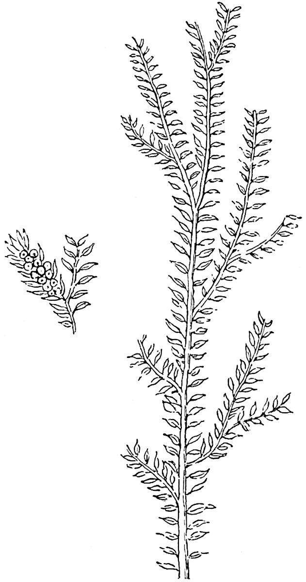
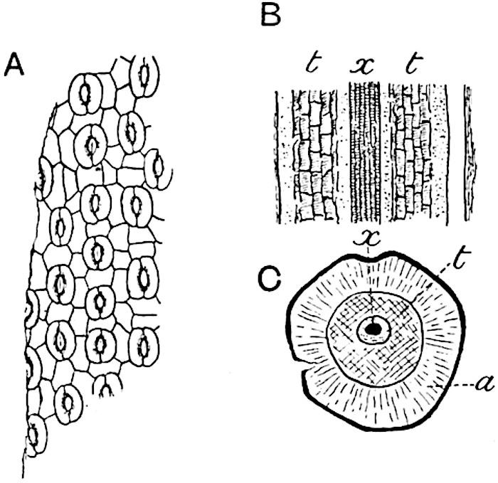
Selaginellites primaevus (Gold.). Fig. 135, A, fig. 138. 1855 Lycopodites primaevus, Goldenberg[227] . 1870 Lycopodium primaevum, Schimper[228] . 1907 Selaginellites primaevus, Halle[229] .
F��. 138. Selaginellites primaevus (Gold.). (After Goldenberg.)
In habit this species, first recorded by Goldenberg from the CoalMeasures of Saarbrücken, is similar to S. Suissei Zeill.
The drawing reproduced in fig. 138 is a copy of that of the typespecimen: another specimen, named by Goldenberg, is figured by Halle in his recently published paper. The leaves appear to be distichous: no smaller leaves have been detected, though Halle is inclined to regard the plant as heterophyllous. The sporophylls, borne in slender terminal strobili, are smaller than the foliage leaves and spirally disposed (fig. 138; smaller specimen). Halle succeeded in demonstrating that some of the sporangia
contained a single tetrad of spores, each spore having a diameter of 0·4–0·5 mm. No microspores were found, but it is clear that the species was heterosporous and that it agrees with recent species in having only four spores in the megasporangium.
Selaginellites elongatus (Gold.). Fig. 135, B, D.
1855 Lycopodites elongatus, Goldenberg[230]
1870 Lycopodium elongatum, Schimper[231] .
The shoots of this species resemble the recent Lycopodium complanatum; they differ from those of Selaginellites primaevus in their long and narrow branches which bear two forms of leaf. The longer leaves, arranged in opposite pairs, are slightly falcate; the smaller leaves are appressed to the axis and have a triangular cordate lamina. Another peculiarity of this species is the occurrence of sporangia in the axil of the foliage leaves, a feature characteristic of the recent Lycopodium Selago. In recent species of Selaginella the sporophylls are always in strobili. No microspores have been found nor the walls of megasporangia, but tetrads of megaspores were isolated by Halle: the spores have three radiating ridges (fig. 135, B) connected by an equatorial ridge. Halle estimates the number of spores (0·45 mm. in diameter) in a sporangium at 20 to 30. In size as in number the spores exceed those of recent species and agree more nearly with the megaspores of S. Suissei.
It would seem to be a general rule that the spores (megaspores) of the fossil herbaceous species exceeded considerably in dimensions those of recent forms and on the other hand were smaller than those of the Palaeozoic arborescent species.
There can be little doubt that some of the Mesozoic and Tertiary species included under Lycopodites agree more closely with the recent genus Selaginella than with Lycopodium, but this does not constitute an argument of any importance against the restricted use of the designation Selaginellites which we have adopted. From a botanical point of view the various records of Lycopodites and Selaginellites have but a minor importance; they are not sufficiently numerous to throw any light on questions of distribution in former periods, nor is the preservation of the material such as to enable us to compare the fossil with recent types either as regards their anatomy or, except in a few cases, their sporangia and spores. The Palaeozoic species are interesting as revealing less reduction in the number of spores produced in the megasporangia. Among existing Pteridophytes the genus Isoetes agrees more closely than
Selaginella, as regards the number of megaspores in each sporangium, with such fossils as Selaginellites Suissei and S. elongatus.
It would seem that in most Palaeozoic species heterospory had not reached the same stage of development as in the recent genus Selaginella in which the megaspores do not exceed four in each sporangium. In Selaginellites primaevus, however, the heterospory appears to be precisely of the same type as in existing species.
Lycostrobus.
The generic name Lycostrobus has recently been instituted by Nathorst[232] for certain specimens of a lycopodiaceous strobilus, from the Rhaetic strata of Scania, which he formerly referred to the genus Androstrobus[233] .
Lycostrobus Scotti Nathorst. Fig. 139.
The fossil described under this name is of special interest as affording an example of a Mesozoic lycopodiaceous cone comparable in habit and in size with some of the largest examples of Palaeozoic Lepidostrobi, the cones of Lepidodendron The Swedish fossil from Upper Rhaetic strata of Helsingborg (Scania) was originally designated Androstrobus Scotti, the generic name being adopted in view of the close resemblance of the form of the strobilus to the male flower of a Cycad. A more complete examination has shown that the bodies, which were thought to be pollen-sacs— though Nathorst recognised certain differences between them and the pollen-sacs of lycopods—are the megaspores of a lycopod. Microspores have also been identified. The axis of the cone has a breadth of 2 cm. with a peduncle which may be naked or provided with a few small scales; the sporophyll region of the axis reached a length of at least 12 cm. The spirally disposed sporophylls terminate in a rhombic distal end which may represent the original termination or they may have been prolonged upwards as free laminae. Each sporophyll bears on its upper face a single large sporangium containing either megaspores or microspores: the megaspores,
0·55–0·60 mm. in diameter, are finely granulate and bear small warty thorns or more slender pointed appendages. The microspores, after treatment with eau de Javelle, were found to measure 36–44μ while others which had been treated with ammonia reached 54μ in diameter. Nathorst describes the microspores as occurring in spherical groups or balls, which it is suggested may be compared with the groups of spores separated by strands of sterile tissue (trabeculae) in the large sporangia of Isoetes (cf. fig. 133, H). If this comparison is sound it would point to a more complete septation of the sporangium in Lycostrobus than in any recent species of Isoetes The size of the strobilus would seem to indicate the persistence into the Rhaetic era of an arborescent lycopodiaceous type; but the appearance and manner of preservation of the axis is interpreted by Nathorst as evidence of a herbaceous rather than a woody structure. He is disposed to regard Isoetes as the most nearly allied existing genus.

F��. 139. Lycostrobus Scotti, Nath. (After Nathorst; ⅘ nat. size.)
The comparison made by Nathorst with Isoetes is based on a resemblance between the spores of the two genera and on the evidence, which is not decisive, of the existence of sterile strands of tissue in the sporangia of Lycostrobus. This similarity is however hardly of sufficient importance to justify the inclusion of the Rhaetic strobilus in the Isoetaceae. In size and in the arrangement and form of the sporophylls the cone presents a much closer resemblance to Lepidodendron than to Isoetes. It is probably advisable to regard this Rhaetic type simply as a lycopodiaceous genus which we are
unable, without additional information, to assign to a particular position.
The opinion expressed by Professor Fliche[234] that the plant described by Schimper and Mougeot as Caulopteris tessellata, a supposed tree-fern stem, from Triassic rocks of Lorraine, is more probably a large lycopodiaceous stem, either a Lepidodendron or a new genus, is worthy of note in reference to Nathorst’s account of Lycostrobus.
In habit the fossil strobilus may be compared with the Triassic genus Pleuromeia, but the position of the sporangia on the sporophylls constitutes a well-marked difference. The most important result of Nathorst’s skillful treatment of this interesting fossil by chemical microscopic methods is the demonstration of the existence of a large heterosporous type of lycopodiaceous cone in a Rhaetic flora.
Poecilitostachys.
Under this generic name M. Fliche[235] has briefly described a fertile lycopodiaceous shoot from the Triassic rocks of Epinal in France: the type species Poecilitostachys Hangi consists of a cylindrical axis, 10 cm. × 5 mm., deprived of leaves and terminating in a rounded receptacle bearing a capitulum of bracts or fertile leaves. Detached megasporangia containing small globular bodies found in association with the capitulum are compared with the megasporangia of Isoetes
CHAPTER XV.
Arborescent Lycopodiales.
A���� the best known plants in the Palaeozoic floras are the genera Lepidodendron and Sigillaria, types which are often spoken of as Giant Club-Mosses or as ancestors of existing species of Lycopodium and Selaginella. Of these genera, but more particularly of Lepidodendron, we possess abundant records in a condition which have made it possible to obtain fairly complete information not only in regard to habit and external features but as to the anatomical characters of both vegetative and reproductive shoots. The structure of Lepidodendron differs too widely from that of recent Club-Mosses (species of Lycopodium) to justify the statement that this prominent member of the Palaeozoic vegetation may be regarded as a direct ancestor of any living plant. There is at least no doubt that Lepidodendron and Sigillaria must be included in the Pteridophyta. The description by Dr Scott[236] of the genus Lepidocarpon, founded on petrified specimens of strobili, demonstrated the existence of a type of lycopodiaceous plant in the Carboniferous period distinguished from all living representatives of the group by the possession of integumented megaspores, which may fairly be styled seeds. Lepidocarpon and another seed-bearing plant Miadesmia are described under a separate heading as lycopodiaceous types characterised by an important morphological feature, which among recent plants constitutes a differentiating character between the Pteridophytes and the Phanerogams.
The genus Lepidodendron included species comparable in size with existing forest trees. A tapered trunk rose vertically to a height of 100 feet or upwards from a dichotomously branched subterranean axis of which the spreading branches, clothed with numerous rootlets, grew in a horizontal direction probably in a swampy soil or possibly under water. A description by Mr Rodway[237] of Lycopods on the border of a savannah in Guiana forming a miniature forest of Pine-like Lycopodiums might, with the omission of the qualifying adjective, be applied with equal force to a grove of Lepidodendra. The equal dichotomy of many of the branches gave to the tree a habit in striking contrast to that of our modern forest trees, but, on the other hand, in close agreement with that of such recent species of Lycopodium as L. cernuum (fig. 123), L. obscurum (fig. 124) and other types. Linear or oval cones terminated some of the more slender branches (fig. 188) agreeing in size and form with the cones of the Spruce Fir and other conifers or with the male flowers of species of Araucaria, e.g. A. imbricata. Needle-like leaves, varying considerably in length in different species, covered the surface of young shoots in crowded spirals and their decurrent bases or leafcushions formed an encasing cylinder continuous with the outer cortex. The fact that leaves are usually found attached only to branches of comparatively small diameter would seem to show that Lepidodendron, though an evergreen, did not retain its foliage even for so long a period as do some recent conifers.
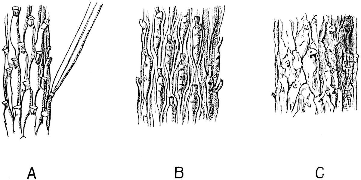
By the activity of a zone of growing tissue encircling the cylinder of wood the main trunk and branches grew in thickness year by year: the general uniformity in size of the secondary conducting elements affords no indication of changing seasons. As the branches grew stouter and shed their leaves the surface of the bark resembled in some degree that of a Spruce Fir and other species of Picea, in which the leaf-scars form the upper limit of prominent peg-like projections, which, at first contiguous and regular in contour, afterwards become less regular and separated by grooves (fig. 140) and at a later stage lose their outline as the bark is stretched to the tearing point (fig. 140, C). The leafless branches of Lepidodendron were covered with spirally disposed oval cushions less peg-like and larger than the decurrent leaf-bases of Picea, which show in the
upper third of their length a clean-cut triangular area and swell out below into two prominent cheeks separated by a median groove and tapering with decreasing thickness to a pointed base, which in some forms (e.g. Lepidodendron Veltheimianum, fig. 185, C, D), is prolonged as a curved ridge to the summit of a lower leaf-cushion.
F��. 140. Picea excelsa. Shoots of different ages showing changes in the appearance of the leaf-cushions: a leaf attached to a cushion in fig. A. (Slightly enlarged.)
A portion of the cushion below the triangular leaf-scar often shows transverse gaping cracks or depressions (fig. 185, C) such as occur on a smaller scale on the older cushions of a Fir twig (fig. 140). Secondary thickening, as in recent trees, is not confined to the vascular cylinder but at an early stage, frequently before there are any signs of secondary wood, the outer region of the broad cortex becomes the seat of active cell-formation which results in the addition of a considerable thickness to the bark. At a later stage of increase in girth, the leaf-cushions are stretched apart and the original surface-features become obliterated by vertical cracks and by the exfoliation of the superficial tissues[238] .
Some species of Lepidodendron produced branches characterised by spiral or vertical series of scars; these in older shoots were replaced by depressions having a diameter of several inches and
comparable in appearance, as also perhaps in manner of formation, with the scars left on the stem of a Kauri Pine (Agathis australis)[239] on the abscission of lateral branches by a natural process. These shoots, known as Ulodendron, are described in a subsequent section. (page 128.)
A fully-grown Lepidodendron must have been an impressive tree, probably of sombre colour, relieved by the encircling felt of green needles on the young pendulous twigs. The leaves of some species were similar to those of a fir while in others they resembled the filiform needles of the Himalayan Pine (Pinus longifolia). The occasional presence of delicate hyphae in the tissues of Lepidodendron demonstrates susceptibility to fungal pests.
Architecturally, if one may use the term, Lepidodendron owed its power of resistance to the bending force of the wind to its stout outer bark formed of thick-walled elements produced by the activity of a cylinder of cortical meristem (figs. 148, 172, etc.). The vascular axis, of insignificant diameter in proportion to the size of the stem (figs. 152, 153, 172, 181, A), must have played a subordinate part, from a mechanical point of view, as compared with the solid mass of wood of a Pine or an Oak.
Within the compass of a text-book it is impossible, even if it were desirable, to include an account of the majority of the species of the widely distributed Palaeozoic genus Lepidodendron. In spite of the great number of known species of this common member of Carboniferous floras, our knowledge of the type as a whole is deficient in many points, and such information as we possess needs systematising and extending by comparative treatment based on a re-examination of available data.
In order to appreciate the meaning of certain external features characteristic of Lepidodendron stems it is essential to have some knowledge of the internal structure.
A dual system of terminology has been unavoidably adopted for species of Lepidodendron: the majority of specific names have been
assigned to fossils known only in the form of casts or impressions, while petrified fragments, which unfortunately seldom show the surface-features, have received another set of names. A glance at the older palaeobotanical literature reveals the existence of several generic designations, which fuller information has shown to have been applied to lepidodendroid shoots deprived of some of their superficial tissues before fossilisation and differing considerably in appearance from the more complete branches of the same species[240] . It has in some instances been possible to correlate the two sets of specimens, casts or impressions, showing external features, and petrified fragments. We may reasonably expect that future discoveries will enable us to piece together as definite specific types specimens at present labelled with different names.
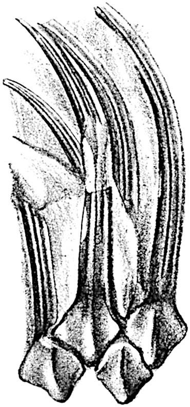
A well-preserved leaf-cushion of a Lepidodendron—the most obvious distinguishing feature of the genus—is rhomboidal or fusiform and vertically elongated (fig. 146, C, E; fig. 185, C, D): in exceptional cases it may reach a length of 8 cm. and a breadth of 2 cm. The cushion as a whole represents a prominent portion of the stem or branch comparable with the elevation on the twig of a Spruce Fir and the leaf-base of a Lycopodium (cf. fig. 121, A, lower portion) which appears in a transverse section of a branch as a rounded prominence (cf. Lycopodium, fig. 125, A and H). Disregarding differences in detail, a typical Lepidodendron leafcushion is characterised by a clearly defined smooth area often situated in the middle region (fig. 146, C, s). This is the leaf-scar or place of attachment of the base of the leaf which was cut off by an absciss-layer while the branch was comparatively young, as in recent forest trees and in some species of Ferns. On the leaf-scar are three smaller scars or cicatricules, the central one is circular or more or less triangular in outline, the two lateral scars being usually oval or circular. The central pit marks the position of the single vascular bundle which constituted the conducting tissue connecting the leaf with the main vascular system of the stem. The two lateral scars (figs. 145, A, p; 146, C, s; 147, p) represent the exposed ends of two strands of tissue, the forked branches of a strand which pass from the middle cortex of the stem into the leaf; this is known as the parichnos, a name proposed by Professor Bertrand in 1891[241] .
The specimen shown in fig. 141 shows the linear leaves attached to their respective cushions.
F��. 141. Lepidodendron Sternbergii. From a specimen in the British Museum (No. v. 1235) from the Coal-Measures of Shropshire. (Nat. size.)
The lamina has a well-defined median keel on the lower surface and on either side a groove in which sections of petrified leaves have demonstrated the occurrence of stomata (cf. fig. 142).
ii. Leaves and Leaf-cushions.
All Lepidodendron leaves, so far as we know, possessed a single median vein only. In some species, as for example in Lepidodendron longifolium Brongn., they have the form of long and slender acicular needles very similar to those of Pinus longifolium; in L. Sternbergii (fig. 141) they are much broader and shorter. In external form as in internal structure it is often impossible to distinguish between the leaves of Lepidodendron and Sigillaria. The distinguishing features enumerated by the late M. Renault cannot be employed, with any
great degree of confidence, as diagnostic characters. In transverse section the lamina of a Lepidodendron leaf presents the same appearance as that of the Sigillarian leaves represented in fig. 142. Near the base the free part of the leaf is usually sub-rhomboidal in section with short lateral wings, a ventral keel and two stomatal grooves (fig. 142, A, B, g). The form and arrangement of stomata are shown in fig. 143, A, which was drawn from a piece of a leaf shown in surface-view in a section lent to me by Professor Weiss. It should, however, be pointed out that the leaf cannot be certainly identified with Lepidodendron rather than with Sigillaria, but as the leaves of these two genera are constructed on the same plan the identification is of secondary importance.
F��. 142. Leaves of Sigillaria in transverse section. A, A′. Section in the Manchester University Museum (Q. 631). B, C. Sections in Dr Kidston’s Collection.
The single xylem bundle consists of primary tracheae only, at least in such laminae as have been identified as Lepidodendroid. Surrounding the xylem strand occur delicate parenchymatous cells in some cases accompanied by darker and thicker-walled elements. As in Sigillaria, the leaves of which are more fully described on page 210, a fairly broad sheath of wider and shorter scalariform or spiral transfusion tracheids surrounds the conducting strand (figs. 142, t; 143, B, C, t). As Renault shows in the case of Lepidodendron esnostense[242] , the small leaves of which are 1·5–2 mm. broad at the base and several centimetres long, the stomatal grooves and keel die out towards the apex when the lamina assumes a more nearly circular form (fig. 143, C).
F��. 143.
A. Stomata in surface-view (Lepidodendron?). a, parenchyma; t, transfusion tracheae; x, xylem. (Manchester University Collection R. 723).
B, C. Lepidodendron esnostense Ren. (After Renault.)
The area of the cushion excluding the leaf-scar is spoken of by some writers as the field. Below the leaf-scar the kite-shaped cushion tapers to a gradually narrowing basal position: in Lepidodendron Veltheimianum, a species characteristic of Lower
Carboniferous strata, it is seen to be continuous, as a ridge with sloping sides, with a lower cushion (fig. 185).
Below a leaf-scar the cushion frequently shows a pair of oval areas on which a fine pitting may be detected in well-preserved impressions, these oval scars, as seen in fig. 185, D, are practically continuous at the upper end with the parichnos scars on the leafscar area; this is explained by the fact that these infra-foliar scars also owe their existence to patches of lacunar, aerenchymatous tissue in close connexion with the parichnos[243] .
Shortly before entering the base of the leaf-lamina the parichnos divides into two arms which diverge in the outer cortical region right and left of the vascular bundle, and passing obliquely upwards they come close to the surface of the leaf-cushion just below the leafscar. The diagram—fig. 144, B—shows a leaf-trace, lt, in the leafcushion, as seen in a diagrammatic drawing of a vertical radial section of a stem, the dotted lines, p, p′, show the two parichnos arms which are represented as impinging on the surface of the leafcushion at p′, and then bending upwards to pass into the leaf-base right and left of the vascular bundle or leaf-trace For convenience the arms of the parichnos are represented in one plane though actually in different vertical planes.
Fig. 144, A, shows the difference between a view of the original surface of a Lepidodendron, as at a, where a leaf-cushion with a leaf-scar is seen, and a view of an impression representing the outer cortex, b, a short distance below the surface. The surface b, in fig. 144, A, corresponds to the face d-e in the diagrammatic longitudinal section fig. 144, B: the outline of each cushion is clearly visible and in the centre is seen the leaf-trace, lt, with its parichnos.
The surface-features, a (fig. 144, A), have been impressed on the rock, c, (fig. 144, B) in which the specimen was entombed and by the removal of the cast of the stem, that is the thickness b to e in fig. 144, B, the form of the leaf-cushion is revealed. The presence of the two infra-foliar parichnos scars at p′ (fig. 144, A) is explained by the diagram, fig. 144, B, p′.
The relation of the parichnos to the oval scars below a Lepidodendron leaf-cushion has been worked out in detail by Weiss who shows that, at least in some species, the two arms do not bend downwards as shown in the diagram, fig. 144, B, but pursue a straight gradually ascending course as seen in fig. 145, A. Just below the leaf-scar region of the cushion each arm comes into association with a group of lacunar, aerenchymatous tissue, such as occurs in the roots of certain Mangrove plants, and it is this aerenchyma which is exposed on the two oval depressions below the leaf-scar The structure of this aerenchyma is shown in fig. 145, B; it consists in this species (L. Hickii Wats.) of stellate cells which would constitute an efficient aerating system. Probably, as Weiss suggests, these patches of aerenchyma were originally covered by an epidermis provided with stomata, and it is owing to the destruction of this superficial layer that the two oval scars often form a prominent feature on Lepidodendron leaf-bases[244] . The diagram reproduced in fig. 144, B, may be taken as practically correct, as the patches of aerenchyma described by Weiss do not differ essentially from the parichnos tissue.
Welcome to our website – the ideal destination for book lovers and knowledge seekers. With a mission to inspire endlessly, we offer a vast collection of books, ranging from classic literary works to specialized publications, self-development books, and children's literature. Each book is a new journey of discovery, expanding knowledge and enriching the soul of the reade
Our website is not just a platform for buying books, but a bridge connecting readers to the timeless values of culture and wisdom. With an elegant, user-friendly interface and an intelligent search system, we are committed to providing a quick and convenient shopping experience. Additionally, our special promotions and home delivery services ensure that you save time and fully enjoy the joy of reading.
Let us accompany you on the journey of exploring knowledge and personal growth!