Axial spondyloarthritis and ankylosing spondylitis (the facts series) 2nd edition muhammad asim khan
Axial Spondyloarthritis and Ankylosing Spondylitis (The Facts Series) 2nd Edition Muhammad Asim Khan
Visit to download the full and correct content document: https://ebookmass.com/product/axial-spondyloarthritis-and-ankylosing-spondylitis-thefacts-series-2nd-edition-muhammad-asim-khan/
More products digital (pdf, epub, mobi) instant download maybe you interests ...
Dry Beans and Pulses Production, Processing, and Nutrition, 2nd Edition Muhammad Siddiq
Ankylosing Spondylitis and Axial Spondyloarthritis
E also available in the facts series
Lung Cancer: the facts
THIRD EDITION
Falk and Williams
Psoriatic Arthritis: the facts
Gladman and Chandran
Schizophrenia: the facts
FOURTH EDITION
Glatt, Faraone, and Tsuang
The Pill and other forms of hormonal
contraception: the facts
SEVENTH EDITION
Guillebaud and MacGregor
Myotonic dystrophy: the facts
SECOND EDITION
Harper
Alzheimer’s and other Dementias: the facts
Hughes
Lupus: the facts
SECOND EDITION
Isenberg and Manzi
Angina and Heart Attack: the facts
Jevon
Ankylosing Spondylitis and Axial
Spondyloarthritis: the facts
SECOND EDITION
Khan
Borderline Personality Disorder: the facts
Krawitz and Jackson
Inflammatory Bowel Disease: the facts
Langmead and Irving
Back Pain: the facts
Lee, Brook, and Daniel
Stroke: the facts
SECOND EDITION
Lindley
Alcoholism: the facts
FOURTH EDITION
Manzardo, Goodwin, Campbell, Penick, and Gabrelli
Prostate cancer: the facts
SECOND EDITION
Mason and Moffat
Diabetes: the facts
Matthews, Meston, Dyson, Shaw, King, and Pal
Skin Conditions in Young People: the facts
McPherson
Essential tremor: the facts
Plumb and Bain
Osteoarthritis: the facts
SECOND EDITION
Prieto-Alhambra, Arden, and Hunter
Huntington’s Disease: the facts
THIRD EDITION
Quarrell
Panic disorder: the facts
THIRD EDITION
Rachman and de Silva
Obsessive-compulsive disorder: the facts
FOURTH EDITION
Rachman and de Silva
Post-traumatic Stress: the facts
SECOND EDITION
Regel and Joseph
Tourette syndrome: the facts
SECOND EDITION
Robertson and Cavanna
Breast Cancer: the facts
SECOND EDITION
Saunders, Jassal, and Lim
Dyslexia and other learning difficulties: the facts
THIRD EDITION
Selikowitz
Down Syndrome: the facts
THIRD EDITION
Selikowitz
ADHD: the facts
THIRD EDITION
Selikowitz
Sleep problems in Children and Adolescents: the facts
Stores
Motor neuron disease: the facts
Talbot and Marsden
Cystic fibrosis: the facts
FOURTH EDITION
Thomson and Harris
Thyroid disease: the facts
FOURTH EDITION
Vanderpump and Tunbridge
Depression: the facts
SECOND EDITION
Wassermann
Cosmetic surgery: the facts
Waterhouse
the facts
Ankylosing Spondylitis and Axial Spondyloarthritis
SECOND EDITION
MUHAMMAD ASIM KHAN, MD, FRCP, MACP, MACR
Professor Emeritus of Medicine, Case Western Reserve University, Cleveland, Ohio, USA
Great Clarendon Street, Oxford, OX2 6DP, United Kingdom
Oxford University Press is a department of the University of Oxford. It furthers the University’s objective of excellence in research, scholarship, and education by publishing worldwide. Oxford is a registered trade mark of Oxford University Press in the UK and in certain other countries
All rights reserved. No part of this publication may be reproduced, stored in a retrieval system, or transmitted, in any form or by any means, without the prior permission in writing of Oxford University Press, or as expressly permitted by law, by licence or under terms agreed with the appropriate reprographics rights organization. Enquiries concerning reproduction outside the scope of the above should be sent to the Rights Department, Oxford University Press, at the address above
You must not circulate this work in any other form and you must impose this same condition on any acquirer
Published in the United States of America by Oxford University Press 198 Madison Avenue, New York, NY 10016, United States of America
British Library Cataloguing in Publication Data
Data available
Library of Congress Control Number: 2022938866
ISBN 978–0–19–886415–8
DOI: 10.1093/oso/9780198864158.001.0001
Printed and bound by CPI Group (UK) Ltd, Croydon, CR0 4YY
Oxford University Press makes no representation, express or implied, that the drug dosages in this book are correct. Readers must therefore always check the product information and clinical procedures with the most up-to-date published product information and data sheets provided by the manufacturers and the most recent codes of conduct and safety regulations. The authors and the publishers do not accept responsibility or legal liability for any errors in the text or for the misuse or misapplication of material in this work. Except where otherwise stated, drug dosages and recommendations are for the non-pregnant adult who is not breast-feeding
Links to third party websites are provided by Oxford in good faith and for information only. Oxford disclaims any responsibility for the materials contained in any third party website referenced in this work.
Dedication
I dedicate this book to my father, Umar Khan, my mother, Hameeda Khanam, and to her father, Sadr-ud-Din Khan (a high school principal who retired as inspector of schools) for having inculcated in me the passion to pursue knowledge and impart it to others. They all worked tirelessly to re-establish when we became refugees, uprooted from our ancestral lands when I was a little over three years old. Therefore, I also dedicate this book to all the refugees like me in this world who may still be longing for a home, and most of them also happen to share my faith.
Advance praise
‘Written for patients by a patient who is also a leading authority on spondyloarthritis, this book is an essential reference and reading for people living with axial spondyloarthritis— with ankylosing spondylitis as its prototype—and their caregivers who want to learn about the disease and how to manage it well.’
Michael Mallinson, Patient Advocate and Volunteer, Axial
Spondyloarthritis International
Federation (ASIF)
Preface
There have been tremendous advances in clinical understanding, early disease recognition, and more effective management of ankylosing spondylitis (AS) and related diseases. The advent of newer imaging methods, such as magnetic resonance imaging (MRI) and very low-dose computerized tomography (CT), have also facilitated early diagnosis and initiation of increasingly more effective (but costly) drugs called “biologics” (administered by injection under the skin or by intravenous infusion), and most recently JAK-inhibitors that are taken orally as tablets. These drugs target inflammatory proteins (cytokines), such as tumor necrosis factor (TNF) and interleukin-17 (IL-17).
All these new development and progress in diagnosis and management of AS and related diseases has necessitated this second edition of the book. I have updated its title by adding the term “Axial Spondyloarthritis” that requires some explanation.
The term “spondyloarthritis” (or SpA) refers to a family of chronic inflammatory non-contagious (non-infectious) forms of arthritis involving the spine and the limbs that share many of their clinical features and genetic predisposing factors. Those patients with SpA who primarily have inflammation of the joints and ligaments of the back and neck (i.e., the axial skeleton) are now sub-classified as having “axial spondyloarthritis” (axSpA); whereas those with involvement predominantly of the peripheral (distal) limb joints (other than hip and shoulder joints that are part of the axial skeleton), are labeled as having “peripheral SpA.” The typical example of axSpA is AS, while that of peripheral SpA is psoriatic arthritis (PsA).
X-ray evidence of damage to the sacroiliac joints (sacroiliitis) is required for the diagnosis of AS. We first reported in 1985 that one can clinically recognize the disease even when there is no X-ray evidence of sacroiliitis. We called it “spondylitic disease without radiological evidence of sacroiliitis.” It is now called “non-radiographic axSpA” (nr-axSpA). The name axSpA encompasses both nr-axSpA and AS.
Most of the current knowledge about axSpA was gained when the disease was called AS, and this is my reason for the use of both AS and axSpA in the title of
this second edition. It is my hope that the third edition of this book will simply be titled Axial Spondyloarthritis: The Facts. The term “radiographic axSpA” (raxSpA) has sometimes been equated with AS but I have preferred to use the term AS in this book as it is the most widely accepted name.
This book is written in a clear and accessible style with American English spellings for patients and their families and friends. But it is also ideal for students and healthcare professionals of all levels who are looking for concise and practical information on all aspects of AS/axSpA. Discussions about the associated forms of SpA, including PsA, reactive arthritis, inflammatory bowel disease associated SpA, juvenile SpA, and some other diseases that may be confused with SpA (the so-called disease mimickers or look-alikes) are also discussed. A glossary of medical terms, a list of abbreviations, links to patient support groups and other helpful organizations, a list of medical references for further reading, and an index are also included.
People who are knowledgeable about their disease show more self-responsibility, comply better with recommended treatment, and are more likely to make positive behavioral changes that will help them achieve an improved health status in the long run. I hope this book will serve people living with AS/axSpA and their families and friends in their need for self-education.
Lastly, I would add that this book provides a general information that cannot replace the care and knowledge provided by your professional healthcare providers. You need to consult them if you have questions as you read this book.
Muhammad Asim Khan Professor Emeritus of Medicine
Case Western Reserve University, Cleveland, Ohio, USA
Acknowledgments
I am most grateful to my wife Mastoora and my sons Ali and Raza for their help; and also very thankful to the many patients, colleagues, and students who have, over the years, enhanced my knowledge of ankylosing spondylitis, an illness I have myself lived with for more than sixty-six years.
Abbreviations
3D three-dimensional
AAU acute anterior uveitis
ACE angiotensin-converting-enzyme
ACR American College of Rheumatology
ADL activities of daily living
AI artificial intelligence
AS ankylosing spondylitis
ASAS Assessment of SpondyloArthritis international Society
SAPHO synovitis, acne, palmoplantar pustulosis, hyperostosis, and aseptic osteomyelitis
SASSS Stoke Ankylosing Spondylitis Spine Score
SEA seronegative enthesitis and arthritis
SF- 36 Short-Study Form-36
SI sacroiliac (or sacroiliitis)
SI joint or SIJ sacroiliac joint
SpA spondyloarthritis or spondyloarthropathy
THA total hip arthroplasty
THC tetrahydrocannabinol
THR total hip joint replacement
TMJ temporo-mandibular joint
TNF tumor necrosis factor
TNFi tumor necrosis factor inhibitor
ts-DMARDs targeted synthetic-DMARDs
UC ulcerative colitis
VAS visual analog scale
WBC white blood-cell count
WHO World Health Organization
WLQ- 25 Work Limitations Questionnaire-25
WPAI Work Productivity and Activity Impairment
xvi
1 What is ankylosing spondylitis?
% Key points
◆ Ankylosing spondylitis (AS) is a chronic (long-term), slowly progressive, and painful inflammatory arthritis of the sacroiliac (SI) joints and the spine that can lead to gradually progressive impairment of spinal mobility.
◆ It affects both males and females, and the symptoms typically start during adolescence and early adulthood, and it is very uncommon for the symptoms to begin after age 45.
◆ The inflammation can also involve the hip and shoulder joints, and less often other limb joints, such as knees, ankles, and heels.
◆ One or more episodes of acute eye inflammation (acute anterior uveitis) can occur in >30% of patients, and 6 to 10% suffer from psoriasis and/or inflammatory bowel disease.
◆ The disease is observed worldwide with very variable prevalence, e.g., it affects one in 200 (0.5% prevalence) adults of European ancestry, but is very uncommon in most of the sub-Saharan African populations.
◆ Its cause is not fully understood but is largely genetically determined, and there is a strong association with a gene called HLA-B27.
◆ There is no cure as yet but most patients can be very well managed if diagnosed and treated early with increasingly effective, though costly, drugs that markedly reduce the risk of irreversible structural damage and help patients pursue a very active and productive lifestyle.
What is ankylosing spondylitis? · the facts
Introduction
Ankylosing spondylitis (AS) is a chronic (progressive) painful inflammatory rheumatic non-contagious (non-infectious) disease that involves the back, i.e. the sacroiliac (SI) joints and the spine, that often results in some degree of stiffness (decreased flexibility) of the spine (Figure 1.1). The word ankylosing comes from the Greek root ankylos, meaning bent, although it has now come to imply something that restricts motion (stiffening) and may ultimately result in fusion. When the joint loses its mobility and becomes stiff it is said to be ankylosed. The word spondylitis means inflammation in the joints of the spine, and is derived from spondylos, which is the Greek word for vertebra, and -itis, which implies presence of inflammation. The name therefore suggests that AS is an inflammatory disease of the spine that can lead to stiffening of the back. It is important to point out that the words spondylitis and spondyloarthritis should not be confused with spondylosis, which relates to wear and tear in the spinal column (degenerative disc disease) as we get older.
Ankylosing spondylitis in history
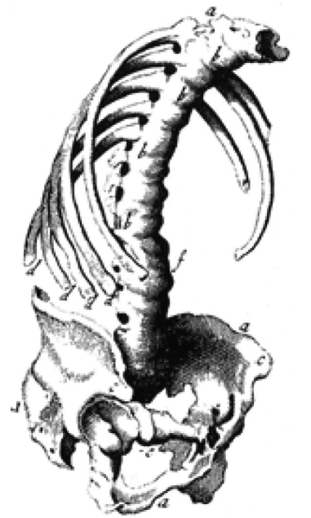
Skeletal specimens in several museum collections testify to the existence of AS from the earliest times. But its first definite anatomical description can be credited to Bernard Conner (1666–1698). He was an Irish physician studying medicine in France when some farmers brought him a skeleton they had found in a cemetery. He wrote in his report, accompanied by the drawing of the skeleton (see Figure 1.2), that the bones were “so straightly and intimately joined, their ligaments perfectly bony, and their articulations so effaced, that they really made but one uniform continuous bone.”
The clinical descriptions of the disease date from the late nineteenth century, with a series of publications in the 1890s by Vladimir von Bechterew (1857–1927) in St Petersburg, Russia, Pierre Marie (1853–1940) in France, and Adolf Strümpell (1853–1926) in Germany. Report of the earliest X-ray examination of a patient with AS was published in 1899, and the characteristic X-ray finding of obliteration of the SI joints was described in 1934.
The name ankylosing spondylitis is widely in use in English speaking countries, with its translation spondylarthrite ankylosante in French, spondylitis ankylopoëtica in Dutch, and espondilartritis ankylosante in Spanish. Older names include Morbus Bechterew (Bekhterew’s or Bekhterev’s disease), Bekhterev-StrümpellMarie disease and Marie-Strümpell spondylitis. Although the use of eponyms is now discouraged for naming a disease, the term: “Morbus Bechterew” is still being widely used in German-speaking countries. Other names that have been used include spondyloarthritis ankylopoëtica, pelvospondylitis ossificans, and
Figure 1.1 Sites that may be involved in AS. The most involved sites are the sacroiliac joints and the spine. They are marked by rectangles. Other, relatively less commonly involved sites are hip and shoulder joints, and less often the knee joints. These sites are marked by circles.
Reproduced with permission from Khan MA. “Spondyloarthropathies” in Hunder G (ed), Atlas of Rheumatology. Philadelphia, PA: Current Medicine Philadelphia, 2005, 151–80.
Figure 1.2 First representation of a skeleton with AS in its final state by Bernard Conner, London, 1695.
the laymmen’s terms bamboo spine and poker back. In the US the disease was wrongly called “rheumatoid spondylitis” up until early 1960s because of a mistaken belief that it was just a variant of rheumatoid arthritis (RA).
Structure of the spine
The spine, also called the spinal or vertebral column or the backbone, consists of 24 vertebrae that are stacked one above the other, separated from each other by intervertebral discs that act as shock absorbers during mechanical stress, and are held together by strong ligaments and 110 small joints. The spinal column is shown in lateral and front views in Figure 1.3, divided into three main sections:
◆ the upper part (the neck or cervical spine) has 7 vertebrae and is the most mobile part of the spine.
Atlas Axis
Cervical ver tebrae
oracic ver tebrae
Lumbar ver tebrae
Sacrum
Coccyx
Figure 1.3 The vertebral (spinal) column.
What is the sacroiliac joint?
Atlas Axis
Cervical ver tebrae
oracic ver tebrae
Lumbar ver tebrae
Sacrum
Coccyx
◆ the middle part (thoracic spine) has 12 vertebrae, and each has a rib attached to the vertebra on either side by two joints called costo-vertebral and costotransverse joints. The 12 ribs on either side make up the chest wall, and they are attached in the front to the breastbone (sternum) by costo-chondral junctions. The term costo stands for the rib and chondral stands for cartilage.
◆ the lower part (lumbar spine) has 5 vertebrae, the lowermost (5th) lumbar vertebra sits on a bone called the sacrum.
What is the sacroiliac joint?
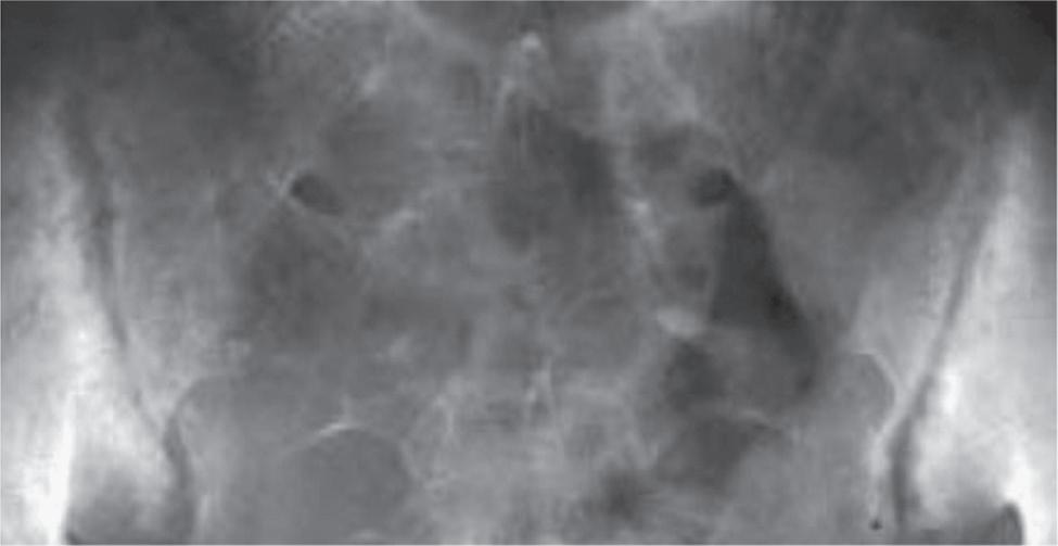
The sacrum bone looks like a keystone in the circular bony pelvis formed together with the right and left pelvic bones. It is attached on either side to ilium (the major part of the pelvic bone) by joints called the sacroiliac (SI) joints (Figure 1.4). The inflammation usually starts first at these two joints in patients
Figure 1.4 The sacroiliac joint: (a) location of the right sacroiliac joint marked by the line separating the sacrum from the ilium, as viewed from the front; (b) pelvic X-ray showing irregularities (bony erosions) of both right and left sacroiliac joints, a characteristic feature of AS.
(b) Reproduced with permission from Khan MA. “Spondyloarthropathies” in Hunder G (ed), Atlas of Rheumatology Philadelphia, PA: Current Medicine Philadelphia, 2005, 151–80.
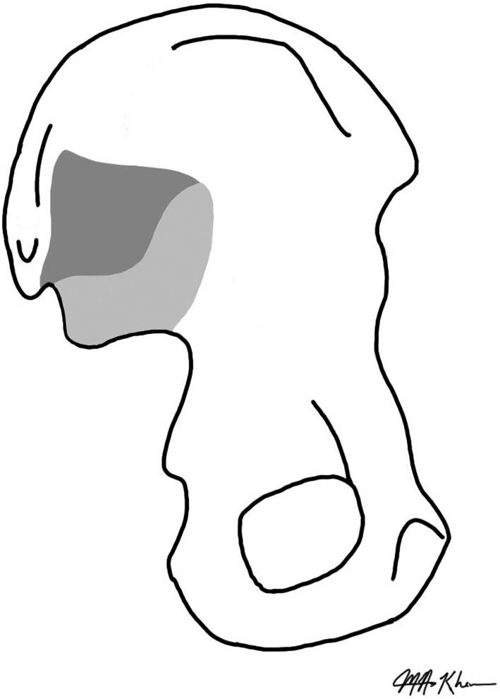
with AS. Figure 1.5 shows a simplified sketch as viewed from inside the pelvis of a removed left pelvic bone. The darker area depicts the site for attachment of ligamentous and the lighter area (shaped like a boomerang) represents its “synovial” part. The adjacent lower-back part of the pelvic bone that bears our weight when we are sitting down is called the ischium or the gluteal tuberosity (cushioned by the overlying buttock muscles) of the pelvic bone. The front part
Figure 1.5 The sacroiliac joint area, as visualized from the inner side of the separated left pelvic bone, is shown in two shades of gray; the darker area depicts the ligamentous part and the lighter area (shaped like a boomerang) forms its “synovial” part. Just behind the ligamentous part is the area called the ischium (or the gluteal tuberosity) that bears our weight when we are sitting down. The front lower part of the pelvic bone is called the pubis that forms a junction (called the pubic junction or pubic symphysis) with the other pelvic bone.
of the pelvic bone is called the pubis that joins with its counterpart from the other side to form the pubic junction (or pubic symphysis).
What is the axial skeleton?
The axial skeleton includes the SI joints, the spine (including the neck), the “root joints” of the limbs (hip and shoulder joints), all the joints and ligamentous structures in the spine, the attachment of the ribs to the back and to the breastbone in the front, as well as attachment of the collar bones (clavicles) to the shoulder girdles (by acromioclavicular joints ) and the breastbone (by manubrioclavicular joints ). The sites of attachments of the ribs to
What is ankylosing spondylitis? · the facts
the breastbone are called costochondral junctions . These sites can be involved in AS, and so can the junction between the two parts ( manubrium and sternum ) of the breastbone.
The disease symptoms and severity vary from person to person. The disease in some patients may be relatively mild or stay limited to the SI joints and the lumbar spine.
Chronic inflammatory back pain and stiffness
Symptoms typically start during the third decade of life (with a wide age range from 12 to 45), with the mean age of about 24 years (even younger in some of the developing parts of the world). The leading initial symptom is chronic (lasting for more than 3 months) low back pain and stiffness of gradual (insidious) onset that results from inflammation of the SI joints and the lumbar spine. It can initially be felt in the lower back or buttocks area and some patients may complain that their pain initially alternated from one side to the other (alternating buttock pain). The pain may also be felt down the upper part of the back of the thigh but it does not radiate all the way down the lower legs.
The pain and stiffness get worse with prolonged physical inactivity and rest, especially at late hours of the night and early morning. They persist for more than half an hour after waking up, and they ease up after taking a hot shower, walking around, or exercising, but not with rest. To remember the 5 salient features of inflammatory back pain (IBP) we have proposed a mnemonic—“IPAIN”—that makes it easier to remember (Box 1.1). It is unlike the common (ubiquitous) back pain or sprain, often called mechanical or non-specific back pain.
Box 1.1 IPAIN: a mnemonic for inflammatory back pain
IPAIN
Insidious onset
Pain at night (with improvement upon getting up)
Age at onset <45 years
Improvement with exercise
No improvement with rest
Reproduced with permission from Ozgocmen S, Akgul O, Khan MA. “Mnemonic for assessment of the spondyloarthritis international society criteria” J Rheumatol. 2010 Sep;37(9):1978.
Some people may complain of only transient episodes of lower-back pain with remission periods in between before their symptoms become persistent. In others, the first symptoms may not be in the back, but they may complain of painful heels and limb joints, especially hip and/or shoulder joints, called the girdle joints. In some such cases it may be difficult to distinguish the disease from some other rheumatic diseases when there is no back pain present, but the typical back symptoms generally do develop later. Some patients, especially females, may present with neck or upper back pain and stiffness, tenderness of the anterior (front) chest wall, or chest pain that is accentuated on coughing or on taking a deep breath due to inflammation of the joints that attach the ribs to the spine and anterior chest wall. Others may present with inflammation of other structures, such as the eyes, the gut, and the skin, and so their first medical visit may be an eye doctor (ophthalmologist) for an episode of painful acute inflammation of the eye (acute anterior uveitis or AAU for short), discussed in Chapter 6, a gastroenterologist (for symptoms of inflammatory bowel disease (IBD)), or a skin specialist (dermatologist) for psoriasis, as discussed in detail in Chapter 10.
Current terminology
I have explained in the Preface that AS belongs to a group of diseases under the term spondyloarthritis (or SpA for short). Since AS is predominantly a disease of the axial skeleton, it falls under the newly proposed term axial SpA (abbreviated as axSpA). A term—radiographic axSpA (r-axSpA)—has sometimes been equated with AS due to the required presence of definite bony structural damage of the SI joints as detected on radiographic (X-ray) examination. But in this book I have used the term AS instead of r-axSpA because it is the most widely used name for this condition.
SpA patients showing involvement predominantly of their “peripheral” limb joints (other than hip and shoulder joints) are labeled as having predominantly peripheral SpA, and its best example is psoriatic arthritis (PsA). These two subtypes of axial and peripheral SpA, however, do show some overlapping clinical features (see Figure 2.1), and they also share some of the underlying genetic predisposing factors. Moreover, as discussed in Chapter 10, patients suffering from AAU, IBD, or reactive arthritis have increased risk of developing SpA, either the axial or the peripheral form or both.
Prevalence
AS is present worldwide but with variable prevalence and incidence that are strongly dependent and are directly correlated to the prevalence of HLA-B27
What is ankylosing spondylitis? · the facts
in the general population, as detailed in Chapter 4. For example, it affects approximately up to 1 in 200 (0.5% prevalence) adults of European ancestry, and among northern Arctic communities and Chinese with a wide variation from 0.2 to 0.5%. It is very uncommon in sub-Saharan African populations. I may add that the prevalence figure for the whole group of SpA is even higher as it includes PsA, and SpA associated with IBD and reactive arthritis.
What is its cause?
The cause of AS/axSpA is not yet fully known but carries strong genetic predisposition. Most patients possess a normal gene called HLA-B27, that is also present in a very small percent of the general population. (Please note that the term HLA-B27 is italicized only when referring to its gene, but not when it refers to the protein molecule produced by the gene). The genetic predisposition cannot be pinned down to this one gene because many additional genetic factors or genes are also involved (discussed in Chapter 3).
The role of gut microbiome
There are trillions of microorganisms (microbes) comprising viruses, bacteria, and fungi that are present at our “barrier surfaces” (e.g. our gut lining and the skin) that form what we call the human “gut microbiome” and “skin microbiome,” respectively. They have evolved to live with us in harmony. The gut microbiome helps digest and convert our food into energy that they also require to survive, and make some key vitamins, such as vitamin K. They also develop and support our immune system to form a frontline of defense to protect us against the harmful microbes. Their effect seems to extend beyond the gut and affect many body functions, even general mood and sleep via the vagus nerve that provides communication between the gut and the brain (the so- called gut– brain axis).
There is a disruption and decreased biodiversity of the gut microbiome in patients with AS, and approximately 60% of the patients have asymptomatic mild gut inflammation, especially among those who also have peripheral joint involvement. Evidence is emerging that the HLA-B27 gene may have some influence on gut microbiome even in unaffected people. But the presence of asymptomatic gut inflammation in AS patients does not show any clear association with HLA-B27. This supports the existence of some common link between gut inflammation and AS, even independent of HLA-B27.
There are an increasing number of other diseases in which disruptions of the gut microbiome is implicated, ranging from IBD, RA, obesity, diabetes, and colon cancer, while disruption of the skin microbiome may be involved in
Immune system and its role
psoriasis and PsA. It is also important to know that medications, such as antibiotics, can alter the gut microbiome, and conversely the gut microbiome can metabolize some of the antibiotics and other drugs we take.
Immune system and its role
We possess two main immune strategies against microbial infections: innate immunity and adaptive immunity. The innate (also called non-specific) immune response is broad and does not need to learn what is dangerous and mostly includes macrophages, neutrophils, and dendritic cells that are equipped with a kind of sensor called pattern recognition receptors (PPRs) that help them quickly trigger defensive strategies by detecting pathogen-associated molecular patterns (PAMPs) that represent overlapping similarities (resemblances) across many disease-causing (pathogenic) microbes. The innate immune response in many cases clears the infection before the adaptive immune response is ready to help out.
The adaptive immune system is more specific because it will recognize exactly the kind of infecting microbe in order to use the exact defensive mechanism needed to get rid of it. This is carried out mainly by lymphocytes, and they are split up into B cells that produce antibodies and T cells that help in getting rid of virus-infected cells. But it takes 10 to 15 days for the T and B cells to acquire that capability and expand in numbers.
The immune cells belonging to both the innate and the adaptive response also build up their memory against the infection so that next time around they more quickly mount a specific (tailor-made) immune response. This adaptive response, that was previously wrongly thought to happen only as part of the adaptive immune system, is called trained immunity by which the innate immune cells also improve their ability to deal with the infection the second time around.
Our intestines have more than 350 square feet of inner lining (mucosal membrane or lamina propria) that is a major site of development of adaptive immunity. A subset of cells in the gut lining or in the adjacent (mesenteric) regional draining lymph nodes perform a function like that of the thymus gland (located behind the breastbone but in front of the heart that shrinks at the approach of puberty). The mesenteric lymph nodes delete T lymphocytes that can potentially attack body’s own organs by a process called “suicidal death” or apoptosis. Failure to delete such cells from the gut wall can potentially lead to autoimmune diseases where the body damages itself instead of the harmful invading microbes. Thus, such autoimmunity can also result in sub-clinical (asymptomatic) gut inflammation or even florid IBD.
What is ankylosing spondylitis? · the facts
Any specific environmental trigger for AS remain unknown. Therefore, it has also been proposed that some foreign (non-self-derived) small protein molecules (peptides) may trigger the self-peptide-activated T cells during early life, and this may lead the autoimmune and inflammatory process in later years that results in a disease, including AS.
Laboratory-raised rats genetically engineered to carry the human HLA-B27 gene have advanced our understanding of how it may predispose humans to AS and related SpA. These so called “HLA-B27 transgenic” rats have been developed in research laboratories that spontaneously develop an inflammatory arthritis that shares many features with the human disease, including sacroiliitis. This illness occurs if they develop diarrhea after they are removed from germ-free (sterile) environment and kept in their natural normal environment. Interestingly, some of these rats without diarrhea develop psoriasis-like skin and nail lesions.
Studies of a mouse model for AS have demonstrated that mechanical strain may trigger inflammation at sites of attachments of ligaments and tendons to bone, known as entheses (singular is enthesis), and lead to new bone formation that occurs in AS. This is a self-directed inflammation whereby local factors at sites predisposed to disease initiate activation of innate immune cells, including macrophages and neutrophils, that can promote reactive bone remodeling and new bone formation. Thus, biomechanical stress and possible microdamage at entheses and resultant autoinflammatory response independent of adaptive immune system has also been proposed to have a role in development of AS.
Men versus women
AS is diagnosed 2 to 3 times more commonly in men than in women, but there is a preponderance of women among patients with non-radiographic form of axSpA (nr-axSpA), as discussed in the next chapter. Women have a lower intensity of inflammation, but they do not differ from men with regard to physical functional impairment and overall health status. This subject and the prevalence of the disease in women are discussed in Chapter 5.
There is no difference in the age of onset, but the diagnosis in women is delayed significantly longer than among men. For example, a recent review that included 23,883 patients (32.3% were women) from 42 publications reported 8.8 years (range 7.4–10.1) of delay in diagnosis for women versus 6.5 years (range 5.6–7.4) for men. Other factors consistently reported to be associated with longer delays are the patients’ lower education levels, younger age at symptom onset and absence of extra-articular (or extra-musculoskeletal)
manifestations. Presence of extra-articular manifestation (e.g. recurrent episodes of acute anterior uveitis, presence of psoriasis or IBD), HLA-B27, or a family history of SpA increases the chance of early diagnosis. It is of interest that studies from high-income countries (as defined by the World Bank) have reported longer delays than those from middle-income countries. It is also worth mentioning that in contrast to AS/axSpA, diagnostic delay in PsA was reported to be only 2.6 years.
In an online survey of 2,846 European patients with SpA, women reported higher number of visits to the primary care physicians, physiotherapists, and osteopaths. Neck and upper-back pain, anterior chest wall pain and tenderness (costochondritis), or limb joint involvement may be the main presenting manifestations among women. They also tend to have more anxiety, depression, peripheral joint symptoms and widespread pain. The presence of such clinical features may lead to a misdiagnosis of fibromyalgia (fibrositis) in women suffering from AS/axSpA. There is also a slower and relatively incomplete progression of spinal fusion (ankylosis) among women. This may mean that it takes longer for regression of their pain that often follows complete spinal bony fusion. There are also differences in levels of proinflammatory cytokines and immunological responses. Both men and women respond to treatment with biologics, but women achieve less than a desired level of response and also show a higher discontinuation rate as compared to men.
Hormonal status and fertility are normal in both sexes. Pregnancy usually does not alleviate or change symptoms of AS or it may only cause a temporary aggravation. Childbirth is normal in the absence of severe hip disease. The growth and development of infants and young children are comparable to those of other mothers unaffected with AS. As discussed in a later chapter on management, certolizumab pegol (Cimzia®) is one of the tumor necrosis factor (TNF) inhibitors that can be continued during pregnancy as it does not cross the placenta and therefore the fetus is not exposed to this drug.
Delay in diagnosis
There is an unacceptable delay in the diagnosis of AS/axSpA, partly due to lack of adequate knowledge of many health care providers about its variable clinical presentation, and absence of any validated diagnostic criteria. Need for an early diagnosis, including that of nr-axSpA has now become more important because of the availability of increasingly more effective therapies, especially if they are started at early stage of the disease. An early diagnosis also helps avoid unnecessary investigative procedures and inappropriate treatments.