Autopsy and Case Reports 2013; 3(4): 23-8
echocardiography revealed diffuse left ventricle hypokinesia; mitral and tricuspid valves insufficiency, and the presence of the pacemaker’s electrode located in the right ventricle. Eight days after surgery the patient experienced sudden respiratory discomfort and died soon after.
AUTOPSY FINDINGS On external examination, the anthropometric measures revealed the body of a man 163 cm in height with a weight of 69 kg. A recent surgical scar measuring 6.5 cm replaced original topography of the external genitalia. At the opening of the thoracic cavity a small amount of pleural effusion was present bilaterally, and a thick pericardium had adhered to the pleura. The heart was enlarged and weighted 530 g (reference mean value [RMV]: 327 g). On sectioning the right chambers, a whitish tumoral structure loosely adhered to the endocardium (Figure 1) was found involving the atrium and the ventricle trespassing the tricuspid valve and covered the pacemaker electrodes. The gross appearance of these structures resembled infectious endocarditis. After microscopic examination, it was clear that the cardiac involvement was represented by neoplastic tissue. On the left ventricle, the papillary muscles were hypertrophied. Histological examination showed myocardial sclerosis and an unspecific inflammatory infiltrate of lymphocytes and eosinophils permeating the myocardiocytes. The tumoral mass was attached to
Siqueira SAC, Tomikawa CS
the endocardium with focal invasion. On microscopy, marked necrosis was present, but preserved areas of squamous cell carcinoma were evident (Figure 2A and 2B). The right and left lungs weighed 660 g and 510 g (RMV: 450 g and 375 g), respectively. The external surface of both lungs was smooth and anthracotic. On sectioning, the parenchyma was reddish and firm. The histologic examination showed several tumoral thrombi (Figure 3), diffuse alveolar damage with hemorrhage, and foci of pneumonia with micro abscesses besides pleural thickening. Mediastinal lymph nodes were enlarged, and the anthracotic pigment was replaced by a whitish firm infiltration, which was represented by neoplastic tissue (Figure 4). The bone marrow was hypercellular at the expense of all cell lineages, but no neoplastic invasion was found. Additional findings comprised non-disrupted, calcified atherosclerotic plaques along the abdominal and thoracic aorta, basilar artery and left coronary artery; an old cerebellar infarction; and nodular prostatic hyperplasia. Gross and microscopic examination of retroperitoneal lymph nodes, and remaining organs were unremarkable. Respiratory insufficiency due to pulmonary tumoral emboli and pneumonia were considered to be the immediate cause of death. Right-sided cardiac metastases were blamed as the source of tumoral emboli.
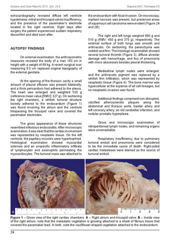
Figure 1 – Gross view of the right cardiac chambers. A – Right atrium and tricuspid valve; B – Inside view of the right atrium, note that the metastatic vegetation is growing attached to a sheet of fibrous tissue that covered the pacemaker lead. In both, note the cauliflower-shaped vegetation attached to the endocardium.
24