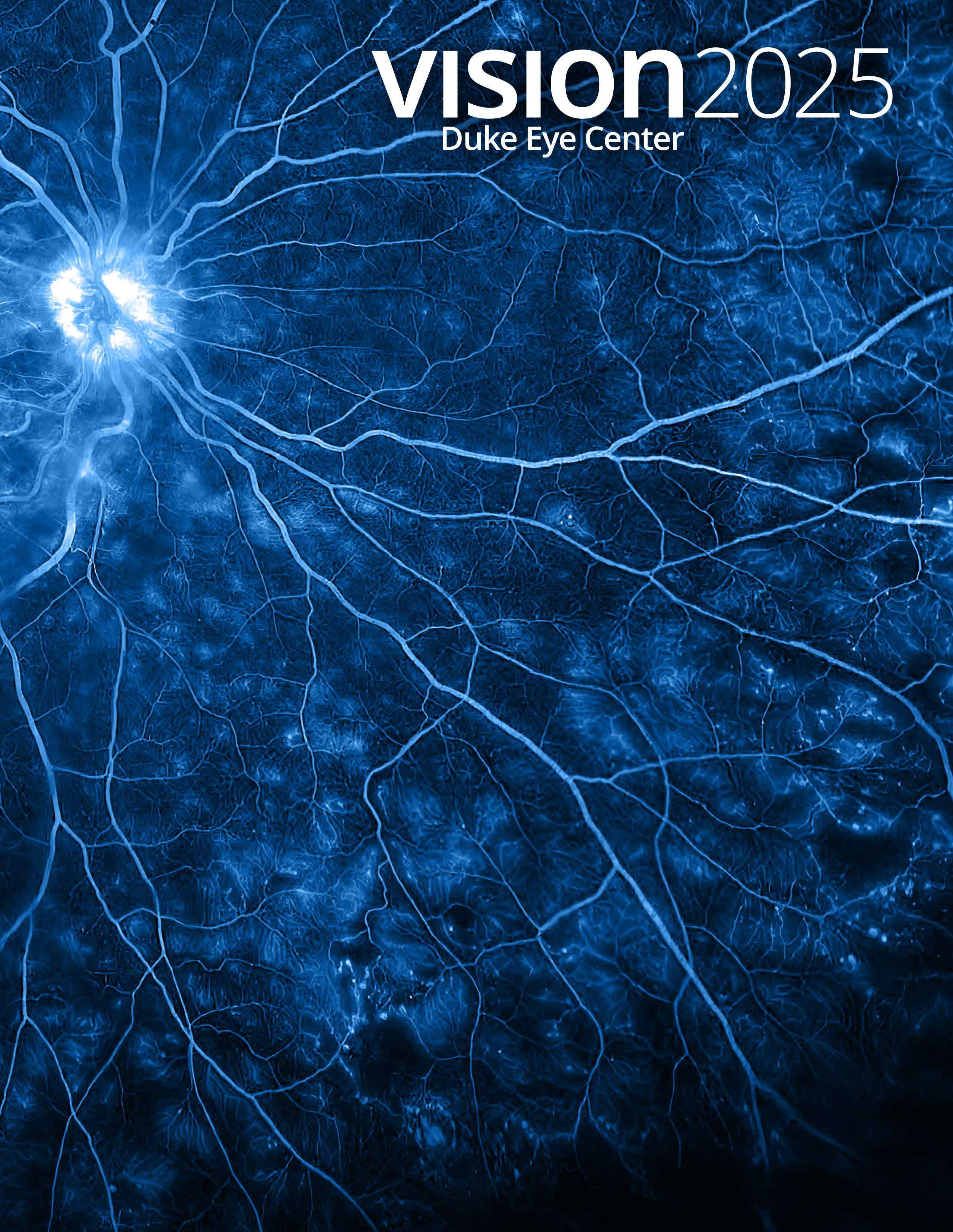
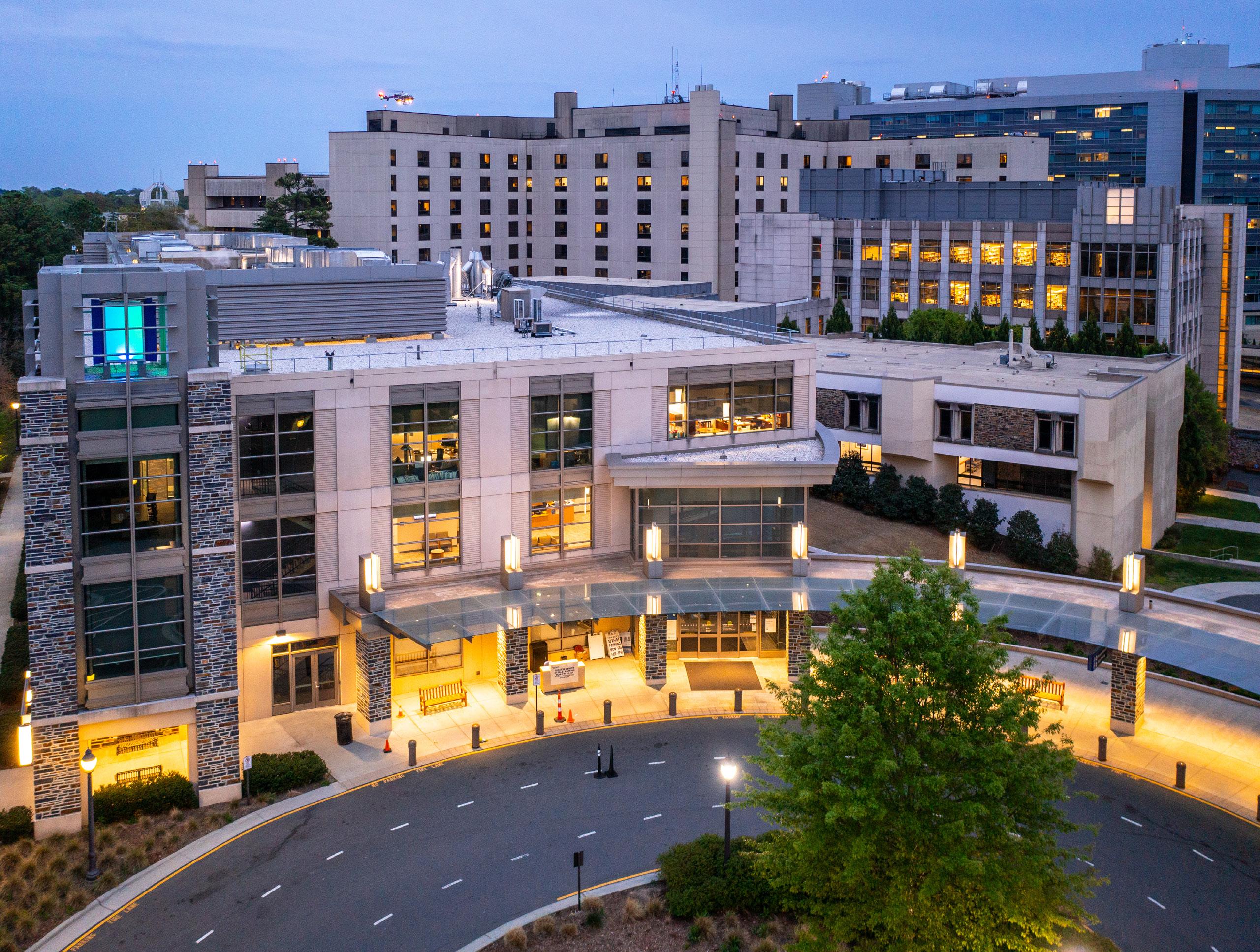
Cover Image: Persistent disc leakage and peripheral ischemia captured during the early phase of an angiogram. John Head, CRA, CPT, Duke Opthalmic Photographer.
Editor Tori Hall
Assistant Editor Jessica Carroll
Writers Vicki Frye, Wendy Graber, Tori Hall and Lori Malone
Art Direction Pam Chastain Design Photography Huth Photo; John Head, CRA, CPT, Duke Ophthalmic Photographer; Mary Harris, COA; Novie Marburn, Sarah Rose Smale, Austin Yeargan, MD
In the midst of our demanding days, it’s easy to lose sight of the profound impact our work has on the lives of our patients. This year, I want to share a story that embodies the work our people do to save vision and improve lives.
Last November, I received an email from a former patient, Dr. Mary Diggs-Garris. The subject line read, “YOU SAVED MY LIFE IN 1995.” Intrigued, I opened the email to find a powerful and moving message about her treatment at Duke for retinoblastoma when she was just three years old, and her remarkable journey since then.
Mary’s mother, Nancy Chambers, was understandably terrified when she learned of her daughter’s diagnosis — eye cancer at such a young age. She was informed that the radiation treatment might affect Mary’s development in ways that were uncertain at the time.
As I continued reading, Mary shared that she is now a successful educator in North Carolina, recently earned her doctorate in education, and is a wife and mother of two. She expressed her gratitude, crediting me and the team at Duke for saving her life and contributing to her achievements as an adult.
This heartfelt message was a poignant reminder of the lasting impact that physicians, researchers and staff can have on their patients’ lives, often in ways we might never anticipate. I was deeply honored that Mary remembered me 30 years after her treatment and that our work had such a profound influence on her life. These moments of recognition are vital, reminding us of the difference we make.
As you read this issue of VISION, you will encounter similar stories of how the incredible work being done at Duke Eye Center continues to transform lives today and will have lasting effects on patients and their families for many years to come.
Warmest regards,
Edward G. Buckley, MD
Chair, Department of Ophthalmology
Vice President for Duke-National University Singapore Affairs
James and Joy Gills Professor of Ophthalmology Professor of Pediatrics
Duke University School of Medicine
“All in all, my quality of life is amazing! I am beyond blessed! Thank you for not giving up! Thank you for everything!”
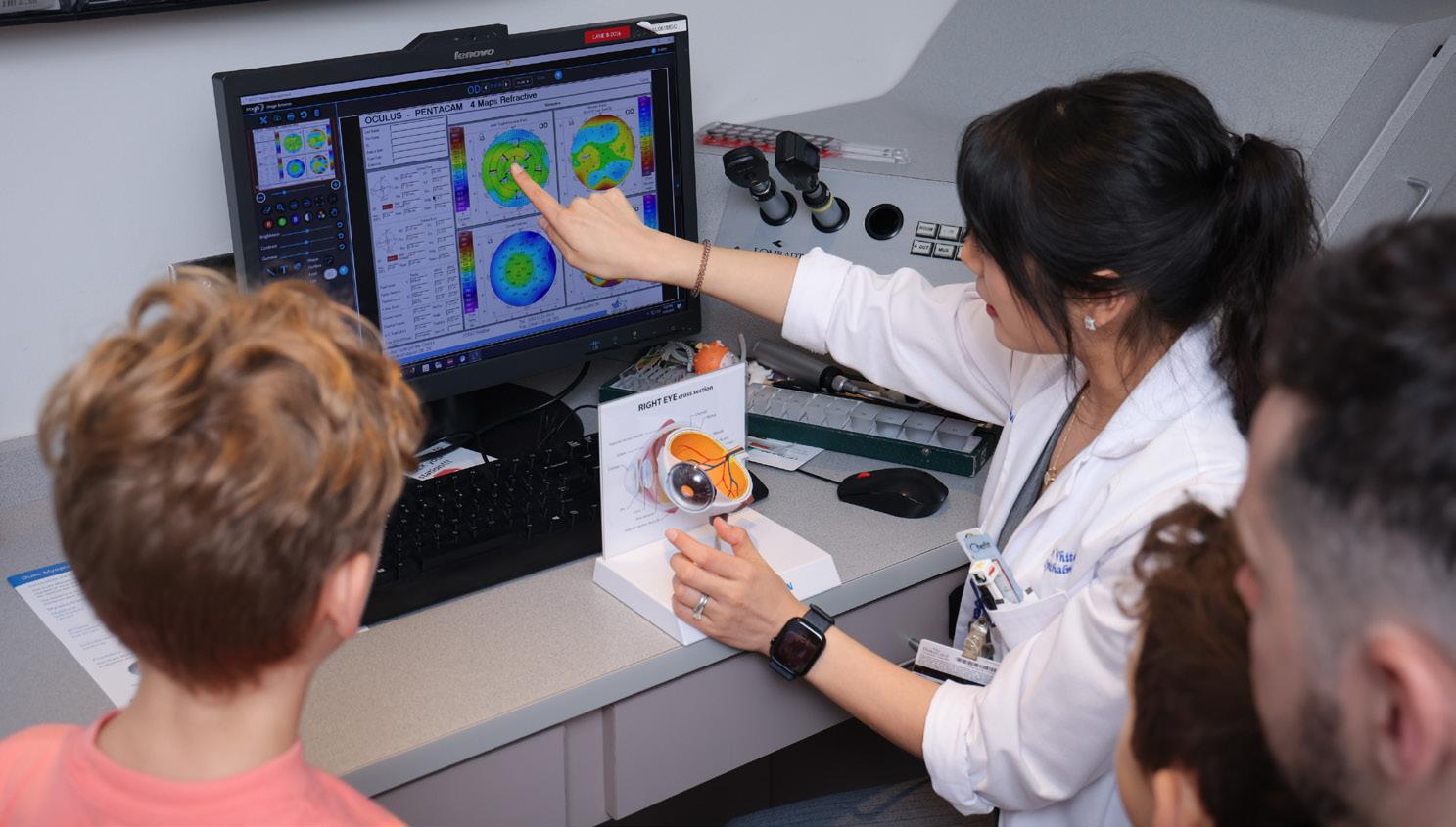
UVEITIS IS A COMPLEX INFLAMMATORY DISEASE of the eye that poses a significant risk of permanent vision impairment and blindness if left untreated. Characterized by symptoms including eye pain, redness, decreased vision and light sensitivity, uveitis can stem from various causes, ranging from autoimmune disorders to infections. Treatment is often complex and has to be tailored to each patient’s distinct inflammatory profile.
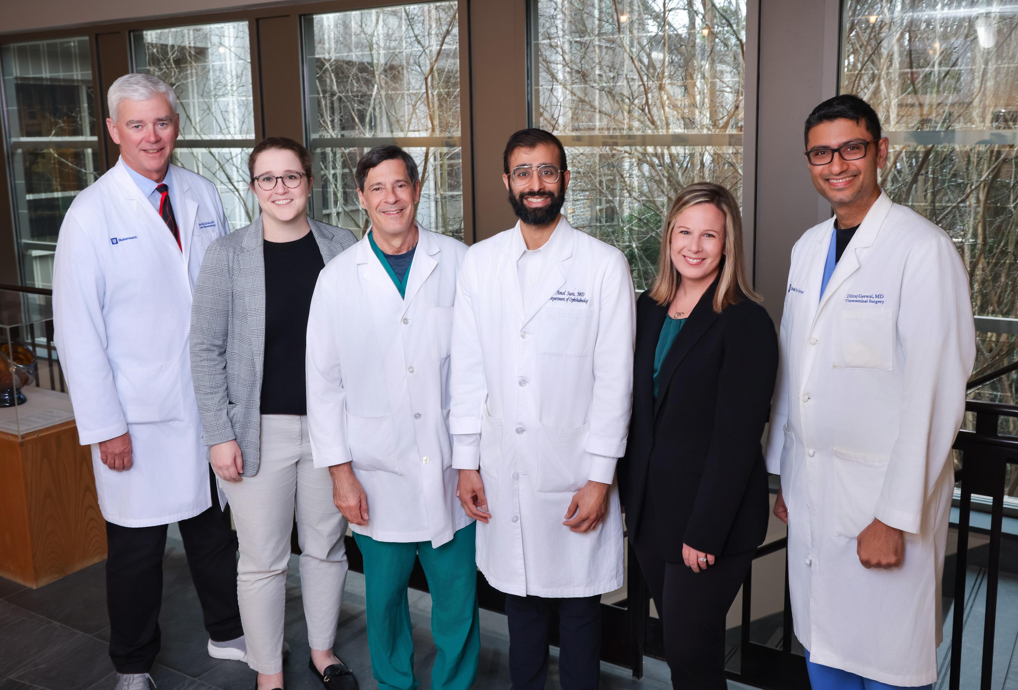
The uveitis and rheumatology team, Rex McCallum, MD, FACR; Mary Buckley, MD; Glen Jaffe, MD; Amol Sura, MD; Lisa Carnago, FNP-C; Dilraj Grewal, MD, created the Duke Multidisciplinary Uveitis Clinic, a comprehensive patientcentered approach to addressing both ocular health and autoimmune issues simultaneously.
Understanding the need for specialized support, Duke Eye Center is home to the Duke Multidisciplinary Uveitis Clinic, distinguished for its innovative treatment protocols and collaborative, consolidative approach. By integrating rheumatology with ophthalmology, patients benefit from comprehensive care for the entire spectrum of inflammation control. The collective efforts of skilled specialists have positioned the clinic at Duke Eye Center as a leader in uveitis care. Glenn Jaffe, MD, Robert Machemer M.D. Distinguished Professor of Ophthalmology and Division Chief Vitreoretinal Disease, emphasizes the importance of this unique model, stating, “It is crucial to have rheumatologists and ophthalmologists working side by side. Patients with uveitis often have systemic diseases that require a comprehensive treatment approach with immunosuppressive medications, and that’s exactly what we provide here at Duke.”
The clinic’s foundation is built on the understanding that the diagnosis and management of uveitis is complex and may be part of an autoimmune condition which impacts other parts of the body. Uveitis often coexists with systemic diseases like sarcoidosis, multiple sclerosis, vasculitis and psoriatic arthritis. This underscores the importance of a comprehensive patient-centered approach to addressing both ocular health and autoimmune issues simultaneously. The Duke Multidisciplinary Uveitis Clinic facilitates this integrated method by bringing together rheumatologists and ophthalmologists in the same room to consult and treat patients concurrently, streamlining the diagnostic and therapeutic processes.
A Comprehensive and Coordinated Approach
Unfortunately, it is common for patients with uveitis to experience lengthy delays when attempting to coordinate care across different specialties. Traditional models often necessitate separate appointments for ophthalmology and rheumatology, leading to gaps in communication that can jeopardize patient outcomes and lead to inadequate treatment.
“The complexity and chronicity of uveitis demands constant collaboration. We identified that many patients were struggling to manage their conditions due to poor communication between specialists,” said Amol Sura, MD, assistant professor of ophthalmology. “By working together in the same room, we eliminate those barriers.”
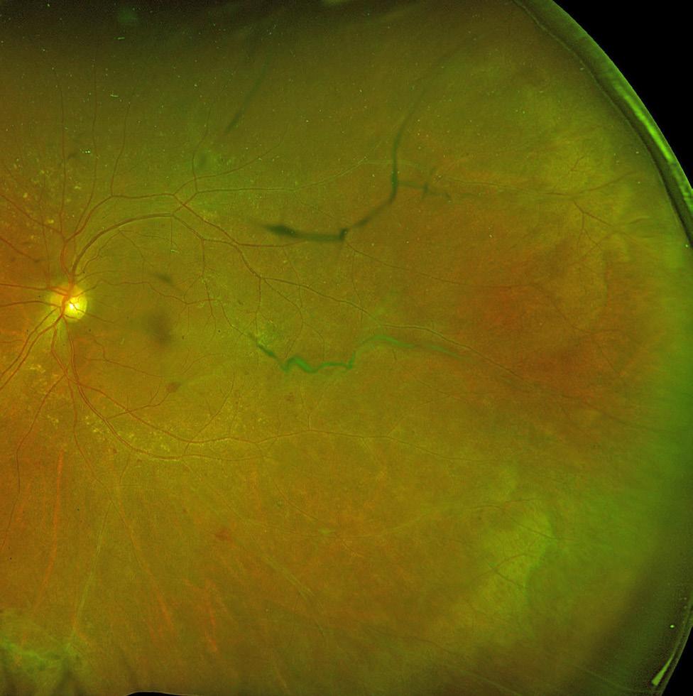
Image shows retinal vasculitis, posterior vitreous detachment of eye.
At the Duke Multidisciplinary Uveitis Clinic, patients see both their uveitis specialist and a rheumatologist, allowing for coordinated discussion of treatment strategies and adjustments. This integrated process is particularly beneficial for patients with multifaceted medical histories, where the relationship between ocular and systemic diseases impacts care decisions. Rex McCallum, MD, FACR , professor of medicine, underscores, “This is a model that works well for our patients. We simplify the process significantly, making it not only efficient but also effective.”
The clinic’s structure is built on a foundation of trust, knowledge-sharing and continuous education. Its physicians actively engage cross functionally, ensuring that they remain at the forefront of uveitis research and treatment advancements. Their shared mindset has not only led to a rise in patient volumes across several adjoining states but has also enriched the education of fellows and trainees, bolstering Duke’s reputation as an elite training ground for future leaders in ocular inflammation.
Unique Facilities and Expedited Diagnosis
Well-appointed facilities at Duke Eye Center enable the robust treatment patients undergo at the uveitis clinic. For example, an on-site blood collection facility allows for on-premises testing to identify underlying systemic causes of uveitis. This capability, along with others such as imaging,
enables the clinic to deliver timely diagnoses and treatments, eliminating the need to defer care while waiting for follow-up testing at another location.
“One of the greatest advantages of the Duke Uveitis Clinic is our ability to access diagnostic resources without delay,” said Dilraj Grewal, MD, associate professor of ophthalmology, “We’re able to complete blood tests, imaging and fluid samples all on the same day, expediting the path to diagnosis and disease control.”
This comprehensive framework also includes simultaneous follow-up appointments with both specialists, fundamentally transforming the traditional patient experience. Frequent assessments enable consistent monitoring of disease progression, allowing necessary adjustments to occur fluidly and without unnecessary lapses. With uveitis often requiring frequent patient visits — typically every three to five months — this consolidated model is a paradigm shift that decreases patient barriers to care, improves communication and prompt care coordination to improve outcomes for people with uveitis.
Leading the Charge in Uveitis Research
The team at the Duke Multidisciplinary Uveitis Clinic is committed to enhancing the understanding of uveitis through groundbreaking research initiatives. For over three decades, the clinic’s specialists have played pivotal roles in clinical trials that have reshaped the treatment landscape.
To achieve further advancements and improve patient outcomes, a comprehensive uveitis registry is in development. This landmark initiative will meticulously track patient outcomes, including quality of life metrics, treatment efficacy and safety. By prioritizing patient needs and preferences, the registry will aim to identify innovative avenues for research and optimize disease management tactics. As Lisa Carnago, FNP-C, expressed, “Our goal is to capture the perspective of patients and understand what truly matters to them. This will guide us not only in refining therapies and improving quality of care but also in understanding the holistic impact of uveitis on their lives.”
This registry will be a historic change to the way patient data is analyzed and utilized. Unlike traditional registries that often focus
“ It is crucial to have rheumatologists and ophthalmologists working side by side. Patients with uveitis often have systemic diseases that require a comprehensive treatment approach with immunosuppressive medications, and that’s exactly what we provide here at Duke.”
Glenn Jaffe, MD
Division Chief,
Vitreoretinal Disease
solely on clinical outcomes, the Duke uveitis registry will incorporate patient experiences, perceived quality of life factors and social determinants of health using multiple methods. This comprehensive approach will empower researchers and clinicians to tailor treatments more precisely to the individual needs and concerns of each patient.
One of the clinic’s primary objectives is to investigate how various therapeutic regimens and interventions influence patient quality of life over time. By engaging patients in an ongoing dialogue, the registry aims to inform clinicians and significantly enhance individuals’ wellbeing beyond clinical measures. “Our aspiration is to advance a holistic understanding of the disease, incorporating patient insights into their treatment preferences and priorities,” asserts Sura. “This authentic, patient-centered approach will truly set us apart and drive improvements in uveitis care.”
The Duke Multidisciplinary Uveitis Clinic stands committed to managing the complexities of uveitis while understanding the patient’s perspective. By fostering collaboration among specialists and streamlining the care process, the clinic stands at the forefront of improving patient outcomes and delivering research advancements. The impending uveitis registry will deepen our understanding of this intricate disease, ensuring patient voices are central to evolving treatment strategies. At Duke Eye Center, we are dedicated to our mission of curing eye disease worldwide and improving the lives of those battling complex eye diseases. With the continuing support of our community, we look forward to continuing to lead the charge in pioneering comprehensive, patient-centered uveitis care.
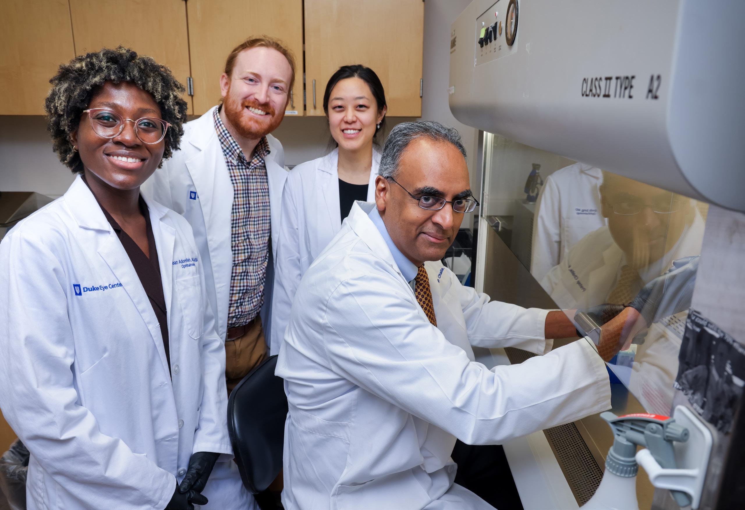
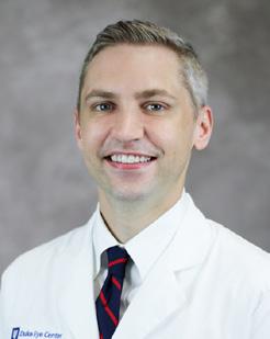
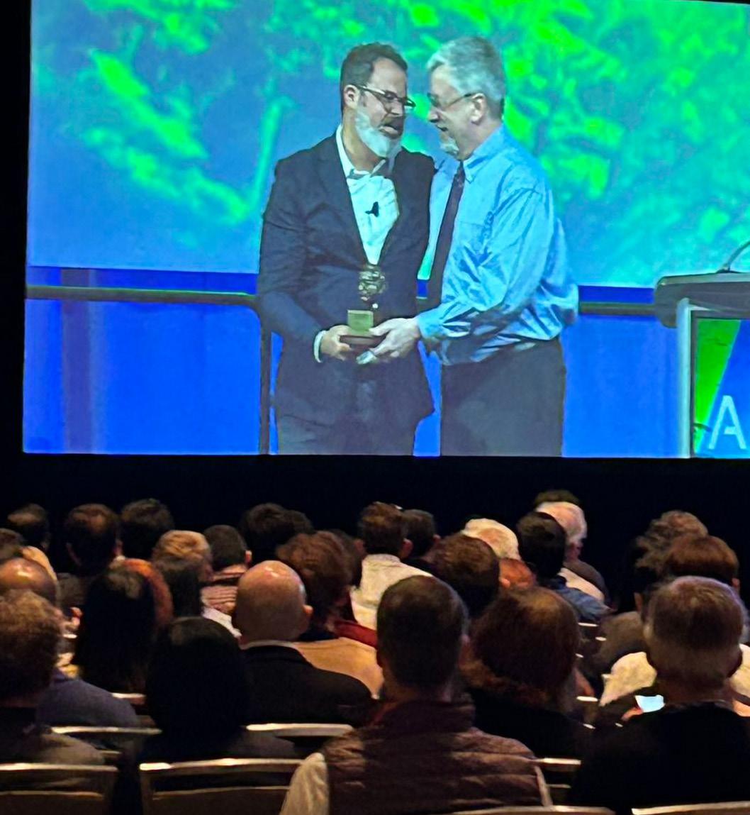
Pratap Challa
Recognized with National Award for Excellence in Residency Program Leadership
The Duke Ophthalmology residency program equips trainees with comprehensive skills and knowledge in surgery, clinical care, and research, preparing them to excel in any career path they choose.
For more than 20 years, Pratap Challa, MD, professor of ophthalmology, has led the Duke ophthalmology residency program that consistently ranks in the top 10 nationwide, and produces a high proportion of clinicianscientists who go on to become leaders in the field.
“His tenure, marked by sustained excellence and innovative leadership, places him among the most dedicated and impactful program directors in the field,” said Edward Buckley, MD, the James Pitzer Gills, III, M.D. and James and Joy Gills Professor of Ophthalmology in the School of Medicine and the chair of the department.
In recognition of his achievements, Challa received the 2024 Straatsma Award for excellence in resident education from the American Academy of Ophthalmology and the Association of University Professors of Ophthalmology.
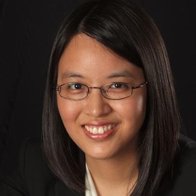
“I’m really glad he got this award,” said Jessica Chow,
MD, who trained under Challa from 2007 to 2011 and now directs the ophthalmology residency program at Yale School of Medicine. “It’s very well deserved.”
Challa said he’s excited to receive the Straatsma award, but is quick to spread the credit around. “We attract great residents and trainees and part of the success goes to them as well as the faculty, who work very hard to train them,” he said.
Training Clinician-Scientists
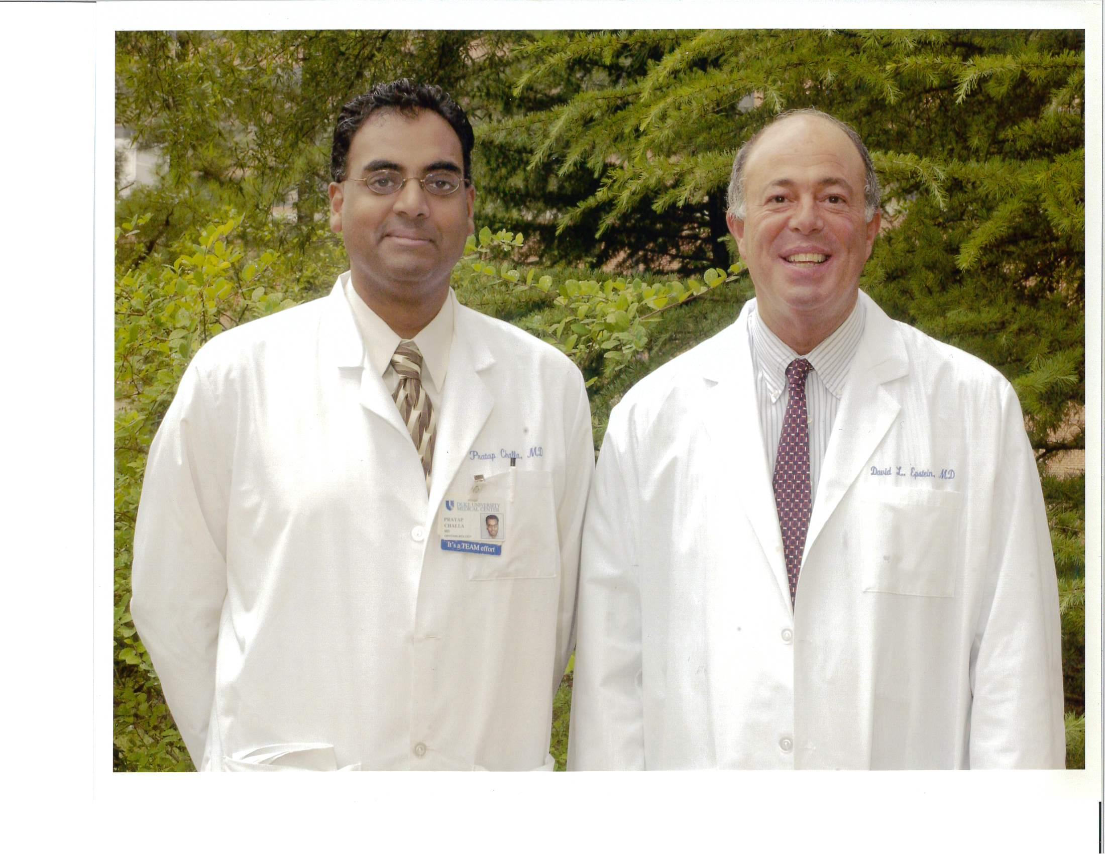
When Challa became the director in 2001, then-chair David L. Epstein, MD, charged him with increasing the number of Duke trainees becoming clinician-scientists. Challa has done just that. When he arrived, 20–25 percent of trainees went into academia; today it’s 50–60 percent.
“We have a broad comprehensive training not just in clinical and surgical areas, but also in the investigative side of medicine, asking new questions
“He’s extremely supportive and always believes in his residents. He always had our backs and I try always to do the same for our residents, I learned that from him.”
Jessica Chow, MD, HS’07-’11, Residency Program Director Yale Ophthalmology
and discovering new things,” Challa said.
A dedicated research block in the third year allows trainees to get a jumpstart on their careers by applying for research grants, collaborating with people outside the department, and publishing their work.
“The goal is to give them the skill set they need to be able to ask a question of significance, form a hypothesis, find a way to test the hypothesis, and add new knowledge to the field,” he said.
And it works: “We average over two publications per resident during their time here, which is quite impressive.”
When it comes to adding new knowledge to the field, Challa practices what he preaches. As a glaucoma researcher, his work with Duke colleagues, David L. Epstein, MD, Vasantha Rao, PhD and Eric Toon, PhD,
David L. Epstein and Pratap Challa, MD in 2001.
led to the development of Rhopressa, approved as a treatment for glaucoma by the FDA in 2017. Challa also studies the effectiveness of residentperformed cataract surgeries and how different patients respond to treatments.
Modeling Excellence
Former residents said Challa is a role model for both trainees and colleagues. “He himself is an outstanding clinician, surgeon, educator, researcher, and colleague,” said Kelly Muir, MD, MHSc, associate professor of ophthalmology. Muir completed the Duke residency program in 2006 and now directs Duke’s ophthalmology fellowship program. “So he’s an ideal role model for the trainees to look up to and try to emulate.”
As director of the Yale residency program, Chow said she admires and is inspired by Challa’s leadership style. “He’s extremely supportive and always believes in his residents,”
she said. “He always had our backs and I try always to do the same for our residents, I learned that from him.”
She also aims to match his calm. “He never got flustered in the operating room,” she said. “I try to emulate that style because it’s really easy to make residents nervous.”
Muir added, “What most of us who have worked with him know is that he is cool as a cucumber. You cannot ruffle his feathers, or if you can, I haven’t seen it.”
Challa is just as supportive of his colleagues as he is of residents, Muir said, stepping up to help in ways large and small, whether he’s lending his expertise on a complex case or covering for a colleague unexpectedly called out of town. “He’s always there to take care of patients, residents, and colleagues,” she said.
In addition to taking care of his own patients, Challa has no
doubt improved the lives of the innumerable patients cared for by all the ophthalmologists whose training he has overseen in the past two decades.
“Our goal is to train the best and the brightest and have them take great care of patients and improve the way we take care of patients,” Challa said.
“What most of us who have worked with him know is that he is cool as a cucumber. You cannot ruffle his feathers, or if you can, I haven’t seen it.”
Kelly Muir, MD, MHSc
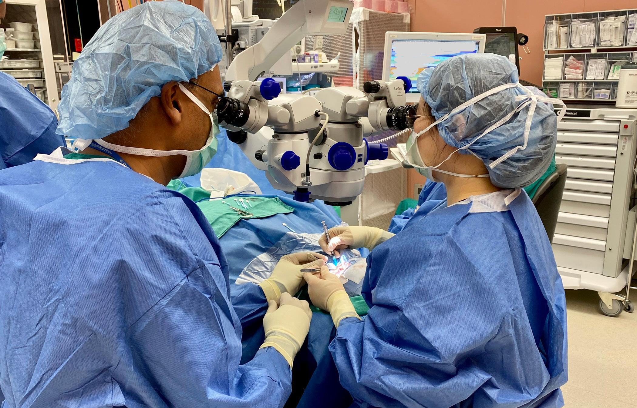
Challa teaching former resident Sarah Xu, MD in the OR.
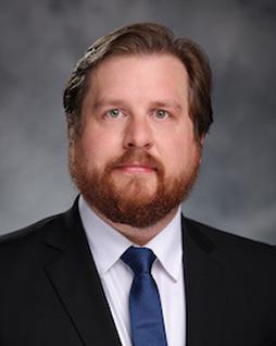
Fellow Turned Inventor Gives Glaucoma Care an Upgrade
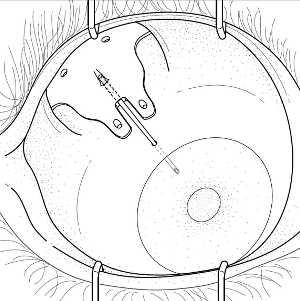
Rami Gabriel, MD, HS ’20-’24 became an expert in administering eye drops during his training, especially to young pediatric patients. He remembers one young patient, not even in elementary school yet, who had just received a glaucoma drainage device (GDD) to help control increased eye pressure from the glaucoma claiming their vision. This patient had an especially difficult time with drops following surgery. When it was time for the drops, they’d squeeze their eyes shut with all their might. Gabriel knew how delicate the ocular tissue was post-surgery and how careful he needed to be. “But once they start squeezing, you’re at a loss,” says Gabriel.
Gabriel encountered patient pain points like this throughout his Ophthalmology residency at Duke Eye Center, and an idea to help glaucoma patients, and their surgeons, began to form. Now a Cornea Fellow at Duke Eye Center, Gabriel is inventing an improved GDD to cut down on eye drops for patients, make surgery easier, and ultimately tackle glaucoma-induced blindness.
Glaucoma is the second leading cause of blindness globally and affects over 3 million Americans. It is characterized by improper fluid drainage in the eye, leading to increased pressure, optic nerve damage, and irreversible vision loss. Early detection and intervention are crucial, with initial treatments typically involving medicated eye drops or laser therapy. When these fail, surgery, including GDDs, becomes necessary.
BY K. LOU WARD
From the filed patent, a drawing excerpt showcasing an implant on the plate side of the GDD temporarily sealing the tube.
GDDs help divert fluid to a reservoir where the body can reabsorb it, but patients often still need medicated eye drops post-surgery. Gabriel observed the difficulties patients, especially children and the elderly, faced in maintaining their eye drop regimen. This inspired him to create a GDD that releases glaucoma medication directly into the eye, reducing the burden on patients and improving surgical outcomes.
Gabriel’s improved GDD design incorporates hydrogel, a dissolvable polymer loaded with glaucoma medications that release over time which should limit or eliminate the need for post-surgery drops.
As a resident, Gabriel assisted in surgery by preparing the tube component of the GDD while his mentor, Leon W. Herndon Jr, MD, performed the surgery. Gabriel noticed that tying off the tube to secure the GDD was a cumbersome step that could delay surgery. This led him to further improve GDD glaucoma treatment, this time for surgeons. “You want to have a successful surgery every single time,” says Gabriel. His innovation to enhance GDDs not only eases the patient’s burden but also simplifies the surgical process by eliminating the need to tie off the tube component of the GDD.
Leon W. Herndon Jr, MD
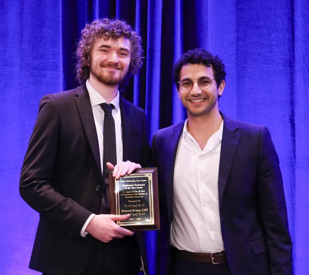
Challa presented Gabriel with the 2024 Ocular Innovation award at the annual Resident and Fellow Awards Banquet.
Gabriel’s improved GDD design incorporates hydrogel, a dissolvable polymer loaded with glaucoma medications that release over time which should limit or eliminate the need for postsurgery drops.
Gabriel found that Duke Eye Center’s emphasis on mentorship, research, and innovation was fundamental in developing his solution for glaucoma care.
With the help of mentors Herndon and Pratap Challa, MD, both professors of ophthalmology and glaucoma experts, Gabriel refined his device. “I’ve been very lucky to have some amazing mentors,” says Gabriel of Herndon and Challa. “Dr. Challa, residency director and my primary mentor, has been teaching glaucoma for over 20 years, and it’s one of the most prestigious programs to learn glaucoma from.”
Gabriel and Challa connected with Dennis Thomas, PhD, now-retired associate director of licensing at the Duke Office for Translation & Commercialization (OTC), to secure a patent and develop a commercialization approach by connecting them with potential industry partners. Players in Duke’s innovation space like OTC are experts in bringing academic ideas to the market, and they bring expertise in navigating the circuitous path of research translation.
For Gabriel and Thomas, patient outcomes are the north star of their commercialization and patenting strategy that will bring this GDD to the clinic. Rather than designing a brand-new device, they’re approaching this as an improvement to existing treatment that is made up of constituent parts already approved for safe use by the FDA. Their hope is to get this GDD safely to patients as soon as possible.
With the support of his mentors and Duke’s innovation infrastructure, Gabriel was able to devise a new device that eliminates a key barrier for patients — eye drop burden — and improves surgeries for physicians. Like many innovative clinicians at Duke Eye Center, Gabriel’s inspiration was borne out of caring for his patients. As he continues to work with OTC on his drug-eluting GDD, he and his fellow clinicians at the Duke Eye Center continue to pursue novel translational ideas that can improve clinical practice at Duke and patient care everywhere.
Gene Therapy Halts Vision Loss from Retinitis Pigmentosa
Duke Eye Center One of Few Eye Centers Offering Gene Therapy for RP
Tyler Wilfong was born with retinitis pigmentosa (RP), a rare collection of inherited eye diseases that slowly robbed his vision. Last year, he went to the Duke Eye Center, one of the few eye hospitals in the country performing gene therapy to halt the disease’s progression. Now Wilfong can work again and watch his baby boy grow. “It’s been life changing,” he said.
Retinitis Pigmentosa Causes Progressive Vision Loss
Wilfong of Lincolnton, NC, was 8 years old when his parents noticed he was having trouble finding a toy on the floor. He was diagnosed with retinitis
pigmentosa, a condition that damages the retinal layers and causes vision loss over time. According to Lejla Vajzovic, MD, FASRS, a retina specialist at the Duke Eye Center, the first signs of RP are loss of night vision, then loss of peripheral vision, which narrows the field of vision. Eventually RP leads to tunnel vision and central vision loss. Wilfong, now 36, described his world as darker as he grew up. “Everything was really blurry. In low light situations it was like blindness. I couldn’t play high school football because I couldn’t see under the lights. Everything in the gym is brown, dark brown hard wood, dark brown bleachers. It was hard to see.”
Afraid of Losing His Sight
While he had a driver’s license for about three years and never had an accident, Wilfong eventually had to stop driving. He was able to do some carpentry work but that had to stop too. “I felt like I was losing my independence. It had gotten to the point where I couldn’t walk around the grocery store. What’s bright to other people wasn’t bright to me.”
As the condition worsened, Wilfong said he was afraid “I was going to lose my sight.”
Because the disease is inherited, Wilfong underwent genetic testing in Winston-Salem to determine if his family members were affected. While none had RP, the testing uncovered that Wilfong’s mother and father carried a specific mutation in the gene referred to as RPE65. His local eye doctor knew that people with this gene mutation could qualify for gene therapy treatment.
The eye doctor referred Wilfong to Duke, one of the few eye centers in the country to offer the gene therapy known as Luxturna (voretigene neparvovec). Wilfong had the procedure on his left eye in 2023.
How Gene Therapy Halts Cell Degeneration
A healthy, artificially engineered RPE65 gene is packaged into a harmless virus that cannot cause disease and acts as a delivery vehicle. The virus is surgically injected precisely into the area of damaged cells responsible for vision loss. “The new healthy gene helps decrease the degeneration of those cells and helps stop the process that has been causing them to slowly die over time.”
Lejla
Vajzovic, MD, FASRS
A healthy, artificially engineered RPE65 gene is packaged into a harmless virus that cannot cause disease and acts as a delivery vehicle. The virus is surgically injected precisely into the area of damaged cells responsible for vision loss. “The new healthy gene helps decrease the degeneration of those cells and helps stop the process that has been causing them to slowly die over time,” explained Dr. Vajzovic. While every patient’s response is different, Dr. Vajzovic said clinical trials show the therapy can decrease the progression of disease and improve vision.
Gene Therapy for RP Has Been Life-Changing
The first thing Wilfong noticed in the days after the procedure was how many fingers he held up on his hands. Eventually the world became brighter, and he could see more details in low light situations. “Before, if there were no lights on, I couldn’t see inside the house.”
Now, he can find his new baby’s pacifier when it drops, see his baby’s mouth to feed him a bottle, and change his baby’s diaper without help. So far, his baby’s vision appears unaffected. “It’s been life changing,” Wilfong said. “Duke really came through. I’m looking forward to getting the other eye done.”
Duke Eye Center at the Forefront of Inherited Retinal Disease Research and Treatment
Gene therapy for RP is confined to a select few centers because of the complexities involved. The virus with RPE65 gene must be formulated in a specialty pharmacy. It must be thawed and mixed appropriately and used within hours by surgeons who are specially trained in the procedure.
Because inherited retinal diseases are so rare, it’s important to go to an eye center like Duke, where experts are specially trained to examine and diagnose people with RP and other rare inherited retinal diseases. The team includes genetic counselors who help diagnose the disorder and provide counseling on the implications of having certain genetic mutations. It also ensures access to advanced treatments and research.
As one of the leading eye centers in the country, Duke is involved in several clinical trials looking at other mutations to potentially be solved with gene therapy.
The Future of Vitreoretinal Surgery
A Look Into the Emerging Role of Artificial Intelligence and Data Science
The field of ophthalmology is undergoing a transformative shift, driven by rapid advancements in artificial intelligence (AI) and data science. At the forefront of this movement is Yannek I.
Leiderman, MD, PhD, professor of ophthalmology, vitreoretinal diseases and surgery, and prominent vitreoretinal surgeon and translational researcher at Duke Eye Center. Leiderman is leading groundbreaking research to integrate AI into surgical procedures, promising to revolutionize the treatment of complex eye conditions such as retinal detachment, diabetic retinopathy and macular surgery. His dedication and passion for enhancing surgical precision and efficiency aligns seamlessly with Duke’s legacy of innovation and commitment to pioneering technologies that positively impact patient outcomes.
A New Era in Surgical Precision
Leiderman — who joined Duke Eye Center in the Fall of 2024 — is concentrating his efforts in a future where AI will become an indispensable ally to surgeons, guiding real-time decision-making and informing treatment plans. By harnessing the power of this technology, clinicians can expect to uncover hidden patterns and trends, enhance precision and optimize surgical techniques, minimize the risk of complications and improve outcomes.
Leiderman is leading groundbreaking research to integrate AI into surgical procedures, promising to revolutionize the treatment of complex eye conditions such as retinal detachment, diabetic retinopathy and macular surgery.
One of the most promising applications of AI in vitreoretinal medicine is the development of surgical guidance systems. These systems can suggest optimal surgical interventions and provide surgeons with immediate feedback on progress during a procedure. By leveraging the power of AI, ophthalmologists can achieve greater accuracy and efficiency in their treatment.
“Microsurgery of the eye is an exacting and demanding activity, and like other endeavors at the limits of human performance that have benefitted greatly from integrating AI, such as aviation and space exploration, AI has the power to analyze large amounts of data quickly and accurately to facilitate a broad array of tasks, and has the potential to transform ophthalmic microsurgery,” shared Leiderman.
The Future of Surgical Training
In addition to its clinical applications, AI is poised to revolutionize surgical training. Leiderman’s research aims to provide tools to help trainees in surgery understand their technical and cognitive performance through analysis of surgical tasks and decision making following a procedure. By analyzing the performance of trainees, AI can help identify areas for improvement and provide personalized feedback. This will enable surgeons to acquire the necessary skills and knowledge to perform intricate procedures with confidence and expertise.
A Collaborative Approach
Leiderman emphasizes the importance of internal collaboration among clinicians, researchers, engineers and computer scientists, as well as with other institutions, to drive innovation across all ophthalmic specialties. The unique academic environment at Duke — with its world-class Department of Biomedical Engineering and School of Medicine in close proximity to each other and operating room suites — provides an ideal platform for interdisciplinary research. By fostering collaboration and knowledge sharing, researchers are able to accelerate the development of new technologies and bring them to the clinic more quickly.
A Brighter Future for Patients
The integration of AI and data science into vitreoretinal surgery holds the promise of a brighter future for patients battling with complex eye conditions. By increasing surgical efficacy, reducing complications and enhancing surgical education, AI-powered technologies will significantly enhance patient outcomes. As Leiderman and his team continue to push the boundaries of innovation, we can look forward to a future where vision-threatening diseases are treated with greater efficacy.
“Duke is recognized as a leader in surgical innovation, and I believe we are poised to continue to revolutionize surgical and clinical care in the dawning era of AI in healthcare,” said Leiderman. “Our ecosystem includes world-renowned surgeon-innovators, biomedical engineers, computer scientists and so many other team members dedicated to the provision of excellent patient care at the leading edge. The application of AI to healthcare is a multidisciplinary collaborative effort, something at which we excel at Duke.”
“Duke is recognized as a leader in surgical innovation, and I believe we are poised to continue to revolutionize surgical and clinical care in the dawning era of AI in healthcare”
Yannek I. Leiderman, MD, PhD
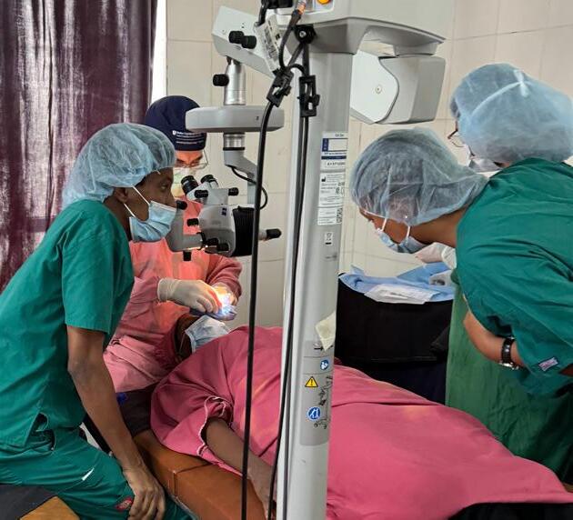
ON A MISSION Making Progress
ISURGERIES ABROAD
t has been a remarkable year for Duke Global Ophthalmology (Duke GO), filled with amazing triumphs and incredible growth — all dedicated to curing blindness worldwide, alleviating human suffering, and transforming not only individual lives, but their families and entire communities.
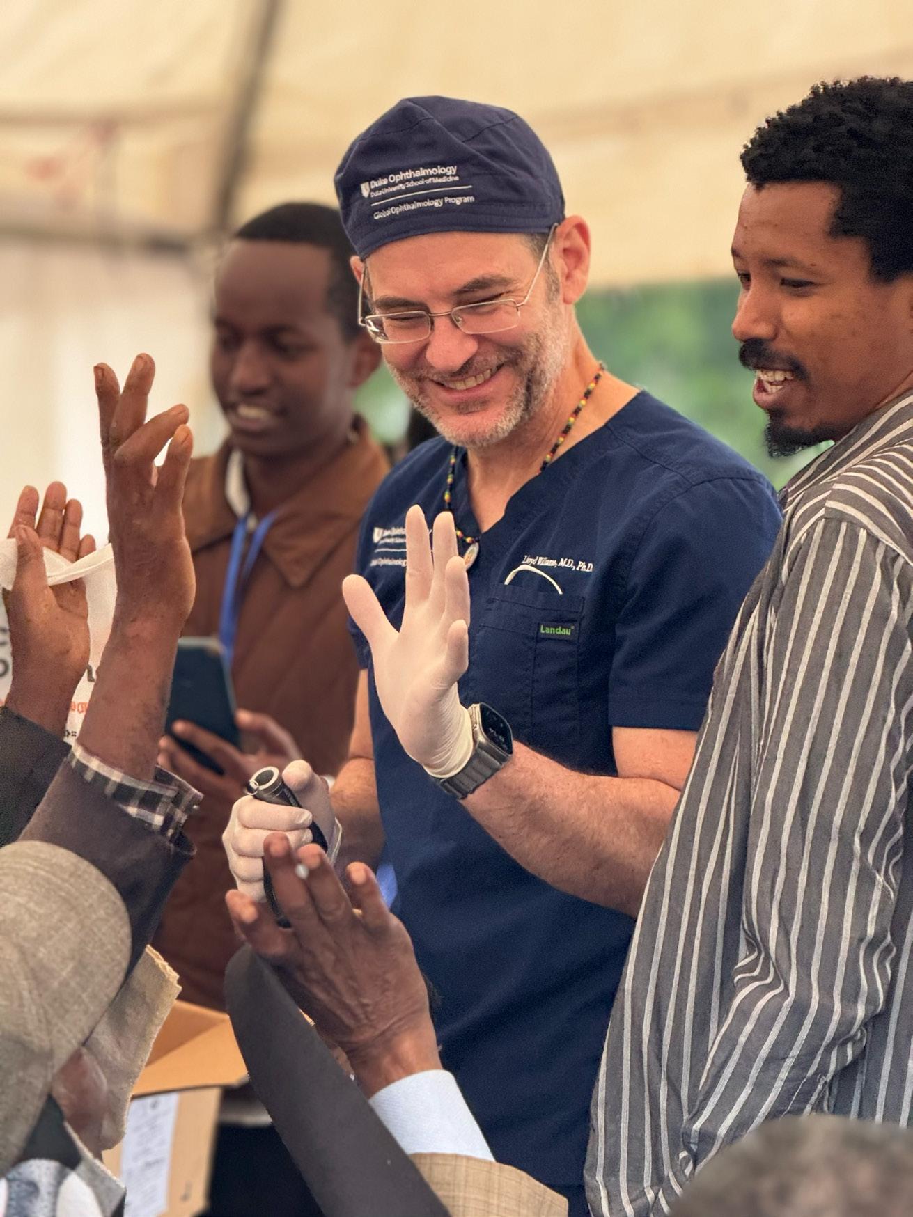
Lloyd Williams, MD, PhD, associate professor of ophthalmology and Duke GO director, leads a tireless team that is having a major impact by restoring vision in several underserved countries across the globe.
Much of Duke GO’s success is due to the generosity of our donors, the addition of a program coordinator, more organized partnerships, and the increased interest of our faculty, staff and trainees to participate in Duke GO. The inaugural global ophthalmology fellow will begin in summer 2025 and will expand capacity to reach more countries and impact more lives.
Duke GO has plans to further expand access with increased faculty and trainee interest to perform cataract and glaucoma surgeries, and corneal transplants globally. Additional priorities include supporting and training international ophthalmologists and staff, as well as establishing eye banks abroad. Duke GO’s commitment to curing blindness worldwide has never been stronger.
EYE SURGERIES LATE 2023 – 2024
DATE LOCATION
December 2023 Akon, South Sudan1
December 2023 Aweil, South Sudan1
January 2024 Yogyakarta, Indonesia2
April 2024 Freetown, Sierra Leone2
May 2024 Roatan, Honduras
August 2024 Arba Minch, Ethiopia1
September 2024 Yogyakarta, Indonesia2
4 EK, 14 ICL
October 2024 Visitors from Indonesia to learn corneal transplant and eye banking 2
October 2024 Freetown, Sierra Leone2 17
November 2024 Visitor from Ukraine to Duke for 1 week learning corneal surgery/transplants
December 2024 Malakal, South Sudan1
cornea transpant in ()
FELLOWSHIP
PARTNERS:
1Cureblindness Project 2 Miracles in Sight and/or CorneaGen 3 Health in Sight
PK = penetrating keratoplasty, EK = endothelial keratoplasty, MSICS = manual small incision cataract surgery, Trachoma = Trabout eyelid rotation procedure for trachoma, ICL = implantable collamer lens.
PARTNERSHIPSAND
ON A MISSION Surgery Cures Blindness
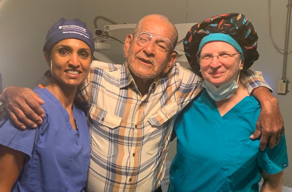
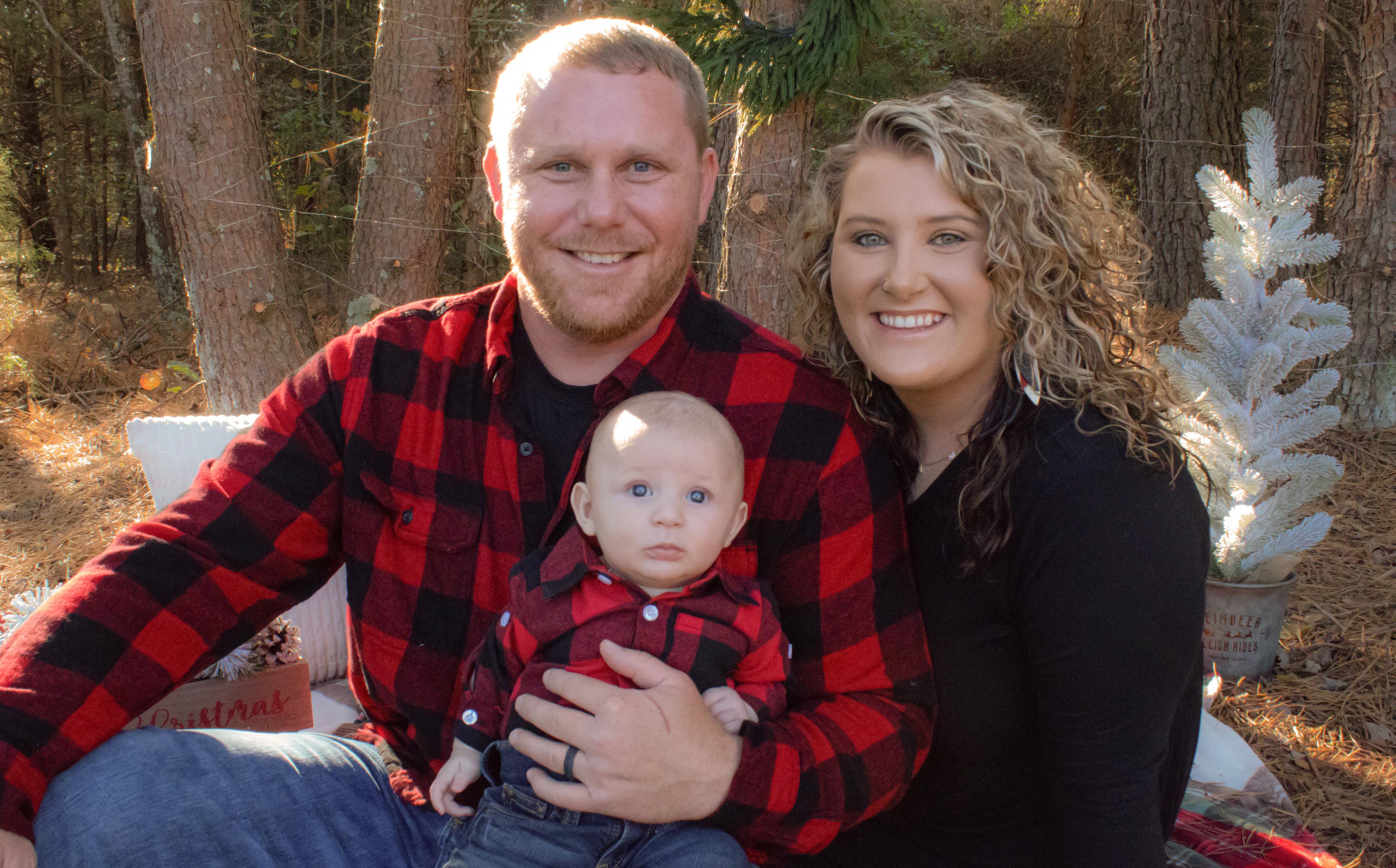
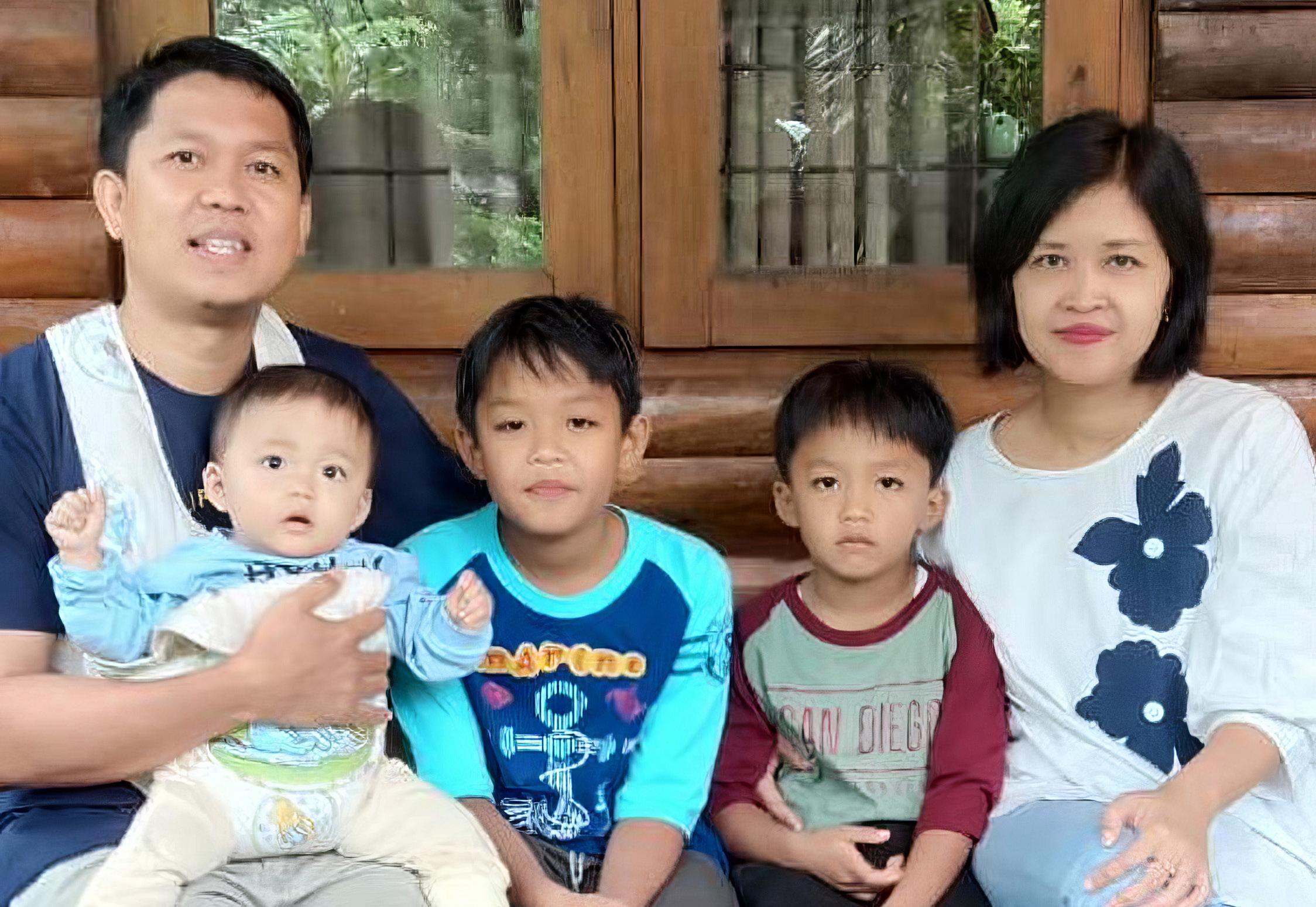
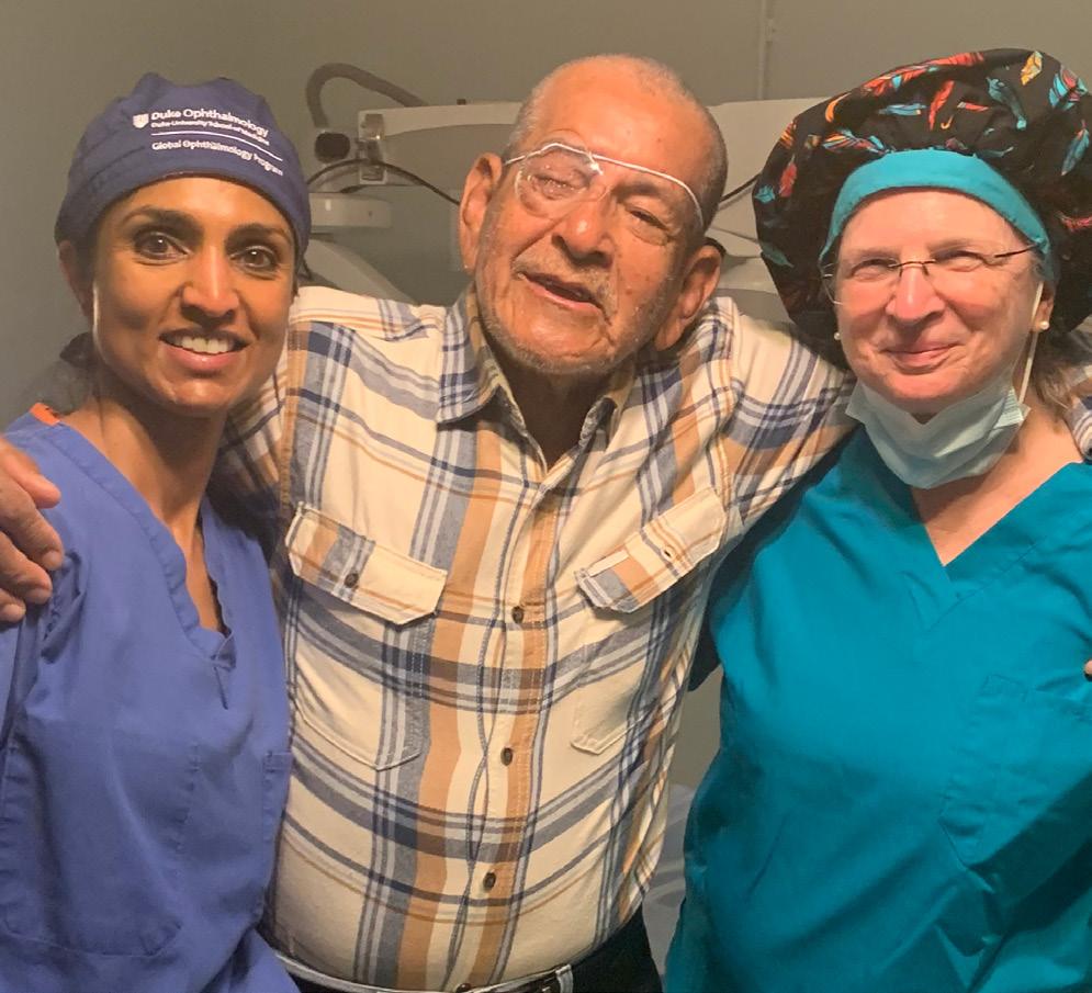
Novie Marburn from Yogyakarta, Indonesia was blind in both eyes for 10 years. She was not able to see her children ages 2, 4, and 7 years old, making it very difficult to care for them. Novi wondered if she would ever be able to see the beautiful faces of her children. Indonesia lacks cornea transplant surgeons and eye banking for Novie to get the cornea transplant needed to regain her vision. During a sight saving trip in February 2024, Lloyd Williams, MD, PhD, met Novie‘s husband Rio, completely by chance. Rio was working as a translator and happened to ask Willams if he could help his wife. Williams
evaluated Novie and realized she was blind in both eyes, but her vision could be restored with corneal transplants in each eye. Novie was getting the sight saving surgery that she needed. Today, Novie now has almost normal vision. She can see her children and care for them with no assistance. This is just one example of thousands, demonstrating the impact of Duke GO.
Rio and Novie Marburn with their children following Novie’s corneal transplants that restored her sight.
Anupauma Horne, MD: “Everyday in ophthalmology we experience small miracles of giving new sight at home but the impact this has in developing communities is on another level. It can mean the difference between suffering and hope for all ages. I love travel because of the immersion in language and culture and the connections that can be formed far outside of what I’m familiar with. Global ophthalmology has allowed me to lean into this even more by challenging me to hone my surgical skills to perform cases that are often far more complex than my daily routine and to do so in unpredictable settings but always leads to such rewarding and impactful results.”
Horne, assistant professor of ophthalmology and chief of Duke comprehensive ophthalmology, leads our efforts in Roatan Honduras with partner Health in Sight.
ON A MISSION Staff Lives
Fekrselame “Metu” Tesfaye, a Duke Hosptal nurse, traveled to her home country of Ethiopia, not to visit family and friends, but to assist with surgeries that restored sight to hundreds of people. It was a lifechanging experience for the patients –and for Tesfaye. “I was shocked,” Tesfaye said. “I myself didn’t know how many blind people there are in Ethiopia. It was unbelievable.”
Are Impacted
Horne with JoAnn Dennis with Health in Sight celebrating with a happy patient after surgery in Honduras.
ON A MISSION Education Expands Global Access
Fellowship
A critical component of Duke GO is the new Global Ophthalmology Fellowship which will begin with the inaugural fellow, Sheena Song, DO, in July 2025. The fellowship is designed to train outstanding young ophthalmologists to specialize in providing care and developing infrastructure overseas. Song will also work with Miracles In Sight to help develop an eye banking program in Indonesia.
“Global ophthalmology is what inspired me to pursue medicine. Duke’s impressive dedication to global health, in addition to Dr. Lloyd Williams’ passion for teaching and global ophthalmology, are what drew me to the program. I look forward to working with the Duke Global Ophthalmology team to make meaningful impact and connections in both the global and local space next year.”
Training International Ophthalmologists
DukeGO surgeons train international ophthalmologists so the local physicians can continue caring for the natives. Thus far, five surgeons have been trained to perform corneal transplants; two in Indonesia, two in Sierra Leone, and one in Ukraine. Training will continue during future trips to increase skill and proficiency.
Nataliya Preys, MD, Ukrainian ophthalmologist, came to Duke to increase knowledge in advanced cornea transplantation techniques to address a growing number of patients with different mechanisms of eye injuries as mechanical and chemical trauma.
Ophthalmologists visit Duke for training. This year, physicians from Indonesia and Ukraine traveled to Durham to observe and improve skills in corneal transplants and learn more about eye banking.
“My observation in ophthalmology was very educational and rich. I observed many surgical cases and methods of treatment and training time on sets of animal corneas in the educational room. At Duke, eye surgery is performed at a very high level! I plan to actively implement the knowledge and experience gained at Duke in my clinical practice in Ukraine.”
Nataliya Preys, MD
Sheena Song, DO
Partnerships and Donors
Thousands of beaming smiles and countless tears of joy wouldn’t have been possible without support from our partners and generous donors including corporations, service organizations, patients and individuals who are committed to the cause. Funding is still a primary focus for Duke GO as it is necessary to continue the success experienced over the last year.
LC Industries, a transformative supporter of Duke Eye Center, provided a substantial gift to help support the infrastructure and travel for Duke GO team members.
Miracles In Sight has been a significant partner and supporter of Duke Eye Center for many years, and also passionate about global ophthalmology. Their generous gift made possible the Duke Global Ophthalmology fellowship. Miracles in Sight also supplies cornea tissue needed for corneal transplants during sight saving missions.
Charlotte International Rotary Club See sidebar.
Cure Blindness Project partnership
Health in Sight partnership
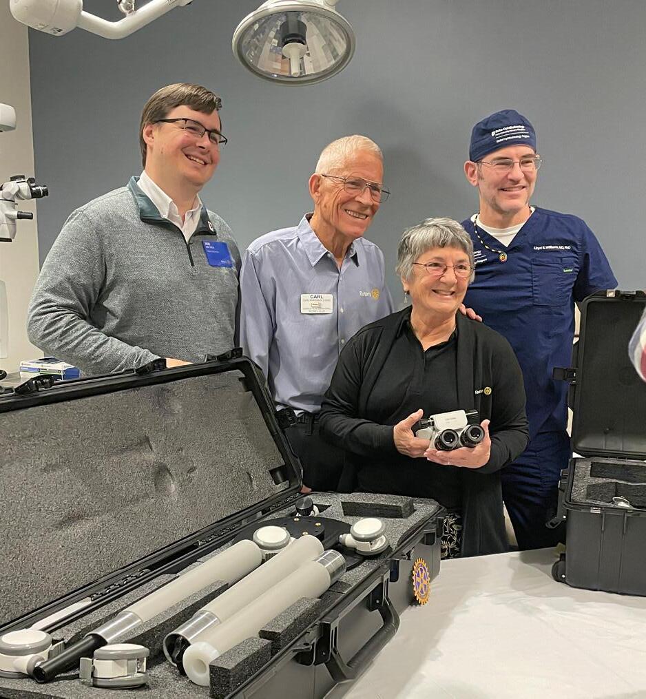
Rotary leader
John Tish, Rotary members, Carl Evans and Ann Evans, and Lloyd Williams, MD, PhD, with the new portable microscope, gifted by Charlotte International Rotary club through a fundraiser for Ann’s 80th birthday. Learn more or donate to Duke GO visit the website
Rotarian Supports Duke GO with Portable Microscope
When Ann Evans, FNP, DrPH, a passionate Rotarian and global health advocate, set her sights on ‘making a difference’ in the growing public health problem of avoidable blindness, she found a mentor in Williams. Evans was in South Sudan in 2022 helping her friend Jill Seaman, MD, 2009 MacArthur Fellow, working with the Himalayan Cataract Project, when she met Lloyd Williams, MD, PhD. “Watching people who were blind have their cataract removed, an $8 lens replacement with a 10-minute procedure, and walk away the following day with sight restored, changed my life,” said Evans. “I saw Williams was passionate about curing blindness; I got infected by his passion.”
Evans, a nurse practitioner and educator, also noted a patient safety issue — the operating room floor was unstable, a dangerous situation for cataract surgery. She submitted to Rotary for a grant to stabilize the floor, which was approved and completed. But the following year Williams returned to South Sudan and the operating microscope was broken. When Evans learned about the broken microscope, she worked with her Charlotte International Rotary Club to raise money. Her daughter established a fundraiser — $80 for mom’s 80th birthday — and the club donated a portable operating microscope to Duke GO.
“I’m really proud of shepherding this project, supporting Williams and his vision for Duke GO, because he has the kind of passion and the connection of the heart and mind that makes good things happen,” said Evans.
Unique Clinic to Treat Myopia
Myopia, also known as near-sightedness, is rising sharply among children worldwide, particularly because children are spending more time indoors focusing on near-work (think screens), and less time outdoors letting their eye muscles relax.
BY MARY-RUSSELL ROBERSON
In Asia, up to 80-90% of middle schoolers and young adults are near-sighted and the U.S. isn’t far behind at approximately 42% in urban areas, which has almost doubled in the last three decades.
That’s a problem not only for the present, but for the future — near-sighted people are at higher risk for multiple vision-threatening problems later in life, including macular degeneration, retinal detachment, glaucoma, and more.
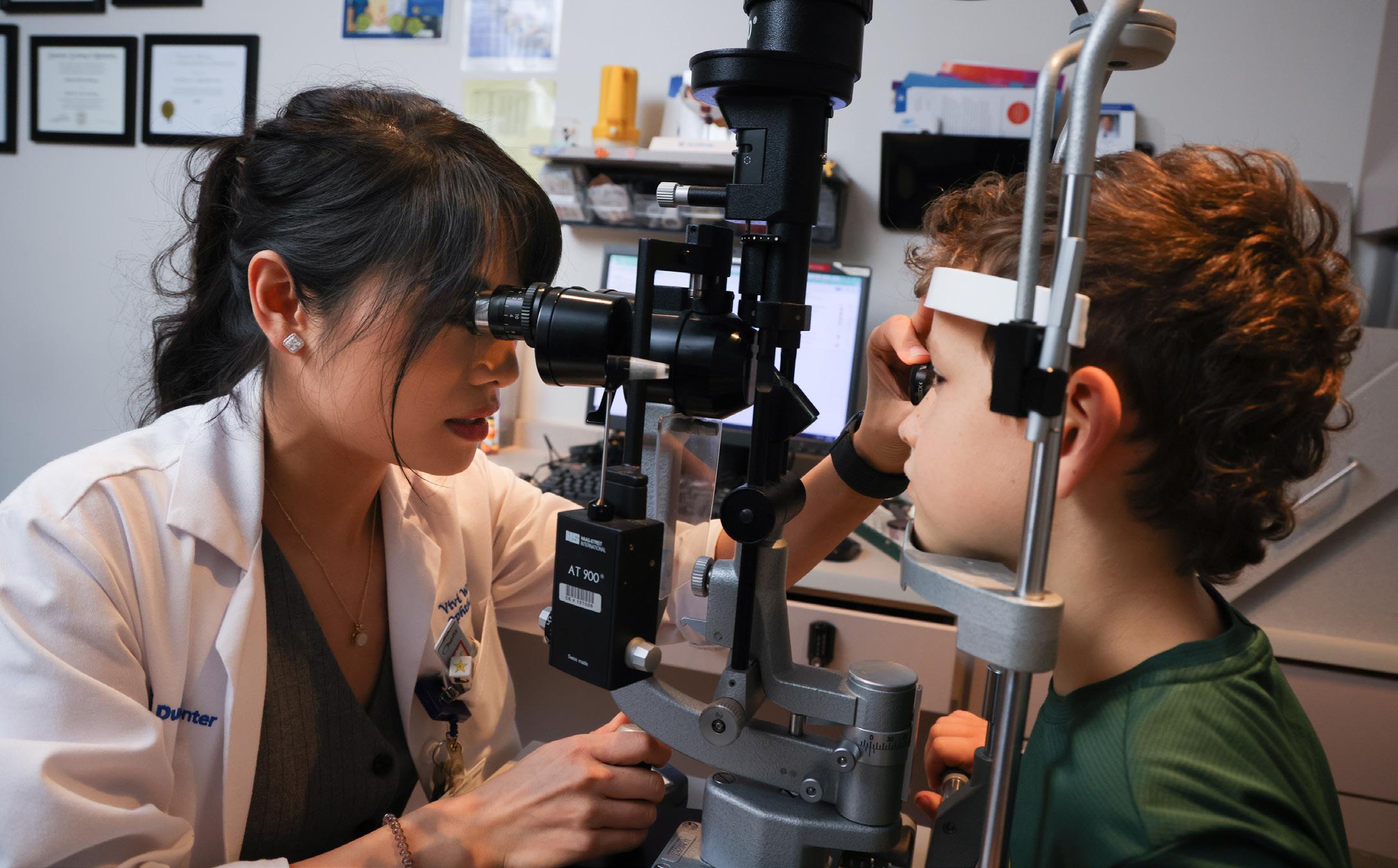
White checking vision during a patient’s visit
The good news? Duke created a unique clinic led by residency trained pediatric optometrists that offers traditional and innovative treatments to slow the progression of myopia in children. “Thirty years ago, doctors could only tell kids to wear glasses, but now we can actually do more to prevent or reduce the lifetime risk of visual impairment for these kids,” said Qiaohui Wei White, OD, MS, FAAO, pediatric optometrist.
The Risks of Myopia
People with myopia often have longerthan-normal eyes. This is because as kids grow and become myopic, the eye’s shape tends to elongate, stretching out the “wallpaper” on the inside of the eye that is responsible for capturing incoming visual information and sending those signals to the brain. This neuro-tissue is known as the retina. If the retina becomes too thin or stretched, it can detach, which can lead to permanent vision loss if not surgically corrected right away.
Severe myopia can also lead to macular degeneration, in which the center of the retina, called the macula, becomes damaged, resulting in the loss of central vision similar to age-related macular degeneration. Near-sighted people are also at higher risk for glaucoma and tend to develop cataracts earlier.
“There is no safe level of myopia,” White said, adding that the longer the eyeball is, as well as the younger the onset, the greater the risk throughout their lifetime.
“At Duke Eye Center, we perform complete, comprehensive testing and discuss with parents which options are suitable for their children, taking into account lifestyle and preference,” White said. “The best method is the one that works for that particular child and that the child will comply with.”
Childhood Treatments for Myopia
As children’s bodies grow, so do their eyeballs. Certain treatments have been shown to slow down the rate at which the eye elongates in childhood, thus reducing the future risk. Three evidence-based treatments are currently well established for myopia management in the United States: prescription eye drops, multifocal soft contact lenses, and nighttime hard contact lenses, known as Orthokeratology. Providers at Duke Eye Center are involved in all three methods and are constantly renewing their knowledge and updating their treatment strategies according to new research results. Scientists have hypotheses to why these treatments are so effective but haven’t yet nailed down the exact mechanism by which these strategies work.
Low Dose Atropine eyedrops
Atropine drops have been used for decades to dilate the pupil for eye exams with great safety. A much smaller dose than that used for pupil dilation, like 0.01% to 0.05% is used as a treatment for myopia, usually given at bedtime.
Multifocal soft contact lenses, including the only FDA approved lenses for “myopia control”, MiSight
These lenses allow clear vision in the center, with a blurry ring in the periphery to provide myopia control effect during daily wear. White said most children don’t notice the blurry ring; those that do, adapt to it quickly.
Ortho-K lenses, or Hard contacts for nighttime
These contact lenses reshape the front of the eyeball (the cornea) during sleep. Children who have this treatment wear the lenses only at night and enjoy clear vision during the day. The reliable safety profile of these lenses in children is supported by solid studies and allows daytime activities without spectacles or contact lenses.
Story continues on page 24
$13,473,941
Duke Eye Center
17,044
Lifestyle Changes to Reduce Myopia
To reduce the chances of developing myopia or to slow its progression, White and others who treat myopia recommend the following lifestyle changes for all children:
• When reading a book or looking at a screen, relax your eye muscles every 20 minutes by looking at least 20 feet away for a few minutes.
• Keep the book or screen a healthy distance from your eyes – at least the length of your forearm.
• Don’t read in dim light.
• Spend at least two hours outdoors each day.
It is not clear what aspect of outdoor time helps — it may be the increased light density, wider wavelength spectrum or the chance to truly relax the muscles used to focus the eye. White said any time of daylight works, shady days or sunny ones. If it is sunny, it’s also important to wear sunglasses to protect the eyes from ultraviolet rays.
Which Kids Should be Treated for Myopia?
“I always tell the parents, if you want to reduce the risk for your kid in the future, myopia treatment is something extra most kids can do sometimes even before developing myopia,” White said. In many
cases, kids can be detected as “Pre-myopia” candidates for myopia treatment, in which cases the kids’ eyes are growing faster than their peers and normative data. She also said the more severe the myopia is, the more important it is to control.
White recommends that children with concerning myopia should be seen at the pediatric myopia clinic to discuss treatments. The team at Duke Eye Center will not only try to reduce the progression as much as we can but we will also distinguish the risk and rule out genetic diseases that may be associated with high myopia. If a genetic syndrome is the underlying cause, the child can be followed more closely by a team of experts, including our pediatric retinal specialists.
“Because of our experience and strongly specialized team, we strive to be the best place to manage myopia,” White said. “[When you visit Duke Eye Center] You are not only seeing me. We can connect you to the strong team behind me.”
“Thirty
years ago, doctors could only tell kids to wear glasses, but now we can actually do more to prevent or reduce the lifetime risk of visual impairment for these kids.”
Qiaohui Wei White, OD, MS, FAAO
White explaining eye images and myopia control options.
Additional Members of the Myopia Management Team
Adriana Ferreira, OD
Nathan Cheung, OD
Duke Eye Center Researchers Identify a New Retinal Structure
Researchers at the Duke Eye Center have made an important — and surprising — discovery in human eye anatomy that has gone unnoticed for decades. Using enhanced imaging technology, they identified a previously unknown part of photoreceptor cells in the retina. Their finding, published in Communications Biology, challenges long-standing assumptions about the retina and opens new avenues for understanding vision and genetic eye diseases.
Most of what researchers know about retinal anatomy has come from exhaustive studies using electron microscopy in the 1960s and 1970s. Eventually, no new major discoveries were being made in the anatomy of the retina, so that chapter of research closed.
But technology kept advancing. Oleg Alekseev, MD, PhD, assistant professor of ophthalmology, and Vadim Arshavsky, PhD, Helena Rubinstein Foundation Distinguished Professor of Ophthalmology, decided to use three-dimensional electron tomography, which creates a 3D map of an object, to look at retinas with unprecedented resolution. “We aimed to take a look at some very small structures,” Alekseev said, “but we found something pretty major.”
Inside the retina are photoreceptor cells, commonly known as rods and cones. Photoreceptor cells convert light signals into electrical messages that the brain decodes
and processes into visual images. Alekseev, Arshavsky, and team found that each rod photoreceptor cell contains a large, mechanically reinforced protrusion, which they’ve termed the accessory inner segment (aIS).
“The function isn’t known yet,” Alekseev said, “but we presume it serves as a physical support to hold the photoreceptor.”
Photoreceptors are long and narrow. The researchers think that the aIS holds everything together and stable, preventing the photoreceptors from collapsing and flopping.
Over 300 known genes make photoreceptors functional.
A mutation in any one of those genes can make the photoreceptor unable to work properly and can lead to irreversible blindness. Genetic testing helps solve only around 70% of cases, but this discovery opens the door to studying additional previously overlooked genes that may be involved in forming and maintaining the aIS.
“We need to explore this structure further,” Alekseev said. “It’s very likely that some diseases are caused by defects in the aIS, and now we have the opportunity to explore those diseases, improve our ability to diagnose patients, and start thinking about treatments.”
The aIS appears to be a uniquely human structure, which could explain some of the discrepancies between human diseases in the eye and mouse models of these diseases. For example, humans with Usher syndrome experience
both blindness and deafness. In mouse models, though, introducing the exact same mutations in the exact same genes produces mice that are deaf, but not blind.
“We speculate,” said Arshavsky, “that some proteins in humans may be functioning within the aIS structure, and whenever you have a discrepancy between a human and mouse phenotype of some gene mutations, this structure comes into play.”
Now that aIS has been discovered, the next step is to understand the molecular composition of its unique parts and see what questions they can answer about human diseases in the eye.
The team found that each rod photoreceptor cell contains a large, mechanically reinforced protrusion, which they’ve termed the accessory inner segment (aIS).
Oleg Alekseev, MD, PhD Vadim Arshavsky, PhD
Recognizing Milestone Anniversaries
This year we will celebrate significant anniversaries for facilities that have been instrumental in Duke Eye Center’s excellence in research, education and patient care.
The Hudson Building, the renovated Wadsworth Building, and the Albert Eye Research Institute (front to back) now make up Duke Eye Center. Duke University Hospital appears in the background.
20 Years of Research Progress
The Albert Eye Research Institute, affectionately known as AERI, opened in 2005 and provided Duke Eye Center with 72,000 square feet to significantly expand research capabilities, pediatric clinical care and educational programs. A transformative gift from Herman and Ruth Albert spearheaded the new five-story building, creating much-needed space for Duke ophthalmology research teams to make groundbreaking discoveries, establishing a separate clinic designed specifically for pediatric patients, and housing a generous auditorium and gathering area for medical education events.
10 Years of State-of-the-Art Care
The Hudson Building at Duke Eye Center opened in June of 2015. The four-floor, 116,000 square foot facility is named in honor of William Hudson, retired CEO of Durham-based LC Industries and past chair of the Duke Eye Center Leadership Council. LC Industries is the largest employer of visually impaired people in the country.
Together with the Albert Eye Research Institute and renovated space in the adjoining Wadsworth Building — the original Duke Eye Center dedicated facility which opened in 1973 — the Hudson Building is the hub of an extensive Duke Eye Center campus incorporating new patient amenities and services from Duke’s expert clinical eye specialists and world-class ophthalmology research at the Duke University School of Medicine.
Donors
Albert Eye Research Institute (AERI)
Ruth & Herman Albert
Kit & Richard Barkhauser
Mary & Jonathan Christenbury
Heather & Jim Gills
Rose & Leonard Herring
Lyn Proctor & Gary Hock
Evelyn Hunter-Longdon
Susan & David Kahn
Roslyn & Milton Lachman & Family
Marianne & Allen Mebane
Roger Milliken
Loraine & Robert Sinskey
The Duke Endowment
Alcon Foundation
A.E. Finley Foundation
Hudson Building
LC Industries, William Hudson, President/CEO
LC Industries Foundation
Heather & Jim Gills
Loraine & Robert Sinskey
Nanaline H. Duke Trust
Kit & Richard Barkhauser
Jack and Judy Brinson Foundation
Catherine Harley
Evelyn Hunter-Longdon
Roslyn & Milton Lachman & Family
Roger Milliken
Phoebe & Gerrish Milliken
Jeannette & Michael Vu
Donors listed generously contributed $100,000 or more to help build these facilities that have transformed Duke Eye Center. Donors are listed by their names at the time of their philanthropic commitments to the Albert Eye Research Institute and the Hudson Building and may not reflect the names used by these individuals and organizations today.
John Karickhoff Bringing the Light of Innovation to Duke Eye Center
In addition to being a talented teacher, surgeon, and humanitarian, John Karickhoff, MD’64, HS‘64-’68, is a visionary. He recently gave $1 million to launch the Duke Ocular Innovation Hub. The hub will serve both as an engine to drive innovation and a repository for innovations developed at Duke Eye Center.
Karickhoff wants every trainee at Duke to think about how they can advance ophthalmology and to consider making innovation their career focus. To him, innovation is at the heart of every advance in medicine.
“I realized that receiving my AB and MD degrees, teaching for 30 years at Georgetown University Medical School, and performing surgery on thousands of people is a worthy career, but none of those activities advanced my profession or will be part of the history of medicine,” Karickhoff said. “It is only innovation, by definition, that advances the art and science of medicine and gives something new to humanity.”
A West Virginia native, Karickhoff graduated magna cum laude from Marshall University while serving as class president his freshmen through junior years and as president of the student body his senior year. After completing eight years of medical training at Duke, he served in Vietnam in the United States Army Medical Corps. In addition to his army responsibilities, he set up a clinic that provided free cataract surgery for Vietnamese civilians. He performed over 500 surgeries, secured donated glasses for his patients, and organized medical
BY WENDY GRABER
Duke Ocular Innovation Hub
teams to provide care in nearby villages. For this work, the Army awarded him the Bronze Star. Karickhoff became the world authority on laser treatment of eye floaters and has invented or designed more than 20 innovations, including devices, like the Karickhoff laser lens, that are used worldwide. His introduction to innovation came in 1979 when he read an article that suggested that people who had good ideas should give them to companies to develop.
“I had an idea and shared it with a company that developed ocular equipment,” said Karickhoff. “They developed a prototype, and we sent it back and forth, improving it with each iteration. I even wrote the advertisement for the device because they didn’t fully understand the device’s importance. And that was the end of it. Then several months later, while attending a national meeting, I stopped by their booth to ask if they had ever sold any. They said, ‘Did we ever. We sold almost $600,000 worth of your device at $475 apiece!’ In the end, they made a lot of money, and I got a framed copy of my advertisement and the knowledge that if I had an idea and worked long enough on it, I could make
something good. That was all the reward I needed.”
That device, the Karickhoff laser lens, is still used today. Karickhoff intentionally donates almost all of his innovations and inventions to benefit ophthalmology. One invention he trademarked, the Karickhoff Mosaic Matcher TM , allows surgeons to quickly and easily estimate the number of corneal endothelial cells prior to surgery — an important factor in determining whether the cornea will remain clear after surgery. The device was freely distributed and is the most widely used device of its kind in the world.
Karickhoff’s unique 40-year research achievement is that he developed an innovation or significant advance for each of the 14 anatomic parts of the human eye, as documented in his book “Laser Treatment of Eye Floaters.” Karickhoff’s eyes light up when he talks about innovation. He wants to spark that same enthusiasm among trainees at Duke.
“Developing something new that no one has ever done before has given me the greatest satisfaction and happiness in my career,” said Karickhoff. “My main hope is that these extremely talented trainees will stop for a second before
they embark on their careers and consider creating something new that will change the course of ophthalmology. To me, innovation is the most important word in medical education.”
The Ocular Innovation Hub will drive innovation through six activities: teaching trainees and faculty how to move an idea to market, recognizing top innovations with annual awards, highlighting exciting innovations from across the field of ophthalmology through a guest lecture series, funding faculty research, funding trainee research (including paying research article publishing fees), and creating a robust repository of Duke ocular innovations.
“I have a keen interest in innovation, given part of my work is exploring new ways to use ocular imaging to improve eye exams and surgery,” said Anthony Kuo, MD, vice chair of technology in the Department of Ophthalmology. “As such, the department and I share Dr. Karickhoff’s vision. The innovation hub and seed funding will provide our faculty and trainees with the resources and access they need to bring their ideas to fruition, adding to the rich legacy of innovation in our specialty.”
Karickhoff, a longtime supporter, chose Duke for the innovation hub because of the excellence of the trainees and his high regard for Duke itself.
“Duke is where I got my professional start in life,” said Karickhoff. “And I knew that much of my education had been paid for by the gifts of previous graduates.”
John Karickhoff, MD’64, HS‘64-’68 recently gave $1 million to launch the Duke Ocular Innovation Hub to drive innovation and establish a digital repository for innovations developed at Duke Eye Center.
Duke Eye Center, opened in 1973, has grown considerably to become an internationallyrenowned academic medical institution. With a long track record of developing innovative treatments, providing excellence in care, and training leaders in the field, it has been consistently ranked as a top ten center for over 30 years.
“Dr. Karickhoff’s generous gift is a vital investment in the future of ophthalmology, empowering our faculty and trainees to
make lasting impacts in the field through innovation,” said Edward Buckley, MD, chair of the Department of Ophthalmology.
“We are immensely grateful for his support, and we hope that others will be inspired to follow in his footsteps, helping to ensure the continued excellence of Duke Eye Center.”
Visit the Duke Ocular Innovation Hub website
Partnership for the Gift of Sight
Family, patient and physician make a winning combination for patient care and research success.
BY MARY-RUSSELL ROBERSON
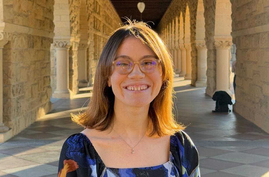
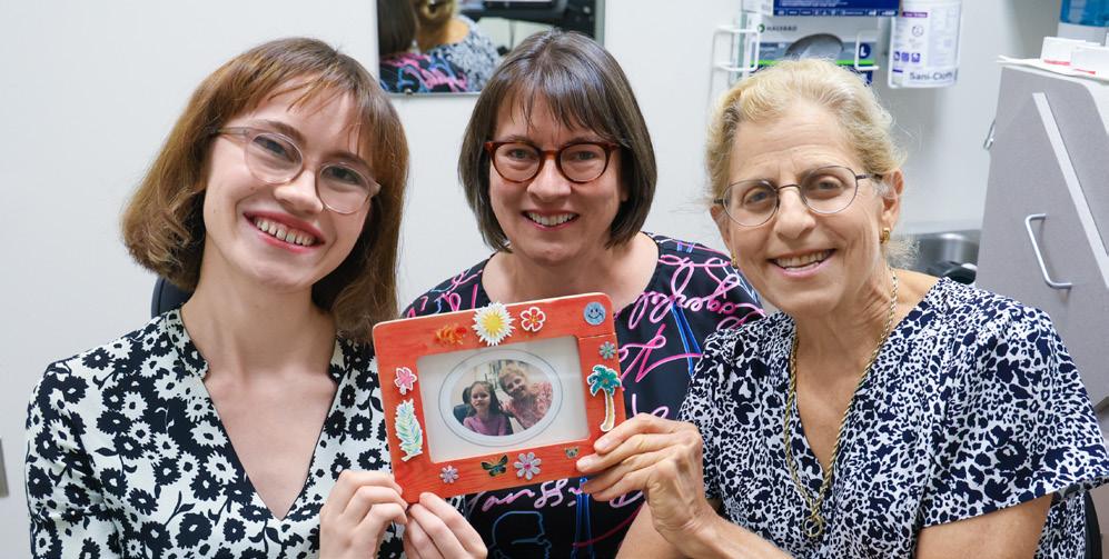
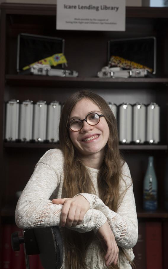
Sharon Freedman, MD, professor of ophthalmology, and the Smale family of Seattle, Washington, have developed a supportive partnership. Sarah Rose Smale, 22, has been a patient of Freedman’s for almost 20 years. During that time, Freedman has helped the family preserve as much of Sarah Rose’s sight as possible and has guided them through multiple chronic and acute eye conditions, often from afar.
Sarah Rose was born blind in her left eye and with a cataract in her right eye, which was removed in order for her to be able to develop some vision. About three years later, Sarah Rose developed a glaucoma specific to infants and children, called glaucoma following cataract surgery.
As soon as Sarah Rose was diagnosed with glaucoma, her parents, Angela and Leonard, set out to find a pediatric glaucoma specialist. They sought someone on the cutting edge of pediatric glaucoma research and practice, willing to measure Sarah Rose’s eye pressure in-clinic in addition to during examinations under general anesthesia, and young enough to manage Sarah Rose’s vision care throughout her childhood. “We wanted the absolute best-match physician,” Angela said, “and we decided Dr. Freedman was that person.”
Sarah Rose was three years old the first time the family flew from Seattle to Duke. “Dr. Freedman explained Sarah Rose’s complex eye conditions, her proposed treatment plan, and the potential risks,” Angela said. “We really connected with her and were very much partners in Sarah Rose’s care after that first meeting.”
Angela and Leonard were impressed with Freedman’s ophthalmic expertise and the way she built a deep, trusting relationship with Sarah Rose through many surgeries and examinations under anesthesia. “You might expect her to experience medical trauma from all these surgeries, procedures, and eye drop medications,” Angela said, “but Dr. Freedman made it such that Sarah Rose actually almost looked forward to her appointments. We used to refer to anesthesia as a ‘hospital nap,’ which helped to build up her resilience.”
Rose, her mom Angela, and Freedman. The photograph they are holding is Sarah Rose at age 6, in a special frame she decorated. Freedman has kept this photo displayed in her office since the Smales sent it to her in 2009.
Parents on the Care Team
In patients with glaucoma, high eye pressures can damage the optic nerve. Angela and Leonard learned how to measure Sarah Rose’s eye pressure at home with an instrument called a Tono-pen. “It was difficult to use at first,” Leonard said. “But we started learning how to use it and sending data back to Dr. Freedman.” That data helped Freedman know when it was time to have Sarah Rose come to Duke for glaucoma surgery. After the surgeries, the data helped Freedman and the Smales adjust Sarah Rose’s medicine as her eye pressure changed. Later, the family switched to a newer instrument, called an iCare rebound tonometer, which was easier and safer because it did not require numbing the surface of the cornea with anesthetic drops before use.
Rose, age 13,
Sarah
celebrating the Lending Library opening in 2015.
Sarah
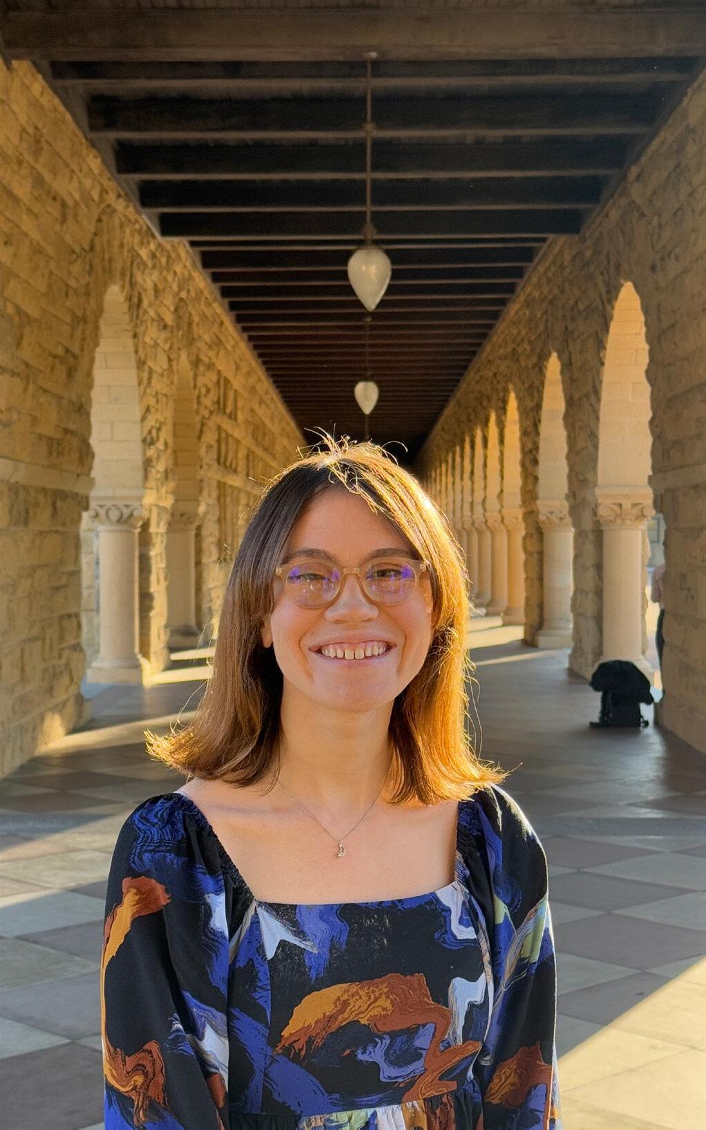
Sarah Rose is a current student at Stanford University, where she is earning both a bachelor’s degree in symbolic systems with a concentration in neurosciences and a master’s degree in computer science.
As she grew older, Sarah Rose realized how significant it was that her parents measured her eye pressures at home. “It helped save a lot of my vision,” she said. “And I really wanted other children with glaucoma to have access to that same level of care.” In 2014, she and her parents started a charity called Saving Kids’ Sight to educate and support other families. With Freedman’s help, Saving Kids’ Sight created a lending library of home tonometry units and provided Freedman with a grant to research the effectiveness of home tonometry in children with glaucoma.
Today, Sarah Rose’s glaucoma is under control, but she has developed significant swelling (edema) in her cornea over the past few years, and she is now legally blind. Freedman is still following her, connecting her with other experts at Duke Eye Center as her needs change. “I really appreciate how she’s shown so much compassion for me and my family,” Sarah Rose said.
Sarah Rose is a student at Stanford University,
where she is earning both a bachelor’s degree in symbolic systems with a concentration in neurosciences and a master’s degree in computer science. For her honors thesis, she’s studying patient and physician attitudes toward home tonometry. Freedman is advising her. “She helped me narrow down my initial project proposal, is an integral part of the project, and is readily available to mentor me throughout the process. Any time I have a question or need feedback, she’s just an email away,” Sarah Rose said.
Giving Back
Over the years, Angela and Leonard have given several gifts to fund specific projects of Freedman’s and they have served on the Duke Eye Center Leadership Council. Angela is currently the chairperson. But they wanted to do more.
After consulting with Freedman, they decided to donate $500,000 to fund a research coordinator position and associated research project costs for five years. In the past, Freedman had to scramble each year to secure funding for this position, typically filled by someone between medical school and ophthalmology residency.
Freedman’s research aims to improve pediatric ophthalmology through new technologies, from home tonometry to optical coherence tomography (OCT) to virtual reality for visual field testing. “My goal is to apply emerging technology to helping children,” Freedman said. “Often children are left behind because they have special testing needs and do not make up a large part of the glaucoma market. But they are pretty important and the Smales have always shared my passion for this mission.”
“Our
gift enables Dr. Freedman to use her time and expertise for the greatest impact, and not having to worry about where she’s going to get the money to pay a research coordinator.”
Freedman pointed out that the Smale family gift will have benefits beyond her own research. “By choosing to support the research coordinator position, the Smales will be facilitating the entire clinical research endeavor of the Duke Pediatric Ophthalmology Division,” she said. “It’s a transformative gift.”
Medical School: George Washington University (Washington, DC)
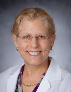
Elizabeth Mathenge, MD, MSc
Assistant Professor of Ophthalmology
Oculofacial and Orbital Surgery
Fellowship: ASOPRS, at Cincinnati Eye Institute
Residency: Howard University
Medical School: Dual MD and MSc in Global Health, Duke University
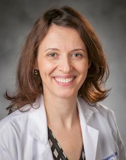
Tali Okrent, MD
Assistant Professor of Ophthalmology
Neuro-ophthalmology
Fellowship: Neuroophthalmology and Oculofacial, Duke Eye Center
Residency: Sackler School of Medicine, Tel Aviv University
Medical School: Ben-Gurion University Medical School for International Health
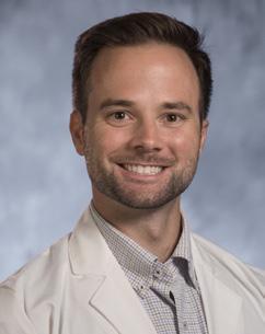
Michael Quist, MD
Assistant Professor of Ophthalmology
Glaucoma
Fellowship: Duke Eye Center
Residency: Duke Eye Center
Medical School: Duke University
Honors and Awards
Lejla Vajzovic, MD, FASRS received the Mentorship Award at the 2024 Vit Buckle Society and was a featured speaker. The Mentorship Award celebrates a sustained career commitment to mentoring vitreoretinal fellows, advancing research, and patient care in the field of ophthalmology. Lindsey Chew, a research scholar, mentored by Vajzovic, won best poster award for her research, Feasibility of Screening for Retinal Disease in Children without Pharmacologic Dilation in the Pediatric Primary Care Setting.
Faculty
Oleg Aleskeev, MD, PhD promoted to Assistant Professor of Ophthalmology.
Alan Carlson, MD Duke Ophthalmic Technician Program Attending Mentor of the Year.
Durga Borkar, MD, MMCi promoted to Associate Professor of Ophthalmology.
Pratap Challa, MD received the prestigious Straatsma Award for Excellence in Resident Education and became a member of the Board of Directors for the American Board of Ophthalmology and represents them on the AAO Council.
Mays Dairi, MD Duke Ophthalmology Golden Globe Award for her strong commitment to resident teaching, in clinic, in the OR, and in didactic lectures.
Thomas Devetski, OD Excellence in Education Award for Outstanding Contribution to Medical Student Education.
Madison Dunning, OD promoted to Assistant Professor of Ophthalmology and earned Fellowship of the American Academy of Optometry (FAAO).
Laura Enyedi, MD elected as AAO Council Deputy Section Leader for Sub-Specialty Councilors, AAO Ophthalmic
Advocacy Leadership Group, received the AAO Senior Achievement Award and Secretariat Award and named Castle Connolly Top Doctor.
Sharon Fekrat, MD, FACS, FASRS Chair, Website Committee, The Retina Society and held several Visiting Professorships; Department of Ophthalmology Tan Tock Seng Hospital, Singapore; Department of Ophthalmology, Asan Medical Center, Seoul, South Korea; Department of Ophthalmology, Northwestern Medicine, Chicago; Mark J Daily MD Visiting Professorship, Department of Ophthalmology, Loyola University Medical Center, Chicago.
Sharon Freedman, MD
American Glaucoma Society Outstanding Educator 2024, AAO Golden iCare Award Recipient, delivered the inaugural lecture for the Edward Buckley Distinguished Lecture in Pediatric Ophthalmology.
>
Nickolas Garson, MD
Duke Ophthalmic Technician Fellow Mentor of the Year award.
Dilraj Grewal, MD, FASRS inducted into Club Jules Gonin 2024, received ASRS Presidential Award, and became Member of Macula Society Executive Committee.
Majda Hadziahmetovic, MD promoted to Associate Professor of Ophthalmology.
Kourtney Houser, MD YoungMD Connect Outstanding Female Leader in Ophthalmology and promoted to Associate Professor of Ophthalmology.
Chicago Ophthalmological Society Meeting, and the Joseph Smiddy, Jr. Memorial Lecture at Wilmer Eye Institute.
Yannek Leiderman, MD, PhD joined the Department of Ophthalmology as Professor of Ophthalmology with tenure and named Castle Connolly Top Doctor.
Paloma Liton, PhD promoted to Professor of Ophthalmology with tenure, elected Vice President, Americas for ISER, and appointed executive editor of Experimental Eye Research, official Journal of ISER.
Anthony Kuo, MD, PhD promoted to Professor of Ophthalmology and Biomedical Engineering, with tenure.
Jeffrey Kozlowski, OD promoted to Assistant Professor of Ophthalmology.
Eleonora Lad, MD, PhD promoted to Professor of Ophthalmology with tenure and received the 2024 ASRS Honor Award, she delivered the Gifford Lecture at the Illinois Society of Eye Physicians and Surgeons and
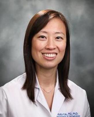
Katy Liu, MD, PhD 2023 David L. Epstein ClinicianScientist Research Award presented by the Chandler Grant Glaucoma Society, the American Glaucoma Society Young Physician Scientist Award, selected to serve on the Council of Vision Editors Fellow (CVEF) Program and one of six inaugural fellows selected for the Council of Vision Editors Fellowship.
Ramiro Maldonado, MD promoted to Associate Professor of Ophthalmology and selected to be a member of the Scientific Advisory Board for the Foundation for Fighting Blindness.
Goldis Malek, PhD promoted to Professor of Ophthalmology and Pathology with tenure, elected to the Board of Trustees for ARVO, appointed co-chair of ARVO’s Women’s Leadership Development Program, invited Program Committee Chair of the Novel Medical Therapy committee of the Foundation for Fighting Blindness
Scientific Advisory Board, selected for Treasurer of the Association for Ocular Pharmacology and Therapeutics (AOPT).
Ryan McNabb, PhD promoted to Assistant Professor of Ophthalmology. Daniel Saban, PhD promoted to Professor of Ophthalmology and Integrative Immunobiology with tenure, Vice Chair of Research Strategy, Scientific Director of the Foster Center for Ocular Immunology, and received the Cogan Award, presented by ARVO.
Terry Semchyshyn, MD promoted to serve as a board examiner for the American Board of Ophthalmology oral board exam this fall. He has now acted as an examiner for over 10 examinations.
Sandra Stinnett, DrPH ASRS Master Contributor Award for outstanding contributions to the Journal of Vitreoretinal Diseases.
Amol Sura, MD promoted to Assistant Professor of Ophthalmology.
Joanne Wen, MD received the American Glaucoma Society Mid-Career Physician Scientist Award.
>
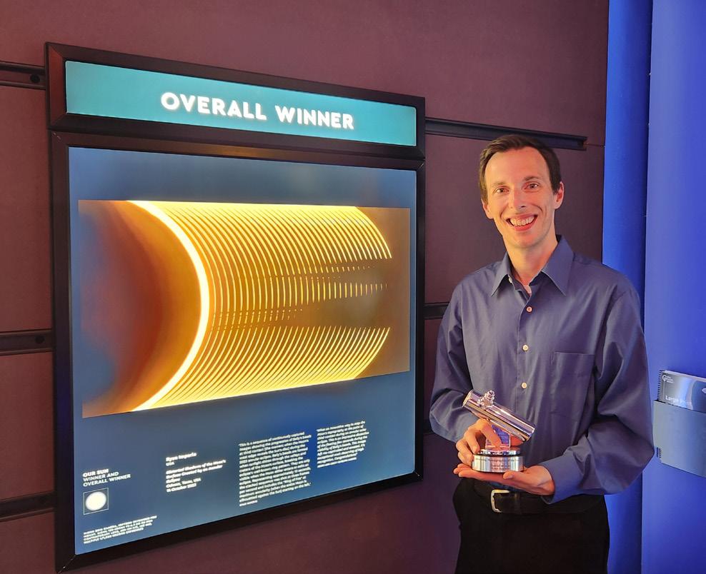
Ryan Imperio, Duke Eye Center Ophthalmic Photographer, was named the 2024 Astronomy Photographer of the year by the Royal Museums Greenwich in London.
Duke Ophthalmic Photographer Earns Global Honor for Capturing Wonders of the Sky
Duke Eye Center Ophthalmic Photographer, Ryan Imperio found a creative outlet in astrophotography, the pursuit of capturing images of celestial bodies and events. This year, Imperio’s images of the 2023 annular solar eclipse on October 14 led to the Royal Museums Greenwich in London naming him the 2024 Astronomy Photographer of the Year.
Ryan’s goal was to photograph ‘Baily’s beads’, a phenomenon that occurs when bright shafts of sunlight peek through the Moon’s uneven surface. He only had seconds to capture the beads as the eastern curve or ‘limb’ of the Moon almost touched that of the Sun, a timeframe so tight that Ryan decided to use an atomic clock for accuracy.
With a skilful camera setup, clockwork precision, dozens of photographs and some clever processing resulted in Ryan’s image “Distorted Shadows of the Moon’s Surface Created by an Annular Eclipse.”
Scan QR code to read the full story about the award and Imperio’s photography technique.
Faculty Promotions
Oleg Alekseev, MD, PhD: Assistant Professor of Ophthalmology
Durga Borkar, MD, MMCi: Associate Professor of Ophthalmology and Associate Professor in Population Health Sciences
Madison Dunning, OD: Assistant Professor of Ophthalmology
Majda Hadziahmetovic, MD: Associate Professor in the Department of Ophthalmology and Associate Professor in Electrical and Computer Engineering
Kourtney Houser, MD: Associate Professor of Ophthalmology
Jeffrey Kozlowski, OD: Assistant Professor of Ophthalmology
Anthony Kuo, MD: Professor of Ophthalmology with tenure
Eleonora Lad, MD, PhD: Professor of Ophthalmology with tenure
Paloma Liton, PhD: Professor of Ophthalmology with tenure and Professor in Pathology
Ramiro Maldonado, MD: Associate Professor of Ophthalmology
Goldis Malek, PhD: Professor of Ophthalmology with tenure and Professor in Pathology
Ryan McNabb, PhD: Assistant Professor of Ophthalmology
Daniel Saban, PhD: Professor of Ophthalmology with tenure and Professor in Integrative Immunobiology
Amol Sura, MD: Assistant Professor of Ophthalmology
Lejla Vajzovic, MD, FASRS: Professor of Ophthalmology with tenure
Qiaohui Wei White, OD: Assistant Professor of Ophthalmology
Lloyd Williams, MD, PhD: Associate Professor of Ophthalmology
Honors and Awards
Lejla Vajzovic, MD, FASRS promoted to Professor of Ophthalmology, Pediatrics, and Biomedical Engineering, with tenure, Vit-Buckle Society Mentorship Award, Real World Ophthalmology Extraordinary Mentor Award, Paul A. Chandler Visiting Professorship at Massachusetts Eye and Ear, Harvard Medical School.
Diane Whitaker, OD Bass Connections Research Award.
Award from the Women in Retina (WinR) Research Conference at Association for Research in Vision and Ophthalmology; mentor, Lejla Vajzovic, MD, FASRS.
Qiaohui Wei White, OD promoted to Assistant Professor of Ophthalmology, earned Fellowship of the American Academy of Optometry (FAAO).
Lloyd Williams, MD, PhD Award of Achievement, presented by the Sierra Leone National Eye Health Programme, Ministry of Health, promoted to Associate Professor of Ophthalmology, President of the North Carolina Society of Eye Physicians and Surgeons.
Fellows
Rami Gabriel, MD 2024 Resident ASCRS Excellence Award, Duke Ophthalmology Ocular Innovation Award, K. Alexander Dastgheib Award, and Golden Apple Teaching Award.
S. Tammy Hsu, MD Real World Ophthalmology Visionary Award, Mina Chung Memorial Research
Mustafa Iftikhar, MD
Best Research Presentation by a Clinical Fellow Award, mentor Dilraj Grewal, MD, FASRS.
Venkatkrish M. Kasetty, MD 2024 Heed Fellow and 2024 VitreoRetinal Surgery Foundation (VRSF) Research Award, mentor, Lejla Vajzovic, MD, FASRS.
Tali Okrent, MD
2024 Hornaday Fellow, recognizing her exceptional ability in clinical, research, and teaching efforts, Best Research Presentation by a Clinical Fellow Award, mentor Sidney Gospe, MD, PhD at Duke Residents and Fellows Day.
Bonnie Sklar, MD Duke Ophthalmology Golden Apple Teaching Award.
James Tian, MD Duke Ophthalmology Golden Apple Teaching Award.
Yuxi Zheng, MD
2024 Edward K. Isbey, Jr. Award for excellence in clinical care, ethics, and research, Duke Ophthalmic Technician Program Resident Mentor of the Year, ASRS Fellows-in-Training Co-Chair, Yannuzzi Rounds Best Fellow Presentation award, presented by the International Society of
Retinal Imaging, 2024 VitreoRetinal Surgery Foundation (VRSF) Research Award.
Residents
Ellie Zhou, MD Duke Ophthalmology Golden Apple Teaching Award.
Kelly Donovan, MD, PhD selected to attend the 2025 Heed Ophthalmic Foundation Residents Retreat, Duke Office of Physician Scientist Development Technician Support Award.
Nicholas Johnson, MD Duke Ophthalmology Golden Apple Teaching Award.
Emily Jung, MD Academic Grant Award, Vit-Buckle Society, mentor, Sharon Fekrat, MD, FACS, FASRS, vice-chair of the GME resident council.
Terry Lee, MD Duke Ophthalmology Golden Apple Teaching Award, travel award for poster presentation at the Harvard Ophthalmology and Mass Eye and Ear 2024 Data Science Symposium, mentor Sharon Fekrat, MD, FACS, FASRS).
Angela Li, MD Robert Machemer Research Award, mentor Glenn Jaffe, MD, selected to attend the 2025 Heed Ophthalmic Foundation Residents Retreat.
Richard Morgan, MD, PhD accepted into the prestigious American Society of Retina Specialists Underrepresented in Retina Mentorship Program, academic Grant Award, Vit-Buckle Society, mentor Sharon Fekrat, MD, FACS, FASRS.
Agnes Owete, MD selected to present at the 2024 AAO Global Ophthalmology Summit.
Cason Robbins, MD American Society of Cataract and Duke Ophthalmology Golden Apple Teaching Award, 2024 Resident Excellence Award presented by the American Society of Cataract and Refractive Surgery.
Richmond Woodward, MD inaugural recipient of the recently established
Edward G. Buckley Resident Educator Award recognizing his contributions to resident education at Duke Ophthalmology.
Medical Students
Lindsey Chew, BS Academic Grant Award from the Vit-Buckle Society to present her work at the 2024 meeting in Miami and best poster award at Vit-Buckle Society for her research, winner of the Duke-National University of Singapore Research Exchange Grant mentor, Lejla Vajzovic, MD, FASRS. National Eye Institute ARVO Travel Grant, invited to speaker at the Keystone
2024–2025 Residents
Third Year Residents (PGY4)
Modupe Adetunji, MD
Kelly Donovan, MD, PhD
Angela Li, MD
Aaron Lindeke-Myers, MD
Richard Morgan, MD, PhD
Cason Robbins, MD
Second Year Residents (PGY3)
Juno Cho, MD
Jinwon (Terry) Lee, MD
Keke Liu, MD
Gregory McElroy, MD, PhD
Esteban Peralta Chacon, MD
James Robbins, MD
First Year Residents (PGY2)
Jean Adomfeh, MD
Nicholas Johnson, MD
Emily Jung, MD
Hazem Mousa, MD
Naveen Karthik, MD
Ailin (Irene) Song, MD
Interns (PGY1)
Ariana Allen, MD
Eli Cehelyk, MD, MPH
Alice Liu, MD
Grace Nipp, MD
Agnes Owete, MD
Jay Rathinavelu, MD
Rodnell Batac, OD Pediatric Optometry
Symposia, Center for Advanced Genomic Technologies Travel Grant mentor, Catherine Bowes Rickman, PhD.
Hemal Patel, BS National Eye Institute ARVO Travel Grant, mentor Sharon Fekrat, MD, FACS, FASRS.
Daniel Wang, BS Duke Ophthalmology
Best Research
Presentation by a Full-Time Research Fellow Award for his presentation, Improving Microbial Keratitis AI Screening Tools Constrained by Limited Data Using Synthetic Generation of Slit Lamp Photos, mentor Anthony Kuo, MD. 2024 VitreoRetinal Surgery Foundation (VRSF) Research Award, mentor, Lejla Vajzovic, MD, FASRS.
The Michelle P. Winn Awards honor individuals and teams who have made significant contributions to diversity, equity, and inclusion over the past year.
38 Michelle Winn Inclusive Excellence Award iMIND Research Team
Led by Sharon Fekrat, MD, FACS, FASRS, and Dilraj Grewal, MD, FASRS, from the Department of Ophthalmology, iMIND Research Team has taken a unique, multidisciplinary approach to enhancing equity and diversity in clinical trials and medical education and training.
iMIND is made up of Duke undergraduates, medical students, ophthalmology residents and fellows, and faculty from several departments. The team’s research involves using eye multimodal imaging to detect and diagnose neurodegenerative disease, including studying gender and racial differences.
They have worked to educate and engage the local community about clinical research to promote diversity in clinical trial cohorts. Their team has produced several national surveys that examine gender, racial, and other potential biases experienced by premedical and medical students, as well as physician residents across the United States. They have also assessed diversity trends in ophthalmology residents over the last decade and analyzed endowed chair allocation across academic institutions to improve equity.
In her nomination letter, Durga Borkar, MD, assistant professor of ophthalmology, praised the team for its innovation: “Their far-reaching initiatives and unwavering commitment to advancing diversity,
equity, and inclusion serve as an example to others within the broader School of Medicine. Their innovative efforts highlight the inclusive thinking exemplified at Duke.”
iMIND Team:
Duke undergraduates: Sejal Patel, Disha Jotsinghani, Annabel Stickel, Alice Haystead, Sanjana Anand, Alexander Richardson, David Bearden, Raj Kundu
Duke MBS student: Lucas McNeill
Duke medical students: Suzanna Joseph, Ariana Allen, Jay Rathinavelu, Jamie Karl, Hemal Patel, Peter Weng, Josh Woo, Wufan Zhao, Alex Choi, Liz Fan, Camryn Thompson, Meredith Cox, Anita Kundu, Angela Hemesath, Praruj Pant, Lily Fahs, Lulu Wein, Claire Washabaugh, Nathan Luzum, Shweta Lodha, Grace Kim, Nicole Schindler
Duke ophthalmology residents: Hazem Mousa, MD; Naveen Karthik, MD; Terry Lee, MD; Cason Robbins, MD; Emily Jung, MD; Sri Meghana Konda, MD; Yuxi Zheng, MD; Richmond Woodward, MD; Aaron Lindeke-Meyers, MD; Richard Morgan, MD, PhD
Duke Faculty: Andy Liu, MD; Kim Johnson, MD; Burton Scott, MD, PhD; Ricardo Henao, PhD; Sandra Stinnett, DrPH; Dilraj Grewal, MD, FASRS; Sharon Fekrat, MD, FACS, FASRS
About the Award:
The Office of Equity, Diversity and Inclusion established the Michelle P. Winn Inclusive Excellence Award in 2016 to recognize individuals who have made significant contributions to diversity and inclusion within the Duke University School of Medicine community. The award is named for Michelle P. Winn, MD, associate professor of nephrology in the Department of Medicine, who passed away in July 2014. Winn was respected and beloved by her colleagues and deeply committed to diversity and inclusion and to the careers of younger physicians and scientists who orbited around her.
Research
Bani Antonio Aguirre, MD received the 2024 VitreoRetinal Surgery Foundation (VRSF) Research Award, and third place poster presentation at the 2024 North Carolina Society of Eye Physicians and Surgeons Meeting, mentor, Lejla Vajzovic, MD, FASRS.
Lauren Cao, BS has been awarded a $10,000 grant by the VitreoRetinal Surgery Foundation (VRSF) Research Fellowship, mentor Oleg Alekseev, MD, PhD.
Sarah Eom, PhD received the 2024 VitreoRetinal Surgery Foundation (VRSF) Research Award.
Nikhil Gadiraju won Best Research Presentation by a Full-time Research Fellow Award for his poster, mentor Lejla Vajzovic, MD, FASRS.
Amit Narawane Duke graduate student, has been awarded a $10,000 grant by the VitreoRetinal Surgery Foundation Research Fellowship, mentor Cynthia Toth, MD.
Jay Nolt Duke pre-med student has been awarded a 2024 Research to Prevent Blindness (RPB) Small Grant Award and 2024 Summer Neuroscience Program fellowship from the Duke Institute for Brain Sciences to conduct Neuroscience senior thesis research in the laboratory of Oleg Alekseev, MD, PhD.
Vahid Ownagh, MD received the Academic Grant Award from the Vit-Buckle Society to present his work during the meeting, mentor, Lejla Vajzovic, MD, FASRS and Research to Prevent Blindness (RBP) grant, mentors: Cynthia Toth, MD, Lejla Vajzovic, MD, FASRS.
Tanu Parmar, PhD accepted into the prestigious National Alliance for Eye and Vision Research (NAEVR) 2024 Emerging Vision Scientist Program.
Sejal D Patel Duke Undergraduate, Conference Grant, mentor, Sharon Fekrat, MD, FACS, FASRS.
Anjali Saini, BS won second place poster presentation at the 2024 North Carolina Society of Eye Physicians and Surgeons Meeting, mentor, Lejla Vajzovic, MD, FASRS.
Myoungsup Sim, PhD received the 2024 Shaffer Prize from the Glaucoma Research Foundation.
Staff
Christiaan Lopez-Miro, CPT received honorable mention, gonio category 2024 American Society of Cataract and Refractive Surgery Ophthalmic Photography Society awards. His image was featured on the cover of the AAO Journal.
Howard Evans, COT received the Ophthalmic Technician of the Year Award, recognizing an outstanding ophthalmic technician who demonstrates excellence in leadership, patient care, and collaboration with trainees in care of patients within the Eye Center.
Jennifer Justice, COMT received the Ophthalmic Technician Mentor of the Year.
Key of Acronyms for Honors and Awards
AAO – American Academy of Ophthalmology
ABO – American Board of Ophthalmology
ASRS – Amercian Society of Retina Specialists
ARVO – Association for Research in Vision and Ophthalmology
WIO – Women in Ophthalmology
ISER – International Society for Eye Research
Duke Vision Scientist Receives Top Honor from ARVO
Daniel R. Saban, PhD was honored with the 2024 Cogan Award, presented by the Association for Research in Vision and Ophthalmology (ARVO). This prestigious award recognizes young researchers that have made significant contributions in ophthalmology or visual science, and that show substantial promise for future contributions in this field.
Saban is a professor of ophthalmology, professor of integrative immunobiology, a faculty network member of the Duke Institute for Brain Sciences, vice chair for research strategy, and scientific director of the Foster Center of Ocular Immunology at Duke Eye Center and Vice Chair of Research Strategy.
His research targets immunity and inflammation of the eye, specifically the cellular and molecular mechanisms that contribute to ophthalmic disease and vision loss.
Saban’s lab demonstrated that dysregulated immune responses could cause a common ophthalmic condition called Meibomian Gland Dysfunction (i.e., a defect eye lid oil glands) that drives dry eye disease. Multiple pharmaceutical companies are leveraging Saban’s findings to develop treatments for this pervasive condition.
Separately, Saban’s lab and collaborators at University College London, identified a novel immune cell activity that causes scarring in blinding diseases of the ocular surface, including ocular cicatricial pemphigoid. The research is being leveraged to develop a new therapy to treat scarring in this autoimmune disease of the eye.
Most recently, Saban’s lab has identified a novel protective immune process that promotes resilience in retinal degenerative disease. The intellectual property from his lab’s findings is the basis of a new Duke spinout company, called Piximune, Inc., that is developing a treatment and novel biomarker for Geographic Atrophy in Age-related Macular Degeneration.
In 2022, Saban and colleagues received a major funding award and became a part of a 5-year
consortium established by the National Eye Institute and the National Institute of Health to bridge the knowledge gap in the biology of corneal nerves, including how immune cells support nerve function. Duke Eye Center and Pratt School of Engineering were named lead sites in collaboration with Jules Stein Eye Institute and Brain Research at UCLA. Saban and the interdisciplinary team are working to unravel genetic, molecular, and functional properties of corneal nerves in animal models and in humans. This research will allow researchers in the future to identify novel pain processes, markers, and targets from whole genome catalogs, as well as establish precise conserved morphological and molecular motifs and reveal structure-function relationships from “real-world” clinical data.
Saban presented the Cogan Award Lecture at the 2024 ARVO Annual Meeting in Seattle, Washington.
Daniel Saban, PhD
Daniel Saban, PhD receiving the 2024 Cogan Award, presented by the Association for Research in Vision and Ophthalmology (ARVO).
Duke Ophthalmic Technician Program Provides a Path to Fulfilling Career
BY MARY-RUSSELL ROBERSON
F
or nearly 40 years, the Duke Ophthalmic Technician Program has trained students to assist ophthalmologists by taking patient histories, checking visual acuity, measuring eye pressure, and more. The program addresses the growing demand for skilled ophthalmic technicians, offering a unique one-year curriculum that combines classroom learning with clinical rotations at Duke Eye Center’s clinics.
“Ophthalmology is very different from other aspects of medicine, so a unique skillset is required for ophthalmic technicians,” said Adam Moses, COMT, program director.
Students benefit from Duke’s advanced resources and expert faculty, preparing them for certification exams. Current students like Mikayla Davis and Ticora Jones-Greene were drawn to the program through personal experiences as patients. They appreciate the program’s comprehensive training, supportive mentors, and the opportunity to make meaningful connections in the field.
Advantages of the Duke Ophthalmic Technician Program
• Students complete their coursework and rotations in just one year instead of two.
• Three months of learning in the classroom as well as practicing clinical skills in the simulated examination area.
• Nine months of rotations through Duke Eye Center’s eight clinics, pairing up with experienced mentors as they learn about various subspecialties, such as glaucoma, retina, cornea, and pediatrics.
• Students have access to cutting-edge resources, including facilities and equipment, as well as the expertise of Duke Eye Center faculty and staff.
After graduating, students are eligible to sit for a certification exam administered by the International Joint Commission on Allied Health Personnel in Ophthalmology, which includes a written test and a skills exam.
Mikayla Davis was drawn to the field because of her own experience with eye problems. “I’ve been a patient of the Duke Eye Center since I was 12,” said Davis. “I know for me personally having technicians that care about you make the tough appointments bearable.” Davis has several eye conditions, including inflammation in her eyes, a swollen optic nerve, and dry eyes. “This program gives us the best chance to learn all we can in a year, to really prepare us well and set us apart,” she said.
Ticora Jones-Greene became interested in Duke’s Ophthalmic Technician program when she was 16 at an appointment at Duke Eye Center. When she started asking questions, the technician gave her a brochure about the program and encouraged her to apply. She was too young then, but she kept it mind for years.
“This program has changed my life,” she said. “As an ophthalmic technician, you play a crucial role, it’s rewarding, and you have a sense of purpose.”
The Duke Ophthalmic Technician Program is continuously accepting applications, providing a pathway for aspiring ophthalmic technicians to gain essential skills and contribute to eye care. Visit the website for more information, dukeeyecenter.duke.edu/OpTech.
Ticora Jones-Greene
Mikayla Davis
Accepting Submissions
Fast Turnaround No Fees
The Duke Journal of Case Reports in Ophthalmology (DJCRO) is an open access, fully online, digital international journal. All submissions must be original, previously unpublished, within the scope of the journal, of good quality, and presented in an educational format with a clear, concise structure. There are no fees for submitting, publishing, viewing, or downloading DJCRO articles.
“DJCRO is an international digital journal that accepts compelling peer-reviewed case reports from across the world in all subspecialties of ophthalmology. This premier journal provides educational benefit to those who would not otherwise have access to such informative medical education. As founder and faculty editor in chief of DJCRO, it has been exciting to launch and grow this journal as it reaches ophthalmologists and optometrists in remote parts of the world and helps them diagnose and manage their patients with uncommon conditions.”
– Sharon Fekrat, MD, FACS, FASRS, Vice Chair of Faculty Affairs, Professor of Ophthalmology, Neurology, and Surgery with Tenure
EDITORS in CHIEF
Sharon Fekrat MD FACS FASRS Faculty, Duke
Margaret Greven MD Faculty, Wake Forest
Richmond Woodward MD Fellow, Harvard MEEI
ASSOCIATE EDITORS
Edward Buckley MD Faculty, Duke
Terry Lee MD Resident, Duke
CORNEA/CATARACT SECTION EDITORS
Esteban Peralta Chacon MD Resident, Duke
Katherine Peters MD Fellow, Vance Thompson Vision
C Ellis Wisely MD MBA Faculty, Duke
REFRACTIVE SECTION EDITORS
Nicholas Johnson MD Resident, Duke
Hazem Mousa MD Resident, Duke
Brent Kramer MD Attending
PEDIATRICS/STRABISMUS SECTION EDITORS
Matthew Olis BS Med student, Nova Southeastern
Jamie Karl BS Med student, Duke
Federico Velez MD Faculty, UCLA
NEURO-OPHTHALMOLOGY SECTION EDITORS
Ariana Allen MD Resident, Duke
Modupe Adetunji MD Resident, Duke
N. Troy Tagg MD Faculty, Duke
OCULOPLASTICS SECTION EDITORS
Kelly Donovan MD PhD Resident, Duke
Richard Morgan MD PhD Resident, Duke
Michael Richard MD Attending, Durham VAHCS
GLAUCOMA SECTION EDITORS
Joshua Woo BS Med Student, Duke
KeKe Liu MD Resident , Duke
Katy Liu MD PhD Faculty, Duke
INHERITED RETINAL DISEASE SECTION EDITORS
Elyse Corpuz BS Med Student, Duke
Emily Jung MD Resident,Duke
Ramiro Maldonado MD Faculty, Duke
ONCOLOGY SECTION EDITORS
Osmel Alvarez BS Med Student, Duke
Gregory McElroy MD PhD Resident, Duke
Miguel Materin MD Faculty, Duke
UVEITIS SECTION EDITORS
Urvi Gupta BS Med Student, Case Western
Yuxi Zheng MD Fellow, Duke
Dilraj Grewal MD FASRS Faculty, Duke
RETINA SECTION EDITORS
Suzanna Joseph BS Med Student, Duke
Alice Liu MD Resident, Duke
Jay Rathinavelu Resident, Duke
Angela Li MD PhD Resident, Duke
Durga Borkar MD Faculty, Duke
PRODUCTION AND SOCIAL MEDIA MANAGEMENT TEAM
Tori Hall Director Marketing, Duke
Jessica Carroll Asst Director Marketing, Duke
Rachel Donaldson – Managing Editor Med Student, Duke