AI Model Uses Retinal Scans to Predict Alzheimer’s Disease Duke study shows potential for a new, accessible way to diagnose the neurological disease
A 14
form of artificial intelligence designed to interpret a combination of retinal images was able to successfully identify a group of patients who were known to have Alzheimer’s disease, suggesting the approach could one day be used as a predictive tool, according to an interdisciplinary study from Duke University.
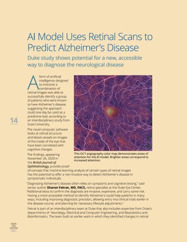
The novel computer software looks at retinal structure and blood vessels on images of the inside of the eye that have been correlated with cognitive changes. This OCT angiography color map demonstrates areas of The findings, appearing attention for the AI model. Brighter areas correspond to November 26, 2020 in increased attention. the British Journal of Ophthalmology, provide proofof-concept that machine learning analysis of certain types of retinal images has the potential to offer a non-invasive way to detect Alzheimer’s disease in symptomatic individuals.
“Diagnosing Alzheimer’s disease often relies on symptoms and cognitive testing,” said senior author Sharon Fekrat, MD, FACS, retina specialist at the Duke Eye Center. “Additional tests to confirm the diagnosis are invasive, expensive, and carry some risk. Having a more accessible method to identify Alzheimer’s could help patients in many ways, including improving diagnostic precision, allowing entry into clinical trials earlier in the disease course, and planning for necessary lifestyle adjustments.” Fekrat is part of an interdisciplinary team at Duke that also includes expertise from Duke’s departments of Neurology, Electrical and Computer Engineering, and Biostatistics and Bioinformatics. The team built on earlier work in which they identified changes in retinal DUKE EYE CENTER
2021


