
MORGELLONS: THE LEGITIMIZATION OF A DISEASE
“The
Symptoms”
By Dr. Ginger SavelyTHE SYMPTOMS
The symptoms of Morgellons disease are unusual and unlikely-sounding and it is not surprising that anyone hearing about them for the first time would be doubtful and incredulous. I know that I was. The thing that made me a believer many years ago was the consistency of patients’ descriptions of the exact same bizarre symptoms. When I first began seeing MD patients in my office there had been no media coverage of the disease and it was not being discussed on the Internet. It was implausible that this diverse group of patients was collaborating about what they were going to tell me! When I started to hear identical symptoms from dozens of people from all walks of life, I began to pay attention and take a closer look at what was going on.
The following list of symptoms is based on data that I have collected over the years from hundreds of patients. There are more symptoms than are listed here but these are the ones that are most frequently reported. All patients have filaments but not all have every one of the following symptoms. Regarding the photographs, I know there will be disbelievers who think that the photographs have been altered or that the items that have already come out of the skin are just debris found in the environment. However, I have seen all these things in my office and know that the photographs you will see and the symptoms I am about to describe are very real.
1.Intense itching (this symptom has been reviewed in the previous chapter).
2.Disfiguring, spontaneously appearing, slow-healing lesions. In a small percentage, these lesions turn into large, deep wounds that will not heal.
3.Tough, difficult-to-remove scabs with keratin projections on the underside.
4.Blue, white, red, and black hair-like filaments extruding through the skin or seen just under the epidermis using magnification. Some filaments are flattened and ribbon-like.
5.Thicker, translucent filaments that are visible without magnification and are very resistant to extraction.
6.Filaments that look like “feathers” because of the many fine projections from either side of the filament.
7.Shards” of keratin that project down into the dermis causing pain until they are removed.
8.Black specks, “fuzz balls” and seed-like objects on clothing, skin, and bed linens.
9.Hyper-pigmented, hypertrophic scars when the lesions first heal. These scars eventually become hypo-pigmented.
10. A waxy film on the skin all over the body as well as gobs of “gelatinous” material.
11. Occasional black tar-like exudate from the pores.
12. Metallic “glitter” on the face and other parts of the body.
13. Crystal-like exudates from the skin.
14. A change in the texture or color of the hair.
15. Hair loss, to the point of total baldness in some.
16. An awareness of tiny flying insects around the body.
17. Blackening and crumbling of the teeth.
18. Fine markings on the skin that appear spontaneously but look like cat scratches or paper cuts.
19. Slightly raised, liner “tracks” on the skin.
20. What look like small “cocoons” coming from the scalp.
21. A soft, mushy “mound” on the crown of the head.
22. Susceptibility to static shocks and inability to wear watches of sit in front of a computer.
23. Systemic symptoms including profound fatigue, anxiety, insomnia, joint and muscle pain, headaches, loss of balance, dizziness, and cognitive disturbances such as loss of short-term memory, and inability to concentrate or comprehend.
24.The constant and unnerving aggravation of feeling as though there are bugs or worms crawling through one’s body, biting, stinging, and causing unbearable discomfort.
As if the symptoms of the disease were not challenging enough, patients are forced to endure ridicule and abandonment by family, friends, and health care providers. Typically,
patients have futilely consulted as many as 20 to 30 different clinicians. With no hope in sight, it is no wonder that most Morgellons patients have depression, anxiety and/or suicidal thoughts and many have ended their lives. A discussion of what is currently known about the symptoms follows.
The lesions
The unusual thing about the lesions of MD is that they appear spontaneously although they look as though aggressive scratching may have caused them. Some patients do not have lesions, but the majority do and somehavevery few while others are literally covered by them. The lesions are very slow to heal and are therefore unlike similar-looking lesions caused by other conditions. Another interesting aspect of these lesions is that they do not tend to develop secondary infections with Staphylococcus aureus or Streptococcus pyogenes, as would be expected if the lesions were caused by scratching.
Some patients report that a certain part of their body is particularly affected and covered with lesions. I have seen a predominance of lesions on the face, arms, hands, back and/or legs of the MD patients I have examined. For many patients the heavily affected area is the scalp. Patients have been known to shave their heads to keep hairs from becoming matted in the seepage from these open sores. Having a shaved head also facilitates application of topical medications or soothing creams to the lesions.
Patients have reported that when they apply a bandage to a lesion or even to a simple cut on the skin, the bandage becomes “shredded” by filaments. Total occlusion of the lesions seems to help them heal, at first, but when the occlusive dressing is removed the lesion and filaments reappear with a vengeance. At least 4 different types of pathogenic bacteria have been found in the lesions. This will be discussed in a later chapter when current research is reviewed.

Image 1: Patients often present to the office looking like this. Color versions of all photographs in this book are available on www.gingersavely.com/morgellons-book
 Image 2: Lesion with black filaments.
 Image 3: Lesions on scalp with hair loss.
The Scabs
The hard scabs or calluses that form over many of the lesions not only contain masses of intertwined filaments of various colors but, most peculiarly, thick, firm, gelatinous projections on the inner side of the scabs as shown in the photos that follow. Using Gomori Trichrome stain, Middelveen and colleagues found these “plugs” (as patients refer to them) to consist primarily of keratin and to gradually harden in consistency over time. Significant pain relief occurs when patients can remove the scabs containing these projections. For this reason, patients are often seen picking, which is perceived as more evidence that the disease is psychopathic when relief of excruciating pain is the actual objective.
Of the following photographs, the first 3 show thin scabs containing intertwined filaments and the next 5 show tougher scabs with keratin projections or casts on the underside. Look carefully at these keratin projections that are found under the scabs of MD patients.
I can think of no other human disease where something like this is seen, and it would certainly not be expected in someone with a purely psychiatric disorder!
The photograph below, Image 4, when visualized in color, shows bright blue fibers intertwined within a patient’s scab. www.gingersavely.com/morgellons-book.


Images 5 and 6
Images 7 and 8: Follicular casts seen at 100x magnification on the underside of removed calluses. from MD patients.


Images 9 and 10: Although these scabs showing keratin “plugs” were removed from one of my patients, they look very much like those seen in cows with Bovine Digital Dermatitis.


Large, deep, non-healing lesions
A picture is worth a thousand words when it comes to this symptom. Photographs on the following pages are not for the faint of heart. Luckily, few MD patients have lesions this deep and large, although most patients have at least one smaller version of a deep lesion, which usually starts out as a circular “hole”. I have primarily seen these types of lesions on the face, but they can be on other parts of the body as well. Not surprisingly, facial lesions this dramatic are quite an embarrassment to the patient.
These deep wounds often appear to go down to the bone and they do not heal for many years. Wound care experts are baffled because every kind of wound healing method imaginable does not work. Biopsies reveal “non-identifiable fibers” and patients are accused of inserting these into their wounds. Interestingly, I have heard of several instances of these large wounds finally starting to heal once a biopsy is performed on
Image 11: A scab with keratin projections underneath as well as a filament that was growing down into the dermis.them. I can only guess that the biopsy, being a new injury to the skin, stimulates the immune system to begin the healing process. There is no doubt that these wounds are like nothing ever seen before and are just one more peculiar aspect of this curious disease.
The following 5 photos are of an ill-fated male patient who suffered a series of these deep lesions. Unfortunately, this man’s profession requires him to meet the public daily, making his appearance a constant source of embarrassment. The first photograph shows a hole on the left side of his face, the first of many deep lesions that appeared later in the same area. The next 3 photographs show the progression as the lesions take over the left side of his face. The fifth photograph shows what the area looked like after all the deep lesions he had suffered there had temporarily healed.

Image 12

Image 13

Image 14

Image 15
Image 16: Same patient after his numerous facial lesions had temporarily healed. Later his face opened up again in several new places.

Following this paragraph is a photograph of one of these deep lesions on the testicle of one of my patients (Image 17). If these wounds are self-inflicted (as many clinicians say they are) can you imagine that any man would want to do this to his testicle? Filaments and thick shards of keratin work their way through this patient’s wound causing him extreme pain. He spends hundreds of dollars per month on bandaging supplies because there is a constant sticky discharge from the lesion. This man had seen many specialists before he saw me and none of them were able to diagnose his problem or help him in any way. Most were downright insulting to him.

The filaments
Originally identified as “textile contaminants” the filaments are now known to be composed of the body’s own proteins, collagen, and keratin, resulting from proliferation and activation of fibroblasts and keratinocytes in the epidermis and stemming directly from these cells. Collagen is the most abundant protein in the human body and is what holds the body together. Keratin is a key component of nails, hair and the outer layer of skin.
A problem for researchers is that textile fibers tend to attach themselves to the lesions and to the Morgellons filaments, adding confusion to the overall picture. Textile fibers actually do look like MD filaments under microscopy. It is only with the use of Gomori Trichrome and other specific stains that filaments can be clearly differentiated from textile fibers.
Filaments come out of MD patients’ bodies in every way imaginable. They are in the mouth, gums, tongue, and inner side of the cheeks. They come out of the eyes, ears, and nose. Filaments are in urine and in seminal and vaginal secretions. They frequently grow out from under fingernails and toenails. Since a gastroenterologist told me that he has seen the filaments in the colons of 2 MD patients, there is every reason to believe they are present throughout the body. Interestingly, whenever an MD patient sustains an injury to intact skin such as an abrasion or even a paper cut, before long the injury becomes a new site of filament proliferation.
Researchers have seen retained cell nuclei at the base of filaments where they stem from the cell. Like human hair, on cross section MD filaments have a cortex and a hollow medulla. Using electron microscopy, the blue Morgellons filaments are shown to have scales much like a normal human hair. In contrast, some MD filaments have a smooth outer surface.
The filaments are thick and tough, fine and hair-like or flat and “ribbon-like”. The thicker ones can be seen without magnification, and I have always compared them to the look of nylon fishing line – translucent, tubular and very strong. These tougher filaments cannot be cut with scissors and are highly resistant to extraction. One of my West Texas patients said, “I wish I could market these filaments because I swear, they are strong enough to pull a tractor trailer”.
The fine filaments are black, white, blue, or red and occasionally even orange, green, or purple. These filaments are nearly impossible to see without magnification. The fine filaments are also strong: it is very difficult to obtain a punch biopsy from an MD patient since the intermeshed filaments will not break.
Patients have told me that of all the colored filaments, the red ones cause the most pain as they move through the skin. The red MD filaments are an unusual color of red and are more of a magenta/pink color. It is unknown how the magenta filaments get their color. The blue Morgellons filaments have been shown to contain granules of melanin,
which may be responsible for their coloration. (Melanin is the pigment that gives color to human eyes, hair, and skin.) Research has shown that the colors of the filaments are actual pigments rather than dye. Since dye is the origin of color in textile contaminants, this is one more piece of evidence that the filaments are not textile, as has been continually suggested by those who do not believe in the disease.
Many patients think that the filaments are alive and are infective agents. This is not the case. The filaments are inanimate objects and are not capable of transferring MD to others. They may be thought of as by-products of the disease process. It is my assumption that there may be an electrostatic charge, which makes the filaments appear to move after they are extracted. Patients often refer to the filaments as worms, an unfortunate mistake that leads to further doubt and mistrust by their medical providers.
When I have tried to extract filaments of all kinds, MD patients describe a pain that radiates over an extended part of their body. The description I frequently hear is that it feels as though there is a connected web of filaments, so that when one is pulled it affects a larger network. These filaments are clearly not being implanted by the patient, as has often been suggested. Once doctors see these filaments for themselves and try to cut or extract them, they will be convinced that they are dealing with something very strange indeed.
Image 18: Black and red filaments are seen in the web between a patient’s thumb and forefinger. The color version of this photograph is on the book’s front cover.

 Image 19: Clear to white filaments are often seen growing from the underside of nails of MD patients.

 Image 20: Black and white filaments as seen under the epidermis at 200x.
Image 21: A flat ribbon-like filament, this one from a vagina.
Image 20: Black and white filaments as seen under the epidermis at 200x.
Image 21: A flat ribbon-like filament, this one from a vagina.
Feather-like filaments
It is not known why certain filaments have this appearance. Following this paragraph are a few photographs of this type of filament extruding from a patient’s skin. When patients report that feathers are coming out of their body, health care providers understandably take this as evidence that they are mentally unstable. Rather than saying that feathers come out of their skin, patients need to be careful to say that they have filaments that vaguely resemble feathers.Image 22


Image 23
“Shards” of Keratin
These objects can vary in appearance but are a source of extreme pain for patients since they project down into the dermis. It is very difficult to remove them, but patients are motivated to do so because once they are removed there is great relief of pain. I have seen hundreds of photographs of these objects through the years that patients have taken after they are finally successful in removing them. These shards are like filaments in the sense that they are made from the body’s own proteins due to a proliferation of keratinocytes. Their size makes them more painful and difficult to remove than the filaments. Images 24, 25 and 26 on the next pages demonstrate the appearance of these keratin shards.


The black specks
Morgellons patients are sometimes accused of sprinkling ground black pepper or coffee grounds on their skin when they complain of “black dots” on intact skin and in lesions. At the 2010 Morgellons disease conference Randy Wymore, M.D. presented slides demonstrating the difference between coffee grounds, pieces of ground black pepper and the black specks of Morgellons disease as seen by electron microscopy. The microscopy slides make it clear that the black specks are no more than black filaments coiled up into a tight ball, appearing upon magnification like a wound-up ball of string. On the other hand, Wymore’s slides demonstrate that coffee and pepper sprinkles have sharp edges showing that they were cut by a grinder.
I have watched these black specks appear suddenly upon the skin of my MD patients. If the patient brushes the specks off of the skin they suddenly reappear. They may be
Image 26: A keratin “shard”.coming through the pores of the skin but there is no proof of this. The black specks are not, as some patients suspect, tiny insects that are responsible for their disease.
 Image 27: A black speck amidst fibers in a skin sample from an MD patient. 70x magnification. Courtesy of M. Middelveen.
“Fuzz balls”
The fuzz balls, like the specks, are tangled masses of fine filaments but are looser with the appearance of tiny “dust bunnies”. The following photo shows one of these fuzz balls at a magnification of 60x with the point of exit from the skin at 12 o’clock. (Image 28).

Gobs of gelatinous material
Morgellons patients report that there is gelatinous material coming out of their lesions and orifices. Clinicians and researchers have received samples from patients that become coveredwith what appears to be cleargelatin after remaining in a container. As mentioned in a previous paragraph, the scabs removed from MD patients’ lesions have gelatinous projections on the underside of the scab, which become harder and more painful the longer the scab stays in place.
Some patients who believe they are infested with biting insects have erroneously confused the gelatinous plugs under the scabs with a type of larva or grub. Research shows that this is not the case; larvae are easy to dissect and identify. Hair follicles of MD patients are covered with gelatin, which can be clear, blue or red. Patients complain that their hair always looks dirty because of the exudate from their hair follicles. The researchers Marianne Middelveen and Dr. Eva Sapi both surmise that the gelatinous material is likely alginate, an element of the biofilm produced by the bacteria found in the lesions of MD patients. I will discuss this further in the chapter on research. The photograph on the next page (Image 29) shows the slimy “goop” (as patients often refer to it) with embedded blue, red, white and clear fibers. As a reminder, the color versions of these photographs may be seen on gingersavely.com/morgellons-book. Following the slime photograph is a photograph of a single gelatinous plug from the underside of a small scab, immediately after having been removed by one of the CEHMDF nurses.


Film on the skin
We can only guess what causes the waxy film that patients report on their skin. Morgellons patients tell me that they feel like they should bathe four to five times a day because shortly after bathing they feel unclean again. I have heard this called “sticky sweats” and some even describe it as slimy. It has been postulated that the alginate that is a part of the gelatinous material may also be on the skin, causing this waxy feeling. We still do not have an explanation for this puzzling symptom.
I have wondered if there may be acidity to this waxy coating. Acidity on the skin would help explain the common complaint of MD patients regarding cotton clothing. Patients report that when they wear cotton socks or T-shirts, they become covered with tiny holes after only one wearing. Many MD patients switch to wearing only clothes made with synthetic fabrics.
Black tar-like liquid coming out of the skin
About one-third of my MD patients report this symptom. Some say that this was one of their early symptoms that gradually disappeared over time. We do not know what this thick black liquid is or why it is present in some but not all MD patients.
One day as I was speaking with one of my MD patients in the office, I recall seeing a bead of black liquid suddenly came out of one of the pores on her face. An MD patient once told me that when she went to the hospital to have a laceration sutured the doctor was horrified to see black fluid coming out of the lesion and stopped mid- procedure, refusing to continue. I have had a few patients show me that their brand-new white socks were black after one wearing due to their feet being wet with “black sweat”. Other MD patients have said that if they scratch their skin after they have been perspiring, they notice black fluid underneath their fingernails.
 Image 31: The following photo shows this black liquid.

Glitter and Crystal-Like Exudates
This is one of the most baffling symptoms that MD patients report. I have seen the fine glitter myself on the skin of my patients, usually visible only under certain light. I think that the larger clumps that resemble clear or opaque crystal are somehow related. Sometimes the little “crystals” are hard (Image 32 on previous page), other times they are so delicate and fragile that they disintegrate into dust. Patients have placed these crystals in bottles to send to researchers, but the bottles arrive empty; the samples appearing to have vaporized. Hopefully, researchers will be able to do x-ray crystallography of these exudates.
Change in the texture of the hair
At the 3rd annual Morgellons disease conference in 2010, Dr. Randy Wymore showed an electron microscope image of several hairs from the scalp of an MD patient that clearly showed a filament coiling itself tightly around each individual hair. This probably accounts for the change in hair texture that patients often complain about, saying that their hair has become thicker and coarser. If black filaments are coating the hair this also explains why MD patients sometimes say that their white or gray hair has turned dark again. “My hair is not my own” is something that I frequently hear from MD patients. Patients make this statement in an attempt to describe what is happening to them but unfortunately, it leads doctors to think that they are mentally unstable.
When black filaments grow out of the scalp and replace hair that is white or light blonde it may appear as though the hair has been dyed black. The short, white hair of one of my elderly male MD patients turned black, leading his friends to tease him about having dyed it. My patient knew that the black “hairs” that his friends were seeing were actually black filaments growing out of his scalp.
A complaint that I hear from patients who have long hair is that it can be perfectly combed and an hour later, even if the patient has been sitting still the whole time, there will be a tangled, matted mess of fine hairs at the nape of the scalp. Facial hair undergoes a change as well. Both male and female patients describe a thick growth of “peach fuzz” on their cheeks, in other words blonde, downy hair that is sometimes growing towards the ears instead of straight down. I have seen this on men, and it is an entirely different type of hair than their normal beard growth.
Image 33: Filaments tightly wound around a human hair at 200x.
Hair loss
Most MD patients report hair loss but only about 5% become totally bald. Some have a patch of baldness, usually on the crown of the head (a photo of this will soon follow). Hair may die and fall out because the filaments grow out of hair follicles and probably compete with hair for nutrients. Another possible reason is that the hair becomes brittle and unhealthy due to being encircled by filaments. My patients who have bought wigs to wear after suffering significant hair loss tell me that filaments growing into them cause a tangled mess, quickly ruining their wigs. I saw this for myself once when a patient removed her wig in my office.
Awareness of tiny insects flying around the body
About half of my MD patients share this complaint. Patients may indeed have an increase of tiny flying insects around them due to a different scent coming from their body but there is no way to know this for sure. Many of my Morgellons patients say that they have noticed a different and strange body odor since the initiation of their symptoms. Plus, there are the open, often oozing lesions that may somehow attract small insects. Unfortunately, some MD patients tell their doctors that these minuscule flying insects are the cause of
their troubles, believing that the insects are laying eggs under their skin. However, none of the researchers believe that these insects are the cause of MD. It is very difficult to convince some MD patients that tiny flying insects are not the reason for their symptoms.
Blackening and crumbling of the teeth
Dentists have called me from all over the United States perplexed as to why patients with the symptoms of Morgellons disease have teeth that are black along the gum lines and falling apart. The teeth are not decaying; they are literally crumbling into pieces or becoming hollow on the inside so that they are nothing more than thin shells. We do not know why this is but I have had a hunch that whatever is causing MD may be leaching calcium from the body. I have also heard patients complain of tiny holes in their fingernails. It would be interesting to conduct research on this looking at the dental records as well as the bone densities of MD patients.
 Image 34: Blackening of teeth close to gum line.
Photo courtesy of Katie Yussuf.
Image 34: Blackening of teeth close to gum line.
Photo courtesy of Katie Yussuf.
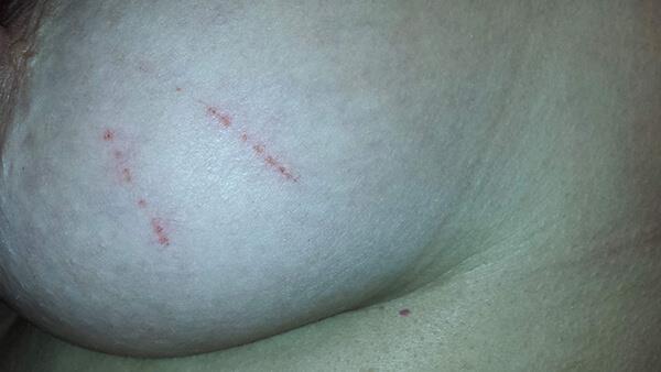
“Scratch Marks” on the Skin
Below is a photograph of a woman’s left breast showing an example of the mysterious “scratches” that spontaneously appear on MD patients’ skin (Image 35). Patients have watched as these markings occur right before their eyes. The explanation for these scratches is unknown but at times they do resemble the “striae” that are typical skin manifestations of Bartonellosis, as seen in the photograph that follows on the next page.

Linear, raised “tunnels” or “burrows”
These raised areas are the same color as the surrounding skin and are not red, as would be expected in someone with dermographism (a hypersensitivity reaction that occurs when someone’s skin is scratched, and a red welt is left exactly where the scratch mark was made). There are no theories as to what causes this type of marking. The photographs on the next page (Image 38 and 37) illustrate this finding.
Image 36: Streaks on an MD patient’s skin consistent with Bartonella striae.

“Cocoons”
Morgellons patients shed material from their scalps that they believe to be "cocoons". Researchers have shown these to be follicular sheaths in which the filaments are forming. Often the filaments are tightly wound around the follicular sheath and can have the appearance of a small, white cocoon. It is important that patients not refer to these objects as cocoons, even if there is a strong resemblance, because this terminology can prejudice health care providers against them. A photograph follows (Image 39).

Soft Mound on the Crown of the Head
Patients with MD often complain of a soft mound on the crown of their head that feels “mushy” to the touch. This mound is a particularly intense area of all the patient’s unpleasant sensations. The soft, mushy feeling of this mound is believed to be due to the gelatinous material present in the base of the hair follicles. Following is a photo of one of my patients who shaved his head allowing this mound to be clearly seen. If a patient experiences partial balding it is almost always in this circular area at the crown of the head. Image 40 shows this mound on the crown of a shaved head.


In the case of only one of my patients this mound “erupted” into a round lesion, with a diameter of about 4 inches, deep enough to expose the skull. For cosmetic reasons, the patient convinced a plastic surgeon to surgically close the wound. A few days after surgery the sutures burst open at which time my patient was accused of pulling them out herself. This allegation both shocked and offended her since she wanted nothing more than to have the unsightly lesion closed and had paid quite a bit of money to see that it was.
Image 41: Mound with lesions, showing loss of hair in that region.Crawling sensations under the skin
The crawling, biting and stinging sensations that MD patients experience are more than likely a combination of two processes. First, as the filaments push through and finally exit the body there is considerable discomfort due to injury to the skin. Patients have told me
“I feel like I’m being stuck with pins but from the inside out”. Also, a large part of the discomfort may be neuropathies caused by inflammation of the peripheral sensory nerves. Damaged small nerve fibers can cause sensations that are perceived as biting, stinging, and crawling.
Due to the inflammatory nature of Lyme disease, Lyme patients who do not have MD often have feelings of crawling, biting, and stinging as well. But, in the case of Lyme disease, these sensations are solely neuropathic in nature and there are no lesions or filaments. That which MD patients experience is far worse, because they actually have clear evidence of filaments working their way through the skin.
Propensity to static shocks and inability to wear watches or sit in front of a computer
I have noted clinically that a significant number of MD patients have below-normal levels of antidiuretic hormone (ADH), the hormone that gives a message to the kidneys to reabsorb some of the fluid that passes through them. Patients with low ADH have a condition called SIADH (syndrome of inappropriate antidiuretic hormone secretion). About 1 in 25,000 people in the general population develop this condition but I see it to some degree or another in about a fourth of my MD patients. People with SIADH feel thirsty all the time and urinate frequently. They cannot stay hydrated because they eliminate fluids as fast as they drink them. These patients have saltier skin, which predisposes them to increased static shocks, a symptom often reported by MD patients.
Saltiness of the skin would also help explain the electrostatic movement that patients report when their filaments appear to move on their own as they protrude from the
epidermis. It may also explain patients’ reported inability to wear watches (because they stop) or sit in front of a computer screen (because they feel increased movement in their bodies when they do). This is all conjecture on my part.
Systemic Symptoms
Almost all the MD patients I have treated have had systemic symptoms along with their skin symptoms. These symptoms are essentially the symptoms of Lyme disease and tickborne co-infections. The malaise and fatigue are profound. It is difficult to sleep, and patients wake up in the morning feeling like a truck hit them. There is mental confusion including attention deficit, processing problems, and poor short-term memory. A new onset of anxiety and panic attacks is typical. There is pain in the joints and/or in the muscles. Many patients have neuropathies – nerves that are painful, burn, tingle, or feel icy cold.
The list of tickborne infection symptoms experienced by MD patients goes on and on and includes cardiac symptoms like chest pain, shortness of breath and palpitations; gastrointestinal symptoms like abdominal pain, nausea, and diarrhea; urinary symptoms like frequent and painful urination; and neurological symptoms like muscle twitching, dizziness, loss of coordination, hypersensitivities, and difficulty walking.
A comprehensive list of the symptoms of Lyme disease, available at www.lymedisease.org will leave the reader amazed to see dozens of symptoms affecting each body system. The fact that MD patients experience so many systemic symptoms that are typical of Lyme and other tickborne diseases strengthens the speculation that MD is a physiological rather than a psychosomatic illness and that it has some kind of connection with tickborne infections.
Summary
After hearing about the symptoms of Morgellons disease you are either eager to continue reading to find out more about this enigmatic disease or you have decided to put this book down, having reached the conclusion that the author has lost her mind. I hope that your decision is not the latter, although I certainly wouldn’t blame you if it were. Please have faith that what you are about to learn will be eye opening, even though at times disturbing and unbelievable. If it is difficult to read about the symptoms of MD and see photographs of its ravages, only try to imagine how dreadful it must be to be one of its victims, suffering not only the pain of the symptoms but the indignity of being labeled a drug abuser or a delusional psychotic. The next chapter will discuss these two diagnoses, the most common misdiagnoses conferred upon Morgellons patients: drug-induced formication and delusions of Parasitosis.