6 minute read
Digital Dermatitis (Heel Warts) in Beef Cattle - An Awareness Campaign


By: Dr. Dörte Döpfer, MSc, PhD, University of Wisconsin - Madison
Digital dermatitis (DD) or hairy heel warts is a common problem in dairy and beef cattle worldwide that leads to ulcerative lesions along the skin-horn border, the so-called coronary band, mostly in the hind aspects of feet. The resulting lameness becomes more pronounced over time and it is crucial to detect the skin lesions as soon as possible before severe, debilitating lameness and outbreaks of DD occur on average 60 days before harvesting.
We have not even begun to estimate the economic losses caused by DD in beef cattle, but for the time being, an estimate of on average $40 per lesion, even the small ones, is a starting point. There is a marked difference in severity of lesions between early season and late season DD lesions on feed yards (Figures 1A, 1B). If we caught DD early, the prospects of successful prevention and control are much better compared to the late season DD lesions..
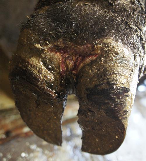
Fig.1A: Early season M2 lesion, about ‘quarter’ size resulting in lameness grade 2 on the Step-Up scale. This type of active lesion can be treated topically and recurrence prevented through well managed foot baths. This type of lesion can be detected in the squeeze chute or during pen inspections while cattle are at the feed bunk or in the alley ways under dry flooring conditions.
What if early season DD were here and nobody was looking for it?
It is the current belief that calves enter the feed yards free of DD, and at the same time one wonders where DD causing the devastating outbreaks of lameness originated from once detected. It seems to “fall from the sky”.
I want to convince you that it is an awareness problem that hinders us to detect DD before the large outbreaks happen. We need to go out and confirm as to whether it is actually true that cattle enter feed yards free of signs of DD and we need to actively seek DD lesions before lameness is apparent.
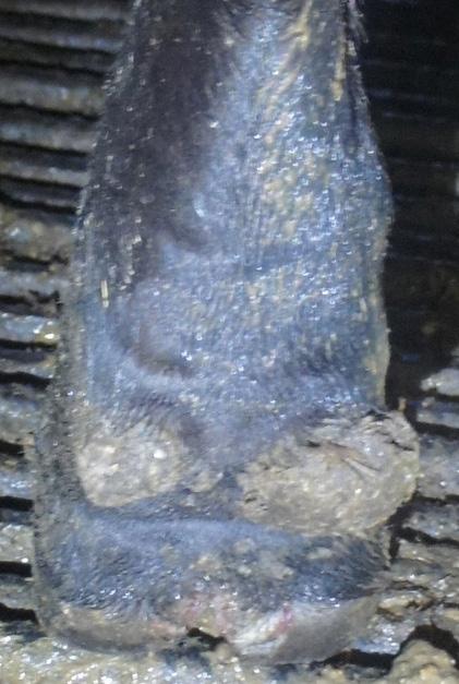
Fig.1B: Late season M2 lesion (15 days before harvesting) with layered heelhorn erosion and crater-like tissue losses’ severe lameness as a consequence (grade 3 on the Step-Up scale).
By means of an anecdote, I want to raise your awareness about how early DD is introduced into feed yards and one route of introduction is that basically cattle with subclinical DD are unloaded from the trucks!
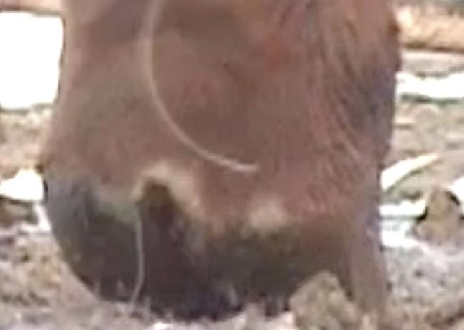
In August of 2015, Dr. Tom Noffsinger showed a 60 second video to an audience of about 100 producers, herdsmen, and pen riders for the purpose of illustrating how abrasive some of the metal flooring types are that those calves had to cross. In 2003, about 60 calves were being unloaded from a truck while the camera zoomed in on the metal bridge that the cattle crossed upon arrival.
This camera setting allowed me to observe the hind aspects of their feet. It was clear that during those 60 seconds from the past, six subclinical chronic DD lesions were being unloaded together with the calves. Figure 2 shows two of the lesions found by means of this old video.

The audience was silent for a moment until awareness surfaced about what this finding implied. Calves as young as the arrival date at the feed yard introduced DD from day 1 into their pens.
In order to explain what this means, I will rewind to provide more information about DD in cattle.
Digital dermatitis (DD), Hairy Heel Warts or Mortellaro’s Disease has been recognized worldwide where intensive cattle husbandry is practiced and DD is considered to be the most prevalent infectious claw disease in cattle.
Most information about DD stems from investigations into dairy cattle. Today, DD is recognized as a serious animal welfare issue that results in costs due to decreased milk production, lameness and reproduction problems, particularly as a consequence of the severe stages of DD. 24 pacdvms.com
The disease manifests itself most often by seasonal outbreaks of lameness on endemically affected farms leading to large investments in labor for the topical treatment of cows and costs for the management of hoofbaths containing disinfectants such as copper sulfate and formalin that are problematic for the environment and health.
Digital dermatitis is considered a multi-factorial disease with a strong bacteriological component and Treponema-like microorganisms (spirochetes) are thought to play a key role in the etiology of DD. Post-peak heifers on slatted floors in free stalls under unhygienic conditions are at higher risk for developing DD. In addition, the severe stages of DD are associated with decreased longevity and lameness.
The clinical course of DD is characterized by a succession of acute and chronic proliferative stages that have been categorized in many ways. Most classification systems for DD lesions distinguish active from non-active and chronic lesions. The details recognized about the stages of DD depend on how the feet of cattle are being evaluated, i.e., standing up in a chute, on the cattle’s side on a tilt table, in the milking parlor or in alleyways and feed yard pens without lifting the foot.
The categorization of DD into M-stages applies to beef and dairy cattle. The freely available DD Check App (for iOS/MAC devices) provides pictures and information about the course of disease as does the “Cattle Lameness” book that can be purchased through for example amazon. com.
In short, the two text boxes below describe the lesions and how to generate types of cattle with regards to recurrence of lesions. Beef cattle in feed yards do often not become old enough to be registered as type III cattle, but chronic cases with repeated M2 stages are numerous.
Box 1
Five M-stages for the clinical stages of DD are (Dopfer et al 1997, Berry et al 2012):
M0: normal skin appearance
M1: small focal circumscribed damage of the epithelium at the skin horn border, <2.0 cm in diameter
M2: circumscribed ulcerative skin defect, a red or grayish surface that can have a white epithelial margin, overlong hair and can be painful to the touch, >2.0 cm in diameter
M3: the healing stage of DD, after the M2 lesion has covered itself with a scab that is not painful to the touch
M4: chronic stage of DD that is characterized by a thickened epithelium (hyperkeratosis) or proliferative growth of the epithelium (heel warts)
M4.1: the chronic stage as described under M4, but with an M1 lesion within its perimeter
Box 2
Based on the recurrence of M2 lesions, steers are typed into (Dopfer et al 2004, Holzhauer et al 2006, Gomez et al. 2015):
Type I: steers that never develop M2 stages
Type II: steers that develop M2 stages once and never again for prolonged periods of time
Type III: steers that develop M2 stages repeatedly, for example every 14 days (known as the core group of “Problem Steers”)
One improvement for the prevention and control of DD is to be aware of the different stages of DD, to monitor for DD stages on a regular basis by means of weekly pen rides or inspection of cattle walking in the alley ways and by generating records of lesions upon receiving cattle through the processing chute.
Recent experimental infections have reproduced epithelial lesions that resemble DD lesions clinically, and are identical to DD histopathologically using pure cultures of Treponemes spp. or wound scrapings of active DD lesions. The fact that Treponema-like microorganisms have evaded bacteriological cultures on occasion is due to their anaerobic culture conditions and their change in morphology over time (spirochetes tend to encyst and not be recognized as spiral anymore which may lead to disposal of a sample as negative).
Reports about many other bacterial species found in DD lesions, the environment and skin biopsies are manifold. The presence of the microbes detected on DD lesions reported is plausible, but their role in the pathogenesis of DD remains speculative.
Complications of DD are recognized: among those are heelhorn erosion, sole ulcers, treponeme infested pre-mammary dermatitis, and non-healing DD. The topical treatments of those complications have different success rates and they include hoof trimming when horn structures are involved, the debridement of the wound combined with antimicrobial topical antibiotics under a bandage and prolonged clinical courses of disease.
In conclusion, after decades of DD being known as an infectious claw disease that leads to severe lameness, the etiology is not clarified, the complications and clinical course of disease are studied in unstandardized ways and no solution for the elimination of this disease is in sight for either beef or dairy cattle.
This has led to extreme frustrations of producers and claw health managers while this animal welfare problem continues to exist and environmentally questionable hoof bathing practices are commonplace in modern cattle practice.
Resources for research about DD are scarce as is commonplace for production diseases. The latter fact delays the study of DD and finding adequate solutions for defining the ‘manageable state of disease’ for endemic DD.
It is clear that there are feed yards where DD is currently not detectable, particularly during certain seasons and in certain regions of the US and Canada. I have spent many hours searching for DD in beef cattle operations and I could not find any visible lesions, including in Holstein cattle and their cross breeds.
But in other operations, subclinical DD lesions are present and the lameness situation during a DD outbreak is simply heartbreaking and irreversible.
We need to train ourselves and our producers and herdsmen to recognize DD earlier than through lameness and the necessary skills can be taught while awareness for DD should be raised!


