
Eurolab June 2024

Page 1


Next-generation DRUG DISCOVERY

FLUID DISPENSING EXPLAINED

Data and its management are a central concern for many healthscience professionals since AI, a burgeoning area of biotechnology, is only as good as the data it uses. For more on this read Through The Looking Glass (page 9), an article exploring how DeepMirror, an innovative UK-based company, is making already discovered research pathways available to health-science customers via its many and varied datasets. In a second data-focused article called Decoding Data Meaning (page 10) we look at why the health-science sector is more advanced than others in its use of data.

A fascinatingly futuristic development called organ-on-achip is explored in Next Generation Drug Discovery on page 38 - this clever product acts as a bridge between human and animal models in the lab.

In addition, the way that 3D printed medical implants might avoid infection or rejection is explored in more detail in Topography And Medical Device Effectiveness on page 14.

As a scientific journal that aims to reflect the concerns of the wider industry, it would be remiss not to include a piece or two on waste reduction, and our articles Improving Lab Sustainability and Walking Off Waste on pages 16 and 18 aim to help improve your green credentials.

Feel free to contact me regarding an article you might want to contribute to Eurolab or any subject you would like to see covered. Either way I'd love to hear from you!

COVER STORY

38 Next Generation Drug Discovery

Organ-on-a-chip technology helps bridge the gap between animal and human models

SPECTROSCOPY

9

10

14

Through The Looking Glass

A new company aims to simplify the relationship between AI and developers

& LAB EQUIPMENT

Decoding Data Meaning Health science companies are making better use of data than those in other sectors, but why?

Topography And Medical Device Effectiveness

An in-depth look at the way 3D printing is driving innovation in life sciences

16

18

Improving Lab Sustainability

Tips on reducing plastic use in laboratories

Walking Off Waste

How to make the most of your sustainability walks

21 Fluid Dispensing Explained

How dispensing techniques can lead to better process control in your facility

22 BEXS Vs EDS

Exploring the differences between BEX imaging and EDS mapping

How microplate readers can help accelerate drug discovery and development

How to break the vector characterisation bottleneck CHROMATOGRAPHY

Boosting Narrow Bore Column Performance

Why sample introduction optimisation will boost narrow bore column performance

The Magic Number

How to better understand a compound's physical structure

PUBLISHER

Jerry Ramsdale

EDITOR

Tips For Pipetting

Nicola Brittain nbrittain@setform.com

DESIGN

Stephanie Taylor, Jill Harris

GROUP HEAD OF MARKETING

Shona Hayes shayes@setform.com

HEAD OF PRODUCTION

Christine Flaxman +44 (0)207 062 2573

BUSINESS MANAGER

John Abey +44 (0)207 062 2559

SALES MANAGER

Darren Ringer +44 (0)207 062 2566

ADVERTISEMENT EXECUTIVES

John Davis, Peter King, Iain Fletcher, Paul Maher

Setform Limited, 6, Brownlow Mews, London, WC1N 2LD, United Kingdom

+44 (0)207 253 2545

Compound Semiconductor analysis

This article visualises doping and topographic variation

Turning to tunable lasers

Why technological advances have lead to research labs using tunable lasers

Give GMO A Chance

How new legislation might increase cultivation of GMO in Europe

Revolutionising Precision Medicine

An overview of advances in oncology research

Cellular Protein Synthesis

Exploring recombinant protein expression using a baculovirus insect cell system

How to achieve error-free pipetting for accurate results

AND LAB CONSUMABLES

The Role Of Relative Humidity

Why controlled humidity is essential for reliable research outcomes 52 Pressing Power

A guide to understanding how head dwell time can impact tablet compression

Dry Granulation

How to better understand the nip angle

Setform’s international magazine for scientists is published twice annually and distributed to senior professionals throughout the world. Other titles in the company portfolio focus on Process, Design, Transport, Oil & Gas, Energy and Mining engineering..

The publishers do not sponsor or otherwise support any substance or service advertised or mentioned in this book; nor is the publisher responsible for the accuracy of any statement in this publication. ©2024. The entire content of this publication is protected by copyright, full details of which are available from the publishers. All rights reserved. No part of this publication may be reproduced, stored in a retrieval system, or transmitted in any form or by any means, electronic, mechanical, photocopying, recording or otherwise, without the prior permission of the copyright owner.

Lab Innovations

Organ-on-a-chip technology will bridge the

GLOBAL INDUSTRY INSIGHTS

Your roundup of world news in the science industry

PRECI AND BIOPREDIC TO PARTNER ON IN-VITRO ASSAY SYSTEMS

Biotechnology company Preci has partnered with Biopredic International, a company specialising in the design and manufacture of human and animal in vitro assay systems to collaborate on the production of pooled suspension human hepatocytes.

Under a license agreement, Biopredic will leverage Preci’s expertise and production capacity in sourcing primary hepatocytes, and combine this with its own IP and know-how in cell pooling.

The partnership will provide drug metabolism and pharmacokinetics researchers with access to large batches of high-performing suspended pooled hepatocytes with extended longevity from multiple donors.

Pooled suspension human hepatocytes play a crucial role in assessing drug metabolism and hepatic clearance, providing a more representative understanding of human hepatic metabolism for predicting drug outcomes and assessing their impact on safety and efficacy.

Despite this, the limited culture lifespan of pooled hepatocytes hinders long-term studies, potentially reducing metabolic competence with repeated use of the same batch.

Anton Hanopolskyi, CEO, Preci, said: “At Preci, we believe that increased diversity and availability of representative cell models will revolutionize the drug discovery landscape. This partnership strengthens our position in the human-derived assays market, providing our customers with access to high quality, reproducible assays, and we look forward to continuing to work with Biopredic to advance our translational models for use in drug metabolism and pharmacokinetics studies.”

MULTIPLEXING FEATURES WILL ALLOW COMPANIES TO PROCESS MULTIPLE DNA SAMPLES

A leading life-sciences company Mission Bio has introduced sample multiplexing features for its Tapestri Platform. These features enable the combination of several samples into a single run, reducing the per-sample costs for single-cell DNA and protein multiomic analysis by up to 60%, according to the company. By allowing researchers to simultaneously process multiple samples, the new features have been designed to o er biopharma companies opportunities for optimising product characterisation and drug development to provide academic researchers with the ability to scale critical single-cell insights, particularly in oncology and genome editing fields. This will lead to transformative advancements in health and disease management.

“At Mission Bio, our goal is to empower more researchers to harness the advantages of single-cell multiomics to accelerate scientific discoveries, from academics using genome editing techniques for disease modeling to biopharma in the cell and gene therapy space,” said Anjali Pradhan, chief product o cer, Mission Bio.

CEO of Preci, Anton Hanopolskyi

PROSTATE

AI TOOL

RECEIVING CERTIFICATION AS MEDICAL PRODUCT

Artificial intelligence innovator FUSE-AI has received EU Medical Device Regulation (MDR) 2017/745 IIa certification for its AI software ‘Prostate.Carcinoma.ai’. As of January this year, the company was authorised to distribute its software as a medical product.

The product aids radiologists by automatically segmenting the prostate gland in MRI scans and independently identifying pathological changes, promising a 30% time saving per patient. This efficiency translates into considerable financial benefits for radiological clinics and practices, fuelling strong interest in the deployment of the AI algorithm.

“This AI software is a testament

to the interdisciplinary collaboration among scientists, AI developers, and radiologists. Receiving certification is a pivotal step, transitioning preliminary agreements into binding contracts and fully leveraging the software’s capabilities in clinical settings. This milestone substantially lowers investment risks into our company,” stated Matthias Steffen, Founder and CEO of FUSE-AI.

This favourable development for “Prostate.Carcinoma.ai” coincides with a prediction that the global market for medical image analysis software will reach a value of $4.545 billion by end of FY 2023, with a Compound Annual Growth Rate (CAGR) of 9.9% according to Reportlinker.com

BROKEN STRING BIOSCIENCES

AND FRANCIS CRICK INSTITUTE TO COLLABORATE ON ALS RESEARCH

London-based genomics company Broken String Biosciences has entered a research collaboration with biomedical discovery institute the Francis Crick Institute to develop novel applications for Broken String’s proprietary DNA break-mapping platform, INDUCE-seq, beyond its established capabilities in geneediting. The research will be focused on leveraging the technology to investigate the impact of genomic instability in the development of amyotrophic lateral sclerosis (ALS). ALS is a progressive and debilitating neurodegenerative disease, causing gradual loss of the ability to control voluntary movements and basic bodily functions.

The collaboration is focused on understanding the contribution of genome stability to ALS, combining the interests of Prof Simon Boulton and Dr Nishita Parnandi at the Crick Institute who are focused on genome stability and DNA doublestrand break (DSB), with Prof Rickie Patani and Dr Giulia Tyzack who are interested in understanding the underlying mechanism of the ALS disease. Recognising the utility of the novel INDUCE-seq platform developed by Broken String’s R&D department,

the teams aim to collaborate to demonstrate and further validate the INDUCE-seq technology in this setting.

The majority of ALS cases (90%) are considered sporadic. While there has been progress to better understand the genes and biological markers associated with the disease, very little is understood about the causes, with current treatment strategies focused on symptom management and slowing disease progression. Combining world-leading research from the Crick with Broken String’s expertise in genomics, sequencing, and bioinformatics, the partnership provides a unique opportunity to expand application of the Company’s INDUCE-seq technology in a key area of clinical unmet need, to support improved diagnosis and treatment of ALS.

The partnership has been secured via the Francis Crick Institute’s Business Engagement Fund, a new initiative supported by The Medical Research Council (MRCUKRI), designed to encourage collaborations with small-tomedium-sized enterprises (SMEs) and strengthen the Crick’s engagement with industry.

EARLY-ACCESS PROGRAM TO HELP WITH VIRAL VECTOR PRODUCTION

Leading provider of premium vector technology and services for the production of biologics ProteoNic Biosciences has launched its LV-2G UNic Early Access Program. This program marks a significant milestone in lentivirus manufacturing optimisation, according to the company, offering access to groundbreaking vector technology designed to revolutionise viral vector production.

The product is aimed at CDMOs, biotechs, and biopharmaceutical companies who would benefit from integrating this cuttingedge vector technology into their existing systems, paving the way for increased viral vector production capacity and substantial improvements in manufacturing cost efficiency.

Frank Pieper, CEO of ProteoNic, said of the launch: “The LV-2G UNic Early Access Program will serve as a launching platform for our viral vector manufacturing innovation. By offering early access to our state-of-theart vector technology, we empower researchers to unlock unprecedented levels of efficiency and productivity in viral vector manufacturing.”

FUSE-AI has been authorised to distribute its software as a medical product

DeepMirror’s bespoke software allows users to tap into AI-driven insights

Through the looking glass

A new company aims to simplify the relationship between AI and drug developers in a bid to reduce complexity and cost, Nicola Brittain reports

Drug discovery is a lengthy and costly process, with pre-clinical development alone costing an average of £35-55m per drug and taking eight years to bring to market (Wellcome Trust, 2023). Drugs take this long to develop because chemists must painstakingly tweak molecules to increase their efficacy - AI promises to help automate this process. Although the promise of AI in this realm hasn’t yet lived up to its initial promise to map the entire human body and its likely responses to pharmaceuticals, predictions around which tweaks are likely to work better than others can reduce overall costs significantly and there are a number of interesting developments in this field; not least a new company called DeepMirror.

Set up as a spin off from the University of Cambridge, this team of experts in physics, biology, and AI recently launched their Early Access Programme after a successful closed beta programme during which chemists were invited to test

the software over several months. The company was set up to develop intuitive design software for the discovery of novel therapeutic drugs. Its bespoke software allows users to tap into AI-driven insights to improve and accelerate molecular design across the drug discovery pipeline through a secure and user-friendly interface which makes AI-powered drug discovery as ‘simple as using a spreadsheet’, according to the company. Users are provided with access to short cuts as found by other pharmaceutical companies during the drug discovery process.

AI-ENABLED DRUG DISCOVERY PROGRAMMES

AI-enabled drug discovery programmes often start with pharmaceutical companies partnering with AI companies to deliver insights for their drug discovery efforts. However, this approach requires extensive crosstalk between the two parties, resulting in long waiting times and considerable resources spent on both sides.

DeepMirror has developed a programme that aims to solve this issue by enabling research and development teams to carry out AI-driven research with workflow integration and without the need to engage external stakeholders, develop internal teams and software, or relinquish any intellectual property.

MOLECULAR PROPERTIES TAKE CENTRE STAGE

In drug development, predicting the properties of drugs (small molecules) before testing them in the laboratory is crucial to reducing the time and resources required to bring safe and effective new drugs to patients. Two main types of property predictions are crucial: properties that describe how ‘drug-like’ a molecule is, such as it’s absorption, it’s distribution in the body, how it gets removed from the body and how toxic it is; and second, properties that describe how good a drug is at binding to its target and exerting an effect against a disease (affinity, potency). Deep Mirror tested 184 AI approaches against 44 public datasets and the research highlighted the need for different AI approaches for different datasets. For example, traditional methods perform better

for low dataset sizes and datasets with affinity measurements, whereas modern AI methods (such as deep learning) perform better at higher dataset sizes and some drug-like property datasets.

The team also found that selecting the right featuriser was also dataset dependent. Featurisers are methods that turn molecular structures into a numerical format that computers can understand. Expert features (properties derived by cheminformaticians) worked best for affinity property datasets; yet molecular descriptors (chemical properties of a molecule) and Natural Language Processing (features derived from letter sequences such as molecules SMILES) worked best for drug-like property datasets.

All in all, the company did not find a single model “to rule them all” in small molecule drug property prediction. By benchmarking a wide range of AI methods across various datasets, Deep Mirror has developed a platform that can intelligently adapt to the specific needs of each dataset.

This adaptability is critical in a field as diverse and complex as drug discovery.

FAST TRACK THE DRUG DISCOVERY PROCESS

The company’s technology to fasttrack the drug discovery process, for example in the hit-to-lead and lead optimisation phases, can predict relevant properties such as drug binding, (bio-)activity, and toxicity, both from user data and from large proprietary curated databases. Laboratory results can be used to refine predictions and generate novel drug candidates for further experimentation, ultimately accelerating the drug discovery process by up to four times as estimated by the Wellcome Trust and the Boston Consulting Group.

THE DEEP MIRROR MISSION

Dr Max Jakobs, co-founder and CEO of DeepMirror, said: “Our mission is to make AI-powered drug design as simple as browsing the web. After

12 months of development and a successful beta-testing programme, we are excited to officially launch DeepMirror to early adopters. We are inviting researchers to get in touch to use our secure and user-friendly AI platform for drug design. DeepMirror has already been used on active drug discovery programmes, resulting in the discovery of novel lead series and inspiring the synthesis of new compounds.”

Dr Andrew McTeague, senior scientist, Medicinal Chemistry, Morphic Therapeutic, said: “DeepMirror is a huge step forward in the democratisation of machine learning models. Its user-friendly interface enables medicinal chemists of all levels to deploy this powerful approach in a fraction of the time. Being able to apply DeepMirror’s platform to any desired endpoint, empowers users to make more informed decisions and to do so faster. We’re always looking for new tools to improve the efficiency of our DMTA cycles and DeepMirror helps ensure that no stone is left unturned." n

Decoding DATA MEANING

Health

science companies are making better use of data than those in other sectors. But why, and what benefit does this afford them?

The IT and OT layers of an organisation have traditionally had very different remits and been run quite separately. However, with automation and virtualisation now ubiquitous, they are no longer distinct. In terms of merging these information types to make use of different data sets across the business, life-science companies are leading the charge, according to Peter Zornio, CTO of technology manufacturer Emerson. He was speaking at the recent Emerson Exchange conference in Dusseldorf. The merging of these technology stacks to make different data sets across the business and the resulting ‘bubbling up’ and sharing of previously siloed data, is leading to improved product time to market, enhanced sustainability, and reduced downtime.

DEFINITION OF THE STACKS

Before we look at why this is happening, it is important to define the types of technology stack to which we are refering. OT (short for Operational

Technology) systems tend to be those closer to the plant level and shop floor. They require a user input, as well as data and information from the specific location. They tend to refer to information created in site specific databases, or related to plant specific operations. Reliability and availability are the most important concerns for OT

systems. OT information has tended to not be shared across a business. However, IT (short for information technology) systems tend to work across sites, do not require user input and are more focused on security and consistency than the former. Their remit might include internet, cloud, SaaS or CRM services as well as monitoring and information reporting.

Historically, the two stacks were distinct and often at logger heads (with different IT staff in charge) but times have changed and companies need a more holistic view of their data to be able to make the right decisions at the right time. Michalle Adkins director of life science consulting, Emerson said of recently developments: “Being able to merge and move data and information from both these stacks has become essential for the smooth running of labs.” She continues: “Pulling data out of bespoke or specific on-site machines can now be given context and be useful to a wider business. This bubbling up of data – or giving it contextual meaning – can be very useful in a number of scenarios.

BENEFITS OF MERGING TYPES OF OPERATIONS

Giving data context can lead to the sharing of operational efficiencies found in one site; sharing of capacity if one site is experiencing downtime; spreading research responsibilities if a product needs to be delivered quickly; and the reduction of repeat experiments in different parts of a business. Similarly, research work can be easily learnt from – particularly important in a large company with international sites; finally and importantly batch issues can be traced back to their root providing a better understanding of production problems.

WHAT IS DRIVING THIS TREND?

As Michalle explained there are several key drivers for this development. These include getting new products to market quickly; a desire for pipeline acceleration; or the need to manufacture multiple products at a given process development facility to bring a new product to market quickly. Operational integrity and the need to be able to reliably deliver products on time, in full, and meeting

Sustainable Laboratory Solutions – No Compromise on Quality

Eco-Conscious Packaging Superior Pipette Tip Refills

Fastrak® and FastZAP™ pipette tip refill systems – reduce – reuse – recycle.

› Lighter, smaller, and minimal packaging

› Made from 95% renewable materials

› Printed with vegetable inks

› Components made form bio-based resin

› Manufactured using 61% renewable energy

› Fast, easy refilling of re-usable racks

Proudly displaying the ACT Verification eco label.

Guaranteeing sustainability accountability, consistency, and transparency. Each label features manufacturing, energy use, packaging, and lifetime rating.

› High-grade resin tips for precision and flexibility.

For more information or to place an order please visit: www.alphalabs.co.uk/fastrak

ENHANCED CONTAINMENT & SUSTAINABILITY

Our specialised Pharmaceutical Manufacturing Industry Collection has been developed to offer a range of products that enable the safe use of powders, chemicals and biohazard materials linked to the research and production of pharmaceuticals. In an evolving market, we help our clients maintain optimal standards whilst providing the best protection for your personnel and applications.

a pre-agreed schedule, can now be almost guaranteed because so many processes are automated. Similarly, equipment issues can be spotted and ironed out early in the process. Releasing products as soon as possible is helped by collating access from all the data across the systems involved. Similarly, sustainability has become increasingly important and being able to move data and information prevents repeat experiments and will improve efficiency. Being able to pinpoint and resolve bottle-necks will also lead to a smoother running facility.

WHY ARE HEALTH SCIENCE COMPANIES MORE ADVANCED?

Although Michalle wasn’t able to say for sure why health science companies are more advanced than other manufacturing sectors in terms of managing and making different types of data available to the wider business (merging their IT and OT systems), she was willing to hazard a guess that it is because there are more siloed parts of a bio-sciences organisation than outfits in other sectors. These will likely include a research facility, a process development facility, a clinical manufacturing arm, and a commercial manufacturing or a contract manufacturing facility (CDMO). Companies also often work with external partners when developing and manufacturing new products. Similarly, it is probably the case that new products are coming on line in

Customer case studies

health science companies more often than in some other sectors and these require nimbleness and agility as well as a concerted push to get products to market. This sharing of data is probably more necessary in international than smaller companies, and many health science companies work at scale. Although companies in the oil and gas sector, for example, are also huge, they deliver the same product consistently. Simiarly, the manufacture of parts in the process industry may not be subject to the same speed of change as those in the health sciences sector - although this is probably not the case for car components or computer chips which are likely to be subject to similar time-to-market pressures.

❝ Being able to move and merge data and information from the IT and OT stacks has become essential for sustainability, efficiency and the smooth running of labs.

DeltaV workstations run on a specific set of preselected Dell computer hardware chosen to provide the best cost performance solution

HOW EMERSON HELPS HEALTH SCIENCE CUSTOMERS

Emerson provides a number of products that can help health companies manage their data. These include the DeltaV Automation System which ‘helps

When asked which companies Emerson works with in the health sciences space, an apposite answer might be be ‘who doesn’t it work with?’ Some 29 of the top 30 health science companies use Emerson technology and these include Thermo Fischer, a company that has spoken about its advances in gene therapy as a result of Deltav Automation; and FujiFilm, a CDMO that increases speed to market using a cloning-across-sites system built on utilising Drug Substance Manufacturing (DSM) modules. These modules rely on Emerson’s DeltaV automation system and two DSM modules that share an Emerson Syncade manufacturing execution system (MES). Other customers include Bayer and Novo Nordisk.

eliminate complexity and project risk by offering contextualised data,’ according to the company. In addition, Emerson has developed other related technology for the health sciences sector that including the DeltaV Spectral Process Analytic Technology (PAT), a distributed process control system that uses spectral analysers to measure reflected light frequencies from on-line product samples.

In 2021, the company also acquired a 55% stake in Aspentech in a bid to accelerate its industrial software strategy. Aspentech currently provides a range of data focused applications including twin technology as well as process automation simulation and other impressive tools to help health science companies deliver competitive advantage and cost savings n

For more information visit https://www.emerson.com/

This diagram shows how data can lead to improved processes

HOW TOPOGRAPHY CAN HELP WITH MEDICAL DEVICE EFFECTIVENESS

An exploration of the way 3D printing is driving innovation in healthcare and life sciences

Healthcare and life sciences are two industries where 3D printing is driving innovation, since it can print micro-precision parts that many medical devices require. Beyond medical devices, large healthcare and pharmaceutical companies are researching the ways that 3D printing can be used for next-generation drug development, like biomedicines, or personalised surgical techniques, like bone grafts.

Many projects also aim to explore the use of topography in optimising device effectiveness.

PROJECT WITH THE UNIVERSITY OF NOTTINGHAM

Last year, The university of Nottingham’s Centre for Additive Manufacturing selected BMF as an advisor for an EPSRC grant-funded 3D printing “Dial Up” project that focuses on“dialing up performance for on demand manufacturing,” where the multidisciplinary research group began to develop a playbook for standardising 3D printing in medtech and life sciences applications. This project runs alongside follow up work funded by an MRC project, the“Acellular / Smart Materials – 3D Architecture: UKRMP2 hub.”

Recently, BMF’s CEO, John Kawola, was asked to serve on the advisory board for another project based in the University of Nottingham’s

Biodiscovery Institute, which has long been a leader in researching new materials and medical devices, as they received a grant to focus on designing bio-instructive materials for translation ready medical devices. The goal of the EPSRC–funded “designing bio-instructive materials for translation ready medical devices” project is to address major compatibility issues of implanted medical devices.

SOLVING THE PROBLEM WITH 3D PRINTING

These projects have differing goals, but have taken thematically similar approaches.

In Dial Up, BMF has taken a screening approach to understand how the process of identifying materials and processes for healthcare products to move quickly from concept to clinic might be automated. This will speed up adoption and streamline the process of making products that will help people with long-term chronic conditions, such as intestinal bowel disease.

The goal is to make an intestinal patch that will allow inflamed intestinal tissue to be regenerated

BMF has taken a screening approach to better understand how to automate the creation of 3D healthcare products

3D printed implants biotech and healthcare

Medical devices can be long-term implants or temporary aids like catheters. Using 3D parts for implants can help to facilitate healthy cell growth while preventing bacterial infections, a common issue with implants. Researchers working on 3D medical implants tend to focus on developing advanced biomaterials that resist infections. Their work aims to create surfaces that naturally discourage bacterial growth while promoting healthy tissue integration. This work not only addresses the immediate challenges of reducing implant infections but also sets a foundation for safer, more reliable medical treatments in the future.

in situ, but this requires BMF’s technology to deliver structures with cell relevant features manufactured at the sizes needed.

Alongside, researchers are exploring how BMF’s technology can be used to create microarchitectures that can control and direct cell phenotype, with the aim of scaling up manufacturing of microparticles that can direct stem cells towards bone or other desired phenotypes. Once again, researchers are seeking the sweet spot between being able to manufacture with feature sizes that cells can respond to and at a scale where commercially viable production is achievable.

TOPOGRAPHIES ARE SIGNIFICANTLY CONTRIBUTING TO IMMUNE ACCEPTANCE

Device rejection is a significant healthcare problem, but researchers have found that physical surface

❝ Device rejection is a signficant healthcare problem but researchers have found that physical surface patterns or topographies, and the materials associated, are significant contributing factors

patterns, or topographies, and the materials associated are significant contributing factors in immune acceptance for implantable medical devices. In the project focusing on devices that counter foreign body response, the research team is utilising BMF’s micro-3D printing technology to scale up findings and produce manufacturingready devices where materials and topologies are tested with semiautomated in vitro measurements.

USING MACHINE LEARNING TO COMPILE RELEVANT DATA

In each of these projects, researchers aim to collect suites of relevant data that can be utilised by artificial intelligence, specifically machine

learning, to build effective models of performance and provide mechanistic insights.

The capability of BMF’s highresolution and micro-precision technology, plus high throughput, makes micro-3D printing ideal for this application. The end goal is to develop new devices or to find new ways to manufacture existing devices that will improve patient care and recovery.

BMF’S HIGH PRECISION P SL TECHNOLOGY IS IDEAL

BMF’s PµSL technology is ideal due to its high-precision, and the manufacturing process allows materials to retain their bioinstructive properties all the way through the production process. These projects will build on BMF’s established work with the University of Nottingham, and it’s an exciting advancement of the partnership to propel innovation across medtech and healthcare, enabling optimised device effectiveness across industries. n

The Fastrak and FastZAP pipette tip refills from Alpha Laboratories use the minimum amount of packaging possible

Sustainable approaches for LAB CONSUMABLES

Tips for reducing plastic use in laboratories

Plastic in laboratories has helped further research for decades however as global governments move to create a more sustainable world, the use of plastic really must be reduced.

Virgin plastic is essential to ensuring the accuracy of many clinical and scientific processes, and the quality and cost has become dependent on this (1). While there have been substantial developments into the production and use of bioplastics, there are still many concerns, regarding; recyclability, price, and consistency (2).

Although we are a long way from removing plastics from laboratories, the race is on to find a suitable alternative that maintains sample integrity in long-term storage or use. The applicability of plastic alternatives is currently limited, but industry developments can address areas of production and supply without compromising the product itself.

Therefore, while the product remains virgin plastic, the manufacturing process and packaging materials should be increasingly sustainable, reducing emissions created by the producer and passed on to the user.

PRODUCT PACKAGING

Product packaging often consists of multiple layers of various materials, providing protection during transport and storage. However, there is often overuse of materials, creating more waste than necessary. It is often believed that an increased ratio of paper-based to plastic packaging incites pro-environmental behaviour (3). However, replacing all plastic with paper-based materials can compromise packaging integrity, putting the product at risk while reducing all unnecessary packaging can lead to a better outcome. Products such as the Fastrak and FastZAP pipette tip refills, from Alpha Laboratories use the minimum amount

of packaging possible, while delivering a high-quality product. Packaging is stripped to the essentials; and through smart product design the fully recycled cardboard shell ensures the stability and integrity of the product on opening. The stacking style reduces the plastic racking and enables reuse of existing tip racks. The minimal weight and size of the packaging decreases associated transport emissions and allows consumers to save space and reduce wastage of materials. In addition, the products are manufactured using 61% renewable energy, further reducing emissions. The ACT label helps customers quantify energy use by detailing lifecycle emissions of the product, from manufacturing, distribution to end-oflife options. Comparisons can be easily made between products, allowing consumers to make informed decisions within their procurement. n

1. Urbina, M., et al. Labs should cut plastic waste too. Nature 528, 479 (2015).

2. Arikan, E.B. and Ozsoy, H.D., A review: investigation of bioplastics. J. Civ. Eng. Arch, 9(1), (2015) pp.188-192.

3. Sokolova, T., et al. Paper Meets Plastic: The Perceived Environmental Friendliness of Product Packaging, Journal of Consumer Research, Volume 50, Issue 3, (2023), pp. 468–491.

Your partner for reliable water activity measurement

Water activity is one of the major parameters when it comes to product quality. It is not only important when it comes to the shelf life of products but also has a great influence on texture, taste and many other properties. Learn more about water activity in diffrent applications: www.novasina.ch/lab/applications

Watch Video to Learn More

DIAGNOSTIC DISPENSING SOLUTIONS

797PCP Progressive Cavity Pumps

• Bond medical device housing

• Dispense and freeze reagents for later use

• Highly precise uid volume accuracy and repeatability at ±1%

Its important to determine the opportunities for optimising management of waste streams

Walking off waste

Here Ashley Davis from Kimberly Clark provides scientists and lab technicians with tips for a waste walk

One of the best ways to begin a waste reduction journey is to conduct a waste walk or a series of waste walks, depending on the size of your organisation. A well-planned walk can help determine the opportunities for optimising management of waste streams and help you figure out what can be diverted.

A waste walk, also known as a Gemba Walk in the long running waste management practice of Lean and Six Sigma, means taking the time to watch how a process is done and talking with those who do the job.

Here are five tips for a successful Waste Walk:

1

Visit your organisation’s receiving area

You need to understand what is coming into your facility and where it’s being used.

2 Map it out in advance

Don’t view your Waste Walk as a casual stroll through your facility. A Waste Walk should be properly planned and supported by all stakeholders at the site.

3 Document key details

Someone should be on hand to capture key information including photos of your waste, collection points and shipping containers and to do this at different times throughout a day.

4 Pay attention to behaviour and practices

Make sure to observe and take note of the behavior of personnel around waste management, waste and material flow throughout the site, as well as the location of all collection bins and disposal fees for waste tonnages.

5 Get leadership buy-in and stakeholder alignment

Leadership on the scope of work and key waste contributors should be involved from the start. Align on outcomes and set timelines for mapping out your waste reduction plan. End users should also be included since they will ultimately be

involved in implementing your waste reduction plan.

What comes next? Once you have as much detail as possible from your Waste Walk, you can begin prioritizing the work ahead. Make sure to assess the:

l Largest volumes of waste

l Largest valued materials

l Easiest solutions

l Most challenging solutions

After this, you need to determine solutions for your waste. If you have in-house experts in waste and recycling, lean on them to help you assess the composition of your materials and your waste streams as well as specific recycling solutions.

If not, try reaching out to a local waste management organisation for ‘simple’ waste such as paper, cardboard and general trash. For other waste, such as rubber, PPE, electronics or polymers, you may need to find a waste consultant who specializes in diverting these more complex waste streams. When choosing a waste and recycling management partner, ensure that they:

l Provide contracts with clearly outlined expectations.

l Supply financial and compliance information.

l Provide access to diversion data. Last, remember that a waste and recycling journey takes time. You can’t get there all at once, nor can you do it alone. Choose partners who will assist you in your journey. This could include manufacturer-led initiatives for recycling certain consumables, such as PPE, and ‘middlemen’ who will help provide your waste with a second life. Whatever you do, take your time, be thorough and choose reputable partners with a proven and verifiable track record of success. n

Look at for wasteful practices in all areas of the business

To learn more about Kimberly Clark’s products and how to recycle them through The RightCycle Programme, please visit www.kimtech.eu

Making imaging elementary

Introducing Unity, the world’s first combined Backscattered Electron and X-ray (BEX) imaging detector.

Accelerate your journey to scientific discovery with instant microstructural and chemical images, acquired simultaneously with the Unity detector.

out how we’re making sophisticated sample analyses simpler and faster than ever before.

Diagnostic application with Nordson EFD Jetting system PICO Pulse XP

Fluid dispensing explained

is article explores a range of dispensing techniques that can lead to better process control in the biosciences

During the assembly of medical devices, point-ofcare diagnostics, near-patient testing products and other life sciences applications, it is essential that dispensing of UV-cure adhesives, cyanoacrylates, silicones, and other fluids are accurately and consistently deposited.

ACCURACY AND REPEATABILITY

Shot-to-shot repeatability and accuracy are critical factors in fluid dispensing, and with particular importance in the manufacture of life sciences products and devices.

Automated robotic dispensing has evolved to support the needs for higher throughput in the assembly of medical devices, point-of-care diagnostics, and near-patient testing products by developing specialised dispensing technologies for automated in-line and near-line assembly.

These technologies provide operators with the ability to set the time, pressure, and other dispensing parameters for an application that improves process control and ensures the right amount of fluid is placed on each part.

ROBOTIC DISPENSING

An important feature of precision

robotic dispensing is Automated Optical Inspection (AOI). When coupled with CCD cameras and confocal lasers, vision-guided automation platforms provide optical assurance of fluid deposit volume and placement accuracy ensuring a conforming deposit.

NON-CONTACT JET DISPENSING

There are two basic types of fluid in-line micro-dispensing techniques: classic needle-based contact dispensing and non-contact jet dispensing. While the best method is ultimately determined by the material and application at hand, for many automated fluid dispensing processes, high-speed jetting technology is a popular alternative to traditional, needle-based contact dispensing.

BETTER FLUID DISPENSING

Point-of-care diagnostics and nearpatient testing products both help doctors make critical decisions, and help patients monitor their health. These devices include HIV tests, blood tests, and pregnancy tests. One system that performs exceptionally well for this application is the proprietary PICO Pulse XP jetting system, due to

its fast-dispensing speed and high precision.

VISION GUIDED DISPENSING

Vision has been used in fluid dispensing applications for more than two decades, and is becoming more prominent as robots, and their control software, get smarter. It permits robotic fluid dispensing systems to deliver faster production cycles, and removes the guesswork from the dispensing process. The latest evolution of fluid dispensing robot systems – exemplified in Nordson EFD’s GV, RV, EV, PRO and PROPlus Series models for 3-axes and 4-axes applications – with their advanced software and vision-guided capability, bring repeatability and precision to automated dispensing systems.

BENCHTOP FLUID DISPENSING

Medical devices sometimes warrant a more personalised and manual fluid dispensing approach in their assembly, compared to employing a more automated, robotic system. Often, the geometry of the parts is too complex to make robotic automation a viable option, or the production volume is too low to warrant the investment.

Benchtop fluid dispensing has proven to be a highly workable and reliable method for assembly of medical devices, and the UltimusPlus line of pneumatic benchtop dispensers from Nordson EFD, was designed to meet stringent fluid dispensing needs of today’s highly precise medical device manufacturing. ■

Operator-based fluid dispensing onto a Kythoplasty catheter

BEXvsEDS

Dr Haithem Mansour and Dr Simon Burgess explore the differences between BEX imaging and EDS mapping

Conventional X-ray mapping, the basis of energy dispersive spectroscopy (EDS), has been around for many years. A backscattered electron image is first generated by the scanning electron microscope (SEM), then an X-ray map is acquired. It requires a prescribed working distance and elevated beam; the map being built up gradually under conditions optimised to ensure accuracy and throughput from a conventional EDS detector.

By contrast, backscattered electron and X-ray (BEX) imaging employs the seamless and simultaneous integration

of these two signals into a single image to understand a sample much more quickly and in more detail, and in essence, generates real-time imaging at a wide range of working distances and imaging conditions. The most important signals, in this case, are electrons (for topography and materials contrast) and X-ray (for constituent elements).

If you spend a lot of your SEM time doing all the exploratory work before collecting any of the data you actually want to analyse or report, then you will find the BEX technique liberating. A SEM without BEX is like driving a car without GPS or watching some films in

black and white; you can, but its either not as quick nor easy as it should be, or it is missing some critical details to achieve a satisfactory outcome or experience.

A UNIQUE NEW DESIGN

To achieve this, Unity, the world’s first BEX detector, is a radically different type of detector which collects significantly more X-rays and is comparatively unaffected by sample height compared with conventional side mounted EDS detectors, due to being located directly below the microscope pole piece.

The unique design of Unity makes BEX imaging a practical technique by combining electron and X-ray sensors in one unit above the sample and below the pole piece – occupying the traditional backscatter detector position in the microscope. Therefore, its X-ray sensors also benefit from this favourable geometry. The result is a much higher signal by ensuring good line of sight at any sample height. An additional advantage of this overhead view is considerably less sensitivity to topography (Figure 1), and this largely removes the problem of shadowing for rough/highly topographic samples.

BUILDING ON CONVENTIONAL EDS

It is the special shape of the Unity sensor head that allows line of site at most working distances. Unity’s signal processing is optimised, so it processes more X-rays faster and more accurately; EDS allows their contribution to BEX imaging to be optimised. Count rates

Figure 1: Battery cathode sample, comparison of outputs: A) EDS map B) BEX element images from the X-ray detectors in the Unity detector; note the significantly higher intensity and data from the shadowed parts of the sample in A, C) Complete BEX image where X-ray and backscatter images from Unity are combined

Figure 2: Multi-signal dataset combined together to produce a detailed picture of the sample

Figure 3: BEX image of an uncoated shell collected in VP mode; note the image quality and level of elemental information revealed from this highly topographic area

of the EDS are much lower under BEX imaging conditions. This suits more analytical tasks better, like automatic peak identification and low energy X-ray measurement.

By combining Unity with a conventional EDS detector, we achieve the best of all worlds, including an accurate element ID to identify the elements of interest in a sample and optimised light element detection (from EDS); a very fast, low artefact X-ray imaging for the majority of elements (from Unity); and quantitative information from EDS acquired at the same time.

From all this data collected from all other sensors, a single, hyperspectral image (Figure 2) is created by the AZtec BEX imaging software, seamlessly, in the background. The best data is selected by the software expert algorithms and presented automatically to the user. In this way, EDS is an important signal source that adds more information to the BEX image.

BEX IMAGING IN LOW VACUUM MODE

BEX enables analysis under conditions not feasible with EDS, such as low

vacuum (LV) mode. EDS in LV mode suffers from artefacts caused by scattering of the beam and low count rates. By contrast, the electron sensors in Unity work very well in LV mode, and at 50-100Pa, it collects clear, high quality, BEX images, including electron and X-ray information (Figure 3).

X-RAY MAPPING WITH UNITY

X-ray mapping will remain an important function of SEM. Unity is fully compatible with the X-ray mapping software in AZtec and can be used in combination with conventional EDS detectors during X-ray map acquisition, with the capability to compare the X-ray maps collected with each detector.

X-ray mapping is also much more efficient when BEX imaging has been used to identify those regions of the sample that require more detailed analysis by a more detailed and accurate EDS approach. n

Dr Haithem Mansour and Dr Simon Burgess are with Oxford Instruments. nano.oxinst.com

Protein-protein INTERACTIONS

Dr Barry Whyte discusses how microplate readers could help accelerate drug discovery and development

Protein-protein interactions, which underpin many crucial molecular events taking place in a cell, are an important area of research and discovery. A typical cell contains thousands of proteins and few of them function alone. It is therefore vital to study how proteins interact with one another and the tools to investigate these interactions have continued to advance over time. Applications amenable to scale can significantly accelerate research and discovery of inhibitors and drugs, for instance. Here, we look at a few examples of how detection technologies in microplate readers provide options for the study of protein-protein interactions in the rapidly emerging area of targeted protein degradation.

DIFFERENT OPTIONS TO STUDY PROTEINPROTEIN INTERACTIONS

Microplate readers support many

useful applications in the life sciences due to their ability to provide ready access to a range of detection technologies. Protein-protein interactions require efficient, highlysensitive assays often with many measurements in a short period of time. For the screening of molecules, it is crucial to have detection technologies that are compatible with automation, and which deliver the required sensitivity for miniaturised assays. Fluorescence- and luminescence-based measurements are often used for this purpose. In addition to detection methods based on Förster’s Resonance Energy Transfer (FRET), fluorescence polarisation is an advanced detection mode that offers exciting opportunities for the

study of protein-protein interactions. ATTECs (Autophagy-Tethering Compounds) are a novel class of bifunctional molecules proposed to hijack the autophagosomal pathway for the degradation of cellular components including proteins. The induction of Atg8 family protein interactions with target proteins of interest offers the possibility for novel targeted protein degradation approaches. LC3A is a member of the human Atg8 protein family and is only found in the autophagosome. The discovery of potent LC3A inhibitors would therefore serve as a handle for the development of ATTECs.1

In the following example, a fluorescence polarisation assay was developed based on the use of a

Figure 1: Adding competing ligands of LC3A to the LC3A-tracer complex results in a displacement of tracer

fluorescently labelled peptide ligand that binds to LC3A. p62 LIR peptide, a peptide ligand of LC3A, was labelled with a Cy5 fluorophore to act as a tracer molecule. The addition of competing ligands of LC3A to the LC3A-tracer complex results in a displacement of tracer and a reduction of the fluorescence polarisation signal (Figure 1). Miniaturisation of this assay allowed for library screening in 1536-

well plates (Figure 2) that was suitable with automation for screening of large compound libraries in days.

In another example related to targeted protein degradation, a NanoBRET-based approach was used to look at the dosedependent ubiquitination of BRD4. Bromodomain-containing protein 4 is a transcriptional regulator implicated in cancer biology and inflammation. Ubiquitination is a crucial step in the action of PROTACs, small molecules that help target unwanted proteins to the ubiquitin-proteasome system. In this case, PROTAC ARV-771 brought a BRD4-labeled HiBiT (a subunit of the NanoLuc luciferase) into proximity with the E3 ubiquitin ligase. Ubiquitin tags on the target protein earmarked it for degradation by the proteasome. Here it was possible to measure the live cell kinetics of ternary complex formation with the E3 ligase as well as the efficiency with which the target protein is ubiquitinated, crucial information to confirm mode of action and to probe ways to improve drug efficacy (Figure 3).

WHAT MICROPLATE READERS BRING TO STUDIES OF PROTEIN-PROTEIN INTERACTIONS

Figure 2: Miniaturisation of the LC3A fluorescence polarisation assay allowed for library screen in 1536 well plates

Figure 3: NanoBRET assay monitoring ternary complex formation in live cell kinetics

Many applications in the laboratory need to be performed at scale and sensitivity and speed are crucial. Highthroughput screening assays need to be compatible with automation to accelerate measurements but must also deliver the required sensitivity. Binding studies for protein-protein interactions provide valuable information on reaction kinetics, dissociation constants and the stoichiometry of interactions. FRET, for example, can be used together with dye-labelled proteins to determine the stoichiometry of protein interactions. Awareness of stoichiometry can be crucial for example in calculations to quantify labelled biomolecules that assemble or are

active in different ratios (dimers, trimers, etc.). Studies on binding kinetics on BMG Labtech microplate readers are facilitated by the MARS (Measurement, Analysis & Reporting) data analysis software.

Multimode microplate readers like the PHERAstar FSX from BMG Labtech offer many features that make them ideal platforms for research applications at scale (Figure 4). Users have flexibility of choice since all commonly-used detection modes are available at the performance level required for screening of molecules. In addition, features such as on-board reagent injectors, simultaneous dual emission for the detection of two emission signals at the same time, and ultrafast sampling rates with detection times of up to 0.01s allow kinetic analysis of interactions in real time at high throughput. Collectively, these features provide the robust high performance needed for automated applications at scale in the modern research laboratory. ■

Figure 4: The PHERAstar is a multimode microplate reader

Modified viruses are used as viral vectors (or ‘carriers’) in gene therapy

Vector VICTORIES

Breaking the vector characterisation

bottleneck with macro mass photometry

Viral vectors play a pivotal role in the advancement of vaccines and cell and gene therapies (CGTs), serving as versatile therapeutic delivery vehicles. Vector characterisation is an important analytical step of the therapeutic production pipeline, but can be a longwinded process, requiring extensive biological (e.g., PCR), cellbased (e.g., infectivity assays), and

physicochemical (e.g., analytical ultracentrifugation (AUC)) testing.

LIMITATIONS OF CHARACTERISATION TECHNIQUES

Conventional characterisation techniques bring limitations: Cellbased vector analysis takes days to perform, while more rapid approaches, like nanoparticle tracking analysis (NTA), provide limited characterisation data. Along with a paucity of in-process analysis tools, these obstacles create a significant bottleneck at the vector characterisation stage for vaccine and CGT programs.

In light of the demand for new innovations to improve the accuracy and speed of vector analysis, macro mass photometry has emerged as a promising solution. Using light scattering to analyse particles, this novel method unlocks a host of valuable vector characterization capabilities, including rapid and accurate determination of full/empty capsid ratios, sample purity and more.

UNDERSTANDING MACRO MASS PHOTOMETRY

Macro mass photometry interrogates two parameters in parallel: particle scattering contrast (a proxy for mass)

and size (diameter). During a typical run, a droplet of sample on a carrier slide is illuminated from below and imaged while being moved vertically (Figure 1).

Each analysis returns size and contrast measurements for every particle, making it possible to determine the size-contrast distribution. This enables users to characterise multiple populations within a sample and assess sample purity and stability.

Developed by Refeyn Ltd., macro mass photometry is a rapid, reliable and convenient approach to inform process development and optimisation for vaccine and CGT development. Macro mass photometry is performed in an all-in-one benchtop instrument that delivers results in several minutes. In the analysis of vectors, the technology can be leveraged to inform:

● Relative population counts

What are viral vectors?

Viral vectors are designed to deliver genetic material into cells and are one of the most e ective methods of gene therapy. Viruses have evolved to develop mechanisms that insert their genomes inside the cells they infect. Modified viruses are used as viral vectors (or ‘carriers’) in gene therapy, protecting the new gene from degradation while delivering it to the ‘gene cassette’ in target cells.

(correlating with physical and infectious titer)

● Particle morphology (size distribution)

● Sample purity

● Stability and degradation

DETERMINING ADV

SAMPLE PURITY

Adenovirus (AdV) characterisation can be challenging due to the coexistence of various particle types in the process. Before downstream

❝ Adenovirus characterisation can be challenging due to the coexistence of various particle types in the process.
Figure 1: Macro mass photometryhow it works
Refeyn’s Two auto mass photometer enables rapid, autonomous, and label-free mass characterisation of biomolecules at a single molecule level.

purification, a typical AdV sample will contain full AdVs, empty capsids, helper viruses, and fragments. Full AdVs are functional and therefore desired, while empty capsids and fragments can decrease therapeutic efficacy. Furthermore, helper viruses can be immunogenic and pose safety concerns.

Since molecular analysis techniques struggle to discriminate between these populations, AUC has become the benchmark for determining empty and full AdVs by their density, but

the approach lacks scalability. Macro mass photometry can efficiently distinguish between particles in a sample, providing convenient means for quantifying impurities and monitoring the process during manufacturing, saving time and resources (Figure 2).

FUNCTIONAL LVV IDENTIFICATION

Following a lentiviral vector (LVV) production process,

the product containing infectious (functional) LVVs commonly retains a significant proportion of non-infectious vectors missing key genetic or protein components. Macro mass photometry can be leveraged to rapidly assess the amount of LVV present in a sample and distinguish LVV populations based on their functionality.

The study data shown (Figure 3) demonstrates a positive correlation between the percentage counts (determined by macro mass photometry) and physical titer measurements (determined by PERT assay). The percentage counts are also found to correlate positively with results from an infectivity assay, with the exception of measurements of the non-infectious samples as macro mass photometry is insensitive to infectivity. Overall, the results demonstrate the valuable utility of macro mass photometry in characterising LVV samples.. n

Figure 2: Sample purity scatter plot
Figure 3: Physical and functional titers
Photometry uses light scattering to analyse particles

Boosting Narrow BORE COLUMN PERFORMANCE

The benefits of high-efficiency, narrow-bore columns can only be fully realised when sample introduction is also optimised. Dr Mark Badger explores a novel way to improve chromatography and reproducibility

Faster analysis times can improve lab productivity, but only if chromatographic performance still allows accurate peak identification and quantification. Narrow-bore GC columns (<0.25 mm ID) speed up analysis because they have greater chromatographic efficiency, which produces tall, narrow, symmetrical peaks that are easy to identify. While using narrow-bore columns can be a good approach to creating fast, highly efficient chromatography with a typical GC-MS setup, their effectiveness is limited by sample introduction.

A variety of sample introduction options are available but split/splitless inlets are the most common. For these inlets, liners with internal diameters of ~2 mm or ~4 mm are typically used. The smaller volume liners (~2 mm ID) transfer sample onto the column faster and in a narrow band, which can improve resolution, decrease the splitless hold time, and minimise adverse interactions, such as adsorption and reactivity. However, lower liner volume means less sample can be injected, which can reduce

sensitivity and reproducibility. In contrast, larger volume liners ( 4 mm ID) provide greater sample capacity, which can improve sensitivity and reproducibility, but their lower flow rates can cause band broadening, wider peaks, poor resolution, and analyte degradation due to the longer residence time in the liner. Ultimately, when choosing between larger and smaller volume liners, tradeoffs must be made in terms of capacity and chromatographic performance.

A MORE BALANCED SOLUTION

To give narrowbore column users more flexibility and allow additional sample to be

injected, without risking backflash or compromising on peak characteristics, Restek has developed an intermediatevolume (IV) liner, that has a 3 mm ID. Compared to smaller volume liners, IV liners allow injection volumes to be doubled because they have significantly more room to contain the solvent vapor cloud. And, since IV liners have faster sample loading capabilities than larger volume liners, good chromatographic performance is maintained because the analytes spend less time in the inlet.

IMPROVED CHROMATOGRAPHY

To assess the performance of IV liners versus larger and smaller liners when used with narrow-bore columns, we compared peak characteristics and resolution of 51 semivolatile compounds analyzed on an RxiSVOCms column (20 m x 0.15 mm ID x 0.15 µm). To avoid exceeding maximum liner capacities, six 0.5 µL injections were made on the IV and smaller liners, and six 1 µL injections were made on the IV and larger liners. When comparing the 1 µL injections, peak area and height were significantly improved (p < 0.05) for most compounds when using the IV liner, making peak identification and quantitation easier and more accurate. As shown in Figure 1, 92% of compounds showed greater average

Figure 1: Effect of liner on chromatographic performance for semi-volatiles on a narrow-bore colum

2: Using IV liners improved peak height and symmetry compared with smaller and larger liners

For instrument conditions, visit www.restek.com and enter “GC_EV1510” in the chromatogram search

peak areas and 80% showed a greater average peak height when using the IV liner. Peak width, resolution, and symmetry were not statistically different from results for the larger volume liner for most compounds, which was attributed to solvent effects causing poor peak shape for some early eluting compounds using both liners. Performance did improve for early eluting compounds when less sample was injected as well as overall for later eluting compounds, which is likely the result of the IV liner’s faster sample transfer onto the column (Figure 2).

For the 0.5 µL injections, the difference was more dramatic: every peak parameter that was investigated showed statistically significant improvements when using the IV

liner versus the smaller volume liner (Figure 1). These benefits can be attributed to the IV liner providing a narrow sample transfer band and maintaining resolution from the solvent peak. Improved performance was particularly beneficial for separating closely eluting isobaric semivolatiles, such as benzo[b] fluoranthene and benzo[k]fluoranthene (resolution of 1.83 vs. 1.93 on the smaller and IV liner, respectively).

INCREASED REPRODUCIBILITY

To evaluate how consistent chromatographic performance was, the relative standard deviations (%RSD) of averaged parameters across all compounds in all experiments were compared across the different liner

Table 1: Of the experimental comparisons evaluated, 86% (43/50) showed better performance or no statistical difference when using an IV liner with narrow-bore columns

sizes and injection volumes. As shown in Table I, the intermediate-volume IV liner demonstrated statistically better reproducibility for peak area and height on narrow-bore columns for both injection volumes and at both partial and full capacity. Results for peak symmetry were also more reproducible using an IV liner, except for the 0.5 µL injections where there was no statistical difference, and the mid-capacity comparison where symmetry was only 0.1% better using the larger volume liner.

OVERALL ASSESSMENT

Table I summarises the statistical results of all the experiments and provides a high-level evaluation of the effects of liner choice on chromatographic performance when using narrow-bore GC columns with splitless injection. Of the 50 comparisons, 43 (86%) showed the intermediate-volume IV liner provided improved (22/50) or equivalent (21/50) performance compared to smaller and larger volume liners. Results were most pronounced for area and height, which are critical for accurate peak identification and quantification. Effects were more varied for width, resolution, and symmetry, but these parameters can also be strongly affected by the other method conditions (isothermal temperatures, column phase, solubility, etc.) and elution time.

Based on this data, IV liners provide more balanced and improved chromatographic performance compared to smaller and larger volume liners. When using IV liners with narrow-bore columns, more sample can be injected compared to smaller liners, reducing the negative effects caused by solvent and liner capacity. In addition, compared to larger volume liners, the sample is introduced onto the column more quickly and in a narrower band, taking better advantage of the column’s intrinsic high efficiency. In conclusion, the intermediate volume of IV liners contributes to fast, sensitive, and highly reproducible analyses, which allows labs to maximize the benefits of narrow-bore columns. n

Figure

THE magic number

How one product delivers a deeper understanding of the physical structure of a compound than traditional GPC/SEC characterisation

Tabsolute intrinsic viscosity and precise Mark-Howink coefficient data. This enables users to unlock information about both chain flexibility and molecular density.

As a result, the determination of macromolecular branching can be based on solid data rather than assumed or projected data, opening the way to a deeper understanding of the macromolecule under study.

and reproducibility can easily be achieved from one day to the next. This triple detector GPC/SEC platform is supported by a software suite that combines operation that aims to be intuitive with all the necessary tools to generate and present results from the collected raw data.

INDEPENDENT TEMPERATURE CONTROL

Each detector in the Trinity has

independent temperature control to ensure that high levels of precision

riple Detection GPC/SEC is a tried and tested technique for the complete and accurate characterisation of macromolecules including proteins, polysaccharides, and synthetic polymers. Most often the technique combines both light scattering and viscometer detectors with a refractive index (RI) detector to deliver invaluable molecular weight and structure information to researchers A powerful new triple detector for Gel Permeation Chromatography / Size Exclusion Chromatography (GPC/SEC) called Trinity has been released by Testa Analytical. [uses software]

limited to molecular weights

The product is aimed at researchers whose focus is not limited to molecular weights but needs a deeper understanding of the physical structure of a compound then the powerful combination of Differential Refractive Index (DRI), Multi-Angle Light Scattering (MALS) and Viscometer detectors.

ABSOLUTE VALUES

FOR MOLECULAR

WEIGHTS

The Trinity Triple Detector delivers absolute values for molecular weights plus

Drawing upon over 30 years of experience, Testa Analytical has established itself as a creator and supplier of innovative, high performance chromatography instrument kits, and detectors with many end user and OEM clients around the world. ■

Detector delivers absolute values for molecular weights

• Outstanding inertness keeps calibrations passing and samples running.

• Consistent column-to-column performance.

Highly complex samples make it tough to see trace-level semivolatiles. But, new Rxi-SVOCms columns are designed specifically to reveal accurate results for the most challenging compounds. Get clear, consistent performance you can count on.

• Excellent resolution of critical pairs for improved accuracy.

• Long column lifetime.

The advancement of semiconductors is among the most vital endeavours in technology

Compound SemiConductor Analysis

WITH CORRELATIVE RAMAN IMAGING

Doping and Topographic Variation Visualised by Thomas Meyer, Judith Beer and Damon StromOxford Instruments WITec

Semiconductors are the materials from which the engines of the information age are built, and their advancement is among the most vital endeavors in technology. The first step in their production generally involves crystal growth and sectioning into thin wafers. The wafers are then altered using methods such as doping to give them specific electronic properties. Access to the subtlest details of these chemical and structural modifications on the sub-micrometer scale is crucial in new device development and final product quality control.

Based on inelastic light scattering by molecules that produces unique energy shifts, Raman spectroscopy can quickly identify material components. In Raman imaging, a spectrum is acquired at each pixel by scanning the sample, which provides local chemical information. Confocal Raman imaging features a beam path that strongly rejects light from outside the focal plane for generating depth scans and 3D measurements.

Raman microscopy is a powerful tool for semiconductor research that can nondestructively acquire highresolution, spatially-resolved information to determine the chemical composition of a sample, visualize component distribution, and characterize properties such as crystallinity, strain, stress or doping. This is particularly valuable for compound semiconductors, which often consist of multiple elements and complex structures.

The measurements below demonstrate the insight that correlative Raman imaging can provide to researchers investigating stress, doping and topographic variation in a large-area wafer measurement, and evaluating a Frank-Read source in a 3D correlative Raman and photoluminescence imaging experiment.

TOPOGRAPHIC RAMAN IMAGING OF A SiC WAFER

To meet the challenge of maintaining nanoscale precision across the surface of a 150 mm (6 inch) diameter Silicon Carbide (SiC) wafer, a WITec alpha300 Raman system was used. This example was outfitted with a large-area scanning stage and a TrueSurface profilometry module to compensate for topographic variations.

Raman imaging revealed alterations in the doping-sensitive A1(LO)mode at 960 rel. cm-1 of the Raman spectrum (Fig. 1A) for a region within the wafer (Fig. 1B). Compared to the bulk wafer area (red), this region contained a higher doping concentration (blue). The sensitivity of the system enabled the detection of minimal shifts of the E2(high) mode at 776 rel. cm-1 which is sensitive

Figure 1: Raman imaging of a 150 mm SiC wafer. A: Raman spectra of two components that di er in the doping-sensitive A1(LO) mode (ca. 990 rel. cm-1). B: Di erent doping concentration (blue) compared to the bulk wafer area (red) color coded according to (A). C: Distribution of stress-sensitive E2(high) peak (776 rel. cm-1), revealing compressive peak shifts in the wafer’s center and tensile shifts toward its edge. (D) Warpage of the SiC wafer with height variations of up to 40 µm. For

to material stress and strain. In comparison to the overall wafer, more central regions were exposed to compressive stress while distal regions were subjected to relatively higher tensile stress (Fig. 1C). TrueSurface compensated for height variations within the sample and allowed the recording of the wafer’s topography and warpage (Fig. 1D) simultaneously along with the Raman spectral information.

CORRELATIVE RAMANPL IMAGING OF A FRANK-READ SOURCE IN G aN

A a possible origin of stress in crystals, including crystalline semiconductors, is a Frank-Read source (FR-source). The term describes the dislocations and repeating wrapping patterns that result from deformations and alterations in a crystal lattice. These can be detected and located with Raman imaging.

In photoluminescence (PL), photons excite electrons, then fall back to a

ground state and re-emit a photon at a longer emission wavelength which is characteristic for each material. For semiconductors, the PL-emitted light can serve as an indicator of its bandwidth, as the energy of excited electrons is reduced to the bandgap minimum in the relaxation process. Here, a WITec alpha300 Raman microscope equipped with the TrueComponent Analysis software

feature was used to generate a highresolution 3D map of an FR-source in GaN (Fig. 2A). The obtained Raman spectra were automatically analyzed to detect spectral differences and identify components. Three different components were found for GaN: The relatively relaxed GaN (red) and two stressed forms (blue, green) within the FR-source. Next, PL signals were analyzed, and the visualized emission peak position (Fig. 2B) shows a different PL fingerprint for the FR-source compared to the overall sample, confirming the alterations in its semiconducting properties.

SUMMARY

The examples shown demonstrate the utility of Raman imaging for characterizing compound semiconductors. The alpha300 Raman system set up for large-area scanning measured doping, stress and topography in a 150 mm SiC wafer and another alpha300 Raman microscope carried out a correlative Raman-PL measurement of GaN that visualized its composition and stress states in three dimensions. Researchers in semiconductor development rely on detailed, conclusive investigations such as these to achieve a comprehensive understanding of their materials and manufacturing processes. The WITec alpha300 line of Raman microscopes offer precise, versatile tools that can help accelerate their rate of advance. ■

Figure 2: Highresolution 3D mapping of GaN with FRsource. A: Raman image generated using TrueComponent Analysis to identify GaN (red) and GaN stressed states (blue, green). B: The color-coded visualization of the photoluminescence (PL) peak position in GaN shows altered PL emission wavelength in the FR-source region.

A.
C.
B.
D.

TURNING TO tunable lasers

Technological advances have meant that many modern research labs are turning to tunable lasers

In biotechnology, light analysis can be used for various applications, such as identifying biomarkers, monitoring bioreactors, characterising biopharmaceuticals, and detecting contaminants. Techniques such as transient absorption are used to study the mechanistic and kinetic details of chemical processes that occur within just a few picoseconds to a femtosecond – the equivalent of one millionth of one billionth of a second.

To conduct this research, labs require fast lasers that can create an excited electronic state of a molecule on this time scale. Since all molecules do not absorb the same wavelengths of light, these lasers must be flexible enough to produce wavelengths across a broad spectrum. Technological advances have meant that many research labs are turning to tunable lasers, or Optical Parametric Oscillators (OPOs).

OPTICAL PARAMETRIC OSCILLATORS

OPOs have long been used in sophisticated test and measurement applications such as mass spectrometry and photoacoustic imaging. Now, these ‘tunable’ pulsed

lasers are being utilised owing to the high resolution and variety of nanosecond wavelength pulses they can produce from visible light to deep UV.

“You don’t want to have to design your chemistry such that it is tailored just to the particular wavelengths you have available in your instrumentation. You want the lasers to be flexible enough to adapt to the chemistry you want to explore,” said Dr. James McCusker, an MSU Foundation Professor in the Department of Chemistry at Michigan State University who leads a research group of PhD students in the study of cutting edge techniques. His group conducts fundamental research on designing and synthesising molecules to absorb the visible part of the spectrum.

“We only want to be limited by our imaginations, not by what

wavelengths we can get out of a particular laser,” added Dr. McCusker, who has worked in the field for nearly 30 years.

ULTRAFAST ANALYSIS

Dr. McCusker and his team of PhD students study a class of compounds known as transition metal complexes. These compounds are based on elements from the so-called transition block of the periodic table. The focus of the group’s research is to understand how a molecule’s structure, composition and absorptive properties relate to their ability to carry out light-induced chemical reactions.

According to Dr. McCusker, one of the primary areas of research relates to solar energy conversion. Transition metal complexes are an important class of molecules in this field of research and can be studied using time-resolved spectroscopy to examine the thermodynamics and conversion efficiencies of solar cells, the potential use of alternative and less expensive earth-abundant materials for processes that can achieve light-to-chemical energy conversion, and for designing molecules whose excited-state properties will enable their use in a wide range of such compounds to enable new kinds of organic transformations of potential interest in the pharmaceutical industry.

“We are pursuing a systematic examination of chemical perturbations to excited-state electronic and geometric structure,” said Dr. McCusker. “As a result, we will be able to develop a comprehensive picture of how transition metal chromosphores absorb and dissipate energy.”

GETTING ON THE RIGHT WAVELENGTH

To facilitate this type of research, fast, pulsed lasers are required to selectively excite a molecule to a specific state to study the compound’s excited-state properties. In the early days of laser development, lasers were constructed to operate at very specific wavelengths. Single wavelength Nd:YAG lasers, for example, are inexpensive and simple to use. However, additional hardware is required to modify a 1064-nm laser before it can operate at a different harmonic frequency for testing such as 213, 266, 355 and 532 nm. This adds to the cost of the laser.

“There are gaps between the wavelengths, and the jump between 1064 nm to 532 nm is significant,” said Dr. Mark Little, technical and scientific consultant for Opotek, adding that testing each of those harmonics increases the cost. The Carlsbad, California-based Opotek offers solutions for specialised applications including photoacoustic, diagnostics, hyperspectral imaging, and medical research.

According to Little, OPO lasers can convert the fundamental wavelength of pulsed mode Nd:YAGs to a selected

frequency This tunability enables OPOs to generate light in a broad range of wavelengths that are amplified within the OPO for a usable output beam.

Dr. McCusker and his research group use the Vibrant 355 II Nd:YAGpumped OPO laser from Opotek which can quickly provide a tunable wavelength output from 300-2400 nm and is used for both time-resolved absorption and time-resolved emission requirements.

Dr. McCusker has relied exclusively on their OPO lasers for the portion of his research program focused on nanosecond time-resolved spectroscopy since 1995 when he was an assistant professor at UC Berkeley.

REACHING INTO DEEP UV

Much of the research conducted by Dr. McCusker and his team, such as solar energy conversion studies, involves designing and synthesising molecules that absorb light in the visible part of the spectrum.

However, he notes that for organic carbon-based or aromatic compounds, researchers may want to study

❝ To facilitate this type of research, fast, pulsed lasers are required to selectively excite a molecule to a specific state to study the compound’s excited state properties.

UV damage to DNA. Since DNA is composed of organic base pairs that absorb in the ultraviolet spectrum rather than the visible spectrum, OPO lasers are required to access those ultraviolet wavelengths for the study of these compounds. n

For more information visit https://www.opotek.com/

More on Opotek

Opotek has developed a diverse array of OPO technologies that ensures wavelengths from the mid-infrared to deep UV can easily be produced. OPO lasers can be designed to generate wavelengths down to 190 nanometers through multiple stages of optical conversion Moreover, unlike typical fixed wavelength deep ultraviolet (UV) lasers, OPO lasers are solid-state and so do not require expensive consumables such as specialised gas or chemical mixtures as the lasing medium.

Opotek’s lazers are solid-state and do not require expensive consumables

Next generation 
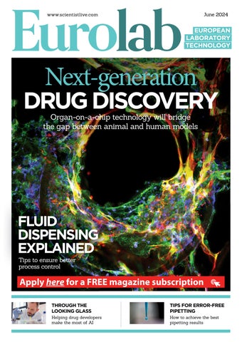
DRUG DISCOVERY

Organ-on-a-chip

technology helps bridge the gap between animal and human models in drug development

Sadly, 90% of drugs entering clinical trials ultimately fail, with insufficient efficacy remaining a leading cause for these failures despite years of R&D and millions of pounds invested. This harsh reality points to the fact that the preclinical tests used to discover and develop drugs do not accurately reflect human outcomes.

Unfortunately, all industry-standard models have flaws; traditional invitro assays are too simplistic and in-vivo animal models are too ‘animal’. The problem is now exacerbated as new advanced drug modalities, with human-specific modes of action becoming more prevalent in drug pipelines. So, what can be done to

overcome these limitations and bring much needed therapeutics to market in fast and cost effective ways - especially for diseases that are becoming more widespread? The answer: Make use of a an innovative new solution called the ‘organ-on-achip’ (OOC).

NEW APPROACH METHODOLOGIES

CN Bio’s in-a-boxsolution helps recreate industry-proven models

The complementary or alternative use of New Approach Methodologies (NAMs), such as organ-on-a-chip (OOC), offer a path forward in situations where model translatability to humans is predicted to be poor, or for testing advanced drug modalities where human-specific targets and pathways are required. The aim of OOC is to accurately replicate human physiology and function in vitro by culturing physiologically relevant combinations of primary human cells together in a perfused environment supplied by fluidic flow. Using OOC, it is now possible to recreate healthy and diseased human organ mimics in the laboratory.

NASH liver tissue showcasing the fibrotic phenotype (yellow colour) associated with the disease

Alternative applications for OOC technology

OOC technology can help oncology researchers study the impact of specific tissue components, investigate the role of the tumor microenvironment (TME), or visualise how cancer cells behave over time when interacting with stromal and immune cells. Similarly OOC is better able to reproduce the complexity that influences cancer behavior in vivo, and appropriately model more recent advancements in cancer therapeutics than conventional in-vitro models. Similarly, OOC can help with inflammatory diseases. Inflammation plays a role in many conditions including Alzheimers and Inflammatory Bowel Syndrome (IBS). However, modeling complex immune response in vitro can be difficult owing to the fundamental differences in the immune systems of animals and humans. OOC allows researchers to incorporate the cellular diversity seen in vivo into a tissue-specific microenvironment that more closely emulates the complex cell-cell interactions involved in inflammation in a closely controlled environment.

NON-ALCOHOLIC STEATOHEPATITIS

One area of growing global concern is the metabolic disorder Non-alcoholic steatohepatitis (NASH), also known as Metabolic Dysfunction-associated Steatohepatitis (MASH). NASH is the most common form of chronic liver disease worldwide. Despite much R&D effort, there is only one approved therapeutic.

This is mainly owing to the inability of traditional in-vivo approaches to accurately predicting the human response to this complex metabolic disease.

CN Bio’s highly characterised and validated PhysioMimix NASH assay is comprised of primary human

❝ Although animal models capture the complexity of a full organism, organ-on-achip technology demonstrates how a disease mechanism, or the effects of a drug, will differ in a human setting

hepatocytes, stellate and Kupffer cells that form 3D microtissues capturing key stages of disease progression, intracellular fat accumulation, inflammation and fibrosis. The OOC model supports development of drugs targeting NASH by enabling the precise mechanistic effects of drug efficacy to be uncovered.

BRIDGING THE GAP BETWEEN HUMANS AND ANIMALS

Whereas animal models capture the complexity of a full organism, OOC demonstrates how a disease mechanism, or the effects of a drug, will differ in a human setting. The combination of the two gives a much broader preclinical insight into a drug’s potential.

In conjunction with the PhysioMimix OOC, CN Bio’s product and service portfolio offer flexible solutions to researchers to fast-track the

incorporation of OOC technologies into drug discovery workflows.

AN ‘IN A BOX’ SOLUTION FROM CN BIO

The company’s ‘in-a-box’ solution provides a straightforward and quick route to recreating its industry-proven models and assays, and through its Contract Research Services (CRS), the team can tailor each experimental design to deliver unique human-translatable insights and actionable data within weeks, while saving significant time and cost versus animal studies.

In December 2023, the company announced its CRS had supported the characterisation of a NASH drug candidate targeting human metabolism. As metabolism in mice is very different from human, OOC offered a path forward. Compound efficacy data derived using this NASH assay was used to support the initiation of Inipharm’s Phase 1 clinical trial for INI-822. The submission represents the first example of an OOC provider’s data supporting the clinical progression of a drug for a complex metabolic liver disease and demonstrates the transformative potential of these models to provide human-relevant insights within preclinical programs. Looking to the future, the body of evidence demonstrating OOC’s superior performance versus traditional approaches is destined to continue growing since these technologies are in a unique position to help pave the way for more much-needed therapeutics to progress into the clinic, and beyond. n

In the US, staple crops like sugar beet, canola (rapeseed), corn, and soybean are almost entirely GMO. However, European regulation and attitudes have meant that almost no GMO crops are grown here. Spain represents the biggest grower but its produce represents less than 1% of global GMO production. Many EU countries such as Germany and France are particularly hostile to GMOs and have banned their cultivation entirely.

Despite this, Europe imports over 35 million tonnes of GM soyabean each year for animal feed, as well as many other crops. And it’s fair to say that attitudes may be changing. Last year, the UK parliament passed the Genetic Technology (Precision Breeding) Act and the EU Commission and Parliament are at an advanced stage on similar laws. These laws cut the red tape around plants produced using “New Genomic Techniques” (NGTs),. Under this new regime, NGT plants are subject to the same level of regulatory scrutiny as plants obtained through conventional breeding and would not be required to undergo the extensive safety testing required for GMOs. However, the EU Parliament does not want to simply update the regulatory framework as has been done in the UK; they also want to exclude NGT plants from being patented.

THE CURRENT LAW

Under the current law, plants produced by conventional breeding cannot be patented in Europe. The EU Parliament has explained that the reason they want to exclude NGT plants from patentability is because they are concerned about giving more power to multinational seed companies over farmers. They also consider that PVRs provide adequate exemptions for breeders but that patent law does not.

The patent sector has been quite critical of this move since it argues that the language of the laws as proposed is vague and too broad. Further criticism has arisen in that these amendments will affect nonEU countries that are part of the European patent system. since the EPC explicitly refers to the EU Biotech Directive (which would be amended under these proposed changes) and

Give GMO a chance

New legislation might lead to increased cultivation of GMO in Europe but a reduction in R&D.

this includes some non-EU countries including the UK and Norway. While it seems likely that these proposed changes would lead to increased cultivation and consumption of GMO plants in Europe, it may nonetheless dissuade further R&D in this space in Europe, particularly from smaller companies. Such companies rely on patents to protect their investment into R&D

as well as to attract funding. Larger companies already have an advantage in having greater resources to help navigate the complex regulatory environment surrounding GMOs. Given the EU parliament’s aim is to avoid giving more power to multinational seed companies, they will need to carefully consider the impact of these new proposed laws before bringing them into force. n

The EU Parliament wants to exclude NGT plants from patentability

IDT ONCOLOGY —empowering

Looking for oncology research insights?

Subscribe to IDT insider and explore IDT ‘s high-performing spectrum of customizable solutions.

confident insights. We'll help.

PREVOLUTIONISING PRECISION MEDICINE

Advances in oncology research

Courtney Thomas PhD is with Integrated DNA Technologies

recision medicine has created a paradigm shift in oncology research, allowing scientists to identify individualised cancerassociated biomarkers. In this space, next generation sequencing (NGS) has emerged as a transformative tool, offering a range of insights from single nucleotide variants (SNVs) to comprehensive profiles of cancer genomes. This knowledge can be further used to streamline laboratory workflows and resources, optimising assay content and protocols for efficient and accurate genomic profiling of tumours. The integration of NGS data with real-time monitoring approaches fosters a dynamic approach to understanding genetic variations associated with cancer and can contribute to the discovery of new cancer targets biomarkers.

Despite this, there are several factors, e.g., cost and accessibility, that have slowed progress in this field [1].

Investing in equipment for NGS can be prohibitive. Additionally, the large amount of data generated with NGS often requires advanced bioinformatic analyses and interpretation.

Accessibility to the latest oncology research NGS approaches varies drastically by country. In some, like Germany, NGS for oncology precision medicine has been adopted as standard alongside a commitment to

making whole genome sequencing a widely accessible tool. Other countries are working towards integrating small, targeted gene panels into their standard practices [1]. There is a clear need to improve availability of NGS tools globally and to standardise implementation of these approaches. IDT has long recognised the power of NGS and made a commitment to empowering oncology research labs in their pursuit of important insights in precision medicine.

“Over its more than 35-year-history, IDT has been innovating alongside its customers to equip them with the right tools when they need them,” said Steven Henck, PhD, Vice President, R&D at Integrated DNA Technologies.

“Our NGS portfolio, which is comprised of stand-alone library preparation, target enrichment, and normalisation chemistries, as well as connected NGS solutions with secondary analysis software support, have been foundational to some of the world’s greatest genomic discoveries. We’re proud to make these solutions widely available to the research community, which depends on IDT to deliver the critical tools they need to continue advancing cancer breakthroughs.”

This article discusses the spectrum of approaches used in oncology research and highlights the benefits and the limitations of each technique.

SINGLE GENE TESTING

Single gene techniques (also referred to as whole gene testing) are among the most widely available methods. Examples include fluorescence and chromogenic in situ hybridization (FISH), PCR, and micro-satellite instability (MSI) tests [1]. They are well established, allow for streamlined analyses and are also cost-effective, making them widely accessible. For certain types of cancers, PCR and other single gene targeting approaches focus on known mutations that drive oncogenesis –e.g., BRCA1/2 in breast cancer [2,3]. These approaches can fail to capture the full picture of mutations driving tumourigenesis since some cancers are the result of an accumulation of mutations across multiple genes.

SMALL TO LARGE PANEL TESTING

The limitations presented by single gene targeting approaches can be largely overcome using small to large gene panel testing. Panels rely on NGS to obtain information about specific genes consolidating relevant targets into a multi gene panel. Small panels (less than 50 genes), and large panels (more than 50 genes) [1] provide more information per sample than a single gene approach.

Small gene panels target a select set of genes that were previously identified as having relevant mutations in specific cancer types. This focused sequencing of a limited number of genes helps streamline data analyses and interpretation, while also providing more accessibility with less cost than large gene panels [1]. Small gene panels can be employed before samples are analysed using approaches that target more genes like large gene panels or comprehensive genomic profiling (CGP).

Large gene panels target a wider

Figure 1. Continuum of approaches used by oncology researchers in precision medicine

range of cancer-associated genes, providing insights into the genetic profile of a tumour tissue or liquid biopsy sample. Increasing the scope of sequencing via large gene panels further reduces the risk of missing key mutations. Relative to the depth of information generated, large gene panels offer consolidated workflows and relatively short turnaround times. Additionally, some gene panels (small and large) are designed to be tumour agnostic, which can be a time saving advantage allowing researchers to cast a wide net to capture relevant biomarkers rather than relying on multiple tumour-specific assays. With increased information generated by small to large gene panels sequencing costs increase along with a decrease in accessibility. This is true across

Approach Description

the continuum of approaches, where there is currently a tradeoff between accessibility, cost, and information gathered (Figure 1). While the scope of small or large gene panels is wider than that of single gene techniques, there is still a risk of missing relevant gene alterations with this approach. Approaches in oncology research that provide an even greater understanding of the genomic signature in cancer include CGP, whole exome sequencing (WES), whole genome sequencing (WGS), RNA sequencing (RNA-seq), and whole genome and whole transcriptome sequencing (WGTS).

COMPREHENSIVE GENOMIC PROFILING

Comprehensive genomic profiling (CGP) is a tumour-agnostic, NGS

Molecular approach that targets known, oncogenic genes to identify mutations

NGS approach that targets < 50 cancer-associated genes

NGS approach that targets > 50 cancerassociated genes

Assay that provides insights into many major, known cancer biomarkers

NGS approaches that either target protein coding regions of a genome (WES), the entire genome (WGS), the transcriptome (RNA-seq), or both the whole genome and transcriptome (WGTS)

method that can provide a more comprehensive view of the cancer biomarker landscape. CGP assays are designed to detect many cancerassociated genomic alterations such as SNVs, indels, copy number variants, fusions, splice variants, as well as other key cancer genomic signatures like tumour mutational burden (TMB) and MSI.

Obtaining this information in a multiplexed, targeted approach saves vital time, and reduces costs as well as the need for sequential biomarker testing. Additionally, because CGP assays are more focused on profiling specific pathologies, there is less risk of incidental identification of unrelated biomarkers–an issue faced by NGS approaches like WES and WGS. However, like other large content approaches mentioned here, the accessibility to CGP is limited.

Well established protocols; simple, streamlined analyses

Cost-e ective for relevant data, simple, streamlined analyses, high-throughput

Cost-e ective for relevant data, relatively simple, streamlined analyses, identification of many biomarkers in parallel, high-throughput

Time and resource saving for comprehensive profiling data, reduces the need for sequential biomarker assays

Comprehensive, identification of cancer biomarkers, highthroughput, no re-design needed when new markers are discovered

Limitiations

Potential to miss relevant variants outside of target

Potential to miss relevant variants outside of targets

Potential to miss relevant variants outside of targets

Less complete profiling of cancer sample relative to WGS/WES, workflows and analysis can be complex to establish, typical fixed content results in reduced flexibility with analysis

Currently high costs (expected to decrease with time) and heavy computing/analysis demands, potential ethical risks with unintended identification of non-cancer disease biomarkers

Table 1. Summary of approaches used by oncology precision medicine

You Are Here You Could Be Here

Spend less time in front of the lab instrument with time-effective solutions from OPSIS LiquidLINE. We provide a large range of instruments with good quality for wet chemistry laboratories. Our product range includes everything from software, digestion blocks, extraction units, and distillation units to complete Kjeldahl analysers. Now we also have an instrument for fiber analysis! Contact us for more information.

WES, WGS, RNA-SEQ, WTS

WES targets the protein-coding regions of a genome (the exome), which contain up to 85% of disease-associated variants [4], making it a cost-effective approach. However, WES can miss relevant variants located outside of the exome in non-coding genomic regions. If this is a concern, WGS can address that limitation as it generates sequence data for the entire genome. Relative to WES, WGS is more expensive and has the potential to miss lower allele frequency variants due to its lower sequencing depth. However, WES is more likely to miss large genomic changes. It is also important to note that sequencing costs have regularly decreased overtime.

Beyond DNA, having information from the transcriptome via RNA-seq provides oncology researchers with a variety of unique insights. RNA-seq captures information concerning splice variants, fusion transcripts, and other transcriptional biomarkers associated with cancer development. RNA-seq results can be heavily impacted by the quality of samples and requires genomic data to obtain meaningful insights. This multi-disciplinary approach is called whole genome and transcriptome sequencing (WGTS) [5]. All these approaches offer broad sequencing data, ensuring that when new biomarkers are discovered, there is no need to re-design or change the sequencing workflow. Though the increase in sequencing data obtained by these approaches reduces the risk of missing important mutations, it also increases the complexity of data obtained. This requires sophisticated

analysis pipelines, and the less-targeted scope of sequencing data could lead to the identification of incidental findings –unrelated to the cancer mutations being targeted.

APPROACHES FOR A TRULY COMPLETE BIOMARKER PROFILE

The decision to use any of these approaches to identify actionable mutations in a sample depends on factors such as sample quality, institutional and country guidelines, and resource availability (Figure 2).

While some methods like CGP assays can stand alone to characterise cancer samples, others are applied in tandem to support applications like tumourinformed minimal residual disease (MRD) research. Here, a tumour tissue sample is sequenced via WES then the variant detection data is used to design a custom gene panel to deeply sequence liquid biopsy samples for traces of those variants in circulating tumour DNA (ctDNA) shed from the solid tumour. For tumour-informed MRD solutions, IDT provides researchers with multiple components for an optimal workflow, including the xGen™ cfDNA & FFPE DNA Library Prep Kit, xGen Exome v2 Hyb Panel, xGen MRD Hybridization Panel, and custom enrichment panels and design services.

For solid tumour CGP solutions, paired Archer™ panels, such as the VARIANTPlex™ Complete Solid Tumour combined with the FUSIONPlex™ Pan Solid Tumour v2, provides a contentflexible comprehensive biomarker profile. These assays are designed to analyse DNA (VARIANTPlex) and RNA (FUSIONPlex) to identify relevant

Figure 2: Overview of oncology research approaches and applications

SNVs, indels, CNVs, ITDs, MSI, and TMB. Additionally, all Archer research assays use the Archer™ Analysis software, allowing researchers to analyse data rapidly and at scale. Ultimately, precision medicine relies on all these techniques (Table 1). IDT is dedicated to supporting researchers by providing expert support throughout the continuum of these methodologies. From small to large gene panels like Archer Research Assays to components for library prep like the xGen cfDNA & FFPE Library Prep Kit and WES enrichment panels like the xGen Exome v2 Hyb Panel–IDT is prepared to help. n

REFERENCES

1. Bayle A, Bonastre J, Chaltiel D, et al. ESMO study on the availability and accessibility of biomolecular technologies in oncology in Europe. Ann Oncol. 2023;34(10):934-945.

2. Miki Y, Swensen J, ShattuckEidens D, et al. A strong candidate for the breast and ovarian cancer susceptibility gene BRCA1. Science. 1994;266(5182):66-71.

3. Wooster R, Bignell G, Lancaster J, et al. Identification of the breast cancer susceptibility gene BRCA2. Nature. 1995;378(6559):789-792.

4. Ng SB, Turner EH, Robertson PD, et al. Targeted capture and massively parallel sequencing of 12 human exomes. Nature. 2009;461(7261):272-276.

5. Nakagawa H, Fujita M. Whole genome sequencing analysis for cancer genomics and precision medicine. Cancer Sci. 2018;109(3):513-522.

n For more information visit www.idtdna.com

AFFORDABLE MICROPLATE DISPENSING IN A SNAP

forcassetteRevolutionarytechnology lower running costs!

WELLJET Reagent Dispenser

The WELLJET dispenser and dispenser stacker together with the EasySnap™ dispensing cassette offer superior user experience and flexibility for applications requiring low cost and precise reagent dispensing at minimal benchspace.

Your choice to save money, space and time!

Cellular Protein Synthesis

Exploring recombinant protein expression using a baculovirus-insect cell system

Proteins are essential for various cellular functions, making them a focal point of scientific inquiry across disciplines. Understanding the structure and function of proteins is crucial for unlocking the ‘mysteries of life’, driving research efforts across several scientific fields. However, obtaining proteins for study can be challenging due to their complexity.

Recombinant protein expression is a method that can help solve this problem since it produces proteins by cloning the gene of interest into an expression vector and introducing it into a host cell. The selection of the optimal host cell system is critical for successful protein expression. One such system is the Baculovirus-insect Cell System (BEVS), which utilises baculoviruses to deliver target genes into insect cells for protein production.

THE BEVS PROCESS

In the BEVS process, a gene encoding the protein-of-interest is inserted into a primary vector and then cloned into a secondary vector known as Bacmid. This Bacmid is transferred into a bacterial strain for preliminary virus production, resulting in the generation of baculovirus (Figure 1). The baculovirus is then amplified in insect cells and used to infect insect cell

lines for protein expression, offering a versatile platform for producing various proteins.

APPLICATION OF BEVS

Insect cells are often used to produce large molecular weight (MW) proteins (MW> 150 kDa) because of their superior folding and posttranslational modification capabilities. One major downside of this endeavour, however, is the structural complexities of such proteins, which often results in relatively low yields (Fig. 2, left). One alternative approach is to express a domain-of-interest rather than the entire protein; however, in the case presented here, the direct expression of two enzyme domains of human fatty acid synthase (FASN) was

Figure 2: Expression of full-length (left), truncated (middle), and methyltransferase plus ketoreductase domain fusion (right) constructs of human FASN. The fulllength protein was prepared as reported by Hardwicke et al.(2014,doi:10.1038/ nchembio.1603), whereas the domain fusion construct was prepared as reported by Lu et al.(2018, doi: 10.1016/j.bmcl.2018.05.014)

not feasible because of low protein yield and heavy degradation (Fig. 2, middle). By extrapolating and fusing the sequences encoding the methyltransferase and ketoreductase domain with a linker, Sino Biological successfully obtained a high-yield construct (Fig. 2, right). The Elute 1 and 2 of this construct were pooled and further purified to yield a final fusion protein with >90% purity. It should be noted that some proteins require certain oligomeric formations to be functional. For example, the hemagglutinin proteins of influenza virus and the spike protein of SARS-CoV-2 exhibit a trimeric format, whereas the human prolyl endopeptidase, FAP, is an intrinsic dimer.

CONCLUSION

Recombinant proteins are fundamental to the study and development of biologics. Insect cells are a superior choice as an expression host because they enable correct protein folding and posttranslational modification, and are suitable for high-density cell culture. In addition, they can produce both secreted and intracellular proteins of various species. ■

Figure 1: Flowchart for recombinant protein expression in insect cells

Changes in the pipette’s angle will affect the hydrostatic pressure within the pipette tip

TIPS FOR PIPETTING

How to achieve error-free pipetting for accurate results

Pipetting is one of the most common laboratory tasks, forming the cornerstone of scientific research and clinical diagnostics. The impact of pipetting errors is often underestimated, but even slight discrepancies in precision – arising from both the instrument and the user – can accumulate over the course of a workflow, resulting in substantial inaccuracies in the final data. Pipetting by hand takes years of practice to perfect, but there are ways to quickly improve the accuracy of manual liquid handling for more reliable results.

MANUAL PIPETTING BEST PRACTICES

Firstly, changes in pipetting angle will affect the hydrostatic pressure within the pipette tip, resulting in an inconsistent aspiration volume. Holding a pipette at an angle of 20 degrees or less from the vertical is therefore paramount for consistent liquid aspiration and dispensing, and for very small volumes of ≤30, the straighter the pipette, the better. After dispensing, there will often be a droplet left behind at the end of the pipette tip. A simple way to make sure the whole sample is dispensed into the target vessel is to

perform a tip touch when removing the pipette from the vessel. There are three ways of doing this: side wall touch off, surface touch off, and direct dispensing into a liquid.

Another common source of error is caused by temperature differences between pipettes, tips and liquids. These can affect the air cushion inside the pipette, leading to volume variations. Incorporating a prewetting step into a pipetting routine helps to ensure that all labware and liquids are in equilibrium, largely overcoming this issue and providing more confidence in the accuracy of measurements. Some electronic handheld pipettes can even be programmed to include a prewetting step, making it easier to achieve precise and consistent results. During repeat dispenses, the first dispense may be too low in volume, and the final dispense could include all the accumulated errors of the previous dispenses. Discarding the first and last dispenses helps to eliminate these inconsistencies, resulting in more reliable aliquots.

ENSURING ACCURATE VOLUME CONTROL

Each pipette has an optimal volume range; for an air displacement pipette,

this is typically 35 to 100 per cent of the device’s nominal volume. Staying within this range is therefore crucial for reducing the amount of air in the cushion, minimising errors and maintaining the best possible accuracy. In addition, poorly fitting tips can lead to leaking, quickly causing inaccurate aliquot volumes. Tips may also become

misaligned or fall off, interrupting workflows and slowing down liquid handling steps. It’s essential to use high quality, compatible pipette tips –ideally from the same manufacturer as the pipette itself, rather than from a universal supplier – to avoid these common issues and promote consistent volume control.

HANDLING NONAQUEOUS LIQUIDS

The calibration of air displacement in micropipettes is performed in a controlled environment with water, so pipetting viscous and volatile non-aqueous liquids – such as glycerol, DMSO, ethanol or Tween 20 – can affect the accuracy of the end result. Viscous liquids attach to the wall of the tip, making it difficult to expel the full volume, while the rapid evaporation of volatile liquids can significantly alter the pipetting volume. Specific techniques are required to ensure the best results when working with these more difficult liquid types.

1

Viscous liquids

a) Hold the pipette upright

b) Use a slower pipetting speed

c) Do not immerse the tip too far into the sample reservoir to reduce the risk of carryover

d) Use low retention, wide bore tips to enable liquids to enter more easily

e) Perform reverse pipetting to prevent incomplete filling or emptying of a pipette tip

2

A common source of error is caused by temperature differences between pipettes, tips and liquids

❝ Pipettes can become less accurate and may cause sample contamination without the proper care, maintenance and storage

PREVENTATIVE DEVICE MAINTENANCE

f) Keep the pipette tip in the solution for longer, 2-3 seconds after aspiration and dispensing, to allow complete liquid transfer

Volatile liquids

a) Hold pipette as upright as possible

b) Use a faster pipetting speed and work quickly to prevent evaporation

c) Prewet to equilibrate air pressure and humidity to reduce evaporation

Pipettes can become less accurate and may cause sample contamination without the proper care, maintenance and storage. The best way to prevent this source of error and frustration is to clean and maintain pipettes regularly. The outside of a pipette should be cleaned daily with a lint-free cloth and 70 percent ethanol, and it’s important to perform a leak test and validate the pipetting volumes on a monthly basis to make sure that the pipette is working as intended. If its accuracy and precision are not within specifications, the device needs to be calibrated before further use. Calibration carried out at least every 12 months will help to keep pipettes working at their best. In addition, if the pipette is dropped, then it should immediately be checked for damage and recalibrated before use to ensure it still meets the specifications. Cleaning, maintaining and regularly calibrating pipettes will give them a long lifetime of reliable activity, and also helps to keep users safe from potential malfunction and contamination.

In summary, good pipetting practices, combined with regular maintenance, help to maintain consistent pipetting, leading to fewer manual errors and contributing to more reliable and accurate results. n

For more information visit https://www.integra-biosciences. com/en

Hold the pipette as upright as possible

Maintaining optimal relative humidity (RH) levels is essential in research laboratories, pharmaceutical cleanrooms, and semiconductor fabrication facilities, where precision, quality, and safety are nonnegotiable. The RH levels present in these facilities directly influence processes, equipment functionality, material integrity, and the well-being of personnel.

The business benefits of controlling RH in these settings are immense, ranging from preventing electrostatic discharge risks to ensuring consistent research outcomes to safeguarding product quality.

One of the primary challenges in all of these environments is the presence of static electricity. Delicate electronic components and sensitive materials are susceptible to damage caused by electrostatic discharge, and fluctuating RH levels can increase this risk. This control of RH is particularly critical in industries such as semiconductor manufacturing, where even slight variations in humidity can substantially impact processes and outcomes.

Research outcomes in biotechnology laboratories heavily depend on stable and controlled conditions. Fluctuations in humidity can lead to variations in experimental conditions, potentially compromising the reliability and validity of research results. Precision humidity control has emerged as a crucial advancement and enhanced sensor technologies and control algorithms enable more precise control over humidity levels.

Boston Scientific Cork Ltd. (BSCL), a manufacturer of heart surgery stents, faced the challenge of ensuring precise and continuous humidification within 1% of the set point. The existing humidifiers were unable to meet this control requirement.

To address these challenges, BSCL replaced existing electrode humidifiers with fourteen DriSteem resistive-element humidifiers in their stent production areas. As required, these humidifiers ensured that the

Managing humidity is essential for many precision facilities

THE ROLE OF RELATIVE HUMIDITY

Here

we explore why controlled humidity is essential for reliable research outcomes

relative humidity (RH) in BSCL’s production rooms remained within 1% of the set point. The steam was effectively dispersed using dispersion panels in the seven air-handling units. The humidifiers were strategically distributed throughout the air handling units, with some units accommodating up to three humidifiers based on the load requirements.

By implementing DriSteem resistive-element humidifiers, BSCL successfully overcame the challenges of maintaining precise control and efficiency in their stent production areas. The new humidifiers ensured continuous and accurate humidification within a tight range of the set point, addressing the limitations of the previous electrode humidifiers. This improvement enhanced the quality of their critical operations and

contributed to cost savings through reduced energy consumption, lower maintenance expenses, and optimised resource conservation. Advanced humidification technologies, such as those offered by industry leaders like DriSteem, play a vital role in addressing these diverse needs. Whether integrating humidity control solutions in existing structures or implementing them in new buildings, factors like the type of indoor space, energy source, required maintenance, and capacity determine the best technology for each building. DriSteem stands out by providing innovative humidification solutions tailored to meet the unique requirements of different environments. n

For more information visit https://www.dristeem.com/

Pressing Power

A guide to understanding how head dwell time can impact tablet compression

Compressing difficult tablet formulations can be achieved via a range of punches or dies. One punch modification, the extended head flat, increases the diameter of the flat area atop the punch head. Typically, this doesn’t require any modifications to the press, and it can be used with cam tracks to meet the Tableting Specification Manual (TSM) or European Union (EU) standards.

Dwell time is dependent on press speed, the pitch circle diameter of the turret, and the head flat’s diameter (Figure 1). Increasing the diameter of the head flat is the easiest way to prolong dwell time without switching

to a press with a smaller turret pitch circle and generally with fewer stations, or without decreasing the turret speed, both of which would decrease production. For example, a Fette 2090 press has a pitch-circle diameter of 410 mm, and assuming an operating speed of 50rpm, a standard B-type punch, TSM domed head flat of 9.525mm (0.375in) would have a dwell time of 8.87 milliseconds.

B-TYPE PUNCH WITH TSM DOMED HEAD

Meanwhile, on that same press, a B-type punch with TSM domed head with extended head flat (13.59mm, 0.535in) would have a dwell time of 12.66 milliseconds (Figure

2). Figure 3 illustrates the differences in dimensions between a standard B-type TSM domed head and TSM extended domed head. The larger head flat increases dwell time by more than 43% without reducing the turret speed.

The extended head flat punch can also reduce the amount of compression force needed to form a tablet at a given breaking force, also known as hardness. Tablet hardness and density are related to both compression force and dwell time. If the amount of time spent under compression increases, the amount of force necessary to maintain the same tablet hardness may decrease. The reduction in required compression force depends on the characteristics of the granulation being compressed. Since the required compression force is dependent on the product being compressed, it’s impossible to

Extended head flat vs. standard head flat

predict accurately how much an increase in dwell time will decrease compression force. The effect of dwell time on compression force can best be quantified during the research and development stage, and understanding the relationship of dwell time to compression force can mitigate issues that commonly arise during scaleup to large production presses.

Geometrically, the design of the extended head flat differs only slightly from a TSM domed head or an EU head. Other than the increased head flat, one notable difference is a reduction in the head’s thickness to allow the larger flat to fit through the same cam profile as that of a standard TSM domed or EU head.

Head wear and the propensity of surface origin fatigue (i.e. head pitting) are affected by the head profile design. The smaller the outside head radius, the higher the contact stress and likelihood of accelerated wear. The difference in stress can be calculated analytically using Hertzian contact stress equations.

USING THE HERTZIAN STRESS EQUATION

For example, using the cylinder-oncylinder Hertzian stress equation to approximate a comparison between TSM Std, TSM Domed, and TSM Extended head profiles assuming a 10” (254mm) diameter pressure roll: Note that contact stresses are reduced with larger pressure rollers and are higher with smaller pressure rollers. For this reason, the old TSM standard head design with a small 5/16” radius between the outside head angle and the head flat was replaced with the domed head design. Punches with extended heads will be less resistant to head wear than domed punches, so care in selection is advised when high compression forces are needed.

The limiting factor in the head flat’s size is the neck diameter of the punch. The neck transfers the force from the head to the barrel and tip and then to the granulation. As you can see in Figure 4, if the head flat gets larger than the neck’s diameter, it won’t have the support required to transfer the force generated when it contacts the pressure roll, which can cause the head or neck to fail.

For mini-tablet production, the size of the head flat affects the side loads

being applied to the punch tips during compression and the likelihood of punch tips bending or buckling. Larger head flats increase side loads. Therefore, a reduced head flat (i.e. 6.35mm) may be advised.

OVAL AND ELLIPTICALLY SHAPED HEAD FLATS

Extended head flats can also be oval or elliptically shaped. Although this design extends the dwell time exactly as a round extended head flat does, it has some drawbacks. The primary drawback is that the oval head flat can pass under the pressure rolls only in one direction along the major axis of the oval or ellipse to extend dwell time. For that reason, punches with an oval head flat must be keyed on the upper and lower punches, even when round, to prevent them from rotating as they pass under or over the pressure rolls. With upper and lower punches requiring keys for oval or elliptical head flats, punch heads can experience accelerated wear because of repeated contact between the punch head and the compression roller at the same spot and no punch rotation.

Additionally, if a turret doesn’t have lower key slots, then round tooling with an oval head cannot be used because the benefit of the extended head flat will be lost if the lower punch rotates. Another limitation is that if two presses have turrets with key slots of different angles, the oval head flat’s punches cannot be interchanged because the different angle will alter the orientation of the head flat with respect to the pressure rolls.

Punch head profiles and the resulting dwell time play an important role in the compaction characteristics of many drug products. Using extended head flat punches can provide a quick and reliable way to increase dwell times and, in some cases, reduce compression force without the need to modify the tablet press or cam tracks. Round, extended head flat punches don’t require keyed tooling for round shapes, as oval ones do, and that allows them on many makes and models of tablet presses interchangeably. n

For more information visit https://natoli.com/

Understanding the nip angle by Barbara Fretter and Michael Schupp

Dry GRANULATION

The Thin Layer Model allows for understanding of some general aspects of powder densification between the rolls in dry granulation and especially the nip angle. Part 1[1] describes the model and how the nip angle can be estimated easily. It also outlines that a stronger densification results inevitably in larger nip angles. Part 2 focuses on the interaction of nip angle and gap and the practical meaning in R&D and Scale-Up.

THE THIN LAYER MODEL

Based on the geometric considerations of the Thin Layer Model it is possible to plot the nip angle versus the densification factor of the powder for different gaps (Figure 1). And an important relation can be derived: to achieve the same densification factor at a larger gap requires a larger nip angle - see Figure 1.

For example, a densification factor of two requires a nip angle of 5o at a

gap of 1mm and a nip angle of 10o at a gap of 4mm. This has some practical consequences for R&D and Scale-Up. Let’s assume that in R&D you have found the right granule properties for your product at certain roller compactor settings. Often small gaps like 2mm or less are chosen in R&D because the available amount of Active Pharmaceutical Ingredient (API) is limited. Additionally, the specific roll force (or a range of it) is defined. Although force and gap are specified for this product, the actual parameter determining the granule properties is the ribbon or at-gap density and they coincide with a certain densification factor. For reproducing the granule properties the at-gap density must be reproduced. This is also valid

when the gap is increased. Because the powder density when being drawn-in is mainly independent of the gap, a general strategy for changing the gap is to keep the densification factor constant. And as shown in Figure 1, increasing the gap at the same densification factor means that the densification must start at larger nip angles. Otherwise, the granule properties will change.

MISTAKES MADE IN DRY GRANULATION

Realising this correlation has some serious consequence for the specific roll force and ignoring it is one of the most made mistakes in ScaleUp. When increasing the gap at the same specific roll force, the resulting at-gap density cannot stay the same. Figures 2 and 3 give the explanation. By using the Thin Layer Model and simple geometric considerations (Figure 2) the solid fraction in each layer can be calculated for different gaps. Assuming that an at-gap solid fraction of 0.7 is the target value which should not be changed if the gap is increased.

Figure 3 shows the progression of the solid fractions for two gaps, 2mm

Figure 1
Figure 2

What is dry granulation?

Granulation is a process in which powder particles are made to adhere to each other, resulting in larger, multi-particle entities, so called granules. If such a process is performed without adding liquids, this is called dry granulation.

In dry granulation, the powder blend is compacted by applying a force onto the powder, which in general causes a considerable size enlargement. The compacts obtained are called ribbons, flakes or briquettes.

In order to obtain the desired granules, the compaction process is followed by a milling step.

and 4mm, towards the assumed at-gap solid fraction of 0.7. It gets obvious, that for each distance the solid fraction for the 4mm gap is larger than for the 2mm gap. Transferring this to the Thin Layer Model (Figure 2) means each horizontal layer must have a larger solid fraction for the 4mm gap than for the 2mm gap. But a larger solid fraction implicates a stronger densification and therefore a higher force which acts onto the rolls. The sum of all forces which act simultaneously onto the rolls must be higher. This is the reason changing the gap without adapting the specific roll force never results in the same granule properties. Unfortunately, this is one of the most made mistakes in roller compaction especially in development and scale-up when increasing the throughput by increasing the gap. Several examples for the influence of

For more information visit https://www.gerteis.com/en/

gap on ribbon or at-gap density can be found in literature, however, unfortunately, not always with the right explanation. Allesø [2] examined ribbons porosities for microcrystalline cellulose at two gaps and three different specific roll forces. His findings (Figure 4) prove the above made considerations: at the same

❝ Increasing the gap at the same densification factor means the densification must start at larger nip angles

specific roll force and a lager gap the resulting solid fraction of the ribbon is lower. The extend of this effect is caused by the densification properties of the material and substance specific.

LARGER GAPS REQUIRE LARGER NIP ANGLES

Based on the Thin Layer Model and geometric calculations an explanation can be given why larger gaps require larger nip angles to achieve the same at-gap density. As consequence, changing the gap without adapting the specific roll force result in granules with different properties. This is an often made mistake in ScaleUp. To achieve the same granule properties upon increasing the gap an increase of specific roll force is mandatory but in its extent substance specific. n

References:

[1] Barbara Fretter, Michael Schupp, Understanding the Nip Angle, Eurolab (Dec 2023), https://content.yudu.com/web/15ex3/0A2nilh/EurolabDec2023/html/index. html?page=52&origin=reader

[2] Allesø,M., et al., Roller compaction scale-up using roll width as scale factor and laserbased determined ribbon porosity as critical material attribute, European Journal of Pharmaceutical Sciences (2015), http://dx.doi.org/10.1016/j.ejps.2015.11.001

Contributors:

- Barbara Fretter, Managing Partner, Solids Development Consult GmbH, PeterJoseph-Lenné Str. 11, 51377 Leverkusen, Germany, +49 214 500 428 83, www.solidsdevelopment.com

- Michael Schupp, Head of Process Engineering, Gerteis Maschinen + Processengineering AG, Stampfstrasse 85, 8645 Jona, Switzerland, +49 55 222 55 22, www.gerteis.com

Figure 3
Figure 4

The IncuSafe Advantage

IncuSafe CO2 Incubators are well known throughout the world for its high-quality. Creating successful cell cultures requires an incubator that has an high level of precision, security and is easy to use. IncuSafe offers a wide variety of cell culture incubators utilizing advanced technology for unprecedented temperature and CO2 (and O2 for some models) control in processing various cell cultures. These CO2 incubators provide long-term performance, are extremely reliable and viable.

Active Background Decontamination

Dual Heat Sterilisation

• Exceptional CO2 and O2 recovery times

• InCu saFe Construction for Germicidal Protection

• Zirconia O2 sensor

• Direct Heat and Air Jacket system

• Dual IR Sensor

• H2O2 decontamination process

• Patented SafeCell UV lamp CO

Following the postponement of an in-person EMC2020 due to the pandemic, EMC2024 will be held in person from Sunday 25th to Friday 30th August in Copenhagen, Denmark.

EMC2024 is the 17th iteration of the show and will bring the world of scientific imaging together in one of Europe’s most beautiful cities. The event will take place at the Bella Center, Center Boulevard 5, 2300.

SHOW DETAILS

The show will feature a series of pre-conference workshops on Sunday 25th August to include sessions on AI in microscopy; optical tissue clearing techniques for 3D lightsheet imaging; volume electron microscopy for the life sciences and many more. All sessions will take place at the venue.

THE OPENING DAY

Attendees will be invited to an opening reception on Monday the 26th with an address presented by Eva Olsson, chair of the Nobel Committee for Physics. This will be followed by the first plenary. Other speakers include Carolyn Larabell professor and vice chair of the department of anatomy at the University of California, and Moritz Helmstaedter director at the Max Planck Institute for Brain Research in Frankfurt, Germany.

The day will continue with six parallel sessions and close with drinks in the Poster and Exhibition area. The last day of the Congress will conclude with a farewell reception.

The show aims to provide a balanced conference program of light and electron microscopy in both physical and life sciences aimed at microscopists, manufacturers, and suppliers. There will also be many opportunities to share new techniques, applications, and technology.

MICROSCOPY RELATED CONFERENCE TOPICS

The congress will address a range of microscopy-related subjects, including dynamic interactions in cells, organoids, tissue, and entire organisms, pathology, immunocytochemistry, and biomolecular labelling, volume electron microscopy in Life Sciences,

EMC2024 FOR OPEN SCIENCE

This European conference aims to bring together European professionals in an open science forum to share insights and techniques

semiconductors, heterostructures, and devices, quantum materials, advances in 3-dimensional image reconstruction, and new instrumentation.

The exhibition is one the largest of its kind featuring more than 100 exhibitors and a Nordic Corner for smaller enterprises, start-ups, and organisations. For more information on opportunities for exhibiting and sponsoring, interested parties should visit the website at the end of this article.

Attendees will also be invited to A Farewell Reception on the last day of

the event - the 29th August - and they may want to book a place at The Gala Dinner on the same day.

OPEN SCIENCE

The EMC2024 show promotes Open Science and encourages attendees to showcase and share their open science methodology, source, data and educational resources to the wider scientific community. n

Lab Innovations

For those at the forefront of scientific discovery

Lab Innovations will take place between the 30th and 31st October 2024 at the NEC in Birmingham. The show describes itself as ‘the main UK event to bring together minds and technologies at the forefront of scientific discovery’.

It also promises ‘access to emerging trends, technologies, and solutions ensuring that your work remains at the cutting-edge of progress and development’.

The event encourages exhibitors to position their brand alongside thought leaders, industry experts and innovators to showcase cuttingedge developments, share insights and catalyse collaboration within the

dynamic landscape of laboratory sciences.

Lab Innovations hosts four stages, each with two full days of conference content scheduled. The programme covers automation, sustainability, AI, technology demonstrations, upskilling and more.

In addition to discussions around these conference themes, delegates can expect a range of technical product advice and demos.

The show encourages attendees to immerse themselves in discussions regarding the technologies that are changing their working lives..

In addition, there will be many opportunities to connect with

The show will foster cross-sector collaborations

industry thought-leaders, experts and visionaries, fostering valuable cross-sector collaborations that will transcend disciplines and deepen connections within the scientific community.

Lab Innovations is running a podcast called Lab Matters, which can be accessed via Spotify,

The show also runs a digital hub which can be accessed via the website - this includes press releases, industry news, a gallery and blogs. n

GREAT FLUIDIC SOLUTIONS

BIOTECH LIQUID µFLOWMETER