Clinical Compendium 2024
April 2024
Published
1 Our Mission...............................................................................................................................................................2 Timeline.....................................................................................................................................................................3 AI-Powered Spectroscopy Technology...................................................................................................................4 Highlights of DermaSensor Study Results and Company Firsts For FDA Clearance...........................................5 DermaSensor Leadership Team..............................................................................................................................6 DermaSensor Clinical Compendium 2024 Highlights............................................................................................7 Device Label: Indications for Use, Contraindications, Warnings and Precautions...............................................8 Glossary of Terms.....................................................................................................................................................10 Clinical Performance of Novel Elastic Scattering Spectroscopy (ESS) in Detection of Skin Cancer .................12 A Blinded, Prospective, Multi-Center Clinical Trial Initial Results Clinical Utility of an Elastic Scattering Spectroscopy Device in Assisting Primary Care Physicians .................13 Detection of Skin Cancers Clinical Capabilities of a Handheld Elastic Scattering Spectroscopy - Artificial Intelligence Device .................14 as an Adjunctive Tool for Evaluating Skin Cancer in Skin of Color DERM-ASSESS III: Validation of a Handheld Elastic-Scattering Spectroscopy Device on Lesions ....................15 Suggestive of Melanoma Skin Lesion Analyzers: Economic Impact of Integrating These Technologies into Dermatology ......................16 Clinical Practice for Detecting Melanoma Summary of Early Clinical Effectiveness Studies...................................................................................................18 Likelihood of Malignancy and Number Needed to Biopsy in the DERM-SUCCESS Clinical Study......................19 Review of Current Available Literature Regarding Techniques for Skin Cancer Diagnosis.................................20 References................................................................................................................................................................22 Table of Contents
Our Mission
To empower clinicians with intelligent, objective solutions by leveraging data-driven, breakthrough technologies to improve skin cancer detection and patient care.
2
2009
Company founding
2011
Star t of melanoma algorithm training study using 1st generation device
2015
Star t of all skin cancer algorithm training study using 2nd generation device
2018
Completion of 4th generation device, star t of studies for CE mark
Mid 2020
CE Mark obtained In E urope, $11.5 million Series A financing to conduct FD A Piv otal Clinical Study
1H 2022
Positiv e results fr om FD A Piv otal Study , $10 million financin
Q1 2024
FD A Clearance and US Launch
2nd

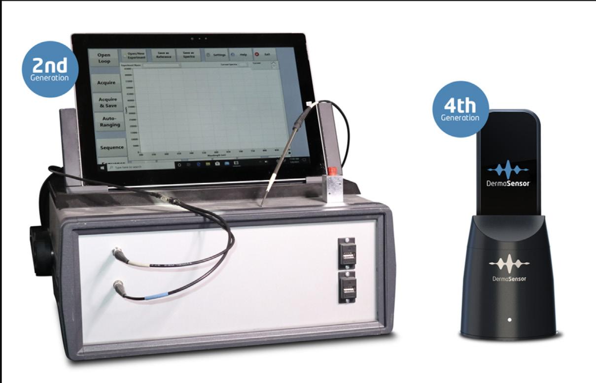
4th
3 Timeline
Generation 2009
Generation 2018
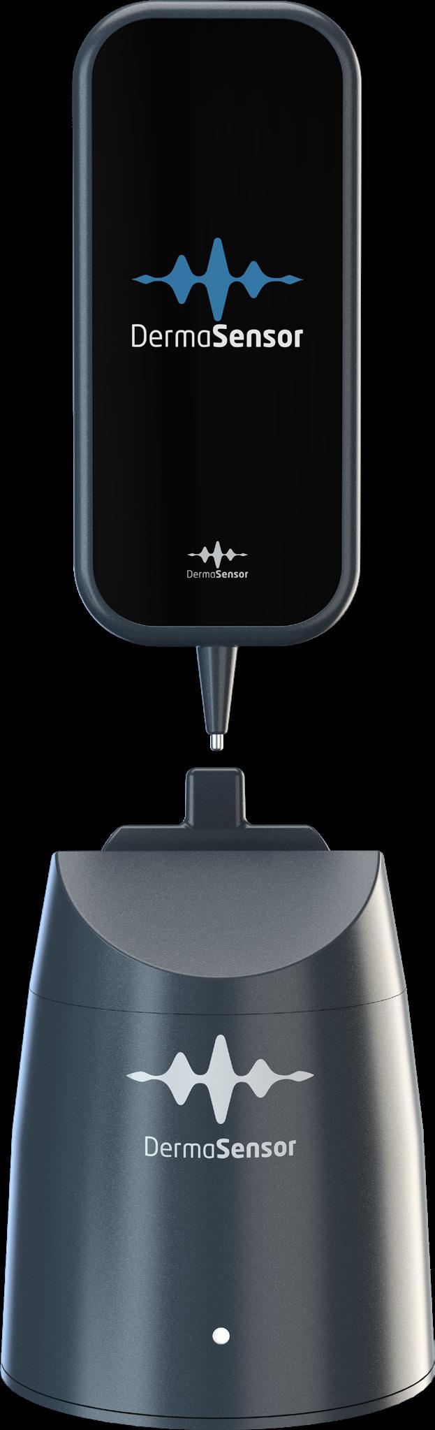
AI-Powered Spectroscopy Technology
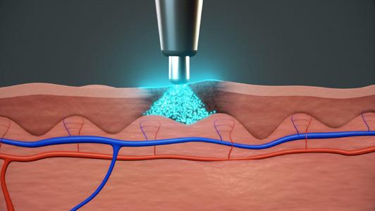
Five Spectral Recordings
Xenon lamp pulses light to take~1mm optical tissue samples of cellular f eatur es.
Algorithmic Analysis
The device receives spectral data across 47 wavelengths, comparing each lesion’s pattern of reflectance to patterns previously shown to indicate malignancy. Spectral signatures of malignancy correspond to histopathology features such as nucleus siz e, the pr esence of condensed chromatin, and melanocyte density
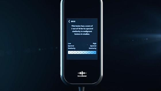
Objective, Probabilistic Output
DermaSensor ’s algorithm analyzes spectral data and deliv ers assessment results within seconds. The output reports a spectral score, with higher scores corresponding to greater risks of malignancy. The AI algorithm was developed and validated on over 20,000 scans and over 4,000 benign and malignant lesions.
4
Highlights of DermaSensor Study Results and Company Firsts
For FDA Clearance
Major Findings From Six Clinical Studies For Regulators
96% sensitivity for detecting all skin cancers1
PCP unaided melanoma sensitivity of 69-70%, device melanoma sensitivity of 90-96%2,3,5
Melanoma sensitivity and accuracy as high as expert dermatologists’ in-person dermoscopic diagnosis; melanoma sensitivity comparable to or higher than that of dermatopathologists from study findings and from discordance rates In literature3
Skin cancer sensitivity and accuracy higher than PCPs’ assessments5,6
Decreased PCPs’ missed skin cancer referrals by 30-52%6
Improved PCPs’ accuracy for managing and confidence in assessing skin cancer6
Ruled out 21-33% of benign lesions biopsied by physicians as suspicious for skin cancer3,5 and ruled out 61-77% of benign lesions that were suggestive of skin cancer to patients*8
Major Product Firsts Achieved Upon FDA Clearance
First skin cancer device designed to help PCPs evaluate suspicious skin lesions
First skin cancer device affordable to PCPs that provides any kind of automated risk assessment
First automated skin cancer device indicated for use with all common skin cancers
First/only currently available automated skin cancer device that uses light (i.e. optical or image-based device)
First FDA Breakthrough device for skin cancer
First FDA De Novo skin cancer device
First medical device of any kind that uses ESS
First automated skin cancer device that is easy to use (i.e. “point & click easy ”)
*This clinical study, called PATIENT-SELECT, Is considered an early effectiveness study by the FDA, not principal effectiveness evidence. PATIENT-SELECT was a screening-like study design of patient-selected lesions (i.e. not solely physician-selected lesions, per the indicated use). The overall device specificity was 61%, the specificity for pigmented lesions was 77%.
5
Experienced DermaSensor Leadership Team


Maurice Ferre MD
Chairman & Co-FounderDr. Ferre is the current CEO of INSIGHTEC, the former CEO of MAKO Surgical and a former VP at GE Healthcare. He has led INSIGHTEC to over a $1 billion valuation and has also had two exits, having sold MAKO to Stryker for $1.65B, and having sold Visualization Technology to GE.
Cody Simmons
CEO & Co-FounderCody is a bioengineer and entrepreneur that has spent all of his career bringing new health technologies to physicians and patients. Prior to joining Maurice to build DermaSensor, Cody led commercial efforts for a Silicon Valley medical device screening startup and held BD and commercial strategy roles at Genentech.
Larry Anderson
Larry is a West Point Graduate with 20 years of medical device experience, specializing in building teams and launching disruptive technologies. Most recently, Larry led the sales team at Acessa Health to a successful exit to Hologic during the COVID-19 pandemic. His prior companies include Ethicon Women’s Health and Urology, Interlace Medical, Hologic, and BG Medical.
Gary Slatko MD
Gary has over 35 years of experience supporting the clinical development, medical affairs, safety surveillance and risk management of biomedical products, including 6 years at FDA. Prior leadership roles include CMO at Aquestive Therapeutics and at ParagonRx, LLC.
Ryan Freiden
Ryan has over 16 years experience founding and scaling companies in consumer goods, healthcare, and enterprise technologies. Recently, Ryan led the growth for a healthcare supply chain technology and previously ran the international manufacturing and distribution of consumer hardware electronics.
6
DermaSensor Clinical Compendium 2024 Highlights
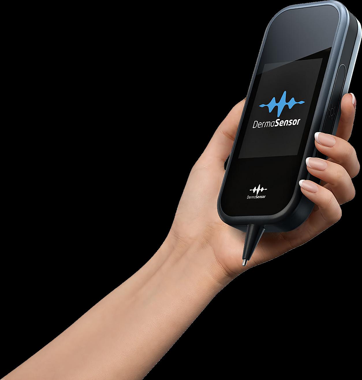
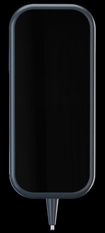
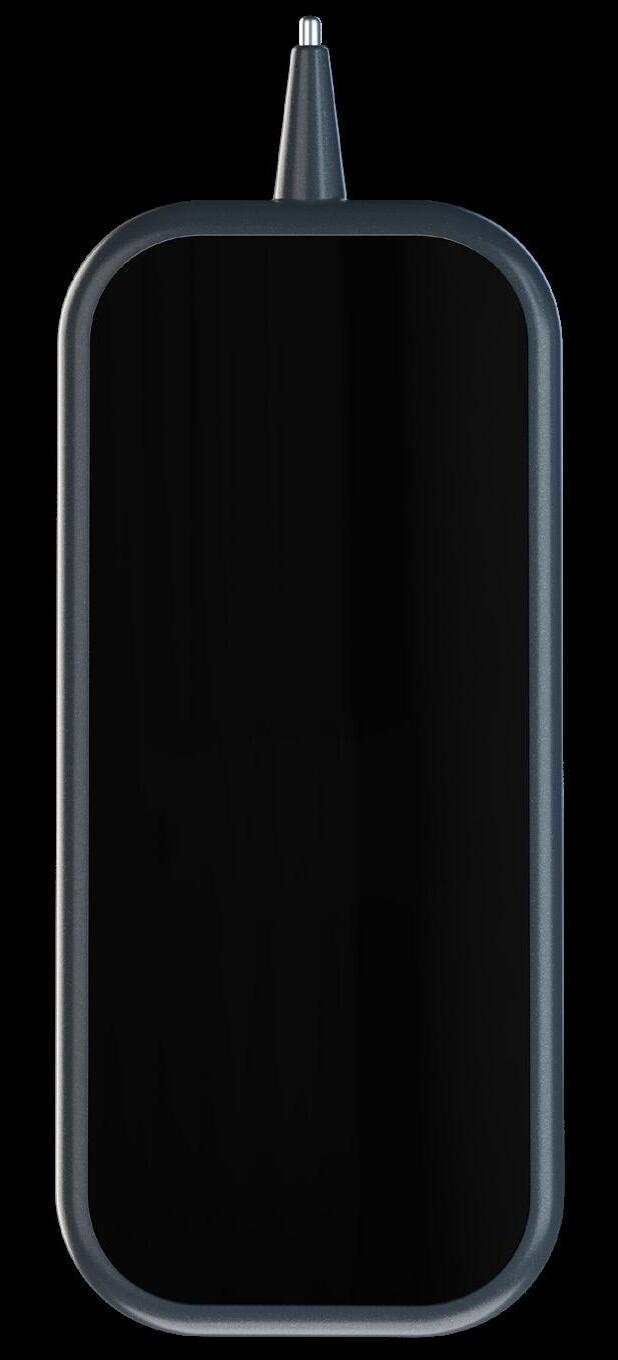
DermaSensor Inc. is a health technology company designing non-invasive tools to better equip primary care physicians for skin cancer detection. The DermaSensor device is an affordable, handheld device that uses spectroscopy and algorithms to evaluate skin lesions for potential cancer in a matter of seconds.
DermaSensor’s mission is to provide broad access to effective skin cancer checks. Key publications in support of DermaSensor are summarized in the pages to follow. Please contact info@dermasensor.com for additional information on clinical data for DermaSensor.
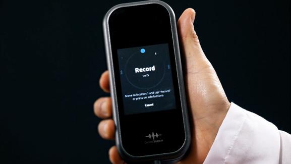
Rapid & Reliable Results
• Non-Invasive, point-and-click spectral recordings
• Immediate, objective results
Easy to Learn and Use
• Just 15 minutes of simple instructions on how to incorporate the device into your workflow
Clinically Effective
• 96% skin cancer detection sensitivity
• >2x as many cancers missed without device use
Cost Efficient
• No capital cost
• One subscription. No surprise fees
7
Device Label: Indications for Use, Contraindications, Warnings and Precautions
With regard to the Indications for Use, while the DermaSensor FDA pivotal validation study (DERM-SUCCESS included 1,579 lesions biopsied b y primary care physicians, and a supplemental melanoma validation study (DERM-ASSESS III) was conducted with biopsied lesions in the dermatology setting, note that the device indication for use is non-dermatologist physicians since the FDA’s clearance was based on a benefit-risk valuation limited to physicians who are not experts in the clinical diagnosis and management of skin cancer.
INDICATIONS FOR USE
The DermaSensor™ device is indicated for use to evaluate skin lesions suggestive of melanoma, basal cell carcinoma, and/or squamous cell carcinoma in patients aged 40 and above to assist in the decision regarding referral of the patient to a dermatologist. The DermaSensor device should be used in conjunction with the totality of clinically relevant information from the clinical assessment, including visual analysis of the lesion, by physicians who are not dermatologists. The device should be used on lesions already assessed as suspicious for skin cancer and not as a screening tool. The device should not be used as the sole diagnostic criterion nor to confirm clinical diagnosis of skin cancer.
CONTRAINDICATIONS
There are no known contraindications.
WARNINGS
• Do not use for the direct diagnosis of skin cancer.
• Always wear gloves during use (or examination).
• Do not use under direct focused light, such as a surgical or examination light or headlamp.
• Do not point the tip of the Handheld Unit directly at the eye.
• Do not attempt to disassemble, repair, or modify the DermaSensor device in any way.
• Do not immerse the DermaSensor device in liquid.
• Do not put the DermaSensor device in an autoclave or low temperature sterilizer.
• Do not buff or use an abrasive cream cleanser that may scratch and damage the tip of the Handheld Unit.
• Do not simultaneously make contact with the patient while touching or holding the Base or power adapter.
• Portable RF communications equipment (including peripherals such as antenna cables and external antennas) should be used no closer than 30cm (12 inches) to any part of the
• DermaSensor device, including cables specified by DermaSensor, Inc.; otherwise, degradation of the performance of this equipment could result.
8
WARNINGS (CONTINUED)
• DermaSensor performance has not been assessed for the following types of lesions; thus, safety and effectiveness have not been established when:
» Lesion is not accessible to the DermaSensor device Handheld Unit and tip (e.g., inside ears, under nails).
» Lesion is on areas of psoriasis, eczema, acne, or similar inflammatory skin conditions that may impede appropriate DermaSensor device tip placement on the lesion.
» Lesion is greater than 15mm in diameter at the widest point.
» Lesion has a targeted area less than 2.5mm in diameter where the DermaSensor device tip cannot be placed entirely within the border of the targeted area of the lesion.
» Lesion has no contiguous area of at least 2.5mm due to ulceration, erosion or liquid discharge (e.g., blood).
» Lesion is covered by a crust or scale, and lesion surface can not be cleared of crust or scale such that there is a contiguous area of at least 2.5mm of cleared intact skin that is free of any crust, ulceration, erosion or liquid discharge (e.g., blood).
» Lesion is obstructed by foreign matter that can not be non-invasively removed (e.g., tattoo, splinter).
» Lesion is not completely cleared of (i.e., free of any remaining residue) dermoscopy oils, makeup, sunscreen, other topical solutions or powders, markings, and staining treatments (e.g., iodine).
» Lesion is located on acral skin (e.g., sole or palms).
» Lesion is located within 10mm of the eye.
» Lesion is on or adjacent to scars, areas previously biopsied, or areas subjected to any past surgical intervention.
» Lesion is located on mucosal surfaces (e.g. genitals, lips).
» Lesion is located in an area with acute sunburn.
PRECAUTIONS
• Caution: United States Federal law restricts this device to sale by or on the order of a physician.
• A complete assessment, including
» the visual evaluation of the lesion,
» clinical considerations such as the patient’s ultraviolet light exposure history, and
» the patient and patient’s family’s skin cancer historyshould be considered, per standards of clinical care, in conjunction with DermaSensor results when making a formal clinical determination about a lesion.
• The performance of the device has not been specifically evaluated in patients with increased risk for skin cancer, e.g., inherited or drug-induced photosensitivity; genetic predisposition to melanoma or BCC; immune compromise; or other medical conditions that increase the risk of skin cancer or its metastasis.
• The device is intended to assist in clinical decisions related only to the skin malignancies melanoma (including severely atypical nevi), SCC, and BCC. It has been tested on each of these three common skin cancer types but has not been tested on rare skin cancer types; thus, it should not be used for lesions that are suggestive of malignancies other than melanoma, BCC and/or SCC.
• The device is intended for use on primary lesions only and has not been tested on lesions that are previously biopsied, recurrent, or metastatic; on scars, tattoos, sunburned skin, or within a hairy area (i.e., dense hair on the scalp); or which are located on palms, soles, mucosal surfaces, genitals, ears, within 1 cm of the eye, or under nails.
• Consistent with the lower prevalence of skin cancer in Fitzpatrick skin phototypes IV-VI, less data is available for sensitivity of the DermaSensor device for melanoma in these patients. The decision to refer patients with suspicious pigmented lesions in this group should be primarily based on clinical concern.
• Protect the tip of the Handheld Unit by storing the Handheld Unit in the Base when not in use.
• To maintain optimal device integrity, follow the cleaning and disinfection instructions before and after use.
• Use of accessories, transducers, and cables other than those specified or provided by the manufacturer of this equipment could result in increased electromagnetic emissions or decreased electromagnetic immunity of this equipment and result in improper operation.
• Use of this equipment adjacent to or stacked with other equipment should be avoided as it may result in improper operation. If such use is necessary, this equipment, and the other equipment, should be observed to verify that they are operating normally.
• Only use the included components and accessories provided as part of the DermaSensor medical equipment system.
• Connection to IT networks including other equipment could result in previously unidentified risks to patients, operators, or third parties. The user should identify, analyze, evaluate, and control these risks.
• Changes to the IT network (e.g., changes in network configuration, connection of additional items, disconnection of items, update of equipment, upgrade of equipment) could introduce new risks that require additional analysis.
• To avoid EMC disturbances, floors should be wood, concrete, or ceramic tile. If floors are covered with synthetic material, the relative humidity should be at least 30%.
9 https://support.dermasensor.com/labeling-guidance
Glossary of Terms
Adverse Event
Adverse Device E ffect
Area Under Cur ve
Basal Cell Carcinoma
Blinded Independent Dermatopathologist
Central Review Corrective and Preventive Action
Direct Data Captur e
Electr onic Case Repo r t F orm
Ethics Committee
Elastic Scattering Spectroscopy
F ood and Drug Administration
Good Clinical Practice
Informed Consent F orm
Institutional Re view Boar d
Instructions for Use
International Council on Harmonisation
International Committee of Medical Journal Editors
Melanocytic P athology Assessment T ool and Hierarchy for Diagnosis
Multi-Reader, Multi-Case
Multispectral Digital Skin Lesion Analysis
Near Infrar ed
Negativ e Predictiv e Value
Non-melanoma skin cancers
10
AE ADE AUC BCC DDC ESS IRB ICMJE MSDSLA NMSC BIDCR FDA IFU MPATH-DX NIR CAPA EC ICF ECRF GCP ICH MRMC NPV
Primary Care Physician
Principal Investigator
Personal Health Information
Positiv e Predictiv e Value
Serious Adv erse Event
Serious Adv erse Device E ffect
Squamous Cell Car cinoma
Treatment Emergent Adverse Event
Unanticipated Adverse Device Effect
Unanticipated Serious Adverse Device Effect
Ultraviolet
11
PPV TEAE PCP SAE UADE PHI SCC PI SADE USADE UV
Glossary of Terms (Continued)
Clinical Performance of Novel Elastic Scattering Spectroscopy (ESS) in Detection of Skin Cancer A Blinded, Prospective, Multi-Center Clinical Trial Initial Results
Stephen P Merry, MD1; Brian McCormick, MD2; David Leffell, MD3;Kiran Chatha, MD, MPH4; Ivana Croghan, PhD1Objective
This study investigated the sensitivity and specificity of a noninvasive, hand-held DermaSensor in evaluating skin lesions compared to the in-person clinical evaluation by primary care physicians (PCPs).
Trial Design

Link to publication : http://doi.org/10.1016/j. jdin.2023.10.011. Link to Publication
This blinded, prospective, multi-center study was conducted at 22 primary care study sites across the United States (18 sites) and Australia (4 sites). Patients with lesions suggestive of skin cancer were clinically assessed by PCPs and then evaluated by the DermaSensor. Patients and PCPs were blinded to device output. All lesions enrolled were biopsied per physician assessments and standard of care. Each lesion’s diagnosis involved 2-5 dermatopathologists, dependent on pathology and discordance. Statistical analyses after study unblinding included standard diagnostic test parameters of the device for detecting skin cancer as well as the influence of lesion and patient factors on device performance.
Enrollment
During study enrollment, five lesions (0.3%) were excluded due to device data capture issues and five lesions (0.3%) due to lack of dermatopathology consensus. A total of 1,005 patients with 1,579 lesions suggestive of skin cancer were enrolled. Among the patients enrolled, 51.4% were female with a mean age of 59 years, and 72.5% of patients were Fitzpatrick Skin Type I-III.
Conclusion
The novel hand-held DermaSensor demonstrated high sensitivity in detecting skin cancer when compared to the gold standard of histopathologic examination. Use of this device have potential to improve PCP sensitivity for skin cancer from 83% to 96% for high-risk lesions. Coupled with clinical exam findings, this device may aid PCPs to improve clinical decisions about suspicious skin lesions (i.e., to refer, or monitor), with this study suggesting device use could rule out 20.7% of suspicious lesions from needing further evaluation. This highly sensitive, noninvasive, hand-held DermaSensor may fill a well recognized void in PCP dermatologic care by providing an objective, point-of-care test for clinical assessment. This device may help increase quality of referrals to dermatology by providing PCPs with an additional instrument to assess lesions for skin cancer risk.
Key Findings
• DermaSensor demonstrated high sensitivity of 95.5% in detecting all skin cancer types when compared to the gold standard of histopathologic examination, with similar sensitivity and specificity across all Fitzpatrick skin type subgroups.
• NPV of the device was 96.6%, meaning a negative “monitor” device result had only a 3.4% chance of being a false negative, i.e. any of the three cancer types.
• The positive predictive value (PPV) for an “investigate further” result was 16.6% (NNB of 6:1).
• The device sensitivity and overall accuracy (i.e. AUC) were both found to be superior to those of the PCPs
• By pairing high sensitivity with clinical exam findings, the device may even rule out 21% of lesions from further evaluation, reducing patient anxiety and optimizing healthcare resource allocation
• Likelihood of malignancy increased with increasing spectral scores. For scores of 1-3, PPV was 6%, which increased to 18% for scores of 4-7 and 40% for scores of 8-10.
12
1 Mayo Clinic, Rochester, MD; 2 Hampton Family Practice, Hampton, VA; 3 Yale School of Medicine, New Haven, CT; 4 DermaSensor Inc.
Clinical Utility of an Elastic Scattering Spectroscopy Device in Assisting Primary Care Physicians Detection of Skin Cancers
Elizabeth V Seiverling, MD1; Thomas Agresta, MD, MBI2; Peggy Cyr, MD3; Laurie Caines, MD4; Vivien L Nguyen, PharmD6; Kiran Chatha, MD, MPH6; Daniel M Siegel, MD5
1 Tufts University School of Medicine, Boston, MA; 2 University of Connecticut School of Medicine, Farmington, CT; 3 Maine Medical Partners Family Medicine, Portland, ME; 4 UConn Health, Farmington, CT; 5 SUNY Downstate Health Sciences University, Brooklyn, NY; 6 DermaSensor, Inc., Miami, FL
Objective
The aim of this study was to assess and compare the diagnosis and management performance of primary care physicians (PCPs) with and without the use of the handheld DermaSensor in detecting skin cancer.

Link to publication : http://doi.org/10.1016/j. jdin.2023.10.011. Link to Publication
Key Findings
Trial Design
In this clinical utility study, 108 PCPs evaluated 50 skin lesions (25 malignant, 25 benign), with and without ESS device output. For each case, high-resolution digital clinical images, the patient’s clinical information, including prior skin cancer history, risk factors, and physical examination results were provided. The PCPs completed a questionnaire about their diagnosis of the lesion, their recommended management decision, and their confidence level in their management decision for each case.
Sensitivity and specificity of PCP diagnostic and management with and without the device output were calculated.
Enrollment Conclusion
PCP participants included U.S. board-certified. internal and family medicine physicians with an even distribution of years in practice (range: 1-21+ years). PCPs were recruited from across the U.S., including urban and rural areas.
PCPs had an improvement in sensitivity in detecting all skin cancer types with device availability, with management and diagnostic sensitivity increasing significantly. There were small, clinically insignificant decreases in device specificity related to diagnosis/referrals. Additionally, the effectiveness analyses observed an increase in PCPs’ overall diagnostic performance (i.e. AUC) and confidence level in their management decisions with the use of the handheld DermaSensor. The findings suggest the use of the DermaSensor output improves PCP skin cancer detection and confidence in skin lesion evaluation.
• Improvement in Sensitivity : Management sensitivity increased significantly from 82.0% to 91.4% (p=0.003) with device output. Diagnostic sensitivity increased significantly f om 71.1% to 81.7% with device output (p=0.008). Specificity deceased from 60.9% to 54.7% (p=0.190) for diagnosis and 44.2% to 32.4% (p=0.026) for referrals.
• Improvement in Physician Confidence: Physicians eporting high confidence in their assessments increased from 73.0% to 81.6% (<0.0001) with device output.
• Improvement in Area-Under-the-Curve: Overall PCP diagnostic performance (i.e. AUC) increased from 0.685 without device output to 0.727 with device output.
13
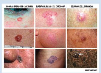
Clinical Capabilities of a Handheld Elastic Scattering SpectroscopyArtificial Intelligence Device as an Adjunctive Tool for Evaluating Skin Cancer in Skin of Color
Genevieve Patrick1, David Leffell, MD2, Stephen P Merry, MD3, Ivana Croghan, PhD3, Harold Rabinovitz, MD4, Armand Cognetta, MD51 Florida State University College of Medicine, 2 Yale University School of Medicine, 3 Mayo Clinic, 4 Medical College of Georgia, 5 Florida State University Department of Micrographic Surgery and Dermatology Oncology
Objective
To evaluate DermaSensor’s performance in patients with skin of color, this sub-analysis of the DERM-SUCCESS study compared device sensitivity and specificity in patients of Fitzpatrick skin type I-III and IV-VI subgroups.
Trial Design
Consistent with the lower prevalence of skin cancer in Fitzpatrick skin phototypes IV-VI, less data is available for sensitivity of the DermaSensor device for melanoma in these patients.
In total, 1,579 lesions from 1005 patients were enrolled in the study. All Fitzpatrick skin types were represented with 72.5% of individuals characterized as Fitzpatrick skin types I-III and IV-VI.
Lesions suggestive of skin cancer were clinically assessed by primary care physicians (PCPs) and scanned by the DermaSensor device.
Limitations of this study involve the unknown impact of the device on the clinical decision to biopsy lesions in skin of color as the study was designed to be double-blinded. Additionally, the sample size for various malignant pathologies across all skin types as well as the racial and ethnic diversity of the study population were limited.
Key Findings
Double-blinded, prospective, multi-center study; 22 primary care study sites across the, U.S. (18 sites) and Australia (4 sites)
Enrollment Conclusion
The DermaSensor demonstrated a high sensitivity of 95.5% in detecting all skin cancer types when compared to histopathologic examination. Additionally, the device correctly classified 20.7% of biopsied benign lesions.
Overall sensitivity was similar between Fitzpatrick skin types I-III and IV-VI, with 96.5% (95% CI: 92.1-98.5%) and 92.2% (95% CI: 82.1-96.8%), respectively. Sensitivity was consistent across Fitzpatrick skin types.
There was little variation in device sensitivity or specificity when comparing patients based on Fitzpatrick skin type subgroups. This is in line with ESS functionality being unaffected by the underlying melanin content of the skin.
Unlike image-based tools which are subject to skin-type bias, the DermaSensor has the potential to augment PCPs exams and improve skin cancer detection capabilities across skin types.
• Unlike image-based tools such as Visual D X which are subject to skin-type bias, skin color does not impact DermaSensor results which has the potential to augment PCPs exams and improve skin cancer detection capabilities across skin types.
• Overall sensitivity was similar between Fitzpatrick skin types I-III and IV -VI, with 96.5% (95% CI: 92.1-98.5%) and 92.2% (95% CI: 82.1-96.8%), respectively. Sensitivity was consistent across Fitzpatrick skin types.
• The specificity of the DermaSensor for Fitzpatrick skin types I-III was 18.7% (95% CI: 16.2-21.5%) and 25.1% (95% CI 20.9-29.7%) for types IV-VI. The device has the potential to rule out 18.7% to 25.1% of biopsies and/or referrals of lesions that were benign.
14
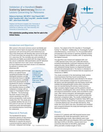
DERM-ASSESS III: Validation of a Handheld Elastic-Scattering Spectroscopy Device on Lesions Suggestive of Melanoma
1 Brigham and Women’s Hospital, USA; 2 University of Leipzig; Germany 3 North Florida Dermatology, USA; 4 University of California, USA; 5 Yale University, USA 6 University of Connecticut, USA
Objective
This study aimed to validate whether the use of an ESS point-of-care test can detect melanoma when dermatologists are evaluating lesions that were concerning for melanoma.
Trial Design

Link to publication : http://doi.org/10.1016/j. jdin.2023.10.011. Link to Publication
Key Findings
Ten dermatology study centers, across the US and Australia, scanned lesions that they found concerning for melanoma. All dermatologists were blinded to the device results. Gold standard comparison for performance of the device and dermatologists was the biopsy result with multiple dermatopathologist review when consensus was not reached during the primary review process. High resolution digital images and the patient’s clinical information, including prior skin cancer history, risk factors and physical exam results, were recorded for each case. After clinical evaluation, dermatologists reported their diagnosis and confidence level, which provided the physician comparison data. The results evaluated were sensitivity, specificity, Negative Predictive Value for melanoma, melanoma + severely atypical nevi, and all high-risk lesions. Area Under the Curve (AUC) was also calculated and modeled and compared between the study dermatologists and the device performance.
Enrollment Conclusion
A total of 311 patients with 440 biopsied lesions were evaluated by the study dermatologists, device and dermatopathology results.
The use of the handheld DermaSensor by physicians, in addition to clinical evaluation, may improve melanoma detection. The device was able to identify 96% of melanomas when compared to dermatopathology results.
While this study was conducted by melanoma specialists, given the device’s similar overall performance to these specialists and its simple, non-invasive use, there is potential for the device to be used to help rule in or out lesion referrals for primary care physicians.
• The 10 dermatology study centers biopsied all lesions that were suspicious of melanoma based on their standard of care clinical and dermoscopic assessment and decision making.
• DermaSensor was able to identify 96% of melanomas when compared to dermatopathology consensus results (at least two dermatopathologists reviewed each high-risk melanocytic lesion case); for both melanomas and several atypical nevi, the device sensitivity was 90.9%.
• Overall device specificity was 33%, thus DermaSensor could have effectively ruled out 33% of lesions as benign while detecting 96% of melanomas.
• The overall negative predictive value (NPV) for a “monitor” result was 98.1% for melanoma, i.e. a negative result had less than a 2% chance of being melanoma. The positive predictive value (PPV), i.e. the likelihood a lesion was melanoma for a positive “investigate further” result, was 10.3% for low 1-3 scores (NNB of 10:1), 20.5% for mid 4-7 scores (NNB of 5:1), and 47.4% for high 8-10 scores (NNB of 2.1:1).
• The overall device accuracy (i.e. Area Under the Curve or AUC) of 0.76 was comparable to that of 0.75 for the Dermatologists.
15
Rebecca Hartman, MD MPH1; Uwe Paasch MD2;Kelly Tepedino MD3, Max Fung MD4, Jennifer McNiff MD5, Jane Grant-Kels MD6
Skin Lesion Analyzers: Economic Impact of Integrating These Technologies into Dermatology Clinical Practice for Detecting Melanoma
1 SUNY Downstate Health Sciences University, Brooklyn, New York, USA 2 TTi Health Research & Economics, Inc., Westminster, Maryland, USA 3 DermaSensor, Inc., Miami, Florida, USA 4 Medical College of Georgia, Augusta, Georgia, USA 5 Northwell Health, Lake Success, New York, USA
ESS, Elastic Scattering Spectroscopy; EIS, Electrical Impedance Spectroscopy
ESS, Elastic Scattering Spectroscopy; EIS, Electrical Impedance Spectroscopy; M, millions; USD, United States Dollars
To assess the economic value of skin lesion analyzers for the evaluation of lesions suspicious for melanoma from the United States commercial payer perspective.
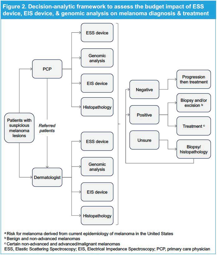
Objective Methods
Model structure
• A decision-analytic model was developed to estimate the total cost of analyzing skin lesions (Figure 2)
• Three technologies that provide an objective melanoma risk result—ESS, EIS, and genomic analysis—were compared with histopathology (standard of care)
• Patients presenting skin lesions to PCPs and dermatologists for examination of lesions suspicious for melanoma were included
Assumptions
• All patients start in the model with a suspicious melanoma lesion, which is retrospectively evaluated
• Test performance is independent of clinical practice (Table 1)
• Diagnostic technologies are used on the patients without evaluating the cost for every lesion on the patient
Outcomes
• Clinical outcomes for diagnostics were evaluated
• Total budget impact, per member per month (PMPM) and per patient per month (PPPM) costs were evaluated over 3 years
One-way sensitivity analysis
• Evaluated the influence of variability in model estimates
ESS device [5] 95.5% 32.5% EIS device [6] 95.9% 31.3% Genomic analysis [7] 91.0% 69.0% Histopathology [8] 92.0% 90.8%
Cumulative budget
years
in 2023 USD)
device 145.8M EIS device 146.5M Genomic analysis 160.8M Histopathology 169.3M
over 3
(millions
ESS
16
Daniel M. Siegel MD,1 Avijeet S Chopra, PhD2 Kiran Chatha MD MPH,3 Harold Rabinovitz MD,4 Alina Bridges, DO5
Technology Technology Sensitivity Specificity
Table 1. Technology Performance Estimates
Table 2. Total budgetary impact
Skin Lesion Analyzers: Economic Impact of Integrating These Technologies into Dermatology Clinical Practice for Detecting Melanoma (Cont.)
Daniel M. Siegel MD,1 Avijeet S Chopra, PhD2 Kiran Chatha MD MPH,3 Harold Rabinovitz MD,4 Alina Bridges, DO51 SUNY Downstate Health Sciences University, Brooklyn, New York, USA 2 TTi Health Research & Economics, Inc., Westminster, Maryland, USA
3 DermaSensor, Inc., Miami, Florida, USA 4 Medical College of Georgia, Augusta, Georgia, USA 5 Northwell Health, Lake Success, New York, USA
Results
Disease Burden
• An estimated 15,882 patients were evaluated based on the estimates of the 2019 National Ambulatory Medical Care Survey
» A total of 5,575 and 10,291 patients were evaluated by primary care physician (PCP) and dermatologists, respectively
• Based on the epidemiology of melanoma, the model estimated that approximately 673,600 examinations were ordered by PCPs and dermatologists for the evaluation of melanoma skin lesions
» 64% of the examinations were conducted by dermatologists
Clinical Outcomes
• The use of the ESS device in clinical practice is estimated to be lower because of the novelty of the technology
• About 80% of surgically assessed lesions clinically suspicious for melanoma were diagnosed as benign, with 3.9% of the clinically suspicious benign lesions undergoing a more advanced procedure (e.g., excision), either initially or following a biopsy as part of their care.
Economic Outcomes
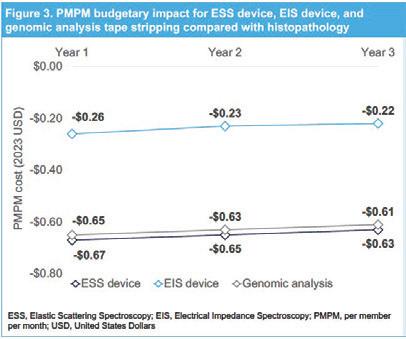
• The use of all three technologies resulted in net savings over 3 years compared with the standard of care, with the highest savings estimated with the ESS device (Figure 3)
• Total budget savings with the ESS device were$8.0 million in the first year, with a cumulative savings of $23.5 million over 3 years compared with histopathology (Table 2)
• Cumulative PMPM savings were $1.95 over 3 years with ESS device, compared with $1.89 for the EIS device and $0.70 with genomic analysis
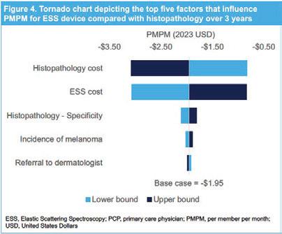
One-way sensitivity analysis
• Results from the sensitivity analyses suggest greater dependence of the budget impact on cost of diagnostic techniques in preference to test performance, disease epidemiology, and direct healthcare resource utilization (Figure 4)
Key Findings
• Elastic Scattering Spectroscopy (ESS) resulted in the highest budget savings compared with the use of histopathology (standard of care) from the US payer perspective over 3 years.
• Availability of ESS has the potential to improve patient care by minimizing avoidable surgical procedures on benign lesions, improving melanoma management (i.e., reducing number needed-to-biopsy) and decreasing the downstream impacts of late-stage melanoma diagnoses.
• Considering that ESS device is non-invasive, easy-to-use, and provides realtime risk results, it has the potential for significant clinical adoption, resulting in considerable cost savings for payers.
• Budget savings with ESS, combined with better clinical outcomes vs standard of care, demonstrate that ESS may be a better diagnostic technique for the evaluation of lesions suspicious for melanoma, as well as potentially keratinocyte carcinomas.
17
Summary of Early Clinical Effectiveness Studies
Summary of Early Clinical Effectiveness Studies
Clinical Study Name Design, Setting and Sample Size
DermaSensor Use in the Assessment of Skin Lesions Suggestive of Cancer (DERM-ASSESS II)
Use of an ESS-based Adjunctive Tool in the Assessment of Lesions Suggestive of Skin Cancer (Skin Center Investigator-initiated Study (IIS))
Use of DermaSensor on Patient-Selected Lesions that are Concerning for Skin Cancer (PATIENT-SELECT)
A Multi-Reader Multi-Case Companion Study to the DERM-ASSESS II Clinical Study (DA-II Reader Study)
US-based, multicenter, prospective, blinded clinical study at four dermatology sites in the US
333 lesions (169 malignant, 164 benign)
Single-site, prospective, blinded clinical study at one dermatology site in New Zealand
410 lesions (50 malignant, 360 benign)
US-based prospective, blinded clinical study at one primary care site in the US
155 lesions (10 malignant, 145 benign)
US-based prospective multi-reader, multi-case study; 57 primary case physicians (PCPs) completed 5,700 lesion assessments
To train the device algorithm and validate clinical performance for melanoma, BCC, and SCC
Device sensitivity of 97.0% (95% Cl: 92.4-98.9%) and specificity of 26.2% (95% Cl: 20.3-33.2%)
To evaluate DermaSensor device performance as an adjunctive tool for evaluating patients with skin lesions suggestive of melanoma, BCC, and SCC
To assess device performance on skin lesions identified by patients as concerning for skin cancer
To assess and compare the management sensitivity and specificity of primary care physicians for the most common types of skin cancer (Melanoma, BCC, SCC), with and without use of the device result
Device sensitivity of 96.0% (95% Cl: 86.3-99.5%) and specificity of 31.4% (95% Cl: 26.6-36.5%)
Device sensitivity of 90.0% (95% Cl: 71.4-100.0%) and specificity of 60.7% (95% Cl: 52.5-68.4%)
PCP sensitivity with device result of 94.2% (95% CI: 91-96%) was superior to PCP sensitivity without device result of 81.4% (95% CI: 77-85%) (p=0.0009).
Management specificity with (31%) and without (36%) device use was not siginificantly different (P = 0.3558)
18
Objective Effectiveness Results
Health System Management Guidelines: Aligning Patient Management Decisions with Malignancy Risk
DERM-SUCCESS FDA Pivotal Study Results
Investigate Further 1-3 6% 17 37% Investigate Further 4-7 18% 5.5 29% Investigate Further 8-10 40% 2.5 16% 19
Device Result Monitor Likelihood of Malignancy Number Needed To Refer Result Frequency Health System’s Management Guidelines For Its Clinicians 3% 29 18%
Title, Authors, Journal
Artificial Intelligence in Medicine
Title: Mapping the landscape of artifical intelligence in skin cancer research: a biblio-metric analysis
Authors: Qianwei Liu, Jie Zhang, and Yanping Bai
Journal: Frontiers in Oncology
Liu Q, Zhang J, Bai Y. Mapping the landscape of artific al intelli-gence in skin cancer research: a bibliometric analysis. Front Oncol. 2023 Oct 13;13:1222426. doi: 10.3389/fonc.2023.1222426. PMID: 37901316; PMCID: PMC10613074.
Title: Artifical Intelligence in the detection of skin cancer
Authors: Eric Beltrami, Alistair Brown, Paul Salmon, David J Leffell, Justin Ko, Jane Grant-Kels
Journal: Journal of the American Academy of Dermatologists
Beltrami EJ, Brown AC, Salmon PJM, Leffell DJ, Ko JM, Grant-Kels JM. Artific al intelligence in the detection of skin cancer. J Am Acad Dermatol. 2022 Dec;87(6):1336-1342. doi: 10.1016/j. jaad.2022.08.028. Epub 2022 Aug 23. PMID: 35998842.
Key Findings
Publications related to AI date back to 1990, and AI in relation to skin cancer research to 1991. From 1991-2023, there have been 512 publications on the topic of AI in skin cancer.
To date, there are no FDA approved imaging or optical products on the market that use AI for the purpose of skin cancer detection other than DermaSensor’s device, despite there being over three decades of research.
According to the global cancer statistics for 2020, skin cancer accounted for 1,198,073 new cases and environmental factors will contribute to the increase in skin cancer worldwide.
Recent advances in artific al intelligence (AI) in dermatology have demonstrated the potential to improve the accuracy of skin cancer detection.
Ultimately, the development and validation of AI technologies, their approval by regulatory agencies, and widespread adoption by dermatologists and other clinicians may enhance pa-tient care. Technology-augmented detection of skin cancer has the potential to improve quality of life, reduce health care costs by reducing unnecessary procedures, and promote greater access to high-quality skin assessment.
*NOTE: No FDA approved AI based devices or tools are in use by dermatologists currently, DermaSensor is the first approved AI based device for PCP use.
20
--
Title,
Findings
Title: Comparative Analysis of Diagnostic Techniques for Melanoma Detection: A Systematic Review of Diagnostic Test Accuracy Studies and Meta-Analysis
Authors: Alessia Blundo, Arianna Cignoni, Tommaso Banfi and Gastone Ciut Journal: Frontiers in Medicine
Blundo A, Cignoni A, Banfi T, Ciuti G. Comparative Analysis of Diagnostic Techniques for Melanoma Detection: A Systematic Review of Diagnostic Test Accuracy Studies and Meta-Analysis. Front Med (Lausanne). 2021 Apr 21;8:637069. doi: 10.3389/fmed.2021.637069. PMID: 33968951; PMCID: PMC8103840.
Title: Dermoscopy, with and without visual inspection, for diagnosing melanoma in adults
Authors: Dinnes et al
Journal: Cochrane Review
Dinnes J, Deeks JJ, Chuchu N, Ferrante di Ruffano L, Matin RN, Thomson DR, Wong KY, Aldridge RB, Abbott R, Fawzy M, Bayliss SE, Grainge MJ, Takwoingi Y, Davenport C, Godfrey K, Walter FM, Williams HC; Cochrane Skin Cancer Diagnostic Test Accuracy Group. Dermoscopy, with and without visual inspection, for diagnosing melanoma in adults. Cochrane Database Syst Rev. 2018 Dec 4;12(12):CD011902. doi: 10.1002/14651858.CD011902.pub2. PMID: 30521682; PMCID: PMC6517096.
Title: A comparison of dermatologists’ and primary care physicians’ accuracy in diagnosing melanoma: a systematic review
Authors: Suephy Chen, Dena Bravata, Evette Weil, Ingram Olkin
Journal: JAMA Dermatology
Chen SC, Bravata DM, Weil E, Olkin I. A comparison of dermatologists’ and primary care physicians’ accuracy in diagnosing melanoma: a systematic review. Arch Dermatol. 2001 Dec;137(12):1627-34. doi: 10.1001/archderm.137.12.1627. PMID: 11735713.
A systematic review of the available literature was performed using PubMed, Scopus and Google scholar databases (2010-September 2020). All human, in-vivo, non-invasive studies using techniques, alternative to dermoscopy, for melanoma diagnosis were included with no restriction on the recruited population.
Based on the SROC curves, optical spectroscopy achieved the best performance in terms of sensitivity (93%, 95% CI 92.8-93.2%) and specificity (85.2%, 95%CI 84.9-85.5%), even though there was high concern regarding robustness of metrics. Reflectance-confocal microscopy, instead, demonstrated higher robustness and a good diagnostic performance (sensitivity 88.2%, 80.3-93.1%; specificity 65.2%, 55-74.2%)
Central message: Optical spectroscopy performs superior to visual examination alone and similar to confocal microscopy which takes years to learn and costs over $100,000.
Dermoscopy is a valuable tool to support the visual inspection of a suspicious skin lesion for the detection of melanoma and atypical intraepidermal melanocytic variants, particularly in referred populations and in the hands of experienced users. Data to support its use in primary care are limited, however, it may assist in triaging suspicious lesions for urgent referral when employed by suitably trained clinicians.
For diagnostic accuracy, sensitivity was 81- 100% for dermatologists and 42-100% for PCPs.
For biopsy or referral accuracy, sensitivity ranged from 82 -100% for dermatologists and 70 to 88% for PCPs
Key
21
Authors, Journal
1. Merry SP, Croghan I, McCormick B, Chatha K, Leffell D. Clinical Performance of Novel Elastic Scattering Spectroscopy (ESS) in Detection of Skin Cancer: A Blinded, Prospective, Multi-Center Clinical Trial [Initial Results]. Cutis 2022 December; 110(6 Suppl):31
2. Manolakos D, Patrick G, Geisse JK, Rabinovitz H, Buchanan K, Hoang P, Rodriguez-Diaz E, Bigio IJ, Cognetta AB, Use of an Elastic-Scattering Spectroscopy and Artificial Intelligence Device in the Assessment of Lesions Suggestive of Skin Cancer: A Comparative Effectiveness Study, JAAD International (2023), doi: https://doi.org/10.1016/j.jdin.2023.08.019.
3. Hartman RI, Trepanowski N, Chang MS, Tepedino K, Gianacas C, McNiff JM, Fung M, Braghiroli NF, Grant-Kels JM, Multicenter Prospective Blinded Melanoma Detection Study with a Handheld Elastic Scattering Spectroscopy Device, JAAD International (2023), doi: https:// doi.org/10.1016/j.jdin.2023.10.011.
4. Hartman R, Paasch U, Tepedino K, Fung M, McNiff J, Grant-Kels J. DERM-ASSESS III: Validation of a Handheld Elastic Scattering Spectroscopy Device on Lesions Suggestive of Melanoma. Poster Presentation, German Skin Cancer Congress, Germany. Aug 2022.
5. Jaklitsch E, Thames T, de Campos Silva T, Coll P, Oliviero M, Ferris LK. Clinical Utility of an AI-powered, Handheld Elastic Scattering Spectroscopy Device on the Diagnosis and Management of Skin Cancer by Primary Care Physicians. J Prim Care Community Health. 2023 Jan-Dec;14:21501319231205979. doi: 10.1177/21501319231205979. PMID: 37933569; PMCID: PMC10631325.
6. Seiverling EV, Agresta T, Cyr P, Caines L, Nguyen VL, Chatha K, Siegel DM. Clinical Utility of an Elastic Scattering Spectroscopy Device in Assisting Primary Care Physician’s Detection of Skin Cancers.J Clin Aesthet Dermatol 2023 April: 16(4 Suppl): s16-17.
7. Patrick G, Leffell D, Merry SP, Croghan I, Rabinovitz H, Cognetta A. Clinical Capabilities of a Handheld Elastic Scattering Spectroscopy-Artificial Intelligence Device as an Adjunctive Tool for Evaluating Skin Cancer in Skin of Color. Podium Presentation, American Academy of Dermatologist’s Annual Meeting, Tampa Bay, FL, Aug 10-13th 2023.
8. Tepedino M, Baltazar D, Hucks C, Chatha K, Zeitouni N. Use of Elastic Scattering Spectroscopy on Patient Selected Lesions that are Concerning for Skin Cancer. Cutis 2022 December; 110(6 Suppl):35-36.
9. Siegel DM, Chopra AS, Chatha K, Rabinovitz H, Bridges A. Skin Lesion Analyzers: Economic Impact of Integrating These Technologies into Dermatology Clinical Practice for Detecting Melanoma. Poster Presentation, Maui Derm Annual Conference, Maui, HI, Jan 22-26th, 2024.
10. Liu Q, Zhang J, Bai Y. Mapping the landscape of artificial intelligence in skin cancer research: a bibliometric analysis. Front Oncol. 2023 Oct 13;13:1222426. doi: 10.3389/ fonc.2023.1222426. PMID: 37901316; PMCID: PMC10613074.
11. Brancaccio G, Balato A, Malvehy J, Puig S, Argenziano G, Kittler H. Artificial Intelligence in Skin Cancer Diagnosis: A Reality Check. J Invest Dermatol. 2023 Nov 18:S0022-202X(23)02964-0. doi: 10.1016/j. jid.2023.10.004. Epub ahead of print. PMID: 37978982.
12. Beltrami EJ, Brown AC, Salmon PJM, Leffell DJ, Ko JM, Grant-Kels JM. Artificial intelligence in the detection of skin cancer. J Am Acad Dermatol. 2022 Dec;87(6):1336-1342. doi: 10.1016/j.jaad.2022.08.028. Epub 2022 Aug 23. PMID: 35998842.
13. Blundo A, Cignoni A, Banfi T, Ciuti G. Comparative Analysis of Diagnostic Techniques for Melanoma Detection: A Systematic Review of Diagnostic Test Accuracy Studies and Meta-Analysis. Front Med (Lausanne). 2021 Apr 21;8:637069. doi: 10.3389/fmed.2021.637069. PMID: 33968951; PMCID: PMC8103840.
14. Dinnes J, Deeks JJ, Chuchu N, Ferrante di Ruffano L, Matin RN, Thomson DR, Wong KY, Aldridge RB, Abbott R, Fawzy M, Bayliss SE, Grainge MJ, Takwoingi Y, Davenport C, Godfrey K, Walter FM, Williams HC; Cochrane Skin Cancer Diagnostic Test Accuracy Group. Dermoscopy, with and without visual inspection, for diagnosing melanoma in adults. Cochrane Database Syst Rev. 2018 Dec 4;12(12):CD011902. doi: 10.1002/14651858.CD011902.pub2. PMID: 30521682; PMCID: PMC6517096.
15. Chen SC, Bravata DM, Weil E, Olkin I. A comparison of dermatologists’ and primary care physicians’ accuracy in diagnosing melanoma: a systematic review. Arch Dermatol. 2001 Dec;137(12):1627-34. doi: 10.1001/archderm.137.12.1627. PMID: 11735713.
22 References
Published April 2024 Contact 1.855.373.6767 DermaSensor.com DermaSensor@DermaSensor.com 80 SW 8th Street #2000 Miami, FL 33130 80-0081.1