C&T
Control & Therapy Series
Centre for Veterinary Education


Issue 314 | March 2024
15
Major Winner—Venous Air Embolism
Adam Gordon
Wound Management in a Pet Goat
36
Cautionary Tails
3
Canine Gastric Carcinoma
43


brand for your business online VETPLUS can help create a powerful Vetplus provides a program to build a digital brand presence that perfectly aligns with your in-clinic brand. Featuring Australia’s #1 veterinary website creator and customisable mobile apps, Vetplus makes it easy to deliver an integrated digital brand experience for your clients. To find out more, scan the QR code or talk to your Boehringer Ingelheim Territory Manager. Boehringer Ingelheim Animal Health Australia Pty. Ltd. Level 1, 78 Waterloo Road, North Ryde, NSW 2113. Toll Free 1800 808 691. Vetplus ® is a registered trademark of Boehringer Ingelheim Animal Health Australia Pty Ltd. Protech ® Duramune ® and Fel-O-Vax ® are registered trademarks of Boehringer Vetmedica Inc. All rights reserved. BI1716TA-VAC-04/23. Proudly supported by Australia’s leading canine and feline vaccine range: Protech ® Duramune ® and Fel-O-Vax ® ... we reinforce our brand advantages both in-clinic and online’ ‘ - Rebecca Porter Marketing Manager, Manly Road 24Hr Veterinary Hospital ADVERTISEMENT
C&T
Issue 314 | March 2024
Control & Therapy Series
PUBLISHER
Centre for Veterinary Education
Veterinary Science Conference Centre Regimental Drive
The University of Sydney NSW 2006 + 61 2 9351 7979
cve.publications@sydney.edu.au cve.edu.au
Print Post Approval No. 10005007
DIRECTOR
Dr Simone Maher
EDITOR
Lis Churchward elisabeth.churchward@sydney.edu.au
EDITORIAL ASSISTANT
Dr Jo Krockenberger joanne.krockenberger@sydney.edu.au
VETERINARY EDITOR
Dr Richard Malik
DESIGNER
ADVERTISING
Lis Churchward elisabeth.churchward@sydney.edu.au
To integrate your brand with C&T in print and digital and to discuss new business opportunities, please contact:
MARKETING AND
DISCLAIMER
All content made available in the Control & Therapy (including articles and videos) may be used by readers (You or Your) for educational purposes only.
Knowledge and best practice in this field are constantly changing. As new research and experience broadens our knowledge, changes in practice, treatment and drug therapy may become necessary or appropriate. You are advised to check the most current information provided (1) on procedures featured or (2) by the manufacturer of each product to be administered, to verify the recommended dose or formula, the method and duration of administration, and contraindications.
To the extent permitted by law You acknowledge and agree that:
I. Except for any non-excludable obligations, We give no warranty (express or implied) or guarantee that the content is current, or fit for any use whatsoever. All such information, services and materials are provided ‘as is’ and ‘as available’ without warranty of any kind.
II. All conditions, warranties, guarantees, rights, remedies, liabilities or other terms that may be implied or conferred by statute, custom or the general law that impose any liability or obligation on the University (We) in relation to the educational services We provide to You are expressly excluded; and
III. We have no liability to You or anyone else (including in negligence) for any type of loss, however incurred, in connection with Your use or reliance on the content, including (without limitation) loss of profits, loss of revenue, loss of goodwill, loss of customers, loss of or damage to reputation, loss of capital, downtime costs, loss under or in relation to any other contract, loss of data, loss of use of data or any direct, indirect, economic, special or consequential loss, harm, damage, cost or expense (including legal fees).
Mirgheshmi
Samin
SALES MANAGER Ines Borovic ines.borovic@sydney.edu.au Best Visuals Winner Winners Winners Major Winner Engage With Your Profession ................................................................... 2 From the Director 2 What's Your Diagnosis? ansWers To C&T No. 6004 Afterhours Dystocia Case in a Multiparous Beef Cow Robert Mills Answer 1 Ross Sillar Answer 2 Andrew Bissett 7 To C&T No. 5993 Cutaneous Cryptococcosis in a Cat Natalie Courtman & Beth McDonald Comment courtesy of Mark Krockenberger Answer 1 Sharnee Lehrmayer Answer 2 Robert Bird ... ............................. ............................................9 Q uestion C&T No. 6007 Natalie Courtman 13 small animal Cautionary Tails , Learning From Mistakes Terry King .................................................................................................... 3 Fatal Venous Air Embolism During Thoracic Limb Amputation in a Dog Adam Gordon 15 Pathology in Practice: Canine Right Atrial Haemangiosarcoma with Widespread Pulmonary Metastases Alexander Teh 18 Abstract: Durable Contraception in the Female Domestic Cat Using Viral-Vectored Delivery of a Feline Anti-Müllerian Hormone Transgene by Lindsey M. Vansandt et al Commentary by Fiona Hollinshead & Alan Conley 20 Feline Orofacial Pain Syndrome Clare Rusbridge ISFM Research Roundup ......................................................................... 23 Feline Arterial Thromboembolism in a Cat Christopher Simpson 28 Save Your Dog’s Hock! Tips for Trainers John Katakasi, David Larratt & Peter Yore ........................................31 Burkholderia Infection in a Cat (Likely Meliodosis) Candice Yeo Comment courtesy of Mark Krockenberger 32 l arge animal Management & Healing of a Large, Necrotic Dog Attack Wound in a Pet Goat Curtis A. Goding 36 general Interested in Diabetic Research? Linda Fleeman 12 Wild Horse Management in Kosciuszko National Park ...................... 22 Aeromonas hydrophila a Likely Causative Agent of Segmental Ulcerative Colitis in a Human Recipient Peter Kerkenezov ....................................................................................34 Perplexity AI Editor's Note .................................................................. 35 PersPective Canine Gastric Carcinoma Ed Hall 43
Engage With Your Profession
The unique C&T Series was established in 1969 by our first Director Dr Tom Hungerford OBE BVSc FACVSc HAD who wanted a forum for uncensored and unedited material, to get the clinicians writing:
"not the academic correctitudes, not the theoretical niceties, not the super correct platitudes that have passed the panel of review… not what he/she should have done, BUT WHAT HE/SHE DID, right or wrong, the full detail, revealing the actual ‘blood and dung and guts’ of real practice as it happened, when tired, at night, in the rain in the paddock, poor lighting, no other vet to help.
The C&T forum gives a ‘voice’ to the profession and everyone interested in animal welfare. You don’t have to be a CVE Member to contribute an article—please send your submissions to Dr Jo Krockenberger. joanne.krockenberger@sydney.edu.au
Join In!
The C&T is not a peer-reviewed journal.
We are keen on publishing short, pithy, practical articles (a simple paragraph is fine) that our readers can immediately relate to and utilise. Our editors will assist with English and grammar as required.
"I enjoy reading the C&T more than any other veterinary publication.
-Terry King, Veterinary Specialist Services, QLD
Thank You to All Contributors
The C&T Series thrives due to your generosity.
Major Winner Prize: A CVE$400 voucher
Fatal Venous Air Embolism
Winners Prize: A CVE$100 voucher
Feline Arterial Thromboembolism in a Cat Christopher Simpson ................................................................
Answer to What's Your Diagnosis?
Afterhours Dystocia Case in a Multiparous Beef Cow
Answer 1 Ross Sillar
Answer 2 Andrew Bissett ........................................................ 7
Cutaneous Cryptococcosis in a Cat
Answer 1 Sharnee Lehrmayer
Answer 2 Robert Bird ............................................9
Best Visuals Prize: A CVE$100 voucher
Management & Healing of a Large, Necrotic Dog Attack Wound in a Pet Goat Curtis A. Goding ...............................36
Visit cve.edu.au for Membership & CE Course info
From the Director

We all know that in veterinary practice, things don’t always go to plan. Sometimes that’s due to variation in individual responses or unforeseen complications. Sometimes (the worst times) it’s due to a momentary lapse in judgement, or a mistake made under time pressure, or choosing a path that with the benefit of hindsight turned out not to be the right one.
One of my very worst days as a fairly new grad was losing a patient post op due to hypothermia. Looking back, I can see that everything was heading in that direction—I was probably too inexperienced to do this particular surgery, I performed it too late in the day (making me more tired and slow), and there were too few staff around to properly monitor during recovery. I was absolutely devastated.
The gutting sorrow of losing a patient was matched by my shame that had I been a ‘better vet’ this wouldn’t have happened, and that forevermore I would be labelled a ‘bad vet’. This fear prevented me from sharing my experience widely until many years later—when really, being open about my experience would probably have been a valuable lesson for others.
So a huge thank you to those in this edition who have openly shared stories of things that have not gone to plan for the benefit of other clinicians and their patients. I know from personal experience it takes a measure of bravery and confidence to do this—but hopefully leads to better outcomes in the long run.
Some fantastic submissions and images this edition – one of my favourites has to be Lenny the goat’s story. There’s no way you can avoid smiling back at him in the last image.

Happy reading!
Simone
Amputation in a Dog Adam Gordon 15
During Thoracic Limb
28
Control & Therapy Series – Issue 314 March 2024 Page 2 Authors’ views are not necessarily those of the CVE
Cautionary Tails
"To err is to be human,” wrote Alexander Pope. “Success is not final, failure is not fatal: It is the courage to continue that counts,” Churchill proclaimed. An African proverb announces that “Only those who do nothing never make mistakes".
—Brookings
Learning From Mistakes...
Terry King BVSc MANZCVS
Emergency & Critical Care Internal Medicine
Specialist Veterinary Services - Queensland Underwood | Carrara | Jindalee
To learn more about Terry’s distinguished career visit: vss.net.au/dr-terry-king.html
C&T No. 6005
1. Spaying a male cat
My only excuse was that the kitten’s name was ‘Mary.’ An embarrassing admission to the owners when they came to pick up their kitty (although they firmly believed ‘he’ was a ‘she.’)
When represented for suture removal from the celiotomy wound, ‘Mary’ had been renamed ‘Martin.’ Preventative measures to prevent this mistake from recurring are obvious.
2. ACP versus Ancylol
Giving a 3.5kg hookworm anaemic puppy 1 mL of equine-strength (10mg/mL) Acetylpromazine SQ instead of Ancylol (Disophenol 35mg/mL) as the 10 mL bottles had similar yellow-coloured contents and were placed close together on the medication shelf (in alphabetical order according to their trade names)—a nearly 3mg/kg of the sedative acepromazine instead of the therapeutic dose of anthelmintic.
My horror a week later when I reached for the Ancylol (Disophenol) for another puppy and realised my mistake! I rang the owner of the initial pup to see how he was going to be told ‘Yeah, he’s good now, Doc, but golly, those hookworms are powerful, he slept for 3 days when he got home from the surgery clinic last week.’
Luckily, disophenol was specifically for ancylostomi asis, and as most of these ‘hookworm’ pups had roundworms as well, they usually got a dose of Pyrantel as well as the ‘hookworm shot.’
Changing the pharmacy positioning of drugs to the generic names (Disophenol versus Acetylpromazine) made it harder to make the same mistake again.

3. Hypokalaemia treatment
Young 5kg Dachshund with chronic vomiting and small-bowel diarrhoea was hospitalised overnight while preparing for endoscopy.
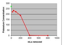
Screening bloodwork had shown significant hypokalaemia (Serum K+ 2.2 mmol/L) and hence IVFs planned for overnight were supplemented with 60mEq/L of KCl per the Potassium charts at a ‘maintenance’ rate of 15mLs/hour which should deliver K+ at about 0.2 mEq/kg/hr, well within the recommended maximum rate of 0.5-1.0 mEq/kg/hr.
The dog was found weak/collapsed in his cage a few hours later with bradyarrhythmia and ‘classic’ ECG changes of hyperkalaemia, the serum level was measured at 9.8 mmol/L.
He recovered uneventfully with unsupplemented saline diuresis.
Presumably, the potassium additive wasn’t mixed properly in the Saline bag.
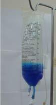
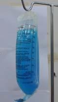
This was proved right, to us at least, a year later when we did an in-house study of adding food dyestained potassium to bags of saline hanging on IV stands as it took some vigorous mixing to get the dye fluid to evenly spread through the whole litre of IVF and we could measure fluid concentrations of up to 220mEq/L in fluids being delivered when the K+ additive was just injected up into the bag, without active agitation.
Control & Therapy Series – Issue 314 March 2024 Centre for Veterinary Education | Est. 1965 Page 3
When we injected 60mEq of KCl (10mLs) into the port of a hung IV fluid bag of IL Saline without vigorous mixing, we could measure >220mEq/L of K+ in the first 100mLs of fluid that would be delivered to the patient, and nearly 200mEq/L for the next 100mLs, with the last 600mLs having 0-2mEq/L K+. This would mean that a 10kg dog getting ‘maintenance’ fluid rates of 30mLs/hr could be getting approx. 0.6 mEq/kg/hr (maximum recommended rate is 0.250.5mEq/kg/hr)
Moral of the story is to mix thoroughly any additive, preferably by agitation of the bag in multiple planes before hanging on the IV stand.
VSS Medication protocol
When adding KCl to a new bag of fluids, flush the injection port by drawing back fluid then reinjecting 2 – 3 times and then invert the bag 5 to 6 times squeezing at the same time to ensure the potassium is distributed evenly. If potassium is added to a bag of fluids that is already connected, you must stop the pump first, add the potassium, flush the injection port, invert and squeeze the bag 5 – 6 times before replacing the bag back on the stand and restarting the pump.
4. Diabetic on Caninsulin given 100 U/mL syringes instead of 40 U/mL
It’s not rare in practice to have a diabetic represent with re-emerging PD/PU/Weight loss and find its diabetes inadequately controlled on its Caninsulin because the owners replenished their insulin syringes, sourcing them cheaper from the local chemist; however, getting 100U/mL syringes instead of the ‘horses for courses’ 40U/mL syringe leads to effectively getting a 2.5x underdose.
The situation could be catastrophic if the opposite occurred, albeit this would be rare, i.e. a dog getting 5 Units of Insulin on a 100U/mL syringe being replaced with a 40U/mL syringe, consequently getting 2.5x overdose.
5. This is a middle-aged cross-bred MN dog (image below) who the owner presented saying,
‘You told me last year to “Keep an eye on it…”
This prompted us to revise our medication protocol for adding KCL to IVFs, including:
Graph showing [K+] potentially fatal amounts in the first 200mLs delivered by gravity with incomplete mixing of the additive, as shown in Figure 1 below.
1 2
Control & Therapy Series – Issue 314 March 2024 Page 4 Authors’ views are not necessarily those of the CVE
Figure 1. shows incomplete mixing of the additive Figure 2. shows more complete (but still inadequate) mixing of the dyed solution with quite vigorous agitation but with the IV bag hung on its stand.
On a slightly different vein, Mike Garvey (AMC, NY City) was here in 1991 with Steve Haskins (UC Davis) giving an Emergency & Critical Care course for the University of Sydney Post-Graduate Committee in Veterinary Science (now the CVE) and in the proceedings you’ll find:
GOLDEN RULES OF EMERGENCY PRACTICE - Michael
Garvey, 1991
1. Attend to the most life-threatening problems first
This requires little explanation. Many cardiac arrests start out as respiratory problems or respiratory arrests. Respiratory problems should be treated first. Cardiovascular problems (including shock and haemorrhage) come in a close second, followed by problems of the CNS and the abdominal cavity. The rest of the body can usually wait until the first four are treated and stabilized.
2. Minimize patient stress at all times
There is a limit to what the critical patient can tolerate in the realm of physical restraint and manipulation. Sometimes things that are generally considered to be indicated must be delayed or abandoned in critical animals. Cage rest is sometimes the best medicine.
3. Expect the unexpected
Unstable patients seem to develop a series of life-threatening complications without warning. In many cases, these events can be anticipated and prevented or minimized. One should always be asking ‘What is likely to go wrong next?’
4. Nature sides with the hidden flaw
In emergency medicine, what you don’t know will hurt you. The more information that you can gather about your patient, quickly and safely, the better your decisions will be. Make good use of simple diagnostic tests, such as glucose sticks, urine dipsticks, BUN sticks, urine specific gravity, PCVs and TPs, EKGs etc.
Editor's Note: Also consider VPOCUS
5. Do not place the patient at risk to achieve a diagnosis
Perhaps this should be a corollary to rule #4. There are times when the only or most appropriate diagnostic test cannot be safely performed in a critical patient. In those cases, it is best to go without the diagnosis. Radiographs often fall into this category.
6. When in doubt, look at the patient
Occasionally, we end up treating the patient’s data or numbers instead of the patient. There is no substitute for careful patient monitoring and frequent patient observation. It was once correctly stated that we make more mistakes because of not looking than not knowing.
7. Left to themselves, things usually go from bad to worse
Conservative management works best in healthy patients. Minimal treatment, followed by waiting to see what happens does not usually achieve a positive result. There is some discomfort in proceeding without knowing everything, but sometimes we must.
8. When you can’t make a diagnosis, treat for the treatable
There are times when it is impossible to narrow down the possibilities to one, and only one, aetiology. This may be because the data will not be available in time or because the needed procedure is too risky for the patient. In these cases, it is appropriate to treat for the best possible disease.
9. When everything seems to be going well, you have obviously overlooked something
This is similar to rule #3 but applies to stable patients. Just because a patient has improved, it does not mean that the battle is over. Monitoring and observation must continue.
10. Don’t panic! The patient is the one with the disease
This is borrowed from a book about human interns, called House of God. Panic leads to poor decision making, a flurry of useless activity, and a tendency toward overtreatment. In an emergency situation, the first pulse that a doctor takes should be his/her own.

This poster is available in A2 format here
Page 5 Centre for Veterinary Education | Est. 1965 Control & Therapy Series – Issue 314 March 2024
Join in!
We offered a $200 CVE voucher to the first person who contributed their Cautionery Tail for this March issue as we hope to make it a regular column. Terry has generously forgone the prize in favour of the next person who sends in their own 'Cautionary Tail'.
Open to Members and Non-Members!
Use the voucher towards memberships (Individual $395, Part-time and Recent graduates $198, Vet Nurse $60, Academic and Students free or Practice Membership: Small $635, Medium $775 and Large $1,295) or enrol in a CVE course.


cve.edu.au +61 2 9351 7979 With our compliments
Dipl.ACVECC and
DACVIM how POCUS differs from consultative (traditional) ultrasound e.g. abdominal ultrasound, echocardiography and more… NEW! VPOCUS: Practical Applications for GPs Distance Education
May - 16 Jun 2024
CVE$200 Learn from the highly engaging tutors Soren Boysen DVM
Serge Chalhoub BSc DVM
6
Answer to What’s Your Diagnosis?
Afterhours Dystocia Case in a Multiparous Beef Cow
Robert Mills
Moonee Beach Veterinary Surgery
e. rob@mooneevet.com.au
C&T No. 6006
Read C&T No. 6004, Dec 2023
The dystocia was due to a foetal malformation known as schistosomus reflexus (SR).
SR foetal malformations result in all four limbs, head and tail bent dorsally with the foetal viscera located externally. The defect occurs early in the embryonic development when the embryonic disc edges reflect dorsally instead of ventrally.
This case was a ventral presentation, as what was protruding from the cow was foetal jejunum. The foetal head, tail and limbs were all directed towards the head of the cow.
The first thing I did was to give the cow an epidural anaesthetic and whilst doing this I told the client that we were in for a reasonably challenging task to remove this from the cow.
I performed a vaginal exam to confirm the diagnosis and then discussed the options, which were foetotomy and vaginal delivery or caesarean.
Foetotomy was performed in this case. If I didn’t have my foetotomy gear in the vehicle then I may have opted for a caesarean delivery but I have personally only dealt with one SR dystocia using this method. I generally feel that foetotomy is the preferred treatment option, as long as it can be done in a reasonable timeframe to make the job economically viable for the producer.
I sent the farmer off for plenty of fresh water and proceeded to extract the foetus.
Viscera was removed first and then the foetotome was then fished out of the vehicle, along with plenty of
obstetric wire and lubricant. The challenging part of the job is the passing of wire around the foetus and reducing the size of the deformed foetus in as few cuts as possible. The foetus was successfully removed in several pieces after an arduous session on the wire.
The use of a Krey hook was extremely helpful to obtain purchase on the awkward foetal parts left after three or four cuts. The cow remained standing in the crush for the entire job and suffered no major vaginal trauma. She walked out afterwards and was given NSAID and procaine penicillin plus oxytocin.
The cow was eating hay when leaving the yards and reportedly went on to make an uneventful recovery.
I hope this makes for an interesting read and am sure there will be some astute cattle obstetricians out there who will be on the ball with this one!

Control & Therapy Series – Issue 314 March 2024 Centre for Veterinary Education | Est. 1965 Page 7
Ross Sillar BVSc
Casino NSW
Retired
e. rosssillar@hotmail.com
This is most likely going to be a Schistosomus reflexus calf.
To confirm the diagnosis, an intra uterine examination is needed. You will find what I best describe as an ‘Inside Out Calf’. The spine is severely bent backwards and fused with the four legs held together in one direction by a pocket of skin. This pocket of skin is inside out with the outer hair layers on the inside. All the organs of the calf’s thoracic and abdominal cavity are attached to the calf but floating freely in the cow’s uterus. It is as if a midline incision is made through the thoracic and abdominal walls and bending the spine backwards the calf is turned ‘inside out.’
These calves are smaller than usual but an embryotomy is needed through the mid spinal area to get the two pieces out.
A very rare condition but I did have three cases in the one herd in one year. This was in a 40 cow Hereford herd and for the last 25 years all replacement bulls and heifers came only from within the herd.
The management of this condition was a lecture on how to not inbreed.
Often these calves are presented as four legs tightly locked together.
Dr Andrew Bissett BVSc(Hons) V2069
Senior Small Animal Veterinarian
Gippsland Veterinary Group
w. gippslandveterinarygroup.com.au
e. AndrewB@gippsvet.com.au
Gotta love these!
What is hanging out of the cow?
Calf intestines.
What is the likely diagnosis?
Schistosomus reflexus
What is the very first thing you are going to do?
Administer an epidural before doing a vaginal examination to confirm the diagnosis.
What are your plans for management of this problem?
Use an introducer to pass embryotomy wire around the calf’s body and then cut the calf into 2 sections. After that each section is usually relatively easy to remove by traction.
What other options will you give the client before proceeding?
The client could consider euthanasia BUT the cost of solving the problem is not prohibitive.
Caesarean can also be performed but I would only reserve this if plan A did not pay off. Caesarean on these presentations usually still requires sectioning the calf to remove through incision and are often difficult. I like to avoid them in these cases!

Best Reply Winners Entitled to a CVE$100 voucher each WebinarPLUS Series for Vets, Techs & Nurses 1 CPD Point each | FREE to CVE Members | Non-members welcome to enrol Tailored TLC for Feline Friends WebinarPLUS NurseEd 15 - 21 Apr 2024 The Master Gland: Pituitary WebinarPLUS 28 Mar - 3 Apr 2024
Answer to What’s Your Diagnosis?
Cutaneous Cryptococcosis in a Cat
(Dec 2023 Issue 313)
Dr Natalie Courtman
Associate Professor of Veterinary Clinical Pathology Veterinary Pathology Diagnostic Services
Sydney School of Veterinary Science.
e. natalie.courtman@sydney.edu.au
t. +61 2 9351 3099
Dr Beth McDonald
Specialist in Veterinary Dermatology University Veterinary Teaching Hospital Sydney.
e. beth.mcdonald@sydney.edu.au
C&T No. 6007
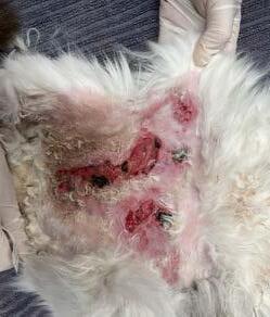
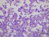
A 5-year-old, 6. 39kg neutered female domestic longhaired cat was referred to the dermatology service of University Veterinary Teaching Hospital Sydney with a two-year history of inflamed ventral abdominal skin and underlying nodular subcutaneous tissues with draining sinuses. The cat had shown a partial response to doxycycline 50mg SID and had also been treated with 0.7mLs of 80mg/mL cefovecin injections subcutaneously (Cerenia, Zoetis). It was then treated for a month with a combination of daily 5mg prednisolone and 7mg/kg cyclosporin. The lesions worsened and this treatment was stopped 3-4 weeks prior to presentation. The cat had been licking at the affected areas.
The skin and subcutaneous tissues were erythematous and extensively thickened over the ventral abdomen bilaterally, extending down both hindlimbs to the stifle region and cranially to caudal thorax. The thickening consisted of coalescing nodules with areas of ulceration and draining sinus tracts, as shown in the image of the ventral skin ( Figure 1).
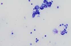
Fine needle aspirates (FNA) were obtained from two subcutaneous nodules and impression smears from the ulcerated surfaces and stained with rapid Romanowsky stain (Rapid Diff, Australian Biostain). Image of the cytology smear is shown in Figure 2.
What Are The Main Differentials For The Gross Skin Lesions?
What Is Your Diagnosis Based On The Cytology?
Answer
Main differentials for the gross skin lesions
Mycobacterial panniculitis and dermatitis; bacterial panniculitis e.g. Nocardia or Actinomyces; fungal panniculitis and dermatitis e.g. Cryptococcus, Sporothrix, Trichosporon, Pythium; sterile panniculitis and dermatitis e.g. idiopathic, vitamin E deficiency and/or excess dietary polyunsaturated fatty acids or secondary to pancreatic neoplasia.
Microscopic findings
The smears are moderately cellular containing inflammatory cells and abundant round yeast organisms with occasional clusters of paired cocci bacteria in a light blue stippled proteinaceous background. The
Figure 1. Image of ventral abdominal skin lesions
Figure 2. FNA from subcutaneous nodule (500x magnification)
Control & Therapy Series – Issue 314 March 2024 Centre for Veterinary Education | Est. 1965 Page 9
inflammatory cells are a mix of degenerate neutrophils, small lymphocytes and macrophages. Macrophages frequently contain yeasts and occasional neutrophils contain bacteria. The yeast are 5-15µm diameter with a thin pink cell wall and prominent unstained capsule (1-2µm diameter) and show rare narrow based budding.
Interpretation
Fungal and bacterial neutrophilic and histiocytic panniculitis/pansteatitis. Fungal morphology consistent with Cryptococcus species.
Diagnosis
The clinical findings and cytology support cutaneous Cryptococcosis with secondary bacterial infection.
Further testing
Routine haematology and biochemistry panel were unremarkable aside from a mild hypoalbuminaemia of 25.9g/L (RI 27-40) which likely reflected inflammation as albumin is a negative acute phase protein. FIV and FeLV ELISA tests were negative.
Acid fast stain of FNA smear – negative
Gram stain of FNA smear – moderate Gram-positive cocci
Cryptococcal antigen titre (LCAT) positive 1:1024
Aerobic cultures on blood agar, MacConkey agar, and BHI at 37 C yielded heavy growth of Enterococcus hirae susceptible to ampicillin, chloramphenicol, doxycycline, amoxicillin/clavulanic acid, gentamicin (high level), marbofloxacin, resistant to trimethoprim/sulpha, cefovecin.
Also cultured was Cryptococcus neoformans, susceptibility testing was not performed. Organisms were identified using MALDI-TOF.
Diagnosis
The clinical findings and cytology support cutaneous cryptococcosis with secondary bacterial infection.
Therapy and Follow Up
The cat was treated with Itraconazole 50mg SID and topically with Otoflush (Dermcare-Vet) and silver sulfadiazine ointment (Flamazine, Smith and Nephew). At recheck two weeks later, she had shown a 20% improvement with the skin remaining erythematous with some reduction in ulceration and minimal change in the subcutaneous nodules. She was changed to Fluconazole 50mg BID. At recheck another four weeks later, she had shown marked improvement as evident
in the image below (Figure 3). There were still multiple palpable nodular lesions in the subcutis but these had significantly reduced in size since the last visit (estimated 70% reduction) and there was one 8mm ulcer. Numerous Cryptococcus organisms were evident on FNA of a nodule and on a scraping of the ulcer. A liver profile biochemistry panel was normal with albumin 28.4g/L (RI 27-40). She was continued on Fluconazole 50mg BID with a plan to recheck in 8 weeks.
Discussion
Cutaneous Cryptococcus infection is an uncommon presentation of Cryptococcosis,1-4 with lesions most commonly seen around the head and neck.4 Cutaneous cryptococcosis is typically associated with firm dermal or subcutaneous nodules that can be ulcerated with serous to mucoid discharge.2, 3 The cutaneous lesions are similar to that of Mycobacterial panniculitis and other infectious causes of pyogranulomatous panniculitis5-10 and also overlap with lesions seen with sterile panniculitis and dermatitis e.g. idiopathic,11 vitamin E deficiency and/ or excess dietary polyunsaturated fatty acids 12 or secondary to pancreatic neoplasia.13
The characteristic cytologic appearance of Cryptococcus is pleomorphic round yeasts with a thin cell wall often with a pronounced unstained mucopolysaccharide capsule and occasional narrow-based budding, associated with variable numbers of neutrophils, macrophages and lymphocytes.4 Diagnosis can be confirmed with culture on birdseed or Sabouraudsdextrose agar and with Cryptococcal antigen titres (LCAT) which are also useful for monitoring response to therapy.4 The most common species causing Cryptococcosis in domestic species are in the C.neoformans-C.gatti species complex, with most cutaneous infections reported as C.neoformans 1, 3
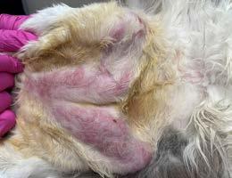 Figure 3. Image of ventral abdominal skin lesions at recheck six weeks after commencing antifungal therapy
Control & Therapy Series – Issue 314 March 2024 Page 10 Authors’ views are not necessarily those of the CVE
Speciation can be achieved with molecular diagnostics e.g. PCR, or use of selective culture media4 or with matrix assisted laser desorption ionization (MALD-TOF).14
Treatment of Cryptococcal infections with antifungal drugs is required for months to years and removal of cutaneous masses may further assist with resolution of infection. Monitoring of LCAT titres can assist with evaluation of response to therapy4 and in this case cytologic review of lesions was useful to identify active infection and thus need for ongoing therapy. Once cytological examination is negative for the organism, LCAT will be used to determine if the infection has been cleared and medication can be stopped.
Enterococcus hirae is a component of the normal mucosa associated microbiota of cats15 and has been associated with ascending cholangitis and pancreatitis in a kitten.16 In this case the presence in the ulcerated nodules was likely to reflect opportunistic infection or colonisation secondary to the cat licking the lesions.
References
1. Nunes Rodrigues, T.C., L.R. Stroobants, and S.I. Vandenabeele, Feline cutaneous nodular and ocular Cryptococcus neoformans in Belgium. JFMS Open Rep, 2020. 6(1): p. 2055116920912560.
2. Medleau, L., et al. , Cutaneous cryptococcosis in three cats. J Am Vet Med Assoc , 1985. 187(2): p. 169-70.
3. Myers, A., et al., Atypical cutaneous cryptococcosis in four cats in the USA. Vet Dermatol 2017. 28(4): p. 405-e97.
4. Lester, S.J., et al., Cryptococcosis: update and emergence of Cryptococcus gattii. Vet Clin Pathol , 2011. 40(1): p. 4-17.
5. Malik, R., et al. , Infection of the subcutis and skin of cats with rapidly growing mycobacteria: a review of microbiological and clinical findings J Feline Med Surg , 2000. 2(1): p. 35-48.
6. Rissi, D.R., K.D. Kirby, and S. Sanchez, Systemic Trichosporon loubieri infection in a cat J Vet Diagn Invest, 2016. 28(3): p. 350-3.
7. Thomson, J., et al. , An atypical cause of sporotrichosis in a cat Med Mycol Case Rep , 2019. 23: p. 72-76.
8. Malik, R., et al. , Nocardia infections in cats: a retrospective multiinstitutional study of 17 cases Aust Vet J, 2006. 84(7): p. 235-45.
9. Love, D.N., et al. , Isolation and characterisation of bacteria from abscesses in the subcutis of cats J Med Microbiol , 1979. 12(2): p. 207-12.
10. Dowst, M., et al. , An unusual case of cutaneous feline pythiosis Med Mycol Case Rep , 2019. 26: p. 57-60.
11. Giuliano, A., et al. , Idiopathic sterile pyogranuloma in three domestic cats J Small Anim Pract, 2020. 61(3): p. 202-205.
12. Niza, M.M., C.L. Vilela, and L.M. Ferreira, Feline pansteatitis revisited: hazards of unbalanced home-made diets J Feline Med Surg, 2003. 5(5): p. 271-7.
13. Fabbrini, F., et al. , Feline cutaneous and visceral necrotizing panniculitis and steatitis associated with a pancreatic tumour . Vet Dermatol , 2005. 16(6): p. 413-9.
14. Firacative, C., L. Trilles, and W. Meyer, MALDI-TOF MS enables the rapid identification of the major molecular types within the Cryptococcus neoformans/C. gattii species complex PLoS One, 2012. 7(5): p. e37566.
15. Gookin, J.L., et al. , Randomized placebo-controlled trial of felineorigin Enterococcus hirae probiotic effects on preventative health and fecal microbiota composition of fostered shelter kittens Front Vet Sci , 2022. 9: p. 923792.
16. Lapointe, J.M., et al. , Enterococcus hirae enteropathy with ascending cholangitis and pancreatitis in a kitten Vet Pathol, 2000. 37(3): p. 282-4.
Comment courtesy of Professor Mark Krockenberger
BSc(vet) BVSc PhD GradCertEdStud FANZCVS (Anatomical Pathology)
This is a nice case of cutaneous cryptococcosis. The cytology images are fairly typical. Narrow-necked budding of encapsulated yeasts in Australia is fairly definitive for cryptococcosis. As the authors point out, the causative organism can be found in the Cryptococcus neoformans species complex or the Cryptococcus gattii species complex and in my experience, also, disseminated cutaneous cases do seem to more commonly be in the C. neoformans species complex.
The positive cryptococcal serology supports the diagnosis of cryptococcosis and can also be used as a reference point to gauge the response to therapy over time. If you use serology in this way, use the same trusted lab and the same test each time as there will be variation between labs and even more between tests. It is likely to take some time to see serological improvement (and sometimes there will be a worsening serologically as the yeast are killed and more capsule is liberated; however, when you are thinking about ceasing antifungal therapy I would always check the cryptococcal capsular serology. I usually prefer it to be negative twice (a month apart) before ceasing therapy. Beware of recurrence following cessation of therapy!
Cutaneous cryptococcosis can be associated with disseminated cryptococcosis, so you may need to think about the disease being present in more organs than just the skin. It is worth considering investigating cases like this to see whether they are localised disease secondary to a penetrating wound and localised immunosuppression or whether they reflect more disseminated disease. This consideration may help you in thinking about prognosis.
The azoles are fungistatic so if you have severe disseminated disease it may be worth considering amphotericin as the initial antifungal agent. Amphotericin is now most easily obtained in the liposomal formulation but the deoxycholate formulation is cheaper (if you use deoxycholate, deliver using this protocol (read perspective 134 below: Antifungal therapy in companion animals -A practical approach). Most strains of both species complexes are susceptible to fluconazole; however, resistance is more likely in some members of both complexes. Culture and identification with the species complexes may help work out whether susceptibility issues are likely. The owners need to be counselled about the long-term therapy required and some cats with disseminated disease may end up on fluconazole for life.
Control & Therapy Series – Issue 314 March 2024 Centre for Veterinary Education | Est. 1965 Page 11
Entitled to a CVE$100 voucher each
Answer 1
Sharnee Lehrmayer
Burvale Heights Veterinary Hospital
Forest Hill VIC 3131
e. info@BurvaleHeightsVET.com.au
Based on the cytology, I would make a diagnosis of cryptococcosis, with associated pyogranulomatous inflammation. There are multiple round bodied organisms with a negative staining capsule, and there are also some inflammatory cells (neutrophils and macrophages) present.
The main differentials based on the gross skin lesions (aside from cryptococcosis) would be another fungal infection, an unusual bacterial infection, in particular Mycobacterium spp. infection (which was actually my first thought when I looked at the photo), Nocardiosis, Actinomycosis, or neoplasia (e.g. epitheliotropic lymphoma). Due to the severity of the lesions (and lack of response to prednisolone and cyclosporine), eosinophilic granuloma complex seemed less likely. Overseas, feline pox virus would be a differential but to my knowledge it is not found in Australia!
Read Perspective 134. Antifungal therapy in companion animalsA practical approach.
Answer 2
Robert Bird BVSc Massey 1990 (Dist) MANZCVS
MVM (CA) GCert SAU-A (Honours)
Vetora Co. NZ
w. vetora.co.nz/
t. 078438822
e. Robert.Bird@vetora.nz
My provisional diagnosis for this cat’s lesions is Cryptococcus neoformans
Differentials would be other fungi e.g. Cryptococcus gattii, deep bacterial subcutaneous infections, sterile nodular panniculitis, subcutaneous foreign body/ ies, and neoplasia.

Interested in Diabetic Research?
Monitoring Diabetes Mellitus with FreeStyle Libre: A Veterinarian’s Perspective
If you’ve used continuous glucose monitoring to monitor diabetes in animals or have ever considered using it, please take 10-15 minutes to complete this survey
Thank you.
Linda Fleeman
BVSc PhD MANZCVS
Animal Diabetes Australia—The only diabetes-specific veterinary clinical service in the world.
Correction
The references in C&T No. 6002 Tincture of Time should read as follows (see bold):
Astute owners most probably notice signs of weakness in their dogs before we detect illness— individualized, early, subtle, repeatable signs exist ( Atwell et al (2016), AVJ 94:110) which owners may simply report as ‘dog seems off’ or is ‘quiet.’ Early disease was associated with dogs being less active, barking less, no jumping etc. ‘Figure of 8’ walking best revealed early signs of weakness e.g., left back leg when dog is turning rightwards.
It is feasible that some ticks in different areas could have different combinations of toxins, in varying proportions. (C T Holland & R Atwell (2019), C&T No. 5771:296, CVE) If true, this could explain varying clinical signs (areas, between seasons etc.), why some very small ticks can be highly toxic, why some very engorged ticks produce no signs (in non-immune hosts), and why some TASproducing dogs can develop TP with placement of ticks.
Reply Winners
Control & Therapy Series – Issue 314 March 2024 Page 12 Authors’ views are not necessarily those of the CVE
Veterinary Pathology Diagnostics Services (VPDS)
The VPDS at the School of Veterinary Science supports the University Veterinary Teaching Hospital (UVTHS), external clients, and DVM, resident, and research training. VPDS offers services for Clinical Pathology, Histopathology, Microbiology, Molecular Diagnostics, Cytology and Parasitology.

Our long-term commitment to quality assurance and continuous improvement was recognised by NATA accreditation of our Clinical Pathology and Histopathology services in 2022. Our services are supported by an excellent trained team of technical staff, as well as two specialist veterinary anatomical pathologists, two specialist veterinary clinical pathologists, and world leading academics in pathology, and infectious diseases. Their position at the forefront of research means state-of-the-art interpretation on mainstream diagnostics, as well as an ability to offer a range of less common or unique diagnostic tools to support the work of small animal practices, such as the confirmatory reference tests for the diagnosis of Feline Infectious Peritonitis (FIP), Toxoplasma and Neospora serology, cryptococcal capsular serology (LCAT), bespoke molecular testing, Immunocytochemistry (IHC) on unusual targets, and project and research development opportunities.
By sending cases to VPDS, you not only get first rate service but you support training of the next generation of veterinary practitioners and specialists. We invite you to participate in the VPDS experience and request our services. We are here to help you and your patients!
For further information contact the VPDS Laboratory
+61 2 9351 3099 (Office)
+61 2 9351 7456 (ClinPath Lab)
Veterinary Pathology Diagnostic Services
Sydney School of Veterinary Science
B14 - McMaster Building, Room 213-220
The University of Sydney NSW 2006
https://sydney.edu.au/science/schools/sydneyschool-of-veterinary-science/veterinary-scienceservices.html
What is Your Diagnosis?
Dr Natalie Courtman
Associate Professor of Veterinary Clinical Patholog
Veterinary Pathology Diagnostic Services, Sydney School of Veterinary Science.
m. +61 2 9351 3099
e. natalie.courtman@sydney.edu.au
C&T No. 6008
A 13-year-old neutered female Staffordshire bull terrier was referred for evaluation of a raised round 2cm pigmented haired left foreleg mass above the carpal pad. Fine needle aspirate smears were prepared from the mass and stained with Wrights Giemsa.
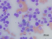
Images of the cytology smear are shown below.
What is your diagnosis based on the cytologic appearance? What is the expected behaviour?
Email your answer to cve.marketing@sydney.edu.au and be in to win a CVE$100 voucher.
Figure 1: FNA from leg mass (200x magnification)
Control & Therapy Series – Issue 314 March 2024 Centre for Veterinary Education | Est. 1965 Page 13
Figure 2: FNA from leg mass (1000x magnification)


01
ADVERTISEMENT
MAJOR Winner
The prize is a CVE$400 voucher
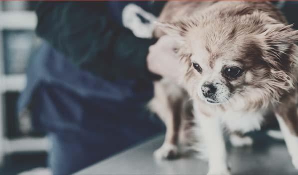
Fatal Venous Air Embolism During Thoracic Limb Amputation in a Dog
Adam
Gordon BVSc(Hons) MVS GCertSAUA
Maroubra Veterinary Hospital, Sydney NSW
t. +612 9344 8722
w. maroubravet.com.au
e. Adam.gordon@maroubravets.com.au
C&T No. 6009
Introduction
Venous air embolism (VAE) occurs when air is introduced to the central venous system, obstructing blood flow and producing an air embolism to the right heart and/ or pulmonary artery. It is a potentially catastrophic complication and is almost always iatrogenic.
Entry of air occurs as a result of a negative pressure gradient between an exposed venous sinusoid near the site of surgery and the right atrium of the heart. Air (or other gases like carbon dioxide) can also enter the venous circulation by creation of a pressure gradient through the use of insufflation of a body cavity or by the use of gas pressurized equipment.
Alternatively, air may enter the venous system through a gravitational gradient, created by having the operative site elevated above the heart.
Previous reported causes
Venous air embolism is well recognized and documented in human medicine.
The majority of published cases involved orthopaedic surgery or neurosurgery.
VAE has also been documented in obstetrics, gastroenterology, arthroscopy, central line placement and removal, interventional radiology procedures and endoscopy.
There are relatively few cases of VAE published in the veterinary literature. Published cases and outcomes of VAE in the veterinary literature occurred during the following procedures:
Pneumocystography—fatal.
Laparoscopy—fatal.
Cryosurgery (with pressurized liquid nitrogen) to treat gingival neoplasms—fatal. Gas insufflation of a retropharyngeal diverticulum—fatal.
Dental extractions involving use of air-driven dental drill—fatal. Thoracic limb amputation—non-fatal. Hemilaminectomy—1 fatal, 1 non-fatal.
Iatrogenic introduction of air through an intravenous catheter during surgery or hospitalization—several fatal, 2 non-fatal.

Case Report
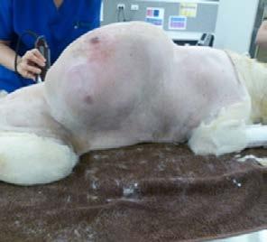
A 10-year-old, 32kg female neutered Labrador dog presented with a large mass over the distal antebrachium and carpus of the left forelimb (Figure 1)
The mass was broad-based, multilobular and was approximately 30cm in diameter. It had been present for 3 years.
Histopathology revealed it to be a myxofibrosarcoma (Grade 1, mitotic index 4 mitotic figures per 10 high power fields).
Left forequarter amputation was chosen as the most appropriate treatment option.
Thoracic radiographs, haematology and serum biochemistry pre-operatively were unremarkable.
Physical examination on the morning of surgery was unremarkable other than the left forelimb mass.
Figure 1. Soft tissue sarcoma of left forelimb
Control & Therapy Series – Issue 314 March 2024 Centre for Veterinary Education | Est. 1965 Page 15
The dog was bright and alert, HR 120 beats per minute, respiratory rate 28 breaths per minute, rectal temperature 38.3°C. Mucous membranes were pink with capillary refill time less than 2 seconds. Thoracic auscultation and abdominal palpation were unremarkable.
The dog was pre-medicated with acepromazine (0.025mg/kg) and methadone (0.25mg/kg) subcutaneously. An intravenous catheter was placed in the right cephalic vein, and anaesthesia was induced with propofol intravenously (4mg/kg). The dog was intubated with a cuffed 11.0mm endotracheal tube. Anaesthesia was maintained with isoflurane in 100% oxygen at a fresh gas flow rate of 1.5L/minute.
Intravenous fluids (Hartmann’s solution) were administered for the duration of anaesthetic and surgery at 5mL/kg/hr. Anaesthetic monitoring consisted of pulse oximetry, end-tidal CO2, ECG, rectal temperature, heart rate, respiratory rate and oscillometric blood pressure.
The dog was placed in right lateral recumbency for surgery. During dissection, a small tear was inadvertently created in the axillary vein. Haemorrhage was quickly controlled through placement of haemostats on the axillary vein proximal to the tear. In the next minute ETCO2 dropped, the pulse oximeter did not give a reading, the patient became cyanotic and suffered cardiac arrest.
Attempted cardiopulmonary resuscitation (external compressions, boluses of atropine and adrenaline intravenously) was unsuccessful and the dog was pronounced deceased after 20 minutes of CPR.
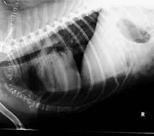
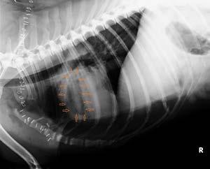
Post-mortem thoracic radiographs were performed. They showed an elliptical radiolucent focus measuring approximately 7cm in height and 3.5cm in length superimposed over the region of the right ventricle in lateral views (Figures 2 and 3).
The clinical presentation and thoracic radiographs support a diagnosis of venous air embolism.
Discussion
In this unfortunate case, the iatrogenic rent in the axillary vein allowed air to enter the central venous system down a gravitational gradient to the right side of the heart. This resulted in outflow obstruction to the right ventricle, hypocapnia, hypoxaemia, hypotension, reduced cardiac output and ultimately cardiac arrest.
The clinical signs and consequence of VAE are dependent on the volume of air introduced and the rate at which it is introduced to the venous system.
The lethal dose of a bolus of air in dogs has been reported as 7.5–15mL/kg body weight.
The majority of published cases of VAE in the veterinary literature had a fatal outcome.
Successful management of VAE requires:
Awareness of risk of occurrence (for instance where the operative site is positioned above the right atrium). Good communication between surgeon and anaesthetist/individual monitoring the anaesthetic. Early diagnosis.
Proactive management if VAE is suspected.
Figure 2. Lateral thoracic radiograph showing air in right ventricle
Control & Therapy Series – Issue 314 March 2024 Page 16 Authors’ views are not necessarily those of the CVE
Figure 3. Lateral thoracic radiograph with air embolus in right ventricle outlined by arrows
Diagnosis of Venous Air Embolism
There are no minimally invasive aids with high sensitivity and specificity for diagnosing VAE. Transoesophageal echocardiography is the most sensitive technique for detecting VAE, being able to detect 0.02mL/kg of air. Precordial doppler ultrasound is another sensitive technique for detecting VAE, whereby the anaesthetist listens for a characteristic ‘drum-like’ or ‘mill-wheel’ murmur associated with VAE.
From a practical standpoint in the veterinary setting, the following methods are most reliable for diagnosing suspected VAE.
Capnography—an abrupt drop in ETCO2 can occur with VAE (though reductions as little as 2mmHg can also be seen). Unfortunately this is not particularly specific, and can also be seen with hypothermia, hyperventilation, reduced cardiac output, pulmonary embolism and bronchospasm. Reduced ETCO2 is generally the earliest indicator of VAE in anaesthetised patients. Pulse oximetry—reduced oxygen saturation on pulse oximetry is considered a late sign of VAE. Blood pressure monitoring— hypotension occurs with VAE. ECG—tachyarrhythmias and ST segment depression may be seen early, followed by bradycardia and cardiac arrest.
Management of Venous Air Embolism
As the diagnosis of VAE is fraught with difficulty and the potential consequences devastating and potentially rapidly fatal, management of VAE must be instituted promptly and usually before a definitive diagnosis is made where there is an index of suspicion of VAE.
The key points in management of VAE are:
Prevent further entry of air into the venous system. In open surgical procedures the operative site should be flooded with saline or covered with moistened sponges. Immediately discontinue any insufflation of body cavities and use of gas-pressurized equipment. Where possible, lower the operative site below the level of the heart.
If nitrous oxide is part of the anaesthetic mix it should be discontined immediately as it can dramatically increase the size of the air bubble due to its increased solubility in blood.
Administration of high FiO₂ 100% oxygen with intermittent positive pressure ventilation (IPPV). Maintain normovolaemia and prevent hypotension—ensure adequate intravenous fluid rates and use of positive inotropes (adrenaline, noradrenaline, dopamine, dobutamine).
Repositioning of the patient—placement into left lateral recumbency either horizontal or head down (Durant’s maneuver) —known as the Trendelenburg position. It was postulated that an obstructing air embolus could be displaced and right ventricular failure prevented by this repositioning. In an echocardiographic study, transoesophageal echocardiography demonstrated that this repositioning relocated intracardiac air to nondependent parts of the right heart. However, there was no corresponding improvement in haemodynamic performance or change in cardiac dimensions. Irrespective of this, placing patients with suspected VAE into the Trendelenburg position remains standard of care in human medicine.
KEY POINTS
The most important lesson learned from this unfortunate case was the importance of immediate communication by the surgeon that there was a breach of the venous system that increased the risk of venous air embolism. Instituting IPPV, continuous monitoring of blood pressure and treating hypotension (if it were present) would have improved the chances of survival, though by no means ensured it, such is the seriousness of the introduction of significant volumes of air in a short period of time.
It is thought that the incidence of VAE in veterinary patients is much higher than has been documented in the literature. Having studied the published literature and perused VIN (Veterinary Information Network), it would appear that lack of due care with intravenous fluid lines and subsequent ingress of air is the most common cause of VAE in companion animals. This is an important reminder to avoid complacency and ensure all staff understand the importance of ensuring all IV lines are primed and capped where appropriate.
Intravenous fluid pumps are definitely not foolproof, and there are reports of the air alarm failing and large volumes of air being infused into the patient with subequent death.
References & Further Reading
1. Mouser PJ, Wilson JD. Fatal venous air embolism in an apparently healthy adult Chihuahua. J Am Anim Hosp Assoc 2015; 51: 176-179.
2. Bautista Diaz-Delgado O, Campagna I. Suspected venous air embolism during thoracic limb amputation in a dog. Vet Rec Case Rep 2020; 8: 1-6.
3. Austin LS, VanBeek C, Williams GR. Venous air embolism: an underrecognized and potentially catastrophic complication in orthopaedic surgery. J Shoulder Elbow Surg 2013; 22: 1449-1454.
4. McCarthy CJ, Behravesh S, Naidu SG, Oklu R. Air embolism: Practical tips for prevention and treatment. J Clin Med 2016; Vol.5(11): 93-105.
5. Lui PW, Lin YM, et al. Spectral characteristics of embolic heart sounds detected by precordial doppler ultrasound during venous air embolism in dogs Int J Crit Illn Inj Sci 2013; 3(1): 73-76.
6. Gordy S, Rowell S. Vascular air embolism. Int J Crit Illn Inj Sci. 2013; 3(1) Jan-Mar: 73–76.
Control & Therapy Series – Issue 314 March 2024 Centre for Veterinary Education | Est. 1965 Page 17
7. Palmon SC, Moore LE, Lundberg J, et al. Venous air embolism: A review J Clin Anesth 1997; 9: 251-257.
8. Gilroy BA, Anson LW. Fatal air embolism during anesthesia for laparoscopy in a dog J Am Vet Med Assoc 1987; 190: 552–554.
9. Thayer GW, Carrig CB, Evans AT. Fatal venous air embolism associated with pneumocystography in a cat J Am Vet Med Assoc 1980; 176: 643–645.
10. Gilroy BA, Anson LW. Fatal air embolism during anesthesia for laparoscopy in a dog J Am Vet Med Assoc 1987; 190: 552–554.
11. Harvey HJ. Fatal air embolization associated with cryosurgery in two dogs J Am Vet Med Assoc 1978; 173: 175–176.
12. Ober CP, Spotswood TC, Hancock R. Fatal venous air embolism in a cat with a retropharyngeal diverticulum. Vet Radiol Ultrasound 2006; 47: 153–158.
13. Costa-Farré C, Torrente C, Bertrana CD, et al. Nonfatal infusion pump-related venous air embolism in a dog. Vet Anaesth Analg 2017; 44: 382–383.
14. Gunew M, Marshall R, Lui M, Astley C. Fatal venous air embolism in a cat undergoing dental extractions. J Small Anim Pract 2008; 49(11): 601-604.
15. Geissler HJ, Allen SJ, et al. Effect of body repositioning after venous air embolism. Anesthesiology 1997; Vol.86 (3): 710-717.
PATHOLOGY IN PRACTICE
Canine Right Atrial Haemangiosarcoma with Widespread Pulmonary Metastases
Alexander Teh BVetBiol DVM
Resident in Anatomical Pathology
Sydney School of Veterinary Science
The University of Sydney
e. alexander.teh@sydney.edu.au
C&T No. 6010
Signalment, History, and Clinical Presentation
An 8-year-old male neutered Border Collie cross presented to the University Veterinary Teaching Hospital Sydney for coughing. He was initially treated with lofenoxal and amoxiclav with no improvement noted.
One week later, he re-presented with worsening respiratory effort and rate, and lethargy. He vomited blood one day later, and his lethargy and respiratory effort continued to worsen despite intranasal oxygen therapy. Thoracic radiographs revealed a diffuse pulmonary nodular pattern.
Haematology revealed a mild mature neutrophilia, and no significant abnormalities were reported on biochemistry. Prothrombin time (PT) and activated partial thromboplastin time (APTT) were within normal limits. Due to the continued clinical deterioration of the patient, he was euthanised and submitted for a postmortem examination.
Post-Mortem Gross Examination
At post-mortem examination, severe pulmonary haemorrhage with multifocal nodules in all lung lobes were observed (Figure 1)
Additionally, there was a focal, dark red, irregularly shaped, slightly firm mass that was protruding from the endocardial surface of the right atrium (Figure 2)
Histopathological Examination
The right atrial mass was consistent with a large, poorly demarcated, densely cellular, invasive haemangiosarcoma (Figure 3). The neoplastic spindloid cells were arranged in short interlacing and haphazardly arranged streams and bundles which frequently formed irregularly shaped clefts, channels and spaces which were variably filled with erythrocytes (Figure 4). The mitotic rate was relatively high, and 54 mitotic figures were identified in 2.37mm2 (equivalent to 10 standard high power [400x] fields). Multiple blood vessels contained tumour emboli demonstrating vascular invasion (Figure 5).
The lungs were disrupted by multiple dense regions of neoplastic cells which appeared similar to those in the right atrial mass ( Figure 6). These presumably represented pulmonary metastases from the primary right atrial haemangiosarcoma. Pulmonary haemorrhage was also observed.
Canine Cardiac Haemangiosarcoma
In dogs, the right atrium is considered one of the most common sites for primary cardiac haemangiosarcoma, in addition to the right auricle and the spleen. Haemangiosarcoma is the most common canine malignant cardiac tumour, comprising approximately 70% of all canine cardiac neoplasms, and the lungs are a common site of metastasis as the pulmonary capillaries are the first filter encountered by tumour emboli released from the right side of the heart. The extensive and severe pulmonary haemorrhage was presumably a consequence of pulmonary metastasis.
Control & Therapy Series – Issue 314 March 2024 Page 18 Authors’ views are not necessarily those of the CVE
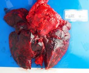
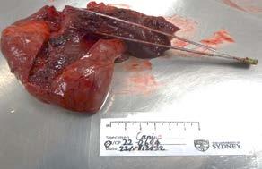
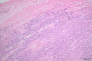
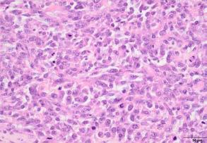
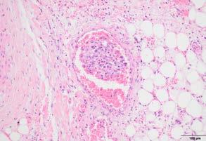
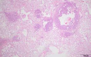 Figure 1. Lungs when removed at necropsy. There was severe pulmonary haemorrhage and multifocal nodules distributed throughout all lung lobes
Figure 2. Right atrial endocardial mass held by the forceps
Figure 3. Right atrial mass consistent with an invasive haemangiosarcoma
Figure 4. Neoplastic cells with scattered mitotic figures
Figure 5. Blood vessel containing a tumour embolus demonstrating vascular invasion
Figure 1. Lungs when removed at necropsy. There was severe pulmonary haemorrhage and multifocal nodules distributed throughout all lung lobes
Figure 2. Right atrial endocardial mass held by the forceps
Figure 3. Right atrial mass consistent with an invasive haemangiosarcoma
Figure 4. Neoplastic cells with scattered mitotic figures
Figure 5. Blood vessel containing a tumour embolus demonstrating vascular invasion
1 2 3 4 5 6 Centre for Veterinary Education | Est. 1965
Figure 6. Lungs with multiple regions of neoplastic cells similar to those in the right atrial mass, presumably representing metastatic spread from the primary cardiac haemangiosarcoma
Durable Contraception in the Female Domestic Cat Using Viral-Vectored Delivery of a Feline Anti-Müllerian Hormone Transgene
Lindsey M. Vansandt et al
Nature Communications (2023) 14: 3140
Abstract
Eighty percent of the estimated 600 million domestic cats in the world are free roaming. These cats typically experience suboptimal welfare and inflict high levels of predation on wildlife. Additionally, euthanasia of healthy animals in overpopulated shelters raises ethical considerations. While surgical sterilization is the mainstay of pet population control, there is a need for efficient, safe, and cost-effective permanent contraception alternatives. Herein, we report evidence that a single intramuscular treatment with an adenoassociated viral vector delivering an antiMüllerian hormone transgene produces long-term contraception in the domestic cat. Treated females are followed for over two years, during which transgene expression, anti-transgene antibodies, and reproductive hormones are monitored. Mating behavior and reproductive success are measured during two mating studies. Here we show that ectopic expression of anti-Müllerian hormone does not impair sex steroids nor estrous cycling, but prevents breeding-induced ovulation, resulting in safe and durable contraception in the female domestic cat.
Commentary by Associate Professor Fiona Hollinshead
BVSc (Hons), PhD, DACT
Associate Professor of Small Animal Theriogenology
College of Veterinary Medicine and Biomedical Sciences
Small Animal Reproduction
Colorado State University, USA
Professor Alan Conley BVSc (Melb), MS, PhD, FRCVS, Hon Dipl ACT
Distinguished Professor
Department of Population Health & Reproduction
John P. Hughes Endowed Chair in Equine Reproduction Director, Clinical Endocrinology Laboratory
School of Veterinary Medicine
University of California, Davis CA 95616
t. (970) 297 4023
C&T No. 6011
The domestic cat is an important target species for population control because millions of free-roaming cats inhabit rural and urban areas worldwide. This is a serious animal welfare problem that has a detrimental impact on wildlife, as well as becoming a growing concern for public health as cats can serve as a reservoir for viruses and parasites that cause zoonotic diseases (Johnson et al., 2019).
The over expression of a reproductive hormone such as AMH as a means of shutting down reproductive physiology is a novel strategy and the results presented in this paper are very promising.
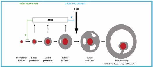
Anti-Müllerian Hormone (AMH) is an interesting target for animal contraception, as the mechanisms as to how increased levels of AMH shuts down ovarian function are elusive. AMH is secreted by the follicular granulosa cells in females (Figure 1) and Sertoli cells in males. It is highly conserved across species in both male and females, making it an attractive target to develop a universal contraceptive product.
However, species differences may exist in whether AMH secretion is primarily from follicles before (pre-antral) or after (antral) becoming gonadotropin-responsive, and in the relative population size of the various follicle classes.
It is fascinating that high levels of AMH inhibited folliculogenesis and mating-induced ovulation in the queen. Further investigations, by studying a much larger number of cats over a longer period of time (ideally the entire lifespan) to elucidate the true efficacy and underlying mechanism(s) to this therapy is
Control & Therapy Series – Issue 314 March 2024 Page 20 Authors’ views are not necessarily those of the CVE
exciting and will open the door to more creative ways to use this potential contraceptive pathway in other species. Wouldn’t it be great if it could be used to limit reproduction of wild horses (brumbies)!
These preliminary data are novel and promising for the development of a non-invasive contraceptive for cats. However, the biological relevance after administration of this contraceptive is hard to answer with this early study as there were very few cats included (only 3 per treatment group = a total of 9 cats) that were observed for a 2-year period. Despite much blood sampling and sequential hormone measurements performed, important information on mechanisms and possible offtarget effects that would be gained from tissue samples was restricted to a single opportunistic occasion on a queen given a ‘first generation’ vector that also induced an immune reaction, potentially complicating results.
The next step that needs to be undertaken to confirm the contraceptive efficiency of this novel AMH transgene delivered utilizing a validated viral vector in the cat is to perform a large-scale trial involving hundreds of male and female cats but with the ability to access and monitor outcome over a lifetime to assess the long-term effects of supraphysiological concentrations of AMH on reproduction and general health.
This seminal report will no doubt open the doorway to exciting research opportunities on a much larger scale.
While contraception of cats with a non-invasive contraceptive method such as novel viral vector delivery of genetically modified targets is very appealing at an ethical and welfare level, as well as the practical application of an intraperitoneal injection which would facilitate the efficient and effective management of
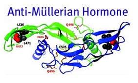
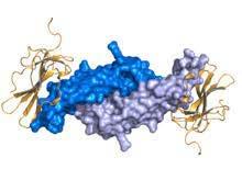 Figure 1. In women, AMH is secreted by the granulosa cells of growing follicles but not FSH stimulated follicles.
Initial recruitment
recruitment
Figure 2. Glycoprotein structure
Cyclic
Preovulatory TRENDS in Endocrinology & Metabolism AMH FSH b a Anti-Müllerian Hormone Control & Therapy Series – Issue 314 March 2024 Centre for Veterinary Education | Est. 1965 Page 21
Primordial follicle Small preantral Large preantral Antral 2-7mm Antral 8-12mm
large populations of wild/stray cats, it is important we don’t ignore the reality of contraception in which failure, even just one failure, has drastic consequences. One reproductively active female cat can exponentially derail population control in a very short period of time i.e. theoretically, if 1 cat has 6 kittens, each having 6 of their own, then leads on to 36 cats and with another generation (within just 3 generations in all) there are potentially 216 cats in the population from just one fertile queen.
Surgical sterilization of both males and females has many drawbacks, primarily the invasive nature of the procedures, the practicality and cost of performing them but, most importantly, the reproductive life of that animal is ended with 100% efficiency, immediately and
permanently. For this reason, surgical sterilization is the ‘gold standard’ for life-long contraception.
This report offers hope of a non-invasive, ethical, welfare orientated, cost effective potentially permanent contraceptive that can be easily and readily administered by a single injection to manage large scale feral animal populations in other species besides cats such as pigs, horses, camels, mice, and rats. It would be even more effective in some feral animal population groups if it was possible to use a viral vector which might work with oral administration.

Wild Horse Management in Kosciuszko National Park
Significant recent media focus has been given to the controversial NSW government decision to introduce aerial culling of wild horses in Kosciuszko National Park. The veterinary profession has an important role in educating the public on animal welfare issues and vets may find they are asked for an opinion. However, it is not an area into which many veterinarians have great insight. To help decipher the evidence around this, an upcoming C&T e-article (composed by experts in wild horse management and welfare) unpacks the welfare impacts of aerial culling and compares and contrasts some of the other lethal control methods.
We’re also interested to know what YOU think by answering the following anonymous poll:

1. With regard to wild horse management, which of the following lethal management options do you consider to be a humane death (tick all that you think apply)
Passive trapping in yards followed by a single gunshot to the head
In situ ground shooting (head shot)
In situ ground shooting (chest shot)
Aerial shooting with < 1 minute chase time (chest shot)
Aerial shooting with > 1 minute chase time (chest shot)
None of the above
2. Are you a veterinarian?
Yes
No

Feline Orofacial Pain Syndrome
Clare Rusbridge BVMS PhD DECVN FRCVS
Professor in Veterinary Neurology at the University of Surrey & Senior Neurologist at Wear Referrals
The ISFM & AAFP are partners with the CVE in delivering the Feline Medicine Distance Education course.
C&T No. 6012
Feline orofacial pain syndrome (FOPS) is a maladaptive pain disorder characterised by behavioural signs of oral discomfort and tongue mutilation, which is episodic, typically unilateral and triggered in many cases by mouth movements. Burmese cats are predisposed and an inherited disorder affecting processing of nociceptive trigeminal information is suspected. Clinical signs are precipitated by conditions causing oral pain, and anxiety and social stress influence disease expression. Clinical signs may be poorly responsive to licensed analgesics but managed with adjuvant analgesics.
Clinical signs
Feline orofacial pain syndrome (FOPS) is characterised by clinical signs that suggest oral discomfort, particularly of the tongue. Owners of affected cats describe exaggerated licking and chewing movements, with pawing at the mouth typically to one side only.
There are two presentations: acute–severe and chronic–episodic. The acute disease is characterised by signs of severe and unrelenting discomfort with mutilation of the tongue or buccal mucosa. The tongue can be so badly lacerated that surgical repair may be necessary and tongue auto-amputation is even possible. The classic presentation is in young teething kittens. The chronic–episodic form occurs in older cats that may have had the acute form as kittens. Signs are similar, but in adult cats, the pain can be paroxysmal and triggered in many cases by mouth movements such as chewing, drinking or grooming. Owing to the severity of the pain, some cats are anorexic or inappetent.
Pathogenesis
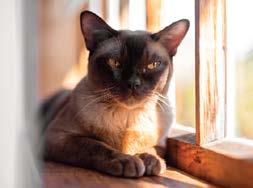
FOPS is seen in a variety of feline populations (including some crossbred cats), although Burmese cats from the UK, Europe and Australasia make up most reported cases.( Figure 1 ).¹
The disease is triggered by conditions causing oral pain, although the extent of disease may be considered minor and less than what a veterinary surgeon would typically associate with clinical signs. In young cats, the disease is almost always associated with permanent


teeth eruption and is self-limiting, with signs resolving within a few weeks. More rarely, signs may be triggered by other oral lesions such as mouth ulceration associated with feline respiratory virus infection. In adult cats, periodontal disease and feline tooth resorption are the more important predisposing causes. The predominance of affected Burmese cats suggests an inherited predisposition. A genome-wide casecontrol association study had a suggested association to a genomic region with a single candidate gene that encodes a multifunctional plasma membrane receptor.2 In the central nervous system, this receptor influences
Figure 2. Burmese cats make up most reported cases of feline orofacial pain syndrome
Figure 1. A diagnosis of feline orofacial pain syndrome is not appropriate for a cat with discomfort due to dental disease
Control & Therapy Series – Issue 314 March 2024 Centre for Veterinary Education | Est. 1965 Page 23
N-methyl-D-aspartate (NMDA) receptor functioning, long-term potentiation and synaptic signalling, and plays a role in neuronal development.3,4 Studies have suggested that it may be implicated in the development of maladaptive pain states, making it a reasonable candidate gene for FOPS.
Key point
Feline orofacial pain syndrome is influenced by anxiety and the cats’s emotional state, with environmental factors shown to influence disease expression.
The expression of FOPS is influenced by anxiety and the cat’s emotional state. A retrospective study found that for one in five FOPS cases, environmental factors influenced the disease expression.1 Individuals with poor social coping strategies in multi-cat households appear to be more vulnerable, but other reported events that have triggered FOPS have included attending cat shows, admission to catteries and veterinary hospitals, builders in the house and death of a primary carer.1 Maladaptive pain states may be influenced by functional and anatomical differences in corticolimbic circuitry.
Diagnosis
There is no definitive diagnostic test for FOPS. Diagnosis is made based on appropriate signalment, elimination of other explanations, and identification of contributory causes. A diagnosis of FOPS is not appropriate for a cat in discomfort because of dental disease or other oral lesions (Figure 2). The dosage of analgesia required for the management of dental disease in cats is often underestimated and under-recognised. One study found that cats with severe dental disease required opioids for up to 72 hours after surgery and still had high pain scores 6 days after surgery.5
Assessing environmental triggers
Spending time establishing the cat’s environment and social interactions, especially with other cats, is paramount. Using a questionnaire or welfare score can be a useful means of ensuring that the correct information is obtained. Cats have a fundamental need to be in control and to be able to access vital resources freely and immediately, without conflict with other cats, humans or other pets.6 Using modern technology, such as video monitoring, can improve understanding of the home layout and ascertain if the cat can traverse their territory, obtain water and food, and use the litter tray without encountering other cats. Points of entry and exit to rooms containing resources and to the outside world (if appropriate) need to be freely accessible. Cats also need undisturbed access to their preferred resting
places. Do not rely solely on the owners’ perception of their cats’ relationships for determining if social tension is present, as signs of conflict can be subtle and easily missed by owners; for example, cats staring at each other, one cat blocking access to resources or stealing a resting place.6 Asking owners to closely observe their cats over a 7-day period to identify social interactions such as allogrooming, allorubbing, nose-touching and sleep-touching helps to determine if the FOPS-affected cat is part of a social group or subgroup or just coexisting with other cats in the household ( Figure 3 ).6,7 Information about visual access points from which the resident cat(s) can observe the outdoor environment and neighbourhood cats, is extremely important as social stress can result from visual as well as actual invasion of the core territory. Questions should be asked to determine whether neighbourhood cats are able to lurk within gardens, on top of sheds, fences or walls, and restrict the resident cat’s free access to its outdoor environment.
Clinical examination
Examination focuses on investigation of the causes of oral or facial pain.
The head should be examined for symmetry (including the masticatory muscles), swellings and lymph node enlargement. The eyes should be examined for vision, ocular discharge, normal tearing, blepharospasm, discolouration and normal ability to retro-pulse the globe. Pain or dysfunction of the temporomandibular joint is assessed when opening the mouth. The oral cavity should be inspected; periodontitis is indicated if there is gingival recession or the tooth is mobile on digital palpation. However, a thorough oral examination can only be performed under general anaesthesia, especially as a cursory inspection of tooth resorption may only appear

Control & Therapy Series – Issue 314 March 2024 Page 24 Authors’ views are not necessarily those of the CVE
Figure 3. Sleep-touching is a sign that cats are part of the same social group
as a zone of inflamed gingiva over the lesion.⁸
A full neurological examination should be performed with attention paid to cranial nerve testing, in particular the trigeminal nerve and closely associated structures. In FOPS, neurological findings are normal.
Oral examination under general anaesthesia
The reader is recommended to review comprehensive guidelines for oral examination published by the WSAVA and to use a dental chart (Figure 4) 8 Signs of FOPS can be worsened or induced by dental work and referral to a specialist may be warranted, especially if specialist equipment, such as being able to take dental radiographs, is not available.
Diagnostic imaging
Dental radiographs are an essential part of a diagnostic work-up for an adult FOPS case. Radiographs are best performed with a dental x-ray machine, and ideally, full-mouth intraoral radiographs to lessen the chance of missed pathology.9 Protocols, methodology and interpretation for intraoral radiographs are reviewed by Niemiec and the WSAVA Global Dental Guidelines.8,10
Other diagnostic investigation
If there are neurological deficits, especially of the trigeminal nerve, then MRI is indicated. MRI is unremarkable in the instance of FOPS but can be useful
to rule out other causes of orofacial pain. There are no specific haematological or serum biochemistry abnormalities in the case of FOPS. However, it is important to obtain at least a minimal database to rule out contributory systemic disease and identify if there are any contraindications to medical management. In addition, determining the cat’s retroviral status is recommended.
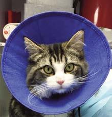
Treatment and prognosis
Any other systemic influences should be addressed. Until discomfort can be controlled, mutilation may need to be prevented by using an Elizabethan collar and/or paw bandaging (Figure 5). If this approach is taken, it must be remembered that using barrier methods of this nature has a negative welfare impact with decreased quality of life, and therefore, stress reduction will be even more important.11
Periodontal disease
In an older cat, it is highly likely that periodontal disease has precipitated the disease and therefore examination under general anaesthesia, intraoral radiographs and appropriate management is recommended. It is important to supply appropriate analgesia and for long enough.5 Referral to a veterinary dental specialist is recommended if the expertise and equipment are not available to extract teeth correctly, especially as alveolar bone compromise can damage trigeminal nerve endings and precipitate a maladaptive pain state. Some cases of FOPS develop following routine dental treatment and extractions.
Early and aggressive therapy for postoperative pain reduces the risk of developing a persistent pain state.12
Figure 4. Example of a dental chart which is available to download at catfriendlyclinic.org
Control & Therapy Series – Issue 314 March 2024 Centre for Veterinary Education | Est. 1965 Page 25
Figure 5. A soft Elizabethan collar may be required to control self-mutilation
With peridontal disease, early and aggressive therapy for postoperative pain will reduce the risk of developing a persistent pain state.12 For cats with severe oral disease, long-term analgesia will be required after dental extractions.
Performing nerve blocks to desensitise the oral cavity completely will theoretically reduce the chance of the dental procedure exacerbating the pain. The maxillary nerve block, where an injection of lidocaine and bupivacaine is made in the direction of the maxillary foramen in the pterygopalatine fossa, should block the pterygopalatine ganglion.13 Long-term analgesia is required after dental extractions in cats with severe oral disease.5
Environmental needs
Addressing environmental needs according to the five ‘pillars’ framework is an essential, not an optional, part of the management of a FOPS-affected cat (Figure 6) 14 The home should be optimised so that the cat feels in control and has free and immediate access to resources. It is recommended to use house plans to identify and discuss problems associated with the distribution of the cat’s essential feline resources; food, water, resting places (including elevated perches and individual hiding places), latrines and points of entry and exit into the territory. Separate resource stations are needed when there is more than one social group, and these should be out of view of other resource locations, allowing an individual cat to avoid others and minimise competition, bullying and stress.14 In multi-cat households, there should be
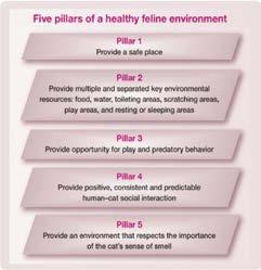
as least as many safe places as there are cats, and these should be separated from each other and have more than one entry so that it is more difficult to block. Cats should also: have free access to safe places outdoors (if possible); be able to engage in pseudo-predatory play and feeding behaviours; have positive, consistent and predictable human–cat social interaction; and have an environment that respects the importance of the cat’s sense of smell. For more details, the reader is referred to the AAFP and ISFM Feline Environmental Needs Guidelines.14
Use of commercially available feline facial pheromone F3 within the home environment, can increase the sensation of safety but will only be beneficial when used alongside environmental modifications. Finally, in multicat neighbourhoods, it may be necessary to prevent visual intrusion using blinds or temporary frosting on the windows, and to prevent physical intrusion using microchip-operated cat flaps (Figure 7)
Pharmacological treatment of FOPS
First-line analgesia for FOPS should be with licensed analgesics; however, because FOPS likely involves disordered somatosensory nervous system processing, this therapy is insufficient in many cases. Maladaptive pain is managed with adjunctive drugs that have antihyperalgesic and antinociceptive activities. Based on anecdotal information, the most useful adjuvant drugs for treating FOPS are gabapentinoids (gabapentin or pregabalin) or phenobarbital. Although the gabapentinoids are generally considered the most useful for management of feline maladaptive pain and can also reduce anxiety, it is the author’s experience that phenobarbital can be more effective for FOPS.1,15 It is not clear why this might be, if indeed it is true. Potentially, phenobarbital works by increasing activity of GABAergic inhibitory circuits; for example, influencing the trigemino- parasympathetic brain stem reflex or perception of noxious stimuli in the prelimbic region. Phenobarbital has the advantage that, for FOPS, once daily dosing may be possible and tablet sizes are more convenient for dosing. In contrast, gabapentin and pregabalin may need to be compounded. However, adverse effects of sedation, such as reduced jumping ability and ataxia, may limit the use of phenobarbital (Figure 8). Adverse effects, such as haematological abnormalities, pseudo-lymphoma and liver compromise, are rare but even less common with the gabapentinoids.16–19 For this reason, the author usually starts treatment with gabapentin, and if this is not effective within 2 weeks, switches to phenobarbital. For acute management of hospitalised cases, drugs such as parental phenobarbital, benzodiazepines, dexmedetomidine, nerve blocks or constant rate infusions of ketamine can be useful.
Tip
Control & Therapy Series – Issue 314 March 2024 Page 26 Authors’ views are not necessarily those of the CVE
Figure 6. The five pillars of a healthy feline environment. Ellis SLH et al. J Feline Med Surg. 10.1177/1098612X13477537. Available at: https://journals.sagepub.com/doi/epdf/ 10.1177/1098612X13477537. Copyright © 2013. Reproduced with permission from SAGE Publications, Ltd.
Cats with ongoing anxiety due to environmental stress should have their welfare needs addressed; however, there may be some cases where mediation with selective serotonin reuptake inhibitors is necessary while environmental optimisation is achieved.
In kittens, signs resolve spontaneously when ‘teething’ is complete, and it should be possible to withdraw medication over a 4–8-week period.
In adults, if the periodontal disease is addressed successfully, then it may be possible to withdraw the adjuvant analgesic; however, as healing after dental extraction can be prolonged, it is recommended that this be delayed until after the cat has been free of clinical signs for at least 4 weeks.
FOPS may occur in bouts over a period of weeks or months, with subsequent spontaneous remission. Over time, however, the disease can become unremitting, with up to 10% of cases being euthanased because of the perceived poor quality of life.
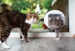
References
1. Rusbridge C, Heath S, Gunn-Moore DA, et al. Feline orofacial pain syndrome (FOPS): a retrospective study of 113 cases. J Feline Med Surg 2010; 12: 498–508.
2. Schurks M. Genetics of migraine in the age of genome-wide association studies. J Headache Pain 2012; 13. DOI: 10.1007/s10194011-0399-0.
3. Gormley P, Winsvold BS, Nyholt DR, et al. Migraine genetics: from genome-wide association studies to translational insights. Genome Med 2016; 8: 86.
4. Esserlind AL, Christensen AF, Le H, et al. Replication and metaanalysis of common variants identifies a genome-wide significant locus in migraine. Eur J Neurol 2013; 20: 765–772.
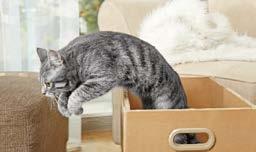
5. Watanabe R, Doodnaught G, Proulx C, et al. A multidisciplinary study of pain in cats undergoing dental extractions: a prospective, blinded, clinical trial. PloS One 2019; 14.DOI: 10.1371/journal.pone.0213195.
6. Overall KL, Rodan I, Beaver BV, et al. Feline behavior guidelines from the American Association of Feline Practitioners. J Am Vet Med Assoc 2005; 227: 70–84.
7. Elzerman AL, DePorter TL, Beck A, et al. Conflict and affiliative behavior frequency between cats in multi-cat households: a surveybased study. J Feline Med Surg 2019;
8. 22. DOI: 10.1177/1098612X19877988.
9. Niemiec B, Gawor J, Nemec A, et al. World Small Animal Veterinary Association global dental guidelines. J Small Anim Pract 2020; 61. DOI: 10.1111/jsap.13132.
10. Tsugawa AJ and Verstraete FJ. How to obtain and interpret periodontal radiographs in dogs. Clin Tech Small Anim Pract 2000; 15: 204–210.
11. Niemiec BA. Feline dental radiography and radiology: a primer. J Feline Med Surg 2014; 16: 887–899.
12. Shenoda Y, Ward MP, McKeegan D, et al. ‘The cone of shame’: welfare implications of Elizabethan collar use on dogs and cats as reported by their owners. Animals 2020; 10. DOI: 10.3390/ani10020333.
13. Kehlet H, Jensen TS and Woolf CJ. Persistent postsurgical pain: risk factors and prevention. Lancet 2006; 367: 1618–1625.
14. Aguiar J, Chebroux A, Martinez-Taboada F,et al. Analgesic effects of maxillary and inferior alveolar nerve blocks in cats undergoing dental extractions. J Feline Med Surg 2015;17: 110–116.
15. Ellis SLH, Rodan I, Carney HC, et al. AAFP and ISFM feline environmental needs guidelines. J Feline Med Surg 2013; 15: 219–230.
16. Gandolfi B, Rusbridge C, Malik R, et al. You’re getting on my nerves! The feline orofacial pain syndrome. Proceedings of the Tufts’ Canine and Feline Breeding and Genetics Conference, Boston, MA, 2013.
17. Ducote JM, Coates JR, Dewey CW, et al. Suspected hypersensitivity to phenobarbital in a cat. J Feline Med Surg 1999; 1: 123–126.
18. Lieser J and Schwedes CS. Pseudolymphoma in a cat on phenobarbital treatment. J Small Anim Pract 2018; 59: 444–447.
19. Djani DM and Draper WE. Suspected phenobarbital-induced fever in a cat. JFMS Open Rep 2019; 5. DOI: 10.1177/2055116919830214.
20. Charalambous M, Pakozdy A, Bhatti SFM, et al. Systematic review of antiepileptic drugs’ safety and effectiveness in feline epilepsy. BMC Vet Res 2018; 14: 64. DOI: 10.1186/s12917-0181386-3.
 Figure 7: Prevent intrusion from other cats via the cat flap
Figure 8. Adverse effects of sedation, such as reduced jumping ability and ataxia, may limit the use of phenobarbital
Figure 7: Prevent intrusion from other cats via the cat flap
Figure 8. Adverse effects of sedation, such as reduced jumping ability and ataxia, may limit the use of phenobarbital
The International Society of Feline Medicine is the veterinary division of the pioneering cat welfare charity International Cat Care. Trusted by vets and nurses, it provides a worldwide resource on feline health and wellbeing, via the Journal of Feline Medicine of Surgery, by fostering an international community of veterinary professionals with a shared vision of feline welfare, and supporting professional development with practical CPD. Additionally, International Cat Care’s website provides a valuable resource of accurate information delivering what both vets and cats would want owners to know.
Research Roundup
Welcome to Research Roundup where we bring you summaries of the latest feline research. Included are articles on the practical use of POCUS in cats with ureteral obstruction, the significance of thrombocytosis and barriers to the measurement of blood pressure. Winner
Entitled to a CVE$100 voucher
Feline Arterial Thromboembolism in a Cat
Dr Christopher Simpson BVSc MANZCVSc
Victoria Veterinary Clinics
Hong Kong
e. simpson_christo@icloud.com
C&T No. 6013
Hugo is a 1-year-old male neutered Ragdoll cat. He was presented to us profoundly dyspnoeic, with bilateral hindlimb (HL) paralysis. The owner reported that no cardiorespiratory or other signs had been present prior to the day of presentation. He had been normal the night before. The owner was awoken by his loud vocalisation in the morning, and he was found severely distressed, dyspnoeic, and paralysed, and brought straight to the hospital.
A brief/abridged clinical examination indicated that Hugo was severely dyspnoeic. Lung sounds were muffled, and cardiac auscultation not possible. His HLs were paralysed bilaterally, without detectable femoral pulses, and with cold/cyanotic footpads.
He was placed in an O2 cage and given butorphanol (0.1 mg/kg IM) as an anxiolytic, while further diagnostics were discussed with the owner.
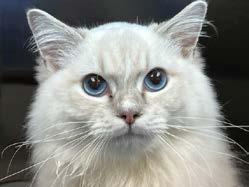
The owner consented to plain radiographs and a pointof-care (POC) thoracic ultrasound.
These both confirmed the presence of a large volume of pleural effusion.
The owner then gave consent for therapeutic and diagnostic thoracocentesis.
IV access was obtained carefully, and Hugo was then sedated with midazolam 0.2 mg/kg + alfaxalone 0.2 mL boluses to effect. Mask O2 was provided during sedation.
The chest was clipped and disinfected, and then 200 mL of pink-tinged fluid was obtained (see Figure 3 ), approximately 100 mL from each side of the chest. During the centesis procedure, Hugo’s respiratory rate and subjective effort both decreased steadily.
Figure 1. Hugo the Ragdoll
Control & Therapy Series – Issue 314 March 2024 Page 28 Authors’ views are not necessarily those of the CVE
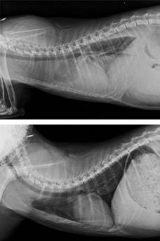
After drainage, a full diagnostic cardiac ultrasound was performed, which demonstrated marked enlargement of the right atrium (RA) and right ventricle (RV) and marked tricuspid regurgitation (TR). There was marked smoke within RA and RV, and a distinct echogenic object suspected to represent a thrombus, trailing from the RA to the RV, traversing the tricuspid valve.
The two main differential diagnoses for these kind of right-sided changes in a cat of this age are:
tricuspid dysplasia, or arrhythmogenic right ventricular cardiomyopathy (ARVC)
There is no specific medical therapy for either of these conditions, and therapy consists or diuresis, with or without centesis, and antithrombotic treatment where relevant.
Diuresis was started with frusemide by injection, initially at 2 mg/kg, then at 1 mg/kg q 2 hrs until the resting respiratory rate (RRR) was < 40 breaths/minute.
Due to the severity of the clinical presentation, including the smoke and thrombus, dual antithrombotic therapy was commenced with clopidogrel 18.75 mg sid and rivaroxaban 2.5 mg sid (as per Lo et al JVIM
2023—Synergistic inhibitory effects of clopidogrel and rivaroxaban on platelet function and platelet-dependent thrombin generation in cats.)
Despite being given an extremely guarded prognosis, Hugo proved to be an extremely stoic and resilient patient. Within minutes of waking up from the centesis procedure he was sitting up in the O₂ cage and ate food when offered.
By the next day he was dragging himself around by the front legs and was trialled out of O₂. Bloods were run to check for any azotemia or electrolyte derangements suggestive of overzealous diuretic use. These showed a mildly increased BUN, and moderate hypokalaemia, but normal CREA.
Hugo continued to eat well, and the decision was made to discharge him on oral frusemide at an equivalent of 1 mg/kg bid, the anti-thrombotics as above, Renal K gel as a potassium supplement, and transdermal mirtazapine.
The owner was instructed carefully on monitoring the sleeping respiratory rate (SRR) as a means of monitoring
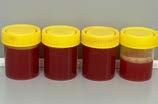
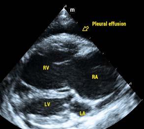 Figure 2. Chest radiographs, before and after paracentesis and diuresis
Figure 3. Fluid obtained by thoracocentesis
Figure 2. Chest radiographs, before and after paracentesis and diuresis
Figure 3. Fluid obtained by thoracocentesis
Control & Therapy Series – Issue 314 March 2024 Centre for Veterinary Education | Est. 1965 Page 29
Figure 4. Massive R-sided enlargement, with smoke visible in RA, and a thrombus faintly visible traversing the TV. (This was more evident on moving images.)
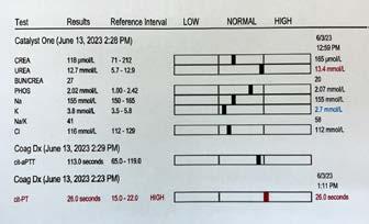
for any recurrence of pleural effusion or pulmonary oedema. The owner was also advised to closely check all the venipuncture sites, shaved skin and mucosal surfaces for any signs of bleeding which may indicate excessive anti thrombotic effect.
At a one-week revisit, Hugo’s improvement was astonishing. He was bright, eupneic, friendly/calm, and walking completely normally!
Repeat bloods (shown below) showed correction of the BUN elevation, the hypokalaemia, and a mild PT prolongation, indicative of a satisfactory degree of antithrombotic effect from the clopidogrel and rivaroxaban.
At the time of writing, Hugo has been on frusemide, clopidogrel and rivaroxaban now for 6 weeks, and is totally asymptomatic. The owner has been diligent in recording the SRR nightly, and this has remained under 30 breath/minute at all times.
As a matter of routine, cats revisiting our clinic for blood tests or repeat basic imaging are giving 50-100 mg of gabapentin at home before revisits.
This reduces the stress of restraint, blood collection, and POC imaging.
The problem in this case – is the pathophysiology does not make sense! The cat has a right sided problem, with the clot present in the aorta likely haven arisen in the RIGHT atrium. But how does the clot in the right atrium end up in the distal aorta? The only logical mechanism would be a connection between the right and left side of the heart – and the defect most likely to do this without causing a murmur or obvious turbulence on colour flow Doppler, would be an atrial septal defect, such as a patent foramen ovale. To find such a defect, one would need a very detailed Doppler study, tweaking the machine to detect low flows and possible a ‘bubble-0gram’. These studies were not clinically justified when the cat was presented in such a critical state.
We plan to monitor the cardiac status with repeat ultrasounds every 3 months until a clear trend is evident, then potentially de-escalate to every 6 months if progression is slow and the owner is confident in observations at home.
The owner will advise us immediately if the SRR becomes elevated at any time in between scheduled revisits.
Figure 6. Repeat bloods
Figure 5. Spectral Doppler study through the right ventricular inlet showing marked tricuspid regurgitation (TR)
Control & Therapy Series – Issue 314 March 2024 Page 30 Authors’ views are not necessarily those of the CVE
Save Your Dog’s Hock! Tips for Trainers
Practical Strategies for PreventingHock Fracture in Racing Greyhounds
Dr John Katakasi BVSc (Hons) CVA CVCHM
Adelaide Plains Veterinary Surgery, SA
Dr David Larratt BVSc IVAS
The Lake Veterinary Hospitals, NSW
e. greyhoundtarsalscreening@gmail.com
Dr Peter Yore BVSc, IVAS
Belmont Veterinary Hospital, NSW
C&T No. 6014
Read Save Your Dog’s Hock! Tips for Trainers
This educational booklet on fracture prevention of the hock (tarsus) in racing greyhounds was distributed to trainers and On Track Veterinarians (OTVs) in Australia and New Zealand—a collaboration of the 3 veterinarians named who have devoted the majority of their careers assisting the welfare of racing greyhounds by diagnosing, treating and searching for prevention of the common serious injuries. They have correlated palpable damage to the joint structure (both soft tissue and bone) with repeatable diagnostic markers observable with radiography and CT scanning.
Specifically written to raise awareness of the signalment and recently documented diagnostic options available
to monitor and prevent hock fracture in racing greyhounds, it contains practical information on joint stability, assessment and a new approach to radiographic investigation, which is also relevant to all veterinarians working with more active and sporty domestic dogs.
This pictorial booklet covers:
1. Examination techniques
2. Strapping guidelines
3. X-Rays.
Across Australia, industry-sponsored orthopaedic repair has dramatically reduced euthanasia; however, the serious injury rate in NSW has increased by 100% in the last 5 years (GWIC, 2023). If these race-related injuries could be prevented, then many dogs would avoid chronic pain and joint dysfunction beyond their racing days.
It has been long established that serious tarsal fracture is linked to a significant loss of bone density (Emmerson et al., 2000) and (Hercock, 2010).
With this knowledge, what action can be taken to drive effective preventative protocols?
Unlike the equine industry, the greyhound industry is not serviced by an educational institution that registers veterinarians as specialists in small animal surgery, sports rehabilitation or imaging. Currently there is no specialisation available for veterinarians who are routinely performing preventative musculoskeletal examinations and imaging (radiography and/or CT) with racing greyhounds.
The CVE provides one of the few pathways for veterinarians in their diverse fields of special interests to share their observations and insights.
The Brutal Reality of Hock Fractures in Racing Greyhounds— Introducing Radiographic Guidelines for the Early Warning of Impending fracture (Larratt, 2023) was published in the C&T Series March 2023 Issue 310 and focused on a new approach in radiographic interpretation and correlation of 4-view tarsal radiography with CT imaging.
Publication led to an invitation to present at the annual conference of the Greyhound, Working and Sporting Dogs Veterinarians (AGWSDV) in October 2023.
The Way Forward
Evolving public attitudes have seen a positive shift in the status of the racing greyhound. The standard outcome of euthanasia for those with race-related fractures is
Control & Therapy Series – Issue 314 March 2024 Centre for Veterinary Education | Est. 1965 Page 31
now greatly reduced. Greyhound fractures are now being repaired all over the country with an estimated 700 race-related serious tarsal fractures occurring in 2023. However, fractures are painful and if avoidable then surely prevention is a superior welfare option.
The release of Save Your Dog’s Hock has stimulated positive response within the greyhound and veterinary industries. A new website has been created that provides further detail on radiographic positioning and interpretation:
www.greyhoundtarsalscreening.com.au
References
Emmerson, T.D., Lawes, T.J., Goodship, A.E., Rueux Mason, C. and Muir, P. (2000) Dual energy X ray absorptiometry measurement of bone mineral density in the distal aspect of the limbs in racing Greyhounds. Am J Vet Res 61, 1214 1219.
GWIC (2023), Analysis of greyhound racing injuries, 1 January – 31 March 2023. https://www.gwic.nsw.gov.au/__data/assets/pdf_file/0003/1144461/1-January-31March-2023-Injury.pdf
GWIC (2024) Greyhound Welfare & Integrity Commission https://www.gwic.nsw.gov.au/
Hercock, C.A. (2010). Specialisation for fast locomotion: performance, cost and risk. Thesis for the degree of Doctor in Philosophy. The University of Liverpool, Liverpool, United Kingdom.
Larratt, D. (2023) The Brutal Reality of Hock Fractures in Racing Greyhounds – Introducing Radiographic Guidelines for the Early Warning of Impending Fracture, C&T Control & Therapy Series, Issue 310; March 2023, Centre for Veterinary Education, The University of Sydney, NSW, Australia.
Yore, P., Katakasi, J. and Larratt, D. (2023) Save Your Dog’s Hock! – Tips for Trainers. https://www.greyhoundtarsalscreening.com.au/tarsal-screening

Burkholderia Infection in a Cat (Likely Meliodosis)
Candice Yeo
C&T No. 6015
A 1.5-year-old female neutered Domestic Short Haired cat (2.4kg) presented to a clinic located in Singapore with a 1 month history of recurrent left hindlimb swelling causing lameness. Prior to presentation, she had been hospitalised for 2 days due to the left hindlimb swelling, lethargy and hyporexia and treated with parenteral clindamycin and amoxicillin-clavulanic acid and a non-steroidal anti-inflammatory drug (tolfenamic acid; Tolfadine).
Blood tests had revealed a moderate hyperglobulinaemia and neutrophilia (no left shift appreciated). Previous radiographs of her left hindlimb showed signs of osteomyelitis. Her previous medical history included recurrent skin disease (bacterial dermatitis and dermatophytosis) that had been managed with prednisolone, cephalexin and itraconazole and she was suspected to have food responsive skin allergies. Prior to adoption, she had been an outdoor cat.
On physical examination, her left hock was markedly swollen with draining tracts noted on the lateral and medial aspect of the hock with a large amount of purulent exudate. The left popliteal lymph node was mildly enlarged, but the rest of her peripheral lymph nodes were normal. She was pyrexic (39.8°C). She was of a good body condition score but was noted to be of a small stature. No other skin lesions were noted, and the rest of the physical examination was unremarkable.
She was admitted for further supportive care with plans for advanced imaging of the affected limb the following day. Computed tomography revealed left tarsal soft tissue swelling around the calcaneal (Achilles) tendon and palisading new bone proliferation around the left calcaneus (yellow arrows in Figure 1). Hence the differentials included inflammatory, infectious disease of the soft tissue, osteomyelitis, or septic arthritis. There was also focal bone loss in the proximal tuber calcanei, suspicious of previous trauma or osteomyelitis. As expected, there was marked hind limb muscle atrophy and left popliteal lymphadenomegaly. The left inguinal lymph node was noted to be enlarged as well.
A culture swab of the exudate was taken and sent to IDEXX laboratories in the USA and results revealed a multi-drug resistant Burkholderia species only
Top Tips for Successful Biopsies TimeOnline OnDemand $150 for Members | $300 for Non-Members Authors’ views are not necessarily those of the CVE
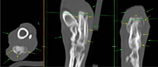
susceptible to doxycycline and imipenem. An oesophageal feeding tube was placed under the same general anaesthesia to facilitate nutrition and feeding of medication.
She was started on doxycycline and continued on antiinflammatories and pain relief and the pyrexia resolved after 2 days. Topical management included manuka honey and bandaging.
Rapid response to treatment was noted, with the swelling resolving within 1 week. Doxycycline was initially continued for 6 more weeks in total; on repeat examination, the left hock was normal and the popliteal enlargement had resolved. There was no pain on manipulation of the affected limb. Repeat blood tests showed the neutrophilia and hyperglobulinaemia had resolved.
2 weeks post stopping the doxycycline, she represented with hock swelling but without any discharge and much less swelling than before. She responded well to restarting of doxycycline and after consultation with a surgeon, we opted for a 6-month course of doxycycline due to concerns of lower diagnostic yield at this point of time. At time of writing, hock swelling has completely resolved.
Discussion
The specific species was not able to be cultured due to biosafety issues. However, Burkholderia pseudomallei is a concern given the zoonotic potential of this disease and given the cat’s history of being an outdoor cat, it could not be excluded as a possibility. Burkholderia pseudomallei is a soil saprophyte present in Southeast Asia and northern Australia¹ and can be fatal, especially in humans with predisposing factors (renal disease, diabetes mellitus, injuries). ² Infection occurs by inoculation, ingestion, or inhalation of the organism
in the environment. Animal-to-human transmission is possible.
Burkholderia can be isolated from various organs (liver, spleen, meninges, pulmonary parenchyma).3,4 Clinical disease can be mild, chronic with abscessation, or can cause acute and fatal disease.³ As melioidosis can present in many forms, it is an important differential for practitioners to consider in endemic areas.
Burkholderia pseudomallei can be cultured on standard media such as blood or MacConkey agar but identification may require more specific biochemical phenotyping, genetic information and specific antibodies as it is commonly misdiagnosed as B. cepacian Polymerase chain reaction can also diagnose B. pseudomallei.³ If cultured, melioidosis may be a notifiable disease in some states.
Owners of infected pets should be educated on the zoonotic potential of this disease. In humans, clinical signs include fever, headache, cough, chest pain, joint and muscle pain and they should seek medical attention if owners have been exposed.
In veterinary clinics, gloves and the washing of hands after handling animals are both recommended. Other precautions include wearing disposable gowns, shoe covers, face shields, face masks and hairnets. This organism is susceptible to disinfectants including 1% sodium hypochlorite, 70% ethanol and formaldehyde.
Unfortunately, treatment of melioidosis requires long courses of antibiotics and this bacteria is inherently resistant to many antibiotics in vitro.⁵ Acute fulminant disease is often difficult to cure and often fatal; owners should be made aware of the poor prognosis. In cats that have been cured, lifelong monitoring for relapse is recommended.⁴
Control & Therapy Series – Issue 314 March 2024 Centre for Veterinary Education | Est. 1965 Page 33
Figure 1. CT images of the cat’s hock. Computed tomography revealed left tarsal soft tissue swelling around the calcaneal (Achilles) tendon (green arrows) and palisading new bone proliferation around the left calcaneus (yellow arrows)
References
Raja NS, Ahmed MZ, Singh NN. Melioidosis: an emerging infectious disease. J Postgrad Med. 2005;51:140–145. doi: 10.5005/jp/books/11617_52.
Currie BJ, Ward L, Cheng AC. The epidemiology and clinical spectrum of melioidosis: 540 cases from the 20 year Darwin prospective study. PLoS Negl Trop Dis. 2010;4:e900.
Sprague LD, Neubauer H. Melioidosis in animals: a review on epizootiology, diagnosis and clinical presentation. J Vet Med B Infect Dis Vet Public Health. 2004 Sep;51(7):305-20. doi: 10.1111/j.1439-0450.2004.00797.x. PMID: 15525357.
O’Brien CR, Krockenberger MB, Martin P, Parkes H, Kidd M, Malik R. Disseminated melioidosis in two cats. J Feline Med Surg. 2003 Apr;5(2):83-9. doi: 10.1016/ S1098-612X(02)00094-3. PMID: 12670433.
Adam W.J.J., Gary L., Dale A. and etc (2001). Antibiotic susceptibility of Burkholderia pseudomallei from tropical northern Australia and implications for therapy of melioidosis. International Journal of Antimicrobial Agents 17:109-113.
Comment Courtesy of Professor Mark Krockenberger
BSc(vet) BVSc PhD GradCertEdStud FANZCVS (Anatomical Pathology)
This case is a good reminder of a couple of things:
1. To suspect the unusual when a case doesn’t respond as expected, and
2. That, particularly in the north of Australia and in SE Asia and less commonly in subtropical Australia, melioidosis can occur in our companion animals. The causative organism can be found widespread in soil and water in those tropical environments. It is a potential zoonosis, particularly when cultured, so let your lab know if you are suspecting it. MALDI-TOF is probably the most commonly used method of species identification now rather than biochemical testing of isolates but, as the author notes, molecular confirmation of identification may be necessary, particularly when looking at the epidemiology of this disease. The distribution of animal cases of melioidosis could be an important part to add to the epidemiology of human melioidosis. This is another case where veterinarians should think One Health! More information will emerge as more veterinary isolates are typed but in the meantime vets should take note of the human literature when they encounter this disease where resistant isolates mostly emerge during long term therapy and initial therapy is relatively standard.
Aeromonas hydrophila
a Likely Causative Agent of Segmental Ulcerative Colitis in a Human Recipient
Dr Peter Kerkenezov
BM BVSc DipAppSc Cert Equine Surgery
e. equivet@nor.com.au
C&T No. 6016
Abstract - 16 September 2023
The concept One Medicine has evolved since its inception by ‘the father of modern pathology’, a Prussian, Rudolf Ludwig Carl Virchow (13/10/1821 –05/09/1902). Veterinary medicine, human medicine and environment are unequivocally interwoven on many fronts. Virchow coined the term ‘zoonosis’ after his discovery of the nematode parasite, Trichinella spiralis A. hydrophila is a bacterial zoonosis and the subject of this essay that endeavours to explain how the pathogenicity of this bacterium (prokaryotic) could possibly be transferred to a mammalian host colonic cell (eukaryotic) by plasmid (a vector) transfection. That is, the bacterium’s capacity to cause disease could be transferred to the recipient host colonic cell lineage when provided the opportunity, predominantly dose and time, and remains there indefinitely long after the perpetrator has been eliminate by antibiotic. Each recipient colonic epithelial cell then undergoes ongoing self-degradation, generating an inflammatory response. The recipient’s only natural defence is an immune response. Local and systemic medicinal intervention is also discussed.
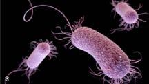
Figure 1. A. hydrophila. Image source: The Atlantic
Read the full article here. Control & Therapy Series – Issue 314 March 2024 Page 34 Authors’ views are not necessarily those of the CVE
Perplexity AI
Editor's Note
Centre for Veterinary Education
C&T No. 6017

Image from The Japan Times
https://www.perplexity.ai/
Perplexity AI is a new AI tool which displays the sources of the information it presents—because the bots occasionally still get it wrong. Phew—they still need a few humans around.
The first thing I tried out was checking the current recommendations for vaccinating cats in NSW, given the current vaccine shortages.
You can see the results below:
The sources referenced include Sydney University, The RSPCA and the AVA.
Not bad. Quicker than logging into and searching VIN.
You don’t need an account, and you can ask follow up questions.
Next I tried ‘antifungal resistance to Malassezia in dogs’ and was immediately provided with several journal articles including:
Peano A, Johnson E, Chiavassa E, Tizzani P, Guillot J, Pasquetti M. Antifungal Resistance Regarding Malassezia pachydermatis: Where Are We Now? J Fungi (Basel). 2020 Jun 25;6(2):93. doi: 10.3390/jof6020093. PMID: 32630397; PMCID: PMC7345795
Of course you still need to use your brain to decide which sources are reputable and current. I think our jobs are safe from AI for a while but for a quick ‘duck out the back and look something up’ it seems like a pretty handy tool.
'What is the current shortage of feline vaccines in Sydney'

Answer
Due to complicated supply problems post-COVID, there is currently a shortage of feline vaccines in Australia. The shortage is expected to continue until early 2024. MSD Animal Health and Boehringer Ingelheim have provided information on the availability of their F3, F4, and F5 vaccines, with some expected to be available in late March 2024 and late April 2024. As a result of the shortage, veterinarians are advised to prioritize the vaccination of previously unvaccinated kittens and cats wherever possible. Adult cats that have had a series of 2-3 kitten vaccines and an annual booster are at much lower risk of vaccine-preventable diseases, so delaying boosters for adult cats until supply normalizes in early 2024 is unlikely to have a significant impact on their health. Boarding establishments are also advised to prioritize the vaccination of at-risk animals and to seek veterinary advice on whether unvaccinated cats can be safely boarded in isolation from other cats. The shortage has led to some animal shelters, welfare groups, and rescue organizations turning away cats and kittens, and has also affected the intake of cats in New South Wales. The F3 vaccine, which protects against feline panleukopenia (cat parvovirus), feline calicivirus, and feline herpesvirus (cat flu), is particularly affected by the shortage. It is considered a "core" vaccine, crucial to protecting cats with an unknown vaccination history, such as strays and kittens, with boosters administered every one to three years throughout the cat’s life 1,2,
Control & Therapy Series – Issue 314 March 2024 Centre for Veterinary Education | Est. 1965 Page 35
3
Management & Healing of a Large, Necrotic Dog Attack Wound in a Pet Goat
Curtis A. Goding BVSc (hons)
Associate Veterinarian
Williams River Vet Clinic
28 Grey St, Clarence Town NSW 2321
C&T No. 6018
Introduction
Wounds are a frequent clinical presentation for small and large animal patients in veterinary practice. Dog-related livestock injuries and mortalities are commonplace in rural Australia¹ and around the world.² Dog bites commonly cause initial trauma through shearing/tearing; however, significant after effects from the crushing forces can cause devitalisation and delayed wound issues.³
Fly larvae infection, or myiasis, is a common complaint in rural areas⁴ and in Australia it is particularly problematic for small ruminants.⁵ Wound myiasis is where wounds from other causes, such as trauma, are infected with
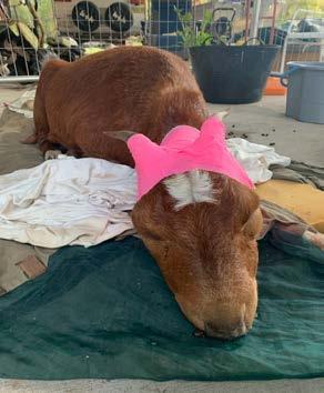
fly larvae and this can cause infection, discomfort and delayed wound healing.⁶
Wound management is a large research area in human medicine with ever-increasing innovation and development.⁷ Many of these therapies are developed using animal-based models and therefore offer a translatable evidence base for veterinary patients.⁸ Veterinary-specific data supporting of the use of certain therapies for animal wound healing can be lacking.9, 10 As such vets can rely on experience with previous successful treatment protocols with their own cases or those of their colleagues to guide decision making.
Wounds heal through overlapping phases simplified as haemostasis, inflammation, proliferation, and remodelling.7,8 Debridement is an essential part of wound healing and can be either autolytic (where the body naturally removes necrotic tissue), or through surgical, manual (wound irrigation or wet-to-dry wound dressings) or biological methods (maggot therapy).11 Manuka honey has been studied extensively as a wound gel preparation in the human field and has a distinct role to play in the healing of certain wounds.12
In January 2022 a caprine patient presented to our clinic after hours for a poorly healing wound and increasing systemic disease complications. He was hospitalised and stabilised, and then the wound was addressed over an 8-week treatment course.
Patient description
Lenny
Signalment: 3.5yo male desexed Anglo-Nubian goat. Weight: 45 kgs and body condition score 2/5
Case history
Lenny was a pet goat who was attacked at home by two dogs in January 2022. He was taken to an emergency equine hospital where the wound was assessed, a penicillin course started, and he was given pain relief. He was managed at home with wound cleaning with the assistance of neighbours until he became increasingly uncomfortable and had difficulty eating and breathing.
Physical examination findings
Initially Lenny presented with a bandaged wound ( Figure 1). He was lethargic and had a rectal temperature of 39.5°C. He had bilateral nasal oedema and serousmucoid discharge. The oedema extended down under his jaw and neck. His abdomen was tucked up and he had
Figure 1. Lenny as he presented to the clinic. Bandage in place and marked swelling of his entire face
Winner - Best Visuals
Control & Therapy Series – Issue 314 March 2024 Page 36 Authors’ views are not necessarily those of the CVE
Entitled to CVE$100 voucher
reduced rumen fill. The wound was approximately 25 cm across from the caudal mandible to the middle of the neck laterally. There were live maggots within the tissues and the wound bed consisted mainly of hard eschar type necrotic tissue. His right ear was missing, and the ear canal was indistinguishable from the surrounding wound. He also had lacerations to his left ear and various puncture wounds along the ventral neck and left-hand side of his face.
Treatment plan
The following treatment plan was devised based on the presence of severe systemic illness and decompensation secondary to the attack and poorly healing wounds.
1. Fluid therapy: A 21g catheter was placed in the right cephalic vein and Hartmann’s given at approximately 5 mL/kg/hr for 24 hours.
2. Antimicrobials: Penicillin was continued, now at 22mg/ kg IM BID, Oxytetracycline was started 15mg/kg IM BID.
3. Anti-inflammatories: 10mg of dexamethasone was given IM once only.
4. Nutritional support: Syringe feeding of small volumes of easily digestible forage replacer for herbivores and electrolyte mix put into his available water.
5. Initial wound care: Clipping of the surrounding skin. Manual removal of all fly larvae then cleaning with diluted iodine solution and gauze swabs.
6. Initial bandage: Application of manuka honey, non-adhesive dressing, cotton wool, vetwrap and elastoplast. This was changed twice daily initially.
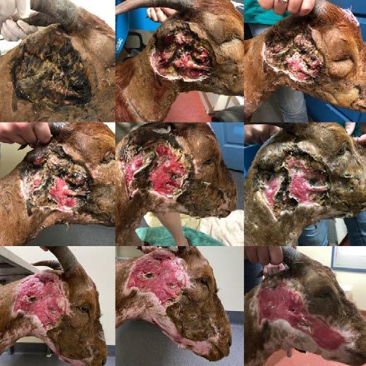
Control & Therapy Series – Issue 314 March 2024 Centre for Veterinary Education | Est. 1965 Page 37
Figure 2. Lenny’s extensive facial trauma and initial healing progress
Approximately four days into his critical care the swelling over his mandible and nose had begun to reduce and Lenny started eating grass spontaneously. His breathing improved significantly, and the nasal discharge resolved. At this point the decision was made to persevere with treatment having set clear endpoints. Realistic expectations were discussed regarding the timeframe for healing and cost, and a mutually agreed plan put in place. The oxytetracycline was stopped and the bandage was no longer changed twice daily.
1. Staged surgical debriding: Performed under a standing sedation of butorphanol (0.1mg/kg) and diazepam (0.3mg/kg) given intramuscularly. 10mL of injectable 2% lignocaine was applied topically to the wound bed. Then a combination of sharp and blunt debridement of visible eschar and scoring of thick, well-adhered necrotic tissue. This procedure was well tolerated overall and ceased when Lenny became uncomfortable.
2. Meloxicam was commenced at 1mg/ kg SC and repeated every 2-3 days.
3. Penicillin in the long-acting formulation was then given at 22mg/kg every 2-3 days.
4. Subsequent manual debriding was performed after bandage removal and the wound cleaned with diluted iodine. The wound was then debrided with either dry gauze swabs or surgical instruments as necessary with consistent positive progress (Figure 2).
5. The bandage was changed every 2-3 days in the fashion previously described with some variation in bandaging material depending on the vet’s preferences. The bandage was eventually adapted to incorporate the right-hand side of the face after delayed necrosis and sloughing of this area.
6. Once the granulation bed was filled in with no remaining necrotic tissue and Lenny had a consistent appetite, the decision was made to leave the bandage off and switch to twice daily cleaning with diluted iodine, drying of the wound bed and the application of an antiseptic, fly repellent spray.
Expected outcome of the treatment plan
It was expected that if the wound healed, it would take a period of months with wound infection considered the main possible complication of treatment. The staff at the clinic had grave concerns initially about the ability of a wound of this size to heal at all.
Actual outcome
The wound had healed entirely two months into the treatment course ( Figure 3 ).
The external ear was completely destroyed but we found the ear canal about halfway through healing and it remains as just a small hole into the horizontal ear canal.
Discussion
Myiasis is a considerable welfare challenge in livestock medicine.4, 13-15 Human cases are treated by complete removal of the larvae through various means and then debridement of necrotic tissue.⁶ Fly larvae have been used as a treatment for necrotic wounds for thousands of years and it remains a utilised method of biological wound debridement in selected human and veterinary cases.9,16 In human cases, the process of maggot therapy is highly controlled whereby sterile larvae, from species of fly where the maggots mainly debride necrotic and not living tissues, are utilised.16 In cases of wound myiasis where the maggots are uncontrolled and allowed to digest living tissue they can cause wound enlargement, infection and severe discomfort.⁶ Therefore, there should be no misconception that uncontrolled, nonsterilised maggots infecting a wound in the field serve any benefit.
In Lenny’s case, normal and efficient healing processes had been disrupted by the time he was presented to our clinic. Manuka honey was selected as the best means to fight localised infection and promote cell proliferation for second intention healing. Manuka honey’s benefit in wound healing is through its anti-bacterial and antiinflammatory properties, as well as lowering wound pH, maintaining moisture, and promoting autolytic debridement.17 There is limited evidence for the use of manuka honey in veterinary settings10 and yet it is widely used in our clinic for all species with wounds. In goats there is only one paper that compares honey and aloe vera for wound healing, and it found the aloe vera to be preferential, although both methods were effective.18 In this case it proved very effective, as one of only a few treatments employed to manage such a large wound. When manuka honey is applied to eschar, the recommendation is to score this tissue to allow the honey’s penetration into underlying tissue to prevent further drying out of the wound and encourage further debridement.19
Bandage-related injuries, particularly swelling and ischemia, are a reported complication of inappropriate bandage application in veterinary medicine.20 Initially Lenny presented with marked swelling to his face and neck, dyspnoea, nasal discharge, and difficulty eating. It is unclear whether earlier bandaging had been applied too tightly or if this swelling was due to localised woundassociated cellulitis or destroyed lymphatic drainage from the trauma. Resolution of this swelling was our priority, and ensuring no recurrence due to subsequent bandaging was one of the challenges in Lenny’s care. The wound was bandaged for three quarters of the total healing time in this case. The bandage served multiple functions: protection from environmental contamination (including reintroduction of fly larvae) and increasing
Control & Therapy Series – Issue 314 March 2024 Page 38 Authors’ views are not necessarily those of the CVE
contact between our chosen wound gel and the wound. The bandage shape and design varied little between the vets and nurses applying it, often incorporating the horns to reducing slipping and with a priority being placed on natural fibre bandaging material for breathability and patient comfort.
The wound was surgically debrided 3-4 times during the treatment course, each time only under a light sedation or conscious. Often a significant amount of necrotic tissue was left within the wound bed. This was a decision that we grappled with and settled on for several reasons to:
avoid general anaesthesia which was thought to be too risky with the level of trauma and swelling present initially;
prevent inadvertent overzealous debridement and the loss of potentially viable wound tissue, which could further increase the wound size; ensure no exposed functional vasculature was damaged during debridement leading to further necrosis in adjacent area; and reduce treatment costs.
Therefore, autolytic debridement or relying on the body’s healing process (enhanced by manuka honey) was relied upon and proved very effective. Necrotic tissue will serve as a nidus for infection and should be removed from wounds to facilitate prompt healing.21 In this case, a staged surgical debridement approach was taken and proved very effective when used in combination with manuka honey, antibiotics and bandaging.
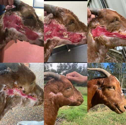
Control & Therapy Series – Issue 314 March 2024 Centre for Veterinary Education | Est. 1965 Page 39
Figure 3. Progress of Lenny’s wound once wound contraction had started
Conclusion
Large and necrotic wounds with myiasis in goats are challenging but can be managed in a general practice setting. Lenny’s large dog attack wound could have been life ending; however, with committed owners, exceptional nursing and persistence and patience on the part of his vets, a positive outcome was achieved.
Consent
This article was written with the consent of Lenny’s owner and all photos were taken with permission by clinic staff or the owner.
References
1. T.J. FPJSK. Predation of livestock by wild dogs in eastern New South Wales The Rangeland Journal 1989; 11:61-66.
2. Montecino-Latorre D, San Martin W. Evidence supporting that humansubsidized free-ranging dogs are the main cause of animal losses in small-scale farms in Chile. Ambio 2019;48:240-250.
3. Kannikeswaran N, Kamat D. Mammalian bites. Clin Pediatr (Phila) 2009;48:145-148.
4. Zumpt F. Myiasis in man and animals in the Old World; a textbook for physicians, veterinarians, and zoologists. Butterworths, London,, 1965.
5. Watts JE, Murray MD, Graham NP. The blowfly strike problem of sheep in New South Wales. Aust Vet J 1979;55:325-334.
6. Francesconi F, Lupi O. Myiasis. Clin Microbiol Rev 2012;25:79-105.
7. Tottoli EM, Dorati R, Genta I et al. Skin Wound Healing Process and New Emerging Technologies for Skin Wound Care and Regeneration. Pharmaceutics 2020;12.
8. Lux CN. Wound healing in animals: a review of physiology and clinical evaluation. Vet Dermatol 2022;33:91-e27.
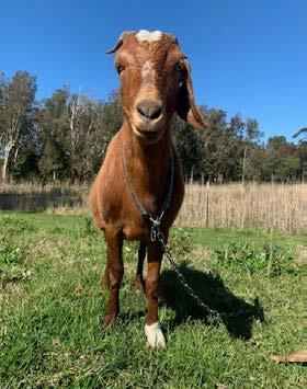
9. Jones G, Wall R. Maggot-therapy in veterinary medicine. Res Vet Sci 2008;85:394-398.
10. Vogt NA, Vriezen E, Nwosu A, Sargeant JM. A Scoping Review of the Evidence for the Medicinal Use of Natural Honey in Animals. Front Vet Sci 2020;7:618301.
11. Schultz GS, Sibbald RG, Falanga V et al. Wound bed preparation: a systematic approach to wound management. Wound Repair Regen 2003;11 Suppl 1:S1-28.
12. Jull AB, Cullum N, Dumville JC et al. Honey as a topical treatment for wounds. Cochrane Database Syst Rev 2015;2015:CD005083.
13. Wall R. Ovine cutaneous myiasis: Effects on production and control. Veterinary Parasitology 2012;189:44-51.
14. Islam M, Maruf A, Mannan M et al. Isolation and identification of associated bacteria and maggots from myiasis affected wounds of cattle and goats in Bangladesh. Journal of Advanced Veterinary and Animal Research 2015;2.
15. Alam MM, Paul S, Ferdousy RN, Tapon MAH, Juyena NS. A Retrospective Study on Occurrence of Myiasis in Ruminants. Progressive Agriculture 2014;24:101-106.
16. Nigam Y, Bexfield A, Thomas S, Ratcliffe NA. Maggot Therapy: The Science and Implication for CAM Part I—History and Bacterial Resistance. Evidence-Based Complementary and Alternative Medicine 2006;3:223-227.
17. Alvarez-Suarez J, Gasparrini M, Forbes-Hernández T, Mazzoni L, Giampieri F. The Composition and Biological Activity of Honey: A Focus on Manuka Honey Foods 2014;3:420-432.
18. Roy K, Rahman A, kar M et al. Evaluation of Topical Application of Aloe vera gel and Honey for Acceleration of Cutaneous Wound Healing in Goat Model. Journal of Bangladesh Agricultural University 2022.
19. Molan PC. Debridement of wounds with honey. Journal of Wound Technology 2009;5:12-17.
20. Anderson DM, White RA. Ischemic bandage injuries: a case series and review of the literature. Vet Surg 2000;29:488-498.
21. Manna B, Nahirniak P, Morrison CA. Wound Debridement. StatPearls, Treasure Island (FL) ineligible companies. Disclosure: Phillip Nahirniak declares no relevant financial relationships with ineligible companies. Disclosure: Christopher Morrison declares no relevant financial relationships with ineligible companies., 2023
Editor’s note:
You can kill maggots by giving the animal ivermectin (that kills everything even if they are not visible). If you want a quicker kill—dissolve Capstar in saline and use it for wound lavage or give a Capstart tablet per rectum—they absorb it very well and the larvae die—easier than removing individual maggots.
Read some previous C&T articles about honey here.
Figure 4. Lenny now fully recovered from his dog attack
Control & Therapy Series – Issue 314 March 2024 Page 40 Authors’ views are not necessarily those of the CVE

Practical Ophthalmology
These highly engaging and extremely experienced ophthalmologists present a rich educational program at the 1-day conference. Perfect your surgical techniques in the wet lab workshop using live dogs and cadavers. Workshop participant numbers are strictly limited to maximise tutor engagement and feedback.
Enrol today to improve and hone your competence and confidence in veterinary ophthalmology.
CVE Member Rewards
Conference
Friday 5 April 2024
6 CPD Points
Workshop
Choose Saturday 6 or Sunday 7 April 2024
14 CPD Points
Sydney
Mark Billson BVSc PhD DVOphthal
DipVetClinStud MRCVS
Allyson Groth BVSc MANZCVS DACVO
David Maggs BVSc (Hons) Dipl. ACVO
Professional Members save $426 (Conference), $185 (Workshop) or $611 (package)
Membership Dues: $395 p.a.
Part-Time Professionals & Recent Graduate Members save $706 (Conference), $465 (Workshop) or $1,171 package
Membership Dues: $198 p.a.
Practice Membership
Small $635 p.a. | Medium $775 p.a. | Large $1,295 p.a.
Find out more + Enrol Control & Therapy Series – Issue 314 March 2024 Centre for Veterinary Education | Est. 1965 Page 41
An Emergency Medicine Conference
Monday 21 October 2024 to Wednesday 23 October 2024
Melbourne | 20 CPD Points

Learn from the experts who share their own hard-learned lessons to help you identify and avoid common mistakes in critical care.
Dominic Barfield
BSc BVSc MVetMed DACVECC
DECVECC FHEA MRCVS
Merrin Hicks
BVSc (hons) MVSc FANZCVS (ECC)
Kate Hopper
BVSc, MVS, PhD, DACVECC
Sam Long
BVSc PhD DipECVN
CVE Member Rewards
Enrol by 23 August 2024 at member-only Early Bird discounts!
Professional Members save $540
Membership Dues: $395 p.a.
Part-Time Professionals & Recent Graduate Members save $1,180
Membership Dues: $198 p.a.
Practice Membership
Small $635 p.a. | Medium $775 p.a. | Large $1,295 p.a.
Find out more + Enrol
Canine Gastric Carcinoma
Ed
Hall, MA VetMB PhD DipECVIMCA FRCVS
Emeritus Professor of Small Animal Internal Medicine
University of Bristol, UK
e. dred.hall@bristol.ac.uk
Perspective. 161
Introduction
Compared to the human situation, malignancies of the canine stomach are rare, comprising <1% of all reported neoplasms.1 This is fortunate, as gastric carcinoma (GC), the most frequent malignancy (i.e. ~65% of all canine gastric cancers), has a very poor prognosis. Consequently, most dogs with signs of gastric disease seen in first opinion practice do not have a GC and the vast majority (excluding gastric foreign bodies and GDV cases) can be treated symptomatically with anti-emetics, H2 receptor antagonist (H2RAs) or proton pump inhibitors (PPIs), and/or dietary modification. However, known breed and age predilections and progression of clinical signs should increase suspicion for a GC.
Signalment
Typically, GC is diagnosed in middle-aged (7-10 years) large breed dogs; it is rarely a disease of geriatrics and is uncommon in smaller breed dogs. Most studies suggest male dogs are only slightly more likely to be affected but there is a clear breed predisposition in collies, especially Rough Collies, and in Belgian Shepherds (Tervuren and Groenendael). Increased incidence is also reported in the following breeds: Staffordshire Bull Terrier, Chow Chow, Labrador and Golden Retriever, Siberian Husky, Bouvier des Flandres, Norwegian Elkhound, and Cairn and West Highland White Terriers. Lundehunds with hereditary lymphangiectasia and atrophic gastritis were also reported to be predisposed to GC, but their gastric tumours are now thought to originate from neuroendocrine cells.
Aetiology
The marked breed predisposition suggests that there is a genetic basis to GC. This hypothesis is supported by the natural history of human GC. There is evidence that Napoleon Bonaparte suffered a perforated gastric tumour, although the role of heavy metal poisons including arsenic causing fatal gastric bleeding and given either as medication or as accidental or premeditated poisoning remains debated.2 Interestingly, Napoleon’s
grandfather, his father, his second brother, and all his three sisters died of ‘gastric disease’ between the ages of 40 and 60. Although GC was only confirmed in some cases, such strong familial associations suggest a genetic cause with perhaps an environmental trigger. For example, the high incidence of GC in Japanese people has been associated with their rice-based diet.
Yet it is now known that the majority of human cases arise from chronic inflammation caused by Helicobacter pylori infection. Infection in childhood is probably acquired in early years from the child’s mother through their close contact. Infection may then, in susceptible individuals and perhaps if infected with a specific pathogenic strain, lead to a progression through chronic gastritis to atrophic gastritis to gastric metaplasia and dysplasia and ultimately to the development of a GC at between 40 and 50 years of age. H. pylori is now classified as a Class 1 carcinogen by the World Health Organisation (WHO). Yet <5% of people infected with H. pylori develop GC, and so other genetic, microbiomial and environmental factors must be involved.³
Dogs often have gastric Helicobacter infections (e.g. H. felis, H. bizzozeroni ), but H. pylori is not present and dogs don’t live 40 years, and there is no evidence that these other gastric spiral organisms can cause GC. However, the concept of an infection being restricted to familial lines is one that is accepted for the perinatal transmission of Demodex infection, and progression from gastric mucosal metaplasia/dysplasia to neoplasia has been documented in Belgian Tervurens.⁴
Pathogenesis
The majority of GCs originate on the lesser curvature of the stomach (see figure 1).
They may then spread to involve the cardia and antrum; the greater curvature is relatively spared until the disease is advanced. As the mass grows it usually ulcerates and bleeds. Consequently, clinical signs of GC are related to the physical presence of a mass that may obstruct gastric outflow, to ulceration of the mass leading to haematemesis and anaemia, and to infiltration of the gastric wall causing gastroparesis. Although an epithelial tumour, the neoplasm tends to spread outwards involving the whole gastric wall (hence a loss of normal layering seen on ultrasound) and invoking a marked fibrous reaction. The wall becomes thickened and rigid, and at post mortem examination, pathologists will describe a classical ‘leather-bottle stomach’. The histological description of GCs allows their classification according to the WHO scheme based on the patterns of cells within the neoplasm, i.e. mucinous, papillary, signet-ring cell, tubular, and undifferentiated. However, this classification is only applicable to full-thickness surgical biopsies,
Control & Therapy Series – Issue 314 March 2024 Centre for Veterinary Education | Est. 1965 Page 43
and is not helpful in predicting biological behaviour or prognosis.
Clinical signs
The initial signs associated with a GC, i.e. vomiting and dysorexia, are indistinguishable from any medical gastric condition. Vomiting is not associated with feeding (assuming the dog is not already anorexic); it often
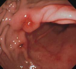
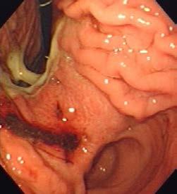
occurs on an empty stomach and can be at any time of day. Further progression of signs may then be masked by symptomatic treatment so that the tumour is quite advanced by the time the diagnosis is considered and made. However, endoscopic screening of young Belgian Shepherds has revealed metaplastic and dysplastic changes can be found in the gastric mucosa before any clinical signs occur.4 As the disease progresses, ulceration of the tumour leads to haematemesis and
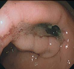
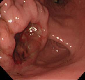 Figure 1a. A relatively early GC arising on the lesser curvature and spreading towards the greater curvature, with two distinct areas of ulceration.
Figure 1b. A more advanced GC with thickening of the angularis incisura and a bleeding ulcer
Figure 1c. A deep non-bleeding ulcer in a mass on the lesser curvature
Figure 1d. A large ulcerated GC involving part of the lesser curvature
Figure 1. Endoscopic views of canine gastric carcinomas (GCs) affecting the lesser curvature.
Figure 1a. A relatively early GC arising on the lesser curvature and spreading towards the greater curvature, with two distinct areas of ulceration.
Figure 1b. A more advanced GC with thickening of the angularis incisura and a bleeding ulcer
Figure 1c. A deep non-bleeding ulcer in a mass on the lesser curvature
Figure 1d. A large ulcerated GC involving part of the lesser curvature
Figure 1. Endoscopic views of canine gastric carcinomas (GCs) affecting the lesser curvature.
Control & Therapy Series – Issue 314 March 2024 Page 44 Authors’ views are not necessarily those of the CVE
possibly melaena, with pale mucous membranes and an increased heart rate. The dog often becomes progressively inappetent and loses weight, and some affected dogs will exhibit profuse drooling. Complete anorexia and cachexia are features of advanced disease and if present in a middle-aged dog of a predisposed breed, the index of suspicion increases significantly.5
Diagnosis
Physical examination. There are rarely any specific signs in dogs affected by a GC although cranial abdominal discomfort may be detected. In advanced disease, a cranial abdominal mass may become palpable, especially if the dog has lost a lot of body condition.
Laboratory testing. There are no pathognomic changes in the haematology and serum biochemistry, although anaemia and mid hypoproteinaemia may indicate chronic gastric bleeding. Dogs with a GC have a significantly lower serum folate concentration, and greater serum C-reactive protein concentration compared with dogs with chronic gastritis but these tests are not diagnostic.⁵
Research studies have demonstrated changes in serum pepsinogen, a variety of cell markers, particularly those associated with epithelial tight junctions, various cytokines and immunological markers, and mutations of tumour suppressor genes.⁶ Whilst these provide insight into the aetiopathogenesis of GC, none have yet been found to be useful diagnostically or prognostically in dogs.
Radiography. Detection of gastric wall thickening on plain radiographs as an indicator of GC is notoriously unreliable. Barium contrast studies may highlight a mass and ulceration but are rarely performed unless gastroscopy is unavailable. Gross metastasis to the lungs is uncommon, so although thoracic radiographs should be taken, they are usually unhelpful.
Ultrasonography. Trans-abdominal ultrasound is much more sensitive than radiography in detecting gastric masses and ulcers. Ulceration may be suspected from defects in the mucosa, and gas tracking withing the wall. Marked thickening of the wall and loss of the normal layering are highly suggestive of neoplasia. However, ultrasonography is not 100% sensitive as luminal gas can partially obscure the view.⁷ Furthermore, wall thickening and loss of layering are sometimes seen with chronic benign gastric ulcers, although enlargement of gastric and hepatic lymph nodes is suggestive of metastatic disease.
Computed tomography (CT). A small study has shown CT to be more sensitive (92%) than ultrasonography (69%) in identifying any gastric tumour.⁷ However, there was overlap in the appearance between different tumour
types, except for gastric lymphoma which had lower mean attenuation compared to the others and did not always cause loss of layering. CT cannot confirm the diagnosis but may help staging and surgical planning by identifying the extent of the primary tumour and any regional or distant metastasis. A definitive diagnosis still requires biopsy and decisions on prognosis should not be based solely on CT findings unless metastatic disease is identified. The most common sites for metastasis are gastric and hepatic lymph nodes, liver, and omentum. Transabdominal metastasis to the duodenum, pancreas, spleen, and adrenals as well as metastasis to the mediastinum, oesophagus, lungs and skin have been reported.
Endoscopy. When performed by an experienced endoscopist, gastroscopy is the most sensitive method for detecting gastric masses. GCs are typically on the lesser curvature and often arise at the angle of the lesser curvature (angularis incisura) (see Figure 1).
With advanced disease the infiltrated gastric wall may be deformed and not insufflate during endoscopy (see figure 2).
Masses arising away from the angularis are more likely not to be GCs (see Differential diagnoses below).
When masses and actively bleeding ulceration are seen on the lesser curvature, GCs can, with experience, be diagnosed with reasonable confidence. The surface appearance of a GC is quite typical, and the ulcer is often a ‘crater on the top of a volcano’, compared with NSAID-induced ulcers which ‘burn into the mucosa’. Endoscopic biopsies may be non-diagnostic if too superficial as the surface of the mass is often covered with a layer of fibrinonecrotic material, and an aggressive biopsy technique to reach deeper tissues is required.
Novel endoscopic imaging techniques. Chromoendoscopy uses stains during endoscopy to highlight dysplastic and malignant changes in the mucosa that are not apparent in white light. More likely to be available to veterinary practice is Narrowband Imaging (NBI) where the red component of the white light illumination is filtered out, leaving just the blue and green components. Consequently, anything that would be red in white light appears black (see figure 3 ).
NBI is much more sensitive for identifying dysplasia, metaplasia, early GC and bleeding points than standard white-light endoscopy.⁴
Surgical biopsy. A definitive diagnosis of GC can be made by full-thickness surgical biopsy at laparotomy. However, by this stage, the extent of the disease through the gastric wall and the presence of regional metastasis often
Control & Therapy Series – Issue 314 March 2024 Centre for Veterinary Education | Est. 1965 Page 45
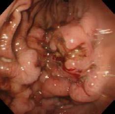
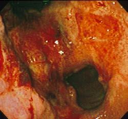
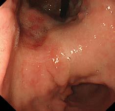
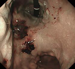 Figure 2b. The whole of the lesser curvature is infiltrated and ulcerated with diffuse bleeding. The angularis incisura is massively thickened and the whole stomach is infiltrated and ulcerated. The stomach was rigid and would not insufflate and the entrance to the antrum was distorted and no longer circular.
Figure 2a. A necrotic mass obliterating the angularis incisura
Figure 3a. An ulcerated GC on the lesser curvature viewed under white light
Figure 3b. The same GC as in Figure 3a. viewed by NBI, highlighting the abnormal mucosa and multiple areas of ulceration and bleeding
Figure 2. Endoscopic appearance of advanced canine gastric carcinoma
Figure 2b. The whole of the lesser curvature is infiltrated and ulcerated with diffuse bleeding. The angularis incisura is massively thickened and the whole stomach is infiltrated and ulcerated. The stomach was rigid and would not insufflate and the entrance to the antrum was distorted and no longer circular.
Figure 2a. A necrotic mass obliterating the angularis incisura
Figure 3a. An ulcerated GC on the lesser curvature viewed under white light
Figure 3b. The same GC as in Figure 3a. viewed by NBI, highlighting the abnormal mucosa and multiple areas of ulceration and bleeding
Figure 2. Endoscopic appearance of advanced canine gastric carcinoma
Control & Therapy Series – Issue 314 March 2024 Page 46 Authors’ views are not necessarily those of the CVE
Figure 3. Endoscopic appearance of canine gastric carcinoma by white light and Narrowband Imaging (NBI)
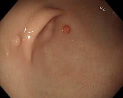
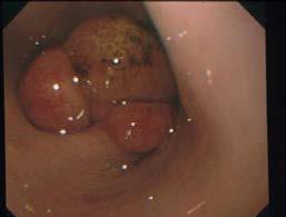
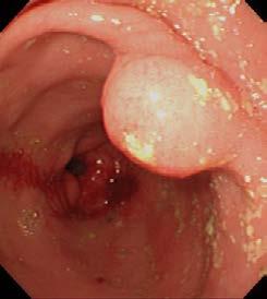
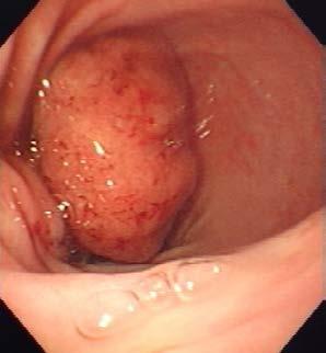 Figure 4c. Chronic hypertrophic pylorogastropathy surrounding the pylorus
Figure 4d. A large pedunculated leiomyoma in the gastric fundus
Figure 4a. Two benign adenomatous antral polyps adjacent to the pylorus
Figure 4b. An extramedullary plasmacytoma. The bleeding near the pylorus is refluxed bleeding from duodenal biopsies
Figure 4c. Chronic hypertrophic pylorogastropathy surrounding the pylorus
Figure 4d. A large pedunculated leiomyoma in the gastric fundus
Figure 4a. Two benign adenomatous antral polyps adjacent to the pylorus
Figure 4b. An extramedullary plasmacytoma. The bleeding near the pylorus is refluxed bleeding from duodenal biopsies
Control & Therapy Series – Issue 314 March 2024 Centre for Veterinary Education | Est. 1965 Page 47
Figure 4. Endoscopic appearance of gastric masses other than gastric carcinoma
leads to premature euthanasia as the chance of curative surgical resection is very small.
Differential diagnoses
Although GC is the most common malignancy of the canine stomach, ulceration is seen more commonly with NSAID administration, whist several benign and malignant masses also occur. In old dogs, one or more small adenomatous polyps are often found.
They are not considered to be of any clinical significance but will occasionally be observed endoscopically to be bleeding. Biopsy may be considered to distinguish them from another but much rarer polypoid mass, the extramedullary plasmacytoma.
Another benign mass that may be mistaken for a GC is chronic hypertrophic pylorogastropathy (CHPG) which can cause outflow obstruction when hypertrophied tissue blocks the pylorus.
Small brachycephalic dogs such as Pekingese and Shih Tzu are most commonly affected by CHPG in middle age, the same time window when GCs develop. The prognosis after successful Y-U pyloroplasty and submucosal resection of the hypertrophied tissue is good.
The second most common masses in the canine stomach are smooth muscle tumours, the leiomyoma and leiomyosarcoma. They tend to arise in the cardia or antrum away from where a GC typically forms. Although arising in the muscle layers of the gastric wall, these tumours may form pedunculated masses and may also ulcerate leading to chronic bleeding. Some smooth muscle tumours may actually be misdiagnosed GI stromal tumours (GISTs) which arise from the Interstitial cells of Cajal (the GI pacemaker cells) and which can only be identified by immunohistochemical cell markers.
However, clinically, GISTS exhibit similar biological behaviour to smooth muscle tumours except they may be susceptible to tyrosine kinase inhibitors such as masitinib and toceranib if not resectable.
Treatment
There is very limited data on the successful use of chemotherapy in treating canine GC and only palliative hypofractionated irradiation has been reported. The mainstay of treatment is still surgical resection. In human medicine, screening of at-risk populations and using novel imaging techniques allows the identification of early GC where radical resection can be curative; this may involve partial or total gastrectomy, removal of all draining lymph nodes and resection of any other affected organs. However, GC in dogs is usually only diagnosed late when complete surgical resection is not viable
especially as it is the lesser curvature that is typically involved and where total gastrectomy is considered too radical. Yet in a multicentre study of 40 GC cases, partial gastrectomy (28 dogs), Billroth I (9 dogs), subtotal gastrectomy (2 dogs), and submucosal resection (1 dog) were reported to have better survival than historical data.⁸ Major postoperative complications did occur in 8 of the dogs, including septic peritonitis secondary to dehiscence in four dogs.
Prognosis
Historically, the prognosis for dogs with GC was grave with a reported overall median survival time (MST) after surgery of only 72 days. Symptomatic treatment with anti-emetics and H2RAs or PPIs to provide short-term palliative relief may be preferable. However, after radical surgery, the median progression free interval was 54 days, and the median survival time (MST) was 178 days (range, 1-1902).⁸ Intraoperative complications were associated with an increased risk of death and administration of adjuvant chemotherapy correlated with an improved survival. However, for dogs with advanced disease showing complete anorexia and cachexia, euthanasia is appropriate.
References
1. Patnaik, A.K., Hurvitz, A.I. & Johnson, G.F. Canine gastrointestinal neoplasms. Veterinary Pathology 14, 547-555 (1977).
2. Kubba, A.K. & Young, M. The Napoleonic cancer gene? Journal of Medical Biography 7, 175-181 (1999).
3. Kumar, S., Patel, G.K. & Ghoshal, U.C. Helicobacter pylori -induced inflammation: possible factors modulating the risk of gastric cancer. Pathogens 10 (2021). https://doi.org:10.3390/pathogens10091099
4. Candido, M.V. et al. Gastric mucosal pathology in Belgian Shepherd dogs with and without clinical signs of gastric disease. Acta Veterinaria Scandinavica 63 (2021). https://doi.org:10.1186/s13028-021-00570-6
5. Seim-Wikse, T. et al. Comparison of body condition score and other minimally invasive biomarkers between dogs with gastric carcinoma and dogs with chronic gastritis. Journal of the American Veterinary Medical Association 254, 226-235 (2019). https://doi.org:10.2460/ javma.254.2.226
6. Hardas, A. et al. Canine gastric carcinomas: a histopathological and immunohistochemical study and similarities with the human counterpart. Animals 11 (2021). https://doi.org:10.3390/ani11051409
7. Zuercher, M., Grosso, F.V. & Lejeune, A. Comparison of the clinical, ultrasound, and CT findings in 13 dogs with gastric neoplasia. Veterinary Radiology & Ultrasound 62, 525-532 (2021). https://doi. org:10.1111/vru.12980
8. Abrams, B. et al. Perioperative complications and outcome after surgery for treatment of gastric carcinoma in dogs: A Veterinary Society of Surgical Oncology retrospective study of 40 cases (20042018). Veterinary Surgery 48, 923-932 (2019). https://doi.org:10.1111/ vsu.13239.
Control & Therapy Series – Issue 314 March 2024 Page 48 Authors’ views are not necessarily those of the CVE
Tar�nga Veterinary Pr�fessional Training � Wil�life Treatment � Care

This course presented by Taronga will support veterinary professionals and veterinary nurses to develop knowledge and skills in native wildlife triage, including first aid, initial treatment and emergency care.
• Supported by the NSW Government Department of Planning and Environment
Semester 2, 2024
Applications Open on 3 June
Email: tarongprofvet@zoo.nsw.gov.au
Visit: https://taronga.org.au/vet-professional-training
• AVA and VNCA certified
• Online Course (20 CPD points)
• Hands-on Workshop (12 CPD Points) –available in NSW, QLD and Vic (see website for details)
• Subsidised positions available to eligible applicants nationally


Workshops available at:
GOLD COAST • A USTRALI A
ADVERTISEMENT


Scan to learn more about Infection Prevention Sterilisation B. Braun VetCare
5, 7-9 Irvine Place Bella Vista NSW 2153, Australia Tel: 1800 251 705 vetcare.au@bbraun.com www.bbraun-vetcare.com.au BIBRAUN SHARING EXPERTISE 1. D-ST22031 lnfographic "Waste Quantity Comparison single-use wrap vs. rigid sterile containersn B. Braun, 2022. ADVERTISEMENT
Level

