
9 minute read
Fatal Venous Air Embolism During Thoracic Limb Amputation in a Dog

Major Winner
The prize is a CVE$400 voucher
Adam Gordon BVSc(Hons) MVS GCertSAUA
Maroubra Veterinary Hospital, Sydney NSW
t. +612 9344 8722
e. Adam.gordon@maroubravets.com.au
C&T No. 6009
Introduction
Venous air embolism (VAE) occurs when air is introduced to the central venous system, obstructing blood flow and producing an air embolism to the right heart and/ or pulmonary artery. It is a potentially catastrophic complication and is almost always iatrogenic.
Entry of air occurs as a result of a negative pressure gradient between an exposed venous sinusoid near the site of surgery and the right atrium of the heart. Air (or other gases like carbon dioxide) can also enter the venous circulation by creation of a pressure gradient through the use of insufflation of a body cavity or by the use of gas pressurized equipment.
Alternatively, air may enter the venous system through a gravitational gradient, created by having the operative site elevated above the heart.
Previous reported causes
Venous air embolism is well recognized and documented in human medicine.
The majority of published cases involved orthopaedic surgery or neurosurgery.
VAE has also been documented in obstetrics, gastroenterology, arthroscopy, central line placement and removal, interventional radiology procedures and endoscopy.
There are relatively few cases of VAE published in the veterinary literature. Published cases and outcomes of VAE in the veterinary literature occurred during the following procedures:
Pneumocystography—fatal.
Laparoscopy—fatal.
Cryosurgery (with pressurized liquid nitrogen) to treat gingival neoplasms—fatal. Gas insufflation of a retropharyngeal diverticulum—fatal.
Dental extractions involving use of air-driven dental drill—fatal. Thoracic limb amputation—non-fatal. Hemilaminectomy—1 fatal, 1 non-fatal.
Iatrogenic introduction of air through an intravenous catheter during surgery or hospitalization—several fatal, 2 non-fatal.
Case Report
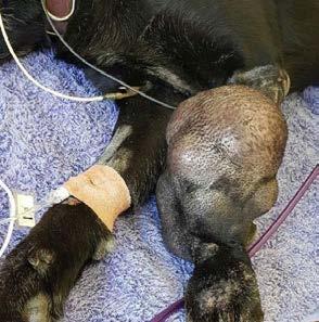
A 10-year-old, 32kg female neutered Labrador dog presented with a large mass over the distal antebrachium and carpus of the left forelimb (Figure 1)
The mass was broad-based, multilobular and was approximately 30cm in diameter. It had been present for 3 years.
Histopathology revealed it to be a myxofibrosarcoma (Grade 1, mitotic index 4 mitotic figures per 10 high power fields).
Left forequarter amputation was chosen as the most appropriate treatment option.
Thoracic radiographs, haematology and serum biochemistry pre-operatively were unremarkable.
Physical examination on the morning of surgery was unremarkable other than the left forelimb mass.
The dog was bright and alert, HR 120 beats per minute, respiratory rate 28 breaths per minute, rectal temperature 38.3°C. Mucous membranes were pink with capillary refill time less than 2 seconds. Thoracic auscultation and abdominal palpation were unremarkable.
The dog was pre-medicated with acepromazine (0.025mg/kg) and methadone (0.25mg/kg) subcutaneously. An intravenous catheter was placed in the right cephalic vein, and anaesthesia was induced with propofol intravenously (4mg/kg). The dog was intubated with a cuffed 11.0mm endotracheal tube. Anaesthesia was maintained with isoflurane in 100% oxygen at a fresh gas flow rate of 1.5L/minute.
Intravenous fluids (Hartmann’s solution) were administered for the duration of anaesthetic and surgery at 5mL/kg/hr. Anaesthetic monitoring consisted of pulse oximetry, end-tidal CO2, ECG, rectal temperature, heart rate, respiratory rate and oscillometric blood pressure.
The dog was placed in right lateral recumbency for surgery. During dissection, a small tear was inadvertently created in the axillary vein. Haemorrhage was quickly controlled through placement of haemostats on the axillary vein proximal to the tear. In the next minute ETCO2 dropped, the pulse oximeter did not give a reading, the patient became cyanotic and suffered cardiac arrest.
Attempted cardiopulmonary resuscitation (external compressions, boluses of atropine and adrenaline intravenously) was unsuccessful and the dog was pronounced deceased after 20 minutes of CPR.
Post-mortem thoracic radiographs were performed. They showed an elliptical radiolucent focus measuring approximately 7cm in height and 3.5cm in length superimposed over the region of the right ventricle in lateral views (Figures 2 and 3).
The clinical presentation and thoracic radiographs support a diagnosis of venous air embolism.
Discussion
In this unfortunate case, the iatrogenic rent in the axillary vein allowed air to enter the central venous system down a gravitational gradient to the right side of the heart. This resulted in outflow obstruction to the right ventricle, hypocapnia, hypoxaemia, hypotension, reduced cardiac output and ultimately cardiac arrest.
The clinical signs and consequence of VAE are dependent on the volume of air introduced and the rate at which it is introduced to the venous system.


The lethal dose of a bolus of air in dogs has been reported as 7.5–15mL/kg body weight.
The majority of published cases of VAE in the veterinary literature had a fatal outcome.
Successful management of VAE requires:
Awareness of risk of occurrence (for instance where the operative site is positioned above the right atrium). Good communication between surgeon and anaesthetist/individual monitoring the anaesthetic.
Early diagnosis.
Proactive management if VAE is suspected.
Diagnosis of Venous Air Embolism
There are no minimally invasive aids with high sensitivity and specificity for diagnosing VAE. Transoesophageal echocardiography is the most sensitive technique for detecting VAE, being able to detect 0.02mL/kg of air. Precordial doppler ultrasound is another sensitive technique for detecting VAE, whereby the anaesthetist listens for a characteristic ‘drum-like’ or ‘mill-wheel’ murmur associated with VAE.
From a practical standpoint in the veterinary setting, the following methods are most reliable for diagnosing suspected VAE.
Capnography—an abrupt drop in ETCO2 can occur with VAE (though reductions as little as 2mmHg can also be seen). Unfortunately this is not particularly specific, and can also be seen with hypothermia, hyperventilation, reduced cardiac output, pulmonary embolism and bronchospasm. Reduced ETCO2 is generally the earliest indicator of VAE in anaesthetised patients.
Pulse oximetry—reduced oxygen saturation on pulse oximetry is considered a late sign of VAE.
Blood pressure monitoring— hypotension occurs with VAE.
ECG—tachyarrhythmias and ST segment depression may be seen early, followed by bradycardia and cardiac arrest.
Management of Venous Air Embolism
As the diagnosis of VAE is fraught with difficulty and the potential consequences devastating and potentially rapidly fatal, management of VAE must be instituted promptly and usually before a definitive diagnosis is made where there is an index of suspicion of VAE.
The key points in management of VAE are:
Prevent further entry of air into the venous system. In open surgical procedures the operative site should be flooded with saline or covered with moistened sponges.
Immediately discontinue any insufflation of body cavities and use of gas-pressurized equipment.
Where possible, lower the operative site below the level of the heart.
If nitrous oxide is part of the anaesthetic mix it should be discontined immediately as it can dramatically increase the size of the air bubble due to its increased solubility in blood.
Administration of high FiO₂ 100% oxygen with intermittent positive pressure ventilation (IPPV).
Maintain normovolaemia and prevent hypotension—ensure adequate intravenous fluid rates and use of positive inotropes (adrenaline, noradrenaline, dopamine, dobutamine).
Repositioning of the patient—placement into left lateral recumbency either horizontal or head down (Durant’s maneuver) —known as the Trendelenburg position. It was postulated that an obstructing air embolus could be displaced and right ventricular failure prevented by this repositioning.
In an echocardiographic study, transoesophageal echocardiography demonstrated that this repositioning relocated intracardiac air to nondependent parts of the right heart. However, there was no corresponding improvement in haemodynamic performance or change in cardiac dimensions. Irrespective of this, placing patients with suspected VAE into the Trendelenburg position remains standard of care in human medicine.
Key Points
The most important lesson learned from this unfortunate case was the importance of immediate communication by the surgeon that there was a breach of the venous system that increased the risk of venous air embolism. Instituting IPPV, continuous monitoring of blood pressure and treating hypotension (if it were present) would have improved the chances of survival, though by no means ensured it, such is the seriousness of the introduction of significant volumes of air in a short period of time.
It is thought that the incidence of VAE in veterinary patients is much higher than has been documented in the literature. Having studied the published literature and perused VIN (Veterinary Information Network), it would appear that lack of due care with intravenous fluid lines and subsequent ingress of air is the most common cause of VAE in companion animals. This is an important reminder to avoid complacency and ensure all staff understand the importance of ensuring all IV lines are primed and capped where appropriate.
Intravenous fluid pumps are definitely not foolproof, and there are reports of the air alarm failing and large volumes of air being infused into the patient with subequent death.
References & Further Reading
Mouser PJ, Wilson JD. Fatal venous air embolism in an apparently healthy adult Chihuahua. J Am Anim Hosp Assoc 2015; 51: 176-179.
Bautista Diaz-Delgado O, Campagna I. Suspected venous air embolism during thoracic limb amputation in a dog. Vet Rec Case Rep 2020; 8: 1-6.
Austin LS, VanBeek C, Williams GR. Venous air embolism: an underrecognized and potentially catastrophic complication in orthopaedic surgery. J Shoulder Elbow Surg 2013; 22: 1449-1454.
McCarthy CJ, Behravesh S, Naidu SG, Oklu R. Air embolism: Practical tips for prevention and treatment. J Clin Med 2016; Vol.5(11): 93-105.
Lui PW, Lin YM, et al. Spectral characteristics of embolic heart sounds detected by precordial doppler ultrasound during venous air embolism in dogs Int J Crit Illn Inj Sci 2013; 3(1): 73-76.
Gordy S, Rowell S. Vascular air embolism. Int J Crit Illn Inj Sci. 2013; 3(1) Jan-Mar: 73–76.
Palmon SC, Moore LE, Lundberg J, et al. Venous air embolism: A review J Clin Anesth 1997; 9: 251-257.
Gilroy BA, Anson LW. Fatal air embolism during anesthesia for laparoscopy in a dog J Am Vet Med Assoc 1987; 190: 552–554.
Thayer GW, Carrig CB, Evans AT. Fatal venous air embolism associated with pneumocystography in a cat J Am Vet Med Assoc 1980; 176: 643–645.
Gilroy BA, Anson LW. Fatal air embolism during anesthesia for laparoscopy in a dog J Am Vet Med Assoc 1987; 190: 552–554.
Harvey HJ. Fatal air embolization associated with cryosurgery in two dogs J Am Vet Med Assoc 1978; 173: 175–176.
Ober CP, Spotswood TC, Hancock R. Fatal venous air embolism in a cat with a retropharyngeal diverticulum. Vet Radiol Ultrasound 2006; 47: 153–158.
Costa-Farré C, Torrente C, Bertrana CD, et al. Nonfatal infusion pump-related venous air embolism in a dog. Vet Anaesth Analg 2017; 44: 382–383.
Gunew M, Marshall R, Lui M, Astley C. Fatal venous air embolism in a cat undergoing dental extractions. J Small Anim Pract 2008; 49(11): 601-604.
Geissler HJ, Allen SJ, et al. Effect of body repositioning after venous air embolism. Anesthesiology 1997; Vol.86 (3): 710-717.
