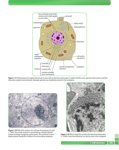
two centrioles close to the nucleus and at right angles to each other
microvilli
mitochondrion
Golgi vesicle
lysosome
Golgi apparatus
ribosomes
rough endoplasmic reticulum
cell surface membrane
nucleolus chromatin nucleus
nuclear pore
smooth endoplasmic reticulum
cytoplasm
nuclear envelope (two membranes) Figure 1.17 Ultrastructure of a typical animal cell as seen with an electron microscope. In reality, the ER is more extensive than shown, and free ribosomes may be more extensive. Glycogen granules are sometimes present in the cytoplasm.
Figure 1.18 TEM of the nucleus of a cell from the pancreas of a bat ( 7500). The circular nucleus is surrounded by a double-layered nuclear envelope containing nuclear pores. The nucleolus is more darkly stained. Rough ER is visible in the surrounding cytoplasm.
Figure 1.19 TEM of rough ER covered with ribosomes (black dots) ( 17 000). Some free ribosomes can also be seen in the cytoplasm.
1 Cell structure
15