Box 1A Biological drawing You need the following equipment:
• • • • •
pencil (HB) pencil sharpener eraser ruler plain paper.
• • • •
always use a pencil, not a pen don’t use shading use clear, continuous lines use accurate proportions and observation – not a textbook version.
Here are some guidelines for the quality of your drawing:
• • • •
arrange label lines neatly and ensure they don’t cross over each other annotate your drawing if necessary (i.e. provide short notes with one or more of the labels in order to describe or explain features of biological interest) add a scale line at the bottom of the drawing if appropriate use a pencil, not a pen.
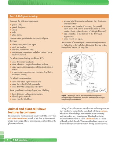
An example of a drawing of a section through the stem of Helianthus is shown below. Biological drawing is also covered in Chapter 15, page 264.
For a low-power drawing (see Figure 1.7):
• • • •
don’t draw individual cells draw all tissues completely enclosed by lines draw a correct interpretation of the distribution of tissues a representative portion may be drawn (e.g. half a transverse section).
For a high-power drawing:
• • •
draw only a few representative cells draw the cell wall of all plant cells don’t draw the nucleus as a solid blob.
• • •
label all tissues and relevant structures identify parts correctly use a ruler for label lines
Some guidelines for the quality of your labelling:
Animal and plant cells have features in common In animals and plants each cell is surrounded by a very thin cell surface membrane, which is too thin to be seen with a light microscope. This is also sometimes referred to as the plasma membrane.
4 4
1 Cell structure
Figure 1.7 The right side of this low-power drawing shows examples of good technique, while the left side shows many of the pitfalls you should avoid.
Many of the cell contents are colourless and transparent so they need to be stained to be seen. Each cell has a nucleus, which is a relatively large structure that stains intensely and is therefore very conspicuous. The deeply staining material in the nucleus is called chromatin and is a mass of loosely coiled threads. This material collects together to form visible separate chromosomes during nuclear division