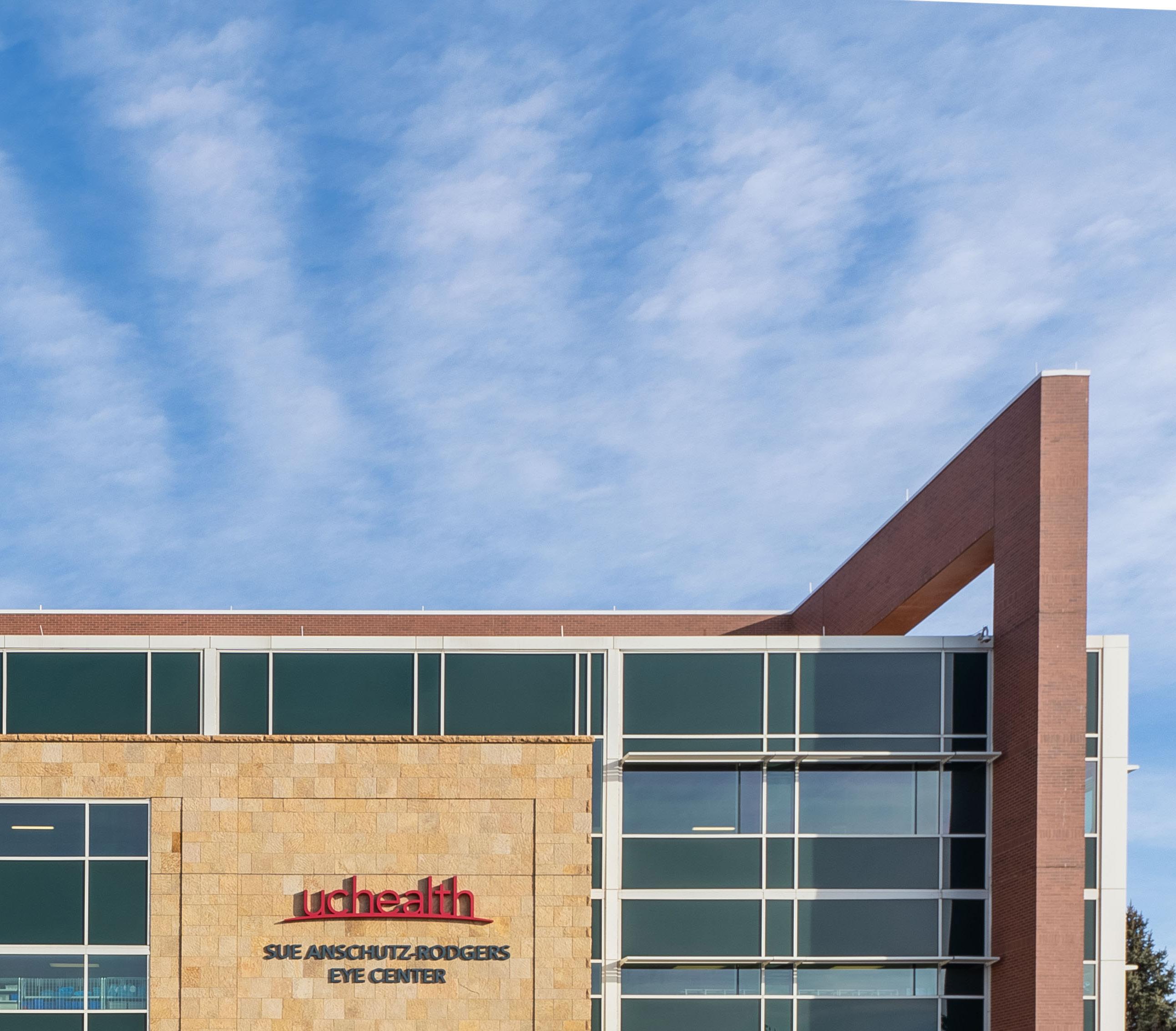
Sue Anschutz-Rodgers Eye Center Annual Report 2023 – 2024
BRINGING SIGHT TO LIFE
Letter from the Chair
See Our Impact
A Novel Treatment for a Patient Facing a Rare Genetic Condition
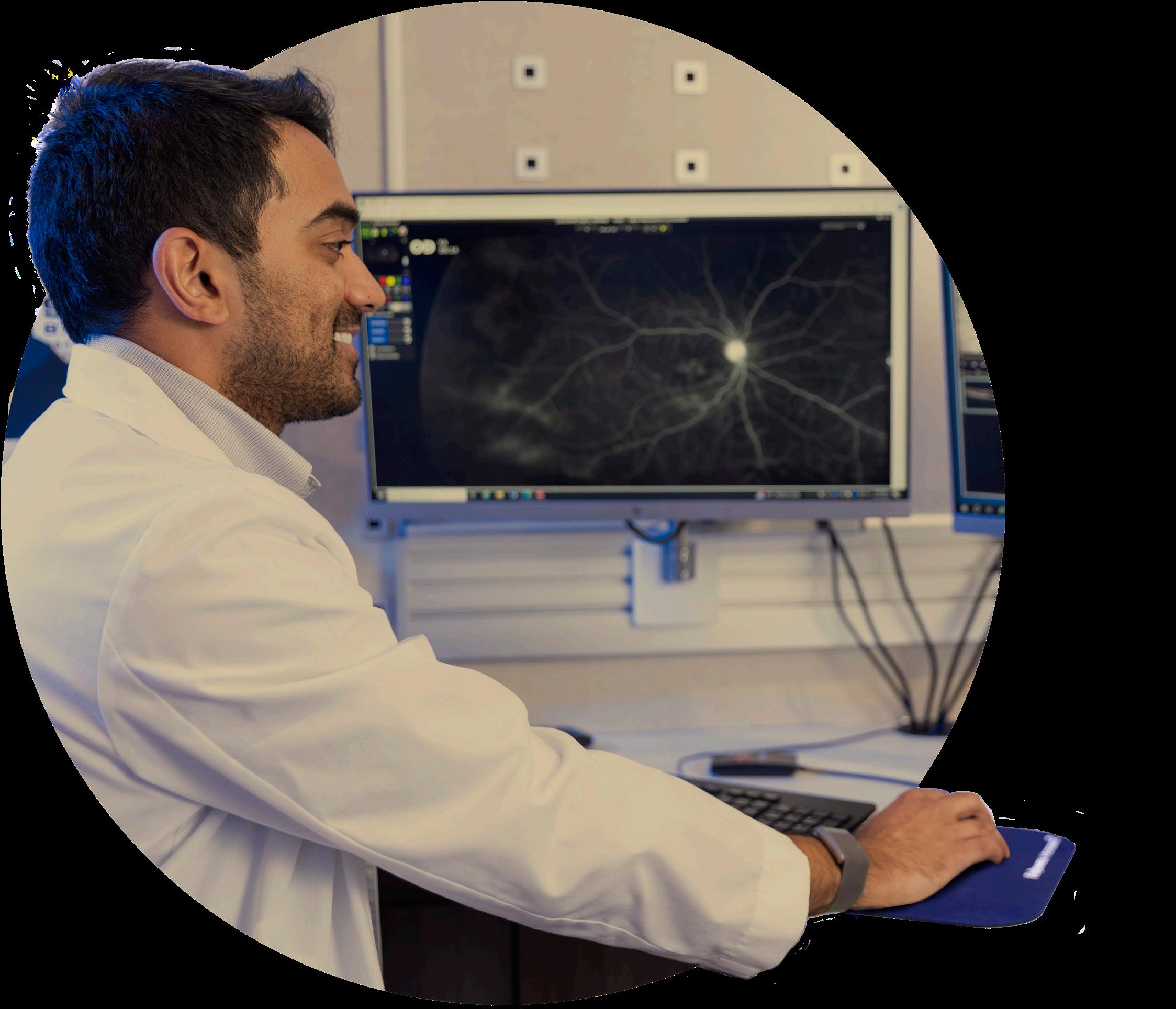
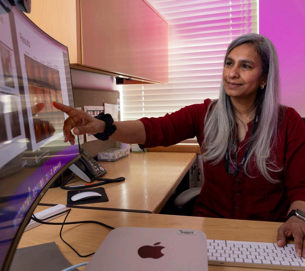
AI and the Future of Ocular Diagnosis
Expanding the Impact of Uveitis and Ocular Inflammation Research
CellSight Reaches New Heights for Research and Innovation
Cochrane Advances Evidence-Based Vision Health Care
Bringing Ocular Health Expertise to Blind and Visually Impaired Children
Forward Thinking in Ophthalmic Health AI
Improving Charting Software for Patients and Physicians
Residency Program
Simulator Upgrades Cataract Surgery Training
Employing Portable Fundus Photography Cameras to Enhance On-Call Imaging
KICKING OFF FISCAL YEAR 2024, WE PROUDLY HOSTED THE LARGEST INTERNATIONAL CONFERENCE ON LOW VISION REHABILITATION EVER ORGANIZED: VISION 2023.
LETTER FROM THE CHAIR
This fiscal year, the Department of Ophthalmology at the University of Colorado School of Medicine showcased our mission as innovators and collaborators in research, education, and translational care through organizing several significant conferences and meetings, attracting experts from around the world to advance vision science and patient outcomes.
Kicking off fiscal year 2024, we proudly hosted the largest international conference on low vision rehabilitation ever organized: Vision 2023. With attendees from 30 different countries, the conference offered nearly 90 hours of education and featured 200 paper, panel, and poster presentations. This event not only showcased cutting-edge research but also fostered invaluable collaborations that will drive future advancements in our field.
More recently, we organized the 10th anniversary of the ROP Update Meeting at Children’s Hospital Colorado. This meeting brought together hundreds of pediatric ophthalmologists and vitreoretinal specialists from across the country to discuss the latest multidisciplinary perspectives on retinopathy of prematurity. The event highlighted advancements in imaging, diagnosis, treatment, and new technologies, reinforcing our commitment to improving treatment options and results for our youngest patients.
Our department also embarked on an exciting new program with our first-ever research retreat for clinical and research faculty leading research initiatives. This retreat was a pivotal moment for us, as we outlined our goals for the next five years with a strong focus on team science, artificial intelligence, and other innovative approaches. The collaborative spirit and shared vision that emerged from this retreat are already propelling us toward new heights in ophthalmic research.
We have continued to receive substantial grant support, which has been instrumental in driving our research efforts. Notably, we have an active grant with Research to Prevent Blindness and secured more than $15 million through the University of Colorado’s Anschutz Acceleration Initiative (AAI). Additionally, we received millions more in funding from prestigious organizations such as the National Eye Institute and The Michael J. Fox Foundation. These resources are enabling us to achieve tangible strides and make a real impact in a short period of time.
As we celebrate these successes, we also look to the future with great anticipation. We are excited to welcome John H. Sampson, MD, PhD, MHSc, MBA, as the new Dean of the University of Colorado School of Medicine. We look forward to working alongside Dr. Sampson to develop one of the top academic eye centers in the country.
Conversely, we also bid farewell to J. Mark Petrash, PhD, who retired this year after more than 15 years of exemplary service as Vice Chair for Research at the Sue Anschutz-Rodgers Eye Center. Petrash’s contributions have been immeasurable, and we are thrilled that he will continue to be part of our community as an emeritus professor and through his role as Executive Vice President of ARVO. We are equally excited to welcome Joseph A. Brzezinski IV, PhD, as our new director of research. His leadership will undoubtedly advance our basic science team to new frontiers.
We are deeply grateful for your continued support and dedication to our mission. Together, we are bringing sight to life, and I am confident that the future holds even greater promise for our department.
Thank you for being an integral part of our journey.
Sincerely,
Naresh Mandava
Sue
Anschutz-Rodgers
Endowed Chair in Retinal Diseases
Professor and Chair, Department of Ophthalmology University of Colorado School of Medicine
ACTIVE CLINICAL TRIALS
TOTAL FUNDING FOR SPONSORED RESEARCH GRANTS* $15.5M IN TOTAL FUNDING RAISED FOR CU DEPARTMENT OF OPHTHALMOLOGY SPINOUT COMPANIES $200M+
TECHNOLOGIES LICENSED TO FIVE DIFFERENT ENTITIES†
#20 IN DOXIMITY’S RESIDENCY NAVIGATOR SURVEY
177,500+ UNIQUE PATIENT VISITS BY CU FACULTY
9,600+ SURGERIES BY CU FACULTY
FACULTY
SECONDARY FACULTY
from FY2022 - FY2024
from FY2019 - FY2024
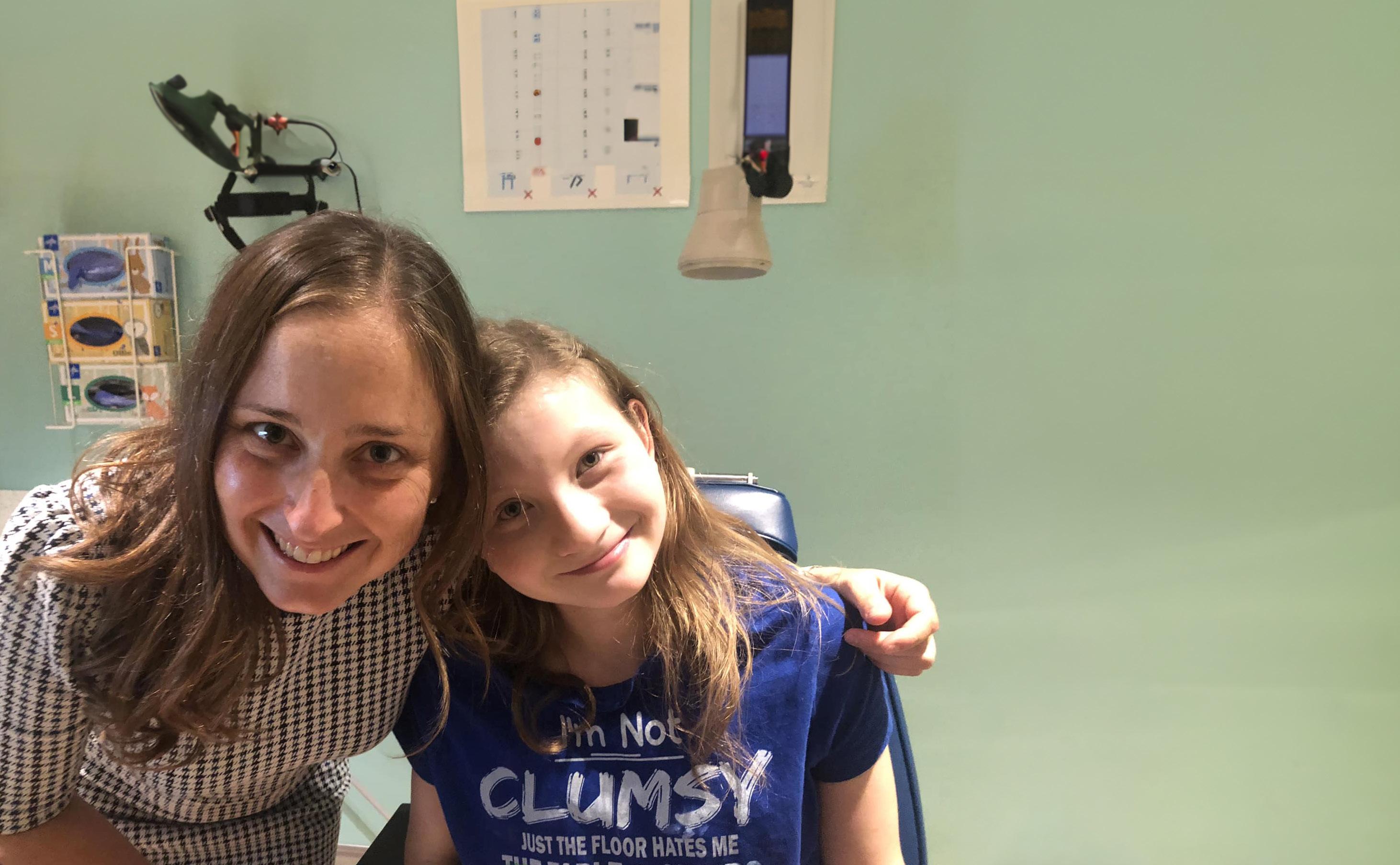
A Novel Treatment for a Patient Facing Rare Genetic Condition
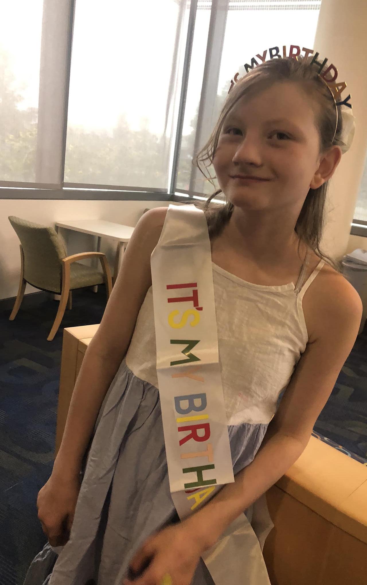
Thirteen-year-old Grace Hoyt received potentially the best birthday gift ever when pediatric ophthalmologists from our department and Children’s Hospital Colorado administered the first treatment designed specifically to slow vision loss associated with posterior column ataxia with retinitis pigmentosa (PCARP), a rare genetic condition that affects vision and the nervous system.
“It’s so incredible that she has this opportunity,” Susan Hoyt says of her daughter, who received the first treatment in August 2023. “We’ve known that Grace is going to go blind, but to have the chance to stop the inexorable march forward is encouraging.”
The new treatment, given with permission from the U.S. Food and Drug Administration as an investigational new drug, is the first start-to-finish personalized medication given at Children’s Colorado developed in partnership with our department.
“This is my first time applying for Investigational New Drug (IND) approval and the first new drug that I’ve given a patient,” says Emily McCourt, MD, associate professor and the Ponzio Family Chair for Pediatric Ophthalmology, who worked with researchers and physicians in the department, the newly created CU Anschutz Investigational New Drug and Device (IND/IDE) Office, Boston Children’s Hospital, and nonprofit drug developer n-Lorem to create the injectable medication that aims to slow down the patient’s vision loss.
“I learned through the process that sometimes you have to phone a friend, and thankfully we have a lot of expertise on the CU Anschutz campus,” she continues. “The IND office was so helpful, as was the research institute at Children’s Colorado.”
A mystery until diagnosis
McCourt met Grace 10 years ago when she was experiencing some motor delays, trouble seeing at night, and falling without responding to pain. One night, Grace put a sippy cup down and couldn’t find it at all.
“She was about a year-and-a-half old when we realized she couldn’t feel pain,” Susan says. “Then, about a year later, we were sitting her in her room one night and she couldn’t find her cup, but it was right in front of her.”
Soon after, McCourt first saw Grace and noticed she had retinitis pigmentosa (RP), a blinding condition of the retina that develops over time. It is rare to experience as a young child, McCourt says. Typically, people who have the disease are diagnosed in their 20s.
“At that time, we knew it was a clue to an overall diagnosis, but it was still difficult to put it all together,” she says. “Meanwhile, Grace was experiencing infections in her fingers along with broken bones that didn’t seem to hurt at all.”
Genetic testing revealed a few mutations, but none that McCourt says really made any sense. “A lot of the time you need two mutations to have a disease, and this patient only had one mutation in any gene that could explain her symptoms.”
The discovery of a second gene mutation, which researchers describe as a deep intronic mutation, only discoverable through whole genome sequencing, led to the PCARP diagnosis.
“It’s a really difficult disease,” says Marc Mathias, MD, associate professor and retina specialist at the Sue Anschutz-Rodgers Eye Center, who assisted McCourt through the process of developing the treatment. “It doesn’t just affect the eyes and vision. There’s a systemic component as well, and the disease can develop slowly in childhood so it might not be recognized early on.”
In the past 50 years, PCARP has only been described in about 20 cases.
One patient, one treatment
Because Grace’s disease has a deep intronic mutation, her form of PCARP is amenable to a certain kind of therapy, called antisense oligonucleotide therapy (ASO), that attaches to RNA.
“Basically, you take these little molecules and put them together and make a drug that binds to the patient’s RNA to help the patient’s body make more normal protein and less abnormal protein,” McCourt explains. “We thought perhaps this therapy could work to slow down or stop the progression of the retinal disease.”
Austin Larson, a CU and Children’s Colorado geneticist who had recently given an ASO drug to another patient, contacted a lab at Boston Children’s Hospital to see what they thought of the possibility of using an ASO therapy for Grace’s retina. The new partners got to work, using cells grown from Grace’s skin biopsy, which showed promising preliminary results.
From there, developing the treatment required testing, making sure the treatment would be safe for Grace, and finally getting the IND application approved by the FDA.
“Safety was goal number one,” Mathias says. “We wanted to do the right thing for the patient and design a study in a thoughtful way that protects her in the end.”
With the drug designed to treat one patient only, researchers had no control group, making it more challenging to show that the drug will be helpful over time, but studying the effectiveness of the therapy was similar to how scientists research drugs that reach bigger populations, too.
“It builds on a lot of experience we have with other clinical trials, and we went through a similar process,” Mathias says.
n-Lorem, a nonprofit foundation dedicated to developing ASO technology to treat nano-rare patients, conducted testing for the trial. Once researchers were satisfied with the therapy, it was off to the FDA to submit an IND application.
“This whole process highlights the fact that medicine is a team sport,” McCourt says.
The power of asking ‘what if?’
Grace is expected to need the medication, through injections into the eyes, for the rest of her life.
“Because she doesn’t feel pain, it’s extremely important that Grace hangs on to whatever vision she has left so she can see her body and evaluate herself for injuries,” Susan says. “I am so grateful to Dr. McCourt for asking ‘what if’ and making the impossible happen for my girl.”
CU ophthalmologists will monitor her progress and watch for vision loss. McCourt and Mathias expect an injection may be necessary every three to six months. The treatment is being manufactured by n-Lorem for the rest of Grace’s life at no cost.
“It’s really taken a village to accomplish this,” echoes Mathias. “There has been support from many players including Grace, who has shown us all the reasons her family calls her Amazing Gracie.”
Learn more about how you can contribute to this specialized care.
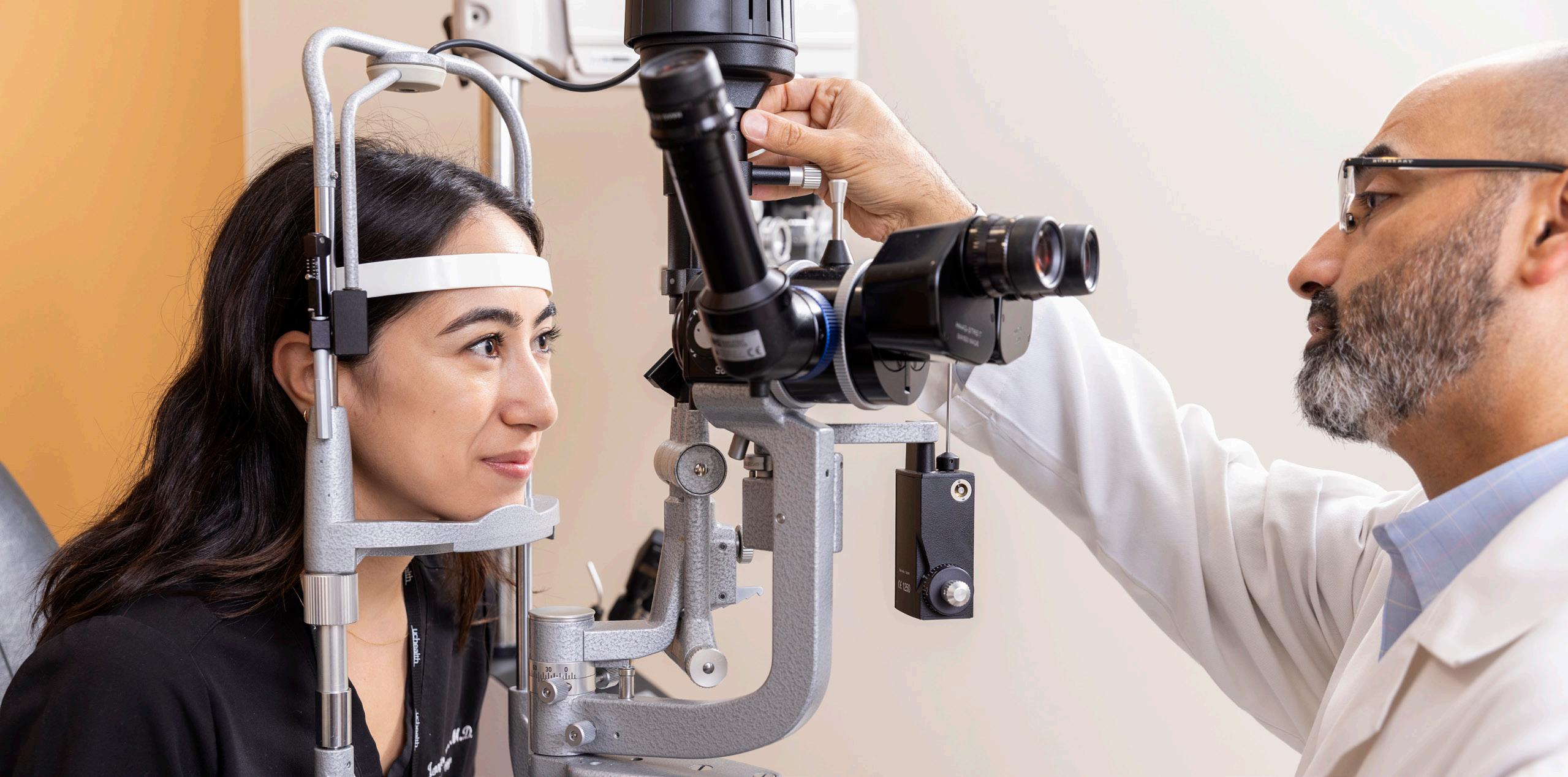
AI and the Future of Ocular Diagnosis
Ophthalmology faculty are at the forefront of figuring how accurate artificial intelligence (AI) is at diagnosing eye diseases.
Large language models (LLMs), such as ChatGPT, have skyrocketed in popularity in the past few years due to their ability to utilize vast amounts of information, but could they be used to diagnose ocular disease?
Ophthalmology researchers at the University of Colorado School of Medicine have begun digging into the question.
Earlier this year, Malik Kahook, MD, professor and the Slater Family Endowed Chair in Ophthalmology, joined a team of researchers to test the accuracy of ChatGPT compared to three senior ophthalmology residents.
ChatGPT gave a correct diagnosis in eight of the 11 cases. The three resident ophthalmologists were correct in six, eight, and eight cases, respectively. In cases with common glaucoma presentation, the residents and ChatGPT were able to give an accurate diagnosis, but cases considered atypical or complex were less accurate for both the LLM and residents.
While still young, the technology could be a promising tool in some instances.
“I anticipate LLMs will aid glaucoma diagnosis and could be of particular benefit in areas where glaucoma expertise is not available,” Kahook says. “This could include low- and middle-income countries with less access to ophthalmologists and subspecialty trained glaucoma experts, primary care settings where ocular disease is not easy to diagnose, and in
residency programs where trainees can benefit from augmenting their knowledge through LLM-based digital assistants.”
With growing accessibility to the public, patients themselves may turn to AI programs for medical advice, just as they have with the rise of the internet.
“These programs are evolving quite a bit,” says Karen Christopher, MD, assistant professor of ophthalmology, who earlier this year participated in a researcher project tasking ophthalmologists with discerning human and chatbot responses to a variety of eye health questions.
The ophthalmologists were able to distinguish between AI and human responses about 61% of the time.
“I was surprised with the results,” Christopher says. “It speaks to how well these AI-generated responses can provide accurate information while mimicking human speech patterns and having empathy that we normally only attribute to humans.”
Even so, Kahook and Christopher say most LLM technology still requires quite a bit of physician oversight to ensure accuracy and reliability, especially given the many nuances involved in ocular diagnosis.
“While this technology is giving some great answers, an ophthalmologist would be able to confirm, add additional information, and give the most accurate advice. It’s our duty to catch information that isn’t fully correct so we can prevent harm to patients as much as possible,” Christopher says.
Expanding the Impact of Uveitis and Ocular Inflammation Research
This year marked a successful chapter in the Center for Ocular Inflammation’s journey to improve patient treatment for uveitis, scleritis, and other ocular inflammations.
The team, led by Alan Palestine, MD, director of the center and professor of ophthalmology, focuses on mechanisms of inflammation and potential treatments that could be used to treat the conditions.
A skilled team grows
The Center for Ocular Inflammation is a collaboration among physicians in multiple specialties, including uveitis, glaucoma, pediatric ophthalmology, ocular surface, neuroophthalmology, and rheumatology. This allows the group to manage complex ocular problems using a coordinated team approach.
This year, Lynn Hassman, MD, PhD, joined the team as a clinician-scientist and assistant professor of ophthalmology, working both in the laboratory and with patients. She received the 2024 Philip and Elaine Ellis New Investigator in Ophthalmology Research Award. This award, funded by Philip Ellis, MD, who served as department chair for over three decades, grants a new recipient each year with $40,000.
“This truly puts Dr. Hassman at the forefront of innovation in this department,” Palestine says.
The center also cemented its dedication to training future uveitis specialists, naming its inaugural fellow, Julia Xia, MD. She has investigated the relationship between uveitis and tuberculosis and studied the effects of micropulse transscleral cyclophotocoagulation in uveitic glaucoma with Mina Pantcheva, MD, associate professor of ophthalmology.
Her future work includes studying pediatric uveitis patients to better define clinical outcomes with Jennifer Jung, MD, associate professor of ophthalmology, and Rebecca Edwards Mayhew, MD, PhD, assistant professor of ophthalmology.
Top-notch research
Several research projects underpinned the center’s success as the team expands its impact and reputation. At the annual American Uveitis Society meeting this year, faculty and residents gave six scientific presentations.
Throughout the year, Amit Reddy, MD, assistant professor of ophthalmology, authored various papers defining which uveitis patients are more likely to respond to local steroid injections for ocular inflammation, while Hassman initiated new collaborations with the CU Division of Rheumatology.
In a collaboration with neuro-ophthalmologist Jeffrey Bennett, MD, PhD, professor of neurology and ophthalmology, Hassman is working to identify site-specific antibodies derived from inflammatory cells harvested within the eye that bind to ocular antigens. This may allow researchers to develop targeted therapies to block specific inflammatory processes.
Genetic stepping stones
The next several years will be significant for the future of ocular inflammation research, starting with genomic sequencing. This year, Palestine and Hassman started the process of completing DNA sequencing on 1,000 patients of the Sue Anschutz-Rodgers Eye Center uveitis biorepository registry. This biorepository will form the basis for a decade of research into the mechanisms of ocular inflammation by looking at genetics, protein expression and inflammatory mediators in these diseases.
“I’m particularly proud of the Center because it’s only through establishing formal processes dedicated to these rare diseases that we will make significant progress,” Palestine says. “We have been fortunate to have the support of multiple patient donors to create an endowment that will support ongoing future efforts.”
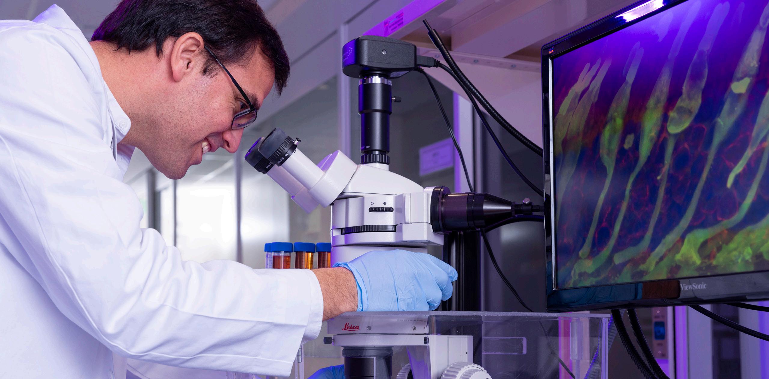
CellSight Reaches New Heights for Research and Innovation
Researchers at CellSight, the Ocular Stem Cell and Regeneration Research Program at the Sue Anschutz-Rodgers Eye Center, continue to advance crucial research and innovations to treat ocular disease while earning top recognition for their work and dedication to scientific discoveries in ophthalmology.
Among this year’s accomplishments, faculty members were awarded millions in grant funding from the National Institutes of Health (NIH) to better understand retinal cell differentiation and mechanisms for successful stem cell-derived 3D retinal transplants in patients with age-related macular degeneration (AMD).
CellSight director Valeria Canto-Soler, PhD, was granted $2.1 million to develop solutions to support cell-based transplantation strategies to regenerate photoreceptors and retinal pigment epithelium (RPE) cells.
Joseph Brzezinski, PhD, director of CellSight’s laboratory of developmental genetics and newly named director of research for the Department of Ophthalmology, was granted an additional $2 million for continued research on understanding how different types of cells in the retina are formed.
“Restoring vision in AMD patients is a big dream, but we are continuously moving closer to that being a reality,” CantoSoler says of CellSight’s work that has garnered important funding and distinction.
Research leaders at the CU Anschutz Medical Campus believe Canto-Soler’s work to restore vision in patients with ocular disease shows promise for delivering life-changing advancements in the next several years. In January, Canto-Soler was named one of nine Anschutz Acceleration Initiative (AAI) winners—a competitive selection process that started with 165 letters of intent and 56 full proposals. With the award, she’ll continue leading a team that’s developing products to aid the more than 2 million Americans with geographic atrophy (GA) regain vision.
The BrightFocus Foundation awarded Miguel Flores-Bellver, PhD, director of CellSight’s ExoSight Lab, with its annual Dr. Joe G. Hollyfield New Investigator Award for Macular Degeneration Research, which is presented to the top-rated New Investigator Grant recipient for exceptionally promising and forward-thinking ideas in AMD, and a three-year grant to study the pivotal role of extracellular vesicles in driving drusen biogenesis.
This year, Flores-Bellver also earned a $150,000 proof-ofconcept grant from the Global Business Development division of Colorado Office of Economic Development and International Trade and its Advanced Industries Accelerator Program, which supports research institutions and startups as they discover and commercialize new life-changing technologies. Flores-Bellver was awarded the funds for his proposal to develop an early detection test for AMD.
The combined work and dedication from CellSight’s investigators is proving successful and paves the way for more important ophthalmologic research.
“These grants and awards, along with our passionate CellSight team, can help accomplish something truly incredible that we believe could one day make a difference in so many lives,” Canto-Soler says.
Cochrane Advances Evidence-Based Vision Health Care
As the world grapples with an overwhelming influx of data, the need for evidence-informed health care has never been more critical. In its 22nd year, the Cochrane Eyes and Vision US Project (CEV@US) remains at the forefront of this effort, dedicated to enhancing vision care through rigorous systematic reviews.
Founded in 2002, CEV@US is a pivotal arm of Cochrane, an international organization renowned for its commitment to synthesizing health care literature. By preparing systematic reviews—highly structured evaluations of evidence—CEV@ US addresses key clinical questions related to eye health and visual impairment. Its mission is to ensure that interventions for preventing, diagnosing, and treating eye conditions are based on the best available evidence.
Systematic review significance
In the past grant year alone, CEV@US has made significant strides. The project published six new Cochrane systematic reviews and updated two existing reviews. Additionally, it released two new Cochrane protocols and five non-Cochrane systematic reviews, with two more under review and one submitted. Their work also includes eight methodological papers, one editorial, and two invited commentaries, along with a podcast that further extends their reach.
Among this year’s top achievements, the project published a systematic review that concludes biosimilars are now proven to be as effective as original anti-VEGF medications. As a result of this work, patients around the world may benefit with many European countries using the review findings to inform practice guidelines.
“There is a huge market for biosimilars because there is a growing population of people who are affected by AMD,” says Tianjing Li, MD, PhD, professor of ophthalmology and CEV@US director. “Knowing that biosimilars are just as safe and effective can help clinicians around the world make informed decisions and feel comfortable with using a more cost-effective treatment for patients who might not be able to afford the original anti-VEGF medicines.”
Enhancing research training
CEV@US has continued to expand its educational footprint, having trained over 138,000 individuals in systematic review methodologies. This education is crucial, as it equips clinicians and researchers with the skills needed to conduct high-quality reviews and apply this knowledge to clinical practice. The project also played a significant role in shaping over 122 clinical practice guidelines in the U.S. and internationally.
This year, CEV@US further strengthened its network by adding two new Centers for Evidence-Based Medicine, bringing the total to 16. They hosted three educational workshops, focusing on the development of Cochrane systematic reviews.
A notable highlight of this year was the continuation of the joint educational program with the American Academy of Optometry. This initiative pairs academy members with experienced mentors from CEV@US, guiding them in publishing Cochrane and non-Cochrane reviews. All six groups involved have published protocols and have their reviews in press or under peer review.
Extending reach around the world
The group’s impact extends beyond its publications. The reviews they produce are utilized by national and international agencies, guideline developers, and health professionals to inform decision-making and support clinical practice. Their findings are disseminated through platforms such as The Cochrane Library, PubMed Central, Wikipedia, and X (formerly known asTwitter), ensuring broad accessibility.
CEV@US also began using the Open Science Framework (OSF) to store supplementary data related to social determinants of health in eye care. This move underscores their commitment to transparency and accessibility in research.
As CEV@US continues its mission, its contributions to evidence-based vision health care remain invaluable. Through partnerships, education, and rigorous research, they are not only enhancing the practice of vision care but also setting a high standard for evidence-based medical research globally.
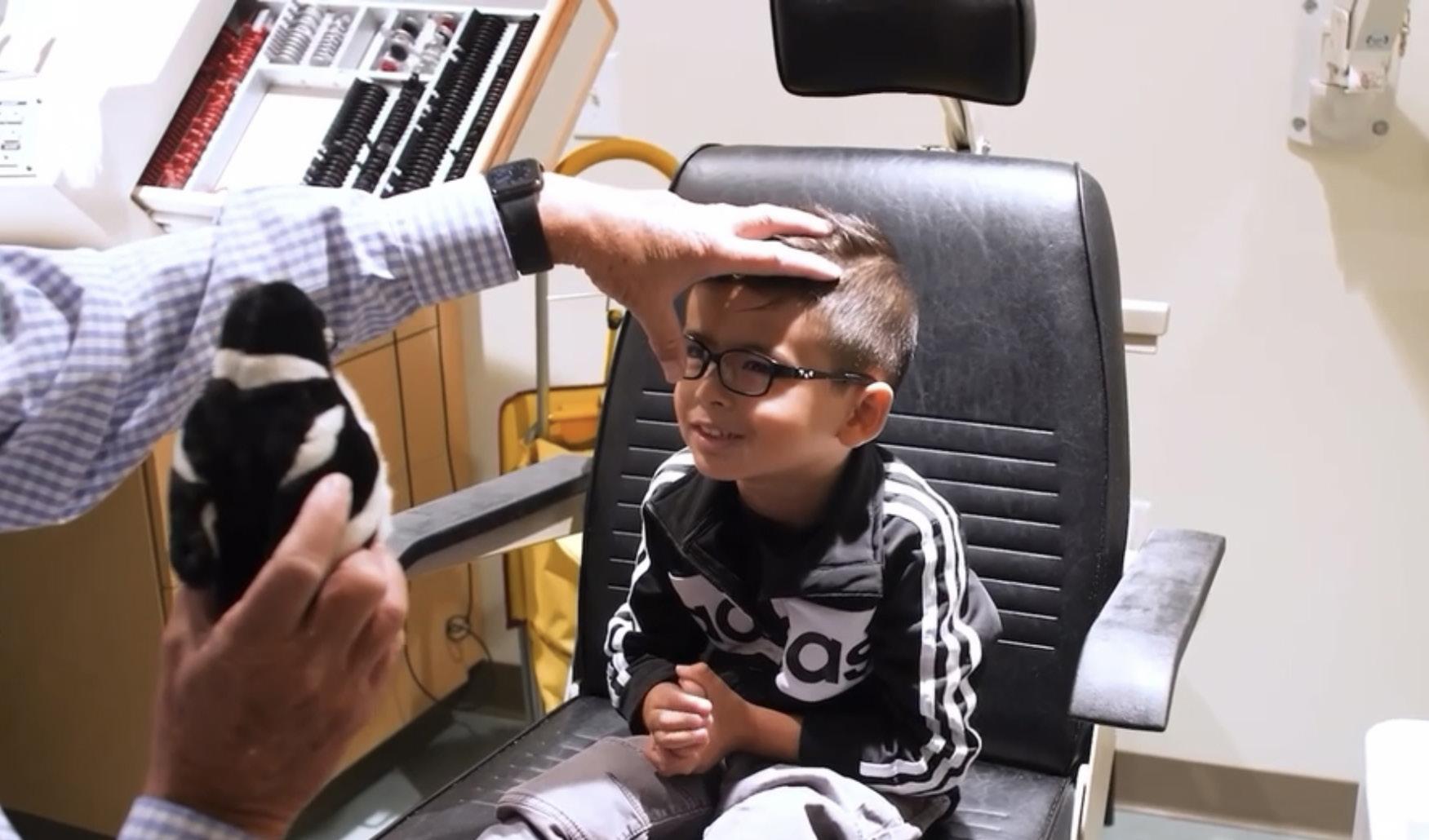
Bringing Ocular Health Expertise to Blind and Visually Impaired Children
For the past year, clinicians in our department have been helping craft a unique experience for visually impaired and blind children and their families.
Lauren Mehner, MD, MPH, assistant professor of ophthalmology, and Emily McCourt, MD, associate professor and Ponzio Family Chair for Pediatric Ophthalmology, each spend one day per month at Denver’s Anchor Center for Blind Children to tend to pediatric patients in an environment that’s familiar, convenient, and comfortable.
The center is the first school for the blind to offer ophthalmology care, thanks to money raised by Colorado ophthalmologist Robert King, MD, an alum of the CU School of Medicine, to create eye exam lanes at the center. Mehner and McCourt’s work builds upon King’s vision and marks a unique partnership with an academic institution.
“These are patients who need us the most,” Mehner says. “They also have access to important services, which make this experience even more special.”
Anchor Center, a nonprofit established in 1982, serves nearly 400 children and their families each year through educational, therapeutic, and ophthalmic services provided through collaborations with our faculty members, Children’s Hospital Colorado, and private practice physicians across the Denver metro region.
“This partnership has created a beautiful canvas where we can bring research, clinical care, and communication with the entirety of a child’s vision team all to one spot,” McCourt says.
Excellence in care
Being able to meet patients in a place where they’re already comfortable and have a teacher certified in visual impairment (TVI) available during appointments is a big perk for patients, their families, and staff at the center.
“This model allows for families to have more time with each doctor and therefore get questions answered and leave the appointment having had a comprehensive exam,”
“These are patients who need us the most.”
says Anchor Center executive director Meghan Klassen. “Because TVIs also sit in on appointments, they get to act as a bridge between the medical and educational side of things. They can help guide the conversations to make sure families understand visual conditions and the implications visual impairments have on overall development, as well as implications for the school setting, which we also share with our team to guide intervention strategies.”
Mehner and McCourt highlight how important comprehensive care is for young patients and what it means that families have access to additional services on site.
“Kids grow and change, and that’s what makes it fun to work with them,” Mehner says. “You get to see their progress and make a difference. Establishing a diagnosis and figuring out what they need early on sets them on a good track. Regular follow-up care and making sure that they can access resources—like a TVI—is so critical.”
A foundation for research
The partnership also presents plenty of opportunity for important research and education.
Last year, Mehner and the center teamed up at Vision 2023, an international conference hosted by our department on low vision research and rehabilitation, to present a workshop for identifying cerebral visual impairment (CVI) patients in the clinical setting. And in 2024, Mehner took her Anchor research experience to the annual meeting of the American Association for Pediatric Ophthalmology and Strabismus to present on CVI referral patterns.
Anchor Center staff say having CU ophthalmologists in the facility encourages more conversations about research and streamlining various processes related to visual impairment. For the past year, for example, Mehner and other CU faculty members have been focusing on whether a screening assessment already validated for older children with potential brain-based visual impairment would be appropriate for younger populations as well.
score, doctors have a better idea of visual impairment. Ideally, this could become a standard of care in NICUs to identify at-risk babies and get them scheduled for the appropriate examinations, make a diagnosis, and match them with services that they need earlier.”
There’s likely more on the horizon, too.
“We’re working on additional funding support to be able to expand these research projects,” Mehner says. “The sky is the limit. We are well-positioned as a big academic center with access to patients, and having this collaboration can really contribute to that body of research.”
“It’s quite quick, non-invasive, and utilizes instruments readily available in a pediatric ophthalmology clinic,” Mehner says of the assessments. “From there, depending on the assessment
Forward Thinking in Ophthalmic Health AI
Since coming to the University of Colorado School of Medicine in 2022 to launch the New Division of Artificial Medical Intelligence in Ophthalmology, Jayashree Kalpathy-Cramer, PhD, has made strides in leading the Department of Ophthalmology and the CU Anschutz Medical Campus in health informatics.
Through her investigations, leadership, and vast experience in engineering, artificial intelligence (AI), and bioinformatics, Kalpathy-Cramer is helping foster groundbreaking research that has the potential to greatly improve ophthalmology care and beyond.
A part in shaping the future
This year, the professor was named director of health informatics at the Colorado Clinical and Translational Sciences Institute (CCTSI), a role she says presents an incredible opportunity to foster collaboration and innovation.
“It is an exciting time for our institution as we leverage informatics and AI to harness the power of data to advance scientific discoveries and translate them into meaningful solutions that impact patient care,” she says.
The institute works to accelerate and catalyze the translation of innovative science into improved, equitable health and patient care for all. It does this in part by providing specialized facilities and staff to conduct clinical research and programs that support early-career investigators and awards nearly $3 million each year in pilot grants and training awards.
“I am truly honored to be appointed to the role of director of Health Informatics at the CCTSI. This role presents an incredible opportunity to continue to foster collaboration and innovation and drive forward our mission of enhancing translational research and education,” she says.
Leveraging AI in disease prediction
Kalpathy-Cramer also directs her time to developing crucial research that utilizes ophthalmic biomarkers.
In January, her project using oculomics as a biomarker for compressive and non-invasive patient health assessment was awarded funds through the Anschutz Acceleration Initiative (AAI), which supports rapid advancement of health care innovations.
Kalpathy-Cramer’s research team, which consists of faculty from across the CU School of Medicine, hypothesizes that by imaging patients at internal medicine, endocrinology, cardiology, and neurology clinics using routine ophthalmic systems supported by advanced AI, they can identify new ophthalmic biomarkers for patients at risk for many systemic diseases sooner than current practice, leading to overall improved outcomes.
Separately, Kalpathy-Cramer is diving into research that could help predict Parkinson’s disease, a progressive disorder that affects the nervous system and causes uncontrollable movements throughout the body, before symptoms even begin.
The Michael J. Fox Foundation recently awarded the researcher $300,000 to begin analyzing clinical data curated at the Sue Anschutz-Rodgers Eye Center using AI to identify biomarkers found in the eye of Parkinson’s disease patients.
Kalpathy-Cramer and her team have extensive data from electronic records at the eye center for research. Working with UCHealth’s Health Data Compass, School of Medicine IT, and guidance from Colorado Multiple Institutional Review Board, plus regulatory, compliance, and informatics leaders on campus, her team has created a large retrospective dataset consisting of images and health records for patients seen at the Rocky Mountain Lions Eye Institute over the last decade in a highly secure research repository.
They are currently using AI to better curate the data through the analysis of structure and unstructured records.
This work may greatly expand the future of disease prediction.
“New developments in machine learning and AI allow us to analyze data at unprecedented scales. Our goal is to utilize the large amounts of retrospective clinical data to better care for our patients in the future,” she says.
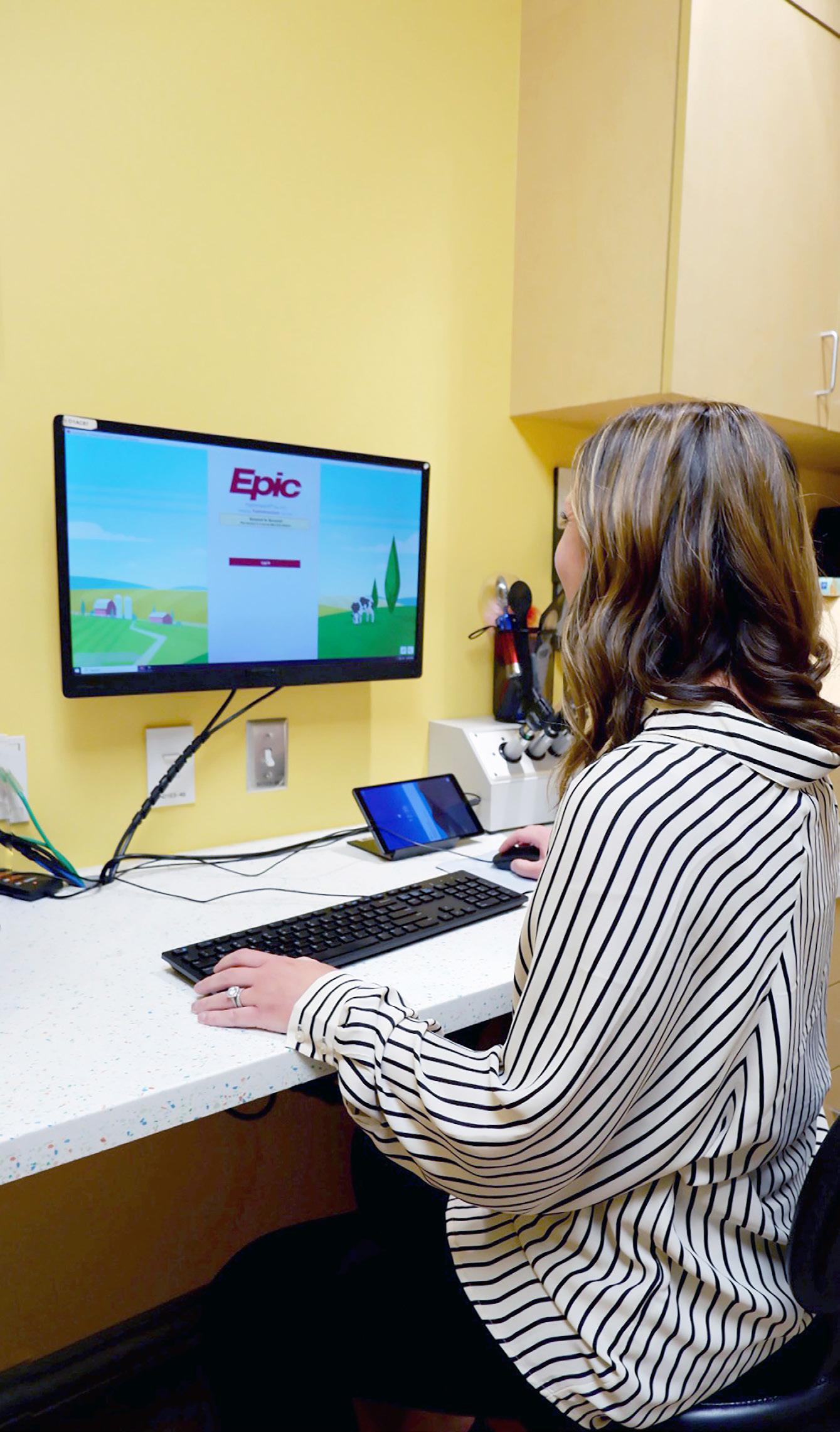
Improving Charting Software for Patients and Physicians
From ensuring timely follow-up care to creating a more welcoming health care setting, members in our department are leading the way on charting customization that enhances the patient experience and ocular treatment.
Instructors and pediatric optometrists Melissa Engle, OD, FAAO, and Erin Major, OD, have each found new and innovative ways to amplify Epic, an electronic health records system they use daily at Children’s Hospital Colorado. Their modifications prioritize their patients’ physical and mental wellbeing and are becoming a model for systems and providers across the country.
Engle, who serves as the department’s director of clinical informatics for pediatric ophthalmology and lead pediatric optometrist at Children’s Colorado, and a team of application analysts recently launched a tracker that automates when and how often a health care provider should follow up with a baby with retinopathy of prematurity (ROP).
“We’re always monitoring babies closely to watch the severity of the ROP,” Engle says. “That’s why it’s so important to have an accurate tracking system, because each baby will have a different schedule for follow-up based on the severity of their exam.”
The tracker works by identifying a baby at risk of ROP—born at 30 weeks or earlier or weighing less than about 3 pounds at birth—and forces a pop-up in the Epic chart, promoting the neonatal intensive care unit (NICU) provider to schedule a ROP screening.
“This consult order data automatically calculates when the baby is due for their ROP screening and places them on a specific weekly report. When our ophthalmologists examine the babies, they document in a customized ROP flowsheet within Epic’s Kaleidoscope ophthalmology module to record essential followup data each week,” Engle says. “This data then pulls into specific ROP weekly reports to ensure each baby is scheduled for the correct follow-up week.”
With more than a year of development, the tracker maintains a 100% success rate, which Engle says is a far better system than the physical ledgers or spreadsheets many providers have been using. The new tracker is being implemented in clinics across the country.
Also in Epic, Major saw an opportunity to modify the software to help health care providers make patients more comfortable in their care.
After completing a career development program focused on health equity and creating an inclusive environment, the optometrist pushed for implementation of an Epic tool that allows a patient’s provider to make a note on how the patient pronounces their name.
Once that detail is entered into the system, any doctor, nurse, or staff member who interacts with the patient can see the note and properly pronounce the name.
Before the tool was implemented, Epic users at Children’s Colorado could use a digital sticky note, but it wasn’t intentionally designed for the purpose of recording name pronunciation. The new tool is integrated into the patient profile and appears when the user hovers over the patient’s name.
“It only takes about 30 seconds to complete, and it can make a big difference,” Major says. “These patients light up when you say their name correctly, especially when it’s commonly mispronounced. It’s a simple thing that we can do as health care providers to make patients feel comfortable and welcome. Your name is such an important part of your identity, and getting it right is huge.”
Sue Anschutz- Rodgers Eye Center Residency Program
The ophthalmology residency program at the University of Colorado focuses on the development of outstanding clinical and surgical skills. Residents experience broad and in-depth training, with extensive exposure to the evaluation and management of common and rare ophthalmic diseases. After the intern year in partnership with the CU Department of Internal Medicine, residents complete three years of ophthalmology training. The residency program has been ranked as one of the top 20 programs in the nation by Doximity.
Resident Surgeries
This year’s six graduating residents completed more than 5,400 surgical procedures during their training. The average number of cases completed by each graduating resident was 902. Below is a breakdown of the average surgical experience.
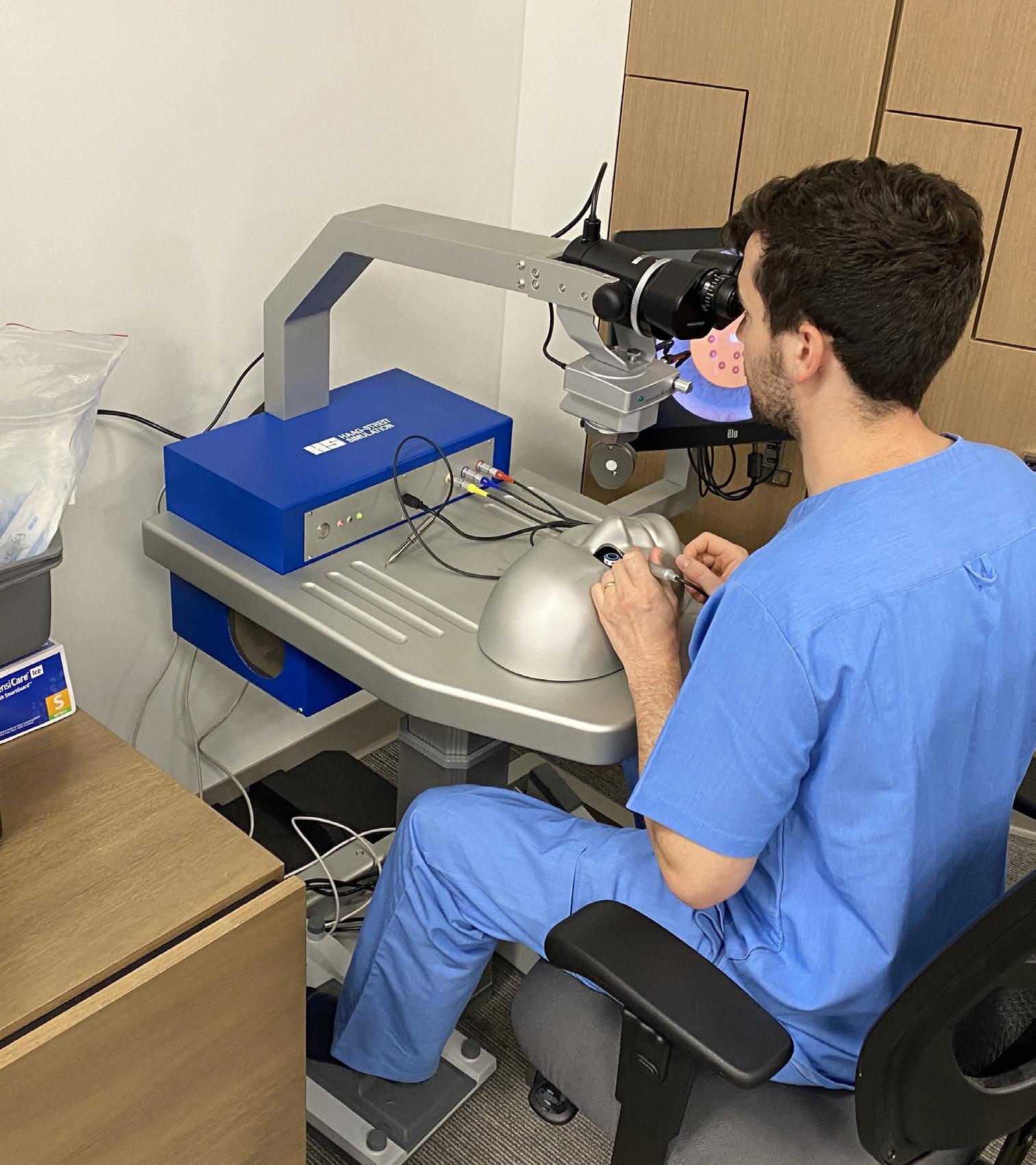
Simulator Upgrades Cataract Surgery Training
By the time ophthalmology residents at the University of Colorado School of Medicine graduate from the program, they’ll have done more than three or four times the number of required cataract surgeries to become practicing physicians. That’s in part due to a new virtual reality surgical simulator purchased by the Department of Ophthalmology this year that allows residents to fine-tune their surgical techniques and conveniently practice cataract surgeries in a lifelike environment whenever it fits into their schedules.
“One of our biggest roles as educators is teaching residents how to teach themselves and how to become constant learners. The way we do cataract surgery now is vastly different than it was a few decades ago, so giving the residents tools that allow them to take ownership of their own surgical skill acquisition is such an important priority,” says Residency Program Director Monica Ertel, MD, PhD.
Prioritizing practice
The simulator—which features a screen, surgery instruments, microscope, and artificial eyeball—starts with basic tasks then gradually gets more difficult, working trainees up to the point of a mock cataract surgery.
“When you’re operating under a microscope, there are a lot of movements with your hands that don’t feel natural at the beginning,” says Ian McClain, MD, a PGY-3 ophthalmology resident. “But you can really become comfortable with moving your hands and getting that piece down with this simulator.”
An added bonus is that the simulator offers training that can reduce hand tremors and expand bimanual dexterity, which is crucial when working in such a small area.
For PGY-3 resident Ari Stoner, MD, receiving feedback with simulator training is especially helpful. “It can be hard for us to really judge how well we did individually, but here you get error messages,” he says. “Knowing where you can improve is important because even if you touch something with your instrument that’s a couple of millimeters away, it can have profound consequences.”
Residents have other options for learning the surgery, too. In a wet lab, they can practice on an animal model eye.
An investment with high returns
When residents eventually reach the operating room with patients, they’ve already completed several cataract surgeries through the wet lab and simulator, and they feel more comfortable with the tools they need to successfully complete the procedure—which is great news for patient care, Ertel says.
Several studies have shown that there are higher rates of complications in an ophthalmologist’s first 50 to 100 cataract surgeries, “so having the residents spend that time on our simulator before the operating room really does result in better, safer outcomes for our patients,” she adds.
An estimated 3.5 million cataract surgeries are performed in the U.S. each year, making it one of the most common outpatient procedures. Residents say rigorous education and support from the department means they can better serve those patients.
“Our faculty members are dedicated to our learning,” McClain says. “The fact that they’re willing to invest in us in this way is huge.”
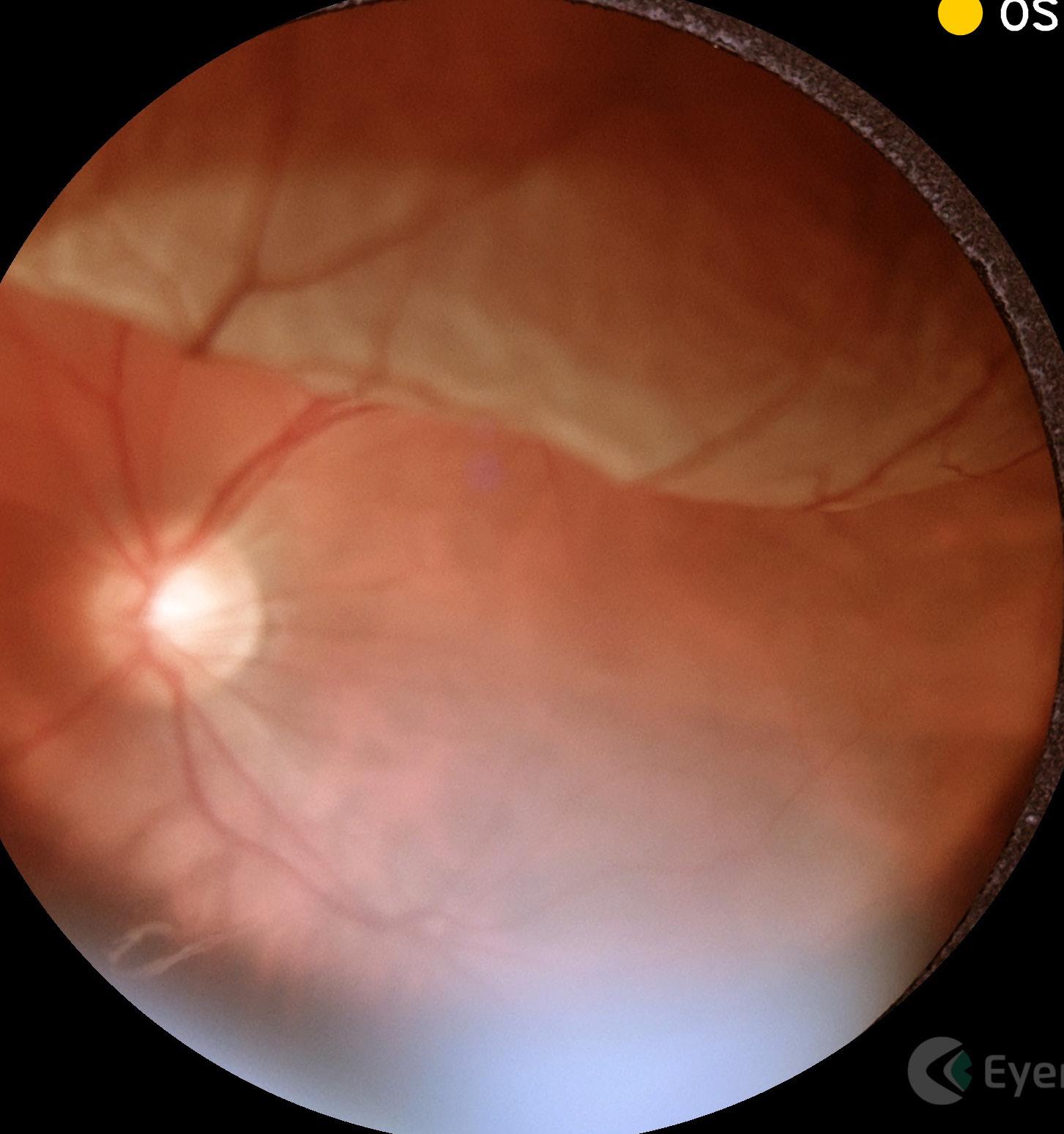
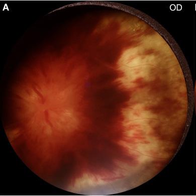
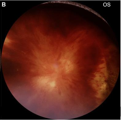
Employing Portable Fundus Photography Cameras to Enhance On-Call Imaging
Ophthalmology residents at the University of Colorado School of Medicine never know exactly what kind of eye or vision complaints will land in their care while on-call in a hospital, but they’re prepared for almost anything.
Now, with portable fundus photography cameras, they have even more cutting-edge tools at their fingertips.
“Ophthalmology is unique because it’s a clinic-based specialty, and we rely on specialized equipment to get the most accurate diagnosis and treatment plan,” says resident Nihaal Mehta, MD. “We have a backpack stocked with dozens of eyedrops and tools that help us perform even seemingly simple tasks, like checking eye pressure, for example.”
More than a year ago, our department added the portable cameras to the on-call residents’ backpacks, marking a new era in resident training and patient care.
The portable cameras are about the size of a smartphone and are equipped with a long lens that brings valuable imaging to wherever a resident is needed. This has enhanced the initial evaluation of patients and facilitated follow-up care. Supervising physicians can now easily see what a resident is seeing, confirming a treatment plan or being able to access more information when a question or concern arises.
“As a retina specialist, I have always been fascinated by the value of an ophthalmic image, not only in making a diagnosis but also in sharing cases with colleagues,” says professor and department chair Naresh Mandava, MD. “The opportunity to deploy a simple imaging technology into a dynamic tertiary care center environment has the potential to improve outcomes for patients as well as decrease the time needed for consultation with colleagues and other experts.”
A new use for a trusted tool
Phelcom gifted the department three Eyer portable cameras, allowing residents to have them ready at whichever hospital they’re stationed for a shift. The addition to their toolbox saves time, especially in fast-paced environments.
At UCHealth University of Colorado Hospital, on-call residents completed 1,200 emergency department consults and 800 inpatient consults in 2023. In addition to those calls, they cross the University of Colorado Anschutz Campus to see patients at Children’s Hospital Colorado or the Rocky Mountain Regional VA Medical Center.
“Residents might see 10 to 15 patients in 24 hours, so it can be really hectic,” Mehta, who is now a vitreoretinal fellow at Emory Medical School, says. “Any tool that can help you while you’re on-call is a huge godsend.”
Eye pathology can be especially difficult to put into words, making these new cameras all the more helpful. The images have increased communication between residents, fellows, and supporting doctors, who might have previously needed to re-evaluate a patient for a second time because they only had a written description of the condition.
“These cameras expedite the process and make it more streamlined when verifying exam findings,” Mehta says.
“It helps us to make a clinical plan, which is also really helpful for the patient.”
The cameras are proving to be worthy in the long term, too.
“These cameras have been helpful in monitoring patients over time,” Mehta explains. “If a patient has bleeding in the back of their eye, you can describe what you see, but a photo a week later can tell you more accurately how the condition is changing.”
In the classroom
Beyond the hospital, residents are finding the tools useful in classroom and academic settings, too.
“This helps us to present at a conference or publish a paper,” Mehta says. He was able to use an image taken with a portable fundus camera to help complete a paper on a rare and remarkable exam finding.
“Because we had those photos, we were able to submit it as a picture case,” he says.
The residents also bring photos from the camera to weekly conferences where they discuss cases.
“Every Wednesday morning, we have a debrief with all the residents on interesting cases from the past week. The addition of imaging to the consult workflows has improved the educational environment for our residents, fellows, and faculty alike,” Mandava says. “In addition, the time needed to gain multiple opinions has been reduced drastically, which is ultimately a benefit to the patients.”
“Deploying a simple imaging technology into a dynamic tertiary care center environment has the potential to improve outcomes.”
ENDOWED CHAIRS
Thank you to those who have made our endowed chair appointments possible, and to the faculty members who carry out our department’s mission. Endowed chairs allow our faculty to engage in specialized research and dedicate their time to elevated academic pursuits.
Endowed Chair in Eye Care Innovation
Sue Anschutz-Rodgers Endowed Chair in Retinal Diseases
Slater Family Endowed Chair in Ophthalmology
Ponzio Family Endowed Chair for Pediatric Ophthalmology at Children’s Colorado
NARESH MANDAVA, MD
MALIK KAHOOK, MD
RICHARD DAVIDSON, MD
EMILY MCCOURT, MD
JAYASHREE KALPATHY-CRAMER, PHD
Endowed Chair in Ophthalmic Data Sciences
SCOTT OLIVER, MD
Vitale-Schlessman Endowed Chair in Retinal Diseases
M. VALERIA CANTO-SOLER, PHD
Doni Solich Family Endowed Chair in Ocular Stem Cell Research
SOPHIE LIAO, MD
Robert H. Bell Endowed Chair in Ophthalmology
PREM SUBRAMANIAN MD, PHD
Clifford R. and Janice N. Merrill Endowed Chair in Ophthalmology
Faculty By specialty
Artificial Medical Intelligence
Jayashree Kalpathy-Cramer, PhD Endowed Chair in Ophthalmic Data Sciences Chief, Division of Artificial Medical Intelligence in Ophthalmology Professor, Ophthalmology
Steve McNamara, OD Research Instructor, Ophthalmology
Praveer Singh, PhD Assistant Professor, Ophthalmology
Basic Science Research
Joseph Brzezinski, PhD Director, Research Director, Laboratory of Developmental Genetics, CellSight Associate Professor, Ophthalmology
M. Valeria Canto-Soler, PhD
Doni Solich Family Endowed Chair in Ocular Stem Cell Research Director, CellSight Director, 3DRet Lab, CellSight Associate Professor, Ophthalmology
Karen Cusato, PhD, PA-C Research Associate, Ophthalmology
Daniel Denman, PhD Assistant Professor, Physiology & Biophysics
Steven Droho, PhD Research Associate, Ophthalmology
Miguel Flores-Bellver, PhD Director, ExoSight Lab, CellSight Assistant Professor, Ophthalmology
Carl Frick, PhD Assistant Adjoint Professor
Uday Kompella, PhD, FARVO, FAAPS
Co-Director and Co-Founder, Colorado Center for Nanomedicine and Nanosafety Professor of Pharmaceutical Sciences, Ophthalmology and Bioengineering
Daniel LaBarbera, PhD Director, CU Center for Drug Discovery Professor, School of Pharmacy
Tim Lei, PhD Assistant Professor, University of Colorado Denver
Andres Lisker, MD Assistant Adjoint Professor, CellSight
Marc Mathias, MD Director, Laboratory of Advanced Ophthalmic Surgery, CellSight Associate Professor, Ophthalmology
Ram Nagaraj, PhD Professor, Ophthalmology
Mi-Hyun Nam, PhD Instructor, Ophthalmology
Daewon Park, PhD
Associate Professor, University of Colorado Denver
Alon Poleg-Polsky, MD, PhD Assistant Professor, Physiology and Biophysics
Natalia Vergara, PhD Director, Ocular Development and Translational Technologies Laboratory, CellSight Assistant Professor, Ophthalmology
Chris Yakacki, PhD
Assistant Professor, University of Colorado Denver
Wenbo Zhou, PhD Research Associate, Ophthalmology
Joel Zylberberg, PhD
Assistant Adjoint Professor, Physiology & Biophysics
Comprehensive Eye Care
Kaleb Abbott, OD, MS, FAAO Assistant Professor, Ophthalmology
Melanie Akau, OD, FAAO Medical Director, Staff Education Instructor, Ophthalmology
Christopher Allen, MD Instructor, Ophthalmology
Brian Bucca, OD, FAAO Chief of Eye Care at Barbara Davis Center Associate Professor of Clinical Practice, Pediatrics - Barbara Davis Center
Angela Demetrulias, OD Instructor, Ophthalmology
Marilyn Dougherty, MD Senior Instructor, Ophthalmology
Joel Goldstein, MD Associate Professor, Ophthalmology
C. Rob Graef, OD Assistant Professor, Ophthalmology
Robert Keyser, MD Clinical Professor, Ophthalmology
Cecelia Koetting, OD, FAAO, DipABO Assistant Professor, Ophthalmology
Stephanie Martich, OD Assistant Professor, Ophthalmology
Andrew Martin, MD Instructor, Ophthalmology
Daniel J. Ozzello, MD Assistant Professor, Ophthalmology
William Roberts, MD Senior Instructor, Ophthalmology
Erin Van Dok, OD Clinical Instuctor, Ophthalmology
Brittany Wright, OD, MS Assistant Professor, Ophthalmology
Cornea, Cataract and Refractive Surgery
Michael Taravella, MD
Chief, Cornea, External Diseases & Refractive Surgery Director, Cornea and Refractive Surgery Fellowship Professor, Ophthalmology
Michael Chen, MD
Ophthalmology Division Chief, Denver Health Medical Center Associate Professor, Ophthalmology
Karen Christopher, MD
Section Chief of Ophthalmology, Rocky Mountain Regional VA Medical Center Associate Professor, Ophthalmology
Richard Davidson, MD Endowed Chair in Eye Care Innovation Co-Medical Director, Rocky Mountain Lions Eye Bank Co-Director, Cornea, External Disease, and Refractive Surgery Fellowship Vice Chair of Clinical Affairs and Quality, UCHealth Eye Centers Professor, Ophthalmology
Michael Erlanger, MD Associate Professor, Ophthalmology
Christopher Gelston, MD Associate Professor, Ophthalmology
Darren Gregory, MD Professor, Ophthalmology
Michael Wildes, MD
Clinical Director, Sue Anschutz-Rodgers Eye Center Assistant Professor, Ophthalmology
Ronald Wise, MD
Medical Director, UCHealth Eye Center - LoDo Associate Professor of Clinical Practice, Ophthalmology
Ophthalmic Epidemiology
Anne Lynch, MD, MSPH Director, Division of Ophthalmic Epidemiology Professor, Ophthalmology
Alison Abraham, PhD, MS, MHS Vice Chair for Education, Colorado School of Public Health Associate Professor, Epidemiology
Tianjing Li, MD, PhD, MHS Director, Cochrane Eyes and Vision US Project Professor, Ophthalmology
Alison Suhsun Liu, MD, PhD, MPH Assistant Research Professor, Ophthalmology
Jennifer Patnaik, PhD, MHS Assistant Professor, Ophthalmology
Riaz Qureshi, MD Assistant Professor, Ophthalmology and Epidemiology
Brandie Wagner, PhD Associate Research Professor, Colorado School of Public Health - Department of Biostatistics and Informatics
Glaucoma
Malik Kahook, MD
Slater Family Endowed Chair in Ophthalmology Vice Chair, Translational Research Chief, Glaucoma Service Co-Director, Glaucoma Fellowship Professor, Ophthalmology
Cara Capitena Young, MD Medical Director, Sue Anschutz-Rodgers Eye Center Operating Room Associate Professor, Ophthalmology
Galia Deitz, MD, MPH Assistant Professor, Ophthalmology
Monica Ertel, MD, PhD Director, Residency Program Assistant Professor, Ophthalmology
Gabriel Lazcano-Gomez, MD, OD
Assistant Adjoint Professor, Ophthalmology
Kaweh Mansouri, MD Professor Adjoint, Ophthalmology
Mina Pantcheva, MD Associate Professor, Ophthalmology
Leonard Seibold, MD Director, Glaucoma Fellowship Professor, Ophthalmology
Jeffrey SooHoo, MD, MBA
Assistant Dean of Admissions and Student Affairs, University of Colorado School of Medicine
Associate Professor, Ophthalmology
Deidre St. Peter, MD Assistant Professor, Ophthalmology
Neuro-Ophthalmology
Prem Subramanian, MD, PhD
Clifford R. and Janice N. Merrill Endowed Chair in Ophthalmology Chief, Neuro-Ophthalmology
Vice Chair for Academic Affairs, Ophthalmology Chair, Faculty Promotions Committee Professor, Ophthalmology
Jeffrey Bennett, MD, PhD Professor, Ophthalmology and Neurology
Mary Labowsky, MD Assistant Professor, Ophthalmology
Maria Nagel, MD Professor, Ophthalmology and Neurology
Victoria Pelak, MD Director, Neuro-Ophthlamology Fellowship Professor, Ophthalmology and Neurology
Oculofacial Plastic and Orbital Surgery
Eric Hink, MD
Chief, Oculofacial Plastic and Orbital Surgery Director, ASOPRS Fellowship in Oculofacial Plastic and Orbital Surgery
Associate Professor, Ophthalmology
Sophie Liao, MD
Robert H. Bell Endowed Chair in Ophthalmology
Associate Chief Medical Officer, UCH - Ambulatory Associate Professor, Ophthalmology
Kia Washington, MD Professor, Surgery - Plastic/Reconstructive
Trevor Williams, PhD Professor, School of Dental Medicine
Caroline Vloka, MD Assistant Professor, Ophthalmology
Ophthalmic Oncology
Scott Oliver, MD
Vitale-Schlessman Endowed Chair in Retinal Diseases Chief, Retina Service Director, Eye Cancer Program Director, Vitreoretinal Diseases and Surgery Fellowship Associate Professor, Ophthalmology
Richard Davidson, MD
Endowed Chair in Eye Care Innovation
Co-Medical Director, Rocky Mountain Lions Eye Bank Co-Director, Cornea, External Disease, and Refractive Surgery Fellowship
Vice Chair of Clinical Affairs and Quality, UCHealth Eye Centers Professor, Ophthalmology
Eric Hink, MD
Chief, Oculofacial Plastic and Orbital Surgery
Director, ASOPRS Fellowship in Oculofacial Plastic and Orbital Surgery
Associate Professor, Ophthalmology
Sophie Liao, MD
Robert H. Bell Endowed Chair in Ophthalmology
Associate Chief Medical Officer, UCH - Ambulatory Associate Professor, Ophthalmology
Prem Subramanian, MD, PhD
Clifford R. and Janice N. Merrill Endowed Chair in Ophthalmology
Chief, Neuro-Ophthalmology
Vice Chair for Academic Affairs, Ophthalmology Chair, Faculty Promotions Committee Professor, Ophthalmology
Michael Taravella, MD Chief, Cornea, External Diseases & Refractive Surgery Director, Cornea and Refractive Surgery Fellowship Professor, Ophthalmology
Pediatric Ophthalmology and Adult Strabismus
Emily McCourt, MD
Ponzio Family Endowed Chair for Pediatric Ophthalmology Chief of Pediatric Ophthalmology Director, Pediatric Ophthalmology & Strabismus Fellowship Associate Professor, Ophthalmology
Mariam Ahmad, MD Assistant Professor, Ophthalmology
Rebecca Edwards Mayhew, MD, PhD Assistant Professor, Ophthalmology
Melissa Engle, OD, FAAO Director, Pediatric Optometry Specialty Care Residency Program Director, Clinical Informatics, Department of Pediatric Ophthalmology Instructor, Ophthalmology
Robert Enzenauer, MD, MPH/MSPH Professor, Ophthalmology
Celeste Gomez, OD Instructor, Ophthalmology
JP Gorham, MD Assistant Professor, Ophthalmology
Jennifer Jung, MD Director, Retinopathy of Prematurity Service Associate Professor, Ophthalmology
Erin Major, OD Instructor, Ophthalmology
Lauren Mehner, MD, MPH Assistant Professor, Ophthalmology
Michael Puente Jr., MD Director, Medical Student Education Assistant Professor, Ophthalmology
Rebecca Sands Braverman, MD Associate Professor, Ophthalmology
Casey Smith, MD Assistant Professor, Ophthalmology
Emma Stahr, OD Instructor, Ophthalmology
Uveitis and Ocular Immunology
Alan Palestine, MD Chief, Uveitis and Ocular Immunology Professor, Ophthalmology
Lynn Hassman, MD, PhD Assistant Professor, Ophthalmology
Jennifer Jung, MD Director, Retinopathy of Prematurity Service Associate Professor, Ophthalmology
Jason Kolfenbach, MD Associate Professor, Medicine - Rheumatology
Mina Pantcheva, MD Associate Professor, Ophthalmology
Amit Reddy, MD Director, Resident Research Assistant Professor, Ophthalmology
Vision Rehabilitation Service
Kara Hanson, OD, FAAO Director, Vision Rehabilitation Service Associate Professor, Ophthalmology
Cade Oost, OD, MS Assistant Professor
David Simpson, OD, FAAO Assistant Professor, Ophthalmology
Vitreoretinal Diseases and Surgery
Scott Oliver, MD
Vitale-Schlessman Endowed Chair in Retinal Diseases Chief, Retina Service Director, Eye Cancer Program Director, Vitreoretinal Diseases and Surgery Fellowship Associate Professor, Ophthalmology
Emily Cole, MD, MPH Assistant Professor
Talisa Forest (de Carlo), MD Director, Medical Imaging Assistant Professor, Ophthalmology
Stuart Fine, MD Clinical Professor, Ophthalmology
Naresh Mandava, MD Chair, Department of Ophthalmology
Sue Anschutz-Rodgers Endowed Chair in Retinal Diseases Professor, Ophthalmology
Niranjan Manoharan, MD Assistant Professor, Ophthalmology
Marc Mathias, MD Director, Laboratory of Advanced Ophthalmic Surgery, CellSight Associate Professor, Ophthalmology
Jeffrey Olson, MD Associate Professor, Ophthalmology
Jesse Smith, MD Associate Professor, Ophthalmology
Professor Emeritus
Phillip Ellis, MD Professor Emeritus
J. Mark Petrash, PhD Professor Emeritus
For administration: 720-848-2500
For appointments: 720-848-2020
Subscribe to department email updates by sending your email address to us at ophthocommunications@cuanschutz.edu
Scan QR code to visit our website www.eyeinstitute.org