

3rd Edition

The goal of this guide is to provide basic facts about ependymoma to increase education and awareness about the rare disease.
We hope this information gives you a better understanding of ependymoma and empowers you when making key decisions for you or your loved one’s care. As you read through the Ependymoma Guide, keep in mind each person’s experience with ependymoma is different and unique. This information is general education and does not replace medical advice. Medical information changes quickly based on scientific developments. Call your physician or care team for medical advice. Please visit www.cern-foundation.org for more information.

This guide was produced and published by the Collaborative Ependymoma Research Network (CERN) Foundation, a program of the National Brain Tumor Society (NBTS). © CERN Foundation. Reproduction is prohibited.
The CERN Foundation is committed to improving the care and outcome of people with ependymoma through community support and research efforts.
Phone: 617-924-9997
Funding for this guide was made possible by the generous support of the DeLotto family and the Childhood Brain Cancer Research Collaborative. The 3rd Edition Ependymoma Guide is dedicated in honor of Eva DeLotto.


Ependymoma is a rare tumor of the brain and spinal cord that affects both children and adults. A collaborative effort between ependymoma advocacy groups across the world was organized in order to prioritize and articulate the unique key issues facing the ependymoma community. The Ependymoma Key Issues tie to the IBTA Brain Tumor Patients’ Charter of Rights in order to amplify the voice of the ependymoma community within the larger brain and spinal cord tumor community and international medical professional network in a cohesive and unified format.
Since there is no established standard of care for ependymoma, we call for local emergency providers and community neurosurgeons to have greater awareness of the unique medical needs of the ependymoma community, access to expert physician-to - physician consultation, and more educational opportunities for community providers on the latest ependymoma research and clinical practices. In addition, families should have a clear understanding of the capabilities and services offered at the facility that relate to the care they require.
Building on the IBTA Charter of RightsClause 2: Appropriate Investigation of Signs and
Symptoms
Care for patients with ependymoma requires significant coordination and collaboration. Ependymoma patients require access to multidisciplinary care throughout the trajectory of their illness and survivorship. Providers should work with the family to identify who is involved in the patient’s medical team, establish a point of contact for each provider, and identify who is the coordinator. The nature and rarity of the disease requires collaboration between all members of the patient’s medical team on an ongoing basis with the goal of prioritizing the patient’s outcome and quality of life.
Building on the IBTA Charter of Rights - Clause 5: Excellent Treatment and High-Quality Follow-Up Care
4
The ependymoma community has an enhanced need for evaluation or consultation by an expert neuro - oncology team at diagnosis and before any non-immediate treatment is done. This can be done in coordination with local providers or outside of that relationship. The family should receive transparent and timely communication throughout the diagnostic process and support when seeking second opinions and information on clinical trials.
The ependymoma community has an enhanced need for evaluation or consultation by an expert neuro oncology team at diagnosis and before any non-immediate treatment is done. This can be done in coordination with local providers or outside of that relationship. The family should receive transparent and timely communication throughout the diagnostic process and support when seeking second opinions and information on clinical trials.
Building on the IBTA Charter of Rights - Clause 3: A Clear, Comprehensive, Integrated Diagnosis
Building on the IBTA Charter of Rights - Clause 3:
A Clear, Comprehensive, Integrated Diagnosis
SURVIVORSHIP & SUPPORT
SURVIVORSHIP & SUPPORT
Building on the IBTA Charter of Rights - Clause 4: Appropriate Support 3 4
It is essential that ependymoma families have a clear understanding of survivorship and known effects of treatment that can impact quality of life. Our personal goals should always be considered in each discussion about treatment and outcomes.
It is essential that ependymoma families have a clear understanding of survivorship and known effects of treatment that can impact quality of life. Our personal goals should always be considered in each discussion about treatment and outcomes.
i.e. , This might include testing like cognitive and neuropsychological evaluation done at the time of diagnosis and when possible, monitored throughout survivorship.
i.e. This might include testing like cognitive and neuropsychological evaluation done at the time of diagnosis and when possible, monitored throughout survivorship.
Building on the IBTA Charter of Rights - Clause 4: Appropriate Support
Key Issues establish a framework upon which to build greater awareness and understanding of the critical issues facing patients and health care providers. 3
Summary Statement: People with ependymoma have an urgent need for increased funding for rare disease research that provides better targeted treatments for the different ependymoma subtypes, increases access to these treatments, and evaluates impact on quality of life. The Ependymoma
Summary Statement: People with ependymoma have an urgent need for increased funding for rare disease research that provides better targeted treatments for the different ependymoma subtypes, increases access to these treatments, and evaluates impact on quality of life. The Ependymoma Key Issues establish a framework upon which to build greater awareness and understanding of the critical issues facing patients and healthcare providers.


A. What is Ependymoma?
B. Brain and Spine Anatomy
C. Ependymoma Statistics
Ependymoma is a rare tumor of the brain or spinal cord. It can occur in both adults and children. Ependymoma is a primary tumor, which means that it starts in either the brain or spinal cord. The brain and spine are part of the central nervous system (CNS). Primary brain and spinal cord tumors are typically grouped by where the cells start.
The most common types of cells in the CNS are neurons and glial cells. Tumors from neurons are rare. Glial cells are the cells that support the brain. Tumors that occur from these cells are called gliomas.
Glial cell subtypes of the CNS include:
• Astrocytes
• Oligodendrocytes
• Ependymal cells
CNS tumors associated with all three types of glial cells are recognized by the World Health Organization (WHO) as astrocytomas, oligodendrogliomas and ependymomas.
Ependymal cells line the ventricles (fluid-filled spaces in the brain) and the central canal of the spinal cord. Ependymomas develop from ependymal cells (called radial glial cells). Ependymomas can form anywhere in the CNS in the supratentorial (top of the head), posterior fossa (back of the head), and spinal cord. Ependymomas often occur near the ventricles in the brain and the central canal of the spinal cord. On rare occasions, ependymomas can form outside the CNS, such as in the ovaries.
Ependymoma can occur anywhere in the brain or spinal cord. The cause of ependymoma is not known.
Ependymoma tumor cells can spread in the cerebrospinal fluid (CSF). They may spread to one or multiple areas of the brain, spine, or both. See diagram on Page 10. Ependymomas rarely spread outside the CNS. In general, tumors form where the tumor cells originate, such as the base of the brain and the bottom of the spinal cord.
Ependymomas occur in both children and adults. Overall, ependymomas occur in males slightly more than females. Ependymomas occur most often in nonHispanic White individuals.
Approximately 1,372 people per year are diagnosed with ependymoma in the U.S.
In the United States, roughly 230 new cases of ependymoma are found in children (0-19) each year.
The majority occur in the back part of the brain (posterior fossa) in the fourth ventricle. They are also less common in the top of the brain (supratentorial) and rarely occur in the spinal cord. See diagram on Page 6.
ependymoma
Ependymomas account for less than 2% of adult gliomas. The majority occur in the spine. For more statistics, see Page 13.


It is important to know where the tumor is located within the brain and spinal cord so you can understand what symptoms may occur and and how the tumor may impact body functions. The biology may also be different based on the location.
During treatment, your health care team will use many terms to refer to locations in the head, neck and spine. The diagrams in this section will help you locate different parts of the brain and spinal cord.
Tentorium Cerebelli
The image below shows a side (lateral) view of the tentorium cerebelli - an extension of the dura matter that separates the cerebellum from the bottom (inferior) portion of the occipital lobes. Tumors below the tentorium are called infratentorial and those above are called supratentorial.
The image below shows a interior (sagittal) view of the brain.
Supratentorial Anatomy
These areas are located in the supratentorial region.
are located in the infratentorial region.
Fossa
The images show views of the lobes: side (lateral), top (axial) and interior (sagittal). For more information about the views, see Pages 15-16.
The brain is divided down the middle lengthwise into two halves called the cerebral hemispheres. Each hemisphere is divided into four lobes.
See the parts of the brain to learn what they do. Knowing the location of your tumor(s) may help you to understand changes in how you act or think. Changes can be due to the impact of the tumor itself or from treatment. For example, if you have a tumor in the temporal lobe, you may have short-term memory loss. Brain tumors, regardless of their location, can cause headaches, seizures and problems with multi-tasking. It is important to address these concerns with your health care provider to evaluate if there are options to help alleviate their impact on your quality of life. Use these charts on the next page to learn more.
3. Higher Mental Functions
Executive function including concentration, coordination of thought and activity, judgement, emotional expression, inhibition
4. Motor Area
Strength on opposite side of body
Brain Function Areas
1. Motor Association Area
5. Sensory Area
Coordination of muscle strength and movement
Sensation on opposite side of body
2. Expressive Speech
1. Motor Association Area
Written and spoken language
6. Somatosensory Association Area
Coordination of muscle strength and movement
1. Motor Association Area
3. Higher Mental Functions
Understanding objects characteristics (for example, texture and temperature and position in space)
Coordination
2. Expressive Speech
Executive function including concentration, coordination of thought and activity, judgement, emotional expression, inhibition
Written and spoken language
7. Global Language
2. Expressive Speech
4. Motor Area
Expressive and receptive language comprehension
3. Higher Mental Functions
Strength on opposite side of body
8. Vision
5. Sensory Area
3. Higher Mental Functions
Executive
Executive function including concentration, coordination of thought and activity, judgement, emotional expression, inhibition
Sight on opposite side, image recognition
Sensation on opposite side of body
4. Motor Area
4. Motor Area
9. Receptive Speech
6. Somatosensory Association Area
Strength on opposite side of body
Understanding words and directions
4. Motor Area Strength
Understanding objects characteristics (for example, texture and temperature and position in space)
5. Sensory Area
5. Sensory Area
10. Association Area
7. Global Language
Sensation on opposite side of body
Short-term memory, emotion
Expressive and receptive language comprehension
8. Vision
Sensation on opposite side of body
6. Somatosensory Association Area
6. Somatosensory Association Area
Understanding
Sight on opposite side, image recognition
Understanding objects characteristics (for example, texture and temperature and position in space)
9. Receptive Speech
Understanding words and directions
7. Global Language
Expressive and receptive language comprehension
8. Vision
10. Association Area
and spoken language 9. Receptive Speech
Sight on opposite side, image recognition
9. Receptive Speech
Understanding words and directions
memory, emotion
Understanding words and directions
Stem Cerebellum
11. Cerebellum Balance and coordination of movement of body, arms and legs
12. Emotional Area
Pain, hunger, “fight or flight” response
13. Olfactory Area Understanding scent
Emotional Area
Olfactory Area
The image on the right shows the side (lateral) view of the different parts of the spinal cord and their functions. The spine is separated into four regions called the cervical, thoracic, lumbar, and sacral. Each spinal region is made up of vertebra. The image below shows the top (axial) view of a vertebrae and spinal cord. The vertebrae are numbered from top to bottom, starting with one and continue. For example, T2 is the second vertebra in the thoracic region.
Tumors are further described by whether the tumor begins in the cells inside the spinal cord (intramedullary) or outside the spinal cord (extramedullary). Extramedullary tumors grow in the membrane surrounding the spinal cord (intradural) or outside (extradural).
Spinal cord tumors, regardless of location, often cause pain, numbness, weakness or lack of coordination in the arms and legs, usually on both sides of the body. They also often cause bladder or bowel problems. Use the images to learn more.
Intervertebral Disc
Arm and hand functions
Thoracic Region (T1-T12)
Chest and abdominal functions
Vertebral Body
Spinal Nerve
Spinal Cord
Cerebrospinal Fluid
Lumbar Region (L1-L5)
Leg, knee and foot functions
Cerebrospinal Fluid
Spinal Cord
Spinous Process
Axial
Lateral
Sacral Region (S1-S5)
Leg, buttocks, foot, bowel, bladder and sexual functions
The spinal cord connects the brain to nerves in most parts of the body. This allows the brain to send messages throughout the body. The network of the brain and spinal cord is called the central nervous system (CNS).
The images show the ventricular system in the brain. Ependymomas can occur anywhere in the CNS. Common locations include the ventricles (fluid-filled spaces in the brain that contain cerebrospinal fluid) and the central canal of the spinal cord.
The image to the right shows the side (lateral) view of fluid in the spine and brain. The image below shows the ventricular system in the brain.
Lateral Ventricle Lateral Cerebrum Fourth Ventricle Third Ventricle Cerebral Aqueduct Cerebellum Brain Stem Lateral Spinal Cord Cerebrospinal FluidThis section shares information about how often ependymoma occurs. Statistics come from the collaborative work of two groups: The Collaborative Ependymoma Research Network (CERN) Foundation and Central Brain Tumor Registry of the United States (CBTRUS). Data is from the 2023 published report, CBTRUS Statistical Report on Primary Brain and Central Nervous System Tumors Diagnosed in the United States in 2016-2020
CBTRUS is a not-for-profit research corporation. Its mission is to gather and spread current epidemiologic data on all primary brain and central nervous system (CNS) tumors. This lets people see incidence and survival patterns, evaluate diagnosis and treatment, facilitate etiologic studies, establish awareness of the disease, and ultimately, prevent brain tumors.
• Incidence: The occurrence of newly diagnosed disease that appears in a particular population during a specified period of time (typically yearly). In this report, the crude incidence rates are calculated by counting the number of people with newly diagnosed primary brain and CNS tumors and dividing by the total population at risk (e.g. the total population in a state or collection of states) and are usually expressed per 100,000 persons. Incidence rates can also be calculated specifically for those diagnosed with ependymoma.
• Age-adjustment: Age-adjustment is a procedure designed to minimize the effects of differences in age composition when comparing incidence and mortality rates among different populations.
Age-adjustment of rates in this report is calculated by the direct method adjusting to the 2000 U.S. standard million population.
• Prevalence: The number or proportion of people with a particular disease or attribute (in this case, both those with a newly diagnosed brain or CNS tumor and those who may have been diagnosed in the past and are living with the disease) in a given population at a specific time.
• 5-year survival rate: The probability that a patient will survive for 5 years from the date of diagnosis (in this case, diagnosis of a brain or CNS tumor).
For all primary brain and other CNS tumors, CBTRUS estimates that the average annual age-adjusted incidence rate is 24.83 cases per 100,000 persons for the years 2016-2020.
An estimated 93,470 new cases of primary brain and other CNS tumors are expected to be diagnosed in the U.S. in 2023.
An estimated 5,230 new cases of childhood primary brain and other CNS tumors are expected to be diagnosed in the U.S. in 2023.

Ependymomas (ependymoma, NOS [not otherwise specified], epithelial ependymoma, cellular ependymoma, clear cell ependymoma, tanycytic ependymoma, anaplastic ependymoma, ependymoblastoma) and ependymoma variants (myxopapillary ependymoma) are rare, and represent 1.5% of all primary brain and other CNS tumors.
Approximately 1,372 ependymomas and ependymoma variants are diagnosed per year.
In adults 20+ years, ependymomas accounted for 1.4% of all tumors diagnosed.
Approximately 1,142 adults are diagnosed with ependymoma per year.

The incidence of ependymoma variants is higher in males than females. The incidence of both ependymomas/anaplastic ependymomas and ependymoma variants was higher in non-Hispanic Whites and non-Hispanic Blacks.
In children aged 0-14 years, ependymomas accounted for 5.2% of all tumors diagnosed.
Approximately 179 children are diagnosed with ependymoma per year.
In children aged 15-19 years, ependymoma accounted for 3.3% of all tumors diagnosed.
Approximately 52 teenagers are diagnosed with ependymoma per year.
The incidence of ependymoma variants is higher in males than females.

For tumors involving the spinal cord, spinal meninges and cauda equina, ependymomas accounted for 19.6% of all tumors diagnosed in adults ages 20+ years and 17% of all tumors diagnosed in children ages 0-19 years.
Approximately 681 people are diagnosed with spinal cord ependymoma per year.
An estimated 1,323,121 persons in the U.S. were living with a diagnosis of primary brain and CNS tumor in 2019.
For children (ages 0-14 years), an estimated 28,219 were living with one of these diagnoses in the U.S. in 2019.
The five-year relative survival rate for all primary brain and central nervous system tumors is 76.3%. For children aged 0-14 years, the 5-year relative survival rate is 83.2%.
For those with ependymoma, the overall 5-year relative survival rate is 90.5%. 5-year relative survival rates are highest for those ages 15-39 years (94.8%), and decrease with increasing age at diagnosis with a 5-year relative survival rate of 90.9% for those ages 40+ years. For children ages 0-14 years, the 5-year relative survival rate is 80.3%.
Please recognize that these numbers are helpful for general information related to ependymoma, but may not be meaningful for any one individual. Please discuss any questions or concerns that you have about your own diagnosis with your treating physician.

Anaplastic ependymoma


Ependymoma in the spine of an adult patient
4th ventricular ependymoma, pediatric patient, T1 weighted with contrast


Ependymoma in the spine of a pediatric patient
A. How is Ependymoma Diagnosed?
B. Pathology
C. What To Ask Your Doctor
D. Coping With The Diagnosis
Usually, people with ependymoma are diagnosed after showing symptoms. Symptoms may start slowly and may not be diagnosed until they worsen.
Other times, problems occur suddenly and result in an urgent trip to the emergency room or clinic. An exam by a health care provider often shows neurological problems. A common diagnostic test that identifies potential neurological problems is a magnetic resonance image or MRI. Although sometimes, CT scans (computed tomography) might be the first test that is done. A CT scan uses X-rays.
An MRI is typically the preferred test for people who may have a brain or spinal cord tumor. An MRI can generate highly detailed pictures of internal organs and soft tissues. MRIs do not use radiation. The MRIs use magnets, radio waves, and a computer to make a series of detailed images of areas inside the brain and spinal cord.
After the initial testing, more diagnostic tests may be needed. If the MRI shows a possible primary central nervous system (CNS) tumor, patients normally do not need other imaging tests of the body. This is mainly because ependymoma tumors do not tend to spread outside of the CNS. MRI of the brain and spine is not only used as a baseline test, but may also be used to evaluate the whole CNS.
Three different views are typically taken of the head, neck, and/or spine for a tumor diagnosis. Contrast agents might be used to show abnormal tissue more clearly. A substance is injected into a vein and travels through the bloodstream. The patient must lie still inside the large round MRI machine for about an hour.
An MRI machine produces a loud noise. There might be earplugs or headphones offered to help decrease the disturbance of the noise. Some children may require sedation for the MRI. After the images are taken, the medical provider or radiologist will read the digital images and create a report and share the results with you.
TIP: Ask for a digital copy of your MRI in case you seek a second opinion at a different institution.
MRIs use three different views (see below) to show the location and position of a tumor. This helps your medical team compare the tumor with normal brain or spinal cord structures. Using these different views, different scans can be done to produce exact images that best show the tumor and the area around the brain or spine. New techniques are being developed and tested on a regular basis.


The three types of MRI ‘views’ or images include:
• Sagittal:
This is a vertical plane that separates the body vertically from head to toe and left and right sides.
• Coronal:
This is a vertical plane that separates the body vertically from head to toe and front to back.
• Axial:
This is a straight line that separates the body horizontally to create top and bottom halves of the body.
A neurological exam is an evaluation of a person’s nervous system that can be done in a medical office. A series of question and tests are performed to check the brain, spinal cord, and nerve function. The exam checks a person’s mental status, motor and sensory skills, balance and coordination, reflexes, and functioning of the nerves.
A procedure used to collect cerebrospinal fluid (CSF) from the spinal column is called a lumbar puncture (LP) or spinal tap. The LP is a medical procedure in which a thin needle is inserted into the spinal column to collect CSF for diagnostic testing. The sample of CSF is checked under a microscope for signs of tumor cells.
Sometimes a lumbar puncture may be necessary if there is concern that the tumor has spread into the CSF. Direct testing of the CSF is done to look for the presence of tumor cells. In addition, an MRI of either the brain or spine may be ordered in addition to the baseline test to evaluate the entire CNS.


Pathology is the examination of tissues and body fluids in order to make a diagnosis. This involves taking a tissue sample of the tumor when a biopsy or surgery is done to remove the tumor. The sample is then sent to a pathologist for review. Once the diagnostic lab report is completed, it is sent to your doctor who shares it with you.
Ependymoma is less common than other brain and spinal cord tumors. In addition, it is sometimes difficult to distinguish it from other tumor types. This can make it difficult to diagnose or a delay might occur. It is not uncommon for ependymoma tissue to be sent out to a different laboratory for a second opinion. Seeking a second opinion from a neuropathologist is always a good option when dealing with a rare disease. Contact the CERN Foundation if you would like help navigating a second opinion by emailing administrator@cernfoundation.org.
TIP: Ask your clinician what institution or laboratory your pathology report came from. Receiving an accurate diagnosis is important. Seek a second opinion.

Tissue samples are reviewed under a microscope by a pathologist to determine the grade and tumor type.
The World Health Organization (WHO) made a significant updates in 2016 and 2021. In addition to the tissue review under a microscope, molecular changes in the tumor are used to further refine the diagnosis. The WHO continues to further refine and update these molecular classifications of ependymoma tumors.
TIP: Ask your medical team if your tumor was diagnosed according to the latest WHO guidelines for ependymoma
Unlike other cancers, primary brain and spine tumors generally do not spread (metastasize) outside of the central nervous system. For this reason, the TumorNode-Metastasis (TNM) staging system widely used for most “solid tumor” cancers is not useful for primary brain tumors.
The WHO classification defines and classifies ependymoma tumors based on tumor location, grading and molecular subgroups.
There are three important tumor locations included in ependymoma classification: supratentorial, infratentorial, and those located in the spine.
There are several types of ependymoma. From CERN supported research, we were able to further molecularly classify these rare tumors. Molecular analysis allows us to look inside a cell to identify important DNA or RNA changes and markers. The classification of molecular groups and subtypes for ependymoma are constantly evolving.
The 2021 5th edition of the WHO classification further refines several subgroups based on molecular genetic features. DNA methylation profiling has determined that there are 10 distinct different types of ependymoma tumors.
TIP: Ask your treating medical team if your tumor tissue has been diagnosed according to the latest WHO criteria.
Subtypes that have been previously recognized by the WHO classification include: subependymoma, myxopapillary ependymoma, as well as morphologic variants.


Ependymoma is a heterogeneous disease, meaning there is great diversity within the disease. We know about these differences because of the extensive work that has been done in molecular classification by an international group of collaborative scientists. Molecular analysis allows us to look inside a cell to identify important DNA or RNA changes and markers. The classification of molecular groups and subtypes for ependymoma is constantly evolving. Based on DNA methylation profiling, different types (molecular groups) of ependymoma are recognized and are distinguished according to location, pathology, and distinct molecular features. Understanding these important molecular differences will help guide future clinical protocols designed to identify targeted therapies.
WHO Grade 1WHO classification of ependymomas:
• Grade 1: Subependymoma
• Grade 2: Myxopapillary Ependymoma & Ependymoma (Classic)
• Grade 3: Ependymoma (Anaplastic)
WHO Grade 1 - Subependymoma
• Slow growing noninvasive tumor
• Are less cellular masses usually attached to the ventricle wall (cerebrospinal fluid filled cavity in the brain).
• More common in adults and older men
• Associated with long-term survival
• Surgery can be potentially curative
TIP: Prognosis refers to progression free and overall survival rates. It does not take into consideration quality of life factors or give specific details for those statistics.
Subgroup
(ST-SE) Supratentorial Subependymoma
(ST-YAP1) Supratentorial Ependymoma with YAP1 Fusion
(ST-ZFTA) Supratentorial Ependymoma with ZFTA
(PF-SE) Posterior Fossa Subependymoma
(PF-A) Posterior Fossa A
(PF-B) Posterior Fossa B
(SP-SE) Spinal Subependymoma
(SP-MP) Spinal Myxopapillary Ependymoma
(SP-EP) Spinal Ependymoma
(SP-MYCN) Spinal Ependymoma with MYCN
Supratentorial ependymoma can now be further classified as ZFTA fusion, which was previously knows as RELA.
WHO Grade 2 - Myxopapillary Ependymoma
• Slow growing
• Commonly occurs in young adults in the spinal cord, sometimes in the bottom of the spinal cord, an area referred as “cauda equina”.
• Tend to have good long-term survival after surgery
WHO Grade 2 - Ependymoma (Classic)
• Most common brain tumor in young children
• Most common type of spinal glioma in adults
• Often develop in the ventricles when intracranial
• Can potentially recur as a higher grade tumor even after treatment
WHO Grade 3 - Ependymoma (Anaplastic)
• Show evidence of increased tumor cell growth compared to conventional ependymoma
• Show evidence of new blood vessel formation to support active growth
• Often require additional treatment after surgery and can recur
Molecular
Balanced Genome
YAP1-Fusion
C11orf95 Fusions; Chromothripsis, CDKN2A/B Loss
Balanced Genome
EZHIP Mutations; H3K27M Mutations; Chr. 1q Gain
Chromosomal Instability
Chr. 6q Deletion
Chromosomal Instability
NF2 Mutations
MYCN Amplification; Chr. 2p
*Kresbach C, Neyazi S, Schuller U. Updates in the classification of ependymal neoplasms: The 2021 WHO Classification and Beyond. Brain Pathol. 2022; 32:e13068. CERN Foundation, a program of the National Brain Tumor Society.
Histopathology
Subependymoma (WHO I)
Ependymoma (WHO II/III)
Ependymoma (WHO II/III)
Subependymoma (WHO I)
Ependymoma (WHO II/III)
Ependymoma (WHO II/III)
Subependymoma (WHO I)
Myxopapillary Ependymoma (WHO II)
Ependymoma (WHO II/III)
Ependymoma (WHO III)
You may need help understanding your diagnosis to make care and treatment decisions. An ependymoma diagnosis requires a multi-disciplinary approach to care. Each doctor has expertise in different parts of your care, so it’s important to learn about your tumor so you know what questions to ask. Taking an active role in your health care requires open communication with your doctors in order to receive the best possible care.
What is the exact type and grade of my ependymoma?
Ependymoma is a class of tumor that can occur in the brain or spinal cord. It includes different subtypes and grades. For example, ependymoma may be defined by grade (I, II, or III) or may have an added name such as myxopapillary ependymoma (a grade II tumor) or PFA ependymoma (a grade II or III tumor). The grading system describes microscopic features that may predict how aggressive a tumor may be. It is not clear how well grade and molecular features predict the outcome of ependymoma.
is the size of my tumor?
Do I have one or multiple tumors?
In addition to the tumor size, you should also ask about the amount of tumor spread into nearby tissue and whether the tumor has spread to other areas of the central nervous system (CNS). Doctors use the tumor subtype, grade and stage to plan treatment.
How many patients have you treated with ependymoma?
It is important that you make sure your medical team is qualified to oversee your care and have experience treating patients with ependymoma. For more information on second opinions and referral options please visit www.cern-foundation.org/ referrals.
What happens to my tumor tissue, and will I have access to it in the future?
Often and with a patient’s permission, tissue is stored in a tissue bank for future testing. For example, results from certain tissue tests may tell your doctor if there are abnormal findings. Also, results could tell your doctor if you would benefit from a targeted drug or if you are eligible to participate in any clinical trials.
is my prognosis?
Your prognosis is what the doctor thinks will happen with your cancer – your chance of recovery, the expected course of the cancer, or the length of time you will be sick. This all depends on the subtype and grade of ependymoma, treatment you can have, your age and general health.

Are there other questions I should ask? Visit the Education section of the CERN Foundation website to get more questions for each stage of your experience.
Coping with an ependymoma diagnosis can feel overwhelming. We can’t always control everything that happens to us but we can decide how we let unexpected news affect our life moving forward.
The following coping mechanisms can help you cope with your diagnosis in a healthy way, at your own pace.
Communication is key and can’t be expressed enough. This is a very difficult time for everyone involved. Learn how to communicate with your health care team to ensure your needs are met. Communicate your concerns to your health care team.
The mental and physiological needs can be managed. Sometimes, there is an expectation that physicians should know what you’re experiencing but that is not always the case. Everyone is unique, so communicate your needs so your treatment and care can be tailored to you.
Keep a record of care, writing down symptoms and side effects you are experiencing between appointments and share this with your health care team.
Communicating with family and friends about a spinal or brain tumor can be painful and difficult. Sharing with supportive people will help you move forward.
You decide how much information they should know, but be honest and decide who you feel comfortable being honest with. Those close to you should be familiar with your course of treatment, so they can understand what you will be going through, both physically and emotionally.

Journaling can be helpful to express your thoughts and feelings when they present themselves. The positive thing about this is, it’s your own personal record of your journey.

Journaling is cathartic and helps you release any thoughts you may be having instead of letting them build up.
The last thing you want is everything to build up inside and not have a way to deal with your emotions. Sometimes when this happens, you explode in unexpected situations or towards innocent bystanders (spouse, children, health care team, friends, etc.).
Another good thing about having a journal is it gives you time to think about steps you can take to resolve a concern that has been plaguing you.
During this time, you can be overwhelmed with everything going on and it can be difficult to focus on one issue at a time.
Learn to be mindful and present in your current state.
Take each day in and focus on your senses (sight, hearing, touch, smell, taste) in each moment. This reduces negative thoughts and things you can’t control and keeps you present in your current situation so you are able to enjoy the moment you are in.
Balance is a key component in maintaining stability. Prior to being diagnosed, you possibly had a routine. Continue to have a routine while coping with your diagnosis. Don’t over exert yourself. Do things within your physical abilities.

Have a life that you can be happy with outside of appointments and treatment visits.
One of the things that occurs when dealing with any type of life-changing news is sometimes we tend to shut down or isolate, not knowing how to deal with the news and having concerns over our future. Having a schedule or routine gives you a purpose. It motivates you to do what you can, when you can and gives you a sense of hope.
Learn ways to deal with unexpected news throughout this process, from when you initially get diagnosed to receiving treatment, to being in follow-up appointments after completing treatment.
Incorporate stress-relieving techniques to use to ease your anxiety about upcoming appointments or treatment.
Learn to practice these techniques that you’re able to incorporate in your daily routine. Try guided imagery, progressive muscle relaxation or diaphragmatic breathing. Seek out the guidance of a counselor or therapist who can help you implement more techniques into your daily routine.
A brain or spinal cord tumor diagnosis can cause a patients and care partners to reevaluate the role of spirituality in their lives. Many people with cancer look more deeply for meaning in their lives. Spirituality can include faith or religion, beliefs, values, and “reasons for being”. Religious and spiritual values can be important to patients coping with cancer. Many patients with cancer rely on spiritual or religious beliefs and practices to help them cope with their disease. Serious illness and suffering can also cause spiritual distress. Spirituality can mean different things to everyone. It is a very personal issue. *Source: Spirituality in Cancer Care (PDQ®) – Patient Version, published by the National Cancer Institute.
Set realistic goals for yourself. Learn to breakdown a big goal into smaller compartments to achieve it. Make sure to communicate your goals to your family and medical team so they can honor your decisions.
Use the SMART technique to set your goals:
• Make your goal Specific
• How is it going to be Measured?
• Is it Attainable?
• Be Realistic about being able to accomplish this goal
• Timely - How long will you give yourself to complete your goal?
One of the most important lessons from goal setting is rewarding yourself throughout this process. If you were able to get out of bed, walk to the mailbox, or wiggle some of your fingers today, be proud of that. We tend to wait until we have attained a big goal before fully celebrating our accomplishments.
Additional resources are available at: cern-foundation.org

Learn more ways to manage your self-care: cancer.gov/nci-connect


A. Treatment for Adults
B. Treatment for Children
C. What to Expect During Treatment
D. Recurrence
Many factors impact decisions about the treatment of ependymoma including the tumor location and grade, and the age of the person.
Children and adults tolerate treatments differently. As a result, the treatment for each age group may be very different.
Your treating doctor will work with you to determine your treatment plan. If you get a second opinion, the two doctors may have different plans. Therefore, it is important for you to understand all options and possible side effects so you can make the best decision for yourself.
To make the best treatment decisions, you need to understand all of your options.
One option may include a clinical trial. Learn about the basics of participating in a clinical trial by visiting www.cern-foundation.org. Talk to your doctor to see if a clinical trial is an option for you.

Partnering with your medical team is an important part of beginning your treatment path and determining your personal plan.
Knowing what to ask your doctor and what questions to ask about treatment are the first steps in your journey through treatment.
Your Medical Team
There are several members of your medical team that may be involved in your care.
Neuro-oncology team members typically include:
• Neuro-oncologist – a doctor who treats brain or spinal cord cancer
• Neurologist – a doctor who manages disorders of the nervous system
• Neurosurgeon – a doctor who operates on the brain or spine
• Radiation oncologist – a doctor who administers radiation therapy
• Neuropathologist - a doctor who examines CNS tissue to determine diagnosis
• Neuro-ophthalmologist - a doctor who deals with diseases that manifest in the visual system
• Psychologists and social workers - offer emotional support and assists in managing the practical and financial impact of a tumor
• Nurses and nurse practitioners - oversee the management of patient care as recommended by doctor
Cancer treatment for adults may include one or a combination of the following:
When possible, removing the brain or spinal cord tumor is the first step in treatment. Surgery serves two important purposes: (1) removes as much of the tumor as possible and (2) gives a biopsy (sample) of the type of the cells for diagnosis.
External beam radiation treatment is often used to treat ependymoma.
This treatment uses beams of X-rays, gamma rays or protons aimed from outside the head or spine at the tumor. The beam kills cancer cells and shrinks tumors. The treatments are given daily as an outpatient over several weeks. There are several methods of delivering radiation treatment. Conformal radiotherapy and intensity-modulated radiotherapy (IMRT) are computer assisted techniques that are more precise. Proton beam radiotherapy is another technique used. It may provide additional protection of the normal structures compared to other X-ray techniques.
Stereotactic radiosurgery (SRS) is another technique for delivering radiation therapy. SRS allows precisely focused, high-dose X-ray beams to be delivered to a small, localized area of the brain. It is administered over 1-3 days. It requires very precise planning and treatment delivery. It can be delivered with a variety of different machines including: linear accelerator, Gamma Knife® or Cyberknife®.
Chemotherapy is drug treatment for cancers or tumors. There are many types of drugs used to treat cancer. Traditional chemotherapy are called cytotoxic agents designed to kill growing tumor cells. These agents often have side effects such as hair loss, nausea and vomiting and can cause a decrease in blood counts. However, now there are more types of drugs available. These include drugs called signal pathway modulators. These drugs target molecular pathways in cancer cells that cause the cell or pathway to survive. These drugs can either be chemical agents or antibodies targeting proteins in the pathways. Examples are drugs that impact formation of blood vessels in the tumor or that allow invasion of the tumor into surrounding normal tissue. These drugs typically have different side effects. For example, some cause skin rash, others cause diarrhea. More research on the role of targeted therapy for ependymoma is needed.
One of the great challenges in knowing what treatment is best is the rarity of the disease. Most doctors see fewer than a handful of patients with ependymoma each year.
A primary goal of the CERN Foundation is to support research efforts that advance the understanding of ependymoma.
The CERN Foundation is designed to be a referral source so that patients can find the best treatments available.
The standard therapy for low-grade ependymoma is complete surgical removal. If this cannot be safely done or if there are other concerns, radiation therapy is usually given after recovery from surgery.
Complete surgical removal may not be possible because of the location of the tumor and the concern for damage to surrounding brain or spine tissue during the operation. Radiation treatment is often recommended for patients where the ependymoma could not be completely removed. Additionally, there are conflicting opinions whether all patients with lowgrade ependymoma should receive radiation to the area of tumor regardless of the extent of tumor removal.
For patients with the more aggressive ependymoma, the standard of care is to attempt a complete removal of tumor by surgery. After surgery, radiation is usually given to the area of tumor. If there is tumor spread into other parts of the central nervous system or spinal fluid, the brain and spinal cord may need radiation treatment. Chemotherapy may be given in addition to radiation. Due to the rare occurence of ependymoma, there is limited information on the role of chemotherapy for treatment in both pediatric and adult patients. Consult with your medical team or seek a second opinion to discuss the role of chemotherapy for your case.
Some patients research alternative and complementary therapies on ependymoma. Some options include massage and diet. Talk to your doctor about guidance.
Your Child’s Medical Team
There are several members of your child’s medical team that may be involved in your care.
Neuro-oncology team members typically include:
• Pediatric neuro-oncologist – a doctor who treats brain or spinal cord cancer
• Pediatric neurologist – a doctor who manages disorders of the nervous system
• Pediatric neurosurgeon – a doctor who operates on the brain or spine
• Radiation oncologist – a doctor who administers radiation therapy
• Neuropathologist - a doctor who examines CNS tissue to determine diagnosis
• Neuro-ophthalmologist - a doctor who deals with diseases that manifest in the visual system
• Psychologists and social workers - offer emotional support and assists in managing the practical and financial impact of a tumor
• Nurses and nurse practitioners - oversee the management of patient care as recommended by doctor
• Child life specialist - help families cope with the challenges of hospitalization, illness, and disability
Cancer treatment for children may include one or a combination of the following:
Removing the tumor is usually the first step in cancer treatment if possible. If all visible tumor is removed there is a better chance for long-term survival. In children, staged surgeries are frequently used.
In staged surgeries, instead of trying to remove the tumor all at once, neurosurgeons will take a small part out first. Then they shrink the tumor with chemotherapy. After a couple of rounds of chemotherapy the surgeon may go back and remove the rest of the tumor. It is preferable to remove as much of the tumor prior to starting radiation therapy.
Radiation treatment is used frequently to treat ependymoma. This process uses external beams of X-rays, gamma rays or protons aimed at the tumor to kill cancer cells and shrink tumors. The treatment is usually given over a period of several weeks. Delivery techniques target the tumor while protecting nearby healthy tissue.
Radiation treatment in children can have serious long-term effects on the brain and other organs. It is important to have treatment performed at a center that specializes in ependymoma. It is also important to discuss the potential side effects and complications with your child’s doctor.
Chemotherapy is often administered through a special, long-lasting IV catheter called a central line. Chemotherapy may require frequent hospital stays. Although chemotherapy has many short-term side effects, it has fewer long-term side effects than radiation therapy.
Some patients research alternative and complementary therapies on ependymoma. Some options include massage and diet. Talk to your doctor about guidance.
Additional resources are available at:
www.cancer.gov/types/brain/hp/childependymoma-treatment-pdq - Childhood Ependymoma Treatment (PDQ®) – Patient Version
Patients have unique experiences with brain and spinal cord tumor treatment. Some patients tolerate chemotherapy and take the prescribed treatment schedule while some patients do not.
Side effects from brain or spine cancer treatment can have a large impact on your life and family interactions. Do not underestimate the importance of partnering with your medical team.
It is important for both the patient and caregivers to discuss any changes they see during treatment with their health care providers. Have an open dialogue throughout treatment.
Each type of treatment comes with its own set of side effects.
In some cases, the side effects are extreme.
• Neurologic problems
• Bleeding
• Blood clots
• Infection
• Stroke
• Seizure
• Swelling of the brain or spine
• CSF (cerebrospinal fluid) leak
• Nerve damage
• Paralysis of muscles
• Wound (surgical incision) healing problems
These side effects can be minimized when procedures are performed in specialized centers where an experienced neuro-oncology team working in the most technologically advanced settings, can provide the most extensive resections while preserving normal tissue.
Tumor cells are fast-growing. Chemotherapy is designed to attack fast-growing cells, rapidly dividing cells. However, some normal cells, are also fast-growing (such as hair follicles, bone marrow and stomach cells) and so are often affected by chemotherapy. Patients may experience:
• Hair loss
• Nausea/Vomiting
• Diarrhea
• Low blood counts
• Low red blood cells (anemia): fatigue
• Low white blood cells (leukopenia or neutropenia): risk of infection
• Low platelet count (thrombocytopenia): risk of bleeding
Chemotherapy can cause fatigue due to anemia. It can increase risk of infection. These side effects can be effectively managed under most circumstances with standard medical approaches. For young adult populations, consultation with a fertility specialist may be beneficial.
Radiation treatment often produces inflammation. This can temporarily intensify symptoms and dysfunction. Steroids are sometimes used to control inflammation. Patients who receive radiation treatment to the head may experience these side effects:
• Redness and irritation in the mouth
• Dry mouth
• Difficulty swallowing
• Changes in or loss of taste
• Nausea
• Fatigue
• Hair loss
• Short- and long-term cognitive and neurologic problems
Other side effects may include:
• Skin changes such as redness, flaking and swelling
These side effects can be effectively managed under most circumstances with standard medical approaches.

Children experience additional side effects because of their developing bodies.
Radiation may cause these side effects in young patients:
• Damage to normal brain and spinal cord structures, causing learning problems or slow growth and development
• Increased risk of developing brain or spinal cord tumors later in life
Finding the delicate balance between giving enough therapy to eliminate the cancer, but not so much as to damage healthy cells and cause unnecessary side effects is one of the most difficult challenges in treating tumors in children. It is important to understand the potential side effects and all treatment alternatives.
Even after the best treatment, ependymomas can regrow and recur.
For this reason, routine check-ups with your neurooncologist are highly recommended. Your doctor will order routine MRI scans for the brain and spine. Make sure to understand the proposed MRI schedule and ask questions if you have any. Most commonly, the regrowth occurs in the same spot as the first tumor. It is possible for a new tumor to grow somewhere else within the central nervous system (CNS). The time from treatment to tumor regrowth can be varied. Often patients are tumor-free for years before testing shows tumor regrowth or a new tumor.
Consult with your provider about follow-up care for ependymoma and the suggesting schedule for MRIs. It is important to understand the plan for frequency of imaging and if you will receive brain, spine, or both. The National Comprehensive Cancer Network (NCCN) guidelines could be referenced for more information.
If you have learned of a recurrence of ependymoma the same treatment options may be available. Talk with your doctor about all of your options, including clinical trials. Clinical trails evaluate the safety and effectiveness of investigational cancer therapies. All standard treatments are a result of past clinical trials.
National Brain Tumor Society
– Clinical Trial Finder www.trials.braintumor.org
The NBTS Clinical Trial Finder is intended to help raise awareness and increase participation in clinical trials to facilitate brain tumor research and accelerate the development of new drugs and treatments for patients.
CERN Foundation www.cern-foundation.org
Includes a current list of CERN supported clinical trials available for ependymoma.
The IBTA www.theibta.org/clinical-trials-registry
Includes a current list of the major international or national clinical trials available online.


A. Common Symptoms
B. Fatigue
C. Pain
D. Sleep Disturbance
E. Palliative Care
The symptoms that patients develop from brain and spinal cord tumors depend on the location of the tumor(s) within the central nervous system (CNS). Most of these tumors are slow growing. So symptoms may develop slowly and worsen over weeks or months.
In general, symptoms from brain and spinal cord tumors can be divided into two groups. The first group, called generalized symptoms, is more common with brain tumors. They usually are related to increased pressure within the brain. The second group of symptoms, called focal symptoms, depend on the location of the tumor within the brain or spinal cord. The location will affect the brain or spinal cord function in that area.
Increased pressure occurs when the tumor presses on ventricles of the brain (for a brain tumor) or along the spinal axis (for a spinal tumor).
Brain cancer symptoms associated with increased pressure may include pain, vomiting, changes in sight, confusion, sleepiness, ataxia, incoordination, and imbalance.
For spinal tumors, the pressure may also result in pain. The pain may be over site of the tumor, or along the nerve paths. Focal symptoms are dependent on the tumor’s location in the CNS.
People living with ependymoma commonly report fatigue, pain and sleep disturbances during the course of illness.
This has been shown through research conducted by the CERN Foundation as part of the Ependymoma Outcomes Project.
• Headache or pressure in the head
• Nausea or vomiting
• Vision changes
• Weakness or numbness and tingling on one side of the body
• Problems with thinking, remembering or speaking
• Back pain
• Weakness in the arms or legs
• Numbness or tingling in the arms, legs or trunk
• Problems going to the bathroom or problems controlling bowel or bladder function
Fatigue is common in people with all types of tumors and those who are undergoing treatment such as radiation and chemotherapy.
Regular exercise has been shown to improve fatigue in people with other cancers. Exercise should be done in moderation. Do not exercise to the point of feeling winded or exhausted.
It is important to talk with your doctor about any symptoms that are bothering you.
Symptoms that are common based on location can be seen on the diagram on Page 8.
Walking is an exercise that has been shown to be helpful in other cancers. Talk to your health care professional about specific exercise recommendations for you.
Short naps or breaks in activity may help you manage daily tasks.
If you nap, don’t overdo it. In general, 30 minutes of napping or less are recommended so you don’t disrupt your sleep during the night.
If you have difficulty sleeping at night or require more rest than 30 minutes during the day, talk to your health care professional.
It may be helpful to spread your activities during the day. Or you may want to plan a rest period before strenuous activity. Prioritize what you want to do and what you need to get done during the day.
If there are certain tasks that are difficult for you to do or drain your energy, accept help from family or friends. Don’t push yourself to do more than you can manage.
Eat well and drink fluids
Food and fluids are the fuel to help you complete the tasks that you need to do. If you have lack of appetite or have had a change in weight, talk to your health care professional.
Fatigue is a problem for many people with tumors and who are getting treatment. Consider joining a support group. See Page 38 for recommended support organizations.

Talk to your doctor about medications that can help with managing symptoms.
Pain is a common symptom of ependymoma. People with tumors in the brain may experience headache pain, whereas those with spine tumors may experience pain along the spine or radiating into the arms, legs or buttocks.
Only you know what your pain feels like. Good communication with your treating doctor is key.
Be sure to share with your health care team the following information: what the pain feels like; what makes it worse; what makes it better; if your current pain medication provides relief and for how long; and how the pain affects your life.
Consider keeping a pain record that you can bring to the doctor to share the pattern of your pain.
Ask what medicine is available to help with the pain; when to take the medicine and for how long; and what to do if it doesn’t help. You can also ask for a consultation with a pain specialist.
Other treatments are also available and may provide relief. Some examples are relaxation, biofeedback, acupuncture, physical therapy, and counseling.
Talk to your health care provider about the use of these techniques and how to access them in your area.
Track any symptoms you are experiencing and log what you are doing to manage it daily to share with your provider.
Sleep disturbance is a common symptom that occurs in people with brain and spine tumors.
The most common type of sleep disturbance is insomnia. Insomnia occurs if someone has trouble falling or staying asleep.
According to the guidelines published by the American Academy of Sleep Medicine, the following information may be helpful in addressing sleep disturbance.
Recognize it
• Complaints of difficulty initiating sleep, maintaining sleep, or waking up too early
• Sleep difficulty occurs despite adequate opportunities for sleep
• Fatigue
• Difficulty concentrating or remembering
• Difficulty interacting with others or completing work
• Irritability
• Decreased motivation
• Daytime sleepiness
• Headache or problems with stomach or bowels
• Worries about sleep
Identify causes
Often, there are other things that impact the person’s ability to sleep.
Work with your physician to identify and treat underlying causes.
Examples include excessive caffeine, stress, sleep apnea, pain, or side effects of medications like steroids.
Ways to help you sleep
• Use fixed bed and wake times
• Relax before going to bed
• Avoid clock-watching
• 20 minute ‘toss and turn’ rule (If you aren’t asleep in 20 minutes, get out of bed and do another relaxing activity for 20 minutes then try to go to sleep again)
• Avoid daytime naps
• Avoid caffeine, alcohol and nicotine within six hours of going to bed
• Exercise regularly but not within 20 minutes of going to sleep
Additional resources are available at: www.cancer. gov/rare-brain-spine-tumor/living/symptoms
Often, when people hear palliative care they think hospice. Though hospice may be part of some palliative care plans, it is only one part.
Both aim to provide comfort, but palliative care often begins at diagnosis and can continue throughout treatment and even after. The goal is to improve the quality of life for the patient.
Palliative care will address the symptoms of a serious illness as well as the side effects of medical therapies used to treat the illness, such as nausea, pain, anxiety, insomnia, lack of appetite and fatigue.
Palliative care not only helps with treating symptoms and improving quality of life, it can also help with understanding treatment options. These services can be offered in a variety of settings and may be covered by insurance, Medicare, and Medicaid—check with your insurance provider to see what they cover.
Together with your medical team you can develop a palliative care plan.
Learn more at: braintumor.org/brain-tumors/ diagnosis-treatment/palliative-care


A. About the CERN Foundation
B. CERN Projects
C. Website Resources
E. National Brain Tumor Society
The CERN Foundation has made tangible progress toward a better understanding of this difficult disease and potential treatment therapies. By bringing together a broad range of experts to study this particular type of tumor, we have expanded our knowledge of ependymoma, both scientifically and clinically.
Officially established in 2006, the CERN Foundation is now a designated program of the National Brain Tumor Society and is dedicated to improving the lives of those affected with ependymoma. Over the years, the CERN Foundation has been responsible for the publication of over 50 peer-reviewed papers in leading medical journals thanks to the efforts of an international network of collaborators. This body of research has greatly advanced our understanding of ependymoma and has left a lasting legacy for future investigators to build upon.
Mission: The CERN Foundation is committed to improving the care and outcome of people with ependymoma through community support and research efforts.
Today, the CERN Foundation continues to advance ependymoma research by supporting scientific fellowships, clinical trials, sponsoring professional conferences and symposia, and investigating risk factors and outcomes for the disease. We strive to bring awareness to the rare disease and improve the outcome and care of patients through education, referral support, and supported research efforts.
The CERN Foundation is currently engaged in a range of community outreach programs and support efforts designed to have a positive impact on the lives of children and adults living with ependymoma, as well as their families and care partners.
For almost two decades, the CERN Foundation has been at the forefront of the fight against ependymoma, a rare type of brain and spinal cord tumor that effects both children and adults. Taking a broad investigative approach involving scientists from some of the world’s most respected cancer centers, the work of the CERN Foundation has contributed to a vastly improved understanding of the genetic make-up and biology
of ependymoma. Today, this knowledge is helping scientists around the world develop new therapies and improve the quality of life for those living with the disease.
The concept for the Collaborative Ependymoma Research Network (CERN) Foundation was sparked in 2006 when Dallas Mathile, a patient at MD Anderson Cancer Center, was facing a second recurrence of anaplastic ependymoma. Dr. Mark Gilbert was the treating physician and recognized there was relatively little in the medical literature on this type of tumor. Responding to the need for more information, Dr. Gilbert proposed the creation of an international group of researchers who would, for the first time, join together to take a collaborative approach to investigating this disease. This launch of this effort was made possible by the generous support of Dallas’ loving brother, Clay Mathile.
To help him in this effort, Dr. Gilbert reached out to a core group of colleagues, including Dr. Richard Gilbertson, a renowned expert in the research of childhood brain tumors, Dr. Kenneth Aldape, a leader in the pathology of adult brain tumors, Dr. Terri Armstrong, an expert in clinical care and patient outcomes, and Dr. Amar Gajjar, one of the United States most respected pediatric neuro-oncologists. Together, they assembled a team of laboratory-based and clinical researchers who were not only top in their field, but were also known for their collaborative mind set.
Over the years, the work of the CERN Foundation has greatly expanded the understanding of ependymoma. CERN-funded research has been published in some of the most respected medical journals in the world. CERN supported investigators have led an effort to develop a biologically-based molecular classification system of ependymoma, which in turn is contributing to the development of individualized therapies based on tumor specific targets. This research found differences in outcome in patients with similar tumors: if patients with aggressive tumors can be identified at the time of diagnosis, those patients could be targeted for intensive therapy.
Patients with less aggressive tumors could be spared the potential harmful side effects of intensive therapy, if found to be unnecessary to achieve a good outcome.
These advances would not have been possible without data collected through the Ependymoma Tissue Repository, the largest repository of clinically annotated ependymoma samples in the world.


This tremendous resource that CERN supported, will allow ependymoma researchers to more accurately classify ependymoma tumor types and predict outcomes. The collection will also help scientists to predict with increasing accuracy which drugs, and combination of drugs, might have a therapeutic effect.
In addition, CERN created and supported the Ependymoma Outcomes Project. This research effort is focused on quality of life issues for children and adults diagnosed with ependymoma. The Ependymoma Outcomes surveys allowed researchers to improve the clinical understanding of the experience and current health status of those living with ependymoma. The successful work published through the Ependymoma Outcomes Project has been expanded to include evaluation of risk factors at the National Institutes of Health through NCI-CONNECT.
The CERN Foundation is the leading organization devoted solely to ependymoma.
Alongside the tremendous research efforts, CERN developed patient resources, such as awareness efforts, educational resources, and patient navigation services to empower the international ependymoma community. The Ependymoma Guide is a key tool used by ependymoma families to familiarize themselves with topics associated with the disease. Visit the CERN Foundation website at cern-foundation.org to get the latest information on inspiration stories, coping strategies, news updates, spotlight stories, resources, and much more!
Ependymoma Awareness Day (EAD) was established in 2012 by the CERN Foundation as part of a global effort to shine a light on this poorly understood disease. Our goal with EAD is to increase public recognition of this rare tumor and the urgent need for more clinical studies to improve diagnostic methods, develop better targeted treatments and improve outcomes for those living with this disease.
On EAD, butterflies around the world are released to honor loved ones with ependymoma, recognize care partners and medical workers, and to support ependymoma research efforts. The delicate and beautiful butterfly was chosen to represent the spirit of the ependymoma community as a symbol of hope through change.
In 2016, the CERN Foundation and the National Brain Tumor Society partnered by co-hosting the Ependymoma Awareness Day event held during the annual NBTS Head to the Hill advocacy event in Washington, D.C. Building upon years of collaboration, and the joint creation of the Ependymoma Fund for Research and Education, the CERN Foundation became a designated program of the NBTS in 2020. With CERN’s focus and expertise in ependymoma, the National Brain Tumor Society’s broad platform for advocacy, and a shared commitment to collaborative, treatmentfocused research, we are combining the strengths of each organization to better serve patients and their families and advance ependymoma research toward the development of new and better treatments.
Despite tremendous advances made over the last 10 years in the understanding of the basic biology of ependymoma, new treatments and therapies are still needed. Future advances will only be possible through donations and collaborative relationships between researchers, clinicians, patients, care partners, and industry. The CERN Foundation and the National Brain Tumor Society will continue to serve as a platform for these collaborations and as a catalyst for progress as we work closer towards a cure every day.

Clinically Annotated Ependymoma Tissue Repository
The CERN Foundation is engaged in a range of activities to support patients, care partners, and medical professionals.
The CERN Foundation will continue creating, leading and implementing the highly productive components through Awareness and Outreach, Education, and Professional Engagement and Scientific Research Programs. The spirit of our foundation was rooted in advancing the scientific field and we will use our position in the community to facilitate further research initiatives and discussions among key leaders in the industry about the disease and work with the NBTS to enhance program offerings.
• Ependymoma Awareness Day
• Social Media Activity - Facebook and Twitter
• Website - www.cern-foundation.org
• Monthly Community E-newsletter
• Personalized Support and Navigation Service
• Ependymoma Guide
• Inspiration Stories
• Online Education Events
• Community Blog Article Series
• News Articles
Professional Engagement and Scientific Research
• Ependymoma Fund with NBTS
• Ependymoma Component of the DNA Damage Repair Consortium with NBTS
• CERN Postdoctoral Basic Science Research Fellowship Program
• Professional Meeting Support for the Society for Neuro-Oncology
• Professional Meeting Support for Ependymoma Scientific Meetings
• Ependymoma Tissue Repository
• Official NCI-CONNECT Partner at the NIH
The CERN Foundation is a resource for patients, families and care partners dealing with ependymoma.
We have assembled useful information on ependymoma, treatment options, caring for yourself or your loved one and more. If you or a loved one has just been diagnosed or have been living with the disease for many years, you probably have a lot of questions. We can help you get the facts on ependymoma.


The CERN Foundation website is a comprehensive resource on ependymoma for patients and care partners, as well as medical professionals.
The site contains a wide range of information on ependymoma education including:
• Ependymoma basics
• Diagnosis
• Treatment
• Recurrence
• Symptom management
• Support
• Questions to ask your medical team and more!
There are many brain and spine tumor organizations that have a large amount of helpful information on common issues for all cancer patients, not just people with ependymoma.
Please visit our website for further information and resources on the following topics:
• Ependymoma groups and organizations
• Patient support organizations and information resources
• Cancer resources
• Books from the community www.cern-foundation.org

Brain Tumor Society has a curated list of patient and care partner resources.
www.braintumor.org


National Brain Tumor Society (NBTS) unrelentingly invests in, mobilizes, and unites the brain tumor community to discover a cure, deliver effective treatments, and advocate for patients and caregivers.
Building on over 30 years of experience, we are the largest patient advocacy organization in the United States committed to curing brain tumors
and improving the lives of patients and families. With thousands beside us, our collective voices and actions are a powerful force for progress.
Our donors, volunteers, advocates, and partners fuel our work and accelerate breakthroughs in brain tumor research. We will not stop until we defeat brain tumors—once and for all. Join us at BrainTumor.org.
We drive and influence best-in-class medical research to develop and deliver new innovative treatments and potential cures to brain tumor patients as quickly as possible.
We convene, educate, and unite the brain tumor community. From our personalized support and navigation program, which helps patients and care partners on a one-on-one basis, to our events that convene brain tumor communities across the country, we are there at every step of the brain tumor experience.
We fuel the voice and power of the brain tumor community to advocate and influence public policy.

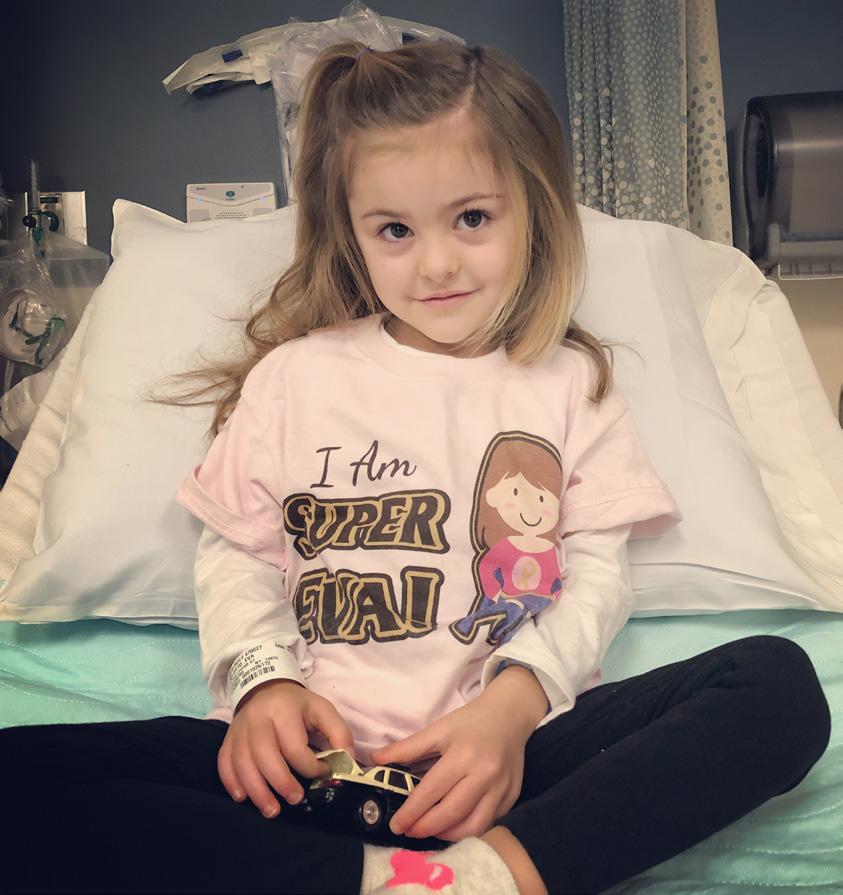
“Shortly after her third birthday, Eva DeLotto was diagnosed with a grade 2 ependymoma brain tumor. She underwent successful surgery to remove the entire tumor. Following her surgery she completed 33 rounds of radiation. Eva continues to celebrate clear scans and is thriving in spite of all she has endured. She is currently a happy and loves to play with her twin sister and older brother. She continues to overcome the obstacles and challenges she faces as a result of her diagnosis with a smile on her face.
“As her parents, we try to focus on her current good health and enjoy each day, but we know that there are many uncertainties in her future. Our hope is to continue to help raise awareness and support new research that is so critical in the treatment and outcomes of all those inflicted with this disease.” - Eva’s Mom and Dad
Funding for this guide was made possible by the generous support of the DeLotto family and the Childhood Brain Cancer Research Collaborative. The 3rd Edition Ependymoma Guide is dedicated in honor of Eva DeLotto.


We would like to extend a sincere thank you to the CERN Foundation Advisory Team that includes the original editors and reviewers of the first and second edition. This includes Editor in Chief Dr. Terri Armstrong, Editor Dr. Mark Gilbert, and Reviewers Dr. Ken Aldape, Dr. Amar Gajjar, Dr. Richard Gilbertson, Chas Haynes, Kristin Odom and Kim Wallgren.
In addition, we would like to acknowledge the following contributors of version three of the Ependymoma Guide - National Brain Tumor Society staff, Dr. Kristian Pajtler, Kelly Hancock and the NCI-CONNECT team for expert review: Dr. Marta Penas-Prado, Dr. Byram Ozer, Dr. Mark Gilbert, Dr. Terri Armstrong, Lisa Boris, and Brittany Cordeiro. Additionally, the Central Brain Tumor Registry of the United States (CBTRUS) Scientific Team for providing statistical oversight, especially Carol Kruchko, Quinn T. Ostrom, PhD, and Corey Neff, MPH. The CBTRUS data were provided through an agreement with the Centers for Disease Control’s National Program of Cancer Registries. In addition, CBTRUS used data from the research data files of the National Cancer Institute’s Surveillance, Epidemiology, and End Results Program, and the National Center for Health Statistics National Vital Statistics System. Contents are solely the responsibility of the authors and do not necessarily represent the official views of the CDC or the NCI.
This guide was produced and published by the Collaborative Ependymoma Research Network (CERN) Foundation, a program of the National Brain Tumor Society (NBTS). © CERN Foundation. Reproduction is prohibited.