A serious concern for lymphedema patients and those at risk
By Dr. Mieke Flour
1
Terminology, definition and causative organisms
Bacterial infection of the dermis and subcutaneous layers, known as cellulitis, is potentially serious. It may spread locally and require hospital admission. In many reference books, a distinction is made between the deeper entity called ‘cellulitis’ and the more superficial ‘erysipelas’. Clinically the distinction is not always clear. Erysipelas is said to have significant lymphatic involvement.
Cellulitis as er ysipelas is commonly due to streptococcal infection (b-hemolytic Streptococcus pyogenes group A, C, G). When the infection is a complication of a pre-existing dermatological disease, a wound, or immunosuppression, staphylococcal (S. Aureus) or mixed infection should be considered. The likely organism will determine the choice of antibiotic treatment. Cellulitis accounts for about 2-3% of hospital admissions in the UK, and is said to occur in 20-30% of cases of chronic lymphedema.
2
Clinical presentation, symptoms and diagnosis
The diagnosis of cellulitis is based largely on clinical signs and symptoms. Systemic and local symptoms may precede skin changes.

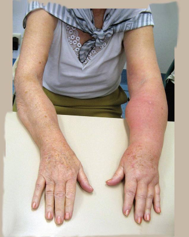
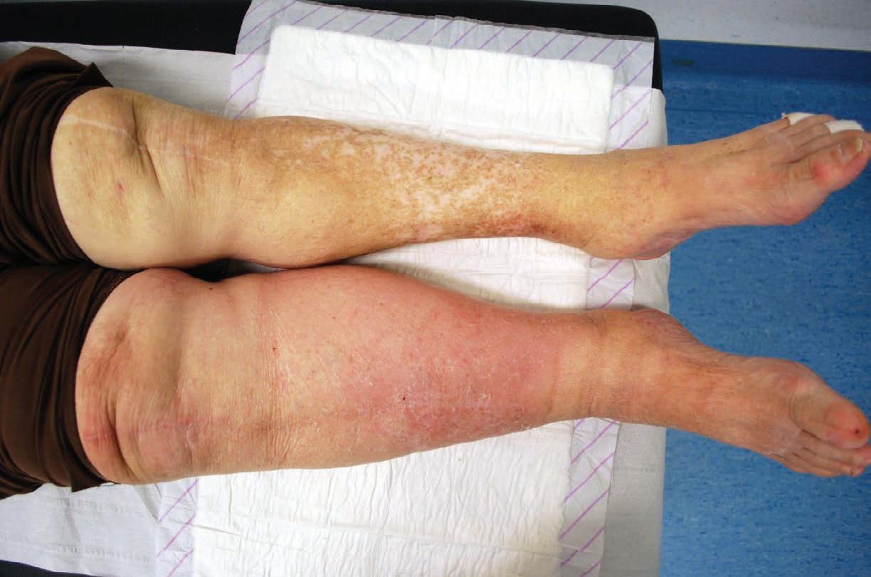
Systemic symptoms may include: sudden malaise, fever, shivering or chills. There may be accompanying headache, nausea and vomiting. In severe cases systemic involvement can be expressed as tachycardia and hypotension. Locally, cellulitis often presents as an acutely red, warm, painful, swollen area.
Systemic and local symptoms may precede skin changes. Systemic symptoms may include: sudden malaise, fever, shivering or chills. There may be accompanying headache, nausea and vomiting.
The edges may be sharply demarcated, and inflammation may rapidly spread peripherally within hours or days. Sometimes red streaks suggestive of lymphangitis develop, and regional lymph nodes are tender on palpation. Although it can occur at many sites, the limbs are most commonly affected, especially in the case of concomitant lymphedema.

Dr. M. Flour is a Dermatologist who was affiliated with the University Hospitals of Leuven, Belgium, until recent retirement. She has been involved in the in- and out-patient care for people suffering from skin lesions due to vascular disorders. For almost twenty years she participated in the multidisciplinary management of the diabetic foot syndrome.
Studies indicate that 66% of cellulitis cases occur in the lower limbs.
Laboratory tests are generally not helpful in establishing the diagnosis. Swabbing intact skin most often does not serve to identify the causative organism. Swabs from nose and throat or other natural niches may indicate carriership, but again may not identify the infectious organism. There may be indication for taking a swab if a culture can be obtained from a port of entry, blister or ulceration in the infected region.
Blood tests may have limited value in diagnosis. While Erythrocyte sedimentation rate, C-reactive protein and an elevated white cell count indicate an inflammatory reaction, leukocytosis is found in only half of cases. An elevated ASO titer, or swab with culture from intact blisters may sometimes confirm a streptococcal or other causative organism. Anti-DNAse B and ASO titers are considered to
FACTS & FIGURES
n 66% of cellulitis occurs in the lower extremity
n There seems to be a significant increase of cases in the summer months
n The risk of recurrent infectious episodes is said to be greater than 50% within a year following a first attack
n Recurrent episodes of cellulitis may start a vicious circle of infection and worsening edema
Cellulitis Treatment Tips
Treat with antibiotics as soon as possible, according to local guidelines
Administer an effective dosage: high enough and long enough
Treatment may be started at home, but in severe or recalcitrant cases – hospitalization is strongly advised
Current guidelines are based on limited but positive evidence of benefit, and on professional consensus.
be good indicators of streptococcal infection: the antistreptolysin titer rises at 7-10 days following streptococcal infection, and will remain elevated for several weeks. Blood cultures, however, are rarely positive, and therefore not routinely done. In immunocompetent patients, frank bacteremia (with positive blood cultures) is only present in approximately 5% of cases. In cases of parasitic infestation an elevated eosinophilia is part of the inflammatory reaction.
Biopsy or aspirate from the deeper layers of skin are of limited value in the diagnosis of cellulitis, but they may be helpful for differential diagnosis. While challenging, it is important to differentiate cellulitis from other acute, potentially more serious infectious conditions that present with local inflammatory reactions.
3Risk factors or predisposing factors
Most cellulitis cases are treated in primary care; however most studies about epidemiology and concurrent conditions associated with cellulitis have been done on hospitalized patients. Therefore the data on incidence, comorbidities and recurrence rates is prone to selection bias. We do know that gender poses an equal risk and interestingly, there seems to be a significant increase of cases in the summer months. Additionally, there appears to be greater risk in older age groups.
Normal skin resists penetration by the micro-organisms that routinely colonise its
surface. Epidermal antimicrobial peptides and Langerhans cells are the most prominent factors in the defensive responses. As the lymphatic system has a role to play in antigen presentation and defense against infection, any breach in this complex lymphatic defense mechanism may increase the risk of invasion by bacteria either through a local portal of entry, or by haematogenous transfer (by the bloodstream) to areas of limited lymphatic clearance.
Numerous local and systemic risk factors exist including: lymphedema, previous cellulitis, a pre-existing wound or dermatologic disease. Systemic risk factors include uncontrolled diabetes or immune suppression. Chronic lymphedema (present in 18% of cases) is considered to be the most important risk factor for cellulitis. Furthermore, it presents an increased risk, greater than 50% within a year following a first attack, of recurrent infections. Obstruction of lymphatic drainage, which may occur secondary to a tumor, excessive skin folds, fat masses or trauma to the lymphatics (surgery, radiotherapy), may predispose one to cellulitis. Chronic disturbance in lymph flow
and function results in an impairment of the innate and adaptive immunologic defense against infection. Additionally, the resulting secondary skin changes may compromise skin barrier integrity and homeostasis, and may thus permit local overgrowth of fungi, yeasts and microbes. Interestingly, several studies found lymphatic insufficiency in the unaffected limb in people with erysipelas. This may point to an underestimated pre-existing lymphatic condition (Damstra 2008, et al).
4
Recurrent cellulitis
Recurrent episodes of cellulitis may start a vicious circle of infection and worsening edema. The mechanism is likely multifactorial. Hypotheses as to the cause of recurrent infections include a failure to completely eradicate the infectious agent, a state of local immune deficiency related to lymphatic insufficiency, and natural carriers of streptococci. Statistics on recurrence rates differ between data gathered in a primary care setting and in hospitalized patients. In a Belgian primary care study (Bartholomeeusen, 2007), 16% were reported to have one or more recurrences.
5. Management of cellulitis
Cellulitis progresses rapidly in severity and extent, herefore treatment should be prompt and effective. Treatment aims at resolution of symptoms, prompt admission to hospital (if equired), and avoidance of sequelae. General measures to reduce morbidity are: bed rest, elevation of the af fected limb, skin care, wound care, and analgesia. Antibiotics are required to treat cellulitis, but type, administration route and course of treatment varies. Many lymphedema specialists consider streptococci to be involved. Antibiotic treatment guidelines vary by country, population, level of health care, issuing organization, and whether treatment is for cure or for prevention. Even though
most patients with cellulits are treated in a home environment, recommendations are largely based on a few RCT’s conducted with hospitalized patients. Many organizations and associations throughout the world have published clinical practice guidelines and documents, including CREST, British Lymphology Society (BLS), and the International Society of Lymphology (ISL).
Oral or systemic penicillin is commonly used against streptococci. Flucloxacillin may be preferred to treat staphylococcal infection in some countries because it is also effective for streptococci, with a low minimal inhibitory concentration (MIC). The CREST guidance in 2005 puts it as first-line treatment, with clarithromycin in case of penicillin allergy. Due to its greater tissue penetration, a large consensus g roup in the UK has chosen amoxicillin for the management of cellulitis in lymphedema (Keeley V, 2009). This group recommends oral amoxicillin 500mg every 8 hours for a minimum of 14 days. For pa-
is the drug of choice prescribed at 300mg every 6 hours for at least 14 days. Along with monitoring systemic symptoms, response to treatment can be easily tracked by marking the periphery erythema and ensuring that the involved area, over time, is diminishing in size.
systemic involvement, oral outpatient treatment is reasonable. However, hospitalization for systemic treatment is advised for the following: unclear diagnosis, serious concurrent illnesses, septicemia (hypotension, tachycardia, high fever, continued on page 12
Academy of Lymphatic Studies

Lymphedema Management Seminar (31 hours)* $895
This 4-day course serves as an introduction to the management of uncomplicated lymphedema affecting the upper and lower extremities, using Manual Lymph Drainage (Vodder Technique) and Complete Decongestive Therapy.
Complete Lymphedema Certification (135 hours)* $3150
This course provides certification in Manual Lymph Drainage (Vodder Technique) and Complete Decongestive Therapy according to the rules and regulations set by the Lymphology Association of North America (LANA).

Certification in Manual Lymph Drainage (40 hours) – NEw! $1150
This 5-day course will certify therapists in the MLD Techniques developed by Emil Vodder, Ph.D. and will enable you to create treatment sequences for the management of edema including post-operative/post-traumatic edema and pathologies such as fibromyalgia, migraines, chronic fatigue and more.
courses.

In the presence of acute infection, lymphedema management should be limited to limb elevation. Compression, should be postponed until the acute infection is resolved.
confusion, vomiting), cellulitis of the face, cellulitis secondary to wounds, and if the patient is unresponsive to 2-3 days of oral antibiotics. On admission, keep in mind that general (and local) signs and symptoms may be masked or modified by previous treatment. In many cases management will require a change of antibiotic regimen. Hospital

antibiotics such as amoxicillin 2g every 8 hours, lucloxacillin or benzyl penicillin 1.22.4g every 6 hours. Clindamycin 600mg every 6 hours is chosen if the patient is penicillin-allergic. Response to treatment should be apparent within 48-72 hours. Clinical signs of improvement include resolution of fever and inflammation. As symptoms subside, the treatment regiment may change to oral antibiotics. Sometimes this will mean an extended course of the same antibiotic but at a reduced dosage. Another option is to rotate the antibiotic either because there is no oral equivalent, or because the clinical response is considered incomplete. A 7-14 day course of treatment is accepted practice. Whilst treating the acute infection, management of pain may also be indicated. Acetaminophen is an appropriate analgesic. NSAIDs and other anti-inflammatory agents should be avoided as they tend to mask inflammation and, with severe infection, impede an accurate differential diagnosis.
If a patient has had two or more episodes of cellulitis in a year, there may be consideration for prescribing prophylactic antibiotics. While there is limited evidence and guidance for this practice, research suggests a reduction in overall number of episodes. French guidelines (Vaillant, 2007) recommend intramuscular benzathine penicillin 1.2-2.4 million units every 2-3 weeks, or oral penicillin V 2-4 million units in 2-3 doses a day. Patients suffering from recurrent infections often quickly recognize the initial signs. They may be advised to carry a twoweek supply of oral antibiotics with them while travelling.
6
Managing lymphedema in the presence of cellulitis
impairment of lymphatic function. Therefore treatment should not only focus on the infection, but also on long-term management of lymphedema, which is essential to prevent recurrence of cellulitis and further aggravation of lymphatic function. LP
References
CREST (Clinical Resource Efficiency Support Team) Guidelines on the Management of Cellulitis in Adults, CREST, 2005. http:// www.acutemed.co.uk/docs/Cellulitis%20 guidelines,%20CREST,%2005.pdf
Damstra RJ, van Steensel MAM, Boomsma JHB, Nelemans P, Veraart JCJM () Erysipelas as a sign of subclinical primary lymphoedema: a prospective quantitative scintigraphy study of patients with unilateral erysipelas of the leg. Br J Dermatol 2008:
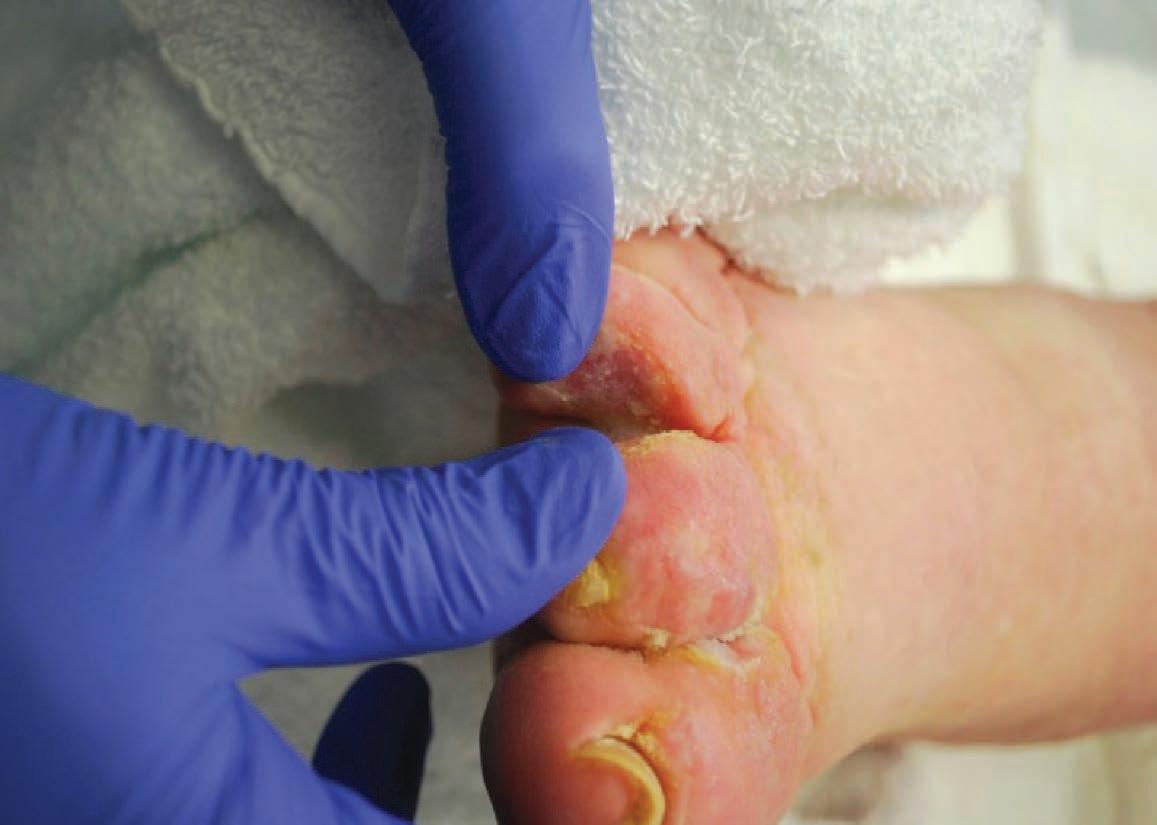
In the presence of acute infection, lymphedema management should be limited to limb elevation. Compression, should be postponed until the acute infection is resolved. Initial compression therapy postinfection will need to be applied in a supportive rather than compressive manner to accommodate individual patient needs and compression tolerance. Effective skin and nail care is essential. Any concurrent skin conditions must be addressed or they may continue to offer a port of entry for pathogens. Cellulitis may reveal an underlying (and perhaps previously unknown)
tholomeeusen S, Vandenbroucke J, Truyers C, Buntinx F. Epidemiology and Comorbidity of Erysipelas in Primary Care. 2007;215:118-122.
Vaillant L. Erysipelas and lymphedema. Phlebolymphology. 2007;14(3):120-124.
Keeley V, Riches K. Cellulitis treatment for people with lymphedema: UK audit. Journal of Lymphoedema, 2009, Vol 4, No 2, 17-28
Editor’s Note: Management of cellulitis in lymphedema may differ from that of cellulitis related to other conditions. There are no Canadian lymphedema-specific guidelines at this time. Recent Canadian guidelines focus on MRSA infections. For 2013 lymphedemaspecific guidelines published by the British Society of Lymphology see: http://www.lymphoedema.org/ Menu3/Revised%20Cellulitis%20 Consensus%202013.pdf
Photo: CanStockPhoto
Photo: M. Flour