
Breast edema/lymphedema during and after breast cancer treatment Identification and treatment
By Lesli Bell
Incidence
288,000 new cases of invasive breast cancer were diagnosed in the United States in 20221 and 28,600 in Canada13. Everyone enters the breast cancer journey with some surprise, to say the least. For 2.26 million people a year worldwide1 it will be the first time they have to navigate this new terrain. No matter what, when the “cancer” word enters your life there are fears, questions and the unknown. Thankfully there are caring oncologists, surgeons and radiation oncologists that have expertise in treating breast cancer. This article will discuss breast edema, an area that is not well-documented and defined but affects almost all breast cancer patients to some extent.
Types of breast-conserving surgery
Breast-conserving (BCS) surgery (lumpectomy, or tumorectomy, rather than total mastectomy) is increasingly popular and has many benefits. However few people emerge from the treatment process without some sort of change or consequence. Some are minor, some are major and some don’t get talked about as much as they should. Lymphedema as a consequence of BCS is receiving much more attention these days, but is primarily described in the literature as pertaining to the arm.

Breast edema
One of the less discussed consequences of breast cancer treatment is breast edema and breast/truncal lymphedema. A systematic review9 noted that “The incidence of breast edema in breast cancer patients following breast conserving surgery (BCS) and radiotherapy is very broad, namely 0–90.4 %”2. There are multiple issues that the patient may contend with including pain, heaviness, lymphedema, radiation fibrosis, sexual issues, shoulder impairments, and ‘cosmetic unhappiness’ just to name a few.
Breast edema occurs in the postoperative phase and is expected. When breast edema lingers much longer than expected, we call it breast lymphedema. Both are physiologically due to lymphatic overload of the tissues3
Regardless of how long it has been present, it is appropriate to treat post operative and/or post radiation edema if it is bothersome, problematic, or painful for the patient. One of the difficulties in documenting and researching breast edema and breast lymphedema is that there is no consistent and objective way to measure it2 Without consistent measurement, it is hard to prove positive change with treatment. However, there are still clinically effective treatments that are safe and consistent with how we presently treat lymphedema of the upper extremity.
Lesli Bell received a BSc in PT (California State University), a doctorate in PT (Regis University) and her lymphedema certification (Lerner). She has owned a private practice (Winooski VT) since 1987, specializing in orthopedics, chronic pain and swelling disorders. Lesli co-developed the Bellisse Compressure Comfort Bra (1999) for patients with breast and chest wall edema/lymphedema.


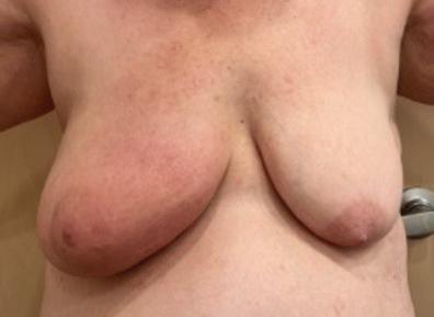
Examples of breast edema
Under best circumstances after the diagnosis of breast cancer, the patient will be evaluated by the medical team, including an oncologist, a surgeon, and a radiation oncologist. If prospective surveillance is included in this evaluation, a therapist specializing in cancer-related lymphedema or in oncology will also assess the patient.
Common symptoms and signs of breast and truncal edema/lymphedema include an increased volume of the breast, “peau d’orange” (inverted hair follicles due to edema in the area giving the skin an orange peel appearance), heaviness of the breast, redness of the skin, breast pain, skin thickening, hyperpigmented (darkened) skin, and a positive pitting (edema) sign2,7
The risk of developing upper extremity lymphedema increases with higher BMI, increasing number of lymph nodes removed, seroma, cording, a history of radiation and surgery, and some types of chemotherapy11 These are likely risk factors for chest wall edema/lymphedema as well. Breast cancer treatment affects the skin, breast tissue, and connective tissue of the trunk and less frequently some internal organs. “Breast edema/ lymphedema appears to be very common, and a source of problems for many patients recovering from breast cancer treatment”4
Prospective surveillance
Under best circumstances after the diagnosis of breast cancer, the patient will be evaluated by the medical team, including an oncologist, a
surgeon, and a radiation oncologist. If prospective surveillance is included in this evaluation, a therapist specializing in cancer-related lymphedema or in oncology will also assess the patient. Stout et al published a paper “A Prospective Surveillance Model for Rehabilitation for Women with Breast Cancer” in 2012. “Prospective surveillance identifies time points during breast cancer care for assessment of and education about physical impairments. Implementation of this model may influence incidence and severity of breast cancer treatment-related physical impairments. The model seeks to optimize function during and after treatment and positively influence a growing survivorship community”4. The paper describes a cost savings and significant reduction in patient complications and fears over the first year or two of recovery. At this time, it is unknown how many institutions have implemented this program in their breast cancer centers. Prospective surveillance assessments might include measurements of both arms, assessment of any older injuries or limitations to the arms, shoulders, or spine, and education regarding rehabilitation, lymphedema and how to navigate any pre-morbid conditions during breast cancer treatment4. Bioimpedance Spectroscopy (BIS) has been demonstrated to identify lymphedema in the extremities and is a recommendation in the clinical practice guideline of the Academy of Oncologic Physical Therapists5. BIS identifies small lymphedema related fluid changes in the limbs by assessing extracellular fluid and aides in the detection of lymphedema much earlier than using a tape measure6. Tissue Dielectric Constant (TDC) values are quantifiable measures of localized skin tissue water and may be able to detect breast/trunk lymphedema. “TDC may be a beneficial tool in the early detection of breast cancer–related trunk lymphedema, which could trigger intervention”7. Both evaluations are most
Examples of cording, peau d’orange, scarring fibrosis in the axilla


meaningful if there is a baseline assessment before breast cancer treatment. If a patient does not have access to a prospective surveillance program, self-taken pretreatment measurements of the upper extremity have been validated by Rafn et al8 The patient can use a common tape measure to take measurements of both arms at the wrist, mid forearm, and mid upper arm. Measure longitudinally along the arm where these lines are and make a mark8. Or pick an identifying spot, mole, or scar that allows one to consistently measure the circumference of the limb in the same place. Take note of the shape and consistency of the tissue on the limb and take note of the visibility of veins in the forearm and hand. Look at the shape and position of the nipple and compare it to the unaffected side. One may even want to take a pre-treatment picture of the chest wall for comparison in the future. It would be wise to revisit and retake these measurements every four weeks after breast cancer treatment has started or if you feel changes such as heaviness, thickening of skin, pain in your breast, trunk, or arm. If there is greater than a one-centimeter change in these areas compared to the unaffected side, consultation with a lymphedema therapist would be recommended.
Treatment
Despite the benefits of breast conserving surgery some women will be troubled by breast edema that may cause an unsatisfactory cosmetic result influencing their quality of life2 After any kind of surgery there will be expected edema during the healing process with signs of improvement in 3-4 weeks or at least be changing in a positive direction. If lymph nodes were removed, either for sentinel lymph node biopsy (SLNB) or axillary node dissection, the swelling may take a little longer to reduce.


JOBST JoViPak in combination with the JOBST JoViJacket is designed for lymphedema compression therapy during the night. The 4 blend foam mix and stitched channels are designed to create multiple pressure points and stimulate lymphatic drainage. These garments for edema management come in various styles and, with the addition of specialized pads, are designed to provide comfort and control from head to toe.
JOBST® JoViPak®. Full body solutions for reliable lymphedema management during night-time
Some of the specialized techniques for scars and skin thickening might include cold laser, elastic taping and manual scar stretching.
Radiotherapy may aggravate the swelling process, causing sudden or slow increases in breast edema early in the radiation process, but more commonly occurs toward the end of treatment or after radiation is complete. It is usually characterized by skin thickening, which usually peaks at 4-6 months9. Scarring, fibrosis and peau d’orange can linger even longer.
Appropriate treatment should yield a decrease in breast swelling, truncal fullness, skin thickening, scarring and pain9. Almost all swelling responds to lymphatic treatment and compression and initiating early treatment may help expedite the healing process. This may include complete decongestive therapy (CDT), which consists of manual lymphatic drainage, exercise, infection risk reduction strategies, and compression garments. Soft quilted shredded foam pads may be beneficial for swelling, scarring, peau d’orange and fibrosis. These quilted foam pads are made from very soft fabric, filled with different densities of chipped or shredded foam. These pads can be worn under the compression bra or vest.
Some of the specialized techniques for scars and skin thickening might include cold laser12, elastic taping and manual scar stretching. Reducing scarring, skin thickness, pitting, peau d’ orange, and fibrosis can significantly improve the ability of fluid to move out of the congested area and may

reduce severity of edema and future problems with lymphedema.
Development of redness, streaking, extreme heat, and localized pain in the chest, breast or arm should always be evaluated by a physician, as it may be first signs of an infection that may require antibiotics. However, the pinkish and redness in the breast or arm may also be signs of tissue inflammation from pooled fluid in the area. If this is the case, then antibiotics will not work to reduce the swelling. Infection and inflammation may be hard to distinguish, and usually physicians will err on the side of safety, treating it as if it were an infection. If it is inflammation, treatment by a lymphedema therapist can often be very helpful by reducing the swelling.
Chest wall compression/vest/bra
Most patients will wake up with some sort of chest compression vest or bra on following surgery to hold dressings in place. How long to use this compression is often unclear. When lymph node dissection has been performed, and/or swelling is persistent, compression might be beneficial for quite a bit longer than a few weeks post-operatively. Breasts are largely composed of adipose and glandular tissue, which make it easy for fluid to accumulate. There is also no muscle pump in the breast to help move lymphatic fluid. A compression bra or vest should fit well, be snug but comfortable, and feel secure, similar to a well-fitting “speedo” swimsuit. The bra or vest should also not impede breathing. One should avoid compression garments that create a tourniquet effect (especially under the breasts and around the trunk). The breasts should be comfortably lifted and compressed to the chest wall (reducing the pendulous position). This will assist with

fluid draining from breasts and inhibit refilling. Wearing the compression vest/bra for several hours during a 24-hour period, whether it is day or night (with or without the quilted shredded foam pad) may help manage the breast swelling. It is also ‘OK’ to wear traditional bras when desired appearances of clothing are important. Paying attention to the response of the swelling will help dictate how many hours of compression are optimal. When vascular reconstructive surgery is performed on the chest wall or breast, compression of the chest wall will need to be discussed with the surgeon.
In conclusion
While surgical and oncological treatment is lifesaving, treatment for breast edema and lymphedema can preserve quality of life. Breast edema and breast lymphedema deserve treatment when it is bothersome, painful, and/ or there is a significant increase in volume. If there is scarring, fibrosis, and cording, treatment with a lymphedema trained therapist using manual techniques and CDT, a wellfitting compression vest/bra, and quilted shredded foam pads can be very beneficial. The lymphedema and oncology trained therapist can be an enormous asset to the patient as they move through the breast cancer journey, allowing early identification of impairments, and treatment before problems become chronic. A patient should freely discuss any of these conditions with the treating physician and consider asking for a referral to a specialized therapist who can help them navigate the journey of breast cancer treatment and beyond. LP
References can be found at https://canadalymph.ca/pathwaysreferences


