Polyp Growth Patterns of the Deep Sea Coral Genus
Narella Due to Varying Environmental
Kaela Harrison
Senior Thesis | 2025

Kaela Harrison
Polyp Growth Patterns of the Deep Sea Coral Genus Narella Due to Varying Environmental Factors
Abstract
This study looked at two samples of deep sea Narella coral from different oceanic sites in the Pacific Ocean off the coast of Costa Rica. The samples were collected between two thousand and three thousand meters below the surface of the ocean, with roughly an 800-meter distinction in depth between them, already giving them a distinct environmental factor difference that this study draws interest from. The question is whether or not these two samples have a statistically significant relationship in growth patterns. In other words: Does a difference in depth, O2 concentration, O2 saturation, salinity, and temperature change the size of Narella polyps, and if so, do the polyps get smaller or bigger as these factors change? This study used the ImageJ software to calculate the surface areas of coral samples, and the findings suggest that the size of Narella coral polyps do change under unalike environmental conditions as one sample’s polyps were significantly bigger than the other. This study took interest in Narella polyps specifically because of the unexplored world of the deep sea. Oceanography is a relatively new study, only starting around 1872 (Webb, 2021); this means that there are still so many things that are left undiscovered or very recently discovered in the ocean. How Narella corals manage to live so deep in the ocean without light started the many questions that a few of are addressed in this study. The findings of this study suggest that the size of Narella coral polyps do change under unalike environmental conditions.
Introduction
An important question to answer before understanding this study is “What are corals?” in general. Surface corals are animals that live in colonies. An individual coral is called a polyp, and they are small, round growths off the “main body” or “skeleton” of the coral. Polyps also help the coral eat, reproduce sexually and asexually, build the coral body, and more. The skeleton of the coral is made up of Calcium carbonate (CaCO3) that the polyps secrete as a part of their function (Webb, 2021). Polyps in a colony are often genetically identical given the coral’s ability to reproduce asexually, although polyps inside a single colony may have different reproductive activity (Goffredo et al., 2011).
For eating (see Figure 1), the polyps help in two different ways. Corals that reside in the photic zone are both autotrophs and heterotrophs, or mixotrophic, which are both essential states of their being for survival. So, corals can both perform photosynthesis when they have access to light (autotrophic), and can capture and eat various microscopic phytoplankton floating in the water for their energy source (heterotrophic). In order to gain energy from photosynthesis, most corals have a symbiotic relationship with zooxanthellae (microscopic algae) that live in the coral tissue. The exchange that occurs between these two organisms is that the zooxanthellae provide the coral with energy from photosynthesis, and in turn the coral provides shelter for the microscopic algae. The zooxanthellae is actually what makes up the color in the coral tissue, and this is where the phenomenon of “coral bleaching” comes into play. When the water gets too warm or unstable, the coral ejects the zooxanthellae, and since the zooxanthellae provide the color to the coral’s tissue, the absence of the symbiotic dinoflagellates is the cause of the corals becoming a blanched white color. Anyway, corals can also eat heterotrophically, in a similar way to jellyfish, to whom they are closely related (Webb, 2021). Corals and jellyfish are part of the same large group of animals known as cnidarians. This name derives from the Greek
“knide,” which means “nettle,” as both jellyfish and coral polyps carry nematocysts – stingers on the ends of their tentacles that can spear, paralyze, and help capture prey. This information helps this study compare and contrast surface corals and deep ocean corals.


1: Image of a surface coral. Left coral represents the coral in its normal state before feeding, while right coral shows the contracted polyps of the corals after feeding.
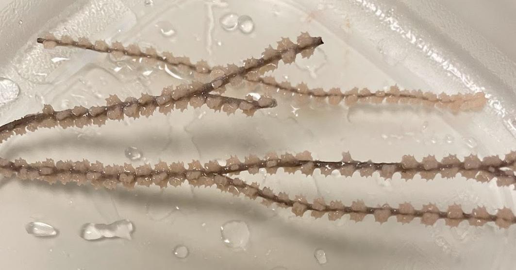
The coral this study focuses on is from the deep sea genus Narella (see Figure 2). The genus gets this name supposedly because its polyps resembled small noses, as the actual term Narella is derived from the Latin word “naris,” meaning “nostril” (Cairns et al., 2019). Deep sea corals are different from the common surface corals. They live thousands of meters below sea level, performing an uncommon feat of living without any light, meaning without the usually vital use of photosynthesis. This also means that deep sea corals are asymbiotic, with no trace of any of the symbiotic dinoflagellates known as zooxanthellae seen in shallow water corals. There are different types of deep sea corals, otherwise known as cold water corals, though, not just one.
There is the “Scleractinia” subclass, encompassing stony corals, the “Antipatharia” subclass, for black corals, the “Stylasteridae” subclass, being hydrocorals, and the “Octocorallia”
subclass, containing soft corals, sea fans, gorgonian, and bamboo corals (Houlbrèque et al., 2015). Narella is classified under the broader subclass of “Octocorals” (Octocorallia). Narella is also part of the family Primnoidae, which consists of the diverse family of deep-water Octocorals that has 32 genera and approximately 207 species (Cairns et al., 2007). Furthermore, Octocorals has three distinct orders within it, being “Helioporacea” meaning blue corals, “Pennatulacea” encapsulating sea pens, and “Alcyonacea” previously known as “Gorgonacea” containing soft corals and gorgonians (Breedy, 2009); Narella is part of the Alcyonacea order (Cairns et al., 2007). The species under the genus Narella are still being discovered, categorized, and sorted. However, it is known that Narella corals can be found between 141 meters and 4594 meters, being the shallowest and deepest collections of Narella species on record, respectively. This study centers on the Narella corals on the deeper end of this scale. It is a genus known to be very rich in species, with 32 known species at the time of Cairns’ review. Cairns predicts that many more species will be discovered. The species characterized under Narella can be found all around the world’s oceans. Known species have been found in the waters around Alaska, Japan, the Hawaiian Islands, the western Pacific –primarily in the Indonesian region, the eastern Pacific, Antarctica, the southwest Indian Ocean, and the Atlantic. The species Cairns gathered are titled N. bayeri, N. cristata, N. arbuscula, N. abyssalis, N. alaskensis, N. irregularis, N. biannulata, N. megalepis, N. compressa, N. bowersi, N. ornata, N. nuttingi, N. allmani, N. clavata, N. horrida, N. obscura, N. orientalis, N. parva, N. dichotoma, N. studeri, N. grandiflora, N. leilae, N. ambigua, N. gaussi, N. gilchristi, N. regularis, N. versluysi, N. bellissima, N. pauciflora, N. laxa, N. spectabilis, and N. alvinae (Cairns et al., 2007). The Narella species used for this study is yet to be identified. The deep Narella corals, being asymbiotic, live in nutrient-rich seawater and are known to eat copepods, zooplankton, phytoplankton, and marine snow – dead organic
material and overall detritus that sinks from the epipelagic zone (top 200m) of the ocean using their polyps (Houlbrèque et al., 2015). Along with a basic understanding of what Narella corals are and how they function, it is also important to understand the general environment they live in.

The area of ocean known as the “deep ocean” (below 200 meters) is a total of 95% of the ocean’s volume, making it one of the largest and least explored biomes on the planet. Less than 0.0001% of the deep ocean has been explored and investigated. This means that the surface of the moon is a better known domain than the floor of the deep sea. It is known that there are many dangerous and/or specialized habitats at the bottom of the ocean though, like mud volcanoes, sharp volcanic rock formations, seamounts, canyons, and hydrothermal vents. In some of these areas the ability to perform chemosynthesis is needed to survive. Chemosynthesis is where creatures live off of the synthesis of chemical compounds instead of light energy or heterotrophic tendencies, and it most commonly takes place around hydrothermal vents, mud volcanoes, whale falls (sunken whale carcasses), and cold-water hydrocarbon seeps (Danovaro et al., 2017). The deep sea is not a very hospitable environment to live in. With the previously mentioned lack of light, as well as the high pressures, cold temperatures, and dangerous small habitats, the deep is mostly reserved for a class of animals called “extremophiles,” animals that live in severe or “extreme” conditions. The species that flourish down so deep are mainly microbes, but it is also common to find various crustaceans, invertebrates such as cephalopods, and evidently many coral species at the deep seafloor (Webb, 2021). This study focused on one type of deep sea coral.
This study focused specifically on two samples of Narella corals: Sample 57 and Sample 76. The samples were collected from two different sites with visually similar environments: rocky shelves making up the terrain. This is because of the common occurrence of currents in areas of canyons, slopes, and seamounts, also known as the current acceleration hypothesis. The currents bring an abundance of food sources for the corals to eat, making rocky shelves a common place for corals to grow (Houlbrèque et al., 2015). Even though the two samples’ physical environments
were similar, there are plenty of environmental factors at play that aren’t visible that are important to investigate. These factors include depth, O2 concentration, O2 saturation, salinity, and temperature; in all of these measurements small variances in numbers can have a potential effect on the sizing of coral polyps. The main question that was addressed was whether or not a difference in the environmental factors would change the size of the coral polyps. This study hypothesized that a certain change in the above mentioned factors would either make polyp size larger or smaller. The null hypothesis is that no matter how the factors change, polyp size will remain the same.
The Environmental Factors; Definitions and Explanations:
Depth is defined as how many meters below sea level the collected corals were. The samples were collected with a large difference of 803.3 meters between the two of them, only changing the amount of pressure in the two environments. It does not affect the amount of light they get, as light only reaches to about 200 meters below the surface (Webb, 2021). O2 concentration is the measured amount of oxygen present in the water and is measured in micromolars (μM), while O2 saturation is the percentage of oxygen dissolved in the water compared to the maximum amount that can be dissolved at a given temperature and pressure, and is measured in percent (Bozorg-Haddad et al., 2021). Salinity is the quantity of salt in the water. The average salinity of ocean water is around 35 PSU (Practical Salinity Unit, defined by 1 gram of salt per 1000 grams of water). The temperature of the water is how cold it is, measured in Celsius. Both coral samples grow in unusually cold conditions.
Study Sites: (See Figure 3 & Table 1)
Collection Site 1: Sample 57
Sample 57 was collected at a depth of 2829.12 meters, an O2 concentration of 134 μM, an O2 saturation of 33.8%, a salinity of 35.2 PSU, and a temperature of 1.82 °C (35.276 °F).
Collection Site 2: Sample 76
Sample 76 was collected at a depth of 2025.82 meters, an O2 concentration of 114 μM, an O2 saturation of 28.2%, a salinity of 35.0 PSU, and a temperature of 2.19 °C (35.942 °F).
Table 1: Data collected from each site.

Sample 76 off the coast of Costa Rica.
Methods:
Coral Collection:
The two coral samples, Sample 57 and Sample 76, were collected from depth using the SuBastian ROV onboard the R/V Falkor(Too), which can dive down to 4500 meters in depth, during two separate dives. Since deep sea corals are thousands of meters below sea level, they cannot be accessed by divers, and require the advanced technology of ROVs, for example. Underwater collection was executed by using a mechanical claw on the SuBastian ROV to carefully place corals into a quiver– a temperature regulated compartment containing seawater from the collection sites. The corals were brought on the ship and stored in a 4 degree Celsius cold room, and placed in incubation (Farrell, 2025).
Coral Documentation:
The collected corals were delicately cut into coral fragments in such a way as to attempt to keep a similar distribution in polyp count in each section. Making sure that the overall samples were kept consolidated, the samples were stored in labeled vials upon the ship. The samples were run through a respirometer test, oscillating in the dark, for roughly 6 hours. Each fragment was then placed on a workbench, lined up next to a ruler and a piece of paper signifying which coral sample it was from. They were documented through pictures by an iPhone camera and kept preserved in their vials for further study.
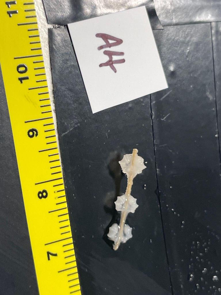
Coral Measurement:
In order to obtain the sizes of the polyps, the surface area was found using a program called ImageJ. In order to use this program, the pictures of coral fragments with rulers in them for reference size are opened as downloaded files individually through ImageJ (see Figure 4). First, the number of polyps in the image are counted and recorded. Then, using the line tool (button with a symbol of a line segment), a straight line is drawn across the ruler for the distance of one centimeter to input it into the reference units, so the program knows how to measure the image. The freehand tool (button with a symbol shaped like a kidney bean) is then used to carefully trace the polyps in full circles. Finally, the Measure function is used to output the surface area encapsulated by the freehand tool.
Data Analysis:
After collecting all the surface areas of the polyps in the corals samples, then it came time to find the surface area to polyp ratio. The surface area to polyp ratio is determined by taking the
calculated surface area value for each image and dividing it by the number of polyps in that image (general formula: surface area / number of polyps). This process provided a data point for each image of coral, which were kept grouped in the separate samples of 57 and 76. Each group of data points was then put through an unpaired T-test to gain the results.
Results and Discussion:
From the results of the unpaired T-test, there is a significant difference between the surface area to polyp ratios of the two samples with a 95% confidence interval (see Figure 5). It is a statistically significant difference because – by conventional criteria – the two-tailed P value equals 0.0003 representing extreme statistical significance.
Ultimately, Sample 76 had larger polyps than Sample 57. This leads the study to hypothesize (as there is not enough evidence to claim) that the environmental factors do, in some way, affect the size of the polyps.

Figure 5: Data plot displaying the averaged surface area to polyp ratio, or overall size of the polyps, including individual data points, medians, and margins of error.
Possible Environmental Factor Implications (Comparison):
Since Sample 76 has significantly larger polyps than Sample 57, this study turns toward all the environmental factors that were taken into account during collection for reasoning. Sample 76 was collected at a lesser depth of 2025.82 meters than Sample 57, which was collected at a depth of 2829.12 meters. This could produce a correlation that at greater depth, polyp size gets smaller. Sample 76’s site had both lower O2 concentration and saturation at 114 μM and 28.2% respectively, while Sample 57’s O2 concentration and saturation was at 134 μM and 33.8% respectively. This means that higher O2 concentration and saturation could make polyp size smaller. Sample 76 had a lower salinity than Sample 57, where Sample 76’s salinity level was at 35.0 PSU and Sample 57’s salinity level was at 35.2 PSU. Lower salt content in the water could lead to larger polyps. Finally, Sample 76’s site had a higher water temperature at 2.19 °C than Sample 57’s site, whose water temperature was at 1.82 °C. Higher water temperature could also produce larger polyps. Although these factors could be acting individually, it is more likely that these factors work in tandem to change polyp size, and that some factors may not affect polyp size at all. Until further, more controlled study is conducted, this study puts forth the previously stated series of hypotheses as forward progress in comprehension of polyp growth.
Related Studies and Further Findings:
James Palardy’s study on colony and polyp biometry theorized that smaller polyps are better at catching food like zooplankton, leaning into their heterotrophic side while larger polyps are better at photosynthesis, because there is more surface
area, and therefore more space for the symbiote zooxanthellae to inhabit the coral’s tissue, as well as addressing a few other environmental factors. They used four environment types to experiment: warm shallow water, warm deeper water, cold shallow water, and cold deeper water. The study concluded that feeding rates and behaviors do not have an effect on polyp size, eliminating that environmental factor, but continued to support the hypothesis that depth and temperature affect polyp size, where deeper warmer waters make polyps bigger (Palardy et al., 2005). This corresponds to the results of this study, supporting the idea that deeper corals need to have smaller polyps in order to maintain the same carbon fixation that higher, bigger polyps can obtain using photosynthesis. However, Palardy’s study was centered on shallow water corals, investigating corals between one and six meters below sea level. As previously stated, deep water corals are very different from surface corals, so there is not a direct correlation between the two studies. This does raise a question, though, of why would the polyps still change in a similar way in the deep ocean despite the lack of light?
Stefano Goffredo’s study looked closely at the relationship between colony sizes and polyp sizes. There were samples of small, medium, and large colonies placed at depths between seven and ten meters below the surface. There was no significant polyp size change found between small and medium sized colonies, but the polyps were less voluminous and lighter in large colonies compared to both the small and medium ones. This supports that changes in the general coral body would also change the polyp size. Goffredo’s study also found that polyp population size in a colony and placement of the polyp on the colony would change the size of the individual polyps (Goffredo et al., 2011). Even though they don’t directly apply to the study discussed in this paper, these findings are important because learning about coral formation and polyp growth with an inward look at corals gives the basics of polyp growth and provides a starting point to expand the
knowledge to an outward look. Once again, though, the corals investigated in Goffredo’s study were surface corals, making a gap between the correlations of the two studies due to the differences between surface corals and deep sea corals.
Hindrances:
This study had a few drawbacks. One of these limitations was the use of the ImageJ program to measure the surface area of the polyps. There is a margin of error in the measurements because the ImageJ method (Planar Projection Photography) of finding surface area is one of the least accurate according to Naumann, as it is only a 2D capturing of a 3D object.
There are many other methods of finding surface area in corals, such as Wax Coating, Simple and Advanced Geometry, and Computer Tomography. They would have been preferable for accuracy (Naumann et al., 2009). There is also SLS (Structure Light Scanning) and OCT (Optical Coherence Tomography) (Jaffe et al., 2022). However, there are downfalls to these methods as well. SLS has trouble taking measurements of corals with microstructural features, and any shadows cast by the coral will mess up the numbers. OCT is very new to coral reef science and it allows for accurate measurements, but it is not a space or cost effective option. Wax Coating is an invasive process, and the coral fragments used needed to be preserved for further study. Simple and Advanced Geometry could not have applied because the corals were not documented in such a way that the calculations could be done. Computer Tomography would be a precise way to find the surface area, but it is also not a cost effective method. Given the resources available to the lab, despite not being the most accurate, Planar Projection Photography was the most realistic method to pursue.
Another drawback this study experienced was the problem of multiple variables. Almost every variable that was measured –out of physical environment, depth, O2 concentration, O2
saturation, salinity, and temperature – changed. Therefore it was difficult to single out a variable to claim what was actually causing the polyps to change size, and the study could only infer the possibilities. On top of this, it is also likely that it could be a combination of factors. Despite not being able to make concrete conclusions, the results of this experiment give rise to some interesting new questions and ideas on ways to proceed.
Future Directions:
The next logical steps for this study to take would be to perform controlled experiments. Grow corals in recreated deep sea ocean conditions, isolating only one environmental factor to change while keeping the others the same. Then the study could take into account more environmental factors, such as food types, water flow, etc., and see how those factors change polyp growth. It would be wise to look at other aspects of coral growth other than just polyp growth, such as colony growth, and learn more about the deep sea corals’ feeding and reproduction habits. More ways to expand this experiment would be to take a closer look at coral genera other than Narella, the most readily available being the genus Tharella, and to explore more of the deep sea ocean ecosystem to get a better understanding of the conditions the corals live in and the other life they live around.
Importance:
Not much is known about deep sea coral growth and their general living conditions in the ocean, so this research is exploring new territory and providing more information about what exists within the mainly unexplored depths of the ocean. Every detail discovered about life in the deep ocean is vital to piecing together the puzzle of its ecology. Coral polyp size has apparent relations to how corals feed and reproduce, which we don’t have a complete comprehensive understanding of either. In a larger sense, another reason this research is valuable is because of global climate change
affecting the deep sea. It is hypothesized that the deep sea environments are more sensitive to even slight changes in temperature and other factors, causing widespread extinction events or otherwise forcing deep ocean species to adapt to conditions faster than they are used to (Danovaro et al., 2017). Therefore, understanding the functions of deep sea corals is important in the attempt to understand how climate change might affect them, as well as what might help them survive despite such change.
Conclusion:
The final results of this study have resolved that the environmental factors present (and recorded), depth, O2 concentration, O2 saturation, salinity, and temperature, are likely the cause of a change in polyp size, as there was a significant difference between the two samples of the study. Essentially, coral colonies in varied environments will have varied growth patterns. Despite not knowing exactly how individual environmental factors affect the growth of polyps, there was progress made in the area of understanding features and functions of deep sea corals. Along with this, the acknowledgement of a significant difference in polyp growth in different environments opens up the possibility for further investigation in diverse ways, as mentioned in the section “Future Directions.” More research of the deep sea is needed, and in order to do this, methodological and technological advancements are required. Once it is realistic for advanced technologies to withstand the pressure and various other dangers of the deep ocean, exploration and understanding of it will make huge leaps and make waves throughout the scientific community. Until then, there is still 99.9999% of the deep ocean to hypothesize about, discover, and protect.
References:
Bozorg-Haddad, O., Delpasand, M., & Loáiciga, H. A. (2021). Water quality, hygiene, and health. Economical, Political, and Social Issues in Water Resources, 217–257. https://doi.org/10.1016/b978-0-323-90567-1.00008-5
Breedy, O. (2009). Octocorals. Marine Biodiversity of Costa Rica, Central America, 161–167. https://doi.org/10.1007/978-14020-8278-8_13
Cairns, S. D., & Baco, A. (2007). Review and five new Alaskan species of the deep‐water octocoral Narella (Octocorallia: Primnoidae). Systematics and Biodiversity, 5(4), 391–407. https://doi.org/10.1017/s1477200007002472
Cairns, S. D., & Taylor, M. L. (2019). An illustrated key to the species of the genus Narella (Cnidaria, Octocorallia, Primnoidae). ZooKeys, 822, 1–15. https://doi.org/10.3897/zookeys.822.29922
Danovaro, R., Corinaldesi, C., Dell’Anno, A., Snelgrove, P. (2017, June 5). The deep-sea under global change. Current Biology Magazine, 27.
Farrell, A. (2025). Breathe deep: Stimulated respiration by deepsea octocorals. [Unpublished master’s thesis]. Boston University.
Goffredo, S., Caroselli, E., Gasparini, G., Marconi, G., Putignano, M. T., Pazzini, C., & Zaccanti, F. (2011). Colony and polyp biometry and size structure in the orange coral astroides calycularis (Scleractinia: Dendrophylliidae). Marine Biology Research, 7(3), 272–280.
https://doi.org/10.1080/17451000.2010.492222
Houlbrèque, F., Rodolfo‐Metalpa, R., & Ferrier‐Pagès, C. (2015). Heterotrophic nutrition of tropical, temperate and deep‐sea corals. Diseases of Coral, 150–163.
https://doi.org/10.1002/9781118828502.ch10
Jaffe, J. S., Schull, S., Kühl, M., & Wangpraseurt, D. (2022). Noninvasive estimation of coral polyp volume and surface area
using optical coherence tomography. Frontiers in Marine Science, 9. https://doi.org/10.3389/fmars.2022.1049440
Naumann, M. S., Niggl, W., Laforsch, C., Glaser, C., & Wild, C. (2009). Coral surface area quantification–evaluation of established techniques by comparison with computer tomography. Coral Reefs, 28(1), 109–117.
https://doi.org/10.1007/s00338-008-0459-3
Palardy, J., Grottoli, A., & Matthews, K. (2005). Effects of upwelling, depth, morphology and polyp size on feeding in three species of Panamanian corals. Marine Ecology Progress Series, 300, 79–89.
https://doi.org/10.3354/meps300079
Webb, P. (2021). Introduction to Oceanography.