MASSMATTERS


Please renew on line by visiting the BMSS Website or why not encourage colleagues to join the BMSS now if they have not already done so? Membership benefits include:
• Reduced delegate registration fees for BMSS meetings as well as reduced rates for certain other linked society meetings.
• Access to funding opportunities to support your mass spectrometry development.
• Reduced rate subscription to the European Journal of Mass Spectrometry.
• 20% off relevant book titles when purchased via Wiley Publishing.
New full members - will also receive the latest copy of Mass Matters, and further editions of the newsletter are sent out as they are published throughout the year. New student members – are offered the same benefits as full members whilst also receiving a copy of Edmond de Hoffmann and Vincent Stroobant’s book – “Mass Spectrometry – Principles and Applications”.
Any membership queries should be directed to: Lisa Sage, BMSS Administrator T: (01606) 810562 E: admin@bmss.org.uk

Disclaimer: Articles published in Mass Matters are not peer reviewed. Authors are solely responsible for the accuracy and content of their articles. Authors may request that final proofs of their articles are approved by the author prior to publication. If no such request is made, the editorial team will proof read the articles for spelling, grammar and general layout only. Authors must obtain any relevant permission prior to submission, including the permission of image copyright holder where applicable. The authors retain copyright to articles published in Mass Matters. All images are copyright to their respective owners. Opinions expressed in articles published in Mass Matters are solely those of the original authors and not necessarily represent those of the BMSS or its executive committee.
Hi Everyone,
Welcome to the Summer Edition of Mass Matters, I hope you’ve seen more of the sun where you are than I’ve seen. As you can see we have published Mass Matters in colour and we would love to hear your feedback as to whether this should be a permanent move
As you will be aware it is the 150th anniversary of the periodic table so we’ve looked at some of the history of it, some versions applicable to mass spectrometrists and some of the techniques we use to study elements. We also have a thought-provoking article on poster presentations which I’m sure will stimulate much debate and also influence how we organise meetings in the future.
We are all looking forward to the 40th Annual Meeting in Manchester, our largest ever meeting. We have some fine plenary speakers for you – Prof. Jonathan Amster, Dr Angela Lamb and Prof. Gary Glish. In addition to the usual range of sessions we also have the Rosalind Franklin Institute Briefing on Biological MS session. There will also be lots of great science, inspiring talks and interesting posters and plenty of socialising at the ‘Dai Games Symposium’.
There will also be a discussion on the BMSS/UKRI Mass Spectrometry Vision document which I advise people to go along to and contribute to the discussion.
As usual, we also have dates for your diary, ‘hot off the press’ publications from the BMSS membership, conference and travel grant reports and a Spotlight article of a new member to the committee. If you have any news or meeting advertising you would like considering for publication in Mass Matters, please e-mail me at andrew.ray@astrazeneca.com
Andy Ray BMSS Publicity Secretary

Advertise your company in Mass Matters or on our website. For details please contact admin@bmss.org.uk
A warm welcome to the next edition of Mass Matters and the Chair’s report. Summer is here – finally – but after stepping off a plane following 10 sunny days on holiday in Portugal only to be greeted with rain at Manchester airport, I’m not so sure! Hopefully by the time you read this, everyone would have had a good Summer wherever that may have been.
As mentioned in the last report, the BMSS committee have been working diligently on the implementation of the new Website and Society Management System with a view to enhancing member interaction. We are well over the initial ‘teething’ issues and all seems to be working well for travel grant submissions, meeting registrations, abstract submissions and membership renewals. Big thanks to the team behind the scenes who have worked diligently on the successful implementation of the system. The 40th Annual BMSS Meeting at the Royal Northern College of Music is taking great shape for this September in the spiritual home of MS – Manchester! Right up to the deadline for abstract submission, when we received well over 50 on the last day, we have had a great response. At almost 200 submissions, it’s the biggest
ever which looks like it's going to make the 40th Annual BMSS meeting the best ever in terms of science and attendance! Thanks to all for considering presenting your scientific work in relation to Mass Spec at this year’s event. As for the scientific programme, this is coming along nicely under the watchful eye of Neil Oldham, our Papers Secretary. The Maccoll Lecture will be delivered by Professor I. Jonathan Amster, University of Georgia, USA, with a title “Expanding the Repertoire of Ion Activation Methods for Glycosaminoglycans”. The Manchester Lecture will be delivered by Dr Angela Lamb, British Geological Survey, Keyworth, Nottingham, UK with a title "A diet fit for a king? Isotope investigations into the life of Richard III” – which should make very interesting listening. And finally, the Chair’s Invited Lecture will be delivered by Professor Gary L. Glish, University of North Carolina, Chapel Hill, USA with a title called "Differentiating Carbohydrate Stereoisomers with a Simple Mass Spectrometry Method (No Chromatography): Fundamentals and Applications". To learn more about the scientific program, please do visit the website (www.bmss.org.uk/bmssannual-meeting-2019/scientificprogramme/).
We also have a great social programme in place, which will of course compliment the scientific content and allow for some great networking opportunities. On Tues 3rd Sept, we have a Curry Night with Live Music and on Weds 4th Sept there is the official Conference Dinner. As Manchester is also well known for football with two great teams, the event is being held at the National Football Museum which should be a treat for all you football fans!
A note of other important events taking place at the meeting: the MS Short course workshop on the Monday/Tuesday, followed by a Careers workshop as well as a 1.5hr discussion of the BMSS/UKRI Mass Spectrometry document which has been driven via Pete O’Connor from the Univ. of Warwick. If you have time, please do come along to hear more about this important topic affecting the MS community. Just to reference a couple of recent successful SIG events, there was a Celebration of MS and Ion Mobility SIG held at Leeds University on 3rd/4th July and an EFASIG Meeting to be held at Manchester Metropolitan University on 17th July. Big Thanks goes to all the organisers of these BMSS relevant events and to Mark McDowall for his

Ashley Sage, BMSS Chair
Finally, and to re-iterate from the last Chair’s report, if any member of the BMSS would like to consider being on a diverse, inclusive and friendly committee please get in touch to find out what it’s like to serve as a BMSS trustee and to serve it's members. You never know, you may have the chance to be part of continuing 50yr+ history of the BMSS - the world’s 1st Mass Spectrometry Society!
So, here’s looking towards the 40th Annual BMSS meeting in Manchester in September and I look forward to seeing everyone there.
Ashley Sage, BMSS Chair
An inclusive award aimed at all those who have had a sustained input into the society
Eligibility criteria:
• Respected position within the UK mass spectrometry community
• Member of BMSS for a significant amount of career
• Made notable contributions to the Society, attending and or making regular scientific contributions to BMSS sponsored meetings
• See BMSS.ORG.UK for details
3rd - 5th September
15th - 20th September
5th - 19th September

31st May - 4th June 2020
8th - 10th September 2020
The EFASIG2019 meeting was held at Manchester Metropolitan University in the John Dalton Building; the local organiser was Dr David Megson, Department of Natural Science, with assistance from Dr Peter Baugh, EFASIG Leader and Prof. Chris Smith. Overall 13 Universities were represented, 9 vendors exhibited and a number of posters were also presented. The meeting opened with a Keynote from Dr Steve Mudge, Norwegian Institute for Air Research, and Chair of the International Network of Environmental Forensics (INEF) with a presentation on, Defining chemical signatures - is more always better? A case for and against was presented.
Session 1A featured food contamination from plastic packaging, delivered by Dr Robert Bradshaw, Sheffield Hallam, using Imaging MS, which crossed the boundary between analytical forensics and contamination of food through migration of plasticizers. Scott Campbell, Spectral Works, presented on Automated comparison of Coffee Samples by GC-MS using AnalyzerPro®, which focused on sampling of beans selected from four countries
worldwide to illustrate the utilization of custom software.
Rhys Jones, Waters Corporation opened Session 1B with a talk on Atmospheric Pressure Chemical Ionisation Gas Chromatography for Compound Identification. Dr Caroline Gauchotte-Lindsay, University of Glasgow, presented a talk on Non-targeted analysis of organic contaminants in complex environmental matrices: from sampling to data integration. Dr Sean Coomber, University of Plymouth, presented an environmentally oriented talk on Source tracking of active pharmaceutical ingredients in a highly polluted river using direct aqueous injection – HPLC-MS/MS.
Session 2A commenced with Keynote 2 presented by Dr Leon Peters, University of Birmingham entitled Multi Residue High Resolution Accurate Mass (HRAM) Methods for the determination of Ultra-Trace level GC Amenable Environmental Contaminants: Temporal Relationships Revealed in Dated UK Freshwater Sediment Cores.
Session 2A started with Dr Bob Galvin, Bruker Daltonics, presenting on Combination of targeted and non-targeted workflows for the identification of pollutants in river water using passive sampling method, which described an environmental project using a chemcatcher absorption facility to frequency monitor the levels of pollutants in a river system. Dr Ashley Sage, AB Sciex, highlighted the application of Robust and sensitive method for direct analysis of polar pesticides in food and environmental samples by LC-MS/MS. Alan Griffiths, Leco UK, in very appropriate physical & environmental science attire, reviewed Utilising GCxGC to Increase Analyte Identification and Confidence in Non-Target Analysis, which continues to be a highly topical technique for diagnostic analysis.
Session 2B presentations highlighted environmental analysis including custom software, automation and specialist mass spectrometric techniques. Neville Llewellyn, Thermofisher Scientific, discussed developments in The role of the Orbitrap and Triple-Quad
in Quantitative and Qualitative environmental and food analysis: Overview of solutions EU Water Framework Directive (WFD); Forensic data-mining using Compound Discoverer; and rapid semi/quantitative analysis using PaperSpray. Dr Mark Barrow, University of Warwick, introduced a software technique with a highly sophisticated processing capability in a talk entitled, KairosMS: Processing of hyphenated ultrahigh resolution mass spectrometry data. Dr Kathy Ridgway, Anatune, highlighted a development in sample extraction, in Dispersive liquid-liquid microextraction (DiLLME) for both target and non-target analysis for Food and Environmental matrices. Prof. Mike Burrell, Sheffield University, reviewed a project in collaboration with Advion, Mobile Mass Spectrometry - A Comparison of Lab and Field Based Results. Dr Raymond Wong, Shimadzu, demonstrated that SFC remains a chromatographic technique for special applications in his talk on the Determination of pyrrolizidine alkaloids in plant material using SFC-MS/MS.
A rapid multi-class, multi-residue UHPLC-MS/MS method for the simultaneous determination of anticoagulant rodenticides, pesticides and veterinary medicines in wild animals, pets and livestock.
Michael Jeffrey Taylor, Anna Giela, Elizabeth Ann Sharp, Claire Catherine Senior and Devanshi Shashikant Vyas
Analytical Methods 2019, 11, (8), 1087-1101 http://dx.doi.org/10.1039/ C8AY02367K
Environmental, accidental or deliberate contamination or poisoning of animals following the legitimate or illegal use of pesticides, veterinary medicines and anticoagulant rodenticides (rat poisons) is of global concern. This new method and analytical strategy represents a significant technical advance as it facilitates the simultaneous detection, identification and quantitation of over 150 multi-class toxicants/ contaminants in a single and routine experiment.

Two-dimensional mass spectrometry: new perspectives for tandem mass spectrometry van Agthoven, M.A., Lam, Y.P.Y., O’Connor, P.B. et al. Eur. Biophys. J. 2019, 48: 213.
Two-dimensional mass spectrometry (2D MS) is a data-independent method for tandem mass spectrometry in which precursor and fragment ions are correlated without ion isolation. 2D MS is a versatile technique with such varied applications as small molecules, polymer analysis and proteomics (both bottom-up and top-down). Here, the principle of 2D MS and the interpretation of 2D mass spectra are explained in detail.
Using matrix assisted laser desorption ionisation mass spectrometry (MALDI-MS) profiling in order to predict clinical outcomes of patients with heart failure, Cao T.H, Jones D.J, Quinn, Daniel Chu Siong Chan, Narayan Hafid, Helen M et al.
Clinical Proteomics 2018, Volume 15, Issue 28. DOI: https://doi. org/10.1186/s12014-018-9213-1.
Current risk prediction models in patients with heart failure (HF) using clinical characteristics and biomarkers only have moderate predictive value. This study discovered and validated a multiple peptide biomarker model using matrix assisted laser desorption ionisation mass spectrometry (MALDI-MS) profiling that was able to predict clinical outcomes for patients with HF. The findings suggest that a cluster of plasma peptides using MALDI-MS can reliably predict clinical outcomes in patients with HF to help enable precision medicine in HF.
“Subunit pI Can Influence Protein Complex Dissociation Characteristics”
Leney, A.C. J. Am. Soc. Mass Spectrom. (2019). https://doi. org/10.1007/s13361-019-02198-3.
To use tandem mass spectrometry to accurately determine the architecture of protein complexes, we must understand the gas phase complex dissociation mechanisms that may occur. This paper highlights
how differing isoelectric points of proteins within a protein complex can alter their gas phase dissociation behaviours; an important factor to consider when determining topological information on unknown protein complexes.
Top or Middle? Up or Down?
Toward a Standard Lexicon for Protein Top-Down and Allied Mass Spectrometry Approaches.
Lermyte, F., Tsybin, Y. O., O’Connor, P. B., & Loo, J. A. Journal of The American Society for Mass Spectrometry, 2019, 30(7), 11491157
Top-down proteomics continues to generate significant interest. Combining different fragmentation methods with native or denaturing ionisation and novel sample preparation protocols, generates a range of distinct workflows that provide information on different levels of protein structure. Here, we conduct an in-depth review of recent top-down literature and propose a systematic framework to regularise the terminology used in this context.
The utilisation of ion chromatography and tandem mass spectrometry (IC-MS/MS) for the multi-residue simultaneous determination of highly polar anionic pesticides in fruit and vegetables
Laura M Melton, Michael J Taylor, Emily E. Flynn. Food Chemistry, 2019, Volume 298,125028, doi.org/10.1016/j. foodchem.2019.125028.
Osteoblast-Derived Vesicle Protein Content Is Temporally Regulated During Osteogenesis: Implications for Regenerative Therapies.
Davies OG, Cox SC, Azoidis I, McGuinness AJA, Cooke M, Heaney LM, Grover LM. Front Bioeng. Biotechnol.. 2019, 7:92.
Proteomic profiles of osteoblastderived secreted extracellular vesicles (sEV) were investigated across a 3-week in vitro mineralisation period. A declining correlation in sEV-localised proteins as well as a time-dependent divergence in protein content was observed as mineralisation advanced. Of note, increased levels of phospholipidbinding and calcium channelling annexin proteins indicative of progressive variations in the nucleational capacity of vesicles were measured.
Ultrahigh-Performance Liquid Chromatography Tandem Mass Spectrometry with Electrospray Ionization Quantification of Tryptophan Metabolites and Markers of Gut Health in Serum and Plasma-Application to Clinical and Epidemiology Cohorts.
L Whiley and LC Nye et al. Analytical Chemistry - April 2019. A targeted UHPLC-ESI-MS/MS method was developed for the quantification of tryptophan and downstream metabolites from the kynurenine and serotonin pathways. The method employed stable isotope internal standards and was validated following FDA recommended guidelines. Optimisation using 96-well plate format enabled application in largescale clinical and epidemiological population studies.
Systematic Isolation and Structure Elucidation of Urinary Metabolites Optimized for the Analytical-Scale Molecular Profiling Laboratory.
L Whiley and E Chekmeneva et al. Analytical Chemistry - Jul 2019
Annotation of metabolite biomarkers from mass spectrometry based metabolic profiling is critical for their biological interpretation. A systematic multistep protocol was developed for the purification and de
novo structural elucidation of urinary metabolites. Urine first underwent chromatographic purification resulting in highly concentrated purified fractions. Structural characterisation was then completed using downstream QTOF-MS/MS, FTIR-MS and NMR analyses.
Sheath-flow probe electrospray ionization (sfPESI) mass spectrometry for the rapid forensic analysis of human body fluids. Stephanie Rankin-Turner, Satoshi Ninomiya, James C. Reynolds and Kenzo Hiraoka.
Analytical methods 2019. doi: 10.1039/C9AY00698B
Sheath-flow probe electrospray ionisation (sfPESI) mass spectrometry has been developed for the rapid analysis of biofluids of forensic interest with no sample preparation. The sfPESI probe touches the surface of the sample, forming a liquid microjunction to enable analyte extraction prior to inducing electrospray. With this technique, complex biological matrices such as blood, saliva and urine could be readily sampled from a range of surface materials, applicable with both wet and dry samples. Distinct chemical profiles were obtained from all sample types, with an analysis time of approximately ten seconds.
“Pushing the analytical limits: new insights into complex mixtures using mass spectra segments of constant ultrahigh resolving power”
Diana Catalina Palacio Lozano, Remy Gavard, Juan P. Arenas-Diaz, Mary J. Thomas, David D. Stranz, Enrique Mejía-Ospino, Alexander Guzman, Simon E. F. Spencer, David Rossell, and Mark P. Barrow
Chem. Sci., 2019 DOI: 10.1039/ C9SC02903F
A combination of custom-designed experimental and data processing methods was used to develop a new strategy (“OCULAR”) for the characterization of the most challenging complex mixtures. An FT-ICR mass spectrometer was operated at constant ultrahigh resolution (~3 million FWHM across m/z 260-1505) to successfully characterize a non-distillable petroleum fraction, with a record number of 244,779 assignments (RMS error: 0.11 ppm).
Robert Graham Cooks is the Henry Bohn Hass Distinguished Professor of Chemistry in the Aston Laboratory of Mass Spectrometry at Purdue University, West Lafayette, Indiana, U.S.A.
How did you first become interested in mass spectrometry?
My Ph.D. project at the University of Natal (now KwaZulu-Natal) under Prof. Frank Warren involved isolation and characterization of an alkaloid from a mangrove tree. The work was going badly (no NMR instrumentation, no IR or UV absorption signal) when Prof. Carl Djerassi visited from Stanford, took a sample, and 10 days later sent the structure elucidated by MS. A year later I received a scholarship to Cambridge and worked by day on sulfur alkaloids with Peter Sykes and by night on mass spectrometry with Dudley Williams and John Bowie (later U. of Adelaide).
What do you think are your greatest contributions in encouraging young people to pursue mass spectrometry as a career?
I have been fortunate to spend my career in an institution that values analytical instrumentation highly. The infrastructure allowed the development of new instruments, from reverse geometry instruments (the MIKES, developed in the early 70’s with John Beynon and Richard Caprioli) to miniature ion traps and ion soft landing and surface scattering instrumentation through to 3D printed ion mobility instruments today. The Chemistry Department at Purdue University has an instrumentation facility (Jonathan Amy Facility for Chemical Instrumentation) that operates
as the 3rd leg in research projects featuring faculty, graduate students and Amy facility staff scientists. As of now 350 students have been mentored in this way. It is no exaggeration to say that Jon Amy’s vision of decades ago is the secret to Purdue’s high ranking in analytical chemistry.
In what new areas could mass spectrometry make a significant impact - are new approaches and thinking necessary for these applications?
The obvious area for expansion is the use of ions in materials and organic synthesis. I think that preparative mass spectrometry, including surface functionalization by ions, ion soft landing and accelerated bimolecular reactions in charged droplets are important subjects of the future. With my colleague Julia Laskin, the use of MS to modify materials through ion
soft landing has developed quickly. The utilisation of the accelerated reactions which occur at the surfaces of microdroplets as a route to small scale organic synthesis is a topic which is pregnant with possibility. At the moment the publications in this area are just beginning to attract wider attention.
What advice would you give a scientist starting out in mass spectrometry?
Don’t study X where X is anything that other people are doing. Be original!
Where do you see the future of mass spectrometry in the next 10 years - can it still be a career path for young people?
Mass spectrometry is the science and technology of ions. So yes, of course it has future as an expanding
area of science and technology. I see synthesis by mass spectrometry, high throughput analysis, surface modification, as among the future growth areas. I believe the future of mass spectrometry has never been brighter.
Could you describe your lasting memories/most satisfying achievements of your career in mass spectrometry?
I was involved in desorption ionization, including the use of matrices, in the 70’s and at the same time with tandem mass spectrometry for mixture analysis. These are the twin pillars on which modern mass spectrometry rests. I’ve published a thousand papers since then and the most recent of these is perennially my favorite!~

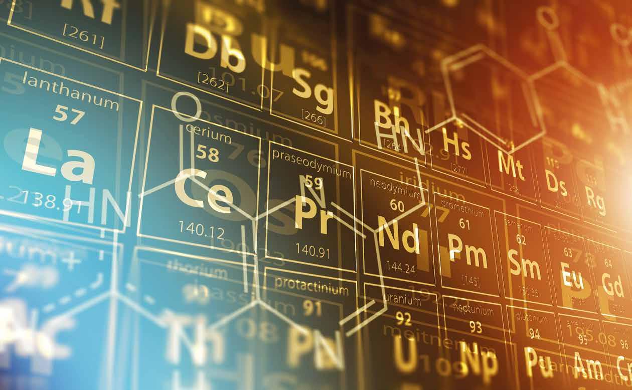
In February 1869, Dimitri Mendeleev produced the first periodic table in a recognisable form as we know it today, building on previous attempts to align elements of similar properties. These earlier attempts included work by John Newlands, Julius Lothar Meyer, William Odling, the telluric screw proposed by Alexandre Béguyer de Chancourtois and the spiral periodic system proposed by Gustavus Hinrichs. The periodic table was updated to the 18-column format by Horace Groves Deming, this was then further developed by Glenn Seaborg who added the row of actinide elements, his work on synthesising actinides winning him the Nobel Prize for Chemistry in 1951.
Since then several other methods have been used to group the elements; Jensen has proposed a 32- column model to avoid interruptions to increasing atomic number and one of the most interesting proposals is the spiral
periodic table of Otto Theodor Benfey. Several three-dimensional models have been proposed such as Courtines' Periodic Classification Wringley's Lamina System, Giguère's Periodic helix and Dufour's Periodic Tree; Stowe's Physicist's Periodic Table has been described as a fourdimensional model (Fig 1). https://upload.wikimedia.org/ wikipedia/commons/c/ce/ Elementspiral_%28polyatomic%29. svg
By DePiep - Own work, CC BY-SA 3.0, https://commons.wikimedia. org/w/index.php?curid=27766488
Mass spectrometry has long been linked with the analysis of the elements, from the Calutron used to purify uranium in the 1940’s to modern inductively-coupled plasma mass spectrometers widely used to quantify metals at low concentrations. Time-of-flight secondary ion mass spectrometry has been widely used for the analysis of surfaces which can include the

elemental composition. Mass spectrometry has come up with its own Periodic Tables, the RSC Analytical Methods Committee has produced a rather wonderful Periodic Table of Mass Spectrometry (Anal. Methods, 2017,9, 50865090 https://doi.org/10.1039/ C7AY90114C), see https://www. msperiodictable.co.uk/ for a downloadable version (Fig 2).
The information provided in a periodic table can be invaluable
for mass spectrometrists; Chip Cody has produced a periodic table that contains isotope graphs with stable isotope ratios, but also displays the exact masses and natural isotope abundances for the stable isotopes (https://www. jeolusa.com/RESOURCES/JEOLPosters-Calendars) and to bring it right up-to-date is also available as an app https://itunes.apple.com/ us/app/jeol-usa-periodic-table/ id1459184462?ls=1&mt=8

Student: Natalie Koch, Rosie Maher, Iris Wagner
Supervisor: Rob Beynon
Centre for Proteome Research, University of Liverpool www.liv.ac.uk/cpr
Twitter: @NatalieKoch91, @rosie_maher, @IW_lovescience, @astacus
@c4pr_liv
We really enjoy scientific conferences – the chance to hear about the greatest and (sometimes) latest developments, and the pleasure of talking science, life, the universe and everything with like-minded colleagues. An opportunity to let a just-starting postgraduate student share their work by presenting and talking through their poster…. what a fabulous way to break into the community!
Inevitably, in many conferences, there are relatively few speaking slots and most science is communicated through the poster sessions. So, this medium of communication - is it effective? Ask that question of a student, or in the case of one of us, a near-retirement academic, how it feels to stand by your poster for an hour and watch people avoid eye contact and swiftly pass by. Or, the poster session that has 200 people juggling plates of food and glasses, packed in a boxy, stuffy room with posters crammed up against each other, often in the herringbone ‘zig-zag’ arrangement that makes access, let alone discussion, virtually impossible. Then of course, we have the poster
sessions where there is plenty of room (Figure 1).
We’ve have been thinking about ways to make posters more effective, and the behaviours that make a poster session more rewarding. There are so many interacting elements to this – the poster itself, the room design, the room layout and even the presence/ absence of refreshments.
‘We need to talk about posters.’. Look at Figure 1 again. How many words on a typical poster there? We guesstimate about 500-1000 words per poster. There are several thousand posters in that meeting, which means that that room could have delivered several million words. We posit that of those posters, virtually none of them will be read fully. Nobody really wants to know if you used 3mM or 6mM of a particular reagent, so why add it? - if they really needed to know, they’ll find you and ask you. Posters are not research papers or thesis chapters, they are visual communication opportunities, and they should be eye catching, colourful and concentrate on the most important messages.

even more appealing and effective?
We’ve discussed this and agreed to limit all our posters to about 200 words (including title and affiliation) and they seem to be much better, as we concentrate on the visual elements. But, can we make them
In spring 2019, our attention was drawn to this YouTube video (www.youtube.com/ watch?v=1RwJbhkCA58) and we realised that there are other interesting solutions out there. This video proposes a bold and imaginative solution, and we’d love to see if it works for our subject areas as well. The video itself is a little slow to develop, but the ideas are readily absorbed, and to our minds, easily adopted. Watch out for posters from CPR over the next few months (especially BMSS Manchester this September), while
we put this to the test, and tell us what you think. Why not go further, have a look at the video, and think about how this might work – this is directed at poster presenters and their supervisors alike. You may experience some pushback from those who only think about traditional posters, but stick with it, and you may be at the vanguard of a small but effective revolution! This is what ‘poster 2.0’ might look like after a reboot.
• A plain language summary of the key conclusion(s) in a huge part of the poster

• A QR code (linking to a web page, full preprint/poster or similar)
• An ‘ammo’ bar (the information/ data/methods) e.g. on the left
• A ‘silent presenter’ bar e.g. on the right– a summary of the poster for quick reading
Look at the example in Figure 2, taken from the video.
Would you be brave enough to do this? In CPR we’re experimenting, and we hope to put some examples on display in BPSR in July and BMSS in September – it would be great if most of the posters were like this! We could really change the way in which we create impact in our posters. The big punchline really makes people stop, and even if they don’t, they get the message.
And presenters, stop being so passive! You put a lot of effort into the science, and the poster. You have
every right to stop passers-by and say ‘can I have a minute to show you my work’, or walk up to one of the ‘big shots’ – (they’re not really big shots, they’re just older, but they share all of your enthusiasm for science!) and say ‘please may I explain my poster’. In virtually every case, you’ll be rewarded by an engaging, knowledgeable, enthusiastic response, and you’ll feel fantastic. Go for it.
These are not particularly radical thoughts and we’re sure some of them have been tried and tested before, but it would be a brave meeting that considered some of these principles and planned for radically new, lively, poster sessions. We can only say ‘why not try?’.
Disclaimer
These thoughts come from one supervisor and three Ph.D students, and reflect our opinions only. Our only goal is to try to create some
(We’ve made these mistakes in the past too).
Give posters time and space – schedule a poster session, don’t jam them in with vendor booths and during lunches and coffees. The poster presenters, predominantly younger, early career scientists, deserve much better.
Use odd/even numbering to manage time for presentation and time for viewing. Schedule times for attendance and if you are a presenter, make sure you are there
Put the posters in a large enough room that there is space for conversations, people flow, and a chance to step back and take a shot of the QR code.
Give the poster presenters a big lapel badge to say ‘it’s me!’ and talk to them. Add poster numbers to name badges so poster presenters can be identified easily.
Supervisors – try to keep a distance form your students and let them go without stabilisers!
UK in particular, perhaps – STOP forcing portrait format posters – find a way to accommodate landscape posters even if you have to pay a bit more for a room/display boards. (If we have to be portrait, here’s a template for the poster format discussed previously in portrait format (https://osf.io/g6xsm/)
discussion about the whole poster experience. Your opinions may differ.
Footnote
Two more QR codes. LH: the YouTube video, RH: the link to the portrait format.

Don’t ‘herringbone’ (zig-zag) the posters – this creates little zones of deadness where nobody can do anything. Make the posters run linearly.
Here’s an idea that has also been used. Try to cluster posters on related topics in groups of, say five. Then, ask a convener (perhaps, one of the supervisors) to gather all the five poster presenters together, and do a quick walk around (maybe glass in hand?), where each presenter tells the others briefly about their posters. The five neighbours will find it easy to talk to each other for the rest of the meeting. (One of us has done this as convener in meetings in the past, and it is a super ice breaker, especially for newcomers to an established meeting).
Schedule posters early – they are a great way of breaking down shyness and creating a better buzz to the meeting
Accept electronic posters (ePosters) that complement traditional paper posters, allowing conference attendees to post comments, questions, or share ePosters during and after the conference. It would also help if ePosters could be searched by ‘buzz words’- 5 or so key words selected by the presenter so related topics can be found easily.
Universal agreement – why do we present posters? Is it to give a summary of a research paper (small figures with walls of text) or is it to engage with passers to entice them over to promote one to one discussion of your work (large colourful figures with little text). Maybe the organisers could be bold enough to set a word limit?
A QR (‘quick response’) code is a 2-dimensional bar code that is often used to link to a web page. Thus, this QR code links to our web page. How can you access this though? The easiest way is to use the inbuilt QR code reader on your phone or other portable device (both iOS and Android). For example, in iOS 11 and later, simply open the camera app, point the camera at the QR code, and you’ll be given an option to be taken directly to the web site. (You could also install QR code reading apps, but beware that some of these sneakily add a monthly subscription – this should not be needed.)
Try it – This QR code points to the CPR web site.

You make a QR code by going to one of three free QR code generators that are on-line. The bottom QR code takes you to a code generator (if you compare them, you’ll see that they look superficially similar, but are different)
Time of Flight secondary ionisation mass spectrometry (ToF-SIMS) is a sensitive surface analysis technique providing detailed surface chemical information (static SIMS). Highenergy (up to 30 keV) ion beams are directed at the sample’s surface, liberating secondary ions. Secondary ions are accelerated into a timeof-flight analyser and separated, producing a spectrum that indicates the sample’s chemical make-up. ToF-SIMS can also be utilised to produce mass spectrometry images via rastering across an area of interest and to interrogate a sample in 3D (depth profiling or dynamic SIMS) using an etching ion beam that sputters layers of material. Depending upon the instrument configuration, either the sputtered material or the exposed material underneath is available for analysis. The technique can provide highly specific chemical information, including molecular information. Applications span many disciplines e.g. physics, chemistry, engineering, life sciences, pharmacy and beyond.
In its infancy in the late 1960’s, the technique utilised monatomic primary ion beams such as Ar+ and Cs+. These promote fragmentation of molecules leading to complex spectra that can be difficult to interpret. However, these early ToFSIMS instruments were particularly well suited to the semiconductor industry, where elemental ion analysis of species including Si+, Ga+ and Li+ are of high interest. Monatomic ion beams continue to offer many uses today for example in battery technology analysis [1].
For the analysis of organic compounds higher mass cluster ion sources such as gold and bismuth (typically 25 keV Au3+ and Bi3+) are routinely utilised. These ‘softer’ ion beams cause a lesser degree of fragmentation and therefore organic molecular ions can be liberated from the sample surface. These can also be used as etching
beams for the analysis of organic compounds and have been a gamechanger, allowing new materials to be interrogated by ToF-SIMS. These advances have increased the range of applications and persuaded many to take advantage of its capabilities [2]. For many years researchers at the University of Nottingham have used ToF-SIMS in collaboration with industrial collaborators to investigate technical challenges in sectors including pharmaceutical, aerospace, cosmetics, petrochemicals and additive manufacturing research.
In large-scale textiles manufacturing, contamination during production may lead to a catastrophic reduction in a material’s aesthetics. This may require the process to be stopped and the material itself wasted, at a very high cost. Working with a local textile manufacturer, Guilford Europe, we analysed the chemical nature of deposits embedded in the weave of their textile during production. Their small size (200–300 µm) diameter made them poor candidates for alternative materials analysis testing methods. Spectra from the deposits were compared to a library of approximately 50 materials used in the manufacturing process. We were able to identify a specific drying agent that was causing the deposits due to a suboptimal washing procedure. The washing procedure was changed to ensure the drying agent did not remain and the problem with deposits was resolved. The reference library of raw materials was used to probe subsequent contamination events, identifying the chemical constituents of leaking pipework and deposits caused by broken machine bearings.
ToF-SIMS can also detect molecular ions for active pharmaceutical agents (APIs), diagnostic ions for polymer delivery agents, surfactants and stabilizers. Hence it has found many applications within the
pharmaceutical sector, including the analysis of API-containing nanoparticles, biomedical implants, tablets and topical medicines. The permeation of topically applied agents is also relevant to the agrochemical and cosmetics industries. In contrast, ToF-SIMS using gas-cluster ion beams enables analysis of the upper surface of a sample and a depth profile e.g. of ex situ skin, producing a 3D profile of the distribution of both endogenous and exogenous chemistries [3]. Such profiles improve understanding of how cosmetics are delivered to the uppermost layers of the skin; this collaborative research will ultimately enable clinicians and others to deliver antibacterial agents, APIs and cosmetics to the skin more effectively.
Despite the many advantages of ToF-SIMS, the technique’s limited mass-resolving power has, in some cases, restricted its range of applications (both industrial and otherwise). This is being addressed with new instrumental advances such as the new 3D OrbiSIMS which uniquely combines SIMS with an orbitrap mass analyser for high mass resolving power and high accurate mass analysis [4]. It is anticipated that this novel instrumentation will be very impactful for the analysis of organic samples such as tissues, allowing accurate mass identification of biological chemical entities. Recently, other hybrid ToF-SIMS instruments have been described that have also expanded the capabilities to include MSMS allowing structural characterisation and confirmation of isomers [5]. Overall, these advances will further expand the range of applications for which SIMS can provide informative data.
Dr David Scurr: david.scurr@ nottingham.ac.uk
Dr Matthew Piggott: matthew. piggott@nottingham.ac.uk
References
1. You, Y., et al., Modified HighNickel Cathodes with Stable Surface Chemistry Against Ambient Air for Lithium-Ion Batteries. Angewandte Chemie International Edition, 2018. 57(22): p. 6480-6485.
2. Passarelli, M.K. and N. Winograd, Lipid imaging with time-of-flight secondary ion mass spectrometry (ToF-SIMS). Biochimica et Biophysica Acta (BBA) - Molecular and Cell Biology of Lipids, 2011. 1811(11): p. 976-990.
3. Holmes, A.M., et al., Dendrimer pre-treatment enhances the skin permeation of chlorhexidine digluconate: Characterisation by in vitro percutaneous absorption studies and Time-of-Flight Secondary Ion Mass Spectrometry. European Journal of Pharmaceutical Sciences, 2017. 104: p. 90-101.
4. Passarelli, M.K., et al., The 3D OrbiSIMS—label-free metabolic imaging with subcellular lateral resolution and high mass-resolving power. Nature Methods, 2017. 14: p. 1175.
5. Fisher, G.L., et al., A New Method and Mass Spectrometer Design for TOF-SIMS Parallel Imaging MS/MS. Analytical Chemistry, 2016. 88(12): p. 64336440.
The BMSS has invested in a tool that is more than just a website! This web-based society management system gives all members their own locker. Anyone can subscribe to the society, register to attend meetings and submit abstracts all through this one portal, then track your status and review receipts in your personal locker room.
Click on the big BMSS logo at any time to access our home page and from there it is just a short click to find out more about the BMSS, to view the latest developments with our annual meeting, to apply for travel grants, to view the latest job opportunities, to see what our special interest groups are up to and to read snippets from Mass Matters on-line and find out how you can contribute articles.
We also have front page news so please share news items with us.
About Us – find out who the committee and advisory board are and what their roles and functions are. Read up on the history of the BMSS and how we came to be where we are now!
Member Benefits – as before, this includes a number of useful links, information about our special interest groups and how to advertise events and jobs with the BMSS (free to members). New to the BMSS website is an enabling space for the LGBT+ group who will be gathering at our annual meeting for their second event.
Meetings – one of the busiest parts of the website this year as this new
web-based system has enabled us to support more meetings than ever before!
Grants – the John Beynon Travel and Conference fund has undergone a few changes and we will see more over the course of this year as we aim to move to enabling applications through the website… watch this space!
Education – Professor Jane ThomasOates is our new BMSS lecturer and we will keep you all updated here as the lecture series develops. Awards – as ever, it is important to recognise the achievements of our members from all walks of life. Find out how to nominate colleagues or to seek recognition yourself!
We are still in the process of developing functionality to best suit
members needs so watch this space for further developments!
We would very much welcome any feedback on the website. Please just email comments to the BMSS Administrator at admin@bmss.org. uk and we will aim to take them all on-board!

The BMSS Ambient Ionisation SIG Meeting and workshop was held at the University of Huddersfield on a cold and snowy January day. The day consisted of 12 interesting talks on both new ionisation sources and novel uses of already accomplished ambient techniques. The poster session included 20 posters detailing more exciting work performed in the field.
Lindsay Harding (University of Huddersfield) presented her work combining hot-stage microscopy and DART-MS (direct analysis in real-time mass spectrometry). Mass spectrometric profiles were used to detect thermal properties of silicone materials across a range of temperatures. Interestingly, this technique can be used as a method to determine a materials coefficient
There were two talks discussing the use of REIMS (rapid evaporative ionisation mass spectrometry). Paul Abu-Rabie (GSK) and ? discussed the use of REIMS in a pharmaceutical environment. Many applications were discussed such as predicting cell line phenotypes and drug response prediction. Joscelyn Harris (University of Liverpool) uses REIMS to monitor bacterial growth and expression of exogenous proteins in E. coli. This research has interesting potential of predicting protein expression in laboratory.
Carl Fletcher (Mass Spec Analytical) used a thermal extraction ionisation source (TEIS?) to directly analyse solids, liquids and gases in real-time. The technique can be successfully applied to drugs and explosives in forensics and also applications in
being excellent, with RSD% of <4% being reported in the literature, quantification using calibration curves, without the requirement for liquid chromatography, is achievable.
Another technique discussed was LESA (liquid extraction surface analysis) and innovative applications. Jana Havlíková (University of Birmingham) discussed her research on developing LESA to detect pathogens in bacterial species. Two strains could be differentiated on sequence variations giving potential biomarkers. Rian Griffiths (University of Birmingham) used LESA in her research to monitor changes in lipid content of cell membranes in Mycobacterium in order to determine disease metabolism.

optimisation in patients and can be used to quantify when using an isotopically labelled internal standard.
Jan-Christoph Wolf used a technique called soft ionisation by chemical reaction (SICRIT) which uses a dielectric barrier discharge-based plasma source. This technique has applications in areas such as pesticides, warfare agents and illicit drugs due to the high sensitivity and has the added advantage of being used in-field. It was described as a “one for (almost) all” ionisation source.
Andrew Ray (AstraZeneca), Luzia Gyr (ETH Zurich) and Max Hecht (University of Tartu) all discussed novel ionisation techniques.
Andrew discussed using the newly developed Open Port Sampling Interface (OPSI) which uses a liquid flow to form a meniscus to analyse samples by touching the surface with the droplet or, dropping liquid into the meniscus. The technique has the unique ability to measure hydrogen/deuterium exchange to determine the chemical structure of compounds, which is not currently possible using other ambient techniques. Luzia used an active capillary plasma ionisation source which uses nitrogen gas to transport all ions directly into the MS. The technique can be used to better understand polar and non-polar compounds due to the dielectric barrier discharge. Max discussed a sample collection technique called ‘sponge spray’ which has the ability to absorb fixed volumes of biological samples, such as blood and urine. The samples were allowed to dry then were analysed directly. This technique shows exciting developments for dosage
Georgia Sanxaridou discussed using SIFT-MS (selected ion flow tube mass spectrometry), combined with supercritical CO2 extraction. A probe is placed at the outlet of the extraction vessel to track the concentrations of the organic compounds. Where it has previously been difficult to detect low concentrations and quantification has previously proven difficult, the addition of SIFT-MS can detect a few parts per billion of solvent with great accuracy.
The final talk of the day was a presentation on the use of DART (direct analysis in real time) for sub-microlitre sample preparation. Brian Musselman (IonSense) discussed using low volume spotting techniques which can considerably reduce matrix interferences when ?directly biological samples such as urine, plasma and oral fluid.
In summary, the meeting was very informative and relayed exciting research and novel techniques. Many ideas were discussed to further improve direct ambient techniques such as reducing matrix effects and the possibility of quantification.
Alison Ashcroft got the meeting started by leading us through some aspects of her amazing career in mass spectrometry, highlighting the technology and methods she has developed along the way and those that she has used throughout. Being one of the first people to take on the challenge of what we now term native mass spectrometry, it was good to see how ion mobility separation and mass spectrometry have grown together over the years at Leeds to inform on many structural biological studies.
Clearly of central importance to Alison has been, and still is, the people she has worked with. This includes the many students who now have their own amazing careers in MS thanks to such an excellent introduction to this field of science and great encouragement, support and enthusiasm along the way; collaborators both with the Astbury Centre at Leeds and beyond; and, the numerous manufacturers Alison has worked for and with over the years. Frank Sobott shared
a story from a time when he was at Oxford and persuaded Alison to dust off and pump down an old Cyphergen instrument to perform SELDI ToF MS on a protein, which Alison kindly and patiently did, despite having told Frank what the outcome was likely to be from that instrument! Alison also reminded us of some Leeds Alumni who went on to develop their own amazing careers in MS: Prof. Ray March, Trent University, Canada; Prof. John Todd, University of Kent, UK, Prof. Dudley Williams, University of Cambridge, UK and Prof. Howard Morris, Imperial College London, UK who came back recently to open the newly refurbished MS labs. Alison clearly made Leeds a great, and fun, place to study mass spectrometry. We heard stories, and saw pictures, of the ‘Mass Spectacular’ team competing in Astbury sports days over the years, where they did Alison proud.
Anton Calabrese and Charlie Scarff took us through some of the scientific achievements from Leeds,
which can be broadly summarised in four themes:
1. Amyloid assembly
2. Membrane protein structure and function
3. Virus capsid assembly
4. Method development
The morning ended with receiving comments from the floor - a great opportunity for everyone to reminisce and share stories of their time working with Alison.
The afternoon saw 4 students from the Ashcroft lab deliver talks on their current endeavours in mass spectrometry, highlighting Alison’s legacy in ion mobility separation mass spectrometry and the applications of native MS. [FD1] The day ended in a lovely meal where everyone had time to sit back, relax and catch up with friends old and new.

Valérie Gabelica opened the meeting with a great tutorial lecture on IM, providing a comprehensive explanation of IM theory and thoroughly detailing how to perform valid IM experiments and report data in an appropriate manner. For those who were not able to attend this meeting, details can be found in a recent paper (Gabelica et al, Mass Spectrometry Reviews 2019, 38, 291) or an extended version of the presentation is available online (10.5281/zenodo.3268737). The scientific talks were then started by Anna Simmonds from the University of Birmingham who showed how you can use IM and modelling to gain an understanding
of the effect of salt-bridges in ECD experiments. Zihao Wang from the University of Oxford then bravely battled the AV sound system and succeeded in delivering a great talk on structural studies on human cardiovascular heat shock protein and how HDX was used to confirm substrate binding sites. Leading us up to lunch, Nico Wortel from MS Vision talked about their history and how the company got involved in native MS while working with Prof. Albert Heck, Utrecht. Although originally a small company that was an instrument service provider, they now modify commercial instruments to meet the applications needs of their customers.
Well done to PhD students Charlie Eldrid (University College London) and Anna Simmonds (University of Birmingham) who agreed to chair the afternoon session with literally no notice! Both did a cracking job of keeping the speakers to time. The first speaker in this session was Matteo Degiacomi from Durham University who told us how integrative modelling can be combined with ion mobility to gain an understanding of protein structure and function. This was followed by Lucy Woods from Bruker who stepped up to the lectern and switched our minds from proteins to lipids, describing how ion mobility can help characterise
them. Emma Sisley, University of Birmingham, then described recent experiments combining LESA and cyclic IMS. The day ended with a great talk from Argyris Politis (Kings College London), who described his recent work using native MS and hydrogen-deuterium exchange MS to characterise antibodies and macromolecular assemblies.
We hope that all who attended enjoyed a great day of talks and networking, and we’re looking forward to the next IM SIG meeting!
The authors of these reports all wish to express their thanks to the BMSS for the award of a travel grant to allow them to attend these conferences.
Travel Grant Recipient:
Cookson Chiu
Department of Chemistry, University of Warwick
Supervisors:
Professor Peter O’Connor
Abstract
Activation of the Ir metallodrugs caused production of reactive singlet oxygen species and extensive oxidation of nearby biomolecules. A whole range of oxidation products, up to 6 per peptide, were observed. The high resolution and mass accuracy and extensive MS/MS capabilities of the FT-ICR MS allowed unambiguous identification of the oxidation sites and the modifications induced. 1O2 was found to cause many methionine oxidation events (to sulfoxide and sulfone), but also oxidation of tryptophan residues, producing diagnostic kynurenine and 3-hydroxy kynurenine moieties. Histidine residues were oxidised to 2-oxo-histidine, which is commonly observed in 1O2 oxidation events.
Bottom-up nLC-MS/MS experiments of drug-treated cancer cells revealed two key targets; aldose reductase (AR) and heat shock protein 70 (Hsp70). Quantitative nLC-MS/MS showed the increase in oxidised products in the drug-treated samples, calculated as 3.0-fold
up-regulation with AR and 5.8-fold up-regulation with Hsp70, together with 9 proteins up-regulated along the glycolysis pathway.
Electron capture dissociation (ECD) of oxidised [Lys3]-bombesin, 815 m/z. Oxidation sites were narrowed down to be histidine and tryptophan, they were oxidised to 2-oxo-histidine and 3- hydroxy kynurenine respectively.
Reacting the Ir(III) photocatalytic drug to A549 lung cancer cells, 3 unique proteins were found to be oxidised. Heat shock protein 70 (HSP70) and aldose reductase (AR) were found to have up-regulated by 5.65-fold and 2.93-fold.
Along the glycolysis pathway, 9 unique important proteins were found to have up-regulated, ranging from 2.1-fold to 5,3-fold, all involved in the conversion of glucose to pyruvate.
What this conference meant to me…
Six of us from the O’Connor group went to University of Lincoln for this year’s EMPW. Lincoln is nice and quiet, and we have met many top scientists from the area and saw a lot of excellent presentations.


Travel Grant Recipient:
Cookson Chiu
Department of Chemistry, University of Warwick
Supervisors: Professor Peter O’Connor
Abstract
Presentation Topic: The Next Dimension In Proteomics
Reversed phase liquid chromatography (RP-LC) is an essential tool for the proteomic studies due to its powerful separation of peptides and proteins prior to MS analysis; however, LC separation of extremely complex samples with widely varying properties suffers from the bias of the chromatography columns applied in LC separation and incompatibility with certain separation conditions such as solvent compatibility, pH, and column chemistry. An alternative analytical method which is able to handle complex proteomic samples and high flexibility in sample conditions is required. Herein, we apply two-dimensional mass spectrometry (2DMS) to study proteomic samples ranging from a standard protein digest to a whole cell lysate digest. In the 2DMS experiments, we show that it is possible to obtain detailed sequence information from peptides without online LC separation. We then compared the results obtained from the 2DMS to the LC tandem MS data and show that 2DMS can achieve similar results to LC MS/MS for identified proteins in the proteomic samples shown. Furthermore, our data show that the 2DMS experiments tend to be more adept at analysing and identifying particularly hydrophilic, basic, or short peptides, which is complementary to those being assigned in LC tandem MS. These
results demonstrate that 2DMS provides an alternative technique for future proteomics.
Two-dimensional mass spectrometry (2DMS) was first introduced by Pfandler et al. in 1987 and further developed and applied by Ross et al. in 1993 using an FTICR mass analyser. In 2DMS, a modulating excitation or de-excitation pulse is used to alter the positions of ions inside the FTICR cell, ions that are modulated to overlap with the position of the fragmentation zone are dissociated with the applied radius-dependent in-cell MS/MS technique, such as infrared multiphoton dissociation (IRMPD) or electron capture dissociation (ECD). Since the fragment ions are modulated with the same frequency as the precursor ion, the modulation frequency can be used to correlate fragments with precursors. In 2DMS, samples are directly infused into the MS which allows a broader selection of sample preparation methods, especially the selection of sample buffers. 2DMS is a dataindependent acquisition technique in that all precursor ions are fragmented during the experiment, and a two-dimensional mass spectrum, showing all precursor and fragment ions, is obtained. Compared to the traditional MS/MS, 2DMS does not require isolation of a precursor ion prior to fragmentation and detection, instead utilising the frequencies of ions in the second dimension to separate the peaks which provides an advantage in the application to complex proteomic samples as discussed below.
2DMS is newly applied to proteomic studies, including both top-down and bottom-up proteomics. Previous studies showed that similar top-down and bottom-up fragments were obtained from both quadrupole precursor isolated MS/MS and 2DMS. Peptides
from digested collagen showed that 2DMS can differentiate and fragment two precursor ions with similar mass-to-charge (m/z) ratios, which, in general, cannot be isolated and fragmented individually by quadrupole mass isolation. Peptide assignment from the 2DMS spectra of digested proteins, such as cytochrome c, calmodulin, and collagen, also showed that 2DMS has the potential for complex proteomic studies without LC separation.
In the presentation, we used a mixture of digested standard proteins and a digested yeast lysate sample to evaluate the performance of 2DMS by comparing the data to proteomic results obtained from a standard nano-LC (nLC) MS/MS approach. Various sample preparation and 2DMS data acquisition methods were used in the mixture of digested standard proteins in order to determine the most suitable workflow for a complex proteomic study. The optimized method was further applied to the digested yeast sample to demonstrate the ability of 2DMS in proteomic studies. From the results, we showed that 2DMS could identify a significant number of peptides in a digested proteomic sample, in which the number of assigned peptides was comparable to the nLC MS/MS data. Furthermore, the identities of proteins and peptides observed in the 2DMS experiments were complementary to the results from nLC MS/MS experiments, thus a better coverage in proteome as well as protein sequence can be obtained through a combination of methods. The solvent and column constraints in LC systems mentioned in above were also shown to be eliminated in our 2DMS experiments without affecting the quality of data. The peptides that were exclusively assigned in the 2DMS experiments suggested this newly developed
method is suitable for complex proteomic studies and associating the data obtained from 2DMS with nLC MS/MS results provides a new capability in future proteomic studies.
Two dimensional mass spectrometry (2DMS) was first introduced by Pfandler et al. in 1987 and further developed and applied by Ross et al. in 1993 using an FTICR mass analyser. In 2DMS, a modulating excitation or de-excitation pulse is used to alter the positions of ions inside the FTICR cell, ions that are modulated to overlap with the position of the fragmentation zone are dissociated with the applied radius-dependent in-cell MS/MS technique, such as infrared multiphoton dissociation (IRMPD) or electron capture dissociation (ECD). Since the fragment ions are modulated with the same frequency as the precursor ion, the modulation frequency can be used to correlate fragments with precursors. In 2DMS, samples are directly infused into the MS which allows a broader selection of sample preparation methods, especially the selection of sample buffers. 2DMS is a dataindependent acquisition technique in that all precursor ions are fragmented during the experiment, and a two dimensional mass spectrum, showing all precursor and fragment ions, is obtained. Compared to the traditional MS/MS, 2DMS does not require isolation of a precursor ion prior to fragmentation and detection, instead utilising the frequencies of ions in the second dimension to separate the peaks which provides an advantage in the application to complex proteomic samples as discussed below.
2DMS is newly applied to proteomic studies, including both top-down and bottom-up proteomics. Previous studies showed that similar top-down and bottom-up fragments were obtained from
both quadrupole precursor isolated MS/MS and 2DMS. Peptides from digested collagen showed that 2DMS can differentiate and fragment two precursor ions with similar mass-to-charge (m/z) ratios, which, in general, cannot be isolated and fragmented individually by quadrupole mass isolation. Peptide assignment from the 2DMS spectra of digested proteins, such as cytochrome c, calmodulin, and collagen, also showed that 2DMS has the potential for complex proteomic studies without LC separation.
In the presentation, we used a mixture of digested standard proteins and a digested yeast lysate sample to evaluate the performance of 2DMS by comparing the data to proteomic results obtained from a standard nano-LC (nLC) MS/MS approach. Various sample preparation and 2DMS data acquisition methods were used in the mixture of digested standard proteins in order to determine
the most suitable workflow for a complex proteomic study. The optimized method was further applied to the digested yeast sample to demonstrate the ability of 2DMS in proteomic studies. From the results, we showed that 2DMS could identify a significant number of peptides in a digested proteomic sample, in which the number of assigned peptides was comparable to the nLC MS/MS data. Furthermore, the identities of proteins and peptides observed in the 2DMS experiments were complementary to the results from nLC MS/MS experiments, thus a better coverage in proteome as well as protein sequence can be obtained through a combination of methods. The solvent and column constraints in LC systems mentioned in above were also shown to be eliminated in our 2DMS experiments without affecting the quality of data. The peptides that were exclusively assigned in the 2DMS experiments suggested this newly developed method is suitable for complex
Members (of at least 12 months standing) may apply for small grants of up to £2,000, with matched funding, to aid their research.
The BMSS Research Support Grant aims to support small research endeavours including, but not exclusive to:
• Generating pump priming data for grant applications or research areas.
• Incoming or outgoing visits to initiate new MS-relevant collaborations/training (standard class travel/accommodation).
• MS taster for new MS users (who must join the society).
• Instrumentation (updates, repairs, add-ons, new developments of existing kit etc.).
• Promotion of industry-academic collaborations.
Full details of eligibility, how to apply and conditions for awarding grants are available on the BMSS website at www.bmss.org.uk.
proteomic studies and associating the data obtained from 2DMS with nLC MS/MS results provides a new capability in future proteomic studies.
What this conference meant to me… I have attended the East Midland Proteomics Workshop (EMPW) since 2015, it is always a nice meeting between all proteomics users in the East Midlands of UK. EMPW is not a huge conference compared to ASMS and BMSS; the interactions between participants, however, are much stronger. The conference covered a wide range of topics, including the workflow and data analysis of proteomics, which are very useful for the proteomics users. Overall, I really enjoy this conference and hope I can attend it again in the coming years.
I have attended the East Midland Proteomics Workshop (EMPW) since 2015, it is always a nice meeting between all proteomics users in the East Midland of UK. EMPW is not a huge conference compared to ASMS and BMSS; the interactions between
participants, however, are much stronger. The conference covered a wide range of topics, including the workflow and data analysis of proteomics, which are very useful for the proteomics users. Overall, I really enjoy this conference and hope I can attend it again in the coming years.


Along with a number of others, I joined the BMSS committee in September 2018 -which was the realisation of an ambition I’d had for a number of years, having stood for election in the past but not been successful (moral of the story…keep trying!). Like my other newly elected members I am excited to be a part of this team, and am very excited about integrating into the committee and getting more involved as time goes by.
I guess at one point, I could claim to be a scientist as I studied Chemical & Analytical Sciences at the University of Wales Swansea (as it was called then) from 1997 to 2000. At the time, the only exposure to mass spectrometry I had was the knowledge that the Chemistry department housed the Mass Spectrometry Research Unit, and it was only when I was reaching the end of my undergraduate studies that I really started to understand how prestigious a group that was. Dai Games approached me and asked if I wanted to consider a PhD and in typical Dai manner, pledged he’d ‘find the funding somewhere’ if it was something I wanted to pursue. That was the first experience I had of the ‘family of mass spectrometry’ that Dai tried to culture and is still how I see the industry today. Fast forward 4 years and I had finished my PhD in 2004, having worked tirelessly on integrating MS with Countercurrent Chromatography (CCC) and achieving the notable first of employing APCI in CCC for the first time. The project was a joint effort between Swansea and Prof Ian Sutherland of Brunel University.
And that my friends, is where my proper scientific career ended! Following Swansea, I joined Waters and spent 5 years or so travelling the world as a customer support engineer helping actual scientists fix broken instruments and tweaking methods to make sure they got the most out
world I moved into a product management team in the old Waters Floats Rd facility and from there took a career swerve and went into marketing – firstly to a strategic role in 2014 looking at the Industrial Chemistry market trends (who doesn’t love a massive spreadsheet?) then into Product Marketing where I looked after Waters’ tandem quadrupole systems with a focus on the IVD & clinical applications. Looking back, I can't really believe I spent 15 years at Waters, the time went so quickly and it really was a fabulous time getting to work alongside some of the ‘household’ names of mass spectrometry – I’m looking partly at you, Mike Morris :-). Mike is another mentor of mine and I was very pleased when he offered to help me mentor a number of students back at Swansea on a number of projects, and pleasingly resulted in Swansea offering me an Honorary Associate Professorship at the Medical School in 2017. Me… a ‘professor’ .. it’s still a bit unbelievable to be honest. One thing it did cement though is my passion for industrial-academic partnership, and how those strong collaborations really get the best out of our small industry. It’s no secret that a lot of my collaboration has been via Swansea, and in particular with Dr Ruth Grodfrey and Prof Gareth Brenton, and it’s been something I have been all too pleased to participate in, and an example of where I hope our industry continues to develop –with strong academic and industrial partnerships forming for the benefit of the science.

My election to the BMSS in September 2018 was quickly followed by a decision to move on from Waters, and in February 2019 I joined Thermo Fisher Scientific in a role that looks after ‘workflow solutions’ for LC and LC/MS for the biopharmaceutical industry. Four months into that role, and things are good, I work closely with a team of very talented scientists, of which I hope

From a personal perspective, I’m fortunate to live in Chester (growing up in North Wales, Chester was always the posh shopping day out). My wife Suzanne and daughter Elizabeth keep me grounded on a daily basis and we spend a lot of our time cooking, growing strawberries in the garden and visiting the many playparks Cheshire has to offer. Living in the North West also allows me to partake in the other great passion in my life alongside family and mass spectrometry, Liverpool Football Club. I’m a season ticket holder and can be found most weekends in/around the Anfield area waving my scarf like a fool shouting ‘Allez Allez Allez’. #wewonitsixtimes
I’m looking forward to the rest of my term with the BMSS and can only hope I can contribute as much as I can.
