Diabetic retinopathy
Diagnose and treat early
Bayer’s Medinfo will help you with all questions related to Bayer products.
Telephone 020 785 21
(weekdays from 9 am to 3 pm)
medinfo@bayer.fi
Bayer Oy Tuulikuja 2, 02100 Espoo PO BOX 73, 02151 Espoo
Telephone 020 785 21
2 DIABETES AND EYESIGHT 3 RISK FACTORS AND PREVENTION 6 SYMPTOMS 8 TREATMENT 10 Contents
Diabetes may affect vision in many different ways. It can, for example, damage blood vessels at the back of the eye, causing bleeding and retinal edema.Good treatment control of diabetes reduces the risk of developing severe retinopathy. Correctly timed treatment of diabetic retinopathy reduces the risk of vision loss significantly.
All patients with diabetes – both type 1 and type 2 – are at risk of developing diabetic retinopathy (DR) and diabetic macular edema (DME). Diabetic retinopathy is a disease of the retina in the eye. On the other hand, macular edema is one form of retinopathy that affects the region of acute vision, i.e. macula in the retina, and thus the ability of the patient to see accurately.
3
Both of these are common complaints. About three percent of patients with diabetes have impaired vision due to DME, and it is the most common reason for visual impairment among the working age population. In Finland, DR causes eight percent of visual impairment among the working age population and four percent among the elderly (Annual of the Finnish Register of Visual Impairment 2016). It is the most common eye disease related to diabetes.
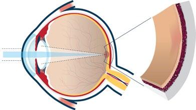
The retina is a light-sensitive nerve layer that lines the back of the eye. The region of acute vision, macula, has the greatest concentration of photoreceptor cells that receive light signals.
4
Retinal blood vessels
Light
Retina
Photoreceptor cells
Macula
Choroid
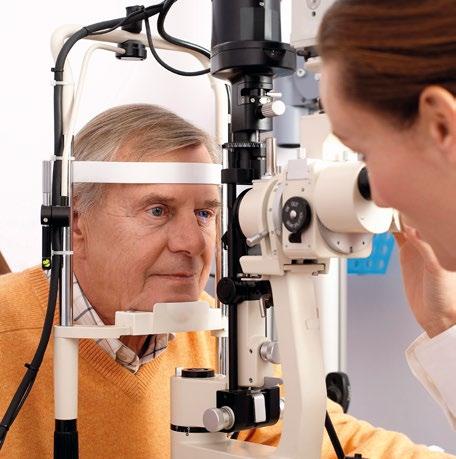
Regular eye examinations are important for diabetics
5
Risk factors and prevention
• Prolonged high blood sugar level (hyperglycemia). Maintaining the blood sugar level as normal as possible may delay or even prevent the development of DR and DME.
• Abnormal cholesterol and triglyceride levels (dyslipidemia)
• High blood pressure (hypertension)
• Renal disease (nephropathy) or cardiovascular disease
• Smoking
• Development of vitreomacular scar tissue. The scar tissue may cause vitreomacular traction and even retinal detachment.
• Pregnancy. Pregnant women with diabetes should always have a thorough eye examination with dilation of the pupil.
• Anemia, sleep apnea, use of medicines that increase the production of insulin, genetic factors, abundant alcohol use and too little exercise
Vision symptoms are rarely associated with the early phase of DR and DME, and loss of vision may happen very quickly. Therefore, talk to your doctor and have regular eye examinations if you have risk factors for diabetic macular edema.
6
The risk of DME can be reduced in many ways:
• Have regular check-ups where factors possibly leading to the development of DME are checked: blood pressure, blood vessels and the condition of the eyes
• Check your values. Keep your blood sugar, blood pressure and cholesterol within the normal range by following the doctor’s instructions. Take the medicines prescribed for you.
The
risk of getting vision impairment can be reduced with healthy living habits

Symptoms
DR is often asymptomatic in its early phases. When it progresses, it impairs vision and causes distortion of lines or colors and blurred vision.
Damaged blood vessel walls and increased permeability
Edema in the retina and macula
Increased VEGF action
Macular edema damages retinal photoreceptors and the patient starts to see in the middle of the field of vision first distortions and later dark areas that obstruct vision.
8
Fovea
Macula
Diabetic retinopathy may lead to diabetic macular edema. Its early phase is typically asymptomatic, and vision symptoms appear only in the more advanced phase.
Diabetic macular edema shows as
• deficiencies in the field of vision
• obscured vision
• blurred vision
• washed out and faded colors


If symptoms of DME appear, it is important to make an appointment with an ophthalmologist as soon as possible.
Even if no symptoms of DR or macular edema have yet appeared, it is important for a diabetic to have regular eye examinations. Regular eye examinations help to identify other problems affecting vision at a much earlier stage. These problems may be caused by diabetes and require treatment.


Treatment
Diabetic retinopathy and macular edema can be treated. Because vision loss may happen very quickly, it is important to go to regular eye examinations before any symptoms appear.
The condition should be diagnosed already in the early phase and treatment started as soon as possible to obtain the best result for vision.
Even though controlling blood sugar levels is important in diabetes, additional treatment is also required. Many different treatment methods are available. Discuss with your ophthalmologist which one will suit you best.
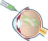
Angiogenesis inhibitors target proteins that trigger the growth of leaking new blood vessels in the eye. The angiogenesis inhibitor is administered by injecting it into the eye with a thin needle. These treatments have been shown to stop the progression of DME and in some cases to restore vision by reducing the growth and leakage of new blood vessels.
Laser therapy is used to prevent the leakage of blood vessels into the retina and macula. In this therapy, the laser beam is directed to the leaking new blood vessels in the retina, which reduces the amount of tissue fluid and slows down leaking. Usually a single
10
Macula
treatment is sufficient but sometimes several laser treatments are required. The aim of laser treatment is to stabilize vision, and it may prevent vision impairment caused by DME.
If blood has leaked to the vitreous humor, the vitreous humor can be removed by performing a vitrectomy. In the treatment of DME, vitrectomy is typically used to remove vitreoretinal traction and/or to improve oxidation of the retina by removing the vitreal fluid of the eye. Vitrectomy may help preserve vision by removing blood.
Corticosteroid treatment can prevent inflammatory reaction and edema in the retina and macula. Corticosteroids are hormones that have been shown to control growth factors, which are assumed to have a role in the development of DME.
These medicines can be administered as injections into the vitreous humor or as active ingredient releasing implants.

11
Diabetic changes in the back of the eye do not prevent you from living a full life. Vision loss is much less common in diabetics today, due to improved treatments and monitoring. Agree with your doctor on an eye monitoring program suitable for you.
AMSLER GRID TEST
If you have vision problems, you can do a simple test with the grid available on the back cover. In this test you should look at the black and white Amsler grid. If the lines seem distorted or in some other way abnormal, this may be a sign of a retinal disease. In such a case contact an ophthalmologist as soon as you can.
1. If you use reading glasses, you should wear them also during the test.
2. Place the chart at a distance of 30 cm from your eyes. Cover one eye.
3. Focus your eye at the black dot in the center of the grid.
4. Repeat the test similarly with the other eye.
5. If the lines in the chart are blurred, curved, irregular or diminished, contact an ophthalmologist immediately.
PP-EYL-FI-0319-1/04-2024 www.nahdaan.fi
