

ARI Activity Report 2023

2 Contents 1 Introduction 4 2 ARI Purpose / Goals / Outlook........................................................................................ 5 3 Funding Summary.......................................................................................................... 6 4 Research Structure & Advisory Committees 7 4.1 AO Research Institute Davos (ARI) Organigram (December 2023) 7 4.2 AO Foundation Executive Committee (AOEC) (December 2023) 7 4.3 AO Foundation R&D Platform 8 4.4 AO Research Institute Davos Advisory Committee (ARI AC)............................. 9 4.5 AO CMF Research Commission (AO CRC).........................................................10 4.6 AO Spine Research Commission (AO SRC) 11 4.7 AO Trauma Research Commission (AO TRC) 12 4.8 AO Vet Research Commission (AO VRC) 14 4.9 AO Research Review Task Force (AO RRTF) 15 4.10 AO Network Preclinical Research (AO NPR) 15 5 ARI Teams / Personnel .................................................................................................16 5.1 Biomedical Development.....................................................................................16 5.2 Preclinical Services 17 5.3 Regenerative Orthopaedics 18 5.4 ARI Administrative Services 20 5.5 Operations standards and safety 21 6 Gender Equality Initiative 21 7 eCM Journal / periodical / ARI conferences...................................................................23 7.1 eCM / ARI Orthopaedics annual conference 24 7.2 eCM conference - Swiss Young Researchers prize 25 7.3 ESB Conference Davos 2023 26 8 Institutional and Professional Relations 29 9 Good News 34 9.1 New noncommercial extramural funding............................................................34 9.2 AO Foundation intramural funding (grants beyond ARI retainer & Clinical Division Research Commission grants) 34 9.3 Awards 35 9.4 ARI new MOU's (Memorandums of Understanding) 35 9.5 Conference organization .....................................................................................36 9.6 Collaborations......................................................................................................36 9.7 Swiss News...........................................................................................................39 10 ARI Medical Research Fellows 40 11 Project Abstracts by Sponsors 50
3 11.1 AO CMF.................................................................................................................50 11.2 AO Spine 52 11.3 AO Trauma 53 11.4 AO VET 72 11.5 AOTC System 76 11.6 ARI AC (AOF Direct Funds) .................................................................................79 11.7 AO Development Incubator .................................................................................97 11.8 AO Strategy Fund 101 11.9 Extramural Projects 104 12 Team Members 132 13 ARI Patents 136 14 Publications & Presentations.......................................................................................139 14.1 2019-2023 Five-year ARI Key Performance Indicators.....................................139 14.2 2023 Published peer reviewed papers (epub & in print) 140 14.3 2022 epub, 2023 in print 147 14.4 Conference paper 148 14.5 Books, Book chapters, Theses 148 14.6 Abstracts published in journals........................................................................149
Abstracts (conference presentations)..............................................................149
Presentations (not in conference proceedings)...............................................157
14.7
14.8
1 Introduction
2023 was an excellent year for output from ARI. On October 2nd the AO Fracture Monitor was implanted into the first human patient in a clinical study. The biofeedback sensor system continuously and objectively monitors bone healing progression. It involves an implantable data logger attached to a standard bone plate and a smartphone app that wirelessly connects with the data logger, downloading the information. This is a significant milestone for this development project in ARI and leads the way to digital monitoring for patient aftercare, a major breakthrough to help improve trauma outcomes. The acquisition of over 5 million CHF of extramural funds demonstrates our excellent scientific reputation and is the highest amount ever. The scientific output in number of publications with 99 peer-reviewed papers (with an average impact factor of 4.4) is on a very high level. 127 abstracts and presentations were made from the 53 active projects within ARI. In 2023 the ARI had 55 permanent and 53 nonpermanent employees (PhD, post docs, fellows, apprentices, internships). 2023 was our first year of the implementation of the ARI Gender Equality Plan, developed by the Gender Equality Working Group. The detailed achievements are listed later in the Activity Report.
After 24 years from the founding of eCM journal, when focusing ARI even more towards the AO mission, we decided that ARI should no longer be a publisher of scientific journals. eCM was ahead of its time when it started in 1999 as the first online only open access journal in the world. The world of scientific journal publishing has followed this mode of scientific publishing with many journals now being Open Access. After careful consideration of several offers to take over the publishing, ARI sold the journal to Forum Multimedia Publishing LLC., part of IMR Press in Q3 of 2023, with the proceeds being set aside for new equipment. We continue to support the journal as editors, but no longer have the administration duties of publishing this scientific journal. We wish Forum Multimedia Publishing LLC. great success in the future.
I take this opportunity to thank the whole ARI and AO NPR team for their motivation and dedication to the AO mission, advancing innovation in orthopedics through translational research and development to improve patient care. Our team trains numerous interns, students, and medical fellows each year dedicating a lot of time to train these enthusiastic young minds. This time is well invested as we see many of these youngsters now grown up in leading positions around the world. The highly motivated ARI team and the great atmosphere within ARI helps make their time here extremely beneficial to their careers. We are also proud of having kept our turnover of permanent employees at 1% showing the treatment of our employees keeps an atmosphere of happiness, productivity, and loyalty. I like to thank all the ARI team for this. I would also like to thank the great cooperation with the other AO Institutes and thank HR and Finance for their continual support
Finally thank you to the AO network who keep us focused to improving patient care, especially to the Clinical division research commissions and ARI Advisory Committee who you can read about in the report. Do not hesitate to reach out to the relevant team members on their projects or leaders on their programs or me.
Sincerely
 Prof Dr R Geoff Richards FLSW, FBSE, FIOR, FORS, FTERM Executive Director AO Research & Development, Director AO Research Institute Davos (ARI)
4
2 ARI Purpose / Goals / Outlook
Purpose
To further the AO's mission, ARI advances innovation in orthopedics through translational R&D (Orthopedics concerns musculoskeletal, spine, and craniomaxillofacial trauma, degenerative musculoskeletal diseases, infections, and congenital disorders.)
Overall goals
• Contribute high-quality applied preclinical research and development (exploratory and translational) focused on clinical solutions and applications.
• Investigate and improve the performance of materials, biologics, and devices for surgical procedures and treatments.
• Foster a close relationship with the AO network, academic societies, and universities.
• Provide a supportive, inclusive, and diverse research environment and mentorship for our employees, scientists, and the AO network.
ARI goals, 2023-2025
• Valorize AO Fracture Monitor together with AO ITC's (AO Innovation Translation Center) Technology Transfer (TT) team.
• Implement the specific-pathogen-free sheep flock in studies.
• Valorize the biphasic plate together with AO ITC's TT team.
• Strengthen and advance research activities in diagnostics and personalized medicine.
• Develop training technologies to support AO Education and the AO network.
• Continue developing 3D (bio)printing and SIM technologies.
ARI principles
• Maintain world-class research and nurture in-house talents for long-term innovation.
• Support the AO network with cutting-edge research and development for clinical problems.
• Continue developing ARI technology portfolio. Translate and valorize ARI innovations together with the AO ITC's Technology Transfer team.
• Maintain our world-class certificates (ISO, AAALAC, GLP).
• Engage with scientific networks and consortia: global (e.g., ORS, TERMIS, ICORS) and European societies (e.g., DKOU, ECLAM, ESB-Biomaterials, ESB-Biomechanics, EORS, TERMIS-EU).
Outlook
The AO Foundation's contract with Synthes was replaced with a new collaboration agreement with Depuy Synthes (DPS) which started new in January 2016. The agreement was renewed in spring 2020 for another 5 years. The ARI is not mentioned within the agreement. The ARI budget is taken from the AO Foundation's endowment funding stream, giving the ARI freedom to operate without direct obligations to the AO Foundation's industrial partners.
2021 marked the start of the new HORIZON EUROPE program. Switzerland's status was reverted from 'To Be Associated' to ' a non-associated third country in the Horizon Europe research program. The endorsement of the Common Understanding in December 2023 by both the European Commission and Switzerland are positive steps towards an association to Horizon Europe. Third Country status continues to apply. The financial guarantee from the State Secretariat for Education, Research, and Innovation (SERI) covers the costs of successful Swiss-based applicants in Horizon Europe projects. Complemented by the national transitional measures, this support limits the erosion of Switzerland’s competitiveness and partially maintains its integration in the European research community. The greater loss for Swiss researchers (including ARI researchers) is not being able to work seamlessly in research projects with peers across Europe.
5
3 Funding Summary
The net result doesn’t reflect the ‘Network Preclinical Research’ (NPR) rollover of CHF 316 K to the 2024 budget. Taking that into account, ARI (including NPR) shows an overspend of CHF -264 K. This overspend is mainly driven by:
• Higher tax than expected (increased number of third-party funded Swiss / EU grants) and increased traveling for ‘Management and Overhead’ (M&O).
• Higher maintenance cost for unexpected repairs and services (including the new SPF Facility costs) and additional staff to maintain the quality standard due to upcoming retirements in ‘Preclinical Services’.
• Lower income in ‘Biomedical Development’.
Income:
The ‘M&O’ achieved higher income due to success of the ESB Congress in Davos (higher number of participants and sponsoring than planned). ‘Regenerative Orthopaedics’ kept a high level of 3rd party grants and generated additional income due to the realization of new ‘Development Incubator’ projects. ‘Biomedical Development’ could not realize the planned income since some ‘Technology Transfer’ (‘TT’) projects didn’t make foreseen progress, and several supplier invoices–planned as expenses on the ARI side–were paid directly by ‘TT’, and, therefore, couldn’t be recharged. ‘NPR’ could generate extra income by charging shared software to other AO units.
Expenses:
The ESB Congress generated higher expenses than planned (CHF ~200 K shown in the ‘M&O’ cost center), but this was absorbed by the higher income achieved. Savings in ‘Regenerative Orthopaedics’ were realized by lower material costs and delayed investments. Higher material costs but lower expenses for scientific fees (3rd party invoices paid by ‘TT’) and personnel could not fully compensate for the lower income in ‘Biomedical Development’. The aging buildings and the age and complexity of the existing machinery are the main drivers for higher costs for needed services and repairs in ‘Preclinical Services’. Additional expenses arose to hire and train further personnel to be prepared for several upcoming retirements. The underspend in ‘Fellowships’ was due to fewer interns that were hired. The lower final cost in ‘NPR’ was driven by two Clinical Priority Program studies that were delayed.
Cost category:
The main cost categories are ‘Personnel Expenses’ with 56% of the total, followed by ‘Material Expenses’ with 11%, and ‘Scientific & Regional Expenses’ with 9%.
6
CHFin,000 abs % abs % abs % abs % 1100Management&OverheadARI 705 13% 1’588 24% 1’389 21% 199 14% 1101RegenerativeOrthopaedics 2’358 42% 2’460 37% 2’250 34% 210 9% 1102BiomedicalDevelopment 1’717 31% 1’635 25% 2’152 33% -516 -24% 1103PreclinicalServices 775 14% 834 13% 818 12% 16 2% 1106NetworkPreclinicalResearch 10 0% 118 2% 0% 118 TotalIncome 5’566 100% 6’635 100% 6’609 100% 27 0% 1100Management&OverheadARI -2’157 12% -2’872 15% -2’546 13% -326 13% 1101RegenerativeOrthopaedics -6’499 37% -6’853 36% -6’953 37% 100 -1% 1102BiomedicalDevelopment -3’071 18% -3’176 17% -3’341 18% 165 -5% 1103PreclinicalServices -2’849 16% -3’052 16% -2’712 14% -340 13% 1104Fellowships -754 4% -732 4% -815 4% 83 -10% 1106NetworkPreclinicalResearch -2’055 12% -2’323 12% -2’665 14% 343 -13% TotalExpenses -17’385 100% -19’006 100% -19’032 100% 26 0% TotalNetResult -11’819 -12’371 -12’423 52 0% 2022Actual 2023Actual 2023Budget VarianceA23/B23
4 Research Structure & Advisory Committees
4.1 AO Research Institute Davos (ARI) Organigram (December 2023)

4.2 AO Foundation Executive Committee (AOEC) (December 2023)
The AO’s Executive Committee reports directly to the AO Foundation Board, and includes the CEO, CFO/COO and executive directors representing key areas of AO activity.

7
4.3 AO Foundation R&D Platform
The AO Research and Development (AO R&D) Platform monitors, reviews, and further develops the overall AO strategy defining clinical needs and implementation on behalf of the AOFB in an advisory capacity. The AO R&D Platform coordinates among research stakeholders to exchange information and develop best practice in operations and evaluation. The platform works closely with the AO Innovation Platform. The AO Foundation Board R&D Representative is the Chairperson of the AO R&D Platform. The chairperson sets the strategy of the platform with support from the AOEC representative for Research & Development and reports directly to the AOFB.
The AO R&D Platform supports the active exchange and mutual discussion about strategies of the AO units with respect to their related goals in R&D. It supports the AOFB in defining general strategic areas and their implementation in an advisory function. It ensures that relevant activities are in line with the AO Mission and strategies as defined by the AOFB. All research stakeholders are finally accountable to the AOFB. The AO R&D Platform monitors, reviews, and further develops the strategies defining clinical needs (in general) and their implementation on behalf of the AOFB and in an advisory capacity. It has no funding and decision authority.
The AO R&D platform met in June 2023 in Sydney, Australia.

R&D Platform as of July 2023
AO Innovation Platform
The AO Innovation Platform (AOIP) serves as a forum for exchange alignment and direction setting purposes among AO stakeholders with an affinity regarding innovation. Like other AO platforms it acts as an advisory group without own decision making or funding authority. It aims to achieve alignment between Institutes and units and individuals regarding their setup and operational execution of innovation related strategies by which it strives to avoid parallel or diverging activities and enhances effectiveness and efficiency. It supports the AOFB in defining general strategic areas and their implementation in an advisory function. It ensures that relevant activities are in line with the AO Mission and strategies as defined by the AOFB. The Chairperson of the AO Innovation Platform is usually the AO Foundation Board member with responsibility for industry collaboration. Currently AOIP meets together with the AO R&D Platform.
8
4.4 AO Research Institute Davos Advisory Committee
(ARI AC)
The AO Research Institute Davos Advisory Committee (ARI AC) provides operational and strategic scientific advice to the ARI on behalf of the AO FB. ARI AC acts as both a sounding board and sparring partner for the Director and mentor group to the Program Leaders, Focus Area Leaders, and ARI scientists. The ARI AC 's tasks and responsibilities include advising ARI on:
Portfolio of competences (skills of personnel and type of equipment)
Strategy and priority setting for direct funds of ARI
Business development and initial advice on technology transfer
Regulatory issues, use of ARI funds, advancement of the ARI capabilities, to assure the efficient use of the infrastructure
The ARI AC comprises the following external members:
Until June 2023: Prof Theodore Miclau Orthopedic Trauma Institute, USA (Chair). Represents ARI AC on the AO R&D Platform and Innovation Platform
From July 2023:
Prof Brian Johnstone, Oregon Health and Science University, USA (Chair). Represents ARI AC on the AO R&D Platform and Innovation Platform (2nd left)
Prof Joost de Bruijn, University of Twente, the Netherlands (3rd right)
Prof Chris Evans, Mayo Clinic, USA (right)
Prof Gerjo Van Osch, Vice dean of Research Erasmus, Rotterdam, NL (left)
Guests
Prof Hamish Simpson, George Harrison Law Professor of Orthopaedics & Trauma, University of Edinburgh (3rd left)
Dr Juerg Gasser, Independent R&D-Consultant for Regenerative Therapies in Bone, Joint and Tendon (previously career until retirement, Novartis) (2nd right).

9
ARI AC as of July 2023 (with guests)
4.5 AO CMF Research Commission (AO CRC)
The AO CMF R&D commission is the international coordination body for all activities of the AO CMF clinical division for research and development of the AO Foundation (AOF). Its mission is to promoting excellence in patient care and treatment outcomes in trauma and musculoskeletal disorders. The AO CRC works closely with the regional craniomaxillofacialrelated AO organizations and surgeon network to establish a cohesive global vision and strategy for AO CMF. It supports the coordination between the surgeon network and the central AO functions and services. AO CRC has focused in building an interdisciplinary team, AO CMF Consortium, to tackle the clinical problem of large bone defect healing, and in parallel also offers funding opportunities for young researchers. This consortium is coordinated by ARI Program Leader and Principal Scientist Prof Martin Stoddart. The AO CRC comprises the following members, permanent guests, and AO representatives:
(Jul 2013 – Jun 2023): Daniel Buchbinder; Andreas Thor; Eppo Wolvius – Chair.
Dr Thomas B. Dodson, AO CMF Research Commission (RC) chair, Seattle, WA, USA
Prof Nils-Claudius Gellrich, AO CMF Technical Commission chair, Hannover, Germany
Dr Rodrigo Pereira, AO CMF RC member (representative AO CMF LAT), Rio de Janeiro, Brazil
Dr Lamont Jones, AO CMF RC member (representative AO CMF NA), Detroit, MI, USA
Dr Patricia Stoor, AO CMF RC member (representative AO CMF ESA), Helsinki, Finland
Dr Chelsea Bahney, AO CMF Research Commission permanent guest, Vail, CO, USA
Dr Catherine Chaussain, AO CMF Research Commission permanent guest, Paris, France
Philipp Büscher, Head AO Network Preclinical Research (NPR)
Prof Martin Stoddart, ARI Program Leader Regenerative Orthopaedics, Davos, Switzerland
Aleksandra Hodor, AO Senior Project Manager CMF, Dübendorf, Switzerland


Back row: Joffrey Baczkowski, Lamont R Jones, Martin Stoddart
Middle row: Philipp Büscher, Andreas Thor, Catherine Chaussain, Daniel Buchbinder.
Front row: Tania Bosque, Chelsea Bahney, Maria Eugenia Pirera, Eppo Wolvius, Thomas Dodson
10
4.6 AO Spine Research Commission (AO SRC)
AO Spine's preclinical research activities are led by Principal Scientist, Dr Sibylle Grad from the ARI. The focus is on intervertebral disc (IVD) degeneration and postoperative spine infection, with a specialization in organ models and biomarkers. The preclinical outcomes are brought to the AO Spine Knowledge Forums (KF), which are expert-driven global clinical study groups, for clinical evaluation. In 2023, there were four preclinical projects being performed:
1. Whole organ model bioreactor: load case experiments were performed with the first-ever bioreactor which houses the culture of a whole IVD and simulates the six degrees of freedom spine biomechanics.
2. Neural cell sensitization: the evaluation of the effect of different IVD loading scenarios on neural cell sensitization and implementation of neural cell responses as an outcome parameter.
3. IVD infection organ model: in collaboration with Balgrist University Hospital, an IVD infection organ model for proof-of-concept testing of antibiotic release is being developed
4. Biomarkers: candidate biomarkers for IVD degeneration are being explored.
The AO Spine Research Commission consists of the following members:
Dr Charles Fisher, Chairperson, Vancouver, Canada
Dr Brian Kwon, AO Spine KF Spinal Cord Injury Representative, Vancouver, Canada
Dr Stephen Lewis, AO Spine KF Deformity Representative, Toronto, Canada
Dr S. Tim Yoon, AO Spine KF Degenerative Representative, Atlanta, GA, USA
Dr Ilya Laufer, AO Spine KF Tumor Representative, New York, NY, USA
Dr Klaus Schnake, AO Spine KF Trauma Representative, Erlangen, Germany
Dr Nelson Astur, AO Spine Latin America Regional Research Officer, São Paulo, Brazil
Dr Shekar N. Kurpad, AO Spine North America Regional Research Officer, Milwaukee, USA
Dr Daisuke Sakai, AO Spine Asia Pacific Regional Research Officer, Tokyo, Japan
Dr Kabir Abubakar, AO Spine Middle East & N Africa Regional Research Officer, Kano, Nigeria
Dr Aron Lazary, AO Spine Europe and S.Africa Regional Research Officer, Budapest, Hungary
Dr Sibylle Grad, ARI representative, Davos, Switzerland

11

The AO Spine Research Commission at the Global Spine Congress 2023 in Prague, Czech Republic. From left to right: Yabin Wu, Sibylle Grad, Jayr Bass, Brian Kwon, Claas Albers, Stephen Lewis, Andrea Montali, S. Tim Yoon, Olesja Hazenbiller, Klaus Schnake, Janneke Loomans, Daisuke Sakai, Maurick Scholten, Waleed Awwad, Laurence Rhines, Aron Lazary, Nelson Astur.
4.7 AO Trauma Research Commission (AO TRC)
The AO TRC is the international coordination body for all activities of the AO Trauma clinical division for research and development of the AO Foundation. The AO TRC partners with external institutes and funds research projects and clinical studies in collaboration with external institutes as part of consortia within clinical priority programs (CPP). It is responsible for clinical guidance of the majority of internal AO funds to ARI.
AO Trauma Research strategy focuses on two fields externally of ARI:
1) To be a knowledge leader, performing large research projects (CPPs) as a consortia with external opinion leaders, experienced clinicians and researchers in collaboration with ARI and AO ITC that help AO Trauma gain scientific knowledge and enhance academic recognition and credibility. Gaining state-of-the-art knowledge serves to promote AO Trauma to maintain its leadership position. To this aim, AO Trauma conducts two CPPs that focus on clinically highly relevant topics. AO Trauma CPP Fracture Related Infections (FRI), led by Prof Stephen Kates (VCU, Richmond, VI, USA) and Prof Edward Schwarz (Rochester University, NY, USA), and AO Trauma CPP Patient Outcome lead by Dr Marylin Heng (Miami, USA). The approval process for these projects includes the AO RRC (Research Review Commission) process without exception.
2) AO TRC provides individual support to young clinicians to increase awareness of research and provide training in the fundamentals of research processes. Within this framework, the AO TRC offers funding programs for smaller projects. These grants follow the AO Foundation Board guidelines in terms of target group (young clinicians < 40 years), access (open to all Clinical Divisions). Out of this pool of young clinicians, new talents are identified. AO TRC also coordinates research symposiums and offers research fellowship programs and offers funding opportunities for research that supports clinical issues. AOTRC has also resources for surgeons who are interested in or are already conducting research.
(Jul 2017 – Jun 2023): Mandeep Dhillon.
AO TRC comprises the following members and AO representatives: Prof Pol Rommens, AO TRC Chair, Mainz, Germany
Prof Peter Giannoudis, AO TRC chairperson-elect, Leeds, UK
Prof Dhaval Desai, AO TRC member (representative AO TAP), Surat, India
12
Dr Joshua Gary, AO TRC member (representative AO TNA), Los Angeles, CA, USA
Dr An Sermon, AO TRC member (representative AO TESA), Leuven, Belgium
Dr Vincenzo Giordano, AO TRC member (representative AO TLAT), Rio de Janeiro, Brazil
Prof Ahmed Kholeif, AO TRC member (representative AO TMENA), Cairo, Egypt
Philipp Büscher, Head AO Network Preclinical Research (NPR)
Dr Alex Joeris, AO ITC Head of Clinical Science, Dübendorf, Switzerland
Prof Geoff Richards, AO Executive Director Research & Development, Davos, Switzerland


13
AO TRC at their meeting in Rio de Janeiro, Brazil in September 2023. From left to right: Tania Bosque, Geoff Richards, An Sermon, Pol Rommens, Vincenzo Giordano, Dhaval Desai, Peter Giannoudis, Alex Joeris, Philipp Büscher, Ahmed Kholeif, Josh Gary, Martin Stoddart.
4.8 AO Vet Research Commission (AO VRC)
AO VET R&D pursues two main goals with its research activities. First one is to perform research activities that help to gain scientific knowledge and enhance academic recognition and credibility. Gaining state-of-the-art knowledge serves to promote the AO to maintain its leadership position. AO VET R&D also provides individual support to young clinicians to increase awareness of research and provide training in the fundamentals of research processes as well as identifying new talents. The preclinical research activities of AO VET are coordinated at ARI by Dr med vet Stephan Zeiter, Program manager Preclinical Services. AO VET R&D also supports the other AO Clinical Divisions as an advisory body (Animal Welfare Advisory Committee (AWAC) and AAALAC).
The AO VET Research and Development Commission comprises the following members and AO representatives: (Jul 2020 – Jun 2023): Junya Ogawa
Prof Kenneth Johnson, AO VET R&D Commission chair, Sidney, Australia
Dr Yukihiro Fujita, AO VET R&D Commission member (representative AP), Tokyo, Japan
Ass Prof Kyla Ortved, AO VET R&D Commission member, Pennsylvania, MI, USA
Dr Kevin Parsons, AO VET R&D Commission member, Bristol, UK
Dr Diego Quinteros, AO VET R&D Commission member, Buenos Aires, Argentina
Philipp Büscher, Head AO Network Preclinical Research (NPR)
Dr Caroline Constant, ARI Preclinical Services Project Leader, Davos, Switzerland


14
The AO VRC at their meeting in Krakow, Poland in July 2023. From left to right: Tania Bosque, Jeffrey Watkins, Kevin Parsons, Caroline Constant, Kyla Ortved, Kenneth Johnson, Yukihiro Fujita, Diego Quinteros, Philipp Büscher.
4.9 AO Research Review Task Force (AO RRTF)
The AO RR TF is an independent peer review body valid for all AO decision-making bodies for grants to all external applicants for AO research funding. The AO RRTF is assigned jurisdiction over many external AO peer review process, while other internal AO Peer Review Policies and expectations govern specific AO Institute research programs, partnering, internal research contracting, and some limited external research funding processes.
Decision-making bodies are defined as bodies that have funding allocation roles within the AO Foundation, including AO Trauma, AO Spine, AO CMF, AO VET, and their respective Research Commissions (RCs). For each Clinical Division (CD) research grant, the decisionmaking body is that respective CD RC.
The current chairperson of the AO RR TF is David Grainger.
4.10 AO Network Preclinical Research (AO NPR)
The goal of the AO Network Preclinical Research (AO NPR) is to gain in efficiency and effectiveness with one central team for all external preclinical research. AO NPR is the international coordination group for all external preclinical research activities of the AO. AO NPR manages and supports the global research commissions of the AO Trauma, AO CMF, and AO VET to establish a cohesive global research vision and strategy for AO F worldwide. AO NPR supports coordination between external partner institutes and AO Institutes and works closely with ARI and the AO Innovation Translation Center (AO ITC)
AO NPR is the entry point for all external research partners for preclinical research. AO NPR promotes excellent research of all AO partners, which are directly or indirectly related with clinical needs in patient care. It helps to strengthen networking among AO clinicians and researchers worldwide, making clinically relevant research attractive for the young generation of AO surgeons.
AO NPR manages the Clinical Priority Programs (CPP’s) of Clinical Divisions and the Research activities of Clinical Divisions AO Trauma, AO CMF, and AO VET together with the research within the regions. AO NPR manages the research governance of the Research Commissions of the Clinical Divisions AO Trauma, AO CMF and AO VET, the Ari Advisory Committee, the AO R&D Platform, and the AO Research Review Commission (AO RRC).
AO NPR is headed by Philipp Büscher. Team members are Tania Bosque, Anna Dönz, Larissa Welti and Anita Anton.
15
5 ARI Teams / Personnel
5.1
Biomedical Development
Program Leader: Boyko Gueorguiev-Rüegg, Deputy: Peter Varga
Team Members: David Ambühl, Gordian Banzer, Jan Barcik, Zoé Beer, Bogdan Bocea, Jan Buschbaum, Paula Cameron, Cherilyn Camichel, Jan Caspar, Daniel Ciric, Mehar Dhillon, Manuela Ernst, Alicia Feist, Konstantin Ganchev, Dominic Gehweiler, Alisa Hangartner, Carla Hetreau, Maximilian Heumann, Nicolas Ion, Alina Jacob, Sascha Lauterborn, Lionel Llano, Rayna Mechkarska, Dominic Mischler, Tatjana Pastor, Guillaume Patt-Lafitte, Christian Peez, Peter Schwarzenberg, Simone Sommer, Jérôme Schlatter, Flurin Spiller, Zubin Trivedi, Antoine Vautrin, Ivan Zderic, Erich Zweifel
Supporting the in-house processes for development and design of medical devices according to EN ISO 13485 and running advanced projects in close collaboration with clinical, scientific, and industrial partners, as well as with the AO Clinical Divisions and the AO Innovation Translation Center, the Biomedical Development Program offers extensive know-how, expertise and experience in the fields of biomechanical testing and computational analyses to improve patient care.
A variety of clinical problems are addressed by development of new concepts, approaches, tools and novel implant systems for surgical applications and research in traumatology and orthopedics. Moreover, digital and hands-on technologies for surgical training and education are developed.
The process of finding optimal solutions to clinical questions is enhanced by capabilities ranging from in silico methods to anatomical labs for quick and effective hands-on work when an anatomical environment is required. Specifically, tailored test procedures with implementation of supplemental radiographs, video and motion tracking systems are applied in diverse experiments on fracture fixation and joint reconstruction. Advancing with state-ofthe-art technologies, powerful numerical methods and comprehensive tools for virtual simulations are integrated to answer various questions with special reference to biomechanical performance of bone-implant constructs. Modalities for medical imaging, processing, and analysis, including CT scanners with a wide range of resolutions and scanned volumes, are interlinked to account for increasingly sophisticated demands for morphological investigations, extract statistical and individual information from medical image data, and extend the knowledge on variations of biomechanical bone characteristics and their role in persisting clinical problems. The capabilities of the Program are completed by the Prototype Workshop offering rapid and high-quality manufacturing of devices, tools, and implants.

16
In vivo radiographs of progressive callus formation over 8 weeks (upper row) agree well with bone healing simulations (lower row) in a sheep tibia osteotomy model.
5.2 Preclinical Services
Program Leader: Stephan Zeiter, Deputy: Nora Goudsouzian
Team Members: Daniel Arens, Mauro Bluvol, Carmen Brazerol, Caroline Constant, Anas Datoussaid, Peter Erb, Lorena Faoro, Loris Faoro, Pierina Faoro, Andrea Furter, Lena Gens, Nilo Hämmerl, Maria Hildebrand, Urban Lanker, Leonie Mollet, Reto Müller, Dirk Nehrbass, Dominic Perren, Christoph Petermann, Lotta Reimann, Jennifer Resnick, André Salvatore, Monika Schneider, James Tapia-Dean, Marie-Theres Thielen, Claudia Zindl
This year, the Preclinical Services team at ARI, encompassing Preclinical Surgery and Tissue Morphology, has once again demonstrated its commitment to animal welfare and preclinical research. With a full range of services spanning from the initial design of projects, conducting the surgical procedures, to the final stages of data analysis and publication, our skilled team has proficiently overseen more than 20 comprehensive studies and performed over 400 surgical interventions using a diverse array of models such as mice, rats, rabbits, and sheep. In 2023, our efforts to assist researchers in enhancing their surgical practices and outcomes continued. In collaboration with the University of Zurich, the "Good Surgical Practice for Rodent Surgery" course was again offered at different locations, aimed at raising the bar for surgical standards and animal welfare. This course together with several online lectures, as well as contributing to the new edition of the book “Anesthesia and Analgesia in Laboratory Animals” are part of our larger commitment to education and training, which saw further expansion through a new partnership with the University of Aberystwyth. This collaboration, along with ongoing partnerships with other universities around the world, provides veterinary students with the opportunity to acquire practical skills and a foundational understanding of research principles at our preclinical facilities.
Our team's dedication also extends to active participation in various professional societies, such as the American College of Veterinary Surgeons (ACVS), the European College of Veterinary Surgeons (ECVS), European College of Laboratory Animal Medicine (ECLAM) and others, ensuring that we remain at the cutting edge of best practices in the use of animal models and reinforcing our role in the advancement of veterinary science and surgery. Our quality management systems, including GLP and AAALAC, ensure the integrity and excellence of our research outcomes. As we continue to advocate for transparency, ethical research practices, and public education on the importance of our studies, we remain dedicated to the principles that have guided our work throughout the years.

17
Femoral large bone defect in sheep: a dummy has been inserted into the defect to ensure correct size prior to allocation of the treatment groups.
5.3 Regenerative Orthopaedics
Program Leader: Martin Stoddart, Deputy: Sibylle Grad
Team Members: Katsuhiko Abe, Marielle Airoldi, Mauro Alini, Ivan Al Saify, Adriana Augurio, Romain Bagnol, Ivana Banicevic, Ance Barzdina, Valentina Basoli, Nicola Di Marzio, Ezgi Irem Bektas Tas, Silvia Berger, Laura Bernhard, Samuel Blackman, Katja Brühl, Simona Casutt, Baixing Chen, Marco Chittò, Eda Ciftci-Dede, Greta Cocchi, Carolina Maria Cordeiro, Elena Della Bella, Oznur Demir, Matteo D’Este, Nicolas Devantay, Pia Fehrenbach, Wenli Feng, Pamela Furlong-Jäggi, Jennifer Gansau, Wei Gao, Nico Giger, Virginia Gobbo, Edin Ivan Gonzales, Geraldine Guex, Alisa Hangartner, Phelipe Hatt, Dacheng He, Shahrbanoo Jahangir, Iris Keller-Stoddart, Aline Klaus, Thomas Krüger, Eliane Kuhn, Marina Kurz, Chiara Lorenzetti, Zhen Li, Junxuan Ma, Maja Markovic, Laura Mecchi, Huan Meng, Danilo Menghini, Ursula Menzel, Gregor Miklosic, Mia Milosevic, Fintan Moriarty, Marcia Mürner, Micaela Natta, Melanie Nonhoff, Pamela Nylund, Victor Palarie, Romedi Parolini, Robert Peter, Predrag Petrovic, Virginia Post, Athanasia Pylostomou, Roots Randriantsilefisoa, Fatemeh Safari, Theresa Schiemer, Maja Schlittler, Maria Schröder, Martin Schulze, Tiziano Schweizer, Amra Secerovic, Tiziano Serra, Claudia Siverino, Astrid Soubrier, Christoph Sprecher, Alica Stegmaier, Shima Tavakoli, Riccardo Tognato, Clemens Unterguggenberger, Jahed Vahid, Daphne van der Heide, Nils Vanvelk, Andrea Vernengo, Sophie Verrier, Nadja Vonlanthen, Esther Wehrle, Liru Wen, Jacek Wychowaniec, Jiangyao Xu, Sara Zarzo, Daniele Zuncheddu, Jovana Zvicer
Biomedical Materials Focus Area
The Biomedical Materials Focus Area is committed to the design of advanced biomaterials and the development of (bio)manufacturing technologies to achieve improved patient care and outcomes in musculoskeletal disorders. Using a variety of chemical approaches, we create responsive biomaterials that react to environmental stimuli and actively interact with cells and tissues. We design biomaterial surfaces and antibacterial delivery systems for prevention and treatment of infections, and we are investigating how materials "talk" to the body at the cellular level by harnessing the inflammatory processes to trigger a healing response and prevent chronic inflammation. We also develop bio-processing technologies for translating tissue engineering approaches to regenerative, patient-tailored precision medicine. By deepening our understanding on how materials dynamically interact with/in the body, and how additive manufacturing and bioprocessing modulate these interactions, we aim to advance orthopaedic patient care.
Bone Biology Focus Area
Bone healing depends on biological factors and the mechanical conditions in the defect region. Despite the advances in fracture fixation, there remains a subset of patients that suffer from healing complications, resulting in delayed healing and non-unions. Currently it is not possible to reliably identify healing complications at an early stage when treatments may be more effective. Within the Bone Biology Focus Area, we study biological factors involved in the different phases of bone healing with a major focus on early immunological, angiogenic and mechano-molecular components.
The immune system is involved in guiding and directing the healing response. We are investigating how modulation of inflammation may be used to enhance the bone healing process, as well as assessing the potential of immune cell characterization to be used as a predictive biomarker of the individual healing potential. Mechano-molecular mechanisms are important for successful bone healing. Via our novel technology we aim to precisely study how mechanics influence molecular mechanism during bone healing in vivo (femur defect loading model in mice) and in vitro (bone bioreactor). In combination with emerging molecular omics techniques, we want to comprehensively characterize the local and systemic mechano-molecular regulation of bone healing. Via this combined in vivo and in vitro approach, we aim for identification of novel therapeutic targets,
18
systemic biomarkers, and mechanical intervention therapies relevant towards translation of personalized medicine approaches for impaired healing conditions.
Disc and Cartilage Biology Focus Area
We aim to investigate mechanisms that lead to intervertebral disc (IVD) damage and evaluate biological treatments for IVD repair and regeneration. Acute and chronic damage to the IVD are major causes of low back pain. However, factors that contribute to loss of IVD function and the underlying pathophysiology are still poorly understood. We have established different organ culture systems with the ability to maintain whole IVDs with the endplates for several weeks under controlled nutrient and mechanical loading conditions. Within our bioreactors, the beneficial or detrimental effects of nutrition, mechanical load, and/or biochemical factors on disc cell viability and phenotype are investigated. The new 6-degrees-of-freedom bioreactor allows us to recapitulate the complex mechanical environment of the IVD. We have developed various defect and degeneration models, allowing us to design and evaluate appropriate biological treatment strategies. These include application of cells, therapeutics, biomaterials, or a combination thereof. Data from ex vivo models are also correlated to in vivo observations to identify markers of IVD health and disorder. To study the potential of new therapies for articular cartilage repair and regeneration, a bioreactor system applying multiaxial load to tissue-engineered constructs or osteochondral explants has been established. The bioreactor mimics the load and motion characteristics of an articulating joint. Chondral and osteochondral defect and disease models enable us to test tailored treatments under physiologically relevant mechanically loaded ex-vivo conditions. Co-culture studies, experiments with human cells, and physiological oxygen concentration enhance the predictability of our ex-vivo studies. Biomaterial-based, chondrogenic and anti-inflammatory therapies are under investigation.
Infection Biology Focus Area
Fracture-related infection (FRI) remains one of the most challenging complications in orthopedic and musculoskeletal trauma surgery. FRI has been convincingly shown to delay healing, worsen functional outcome, and incur significant socio-economic costs. Antibiotic prophylaxis, wound debridement, and postsurgical care can reduce, but not prevent, the incidence of these infections and so novel interventional strategies are required. The musculoskeletal infection team work on in vitro, in vivo and ex vivo studies to better understand, diagnose, prevent, and treat FRI.
A significant portion of the work performed by the Infection Biology team involves collaboration with the preclinical services team in ARI to model FRI in a complex living system and provide robust evaluation of the new interventional technologies under development such as antibiotic loaded hydrogels. This expertise also extends to extramural studies performed with industrial partners to evaluate external innovations in the prevention and treatment of FRI prior to clinical implementation. In parallel to the preclinical in vivo evaluations, greater focus has been applied to the opportunities of working with human materials, either in vitro through basic cell culture studies and also in clinical studies with patients experiencing FRI. Through partnerships with clinician scientists in the AO network, we have gained access to biological materials from patients with FRI in an effort to more accurately study host pathogen interactions and microbiome studies, as two recent examples.
Progenitor Cell Biology and Mechanoregulation Focus Area
The Progenitor Cell biology and Focus area is particularly interested in stem cell therapies for bone and cartilage that could be applied within a clinical setting. We have been identifying predictive markers of donor variation with the aim to predictively identify the potency of cells from individual donors. In the search for biomarkers to determine patient specific healing potential, extracellular vesicles, and non-coding RNA sequences such as miRNA are increasingly being used as a diagnostic and therapeutic tool. The development of a serumbased biomarker approach would dramatically improve patient specific clinical decisions. We also aim to investigate the role of mechanical and soluble factors in the activation of mesenchymal stem cells, and the promotion of differentiation and tissue repair. Mechanical forces can be applied by way of rehabilitation protocols and are able to modify stem cell and
19
macrophage function. Such studies are forming the basis of the emerging field of regenerative rehabilitation. In addition to the effect of load on direct differentiation, it is known that biomechanical stimulation can modulate the cell secretome. Investigating these changes could lead to the identification of new targets, that may be present during articulation. This offers new avenues for potential clinical therapies.
Sound Guided Tissue Regeneration Focus Area
The Sound Guided Tissue Regeneration Focus Area uses sound waves for repair, regeneration, and diagnostics. Spatial patterns of cells, organoids, or inorganic particles can be forced on demand using acoustic surface standing waves, such as Faraday waves. This technology allows tuning of parameters (such as sound frequency, amplitude, chamber shape) under contactless, fast, and mild culture conditions, for morphologically relevant tissue generation. We call this method Sound Induced Morphogenesis (SIM). We use SIM for morphogenesis induction and further explorations in the regenerative medicine and cell therapy fields. Our activities are articulated around the translation of innovative biofabrication technologies for the repair of musculoskeletal disorders and development of cutting-edge 3-D in vitro disease models for drug screening and personalized medicine. To do that, we use our sound wave-based approach and other extrinsic fields (e.g., light, magnetic, electric) for contactless cell assembly and stimulation. Based on this technology, ARI and AODI supported the start-up company Mimix Biotherapeutics.
5.4 ARI Administrative Services
Manager Admin Services: Sonia Wahl, Claudia Barblan
Manager Purchasing: Ulrich Bentz
Team Members: Isabella Badrutt, Simona Ciriello, Nunzia Di Luise, Carla Escher, Gregor Müller, Melanie Rösch, Marisa Vivalda
Administrative support services are essential to the operation of any organization. It includes the tasks performed on a day-to-day basis that keep the institute running smoothly and efficiently. The main goal of the ARI Administrative Services team is to provide an excellent service in all administration and organization fields of the ARI and to numerous AO partners. A highlight for the entire administrative team was the organization and execution of the 33rd Annual Conference of the European Society for Biomaterials 2023 in collaboration with our scientists.
Claudia Barblan (former team member) took over as Manager Administrative Service from Sonia Wahl, who retired end of September and continues parttime as Senior Administrative Coordinator.

20
5.5 Operations standards and safety
Quality Manager: Ulrich Bentz
Successful 2023 routine audit of AO Research Institute Davos:

From April 17 to 18, 2023, the external auditor from the SQS (Swiss Association for Quality and Management Systems) inspected ARI two days for the routine audit of the institute. ARI has passed the routine audit only one minor con-conformity.
The entire ARI is certified according to the international standard ISO 9001:2015. Parts of the Biomedical Development Program are additionally certified to develop medical devices according to EN ISO 13485:2016. ARI is one of the very few academic research organizations to have achieved this certification. ARI is a GLP (Good Laboratory Practice) compliant test facility since February 2016.
The third inspection by Swissmedic took place in May 2021 and ARI has received the renewed statement of GLP compliance on September 30th, 2021, from the Swiss Federal Office of Public Health for the next 3 years. There was no inspection in 2023. We can offer contract research services to all interested customers under GLP, especially if they want to get their medical devices approved by the FDA. Since the achievement of the GLP certification all major commercial studies have been conducted under GLP (without pilot studies).
AAALAC international accreditation of Preclinical facility:

The Preclinical Facility was first accredited by AAALAC International in early 2013. The Association for Assessment and Accreditation of Laboratory Animal Care International (AAALAC), is a private, nonprofit organization that promotes the humane treatment of animals in science through voluntary accreditation and assessment programs. AO Research Institute Davos is one of only 4 accredited institutions in Switzerland, and the only accredited academic Research Institute in Switzerland. In November 2021 we had the fourth AAALAC international site visit and got some great comments on our facility. The final confirmation for the renewal of the accreditation was received February 28, 2022. Reaccreditation site visit is due in 2024.
6
Gender Equality Initiative

Gender Equality working group composition.
Gender Equality (GE) is a fundamental value of the European Union (EU). In 2021, Switzerland adopted the first national strategy for Gender Equality 2030. GE benefits Research and Innovation (R&I) by improving the quality and relevance of R&I, attracting and retaining more talents, and ensuring that everyone can maximize their potential. In January 2022, ARI appointed an internal Gender Equality working group (GEWG) with the aim of establishing a set of commitments and actions. The GEWG has been composed in line with the recommendations of the Horizon Europe Guidance on Gender Equality plans and includes representatives of all major position groups, hierarchy levels, educational backgrounds, and genders from the institute. An initial assessment of the gender equality status quo of ARI has
21
been conducted in 2022. The specific methodological approaches used to carry out the assessment were: 1) identification and review of existing measures promoting gender equality at ARI; 2) collection of sex-disaggregated data about ARI employees; 3) ARI employee survey. The results of the initial assessment allowed to identify the strengths and weaknesses concerning gender equality at ARI and were used as baseline to set up clear objectives and prioritized set of measures.
A summary of the Gender Equality plan 2023-2025 is showed below:
Area 1: Work life balance and organizational culture Objective 1.1: Promoting reconciliation of career and family life. Objective 1.2: Continuing promoting alternative and flexible working arrangements. Objective 1.3: Promoting use of inclusive language around the organization.
Area 2: Gender balance in leadership and decision making. Objective 2.1 Supporting and promoting women in leadership positions.
Area 3: Gender equality in recruitment and career progression. Objective 3.1: Raising awareness on gender issues at different levels. Objective 3.2: Updating the ARI career path.
Area 4: Measures against gender-based violence, incl sexual harassment. Objective 4.1. Preventing chance of gender-based violence, incl sexual harassment.
Area 5: Integrating sex dimension into research content. Objective 5.1: Raising awareness of including sex aspect in research content. Objective 5.2: Setting up standard procedures integrating sex aspect into research content.
Regular meetings are organized: 3-4 meetings per year with the whole GEWG and task forces meetings in between to discuss specific objectives and measures. There is data collection, followed by analysis, reviewing, and reporting.
Dedicated resources allocated by ARI for the GEP include:
1. A dedicated ARI gender equality function composed by one gender equality officer, a team with different expertise, including one human resource representative, and an executive leadership member (director of the institute), publicly supporting the whole function.
2. Earmarked staff time for the whole ARI gender equality function to work throughout the whole GEP cycle.
Earmarked budget to supporting specific measures and areas of the GEP, such as work-life balance and parental leave, as well as staff training, and development will be evaluated and potentially allocated in the next few years.
In 2023, the first year of implementation, ARI organized two workshops events: a first eventGender Equality awareness-raising workshop – where participants were provided with facts, evidence and arguments on various topics relating to gender equality to raise awareness and knowledge on gender (in)equality, with a focus on research and innovation, and a second workshop on unconscious bias, held by Tatjana Topalovic - Senior Program Manager Diversity, Inclusion and Mentorship at AO Foundation - took place for ARI staff in leadership positions. The workshop took the form of highly interactive discussions, in which small groups discussed selected biases, giving them an opportunity to learn more about the topic and reflect on their own attitudes when it comes to diversity in the workplace. ARI also established a series of events named “Inspiring female scientists from ARI network”. In the first edition, Prof Ilse Jonkers - iSi Health Director – KU Leuven Institute for Physics-Based Modeling for In Silico Health – shared her successful career journey as a female scientist, what motivates her, what her values are, and how she has overcome obstacles. This initiative aims to connect earlystage female researchers to successful female scientists, to inspire and motivate them to continue their careers in science, and to build a professional and mentoring network of peers. In addition, the ARI career path for research staff was revised, and a new career path for administrative staff was implemented. Lastly, the working group for sex and gender in research content released the first Standard Operation Procedure on how to include sex aspect into research activities and planned to organize educational activities in the next year. Finally, the ARI Gender Equality initiative was presented to the AO HR management team opening the possibility of establishing a similar GD&I initiative for the whole AO Foundation.
22
7 eCM Journal / periodical / ARI conferences
Editor-in-Chief: R Geoff Richards
Production Editors: Simona Ciriello, Iolo ap Gwynn (external)
Webmaster, Web Editors: Simona Ciriello, R Geoff Richards, Martin Stoddart
eCM Journal was arguably the first Not-for-Profit, open access online scientific peer-reviewed journal in the world (initiated in 1999, implementedwith the launch of the first volume in January 2001). It was created by scientists for scientists and was until August 2023 still run and published fully by scientists. eCM Journal was published by the ARI until August 2023, a Notfor-Profit foundation in Switzerland. All publications are immediately freely available upon publication. Articles are freely accessible to the public without any embargo period, irrespective of who funded the research. This is equivalent to the term "Gold Open Access" where articles are immediately available for others to read, download and share. In 2000, reviewing the first papers before launch of published papers in 2001, eCM initiated a transparent review process (which is common nowadays), naming reviewers within all published manuscripts. Reviewers also have a transparent route for becoming an official listed eCM reviewer (member of the eCM International Review Panel), which is visible on the journal's website. eCM Journal Impact Factor 2022: 3.1; JCR 5-year IF: 4.1; SJR H index: 92; Scopus CiteScore 2022: 6.1
In spring 2023, ARI decided to sell eCM, as we decided that though it is very important for ARI to publish and promote our research results, we do not need to be an actual publisher to achieve this eCM was ahead of its time when it started in 1999 as the first online only open access journal in the world. The world of scientific journal publishing has followed us with many journals now being Open Access. After careful consideration of several offers to take over the publishing, ARI sold the journal to Forum Multimedia Publishing LLC., Part of IMR Press in Q3 of 2023. We continue to support the journal as editors, but no longer have the administration duties or financial burden of publishing this scientific journal. We wish Forum Multimedia Publishing LLC. great success in the future and look forward to a long collaboration. Prof R. Geoff Richards currently continues to serve as the Editor-in-Chief of eCM Journal. Prof Martin Stoddart of ARI has moved from scientific editor to Co-Editor in Chief in August 2023. Apart from two scientific editors who retire, all the remaining scientific editors remain on the journal editorial team, supporting our choice of new publisher. We would like to thank Dr Simona Ciriello who originally joined as a junior pre-production editor in 2016 and worked her way up to production editor during this time along with the duties of webmaster.
eCM Open Access Not-for-Profit online periodical (ARI Abstracts Periodical) eCM Periodical was initiated in 2017, for publishing eCM supplements from the eCM conferences. eCM conferences open access online periodical has been rebranded into two parts, as ARI Abstracts Periodical and ARI Orthopaedics Conference website. All the abstract collections of the eCM periodical have been transferred onto ARI Abstracts Periodical. ARI Abstracts Periodical is open access. It hosts various congress abstract collections of combined individual meeting abstracts in PDF format. ARI Abstracts Periodical is run by Scientists for Scientists at ARI, a Not-for-Profit foundation in Switzerland. The individual abstracts within the abstract collections have been peer reviewed by the respective conference organizers. The collections do not have a DOI, and while the abstracts are not searchable on PubMed, they can be cited, if allowed by the relevant journal. ARI Abstracts hosts all eCM official society meeting abstracts up until July 2023 (now known as ARI Orthopaedics Conference abstracts) along with other abstracts for various congressesas collections of combined individual meeting abstracts in PDF format. The individual abstracts within the abstract collections have been peer reviewed by the respective conference organizers and they are responsible for the content. eCM Periodical has been recorded permanently in the ISSN Register, ISSN: 2522235X from the ISSN International Centre. https://www.ariabstracts.org/
23
7.1 eCM / ARI Orthopaedics annual conference eCM conferences have been rebranded into ARI Orthopaedics Conferences after the sale of eCM journal in July 2023 and all future conferences will go by the ARI Orthopaedics name with the associated new logo.
Organized in Davos by Dr Esther Wehrle, Dr Sophie Verrier and Prof Martin Stoddart, with excellent support from Carla Escher and Melanie Rösch, ARI hosted eCM21: Bone and Fracture Repair in July 2023. Bone has a remarkable propensity to heal and yet complications still can arise. When establishing methods to repair or replace bone, it is worth to consider the fracture healing process as a whole, including aspects such as angiogenesis, neurogenesis and soft tissue. The role of mechanics is well established when considering fracture repair, therefore a greater understanding of biomechanics and how this influences repair at the molecular, cellular and tissue level will aid researchers to formulate new therapies. As technologies improve the use of Omics can provide a greater understanding of the underlying molecular mechanisms and these tools are increasingly being adopted.
Over 2 and a half days and 9 plenary sessions the participants were exposed to the latest clinical and preclinical research relating to bone regeneration and fracture repair. Dr Esther Wehrle and Dr Sophie Verrier welcomed the participants to Davos followed by the opening session on New clinical developments. Day 1 was completed with a session on Mechanobiology, an ISFR Joint Session – Translational fracture healing research – from bench to bedside, and the final session of the day on Innervation, immunology, and angiogenesis. Scientific discussion continued into the evening around the poster boards. Day 2 had two sessions, one on Markers and Omics, with the second being Cell, Drug and Gene Delivery. This was followed by the highly appreciated free afternoon with organized walks to freshen everyone in preparation for the evening Conference Dinner at the beautiful Walserhuus Sertig.

The final day had three sessions. The first on Implants and Materials, the second entitled Fracture repair- more than just bone, and the final session on preclinical models and ex vivo alternatives. In the end of day Awards session, Dr Brett D Crist received the Berton Rahn Research Award winner 2023 for his AO Trauma funded work comparing RIA versus BMC as orthobiologic augments to allografts.
With a total of 14 invited Keynote lectures, and a further 14 talks selected from submitted abstracts, it was a busy meeting with plenty of discussion. The RMS student prize was highly competitive with the three winners being Phelippe Hatt, Daphne van der Heide, and Erin B. McGlinch.
24
The meeting could not have taken place without the sponsors, the Swiss National Science Foundation, AO Foundation, AO CMF, AO Spine, Fujifilm Irvine Scientific and the RMS Foundation. With their help the conference managed to remain cost neutral.
We look forward to next year’s meeting, the first under the banner of ARI Orthopaedics, which will be on Orthopaedic Infection from the 24th – 26th June 2024, Congress Center, Davos, Switzerland.
7.2 eCM conference - Swiss Young Researchers prize
Noée Niggli from the canton of Solothurn was a winner at the Schweizer Jugend Forscht (Switzerland's oldest yearly competition for aspiring young scientists. Niggli not only received the grade "very good" for her paper at the Schweizer Jugend Forscht award ceremony, but also won the special prize from the AO Foundation and, with it, an invitation to the eCM Conference 2023 in Davos.

She mentioned, "It was very exciting to participate in a scientific conference,". She also stated that she gathered a lot of useful information for herself – and for her cats. One of her young cats suffers from a rare disease, the so-called feline inductive odontogenic tumor (FIOT). "My options were to have the cat euthanized or to have it undergo an operation, which involved removing half of the mandible. Niggli opted for the surgery, and the cat is doing well today. "Two papers that were presented at the eCM Conference dealt with similar subjects, albeit in humans. I was able to exchange ideas with experts and found out that prostheses are currently being developed."
AO Research Institute Davos director Geoff Richards was enthusiastic about the youngest participant of the eCM Conference: "Noée Niggli is a cheerful young woman who followed our conference with great excitement." He was particularly impressed that Niggli stepped up to the microphone during the Q&A session. "Her question, which revolved around what happens to the donor site when a bone graft is excised, was smart and scientifically relevant," Richards said. In addition to her award at Schweizer Jugend Forscht, Noée Niggli now has another reason to be happy: She recently passed the entrance exam for studying veterinary medicine and has just started her dream course at the University of Bern.
25
7.3 ESB Conference Davos 2023
The 33rd Annual Conference of the European Society for Biomaterials, ESB2023, took place in Davos from September 4th to 8th, 2023, hosted and organized by ARI, with Prof Matteo D’Este as conference Chair, and Prof Marcy Zenobi-Wong and Prof David Eglin as co-chairs. The selection of ARI to host this landmark event is a recognition of ARI’s reputation in the field. Biomaterials play a leading role in biomedical research and development, whether as degradable or permanent implants, for controlled-release drug delivery, or as laboratory models. As the leading European biomaterials meeting of the year, ESB2023 promoted interactions and collaborations between researchers, educators, clinicians, and industry representatives interested in biomaterials.
The program included 5 invited plenary talks, 4 plenary presentations by the ESB awardees, 61 keynote lectures, 2 translational symposia, 1 Biomaterials Science and Engineering Fellows debate, and 236 oral communications, 67 rapid fire poster communications and 690 posters. With 1200 participants (plus helpers) coming from at least 55 countries, including 225 delegates from overseas, this event had a true global resonance, and demonstrated the strength of the biomaterials community, and the wide participation of young investigators indicated a bright future for the ESB.


The ESB2023 conference was an overwhelming success, and it promoted interactions and collaborations between researchers, educators, clinicians, and industry representatives interested in biomaterials. With this event ARI confirmed it is within the leading institutes in the field and reinforced productive collaborations with colleagues from other European and overseas countries. TheSwiss Society for Biomaterials and Regenerative Medicine (SSB+RM) organized a special session within the conference, presenting Swiss biomaterials research to the international community with national and international speakers. The ESB meeting was previously held in Switzerland in 1993 in Davos, organized by the late Prof Berton Rahn of ARI and in 2009 in Lausanne, co-organized by Prof Geoff Richards of ARI. Especially now, with the current exclusion of CH from the Horizon Europe program, this event was essential to keep international collaborations active and confirm the importance of CH in this research area. Thanks to this conference, experts from the fields of chemistry, biology, medicine, materials technology, and engineering met in Davos to exchange their ideas and findings on the latest advances in the development of biomaterials.
The broad participation allowed the organizers to build a very strong program, consisting of 1054 abstracts being presented in 75 sessions. The program reflected the most recent trends and advances of the field with a series of sessions covering a very wide range of topics
26
Prof Louise Harra, director of the Davos World Radiation Center, and Pascal Kaufmann, CEO of Mindfire and AlpineAI, represented Davos Science City during the ESB2023 opening ceremony.
including biomaterial design, clinical translation, additive manufacturing and biofabrication, sustainable biomaterials, nanomaterials, and in vitro models, among others.
The ESB Young Scientists Forum gave an important contribution to the conference, with a very well attended workshop on day one, and with numerous networking and scientific activities carried out throughout the duration of the conference.
The ESB2023 has been the stage where the present and the future of the field has been displayed, the latest innovations presented, the place where present and future leaders in Biomaterials research met and spent time together to make connections, develop new ideas and research teams to tackle the most important research questions in the field. The conference served as a networking event to reinforce existing collaborations, and to establish new research partnerships between early career researchers and recognized researchers and industrials.

During the conference, four prestigious ESB awards have been presented to recognize prominent contributions in research and education, while students and postdocs competed for the translation, education, presentation awards (https://www.esbiomaterials.eu/ under the sections "Awards" and "Education").
Thanks to the conference ARI scientists were able to significantly strengthen their networks, while the young researchers could build new relationships with other universities and institutions. Organizers from ARI and the SSB+RM received numerous invitations to participate to consortium project applications and for hosting scientists.
The overwhelming success of the conference wouldn’t have been possible without the passionate and dedicated support of ARI’s admin team, supported by the whole institute during the long and complex preparation phase.
The conference has been a success also from the financial standpoint, and we are proud to announce that two generous donations were made to the ESB and to the SSB+RM.
27
Some of the ESB delegates ready for the run around the Davos Lake, blessed by gorgeous weather.

ARI’s admin team receiving a token of appreciation during the ESB2023 closing ceremony.


28
Main lecture hall
Other talents of the organizer Prof Matteo D’Este.
8 Institutional and Professional Relations
Director, Program Leaders & Managers and Focus Area Leaders
R. Geoff Richards has been Director of the ARI since 2009 (having been at ARI since 1991). He is a full Professor at the Medical Faculty of Albert-Ludwigs University, Freiburg, Germany (since 2015). He has an honorary Professorship at Cardiff School of Biosciences, Cardiff University, Wales, GB (since 2007) and an Honorary Professor, Aberystwyth School of Veterinary Science, Aberystwyth University, Wales, UK since 2022. He has Doctor Honoris Causa from the Technical University of Varna, Bulgaria. He is an elected Fellow of the Learned Society of Wales (FLSW) since 2020 (the national academy for arts and sciences of Wales). He is also a Fellow of: Biomaterials Science and Engineering (FBSE) since 2012, International Orthopaedic Research Societies (FIOR) since 2016, Orthopaedic Research Society (FORS) since 2021, Tissue Engineering and Regenerative Medicine International (FTERM) since 2021 He was awarded honorary Fellow in 2019 of his alma mater at Aberystwyth University in Wales. In 2017 Geoff co-founded of the International College of Fellows for Orthopaedic Research at the International Combined Orthopaedic Research Societies (ICORS), where he represents AO Foundation as a executive committee member. Geoff is cofounder and Editor-in-Chief of the Not-for-Profit open access eCM Journal and eCM periodical. He is an Associate Editor of the Journal of Orthopaedic Translation. He has Life Honorary Membership of the Swiss Society of Biomaterials. He is past president of TERMIS Global (Tissue Engineering & Regenerative Medicine International Society). He is Chair of International Fellows of Tissue Engineering and Regenerative Medicine (2022-2024). He is Past Chair of the International College of Fellows for Orthopaedic Research (2022-2025) and is a member of the ICORS executive committee. He is a guest lecturer of the MSc Course Skeletal Repair at the Department of Health Sciences and Technology (D-HEST) of the ETH Zurich. He is the ARI representative to the AO Trauma R&D Commission. Locally, Geoff is President of Science City Davos (since 2021, member since 2013). He was elected to the "Stiftungsrat" (Board of Trustees), Stiftung Sport Gymnasium Davos (Sport Foundation, Gymnasium high School Davos), Swiss Olympic Sport School, Davos in 2022. He is a member of numerous Davos and Graubünden committees including Davos Regional Development Digital Advisory Council
Mauro Alini was the Vice Director of the ARI since 2009 (having been at ARI since 1998) until his retirement in October 2023. He remains at 30% as an Emeritus Research Advisor for ARI. He is an adjunct Professor at the Division of Orthopaedic Surgery of the McGill University, Montreal, Canada. He is a Fellow of: International Orthopaedic Research (FIOR) since 2016, Orthopaedic Research Society (FORS) since 2021, Tissue Engineering and Regenerative Medicine International (FTERM) since 2018. He is co-Editor in Chief of the Journal Orthopaedic Research, Spine. He is on the Assistant Editorial Board of the European Spine Journal. He is a member of the Scientific Editorial Board of the eCM Journal. He is also in the international Editorial Board of the Journal of Orthopaedic Translation and Journal Orthopaedic Research.


29

Boyko Gueorguiev-Rüegg is the Vice Director of the ARI since September 2023. He is program leader of Biomedical Development at the ARI since 2010 (having been at ARI originally in 2003). He is an Honorary Professor at the Technical University of Varna, Bulgaria in the fields of biomedical engineering and biotechnology (since 2016). He is Vice President of the European Orthopaedic Research Society (EORS) and in the board since 2018. He is Honorary Member of the Bulgarian Orthopedic and Traumatology Association and of the Serbian Trauma Association (2019). He is a Member of the Academic Council at the University Multiprofile Hospital for Active Treatment and Emergency Medicine 'N I Pirogov', Bulgaria (2017). He is Honorable Research Fellow of the Institute of Metal Science, Equipment and Technologies with Hydro- and Aerodynamics Centre "Acad A Balevski" at the Bulgarian Academy of Sciences (2022). He is appointed as Associate Editor and Editorial Board Member of the Journal of Orthopaedic Trauma, BMC Musculoskeletal Disorders, and Medicina, Section Editor for Orthopaedic Biomechanics at the Indian Journal of Orthopaedics, Academic Editor at the Editorial Board of Medicine, and Editorial Board Member of International Journal of Orthopaedics. He is the ARI representative to the AO TC System.

Martin Stoddart is a Principal Scientist and Program Leader of Regenerative Orthopaedics at the ARI since 2020 (having been at ARI since 2005). He is a full Professor at the Medical Faculty of Albert-Ludwigs University of Freiburg, Germany (since 2015). He is honorary Professor at the Institute for Science and Technology in Medicine, University of Keele, UK (since 2016). In 2016 he was elected Fellow of the Royal Society of Biology (FRSB) and an ICRS Fellow member. Since 2022 he is a Fellow of the International Combined Orthopaedic Research Societies (FIOR). He lectures on the Skeletal Repair MSc module at the Department of Health Sciences and Technology (D-HEST) of ETH Zurich. He is the Chair of the Orthopaedic Research Society (ORS) LearnORS Committee, a member of the ORS Communications Council and a member of the ICORS steering Committee. He is a Member at large on the TERMIS EU Council, Global Membership Committee and Global Governing Board. He is a member of the International Consortium for Regenerative Rehabilitation Leadership Council. He is Scientific Editor for eCM Journal and became co-Editor-in-Chief in August 2023. He is an editor of BioMed Research International Orthopedics, an editor of Journal of Functional Morphology and Kinesiology, an Associate editor for Frontiers in Bioengineering and Biotechnology, and a member of the Review Editorial Board of Frontiers in Craniofacial Biology. He is the Co-coordinator and organizer of the yearly eCM conferences (now rebranded ARI Orthopaedics Conferences) and a web editor of eCM Journal and eCM periodical (now rebranded ARI Abstracts periodical) He is the ARI representative to the AO CMF Research Commission (AO CRC)
Stephan Zeiter is a program manager of the Preclinical Services at the ARI since 2014 (having been at ARI since 2003). He is the president of the European College of Laboratory Animal Medicine (ECLAM). He is a member of the scientific committee of the Swiss Laboratory Animal Science Association. In Davos, he was the copresident of the Society for Natural Sciences (NGD) until the end of 2023. Stephan is a guest lecturer in the MSc Course Skeletal Repair at the Department of Health Sciences and Technology (D-HEST) of the ETH Zurich. He is ARI`s radiation safety and animal welfare officer. He has been co-founder of the Preclinical Model Section of Orthopaedic Research Society (ORS) and the European Academy of Laboratory Animal Surgery (EALAS).

30
Matteo D'Este is Principal Scientist and Focus Area Leader for Biomedical Materials at the ARI. He is Adjunct Professor at the Département de génie des mines, de la métallurgie et des matériaux of the Laval University, Québec City, Canada. He is a member of the European Society for Biomaterials Council and of the Executive Committee of the Swiss Society for Biomaterials and Regenerative Medicine (SSB+RM). He is lecturer at the Department of Health Sciences and Technology (D-HEST) of ETH Zurich, teaching Biomaterials for the Skeletal Repair and Advanced Hydrogels for the Practical methods in tissue engineering course. Matteo served as Chair of the 33rd conference of the European Society for Biomaterials ESB2023 in Davos, he is Scientific Editor of the eCM Journal and coorganizer of the annual eCM conference (now rebranded ARI Orthopaedics Conferences) on the topics of biomaterials and biofabrication.

Sibylle Grad is a Principal Scientist and Focus Area Leader for Disc and Cartilage Biology at the ARI. She is Adjunct Professor in biomedical engineering at the Department of Health Sciences and Technology (D-HEST) of the ETH Zurich, organizer, and lecturer of the ETH MSc Course Skeletal Repair and co-organizer of the course Practical Methods in Tissue Engineering. She is a scientific editor for the eCM Journal and a co-organizer of the annual eCM conference (now rebranded ARI Orthopaedics Conferences) on the topics disc and cartilage. She is a member of the International Review Board of JOR Spine and associate editor for Frontiers in Bioengineering and Biotechnology. Sibylle Grad is an EUROSPINE EduWeek Faculty member, ICRS Fellow member, ORS Career Development Committee member. She is ARI representative to the AO Spine Research Commission (AO SRC). Locally she is a Board member of Academia Raetica.
Fintan Moriarty is a Principal Scientist and Focus Area Leader for Infection Biology at the ARI. He is a guest lecturer at the Bern University of Applied Sciences, MSc program in Medical Technology. Fintan Moriarty is a lecturer in the MSc Course Skeletal Repair at the Department of Health Sciences and Technology (DHEST) of the ETH Zurich. He is a scientific editor for the eCM Journal and a co-organizer of the annual eCM conference (now rebranded ARI Orthopaedics Conferences) on the topic infection. He is also a member of the Editorial Board of Journal of Orthopaedic Trauma (JOT). He is lecturer in infection biology at the Center for Muskuloskeletal Infections (ZMSI), University Hospital Basel, Basel, Switzerland (since 2022).
Tiziano Serra is a Research Scientist and Focus Area Leader of Sound Guided Tissue Regeneration at ARI. He is Assistant Professor at the Complex Tissue Regeneration Department, MERLN Institute for Technology-Inspired Regenerative Medicine (Maastricht University, NL) and Adjunct Professor at the University of Eastern Piedmont "Amedeo Avogadro", UPO (Novara, Italy) where he held a course of Bioengineering within the Master Degree in Medical Biotechnology. He is co-organizer of the annual eCM conference (now rebranded ARI Orthopaedics Conferences) on the topics of biomaterials and biofabrication.


31
Peter Varga is a Focus Area Leader for Biomechanics and Modeling and Deputy Program Leader for Biomedical Development at the ARI. He has habilitation for Biomedical Engineering at the Medical Faculty of the University of Bern and is the lecturer of the virtual Tissue Biomechanics Laboratory course. He is a guest lecturer in the MSc Course Skeletal Repair at the Department of Health Sciences and Technology (D-HEST) of the ETH Zurich. Peter is a member of the European Society of Biomechanics council.
Esther Wehrle is the Focus Area Leader for Bone Biology at the ARI. She is a lecturer at the Department of Health Sciences and Technology (D-HEST) of ETH Zurich where she lectures on the MSc Courses “Skeletal Repair” and “Bone Biology and Consequences for Human Health”. She is a scientific editor for Scientific Reports and a co-organizer of the annual eCM conference (now rebranded ARI Orthopaedics Conferences) on the topic Bone and Fracture Repair.
Other Professional Relations of ARI Team


Daniel Arens is a member of the credential committee of Specialized Veterinarians in Laboratory Animal Science (SVLAS).
Caroline Constant is a member of the Diversity Equity and Inclusion Committee of the American College of Veterinary Surgery (ACVS). She is the representative to the AO VET research Commission from ARI.
Elena Della Bella is a member of the Teaching Board of the PhD course in Biomedical Sciences and Biotechnology, University of Ferrara (Italy) (Cycle XXXVII, academic year 20212022). Elena achieved the national scientific qualification as Associate Professor in the Italian higher education system for the disciplinary field of 05/F1 - Experimental biology (2023-2034). She is a member of the Scientific Communications Committee of Orthopaedic Research Society (ORS) and member of the ORS International Section of Fracture Repair (ISFR). Elena is Deputy Editor in Basic Science & Molecular Biology for Craniomaxillofacial Trauma & Reconstruction Open Journal (AO CMF journal) and member of the eCM Journal International Review Panel.
Zhen Li is a Senior Research Scientist and Deputy Focus Area Leader for Disc and Cartilage Biology at the ARI (having been at ARI since 2004). Zhen Li is a Visiting Professor at the Medical School of Shenzhen University, Shenzhen, China. She is an ORSSpine Section Board Member since 2020. She is the European Development Committee Member of International Chinese Musculoskeletal Research Society (ICMRS). Zhen Li is the International Editorial Board Member of Journal of Orthopaedic Translation, a member of the JOR Spine Advisory Review Board and eCM Journal International Review Panel. She is also a co-organizer of the annual ARI orthopaedics conference (now rebranded ARI Orthopaedics Conferences) on the topic of Cartilage and Disc Biology.
Junxuan Ma was visiting scientist at the Science Foundation Ireland funded Centre for Research in Medical Devices (CÚRAM), National University of Ireland, Galway for 3 months.
32
Peter Schwarzenberg is Member of the Orthopaedic Research Society International Section of Fracture Repair (ORS ISFR) Membership Committee (2-year term). The aim of the Committee is to promote the section worldwide.
Christoph Sprecher is lecturer at the block course for ETH/ZHAW students at ARI; additionally, he contributed to teaching activities for high school students from the Schweizerische Alpine Mittelschule Davos and the Kantonsschule Chur during TecDay 2023 from Schweizerische Akademie der Technischen Wissenschaften (Swiss Academy of Engineering Sciences) as well as for the Zukunftstag (Future Day).
Daphne Van der Heide is a member of the Swiss Society for Biomaterials and Regenerative Medicine (SSBM+RM). She contributed organizing numerous symposia and networking events for this section of the society.
Sophie Verrier is Principal Investigator in the Regenerative Orthopaedics Program, Bone Biology Focus Area. She a board member of the Swiss Bone and Mineral Society (SBMS). She is part of the eCM International Review Panel (eCM Journal) and also co-organizer of topic specific annual eCM conferences (now rebranded ARI Orthopaedics Conferences)
33
9 Good News
9.1 New noncommercial extramural funding
Swiss Institute for Accident Insurance (SUVA) “Novel Suture Materials for Tendon Transfer Operations and Flexion Tendon Sutures Zone IV-VII” (“Neuartige Nahtmaterialen bei Sehnentransferoperationen und Beugesehnennähte Zone IV-VII”), Main applicant: Tatjana Pastor, Overall budget CHF 21’000.
Swiss Institute for Accident Insurance (SUVA) “Dual Plating Clavicula”, Main applicant: Torsten Pastor, Overall budget CHF 18’896.
International Team for Implantology (ITI) Grant 1799-2023: “An in vitro platform to decipher the correlation between macrophage polarisation and bone remodelling.” Main applicant: Prof Géraldine Guex (University Center for Dental Medicine, Basel, Switzerland). Co-Applicant: Dr Elena Della Bella. Overall budget CHF 45’564.
SLIHI4BONE: “Sustained local ionic homeostatic imbalance to trigger ectopic bone formation and boost orthotopic bone formation (SLIHI4BONE)” (SNF Sinergia grant number: 213520, Marc Bohner, Ralph Müller, Esther Wehrle). Overall budget CHF 1,795’913 CHF, ARI budget CHF 660’066, 2023-2027. ARI personnel: Esther Wehrle, Nico Giger.
DiskedInj (SNSF Weave – Bilateral project with TU Vienna, Austria). “Injectable spheroidloaded microscaffolds for intervertebral disc repair”. ARI Personnel Marcia Mürner, Sibylle Grad. The total budget is CHF 834k over a project duration of 4 years (2023-2027); the ARI budget is CHF 427k.
PANDORA: “Pan-European Educational Platform on Multidrug Resistant Tumors and Personalized Cancer Treatment” CIG Cost Innovation Grant from CA17104. ARI will organize a training School on 3D Models for personalized diagnosis, drug screening in patients with MDR tumors and evaluation of toxicological aspects of new drugs. The ARI budget is EUR 25k. ARI personnel Tiziano Serra, Nicola Di Marzio.
9.2 AO Foundation intramural funding (grants beyond ARI retainer & Clinical Division Research Commission grants)
AO Development Incubator (AODI): AO Fracture Monitor – development phase. Overall Budget CHF 5 3 Mio, 2019-2025. ARI personnel Manuela Ernst
AO Development Incubator (AODI): Growth modulation implant. Overall budget CHF 1.6 Mio, 2021-2024. ARI personnel Jan Buschbaum, Manuela Ernst, Max Heumann.
AO Strategy Fund (AOSF): OSApp – virtual osteosynthesis tool for surgical education. ARI budget CHF 522K, 2020-2024. ARI personnel Dominic Mischler, Peter Varga.
AO Strategy Fund (AOSF): Digitally enhanced hands-on surgical training. ARI budget CHF 482K, 2020-2023. ARI personnel Jan Buschbaum, Daniel Ciric
AO Milestones: Digitally enhanced hands-on surgical training – productization phase. Overall budget CHF 303K, 2022-2023. ARI personnel Jan Buschbaum, Daniel Ciric, Carla Hetreau, Jan Barcik.
AO Milestones: Digitally enhanced hand-on surgical training – development of DEHST extensions and field testing. Budget 2024 CHF 330K. ARI personnel Jan Buschbaum, Daniel Ciric, Carla Hetreau.
34
9.3 Awards
Best Oral Presentation Award: Heumann M, Benneker LM, Constant C, Ernst M, Richards RG, Wilke HJ, Gueorguiev B, Windolf M. Smart Fusion Spine: A novel technique to in-vivo measure spinal implant loads for the assessment of posterolateral fusion – proof of concept in an in-vivo sheep model, 23rd Swiss Society of Spinal Surgery (SGS) Annual Meeting, Visp, Switzerland, 25 August (2023)
9.4 ARI new MOU's (Memorandums of Understanding)
Building from a long-standing collaboration between ARI (Fintan Moriarty, Focus Area leader Infection Biology) and the Beijing Laboratory of Biomedical Materials led by Prof Xing Wang, a memorandum of understanding was signed in June 2023. With support of the Foreign Experts Exchange program of the BUCT for exchange visits to both labs, the MOU will support continuation of research collaboration between both institutes.

ARI director, Prof Geoff Richards, signs a memorandum of understanding with Beijing University of Chemical Technology (BUCT) represented by Prof Mengqui Jia. Also pictured, from left to right: Fintan Moriarty, Shizhong Luo, Zihe Liu and Prof Xing Wang.
ARI and the Traumatology Clinic at the University Hospital Zurich (USZ) have agreed to begin a new strategic cooperation to develop synergies in research, innovation, and education. The initiative is thought to aim on the common goal to further improve patient care and treatment outcomes for trauma and musculoskeletal disorders. The signed MoU has its roots in the ARI Research Fellowship of multiple international fellows and the co-supervising of a previous fellow. Most recently, Dr med Till Berk, who is a senior physician in the USZ’s Traumatology Clinic, participated in the ARI’s Biomedical Development Program.

From left to right: Hans-Christoph Pape, Director of the Traumatology Clinic at University Hospital Zurich, Boyko Gueorguiev, ARI’s Vice Director and Program Leader Biomedical Development, and Till Berk, Senior Physician at the USZ’s Traumatology Clinic.
35
9.5 Conference organization
The 41st Annual Meeting of the European Bone and Joint Infection Society (EBJIS) took place in Basel, Switzerland on 12-14 October 2023. With Fintan Moriarty (ARI) on the local organizing committee, and many past ARI Medical fellows also serving on the committee, EBJIS board, or invited speakers, EBJIS 2023 had many links to the AO. Over three days, more than 800 participants attended themeeting, which was a blend of clinical and basic science. One highlight included the talk about “Debridement, antibiotics, irrigation, and implant retention (DAIR) in a sheep fracturerelated infection (FRI) model” by Claudia Siverino, PhD (ARI). She presented the AO Trauma funded project on a novel approach to infection treatment as well as the potential of the ARI’s fracture monitor, usually used to assess fracture healing, to be used as diagnostic tool for the detection of infection.

Dr Claudia Siverino from ARI presenting at EBJIS 2023 on her work developing new approaches for debridement in fracture related infection.
9.6 Collaborations
ARI’s Dr Claudia Siverino completes research exchange visit with the University of Adelaide
Thanks to the Swiss National Foundation (SNF) via the Scientific exchange research grant, Dr Claudia Siverino had the opportunity to visit the laboratory of Prof Gerald Atkins at the University of Adelaide for three months. During the research exchange, she investigated intracellular osteocyte infection by Staphylococcus epidermidis, which is of lower virulence compared to Staphylococcus aureus but responsible for a significant proportion (30-60%) of late-diagnosed, chronic bone and joint infections. Dr Siverino learned osteocyte culture and differentiation from one of the few laboratories worldwide who have this technique.


36
Dr Claudia Siverino during her Scientific Exchange with the University of Adelaide Health and Medical Sciences Institute
Eurostars project OA_BIO consortium meeting held at ARI, Davos
The OA-BIO EUREKA-Eurostars project team, including 4Moving Biotech (France), Utrecht University (The Netherlands), Chondrometrics GmbH (Germany), and AO Research Institute Davos (Switzerland), met in Davos on 16-17 February 2023 for the annual consortium meeting, hosted by ARI.

Discussion of project members focused on the current progress, results, and further studies of the project that aimed to complete early clinical development (Phase I) of 4P004, a first-in-class disease-modifying osteoarthritis drug, and to identify disease-modifying osteoarthritis imaging and liquid biopsy biomarkers in this face-to-face consortium meeting. The scientists involved in this project from AO Research Institute Davos include Dr Eda Ciftci, Prof Zhen Li, Prof Sibylle Grad, and Prof Mauro Alini.
Osteosynthesis training and workshops for students from ETHZ and ZHAW
On April 14-15, 2023, the ARI hosted a block course for 36 students from the Biomedical Engineering and the Health Sciences and Technology tracks of ETH Zürich, and 10 students in biomechanics from ZHAW Winterthur. The hands-on training in osteosynthesis techniques was guided by leading surgeons and instructors from the cantonal hospital in Chur. Diverse workshops organized by ARI scientists provided insight into ARI's applied and translational research activities. This practical training is an essential complementary addition to the lecture series at ETHZ and ZHAW.


37
Students from ETH (top image) and ZHAW (bottom image) in front of the AO Center Davos.
AO Research Institute – Sun Yat-sen University Webinar Series

The ARI – Sun Yat-sen University (SYSU) webinars were organized by Prof Zhen Li (ARI) and Prof Zhiyu Zhou (SYSU) on 14.06.2023 and 07.12.2023. The goal of this webinar series was to share the most recent research from both institutes and other scientific partners of ARI in China. High quality presentations and active discussions within these webinars facilitate the continuation of current collaborations and build new collaboration initiatives between the institutes. The topics covered by these webinars include recent updates in cell based intervertebral disc (IVD) repair strategies, IVD allograft transplantation, IVD multiaxial loading bioreactor, maintaining hypoxia environment of subchondral bone alleviates osteoarthritis progression, establishment of a joint-in-lab model using ex vivo osteochondral explant and synovium coculture system, and phosphorylation of KRT8 by excessive mechanical load-activated PKN impairs autophagosome initiation and contributes to IVD degeneration.
In-Person Training School on 3D bioprinting for 3D Cellular Models for PANDORA project and the STRATAGEM Cost Action 17104
As partner of the PANDORA project and the STRATAGEM Cost Action 17104 on “New diagnostic and therapeutic tools against multidrug resistant tumors”, ARI organize an in-person training school on 3D bioprinting for 3D Cellular Model. The training themes were 3D bioprinting, contactless methods for generation of pathophysiological multicellular systems / models of tumors and organs, toxicology, pharmacology; single cell analysis; efficacy and toxicology in 3D cellular models. The training took place from September 11th until 12th, 2023. In the 2-days training school, participants had the chance to dive into different 3D bioprinting and biofabrication techniques to generate complex 3D in vitro models of tumors and organs for drug testing. Participants enjoyed a guided tour through the ARI labs, which allowed them to discover all running research activities, lectures held by experts in biofabrication for 3D cell culture models and to participate in group hands-on activities. By actively applying new concepts to their practical tasks, participants had the opportunity to master the course content more effectively.
Most of the participants in the PANDORA school were PhD students, post-docs and young researchers with previous experience with academic research. Participants were eager to adapt the writing and publishing advice from the writing experts to an existing personal project.

The training school was held in combination with another training event that is part of the EU Twinning project “Rising competitiveness of early-stage researchers and research management in Latvia –RISEus2”.
38
Bone and Joint Infections: from basic research to clinical practice - A winter school for medical students of the University of Basel
From 13th – 17th February 2023, medical students of the University of Basel attended a winter school on bone infection in collaboration with the Center for Musculoskeletal Infection at University Hospital Basel. Over one week, the students had operating room visits to observe surgical management of Fracturerelated infection and soft tissue reconstruction. Furthermore, ARI’s Fintan Moriarty hosted small group tutorials on basic science aspects such as scientific paper review, journal club and the basic science of bone infection. Finally, the entire group visited the AO center and received a tour, invited talks by clinicians, and a laboratory workshop on the preclinical infection models of ARI.
9.7 Swiss News

The Swiss Academy of Engineering Sciences SATW has selected ARI's sound-based bioassembly technology for its Technology Outlook 2023. The Swiss Academy of Engineering Sciences SATW is involved in the early identification of technologies on behalf of the Swiss Confederation. The Technology Outlook is a result of these foresight activities and presents forward-looking technologies that will be relevant for Switzerland in the coming years.

Dr Tiziano Serra presented our Sound Induced Morphogenesis at the SATW Vernissage at ETH Zurich on 15.09.2023. "How will we live in the future?" was the central question of the Vernissage Technology Outlook, which focused on impressive showcases from Swiss industry and demonstrated the disruptive potential of innovations. A total of 120 experts from the worlds of science and business contributed to the new Technology Outlook, which describes and classifies more than 30 technologies and around 20 showcases.
39
10 ARI Medical Research Fellows
The ARI's Research Fellowship program again attracted resident and senior surgeons from around the world. Some of the many benefits to a surgeon are:
• Creation of tangible research results
• Possibility of a research publication as a co-author.
• Knowledge about how to approach research challenges.
• Inspiration from being part of a world-renowned international multidisciplinary R&D team.
• Inside knowledge of the AO.
• Enlargement of personal network for future R&D and AO Foundation activities.
• Chance to have a research friend/mentor that is always easy to contact.
Visiting Professor

Ilse Jonkers: KU Leuven, Belgium
ARI Project: Multi-scale insights in the role of mechanical loading in cartilage homeostasis, repair, and disease as basis for regenerative rehabilitation and exercise regimes in osteoarthritis
I joined ARI during my one-year sabbatical. I am a full professor at the Movement Science department at KU Leuven in the Human Movement Biomechanics research group. My research aims to understand the role of mechanical loading in cartilage homeostasis, degeneration, and repair. My early research was mainly related to quantifying joint loading during movement in patients with degenerative joint disease. More recently, my group has been investigating cartilage tissue deformation and structural changes during osteoarthritits, integrating multi-scale computational modeling and medical imaging. This work has sparked my ambition to further unravel how the chondocyte microenvironment changes and how this affects the already compromised mechanobiology of chondrocytes. Being able to follow the bioreactor research at ARI during my sabbatical is a unique opportunity for me. I was already at ARI 7 years ago when I took my first steps into this area of research. Now I would like to deepen my insights in this area by exploring the possibility of learning from stem cell-based approaches to tissue engineering where optimizing the cell microenvironment is also key to establishing a stable chondrogenic phenotype. The approach of mechanically stimulating cells in their native or artificial matrix is a crucial step in bridging tissue engineering approaches from ‘dish’ to ‘patient’. Another aspect that made me choose ARI for my sabbatical is the open research culture, where open collaboration and research sharing are at the heart of scientific progress. I believe that this scientific approach is unique and promotes true collaborative research across Europe and the world: By investing in young research talent and providing them with a springboard to explore and nurture their interest in musculoskeletal regeneration, ARI is continuously investing in the next generation of multi-disciplinary researchers. Add to this the ongoing dialogue with clinical partners, and you have a unique dialogue – with real implications for the future care of patients with musculoskeletal conditions.
40
Research Fellows

Katja Brühl: University Freiburg, Germany
ARI Project: The protective effect of hyaluronic acid and hyperosmolar conditions to artificial biomechanical stress on articular cartilage.
I joined the ARI as a medical research fellow in October 2023 to conduct research for my dissertation on the effects of hyaluronic acid in irrigation fluids on articular cartilage. Given that my studies primarily focus on clinical practice, I was eager to learn more about lab work. I am thankful for the incredible team of multidisciplinary researchers here, who facilitated my swift adaptation to the new work environment by providing invaluable support and expertise. All in all, my time here not only allowed me to gain insights into professional research and delve deeper into the field of cartilage biology, but also introduced me to many amazing individuals from all over the world. Moreover, the picturesque surroundings of Davos offered a perfect counterbalance to the lab work and reignited my passion for skiing. I am truly grateful for this incredible opportunity.

Paula Cameron: University of Bern, Switzerland
ARI Project: A novel highly customizable bone fixation solution (BoneFix)
After recently completing my master’s degree in biomedical engineering at the University of Bern, I started a six-month research fellowship at ARI. My project involved investigating a customizable, light-curable fixation patch as a bone fracture fixation technique. I performed cyclic testing to determine the fatigue properties of the fixation. I truly enjoyed the collaborative atmosphere at ARI, where I was exposed to a lot of new ideas and interesting people. In my free time, I enjoyed learning crosscountry and alpine skiing, and I hope to return to Davos in the future.

Mehar Dhillon: Postgraduate Institute of Medical Education and Research, Chandigarh, India
ARI Project: A novel highly customizable bone fixation solution (BoneFix).
I joined the ARI as a medical research fellow in March 2023 in the biomechanical development department. I am an orthopaedic surgeon from India, and I collaborated with various surgeons and engineers on multiple projects during my 6-month fellowship. I particularly admired how we would collectively keep coming up with new research ideas on interacting with each other, even during our coffee breaks. I would particularly like to thank Boyko Gueorguiev for building such a welcoming and collaborative department that I could freely work on new ideas. The research center in Davos is an embodiment of people from different parts of the world coming together not only to develop exciting new ideas but also lead us to a better future. I feel this is just the beginning of my medical research journey as I plan to keep visiting for future collaborations and the mountains!!

Nicolas Ion: “Lucian Blaga” University, Sibiu, Romania
ARI Project: Stability of different techniques for fixation of distal intraarticular radius fractures and proximal intraarticular tibia fractures. Biomechanical competence of a new suture material for treatment of tendon lesions.
In June 2023, I began my tenure as a medical research fellow at the ARI. My focus during my stay in Davos was on examining the stability of various techniques for fixing distal intraarticular radius fractures and proximal intraarticular tibia fractures. Additionally, I delved into assessing the biomechanical efficacy of a novel suture material for treating tendon
41
lesions. The opportunity to immerse myself in experimental research was invaluable, allowing me to expand my expertise and understanding significantly. Working alongside a diverse, interdisciplinary team of scientists from around the globe was both exhilarating and personally enriching. Outside of work, I indulged in mountain sports amidst Davos' breathtaking scenery. Few places offer the unique combination of mountain living and prestigious research opportunities like Davos. As I prepare to conclude my residency at my home hospital in Sibiu over the next year, I eagerly anticipate maintaining connections and potentially returning to this remarkable environment.

Alina Jacob: Schulthess Clinic, Zurich, Switzerland
ARI Project: Projects in Biomedical Department und Biomedical Imaging.
In January 2023, I was given the opportunity to start as a Medical Fellow at ARI Davos. I was sent and supported by the Spine Unit of Schulthess Clinic, Zurich and joined the biomedical team. The support of my colleagues, who welcomed me as a complete beginner in biomechanics with unfailing forbearance and patience, was incredible. We conducted various projects in the biomedical department and the field of medical imaging. It was an exciting change from clinical work to be part of an interdisciplinary group in experimental research with scientists from all over the world in a unique location. Davos is impressive: it hosts the most prestigious and renowned scientific institutions and conferences in a picturesque mountain setting and at the same time offers untamed nature in the middle of the densely populated European continent – simply a huge playground. It was a privilege to be a medical research fellow at the AO Research Institute in Davos. Thank you for a formative and unforgettable time, the beginning of a promising and valuable collaboration and friendship.

Lionel Llano: Hospital Italiano de Buenos Aires, Argentina
ARI Project: Intercalary Fragments at the Posterior Malleolus Change
Ankle Joint Pressure Distribution – A Biomechanical Cadaveric Study.
I joined the ARI as a medical research fellow in June 2023 for a one-year period. During my research fellowship in Davos my main focus was biomechanical investigation in lower extremity, different fixation types for fractures and the biomechanical implication of cartilage defects in the ankle joint. I highly appreciated the opportunity to gain experience and knowledge in experimental research. Working with an international team of people with different specialization backgrounds represented an enrichment not only for my research at ARI but also has affected my translational research in the future.

Lotta Reimann: University of Veterinary Medicine, Hannover, Germany
ARI Project: Systematic literature review on the choice of control groups in preclinical bone defect models in rats.
I joined the Preclinical Facility as a Veterinary Research Fellow for 11 months and prolonged my stay at ARI for a few months. The Fellowship was a great extension of my previous experience from my Internship at ARI three years ago. Again, I was involved in all running preclinical studies, as part of the anesthesia and surgery team or for postoperative diagnostics and treatments. I worked with five different species of study animals in various infected and non-infected experimental models, mostly related to musculoskeletal disorders. In addition, I started working on my doctoral thesis and I am looking forward to presenting results at the ORS Congress in California next year. I am very thankful for have been part of this supportive and multidisciplinary team of experienced researchers. I learned a lot about planning and conducting preclinical studies and developed an increased fascination and passion for research. Of course, I also enjoyed the beautiful nature of Davos during my stay, whether in winter or summer.
42
Guest Students

Ivana Banicevic: University of Belgrade, Faculty of Technology and Metallurgy, Serbia
ARI Project: Assessing the potential of a 3D in vitro cell culture model based on alginate scaffolds and a perfusion bioreactor for early osteogenic differentiation of bone cells.
I was given a wonderful opportunity to perform a 2-month secondment at the AO Research Institute in Davos as a guest student within the H2020MSCA-ITN project "Precision medicine for musculoskeletal regeneration, prosthetics and active ageing - PREMUROSA” (grant no. 860462).
During my stay, I utilized a three-dimensional (3D) in vitro cell culture model established at the Faculty of Technology and Metallurgy in Belgrade for the cultivation of human osteoblast-like cells and primary mesenchymal stem cells. The objective was to assess the potential of this model to support cell cultures and induce early osteogenic differentiation. Throughout the visit, I received additional training in working in cell culture laboratory as well as in many experimental techniques (i.e. RNA isolation and RT-PCR). Additionally, I had the opportunity to participate in a Journal Club session with other PhD students. I thoroughly enjoyed working at ARI because of the friendly and motivating atmosphere, with everyone being very helpful and professional. Besides ARI, I appreciated the beautiful nature and peacefulness of Davos, where I enjoyed hiking in the mountains or walking around Lake Davos during weekends. Overall, both ARI and Davos provided me with a valuable and memorable experience. I am looking forward to coming to Davos again!

Ance Bārzdiņa: Rīga Stradiņš University, Rīga, Latvia
ARI Project: Baltic Biomaterials Centre of Excellence (BBCE).
I am a PhD student at Rīga Stradiņš University studying in the “Healthcare” program, subprogram “Pharmacy”. During my three-month stay at ARI, I was working on the development of methods for PLGA nanoparticle synthesis that can be applied for both hydrophilic and hydrophobic drug encapsulation. In addition, I learned, what are the main factors impacting the formulation of PLGA nanoparticles (type of PLGA, surfactants, method of formulation, encapsulation of drugs). My stay at ARI greatly improved my research skills and allowed me to learn the practical aspects of several nanoparticle characterization methods, including scanning electron microscopy. The valuable feedback and expertise of the research staff in ARI were very useful in formulating further goals and ideas for my research project. I enjoyed exploring the beautiful nature and scenery of Davos in my free time.

Baixing Chen: KU Leuven, Belgium
ARI Project: Safety and pharmacokinetics of phage therapy in sheep fracture model with infection/ Isolation of novel bacteriophage against Cutibacterium acnes.
I was given the opportunity to conduct my doctoral thesis in the biology infection group with the aim of better understanding the safety and pharmacokinetics of phage therapy in sheep fracture model with infection. Furthermore, I tried to isolate novel bacteriophages against biofilm-related infection caused by Cutibacterium acnes. Staying in Davos again was a great pleasure for me. I enjoyed the countless outdoor activities and made many new friends from all over the world. I hope to come back again.
43

Öznur Demir: Riga Technical University, Riga, Latvia
ARI Project: Investigation into the osteogenic potential of calcium phosphate bone cements enriched with diverse concentrations of bioactive glass.
I am a postdoctoral researcher at Riga Technical University, affiliated with the Baltic Biomaterials Centre of Excellence (BBCE) project. I joined the ARI as a guest researcher on August 14th, 2023, for four-months period. During my visit, I learned a great deal about the osteogenic assessment of my prepared bone cements using advanced characterization techniques. Additionally, I thoroughly enjoyed participating in social activities at ARI with fellow members. Furthermore, I appreciated the natural beauty of Davos and Switzerland, which provided a wonderful environment to spend my spare time with new friends.

Virginia Alessandra Gobbo: Tampere University, Tampere, Finland
ARI Project: 2 months secondment in the context of Marie Skłodowska-Curie Actions (MSCA) Innovative Training Networks –H2020-MSCA-ITN-2019 Precision medicine for musculoskeletal regeneration, prosthetics, and active ageing (PREMUROSA)
In the context of PREMUROSA project, I got the opportunity to spend an interesting and enriching 2 months-secondment in ARI while developing my PhD at Tampere University. The aim of my PhD thesis is to better understand the causes of the poor correlation between in-vitro and invivo characterization of implantable biomaterials, by investigating biomaterial-protein interactions as a function of surface physicochemical properties. In ARI I had the opportunity to learn new techniques of surface modification to investigate a wider range of surface properties, as well as I could approach new characterization techniques for biomaterials for bone tissue regeneration. Moreover, it was enriching also to interact with other researchers and professors, knowing about new approaches to the field and about their practical applications. I also appreciated the frequent seminars where both employed researchers and visitors had the opportunity to introduce their work, for a bidirectional enrichment, as well as the frequent meeting of the journal club, where high quality and innovative papers have been analyzed and discussed. I really enjoyed the multidisciplinary and international environment in ARI, as well as the beautiful nature where I could enjoy numerous outdoor activities.

Dacheng He: Sun Yat-sen University, China
ARI Project: Advanced in vitro organ degeneration models for musculoskeletal research.
I was given the opportunity to conduct my master's thesis in a joined program with ARI and the Sun Yat-sen University.
As a spine surgeon and a PhD graduate from Sun Yat-sen University, I participated in the project "Advanced in vitro organ degeneration models for musculoskeletal research" at AO. My role primarily involved using in vitro models to simulate and study the degeneration of cartilaginous endplate and bone of vertebrae. During this time, I was responsible for data collection and analysis, as well as writing and presenting research findings. Through this project, I gained an in-depth understanding of the complex mechanisms of IVD degeneration and learned a range of advanced experimental techniques and analytical methods. Collaborating with experts at AO provided me with invaluable experience that will greatly benefit my career development. Beyond work, my life in Davos was equally enriching. I enjoyed the local natural scenery and participated in numerous outdoor activities, building deep friendships with colleagues and friends from around the world. These experiences not only enriched my personal life but also broadened my international perspective. Overall, my time at ARI was an invaluable experience that I will always cherish, and I plan to apply what I have learned in my future career.
44

Maja Marković: University of Belgrade, Innovation Centre, Faculty of Technology and Metallurgy, Belgrade, Serbia
ARI Project: Twinning to excel materials engineering for medical devices – ExcellMater (European Union's Horizon 2020 No. 952033). I spent two incredible months from 6th August to 28th September 2023 as a guest researcher at the AO Research Institute Davos. The atmosphere was amazing, and I had the opportunity to meet and work with nice, hardworking, and knowledgeable people from which I learned a lot. I worked on biological characterization of polymer materials – poly(methacrylic acid) (PMAA) hydrogels for controlled release of anti-inflammatory drugs. The PMAA hydrogels prepared by novel and eco-friendly synthesis were tested on bovine chondrocytes in order to determine if these promising materials can be used for treatment of inflammation process. The secretion of nitric oxide was tested by Griess assay; the production of pro-inflammatory cytokines, IL-6 and IL-8 was measured by ELISA; the GAG content in media was tested by DMMB assay and DNA content was tested by Hoechst assay. Proinflammatory gene expression IL6, IL8, TNα and IL1β, catabolism genes including MMP1, MMP3, MMP13, ADAMTS4, ADAMTS5 as well as fibrotic genes COL2 and ACAN were measured by RT-PCR. The experiments based on this in vitro inflammation model complemented my research in designing of green PMAA hydrogels for inflammation treatment. I gained a lot of knowledge regarding bio-characterization of polymer materials and acquired a lot of new skills necessary for analysis of material impact on the cells. The time spent in Davos was such a great experience. I spent such a good time with nice and amazing people and enjoyed in stunning nature of this mountain town.

Danilo Menghini: Collaborative PhD between AO Research Institute Davos and Balgrist Universitätsklinik Zürich
ARI Project: Functionalized hydrogel with annulus fibrosus repair patch to treat post-surgical disc infection.
In my view and based on my experience so far, collaborations among institutions, laboratories, groups, or individual researchers are the key to a successful project. After starting my PhD program at Balgrist in Zürich, I had the privilege of participating in various research groups within the AO Research Institute and conducting experiments in Davos. Over the last 6 months, I focused on loading an injectable hydrogel with an antibiotic and assessing its antibacterial efficacy using standardized protocols. Engaging with diverse groups enriched my experience, allowing me to benefit from the knowledge and expertise of my supervisors and colleagues on many topics. The welcoming atmosphere not only stimulates numerous open discussions but also encourage everyone to help each other with new processes and methods. Moreover, living in the middle of the beautiful mountains does not allow you to get bored as everyone at ARI invites you to do countless outdoors sports and activities. Fortunately, my project is still ongoing and there is still much more to learn and accomplish.

Melanie Nonhoff: University of Münster, Germany
ARI Project: Extracorporeal shock wave therapy (ESWT) on a silver coated and infected disc implanted subcutaneously in mice.
I spent two months at ARI in Davos conducting in vivo tests for my PhD project. As part of a project in Münster, my team is developing an activatable anti-infective bilayer coating for orthopedic implants. To evaluate its effectiveness and potential side effects, we conducted an in vivo study in collaboration with the infection biology and preclinical services teams. During my work in quantitative microbiology, I gained valuable knowledge and experience from my own research and the projects of others. I enjoyed exploring Davos in my free time and would like to return.
45

Christoph Petermann: University of Bern, Vetsuisse Faculty, Switzerland
During my stay at the ARI as a veterinary intern in the preclinical facility, I supported the team during surgery, anesthesia and in the care and nursing of the animals. This was part of my study in veterinary medicine, which I will complete in spring 2024. This enabled me to acquire many practical skills and knowledge about animal species that are rarely taught at university. I was given a very warm welcome and was quickly able to get involved in the day-to-day work. I liked the opportunity to work in an international team with specialists from a wide variety of fields. In my free time, I enjoyed the mountains of Davos and discovered the history of this research location.
Predrag Petrovic: University of Belgrade, Serbia
ARI Project: ExcellMater project, European Union’s Horizon 2020 research and innovation programme, GA 952033.
I had the amazing opportunity to take part in a short-term staff exchange mission at ARI as a guest researcher with a background in pharmacognosy and biotechnology. The primary purpose of my visit was to receive training in working with chondrocyte cell cultures, which included preparing 2D and 3D cell systems, creating an in vitro osteoarthritis model, and using techniques to determine the metabolic activity of chondrocytes. I also had the opportunity to test the ability of some fungal products I am working on to induce changes in chondrocytes'metabolic pathways. The entire experience was greatly enhanced by all the amazing people from the institute who made me feel more than welcomed. As a passionate amateur nature photographer, I am very grateful for the chance to experience summer in the Alps and capture some amazing shots of rare alpine flora and fauna.

Andjela Radisavljevic: University of Belgrade, Innovation Centre of the Faculty of Technology and Metallurgy, Belgrade, Serbia.
ARI Project: ExcellMater project, European Union’s Horizon 2020 research and innovation programme, GA 952033. In 2023, I visited the ARI for two months as a guest researcher through the ExcellMater project. I had the chance to work on the biological characterization of poly(ε-caprolactone) (PCL)-nanofiber-based biomaterials for the first time. My goal was to better understand the potential use of nanofibers in cartilage repair, so I conducted gene expression analysis, DNA, GAG, and NO analyses on my samples. This visit provided me with valuable experience working in a cell culture laboratory, cell thawing and seeding, as well as cell culture maintenance and expansion, which were not readily accessible to me atmy home institution. I gained insight into the biological characterization of biomaterials, which will greatly benefit my future experiments and development of biomaterials. Staying in Davos was a delightful experience. I enjoyed outdoor activities and hadthe opportunityto make friends from all around the world. Lastly, I would like to express my gratitude to my new colleagues for their kindness, selfless assistance, energy, and positive attitude.

Theresa Schiemer: Riga Technical University, Latvia
ARI Project: Coating of calcium phosphate biomaterials. I joined the ARI for one month to visit the facilities and delve into experiments with calcium phosphate materials that I brought from Riga. Shahrbanoo Jahangir guided me in my lab work, taught me how to coat my materials and how to make fabulous confocal microscopy images. I was allowed to join other experiments to broaden my perspective and got the opportunity to be part of a sheep surgery. I was welcomed warmly by all the people in ARI and had the pleasure to spend breaks, mountain walks and indoor climbing with fabulous people from all over the world.
46

Martin Schulze: University of Münster, Germany
I spent two months as a guest scientist for in vivo studies on our development of an anti-infective coating that can be activated using extracorporeal shock-waves. Our microbiologist Melanie Nonhoff was also on board. Together with the team from the Infection Biology and Preclinical Services, we investigated the effectiveness and potential side effects of our new coating in vivo. I was warmly welcomed and integrated into the competent, motivated, and experienced great team. I will remember the impressive mountain scenery view from my desk and the unique opportunity to go for a short run on the cross-country ski trail during my lunch breaks. Our study was also funded by the Else-Kröner-Fesenius Foundation (Germany). This enabled us to take the project a big step forward and gain valuable insights.

Zubin Mukundbhai Trivedi: University of Stuttgart, Germany
ARI Project: Modeling of material injection processes into porous structures applied to vertebroplasty.
I worked on numerical modelling for simulation of vertebroplasty as a part of my PhD. The data for the validation of the model and material characterization was provided by ARI. During my two-month tenure I got hands-on experience. I did rheological measurements for a detailed characterization of the bone cement and measured injection biomechanics. The data generated was crucial to my research. I also worked on image segmentation to study degeneration of intervertebral discs. When I had any free time, I took the opportunity to assist fellow researchers at the ARI so I could observe and experience a side of research that I do not often encounter. And of course, I did not miss out on immersing myself in the beauty of Davos and its surroundings. My time in Davos presented me with novel experiences that I will cherish forever.

Jahed Vahid: Baltic Biomaterial Centre of Excellence, Riga, Latvia
I had the amazing chance to work as a visiting researcher in the biomaterials group for three months as part of a collaborative project between the Baltic Biomaterials Center of Excellence and ARI. During this period, I was mentored by Matteo D’Este and worked closely with Ezgi Bektas to learn techniques such as isolating neutrophils from blood samples and conducting 3D cell culture. Together with Ezgi, we conducted an experiment to isolate intracellular and extracellular metabolites of neutrophils exposed to PCL biomaterial with varying structures, which were then sent to Riga for further analysis. Additionally, I had the opportunity to visit the animal facility at ARI and observe bone surgeries on rats and rabbits, which was a fascinating experience. Overall, the welcoming and supportive environment at AO allowed me to create lasting memories and forge friendships that I still maintain.

Jovana Zvicer: Faculty of Technology and Metallurgy, University of Belgrade
ARI Project: ExcellMater project, European Union’s Horizon 2020 research and innovation programme, GA 952033.
I had the opportunity to participate in a short-term scientific exchange at ARI as part of the ExcellMater project. With a background in chemical and biomedical engineering, including biomimetic bioreactors and biomaterials development and utilization, I lacked experience in biomaterial biological characterization. Thus, the main objective of my visit was to receive training in biological domain, including RNA isolation, cDNA synthesis, PCR techniques, and more. I was fortunate to work with and be trained by postdoc Fatemeh Safari, who was an amazing mentor and colleague. During my stay, I worked extensively, learnt a lot, and thoroughly enjoyed the experience. I'm particularly grateful for the
47
opportunity to not only enhance my professional expertise but also to meet and befriend many incredible people. The diverse and international community at ARI was a refreshing and enriching experience. Additionally, exploring Davos during the summer was fascinating; from clubbing at Jakobshorn and other peaks to enjoying Davos Lake and other natural attractions, it was truly captivating.
Internships

Silvia Berger: Swiss Federal Institute of Technology, Zurich, Switzerland
ARI Project: miDIAG2 - miRNA analysis to discover fracture related biomarkers.
I joined the Progenitor Cell Biology and Mechanoregulation group at ARI during the last semester of my master's degree in Molecular Bioengineering at ETH Zurich. Over three months, I worked on a project identifying miRNAs involved in fracture healing that could be used as diagnostic biomarkers and therapeutic targets. I learned a lot during my internship, ranging from laboratory techniques to experimental planning, and I was able to transfer these skills to subsequent projects. What I particularly enjoyed about my time in Davos was the familiar atmosphere while being surrounded by mountains and likeminded people from all over the world.

Cherilyn Camichel: Swiss Federal Institute of Technology (ETH), Zurich, Switzerland
ARI Project: Simulation of bone fracture healing
As a master’s student specializing in Medical Technology at ETH Zurich, I had the privilege of completing an internship and subsequently my master’s thesis at the ARI. For my projects, I focused on the development of CT-based finite element (FE) simulations to monitor bone fracture healing using various datasets. FE analysis enables the simulation of various loading conditions in subject-specific models, providing insights into the structural biomechanics of the fractured bone. Additionally, the use of in vivo sensor data and mechanical measures, allowed me to validate the corresponding FE models. I am very grateful to have conducted research at this inspiring Institute, situated in the beautiful location of Davos. Outside of work, I enjoyed spending time with my team members on the cross-country tracks during lunch breaks and developed new passions for ski touring and climbing. My time here left me with invaluable skills and cherished memories.

Greta Cocchi: Maastricht University, Maastricht, Netherlands
ARI Project: Biofabrication of a spatially organized vascular network using sound-based bioassembly approach.
As part of my Master’s studies at Maastricht University in Biomedical Sciences, I had the pleasure to be part of the Sound Guided Tissue Regeneration group. I worked on the development of an in-vitro vascular network using Faraday waves to precisely control the spatial distribution of cells and biomaterials. This internship gave me the great opportunity to link my passion for cell biology with theoretical and practical aspects of biomaterials and physical principles of acoustic manipulation. Beside lab work I also had the opportunity to present research findings through my first poster presentation and participated in significant conferences with researchers from all over the world. I also enjoyed the beautiful nature around Davos, engaging in activities such as hiking and running during the autumn months, and transitioning to cross-country and alpine skiing as winter approached. These experiences were often shared with my colleagues, who eventually became close friends. I am very thankful for this whole experience, inside and outside the working environment.
48

Chiara Lorenzetti: Swiss Federal Institute of Technology, Zurich, Switzerland
ARI Project: Biomaterials taming neutrophils for healthy inflammation (BEAN).
I joined ARI as a part of my Master’s degree in Health Sciences and Technology at ETH Zurich for a 3-month internship followed by a Master’s thesis in the Biomedical Materials group. There I worked on investigating the impact of different materials on neutrophils and their subsequent influence on MSCs differentiation. This internship at ARI gave me the opportunity to delve deeper into the field of osteoimmunomodulation and to improve my skills in cell culture and molecular analyses, while working surrounded by an international team of passionate scientists. I enjoyed working in such a friendly environment and contributing to an innovative research project while immersing myself in the splendid natural surroundings of Davos.

Simone Sommer: Swiss Federal Institute of Technology, Zurich, Switzerland
ARI Project: Validation of finite element predictions of bone fracture healing to an in vivo fracture strain sensor
During my three-month internship at ARI, I focused on investigating the results of an in vivo application of a fracture strain sensor coupled with computational simulations of bone fracture healing. During my master’s studies in Medical Technology at the Swiss Federal Institute of Technology (ETH Zurich), I joined ARI's research endeavors with a keen interest in advancing orthopedic techniques. My internship involved conducting finite element analysis as part of computational fracture healing simulations, contributing to our understanding of fracture healing processes. As my internship ended, I was eagerly anticipating the opportunity to commence my master's thesis project, building upon the knowledge and skills gained during my time at ARI. Working within a supportive and collaborative environment, I gained invaluable insights into biomechanics and computational modelling. Additionally, my time in Davos allowed for enjoyment of the scenic surroundings (primarily on cross country skis), fostering a memorable and enriching experience.

Alica Stegmaier: University of Reutlingen, Germany
ARI Project: Optimization of glucocorticoid-induced osteogenic differentiation.
I recently completed my Bachelor`s degree in Biomedical Sciences at the University of Reutlingen. Therefore, I joined the ARI to write my bachelor thesis in the Regenerative Orthopaedics program under the supervision of Prof Martin Stoddart and Dr Elena Della Bella. Throughout my stay at the AO Research Institute Davos, I was involved in the project about the optimization of glucocorticoids use for in vitro osteogenic differentiation with human bone marrow mesenchymal stem cells. I really enjoyed working in such a friendly, familiar, and supportive environment at AO Foundation. I am grateful that I had the opportunity to work in the institute surrounded by Swiss Alps, which also gave the opportunity for hiking and skiing during the whole stay in Davos.
49
11 Project Abstracts by Sponsors
11.1 AO CMF
AO CMF Clinical Priority Program (CPP) Consortium: Instructive bone regenerating hydrogel for translational bone repair (AO CMF BOOST) (started) (ARI consortium personnel: M Stoddart, E Della Bella, T Serra, M D'Este, E Bektas)
Background: As part of a strategy to better utilize funding streams, AO CMF made an open call for collaborative clinical priority program (CPP), with the instruction to ideally to include both ARI with external partners. After an open call eligible consortia were independently evaluated by the AO RRC and the highest ranked consortia was selected.
Goal: Due to the lack of sufficient autograft volume, large bone defects commonly require additional material, both as a void filler and as a source of osteogenic material. This project aims to develop a novel bone forming substitute comprising of a self-assembling peptide system, combined with a bone allograft. The unique aspect of the material is that it can be used to regulate exposure to endogenously produced growth factors, thus improving osteogenesis while at the same time controlling the immune response and inflammation. This is achieved by the incorporation of peptides that can selectively bind, organize, and present specific growth factors (Interleukin-1, vascular endothelial growth factor, bone morphogenetic protein 2). The binding efficiency can be fine-tuned, thus regulating the presentation to cells and subsequent downstream signaling. The graft will be prepared intraoperatively and in addition can also be 3D printed intraoperatively using soundwaves to produce defined patterned sheets that can be sutured into calvarial defects. Furthermore, the artificial bone graft is mixed with bone marrow aspirate concentrate (BMAC) to form a rich intraoperative cellbased implant material that is precellularized. A further challenge in the development of novel bone biomaterials are the methodologies commonly used to test their functionality in vitro. A significant number of materials, if not most, have been tested in vitro with promising results, yet they commonly go on to fail in vivo. This suggests there is a fundamental flaw in the process used to test materials in vitro. With this in mind, a second arm of this study will specifically address how materials are tested in vitro and ex vivo, with in vivo data being reverse correlated to in vitro results in order to establish more predictive early outcome measures. This will be achieved by requiring a detailed analysis of immune regulation, inflammation, and osteogenic differentiation.
Pres:
• Serra T. Biofabrication Winter School.10-13 Jan 2024. Radstad, Austria
• Tognato R. Acoustic patterning of three dimensional osteo-inductive constructs. ESB, European Society for Biomaterials, 04-08.09.2023. Davos, Switzerland
• Serra T. “Controlling multicellular organization by sound”. Technology Outlook 2023, Vernissage – “How will we live in the future?”, 15.09.2023, ETH Zurich, Switzerland
• “Implants from a loudspeaker: Technology Outlook (satw.ch)” Swiss Academy of Engineering Sciences 2023
• Serra T. “From idea to market: controlling tissue organization by sound”, EORS 31st Annual Meeting of the European Orthopaedic Research Society, 27-29.09.2023 Porto, Portugal
• Serra T. Contactless and dynamic tuning of living materials. ESB, European Society of Biomaterials Conference. 09.2022, Bordeaux, France
• Serra T. Controlling morphogenesis by sound. Biointerfaces International Conference 09.2022. ETHZ, Zurich, Switzerland
Pub:
• Tognato R, Parolini R, Jahangir S, Ma J, Florczak S, Richards RG, Levato R, Alini M, Serra T. Sound-based assembly of three-dimensional cellularized and acellularized constructs. Materials Today Bio 22, 100775 doi.org/10.1016/j.mtbio.2023.100775
50
Partners:
• Mata Alavro (D. Eng), University of Nottingham, United Kingdom
• Akdis Cezmi (MD) & Akdis, Mübeccel (MD, PhD), Swiss Institute of Allergy & Asthma Research, University Zurich, Davos, China
• Zhiyu Zhou (MD, PhD) & Yingying Lu (MD, PhD), The Seventh Affiliated Hospital / Orthopaedics Dept., Scientific Research Center Sun Yat-sen University, China
Bottom up printing approach (BUPA2) (Finished) (M Stoddart, A Armiento, P Hatt)
Background: 3D-printed personalized scaffolds are an attractive approach for mandibular bone repair. The challenging loading environment of this site requires biomaterials with suitable mechanical resilience, which may be provided via the addition of flexible materials such as thermoplastic polyurethane (TPU).
Goal: This work aims to create a 3D printable personalized scaffold with a configurable layered composition, enhanced mechanical properties and improved cell adhesion.
Results: Varying material combinations are mixed to obtain a printable ink (RegenHu Discovery®). After printing, surface microporosity and cytotoxicity was assessed using scanning electron microscopy (SEM) and CellTiter-Blue®, respectively. A 3D model of a mandibular defect is derived from CT scans, then sliced and modified with CAD to obtain LEGO®-like structures. The personalized scaffolds are printed as a series of layers incorporating an interlocking mechanism. Scaffolds with precise and interconnected filaments can be printed and SEM images show surface microporosity, while no cytotoxicity is reported in 3T3 cells. Large scale personalized mandibular implants can be successfully printed and assembled.

Partner:
• Zenobi-Wong M (Prof), Institute for Biomechanics, ETH Zurich, Switzerland
Pub:
• β-TCP from 3D-printed composite scaffolds acts as an effective phosphate source during osteogenic differentiation of human mesenchymal stromal cells. Hatt LP, van der Heide D, Armiento AR, Stoddart MJ. Front Cell Dev Biol. 2023 Oct 26;11:1258161. doi: 10.3389/fcell.2023.1258161. eCollection 2023.
• Micro-porous PLGA/β-TCP/TPU scaffolds prepared by solvent-based 3D printing for bone tissue engineering purposes. Hatt LP, Wirth S, Ristaniemi A, Ciric DJ, Thompson K, Eglin D, Stoddart MJ, Armiento AR. Regen Biomater. 2023 Sep 14;10:rbad084. doi: 10.1093/rb/rbad084. eCollection 2023.
51
Figure 11.1.1: Overview schematic of approach.
11.2 AO Spine
Evaluation of anti-degenerative therapies and diagnostic targets for the intervertebral disc (Theranostic follow-on; Printdisc follow-on) (S Grad, A Soubrier, D Menghini)
Background: Disorders of the intervertebral disc (IVD) are multifactorial and require targeted approaches. In early stages of IVD degeneration, physical therapy has shown promising effects in terms of back pain relief. Specifically, traction therapy was demonstrated to improve symptoms and induce beneficial effects on imaging parameters in clinical and preclinical studies. However, effects of traction load on IVD cell phenotype, matrix and water content have not been systematically investigated yet. Another regenerative approach consists in the application of local anti-inflammatory therapeutics to counteract the chronic inflammatory state associated with IVD degeneration. Nevertheless, the effect of anti-inflammatory drugs on the response of IVD cells under inflammatory conditions has not been described yet. A third therapeutic approach consists in the application of antibiotics for prevention of bacteria invasion after a nucleotomy procedure; since it is hypothesized that bacterial infection may increase the risk of developing Modic changes of the endplate.
Goal: The goals of our research are to advance our in vitro cell and organ culture models and to investigate (1) the influence of traction loading on non-degenerative and induceddegenerative bovine IVDs maintained in organ culture; (2) the effect of anti-inflammatory drugs on the sensitization of neural cells by conditioned medium of inflamed human IVD cells (Fig 11.2.1); (3) the feasibility of an antibiotic-releasing hydrogel in a bovine IVD organ culture model.
Results: (1) A new organ model was established consisting of a holding system and biochamber that allow the application of traction forces to bovine IVDs. Cyclic traction could better maintain the IVD height compared to cyclic compression, which may decelerate degeneration. (2) The COX-2 inhibitor celecoxib could alleviate nociceptor sensitization mediated by inflammatory primed annulus fibrosus cells. (3) Hydrogels with different viscosity show a different mechanical response after injection in a nucleotomized bovine IVD ex-vivo. This needs to be considered when designing a drug release system.

Pub:
Figure 11.2.1: Overview of the experimental design and timeline. hAFCs: human annulus fibrosus cells; cxb: celecoxib. ND7/23: rodent dorsal root ganglion cells. Figure created with Biorender
• Ma J, Häne S, Eglauf J, Pfannkuche J, Soubrier A, Li Z, Peroglio M, Hoppe S, Benneker L, Lang G, Wangler S, Alini M, Creemers LB, Grad S, Häckel S. Celecoxib alleviates nociceptor sensitization mediated by interleukin-1beta-primed annulus fibrosus cells. Eur Spine J. 2023 Apr 18;. doi: 10.1007/s00586-023-07672-x.
• Leite Pereira C, Grad S, Goncalves RM. Biomarkers for intervertebral disc disorders and associated back pain: from diagnosis to disease prognosis and personalized treatment. JOR Spine. 2023 Oct 2;6(4):e1280. doi: 10.1002/jsp2.1280.
Partners:
• Jonkers I (Prof), KU Leuven, Belgium
• Dudli S (PD Dr), Balgrist University Hospital, University of Zürich, CH
• Snedeker J (Prof), ETH Zürich, CH
52
11.3 AO Trauma
Temporal sequence of callus stiffening and mechanical callus induction limit (ActiveFixII) (J Barcik)
Background: Despite decades of research on the mechanobiology of fracture repair, certain aspects in the field remain unaddressed. It is widely accepted that strain stimulus (mechanical stimulation) is required to promote callus formation during secondary bone healing. However, previous preclinical studies have provided conflicting results when attempting to quantify the impact of the temporal distribution of mechanical stimulation on fracture healing. Moreover, the lower strain threshold that fosters callus formation remains unknown.
Goal: To investigate (1) the short-term effect of mechanical stimulation on fracture healing, (2) the role of stimulation timing along the healing period (early versus delayed stimulation), and (3) the callus induction strain threshold (callus induction limit) using an established tilting wedge model.
Results: The experimental study phase with a total of twelve Swiss White Alpine sheep was completed in 2022 and has shown significant differences in the callus response between early and late stimulation favoring early stimulation. In 2023, the remaining analyses regarding study questions (1) and (3) were conducted. A short-term effect of mechanical stimulation could be shown in the plasma concentration of bone alkaline phosphatase (BALP) from blood samples collected on post-op days 22 and 29 at the start (9 AM) and end (9 PM) of the daily stimulation phase respectively. The BALP plasma concentration decreased between 9 AM and 9 PM while a slight increase was observed between 9 PM and 9 AM, following a less pronounced but similar pattern as stiffness measurements.
Post-mortem high-resolution CT scans were used to investigate callus formation at different strain levels. It was observed that formation of intracortical callus was present predominantly in the strain range from 0% to 7.5%, while extracortical callus formation followed a different pattern. A follow-up study using the same model is currently ongoing to further investigate the impact of the number of stimulation cycles on bone healing (project Active Fix III).

Pres:
• Barcik J, Ernst M, Buchholz T, Constant C, Mys K, Epari D, Zeiter S, Gueorguiev B, Windolf M. The effect of immediate and delayed mechanical stimulation on secondary bone healing. 2023 EORS (oral)
• Barcik J, Ernst M, Buchholz T, Constant C, Zeiter S, Verrier S. Short-term response of bone related markers over stimulatory and resting periods – case series conducted on sheep. 2023 eCM (oral)
Pub:
• Barcik J, Ernst M, Buchholz T, Constant C, Mys K, Epari D, Zeiter S, Windolf M (2023). The absence of immediate stimulation delays bone healing, Bone 175.
Partners:
• Epari D (Prof), Queensland University of Technology (QUT), Brisbane, Australia
• Klavins K (Prof), Riga Technical University, Riga, Latvia
53
Figure 11.3.1: Virtual slicing of CT scans perpendicular to experimental defect for evaluation of callus formation at different strain levels.
Systematic assessment of the impact of postoperative activity on fracture healing by controlled mechanical stimulation (ActiveFix III) (ongoing) (J Barcik, M Ernst)
Background: It is widely accepted that mechanical stimulus (interfragmentary motion) is integral to the callus formation process during secondary bone healing. While certain aspects of mechanical stimulation, e.g. the magnitude of interfragmentary motion, loading mode or interfragmentary strain, have been studied repeatedly, the impact of temporal factors such as the number and distribution of loading cycles on healing progression have been widely disregarded. However, these factors directly relate to the clinical rehabilitation of fracture patients, but previous experiments often lacked appropriate models to investigate their effect on fracture healing with clinically relevant stimulation protocols.
Goal: To further investigate the role of patients' activity in fracture healing. It is intended to investigate how the number of loading cycles applied per day impacts the formation of fracture callus and healing time. In the frame of this project, the same tilting-wedge active fixator and control unit used and developed during the previous ActiveFix II project is applied.
Results: Following the approval of the local ethics committee, an experimental defect was created in eight Swiss White Alpine sheep and instrumented with the tilting-wedge active fixator. The animals received daily stimulation, administered in ten batches, evenly distributed over the day between 9 AM and 9 PM. The animals were subdivided into four groups differing in the number of cycles applied per batch - i.e. 1, 10, 100, 1 000, resulting in the total number daily stimuli of 10, 100, 1 000, and 10 000, respectively. Preliminary results indicate that differing amounts of postoperative activity simulated by this study do influence the formation of callus tissue. However, further experimental work is currently ongoing to complete the study groups and in-depth analysis of the collected data will be performed thereafter.

Improving rehabilitation protocols of plated long bone fractures (RehabFE) (ongoing) (P Varga, D Mischler, A Valenti, B Gueorguiev)
Background: The persistent prevalence of complications related to bone healing and failure of orthopedic implants presents a significant challenge. The success of surgical bone repair and stabilization hinges on a trio of critical elements: planning before surgery, precision during the surgical procedure, and rehabilitation period following operation. Central to enhancing patient outcomes is the strategic identification and mitigation of potential pitfalls arising after surgery, such as healing disturbances or mechanical implant failures. Leveraging validated computational models using patient-specific data and surgical outcomes may allow for prediction of these failures and further inform about optimal rehabilitation protocols. This individualized strategy promises to decrease the risk of implant failures thus elevating patient care and outcomes.
Goal: To establish and validate a finite element (FE) simulation approach for anticipating fixation failures using an in vivo sheep model in conjunction with AO Fracture Monitor data.
Results: Residual plate bending was observed in six out of nine animals of a previously conducted ovine tibia osteotomy study and quantified using CT scans directly post-surgery and
54
Figure 11.3.2: (a) Intraoperative image of the experimental defect. It incorporates two partial osteotomies directed perpendicular to each other, thus creating a bone fragment in the shape of a wedge. (b) The active fixator instrumented on the defect.
after 39 weeks. The direct post-op situation was virtually reconstructed in subject-specific FE models and biomechanically loaded to the maximum capacity of the fixation construct with the aim to predict the potential of bending. All animals were instrumented with AO Fracture Monitor that continuously measured implant loading. Load amplitudes and cycle numbers were extracted from the sensor data. FE models alone could predict bending / non-bending outcomes in only five out of nine animals. The combination of these models with the sensorbased load data improved the prediction capabilities, correctly capturing seven out of nine animal outcomes, demonstrating the power of combining these technologies and their potential for future prediction of implant failure in clinical cases.

Figure 11.3.3: X-rays of sheep tibia instrumented with AO Fracture Monitor (left). Plate bending could be observed between direct post-OP (left, top) and 39 weeks follow-up X-rays (left, bottom). Virtual replication of the direct post-OP status (right, top) and resulting plate bending after axial loading of the construct (right, bottom).
Pres:
• Mischler D, Windolf M, Gueorguiev B, Varga P. Validated finite element simulations predict overloading failure of osteosynthesis plates. 2023 EORS (oral)
• Mischler D, Valenti A, Varga P. Prediction of overloading failure of osteosynthesis plates using validated finite element. 2023 ESBiomech (oral)
Pub:
• Mischler D, Gueorguiev B, Windolf M and Varga P (2023). On the importance of accurate elasto-plastic material properties in simulating plate osteosynthesis failure. Front Bioeng Biotechnol 11:1268787. doi: 10.3389/fbioe.2023.1268787
Validated simulations of bone fracture healing (SimBo) (ongoing) (P Schwarzenberg, B Gueorguiev, M Ernst, P Varga)
Background: Bone fracture healing is a complex process that relies on both mechanical and biological cues at the fracture site. The mechanical stability is crucial, and any issues can have detrimental effects on healing and lead to delayed or nonunion of the fracture. While our understanding of the mechanical stimuli that guide bone healing has advanced and subjectspecific computational simulations are more accessible, we still do not have a validated healing simulation model that can predict the structural time-course of healing.
Goal: To develop a mechanoregulatory modeling platform and validate it against unique preclinical datasets established at ARI. This is the first time these types of models are validated against an in vivo ground truth measurement. The healing simulations could predict nonunion risk, determine rehabilitation protocols, and assist with implant design and selection.
Results: A mechanoregulatory modeling platform has been developed and preliminary data is promising, demonstrating the ability to predict the time course of healing from mechanical stimuli in specimen-specific ovine osteotomy models. The morphometry across the time course of healing was predicted together with in silico sensor measurements. The same cohort also had in vivo sensor data from the AO Fracture Monitor with corresponding X-rays throughout the healing process as well as post-mortem CT scans. Furthermore, predicted time to healing
55
is similar between in vivo sensors and in silico models. These results are laying to groundwork to optimize and validate the platform to predict a range of healing outcomes.

Figure 11.3.4: a) Cross section views of the progression of healing at different time points along the course of healing. Connective tissue is show in blue, cartilage in red, and mineralized tissue in tan. b) Interfragmentary motion (gold) from the in vivo sensor, virtual displacement (blue) and normalized torsional rigidity (red) from the simulations plotted through the time-course of healing. Stars represent the calculated time to healing from each method and show good agreement in this representative specimen.
Cutting and breaking bones are not equivalent fracture-related infection models: In vivo
Comparison of traumatic fractures mimicking features of the clinical condition with simple osteotomies (FrenchFRI) (ongoing) (C Constant, F Moriarty, N Vanvelk, S Zeiter)
Background: Fracture-related infection (FRI) represents a significant challenge in orthopedic trauma surgery. Growing research interest in FRI prevention and treatment requires clinically relevant preclinical models. However, only 6% of described preclinical models combine all the main clinical features of FRI: a fracture, soft tissue damage, and delay in treatment. Hence, current preclinical models of FRI significantly differ from the clinical situation in humans, which may impact the obtained results. This study aimed to evaluate the combined effect of the etiology and severity of bone discontinuity and traumatic soft tissue injury on the muscle damage and bacterial burden of a clinically relevant FRI preclinical model in rats. We hypothesize that a trauma resulting in a fracture and soft tissue trauma will increase the bacterial burden compared with an osteotomy without trauma.
Goal: to evaluate the combined effect of etiology and severity of bone discontinuity and traumatic soft tissue injury on the animal welfare, inflammatory response, and bacterial burden of clinically relevant FRI preclinical model in rats.
Results: This study described a methodology capable of creating a reproducible traumatic fracture model in rats using a weight being dropped in a controlled apparatus that could be repaired during open reduction and internal fixation with bone plate and screws (Fig. 11.3.5). Of the 22 enrolled rats, 16 completed the study and were included in the analysis (n=8/group). Five rats from group B were euthanized under general anesthesia without fracture fixation because of inadequate fracture configuration, and one rat from group A was euthanized at 2 days postoperatively because of severely reduced general behavior due to pica.
The measured CK and AST serum levels used to evaluate the severity of traumatic soft tissue trauma were significantly increased at 1 day postoperatively compared to before surgery (282±124 vs. 144±77 U/L and 372±172 vs. 153±78 U/L, respectively; p=0.001) and decreased to values similar to baseline at day 3 (p=0.808, 0.168) without being affected by the study group (p=0.059). The severity of the infection (all harvested CFU as combined outcomes) was significantly impacted by the study group (p =0.012; F (3.0, 10) = 6.253; Wilk's Λ = 0.348, partial η2 = .652).
The creation of a bone discontinuity resulting from a surgical bone cut (osteotomy) or from an external trauma (fracture) followed by inoculation with S. epidermidis were both able to
56
establish a reliable FRI when bacterial inoculation with 104 Staphylococcus epidermidis colony forming units (CFU) was performed over the implants prior to surgical closure. In addition, the rats that underwent traumatic fracture creation had significantly increased CFU counts from the soft tissues (8.4±2.5 x10⁶ vs 4.8±1.9 x10⁶ CFU; p=0.002) and total CFU counts harvested from the operated limb (1.5±0.4 x10⁶ vs 1.0±0.3 x10⁶ CFU; p=0.043; Fig 11.3.6).


Figure 11.3.5: Intraoperative (A) and radiographic (B) images of a rat from group B that underwent traumatic fracture creation and repair with peek plate (radiolucent) and screws.

* indicates a p value ≤ 0.05
Pres:
• ICORS 2022 World Orthopaedic Congress, Edinburgh, Scotland
• ORS 2024 Annual Meeting 2024, Long Beach, USA
57
Figure 11.3.6: CFU counts differences between the fracture and osteotomy groups for each sampling locations.
A B
The choice of control groups in preclinical bone defect models in rats - A systematic literature review of current literature (RealRat) (ongoing) (L Reimann, S Zeiter, E Marchionatti, A Steiner, C Constant)
Background: Large bone defects and bone loss are considered one of the biggest clinical challenges for orthopedic surgeons. Fractures, infections, arthroplasties, and tumor resections are among the multiple clinical indications for bone defect repair using bone substitutes. After blood transfusion, bone graft is the most frequent tissue transplanted and is performed over 2 million times a year worldwide. Autologous (autogenous) bone grafting is the gold standard for bone substitution in bone defect repair and involves using the bone from the same individual receiving it. Despite the benefits of autologous bone graft, the additional invasive procedure required for harvesting, associated morbidity, and the inherent limited availability in the patient are motivating the researchers to actively investigate a valid alternative to natural bone.
In vivo animal models are frequently used in research focusing on the healing properties of new biomaterials developed as bone graft substitutes with rats and mice being regarded as one of the first-choice models for the regeneration of bone tissue due to their small size and easy handling. Surgical implantation of substitute materials has been commonly conducted in rodents using tibial or femoral defect models. The potential efficacies can be compared with the baseline effectiveness of the bone regeneration potential with empty defect or to the gold standard, bone graft. Currently, there is some disagreement about whether autologous or allogenic bone grafts should serve as the positive control in preclinical studies. While there is a plethora of new research, many translational challenges exist, and very few artificial bone grafts reach clinical use. We hypothesized that the heterogeneity in preclinical models is detrimental to clinical translation and that the choice of controls (negative and positive) can influence the study outcomes.
Goal: to perform a systematic literature review to summarize the different control groups used to compare new biomaterials developed as bone substitutes (test items) in preclinical bone femoral defect models in rats and to analyze potential pitfalls related to control groups to improve future preclinical research translation ability.
Results: The literature search was performed according to PRISMA guidelines to review the scientific literature from 2017 to 2022 and yielded 1680 results. After eliminating the duplicates and screening titles and abstracts, 346 potentially relevant articles were retained for full-text review. A total of 157 studies were judged eligible and included in the analysis. The surgical method, control groups, and outcomes were inconsistent across studies. The investigation of new test items' efficacy was frequently done in the mid-diaphysis femoral region (n=83/157; 53%) with surgical models not requiring bone fixation (n=80; 51%) and using bicortical bone defects (n=63/157, 40%) as the most commonly used defect model. Overall, 26% (n=41/157) of the studies used bone graft as control and/or test items (Fig 11.3.7), which were allograft (n=26/41; 63%), autograft (n=8/41; 20%) or xenograft (n=7/41; 17%). From these studies, 22% (n=9/41) reported an inflammatory and immune reaction linked to the bone graft, potentially impacting their results.
Less than half of the studies (n=75/157; 48%) included a negative control group only 26% of them (n= n=41/157) included a positive control being either bone graft (n=25/157; 16%; with 12/25 being allograft and 8/25 autograft) or clinically approved bone substitute (n=16/157; 10%). Interestingly and in accordance with our hypothesis, the control groups used to evaluate the test item influenced the study outcomes by impacting the healing comparison (Fig. 11.3.8). In fact, most studies using empty defect as controls to evaluate their test items were able to conclude in the superiority of their test item versus controls (n=63/75; 84%) while very few studies were able to do the same when comparing their test items to positive controls being either bone grafts or other bone substitute (n=12/40; 30%).
58

Figure 11.3.7: Representation of the overall use of bone graft in the 41 of the 157 included studies that reported using bone graft in control groups control and/or within their test items.

Figure 11.3.8: Illustration of how control groups used to evaluate the test items influenced the study outcomes by impacting the healing comparison. Incidence of studies that were able to conclude the superiority of their test item versus controls when using empty defects as controls (left; n=63/75; 84%) versus the studies able to do the same when comparing their test items to positive controls (right; n=12/40; 30%).
Pres:
• ORS 2024 Annual Meeting 2024, Long Beach, USA
Partners:
• Emma Marchionatti, University of Bern, Switzerland
• Adrian Steiner, University of Bern, Switzerland
59
Multiphasic Bone Putty with Dynamic Porosity for Cell Invasion (MEDICI) (ongoing) (R
Randriantsilefisoa, I Al Saify, G Miklosic, EI Bektas, M D'Este)Background: Most currently available synthetic bone graft substitutes function as bone fillers lacking appropriate biological competence. Producing a vascularized bone graft substitute that promotes cell migration and differentiation is crucial to restore native bone structures on large defects. Therefore, we fabricate porous bone graft substitutes made of hydroxyapatite and alginate. These substitutes will incorporate either alginate beads of varying sizes or a PVA negative template as sacrificial components. This approach aims to explore optimal porosities in a bioceramic material to facilitate cell and fluid invasion, thereby supporting the differentiation of human mesenchymal stromal cells (hMSCs) into osteoblasts at critical bone defect sites.
Goal: Introduce a material designed to improve cell and vasculature invasion and preserving mechanical competence for the repair of bone defects.
Results: 6-aminofluorescein tagged-oxidized alginate beads and PVA negative gyroid templates were fabricated. Their degradations were followed qualitatively by microscopy imaging and quantitatively by fluorescence measurements of the beads’ supernatants, while CT scans and weighing measurements were used for the PVA-hydroxyapatite scaffold. Gradual degradation of the beads and release of the encapsulated cells was observed starting from day 2 depending on the oxidation rate of the alginates, which was not observed for the control group, whereas PVA’s gradual degradation was observed from day 1 to day 4. Beads size characterization showed dimensions in the range of 200-500 µm with slightly bigger sizes for the beads containing cells compared to without cells, for both oxidized and non-oxidized alginate, while the porosity created from the PVA template showcased dimensions averaging 1.4 mm. Viability studies of the encapsulated hMSCs in the beads, followed by Live/Dead staining, showed good cytocompatibility of the beads. The same was observed for the gyroid hydroxyapatite bone grafts (Fig. 11.3.9). The formulations were optimized to provide a material cohesive in static aqueous conditions with unchanged shapes for at least 21 days by further addition of CaCl2 for the gyroid graft. When the freshly formed graft material is immersed in solution, hydroxyapatite is produced, while sodium alginate forms a water insoluble gel in presence of calcium ions resulting in the stability of the composite. The studies also showed a good moulding capacity of the putties with and without the beads, showing potential to fill irregular gaps in bone, while the gyroid hydroxyapatite can be configured in any printed shape required as the negative templating allowed a wide variety of geometries.

Figure 11.3.9 (A): Viability and morphology by Live/Dead staining of hMSCs seeded in oxidised alginate beads after 1 day (left) and released from the beads on TCPS after 7 days (middle). On the right, a CT scan image of the obtained construct in one given plane, after beads degradation. (B): Viability and morphology by Live/Dead staining of hMSCs seeded on the PVA-hydroxyapatite gyroid after 1 day (left) and after 7 days (middle). On the right, a CT scan image of the obtained construct from the top view, after PVA degradation. Scale bars: 100 µm.
60
Pres:
• Randriantsilefisoa R, Bektas EI, D'Este M. Filling the Gaps: Dynamic Bone Graft Substitute Embedding Biodegradable Beads Containing Human Mesenchymal Stromal Cells, Society for Biomaterials, San Diego, USA, 19-22 April 2023 (oral)
• Randriantsilefisoa R, Miklosic G, Bektas EI, D'Este M. Filling the Gaps: Dynamic AlginateHydroxyapatite Composite for Bone Tissue Regeneration, European Cells and Materials (eCM), Davos, Switzerland, 10-12 July 2023 (oral)
• Randriantsilefisoa R, Miklosic G, Bektas EI, D'Este M. Filling the Gaps: Dynamic AlginateHydroxyapatite Based Composite Containing Human Mesenchymal Stromal Cells for Critical Sized Bone Defects European Society for Biomaterials 33rd annual conference, Davos, Switzerland, 4-8 September 2023 (oral)
• Randriantsilefisoa R, Miklosic G, Bektas EI, D'Este M. Filling the Gaps: Dynamic Bone Graft Substitute for Critical-Sized Bone Defects. Bioceramics, Solothurn, Switzerland, 17-20 October 2023 (oral)
Partner:
• Loca D (Prof), Riga Technical University, Riga, Latvia
Immunoprofiling of peripheral blood mononuclear cells of patients with bone fracture to diagnose bone healing complications (SACTAK) (ongoing) (P Fehrenbach, C Siverino, TF Moriarty)
Background: Musculoskeletal trauma leading to bone fracture can be severely compromising for the patient, however, modern surgery allows fixation of even highly damaged bone. In most cases the patient successfully recovers, however, a minority suffer from complications including infection and delay or failure of the fracture to heal, a so called non-union. Preoperative blood testing would be valuable in diagnosing infectious causes and facilitate early initiation of appropriate treatment.
Goal: The aim of this study is to profile peripheral blood mononuclear cells (PBMCs) from patients with septic and aseptic non-union and compare with patients with uneventful healing. Patients were recruited from eight level-one trauma centres in Germany, after appropriate ethical approval at local and national level. Blood from healed septic non-union and aseptic non-union patients was taken before surgical revision for routine implant removal, treatment of septic or aseptic non-union treatment. PBMCs were immunophenotyped using highdimensional mass cytometry with a total of 37 markers including innate immune cell populations, as well as activation, differentiation, and exhaustion markers. Results: Septic non-union patients showed significant differences in monocytes and Th1 cells compared to normal healed patients and aseptic non-union patients. These cells are correlated with an immune response to an infection.

Figure 11.3.10: Method overview. Blood samples were taken from patients and PBMCs were isolated. PBMCS were stained with metal-tagged antibodies and measured with CyTOF Helios.
Pres:
• 17th World Immune Regulation Meeting, 5-7 July 2023, Davos, Switzerland (poster)
• Joint Belgian-Dutch Dutch Immunology Meeting 2023, 21 and 22 November 2023, Antwerpen, Belgium (poster)
61
Partners:
• Simon Hackl, Ferdinand Weisemann, Katharina Trenkwalder: BG Unfallklinik Murnau, Germany
• Laura Bürgi: Swiss Institute of Allergy and Asthma Research (SIAF), Davos, Switzerland
• Sebastian AJ Zaat, Esther C de Jong: Amsterdam Institute for Infection and Immunity, University of Amsterdam, Amsterdam, Netherlands
Implant retention in a sheep fracture related infection model: detecting differences in serum metabolites to diagnose fracture related infection (DAIR) (ongoing) (C Siverino, T Schiemer, K Klavins, TF Moriarty)
Background: Fracture related infection (FRI) represents one of the major complications in orthopedic and trauma surgery. The identification of biomarkers that diagnose FRI, would be of enormous value for early intervention. In case of acute FRI, standard blood markers such as white blood cell count, or C-reactive protein can clearly identify the presence of infection. Ideally, blood-based biomarkers would be the best option as they would require a minimally invasive procedure.
Goal: Metabolomics enables to profile a large number of metabolites, hence providing a comprehensive coverage of biological processes and metabolic pathways. Within the DAIR project, metabolomics analyses were performed on serum samples obtained from the sheep of the FRI/DAIR model.
Results: Principal component analysis (PCA) of all time points reveals mild clustering but largely groups overlap. At revision, animals have the largest variability in measured metabolites (Fig 11.3.11 A). A more detailed analyses comparing only on the pre-operative phase vs after infection (revision surgery), highlighted significant differences between the two conditions (Fig 11.3.11 B). A total of 11 metabolites were downregulated after infection, while only one metabolite was upregulated. Among those, L-Cysteine (p-value=0.000002) and 4hydroxyproline (p-value=0.02), are significantly down and up- regulated, respectively (Fig 11.3.11 C, D).

Figure 11.3.11: (A) PCA of the different time points; (B) Volcano plot of revision vs pre-OP. Significance threshold p-value <0.1; FC >1.5 colored, FC >2 annotated.; (C, D) Time course data of metabolites identified in the volcano plot for infected animals. Selection criteria were increase to FC > 2. Significance levels determined with two-way ANOVA with Turkeys multiple comparison. **** p<0.0001, *** p<0.001, ** p<0.01, * p<0.05.
The discovery of metabolic markers of specific conditions, like FRI, would allow to create specific assays for those metabolites of interest and enable more follow up analyses and studies. Up to date, only few studies investigated the role of metabolites in bacterial infection and none in FRI. Further investigation is required to fully understand their role and the interplay between bacterial infection and metabolites. This work can serve as a starting point of metabolites discovery with the final aim of using them as diagnostic markers for clinical use.
62
Influence of the emulsion-based hydrogel (Gedai) on bone healing and infection eradication in a sheep model (dontDAIR) (ongoing) (C Siverino, M D’Este, S Zeiter, TF Moriarty)
Background: Fracture-related infection (FRI) is one of the most challenging complications in orthopedic trauma surgery since it is associated with a prolonged treatment interval, compromised functional outcome as well as recurrent infections. Usually, routine systemic antibiotic treatment is sufficient to eradicate an acute bone infection, but chronic FRI might be extremely difficult to treat depending on the infecting pathogen. At revision surgery, debridement of necrotic bone and soft tissue is crucial for effectively treating FRI. However, due to the spatial distribution of bacterial colonization in the bone and in the surrounding tissues and around the orthopedic implant, it is impossible to ensure the complete elimination of the infection. Therefore, the application of a local antimicrobial treatment represents a great advantage for FRI treatment outcomes. The emulsion-based hydrogel (Gedai) was developed in the last years in ARI and has already shown great potential in the eradication of a methicillin resistant Staphylococcus aureus (MRSA) infection a sheep nail model.
Goal: The aim of this study is to test if the antibiotic loaded hydrogel can also be used in case of a Debridement, Antibiotics, Irrigation, and implant Retention (DAIR) approach in a large animal FRI model with a tibia defect and plating osteosynthesis.
Results: The tobramycin loaded Gedai was first tested in vitro on human fibroblasts. The 7 days experiments showed no significant decrease of cells viability when cells were exposed to the hydrogel containing 2% of Tobramycin (Fig. 11.3.12). The in vivo experiment is ongoing. The effect of the local application of the hydrogel with 2% tobramycin is being compared to the animals only receiving DAIR at revision with systemic antibiotic. Should the EBH prove efficacious in a DAIR approach, it could significantly broaden the potential applications for clinical use of the EBH in future.

Figure 11.3.12: Cell viability of human fibroblasts exposed to 2% Tobramycin over 7 days.
63
A combination enzybiotic and antivirulence approach for the treatment of S. aureus fracture-related infections (Enzybiotic) (ongoing) (M Chittò, V De Maesschalck, J Wagemans, R Lavigne, TF Moriarty)
Background: Fracture-related infection (FRI) presents a significant challenge to the field of orthopedic trauma surgery. Despite the extensiveness of current standard treatments, outcomes remain suboptimal, primarily due to the unique abilities of Staphylococcus aureus to establish and protect itself within the host and resist the effects of classical antibiotic chemotherapy. S. aureus possess virulence factors that enable it to subvert host coagulation pathways to its advantage by encapsulating itself into a pseudocapsule, which protect the bacteria from host defense cells and antimicrobial therapy. Enzybiotic, a novel class of antimicrobial enzymes derived from bacteria and bacteriophages, function by enzymatically digesting bacterial components, leading to rapid cell death. In this study, the efficacy of a staphylokinase, an endolysins phage-derived enzyme, and a conjugated form, was tested on staphylococcal abscess communities (SAC). Staphylokinase, an enzyme converting human plasminogen into plasmin, ultimately leading to fibrin degradation, while the endolysin rapidly cleaves bacterial peptidoglycan, inducing cell lysis and death. For both of those no bacterial resistance mechanism is known to exist. The findings indicated that breaking down the protective pseudocapsule not only enhanced the activity of antibiotics but also increased the access of immune cells to phagocytize the bacteria.


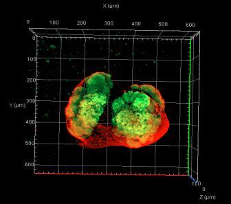


Figure 11.3.13: SAK2 fibrinolytic activity on an in vitro SAC model shown with bright field and CLSM SACs were cultured on transwell inserts in a humidified incubator at 37°C in the presence of HP for a total of 24 hours. After this incubation period, residual plasma was removed, and the samples washed once with PBS. In figure A, B, C, SAC samples are shown before treatment. White arrows highlight the fibrin structure; figure D, E, F, represent samples after a 2-hour 10μM SAK2 treatment.
64
A D E
C F B
Merged
Exopolysaccharide coated material surfaces modulating fracture immune status and enhancing bone healing (EPSIm) (completed) (F Moriarty, D Eglin, R Bagnol)
Background: The gut microbiota and its assortment of surface associated polymers and secreted metabolites have been shown to impact nearly all organs of the human body including bone. Bacterial exopolysaccharides have a wide range of chemical structures close to the polymers present in the human body. They can be useful for tissue engineering applications due to their biocompatibility, ability to be used as carriers for other molecules and materials, as well as specific biological properties. Goal: In this study we incorporate the extracellular polysaccharide (EPS) produced by Bifidobacterium longum subsp longum 35624 in biphasic calcium phosphate scaffolds, as a mean to combine its immunoregulatory action with a clinically relevant calcium phosphate.
Results: The scaffolds incorporating EPS624 have been produced through a freeze-drying process and their physico-chemical and immunological properties characterized. Similar to previously investigated polyelectrolyte coatings, they have shown an immunoregulatory action on human peripheral blood mononuclear cells through IL-10 secretion after 24h. Osteogenic assays on human mesenchymal stem cells revealed the scaffolds and EPS624 showed a nonsignificant trend for increased ALP secretion at day 14, with no positive effect on mineralization assessed by alizarin red staining at day 28. The internal structure of the scaffolds was characterized by computed tomography imaging and showed a highly porous interconnected structure. Fourrier Transformed Infrared Spectroscopy confirmed the presence of both EPS624 and Biphasic Calcium Phosphate absorption bands within the scaffolds, as well as the creation of a new band, indicative of a chemical interaction between the EPS624 and the calcium phosphate particles.
Ex-vivo experiments assessing the effect of EPS624 and scaffolds on rat bone marrow immune cell phenotypes showed they both promoted a switch towards anti-inflammatory and proregenerative phenotypes, especially in the macrophage population whose differentiation was promoted M2-like anti-inflammatory phenotypes. This finding is very positive as such macrophage population have been shown to promote bone healing following a fracture and biomaterial implantation.

Figure 11.3.14: Changes in macrophage phenotypes following 24h exposure to either control RPMI medium (C), EPS624 dissolved in media at concentrations of 1, 10 or 100 ug/mL (E1, E10, E100), and biphasic calcium phosphate/EPS624 scaffolds. Left: Proportion of macrophages after 24h; Middle: proportion of CD86+, M1-like pro-inflammatory macrophages. Right: proportion of ARG1+ antiinflammatory M2-like macrophages.
Pub:
• Bagnol R, Siverino C, Barnier V, O'Mahony L, Grijpma DW, Eglin D, Moriarty TF (2023). Physico-chemical characterization and immunomodulatory activity of polyelectrolyte multilayer coatings incorporating an exopolysaccharide from Bifidobacterium longum. Biomacromolecules, 24(12), 5589-5604, https://doi.org/10.1021/acs.biomac.3c00516
65
Pres:
• Bagnol R, Siverino C, Barnier V, O’Mahony L, Grijpma D, Eglin D, Moriarty TF. Physicochemical characterization of an immunomodulatory bacterial exopolysaccharide coating. Society for Biomaterials, 21/4/23, San Diego, USA (poster)
Partners:
• David Eglin, Ecoles des Mines Saint Etienne, France
• Liam O'Mahony, University College Cork, Ireland
• Tiziano Schweizer, ETH Zurich, Switzerland
• David Marchat, Ecoles des Mines Saint Etienne, France
Establishment of an ex vivo model for Staphylococcus aureus invasion of the osteocyte lacuno-canalicular network (Invibo) (closed) (N Vanvelk, C Siverino, F Moriarty)
Background: Staphylococcus aureus (S. aureus) is one of the primary pathogens responsible for fracture-related infection (FRI). It has developed multiple mechanisms to evade the host immune system and increase its resistance to current treatment strategies. Recently, it has been shown to invade the osteocyte lacuno-canalicular network (OLCN). Goal: This study aimed to develop an ex vivo model to investigate this phenomenon.
Results: Firstly, the tibiae and femora of freshly euthanized mice were infected with a pin containing either wild-type S. aureus or a mutated version that is unable to invade the OLCN (ΔPBP4). While wild-type S. aureus readily invaded the osteocyte lacunae in mouse bones, the ΔPBP4 mutated version was significantly less able to invade (p=0.0005). The presence of bacteria in the OLCN was confirmed by SEM and BB staining was used for quantification of the mean percentage of occupied lacunae. Additionally, TEM imaging of contaminated murine bones confirmed S. aureus invasion of the osteocyte lacunae (Fig 11.3.15 A and B), with only few bacteria in Fig 11.3.15 A or a fully occupied lacuna in Fig 11.3.15 B. The enlargement in Fig 11.3.15 C shows a chain of cocci directed towards a canaliculus. In this study it was proven that S. aureus was able to invade the OLCN in an ex vivo bone model and the antibiotic treatments significantly reduced bacteria in the OLCN. By guaranteeing osteocyte viability for longer periods, this model allows future studies to investigate bacteria-osteocyte interactions.

Figure 11.3.15: Invasion of the osteocyte lacunae by S. aureus in murine bones visualized by transmission electronic microscopy. A. Osteocyte lacuna with two S. aureus cocci, 5000x. B. Osteocyte lacunar space occupied by S. aureus cocci. Note the boxed region displaying deformed cocci entering a canaliculus, 15000x. C. High magnification of boxed area in B demonstrating deformed bacteria, 40000x.
Pres:
• Vanvelk N, de Mesy Bentley KL, Moriarty TF, Siverino C. Establishment of an ex vivo model for Staphylococcus aureus invasion of the osteocyte lacuno-canalicular network, EBJIS 2023, Basel, Switzerland
Partner:
• Karen Bentley, University of Rochester
66
Diagnosis of infected and aseptic non-union: correlating local gene expression and systemic proteomics, miRNA, and immune cells profiles (NUPredict2) (ongoing) (C Siverino, P Fehrenbach, F
Weisemann, TF Moriarty)Background: A critical diagnosis in patients presenting with fracture non-union is the differentiation between infected (INF) and aseptic (AS) non-union. A preoperative diagnosis, without requiring culture of invasive biopsies, would be preferable as intraoperative decisions largely differ between both scenarios. This can be challenging, moreover, in case of low-grade infection lacking clear clinical or radiological signs of infection, or in cases where standard blood markers such as white blood cell count, or C-reactive protein do not show robust and reproducible results. Goal: Some of the aims of this study were to analyze miRNAs expression in preoperative blood samples and immunophenotype peripheral blood mononuclear cells (PBMCs).
Results: MiRNA analyses of the initial cohort of samples (n=10) showed marked differences between HEAL vs AS and HEAL vs INF patients (Fig 11.3.16 A). However, the analyses of all the patients included in the study (n=141) identified only one miRNA as differentially expressed between AS and HEAL when excluding diabetic patients, smokers, and patients with high BMI: hsa-miR-545-5p (Fig 11.3.16 B). The immunophenotyping of PBMCs using high-dimensional mass cytometry revealed significant higher number of monocytes, Treg cells and Th1 in INF samples compared to AS and HEAL patients (Fig 11.3.16 C). Differently Th17 was higher in AS patients compared to INF and HEAL patients (Fig 11.3.16 C).

Figure 11.3.16: (A) Volcano plot of differentially expressed miRNAs in the initial cohort of patients analyzed; (B) Differentially expressed miRNA: has-miR-545-5p in HEAL patients compared to AS when excluding diabetic patients, smoker, and patients with high BMI; (C) Immunoprofiling of PBMCs using high-dimensional mass cytometry.
Pres:
• Siverino C. Towards a preoperative diagnosis of infected non-union with targeted proteomics from human blood plasma, ORS Dallas 2023, USA (poster)
• Siverino C. Towards preoperative diagnosis of infected non- union with targeted proteomics, SVGO-SBMS 2023, Bern, Switzerland (oral)
• Weisemann F. Towards preoperative diagnosis of infected nonunion of femur or tibia with targeted proteomics in blood plasma, EBJIS 2023, Basel, Switzerland (oral)
67
Partners:
• Simon Hackl,Ferdinand Weisemann, Katharina Trenkwalder: BG Unfallklinik Murnau, Germany
• Laura Bürgi: Swiss Institute of Allergy and Asthma Research (SIAF), Davos, Switzerland
Fracture-related infection: testing an antimicrobial and osteoinductive functionalized scaffold in a rabbit model (SugarFRI) (ongoing) (C Siverino, S Zeiter, TF Moriarty)
Background: Fracture-related infection (FRI) remains a clinical challenging complication. Surgical management includes eradication of infection with debridement of affected tissues, dead space management, and systemic antimicrobial therapy. However, the use of local antimicrobials, in addition to systemic therapy offers the prospect of improved therapeutic efficacy. Additionally, debridement often leads to the creation of large bone defects. Therefore, the use of scaffolds incorporating local antimicrobials and bone enhancers like the Bone Morphogenetic Protein 2 (BMP2) would enhance bone healing while treating the infection in a FRI setting.
Goal: This study aims to develop a scaffold with antimicrobial and osteogenic properties. Results: The first validations were performed in vitro and in an aseptic in vivo model (Fig 11.3.17). Histology showed differences in bone bridging between the different conditions. The antimicrobial/osteoinductive scaffold has then been tested in an FRI animal model (Fig 11.3.18). Rabbits were infected with 3x10⁶ CFU for 4 weeks. Bacteriological quantification and evaluation of the influence of infection and of the local application of tobramycin with BMP2 on bone healing is ongoing by histology and CT scans analyses.


68
Figure 11.3.17: Representative histology images of the non-infected rabbits treated with collagen scaffold, unloaded or with BMP2 or BMP2+Tobramycin.
Figure 11.3.18: Study timeline
Short-term Celecoxib Promotes Bone Formation Without Compromising Antibiotic Efficacy in Early Orthopaedic Device-Related Infection: Evidence from a Rat Model (NSAID) (closed) (VS Mdingi, L
Gens, K Mys, S Zeiter, LC Marais, RG Richards, TF Moriarty, M Chittò)This project investigated the effects of non-steroidal anti-inflammatory drugs (NSAIDs) on orthopedic device-related infections (ODRIs) using a rat model to clarify the combined effect of NSAIDs and antibiotics on the bone healing process. The effect of celecoxib (a cyclooxygenase-2-selective NSAID) on bone formation, bone loss, bacterial load, and cytokine levels after antibiotic treatment was measured. Additional comparisons examining the duration of celecoxib exposure and other classes of NSAIDs were also performed. Antibiotic therapy successfully treated the infection in 85.71% (6/7) of cases for the acetylsalicylic acid and ibuprofen group, while the addition of long-term celecoxib treatment reduced efficacy to 45.45% (5/11). Long-term celecoxib treatment significantly reduced both the bone loss by 34% (p<0.0005) on day six and the day 14 periosteal reaction by 0.2760 μm (p<0.0001) compared to the control group. Short-term celecoxib treatment showed similar bone protective effects while avoiding the negative impact on antibiotic efficacy with an 88.9% (8/9) success rate. We could not observe any differences in the tested inflammatory markers from the collected serum over different time points. Our findings highlight the potential benefits of short-term use of celecoxib in preventing bone loss during the early post-infection period without impairing the efficacy of antibiotic therapy. This study suggests that celecoxib may be a valuable addition to the multimodal approach to pain management in orthopedic device-related infections.

Figure 11.3.19: Bone responses in the control (CTL), long-term celecoxib (Cel(L)), and short-term celecoxib (Cel(S) treatment groups of an S. epidermidis infection. Differences in day 6 bone formation. Data shown are the mean ± SEM. A two-way ANOVA with Dunnett’s post-hoc test was performed to determine significant differences between treatment groups and the control group. * p<0.05, ** p<0.01, *** p<0.001, ****
69
miRNA analysis to discover fracture related biomarkers (MiDiag2) (Finished)
(M Stoddart, M Alini, H Schmal, E Della Bella, S Berger, U Menzel)
Background: Biomarkers predictive of fracture healing outcomes would provide a useful tool to allow surgeons to proactively make patient based clinical decisions. Currently, even in highrisk groups, there are no accurate ways to determine the potential of a particular patient to progress to delayed or non-union. Such a tool would enable more reliable patient stratification, thus allowing for earlier diagnosis and increasing the potential success of additional early interventions by the surgeon. In a previous AO Trauma project (MiDiag) we investigated changes in small non-coding RNA in fracture patients and during osteogenic differentiation.
Goal: A panel of prospective microRNA (miRNA) markers were identified, and this now requires further validation. In addition, we aim to use the methods developed, and the database of patient serum non-coding RNA created, to further investigate markers that are mechanically regulated and would be associated with secondary bone healing.
Results: The function of three miRNA was inhibited during chondrogenesis induction in pellet culture, revealing a possible role for two of them in the regulation of chondrogenesis and endochondral ossification. RNA sequencing was performed to determine which genes and pathways were affected by miRNA inhibition. Serum samples from 34 fracture patients (Klinik Diakonissen, Schladming, Austria) were prospectively collected and analyzed for the expression of a selected panel of miRNA, which will be correlated to fracture outcome.

Figure 11.3.20: Schematics of MiDiag2 in vitro work to determine miRNA function in endochondral ossification.
Pres:
• Breulmann FL, Ramasamy S, Herzog M, Pandian GN, Della Bella E, Stoddart MJ. Differentially expressed microRNAs during early endochondral differentiation of human mesenchymal stromal cells as biomarkers for non-union fractures. Orthopaedic Research Society Annual Meeting ORS; 2023 (poster)
Pub:
• Breulmann FL, Hatt LP, Schmitz B, Wehrle E, Richards RG, Della Bella E, Stoddart MJ. Prognostic and therapeutic potential of microRNAs for fracture healing processes and nonunion fractures: A systematic review. Clin Transl Med. 2023;13(1):e1161.
• Isenmann M, Stoddart MJ, Schmelzeisen H, Gross C, Della Bella E, Rothweiler RM. Basic principles of RNA interference: Nucleic acid types and in vitro intracellular delivery methods. Micromachines. 2023;14(7).
• Miclau K, Hambright WS, Huard J, Stoddart MJ, Bahney CS. Cellular expansion of MSCs: Shifting the regenerative potential. Aging Cell. 2023;22(1):e13759.
70
Partners:
• Kubosh EJ (Dr), Department of Orthopedics and Traumatology, University Medical Center Freiburg, Germany
• Breulmann F (Dr), Department of Orthopedic Sports Medicine, Klinikum Rechts der Isar, Technical University of Munich, Germany.
• Zaffagnini S (Prof), Istituto Ortopedico Rizzoli, Bologna, Italy
• Mattiassich G (Priv Doz Dr), Klinik Diakonissen, Schladming, Austria
• Bischofreiter M (Dr), Klinik Diakonissen, Schladming, Austria
Linking mechanics and omics to improve early bone healing (MechOmics) (ongoing)
(E Wehrle, M Stoddart, S Zeiter, S Verrier, M Schröder, N Giger, J Barcik, D Arens, D Gehweiler)
Background: Mechanical loading is a key factor for normal progression of the fracture healing process. Despitethe advances in fracture fixation, there remains a subset of patients that suffer from healing complications, resulting in delayed healing and non-unions. Currently it is not possible to reliably identify healing complications at an early stage when treatments, e.g. mechanical intervention therapies may be more effective. Understanding of the underlying mechanically induced molecular mechanisms on an individual basis could enable wider-scale harnessing of the mechano-sensitivity of the regenerative process in clinical applications. Novel multimodal approaches in small animals have the potential to precisely capture and understand these mechanical-induced biological changes during fracture healing on an individual basis. Within this project we will use and adapt well-established equipment for precisely controlled local application of cyclic mechanical loading in mouse femur defect models.
Goal: To identify systemic biomarkers indicating early deviations from normal healing progression also allowing for initiation and targeted adjustments of individualized mechanical intervention therapies.
Results: Within the project a displacement-controlled loading mode was implemented for femur defect loading models in mice. This allows to longitudinally monitor healing progression via in vivo stiffness measurements. First spatial transcriptomics analyses showed distinct local gene expression patterns for union and non-union defects (Fig 11.3.21).

Figure 11.3.21: Spatial transcriptomics of a non-union (bottom) and union (top) femur fracture in mice. Destained H&E section prior to transcriptomic probe transfer (A), Gene clustering (B).
Pres:
• Barcik J, Schröder M, Arens D, Gens L, Mischler D, Gehweiler D, Zeiter S, Varga P, Stoddart M, Wehrle E. Longitudinal monitoring of healing progression via in vivo stiffness measurements in a mouse femur defect model. 2023 eCM (oral)
• Wehrle E. Individualized omics-based preclinical models for bone healing. 2023 eCM (oral)
• Wehrle E. Spatial transcriptomics of fracture healing. 2023 EORS (oral)
• Wehrle E. Omics-based preclinical models of musculoskeletal regeneration. 2023 EORS (oral)
71
11.4 AO VET
Improved biomechanical properties of Locking Compression Plate designed for large animals using larger diameter locking-head screws: a study on a 20° oblique equine long-bone fracture model (CoreUP) (ongoing) (C Constant, E Marchionatti, S Zeiter)
Background: The development of the Locking Compression Plate (LCP) was a major advancement in internal fixation and its clinical value in equine fracture repair has been proven by its use in nearly all anatomical locations in the horse. Despite improved orthopedic equipment in recent years, large animal long-bone fracture repair remains challenging. Open reduction and internal fixation are associated with poor survival rates because of mechanical failure, usually due to fatigue or a single catastrophic event. Generally, if greater bending loads are applied during a given cycle of loading, fewer cycles are needed to break the implant as a result of fatigue. Screw loosening and breakage is another frequent complication in the clinical application of LCP in horses and occur in almost 20% of LCP stabilization. Screw breakage is influenced by the bending stiffness of the screw which can be evaluated by calculation of the area moment of inertia for the screw core diameter (area moment of inertia (I) = π r4/4). Even a small increase in the core diameter of a screw will greatly increase bending strength (Fig. 11.4.1).
The 5.5mm LCP designed for large animal fracture fixation is a much stronger implant than the 4.5mm LCP. Nevertheless, it is actually less extensively used in equine applications, possibly due to the supposed suboptimal design for horses with the currently available implants. It is believed that the broad 5.5 LCP would probably become a more suitable and even better plate for equine long bones if they were used with larger diameter locking-head screws. Considering these challenges, internal fixation implants more suitable for equine bone, along with improved stability and increased fatigue resistance, are needed to improve the successful outcome of long-bone fracture fixation in horses.
Goal: To compare 6.0-mm locking-head screws (LHS) with a 5.4-mm core diameter resulting in an estimated 26% increase in bending stiffness compared to regular 5.0-mm LHS (Fig. 11.4.1) and modified Locking Compression Plate (LCP) designed to accommodate modified LHS, against standard LCP and 5.0-mm LHS screws in an equine long-bone fracture model. Results: This study is still undergoing testing. After its completion, it will suggest if lockinghead screws with a larger core diameter are more suitable for fracture fixation in equine long bones when using 5.5-mm LCP and if this new construct could improve fixation of fracture in the equine long bone, thus improving successful outcomes of large animal fracture repair and upgrading orthopedic techniques.

Partner:
• Emma Marchionatti, University of Bern, Switzerland
72
Figure 11.4.1: Impact of screw core diameter on bending stiffness evaluated by area moment of inertia.
Biomechanical properties of a new Locking Compression Plate design for large animals to accommodate modified 5.5-mm cortex screws with larger head size: a study on an equine long-bone fracture model (PlateDown) (ongoing) (C Constant, E Marchionatti, S Zeiter)
Background: The main goal of internal fixation is to maintain fracture stability to encourage bone union while maintaining a functional limb during healing. Therefore, successful fracture fixation results when the various loading forces acting on the fracture fragments are resisted by the mechanical stiffness of the bone-implant construct. Given the extreme forces equine patients can apply to fracture fixation devices, surgeons routinely work at the mechanical limits of implants during the repair of adult long-bone fractures. Larger and stronger implants for internal fixation can reduce the incidence of catastrophic implant failure. A stronger plate, the 5.5-mm broad LCP, was specially designed with an increased plate thickness for large animal fracture repair along with the 5.0-mm locking head screw and 5.5-mm cortical screw. Nevertheless, screw loosening and breakage remain a frequent complication in the clinical application of LCP in horses and occur in almost 20% of LCP stabilization and open reduction and internal fixation in equine surgery is still associated with poor survival rates and a high risk of catastrophic failure of the repair or cyclic fatigue failure of the implants. The dimension requirements for human surgical implants, including metal bone screws, are listed under the standards of the International Organization for Standardization (ISO). Current cortical metal bone screws under the ISO9268 requirement6 have a nominal head diameterto-core width ratio between 1.75 to 2. Created for the veterinary market, the 5.5-mm large animal cortex screw (DePuy Synthes) has the same head diameter (8mm) as the 4.5-mm cortex screw. Therefore, the nominal head diameter-to-core width ratio, which is 1.45, is lower than the one of other commercially available veterinary cortex screws. Hence, the relatively small head diameter of the 5.5-mm large animal cortex screw could reduce the full potential of this implant by undermining its ability to produce interfragmentary compression and prevention of movement between plate and bone. Recently, a modified 5.5-mm cortex screw with a 10mm head diameter (head diameter-to-core width ratio of 1.8) was developed and evaluated in vitro by Constant C. et al (Fig. 11.4.2). The design of the modified screws conferred an expanded under-head surface resulting from their 2 mm head diameter increase. The results of this investigation performed at ARI showed that condylar MC3 osteotomies repaired with modified 5.5-mm cortical screws sustained greater maximal hand torque insertion, smaller insertion failure angle and 1.4 fold greater quasi-static failure forces than constructs repaired with standard 5.5 mm screws. The use of larger heads increases the area of pressure under the screw head, thereby allowing for more compression to be generated and decreasing stress risers around the implant head While the features of the modified 5.5-mm cortical screw with a larger area between the screw head and plate could also be beneficial to increase internal fixation stability when a plate is used, the current combi hole of LCP could not accommodate the larger head size of the modified screws and must be modified to successfully implement this new implant.
Goal: compare modified Locking Compression Plate (LCP) designed to accommodate modified 5.5-mm cortex screws with a 10-mm head diameter, against standard LCP and cortex screws for hybrid plating in an equine long-bone fracture model.
Results: This study is still undergoing testing. After its completion, it will suggest if the use of modified LCP to accommodate cortical screws with larger head diameters will improve biomechanical stress distribution around the screw heads and allow better plate-to-bone compression, and therefore have the potential to reduce the incidence of implant failure. Since these new implants should allow to have better distributed stress distribution around them, they should require more cycles before breakage as a result of fatigue. In addition, the ability of a screw to generate a higher insertion torque could increase its ability to obtain a stable construct from decreased micromovements between the LCP and bone in the region of the plate occupied by cortical screws. The increased stability of an LCP construct for hybrid plating would be of significant clinical advantage for long bone fracture repair proximal to the third metacarpus/metatarsus which currently have a poorer prognosis, partly because of the decreased ability to supplement internal fixation with external coaptation.
73

Figure 11.4.2: Detailed drawing illustrating the technical characteristics of standard 5.5-mm cortical screw with an 8-mm head diameter and modified 5.5-mm cortical screws with a 10-mm head diameter. The modified screw head was designed with the same hexagonal screwdriver insert head recess (S; 3.5mm hexagonal), screwdriver insert recess depth (t), and head height (k) as the standard screw. The modified screw shaft was designed with the same nominal diameter (D1; 5.5-mm), core diameter (D2; 3.9-mm) and pitch (P; 2.0 mm) as the standard screw.
From Constant et al. Veterinary Surgery, 2022
Partner:
• Emma Marchionatti, University of Bern, Switzerland
In vivo biomechanical evaluation of a larger diameter angle‐stable interlocking nail in a calf tibial fracture model (Timbits) (ongoing) (C Constant, E Marchionatti, S Zeiter, A Desrochers)
Background: The tibia is a commonly fractured long bone in cattle with a prevalence that can reach 40% of long bone fractures in cattle, regardless of age. While conservative treatment has been reported, severe and potentially life-threatening complications have been frequently described even in newborn calves less than 80kg. Based on those reports, conservative treatments of tibial fracture appear to result in bone healing failure or poor clinical outcomes. Ideally, diaphyseal tibial fractures are internally reduced and repaired using double plating. However, the hexagonal geometry of the bone makes internal reduction and fixation of tibial fracture difficult, and the thin cortices of neonatal calves are not suitable for holding bone screws. In addition, tibial fractures are often a result of high-energy trauma, resulting in comminuted fractures, making its fixation even more challenging (Fig 11.4.3) and explaining, when taken together with the other limiting factors, why high complication rates and overall poor clinical outcomes are frequently reported with internal fixation. Thus, conservative treatment of tibial fractures in young calves using a modified Thomas splint is currently regarded as the option that provides better healing but is associated with severe complications such as open fracture, malunion, delayed union, contralateral limb angular deformity, tendon breakdown, and persistent lameness after fracture healing.
Intramedullary interlocking nail (IIN) fixation has been used successfully in veterinary orthopedics and has resulted in successful outcomes in a large number of cases with similar fractures in small animals. Neonatal calves with thin cortices could benefit from tibial fracture fixation using intramedullary interlocking nail inserted retrograde or normograde through the tibial plateau. However, the IIN veterinary systems are designed for small animals, and the nail’s largest diameter available is significantly smaller than the current AO reference; the recommended use of the largest INN diameter is defined by the narrowest diameter of the inside of the medullary cavity. While IIN may provide an effective and safe technique for the
74
management of tibial fracture in calves, the largest IIN veterinary system available has an 8mm diameter and may not be sufficiently biomechanically stable for fracture healing in calves, especially in unstable fracture configurations.
Goal: to examine the biomechanical properties of intact tibiae from neonatal calves and to compare them to osteotomized bone repaired with a veterinary IIN for small animals and a larger human IIN system more suitable to the diameter of calves’ tibiae.
Results: This study is still undergoing testing. After its completion, it will determine if the largest IIN veterinary systems available or a commercially available IIN human system could be used to successfully stabilize tibial fractures in calves and potentially improve the overall poor clinical outcomes by becoming a surgical alternative and therefore minimizing the risk of unsatisfactory outcomes associated with conservative treatment. The identification of an IIN system already commercially available from the veterinary or human market would facilitate its implementation in large animal surgery and would limit the research and development investment that would be required to market a new nail only for the veterinary market.

Partners:
• Emma Marchionatti, University of Bern, Switzerland
• André Desrochers, Université de Montréal, Canada
75
Figure 11.4.3: Preoperative radiograph of a characteristic complete diaphyseal fracture of the tibia in a 50kg neonatal calf.
11.5 AOTC System
Feasibility of using theAO Fracture Monitor for spinal fusion assessment (SmartFusion) (ongoing) (M Heumann, J Buschbaum, M Ernst)
Background: CT-based monitoring of spinal fusion cases has multiple limitations. Besides the radiation exposure to the patient, the interpretation of CT images is highly subjective. Furthermore, the CTs provide only a coarse visual overview (snapshots) of the fusion process as no continuous data is available. The AO Fracture Monitor is an implantable sensor system allowing for continuous and wireless implant load monitoring of plate osteosynthesis, that may also provide an objective means to monitor the progress of spinal fusion and rapidly react to such complications as implant loosening.
Goal: To investigate the feasibility of applying the AO Fracture Monitor measurement principle for spinal fusion assessment.
Results: Following promising results from previous research, it was decided to split the project in a research and a development pathway. The focus of the development pathway was on adapting the Fracture Monitor implant to meet specific requirements for spinal applications, including optimization of the signal acquisition and usability aspects. The first step comprised redesigning of the sensors’ attachment to the spinal instrumentation. After various design iterations, a prototype was manufactured and biomechanically tested. The new design demonstrated improved attachment mechanics and a considerably better signal strength of the sensor.
Concurrently, a comprehensive investigation was initiated as part of the research pathway to explore the applicability of the monitoring system to a wider range of treatments related to spinal injuries or diseases. In a biomechanical study, the possibility to monitor healing of a complete osseus disruption of a single vertebra – treated without the necessity of intersegmental fusion – was investigated. The study's findings suggest that the healing of three-column fractures can be assessed through implant load monitoring, as there was a significant difference in implant load between the fractured and healed state. This highlights the potential for applying this method not only for spinal fusion cases but also for fracture healing assessment without fusion. Further pilot tests on additional spinal applications, such as multilevel fusion, provided promising preliminary results. However, a more extensive study needs to be carried out to obtain conclusive evidence.

Figure 11.5.1: Synbone model of the construct tested with the Fracture Monitor and strain gauges attached to a posterior instrumentation after an osseus flexion-distraction fracture with additional trauma to the anterior column of the spine (indicated by red line).
76
Pres:
• Heumann M, Benneker LM, Constant C, Ernst M, Richards RG, Wilke H-J, Gueorguiev B, Windolf M. Decreasing spinal implant load indicates progression of posterolateral fusion when measured continuously – in vivo proof of concept in sheep. 4th Spine Loading and Deformation Workshop Berlin, 2023, Berlin, Germany (oral).
• Heumann M, Benneker LM, Constant C, Ernst M, Richards RG, Wilke H-J, Gueorguiev B, Windolf M. Smart Fusion Spine: A novel technique to in-vivo measure spinal implant loads for the assessment of posterolateral fusion – proof of concept in an in-vivo sheep model.
83rd Annual Meeting of Swiss Orthopaedics, 2023, St. Gallen, Switzerland (oral).
• Heumann M, Benneker LM, Constant C, Ernst M, Richards RG, Wilke H-J, Gueorguiev B, Windolf M. Smart Fusion Spine: A novel technique to in-vivo measure spinal implant loads for the assessment of posterolateral fusion – proof of concept in an in-vivo sheep model. 23rd Annual Meeting of Swiss Spine Society, 2023, Visp, Switzerland (oral).
Pub:
• Heumann M, Benneker LM, Constant C, Ernst M, Richards RG, Wilke H-J, Gueorguiev B, Windolf M. Decreasing implant load indicates spinal fusion when measured continuously. Journal of Biomechanics, 2024. 163: p. 111929.
Partners:
• AOTC Spine
• Benneker Lorin (Prof), Sonnenhof Spital Bern, Bern, Switzerland
• Wilke Hans-Joachim (Prof), University Ulm, Ulm, Germany
Validated finite element simulations can predict pedicle screw loosening (ongoing) (D Mischler, P Varga)
Background: Pedicle screw loosening poses a significant challenge in spinal surgery, impacting patient recovery and quality of life. This complication can lead to acascade of issues, including reduced implant stability, persistent pain, and the need for additional surgeries. In osteoporotic patients the risk of screw loosening can be as high as 60%. Addressing the problem of pedicle screw loosening through more flexible materials, like carbon fiber reinforced polytheretherketone (CF/PEEK) optimized using validated finite element (FE) models, aims to enhance the success rates of spinal surgeries, reduce complication rates, and ultimately alleviate the overall burden on patients and healthcare providers.
Goal: To validate an FE simulation approach predicting screw toggling that allows for fast screw design iterations.
Results: Ten vertebral bodies (L1–L5) from five elderly donors were instrumented with CF/PEEK pedicle screws and loaded using increasing ramps. FE models were created based on the intact and instrumented CT scans and the experimental displacements were applied. Peak forces of each ramp were compared with the experimental loads. The predictive capability of the FE models was further compared to a more conventional approach of measuring the bone volume fraction (BV/TV) around the screws. The results demonstrated that the computational models were capable of accurately predicting the peak forces observed during experimental cyclic loading (R2=0.84), whereas BV/TV did not offer reliable predictions (R2<0.05). This indicates the necessity of using specimen-specific, sophisticated FE models over simplistic measures like BV/TV around the screws to predict loosening effectively. These validated models can be used for screw design and stiffness iterations aiming to reduce the risk of screw loosening in low-density vertebrae that are expected to lead to lower complication rates in patients.
77

Figure 11.5.2: Experimental test setup of instrumented lumbar vertebral body (left). Boundary conditions were replicated in the FE models (bottom right) based on the post-OP CT scans.
Partners:
• Zysset P (Prof), University of Bern, Bern, Switzerland
• Kessler F, University of Bern, Bern, Switzerland
• Hüsken L, icotec ag, Altstätten SG, Switzerland
• Tenisch L, icotec ag, Altstätten SG, Switzerland
• Spruit M (MD), Benneker LM (MD), Bransford R (MD), Kumar N (MD), Mazel C (MD), Ryang YM (MD), Watanabe K (MD) and Wolinsky JP (MD), AOTC Osteoporotic Spine Instrumentation Task Force
PHAGESHEEP: Analysis of kinetics, neutralization, and efficacy of different administration routes of bacteriophage treatment in a sheep FRI model (C Peez, L Henssler, B Chen, M Chittò, C Constant, D Arens, S Zeiter, TF Moriarty)
Background: Fracture-related infections (FRI) are among the most devastating complications of surgical fracture treatment. Treatment strategies involve both surgical debridement and local/systemic anti-infective therapy. However, due to rising antibiotic resistance, antibiotic treatment options are increasingly often limited. Recent studies suggest using bacteriophages (phages) as a promising alternative, especially against highly resistant pathogens. Unlike antibiotics with well-established protocols, optimal phage therapy lacks evidence-based guidelines. Understanding pathogen sensitivity, pharmacokinetics, and dynamics is crucial for effective treatment, highlighting the need for further research in phage therapy optimization. Goal: This study aims to evaluate phage distribution, neutralization kinetics, and the optimal administration route (intravenous versus local) for phage therapy in treating fracture-related infections (FRI).
Results: Throughout the treatment duration, there was a progressive increase in the serummediated deactivation of bacteriophages. By the end of the initial treatment cycle, 60% of bacteriophages were neutralized by the sheep serum. After the 4-week latency phase, 50% of bacteriophages were neutralized in the group that had received i.v. phages, contrasting with approximately 25% neutralization in the local treatment group (Fig 11.5.3). Nevertheless, by the end of the second treatment cycle, both cohorts demonstrated a remarkable phage neutralization rate >99%.
78
Figure 11.5.3: Neutralization of phages by the sheep serum over the trial period of phase I.
Partners:
• Anti-infective Global Expert Committee, AO Innovation Translation Center
• Willem-Jan Metsemakers, KU Leuven, Belgium
11.6 ARI AC (AOF Direct Funds)
Bone healing status assessment with implant load sensors – validation by CT imagebased computer simulations (HealFE) (ongoing) (P Schwarzenberg, C Hetreau, D Mischler, M Ernst, P Varga)
Background: An accurate measurement of the healing progress in fractured bones is important for timely diagnosis of complications and recommendations on weight bearing protocols. Fracture status is characterized by the mechanical competence of the healing callus. Clinically available methods use surrogate measures of callus stability, mainly via visual Xrays-based evaluations that are subjective and cannot provide reliable and quantitative assessments of healing status. Two quantitative approaches have been recently developed for mechanical characterization of the healing status of fractured bones and both technologies are available at ARI: 1) AO Fracture Monitor, providing continuous data on implant loading and 2) subject-specific CT-based finite element (FE) analysis at given time points for virtual mechano-structural analysis of the healing callus.
Goal: To evaluate whether the reduction of implant load measured via the AO Fracture Monitor correlates with an in-silico sensor equivalent or the torsional rigidity of the healing callus, both predicted by subject-specific CT-based FE modeling.
Results: Sensor data from previous preclinical studies of an ovine tibial osteotomy healing model have been evaluated and aligned with in vivo CT scans taken through the healing process. From these CT scans, subject-specific FE models have been constructed to simulate the in vivo situation at specific time points. Due to the specimen-specific nature of both sensor data and CT data, the data at each specific time point can be readily correlated. Results demonstrate that the FE models are able to predict the healing trends seen in the AO Fracture Monitor data and can even identify the same delayed and nonunion cases from the preclinical cohort.
79
0 2 4 6 8 10 12 14 16 18 20 22 24 26 28 30 32 34 36 38 40 42 44 46 48 50 0 25 50 75 100 Days N e u t r a l i z e d p h a g e p a r t i c l e s [ % ] ISPLocal ISPi.v 1.PhageTherapyCycle 2.PhageTherapyCycle LatencyPhase
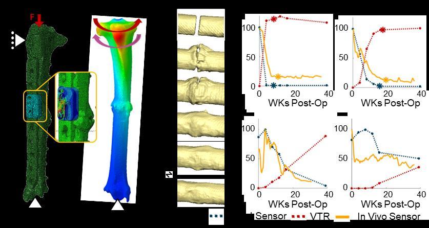
Figure 11.6.1: a) FE model of a representative in silico sensor model with specimen-specific hardware under loading. A zoomed view shows the strain contours of the AO Fracture monitor under the same loading. b) FE model of a representative torsional rigidity model with displacement contours. c) Longitudinal CT scans of the fracture site at different stages of healing. d) Healing completeness curves for each measuring modality with an asterisk highlighting the defined point of healing.
Theses:
• Hetreau C. Bone fracture healing assessment with implantable sensors: correlation between CT image-based computer simulations and in vivo sensors. Master Thesis, ETH Zurich, 2023.
Intercalary fragments at the posterior malleolus change ankle joint pressure distribution – a biomechanical study (L Llano, I Zderic, B Gueorguiev)
Background: With the increased use of CTs in cases with trimalleolar ankle fractures, bone fragments between the posterior malleolus and the rest of the articular tibial plafond surface –described as intercalary fragments (ICFs) – can be recognized. They are detected in 40–55% of the cases with trimalleolar fractures, mostly in Type II according to the Bartonicek Classification.
Goal: To determine the ICF size threshold for a significant change in the pressure distribution at the ankle joint, having a considerable impact on its remaining cartilage.
Results: Eight human cadaveric lower legs were used, preserving the lateral and medial soft tissue structures of the ankle, and the syndesmotic structures. A posterior malleolus Bartonicek II fracture was created with sequential 2 mm, 4 mm, 6 mm, and 8 mm ICFs. The posterior malleolus was fixed with a 3.5 mm third-tube buttress plate and each specimen was mounted in a custom-made frame for axial loading under 700 N in neutral position, 15° dorsiflexion and 20° plantar flexion. Using electronic foil sensors, pressure measurements were performed to define the contact area, centre of force, and peak pressure at the ankle joint in loaded condition. Posterolateral defects of the tibial plafond alter ankle joint pressure characteristics. Regardless of the direction of ankle joint flexion, increasing the ICF defect size results in decreased contact area, increased mediolateral center of force migration and higher peak joint forces. Malreduction or removal of ICFs larger than 2 mm should be avoided to preserve physiological ankle pressure characteristics.
80

Figure 11.6.2: Top left: Setup with a specimen equipped with an electronic foil sensor and mounted for testing in neutral position. Top middle and right: Opening of the malleolus fracture and creation of a 2 mm ICF. Bottom: Mean value and SD of the contact area change per condition.
Partners:
• Jorge Barla (MD), Hospital Italiano de Buenos Aires, Caba, Argentina
• Christian Peez (MD), Department of Trauma, Hand and Reconstructive Surgery, University Hospital Münster, Münster, Germany
81
New dynamic suture material in tendon transfer surgeries – a biomechanical comparative analysis (T Pastor, I
Zderic, B Gueorguiev)Background: Commonly used high-strength sutures for tendon transfer surgeries are designed to withstand high tensile forces and secure the repaired structures in place. However, knot slippage is inevitable when they are heavily loaded, leading to gap formation between the repaired structures. On the other hand, early mobilization after tendon transfer surgery is crucial to avoid commonly observed postoperative soft tissue adhesions. Recently, a new suture was introduced (Dynacord; DC) with a salt-infused silicone core, designed to minimize laxity and preserve consistent tissue approximation.
Goal: To compare the biomechanical competence of DC against a conventional high-strength suture (FiberWire; FW) in a human cadaveric tendon transfer model with an early rehabilitation protocol.
Results: Sixteen tendon transfers (flexor digitorum superficialis (FDS) IV to flexor pollicis longus (FPL)) were performed in 8 pairs human cadaveric forearms using either DC or FW. Markings were made 0.8 cm proximal and 0.7 cm distal to the level of the interweaving zone of the transfer. All specimens underwent repetitive thumb flexion against resistance in 9 intermittent series of 300 cycles each, simulating an aggressive postoperative rehabilitation protocol. After each series, the distance of the proximal marker to the interweaving zone (proximal zone), the length of the interweaving zone (intermediate zone) and the distance of the distal marker to the interweaving zone (distal zone) were measured. From a biomechanical perspective, DC preserved or even increased tissue approximation, and might be considered as valid alternative to a conventional high-strength suture in tendon transfer surgery. DC might allow for a shorter interweaving zone and a more aggressive early postoperative rehabilitation program, possibly avoiding commonly observed postoperative soft tissue adhesions and stiffness.

Figure 11.6.3: Top: Setup with a right specimen treated with DC and mounted for biomechanical testing. Finger II-IV are taped in extension to allow free passage of the flexing thumb. Bottom: Length changes for the intermediate zone after each cyclic test over 9 days, presented for each treatment technique separately in terms of mean value and SD.
82
Pres:
• Pastor T, Zderic I, Dhillon M, Gueorguiev B, Pastor T, Vögelin E, New suture materials in tendon transfer operations. A biomechanical comparative analysis, SGH 2023, Lugano, Switzerland (oral).
Partners:
• Esther Vögelin (Prof), Department for Plastic and Hand Surgery, Inselspital University Hospital Bern, University of Bern, Bern, Switzerland
• Torsten Pastor (MD), Department of Orthopaedic and Trauma Surgery, Lucerne Cantonal Hospital, Lucerne, Switzerland
Nail versus plate in tibiocalcaneal arthrodesis – a biomechanical study (M Dhillon, I Zderic, B Gueorguiev)
Background: Tibiocalcaneal arthrodesis with a retrograde intramedullary nail is an established procedure considered as a salvage in case of severe arthritis and deformity of the ankle and subtalar joints. The current state of the art treatment has demonstrated good primary stability and tolerable soft tissue dissection. Nevertheless, diverse plate systems are still used, especially because they are easier to handle. However, most of them are related to off-label applications with greatly varying results. On the other hand, recently, a significant development in hindfoot arthrodesis with plates has been indicated.
Goal: To compare a plate, specifically developed for arthrodesis of the hindfoot with an already established nail system.
Results: Sixteen paired human cadaveric lower legs with removed forefoot and cut at midtibia were assigned to two groups for tibiocalcaneal arthrodesis using either a hindfoot arthrodesis nail or an arthrodesis plate. The specimens were tested under progressively increasing cyclic loading in dorsiflexion and plantar flexion to failure, with monitoring via motion tracking. Initial stiffness was calculated together with range of motion in dorsiflexion and plantar flexion after 200, 400, 600, 800, and 1000 cycles. Cycles to failure were evaluated based on 5° dorsiflexion failure criterion. From biomechanical point of view, both tested techniques for tibiocalcaneal arthrodesis appear to be applicable.


Partners:

Figure 11.6.4: X-rays of tibiocalcaneal arthrodesis with a hindfoot arthrodesis nail (top left) or an arthrodesis plate (bottom left) together with a visualization of the setup with a specimen equipped with markers for motion tracking and mounted for biomechanical testing (right).
• Mark Lenz (Prof), Jena University Hospital, Jena, Germany
• Kajetan Klos (MD), Gelenkzentrum Rhein-Main, Hochheim, Germany
83
Percutaneous fixation of complex ankle fractures versus classic plating – a biomechanical study (K Ganchev, I Zderic, B Gueorguiev)
Background: Ankle fractures are prevalent, comprising 4% to 9% of all fractures, with up to 50% of the cases involving the posterior malleolus (PM). The gold standard treatment for these fractures considers posterior approaches and plating of both PM and fibula.However, in elderly and comorbid patients this remains controversial due to the need for invasive approaches and soft tissue care.
Goal: To test biomechanically a percutaneous construct, utilizing a fibula nail plus percutaneous anteroposterior screws, versus the traditional double plating.
Results: Sixteen human cadaveric lower legs were used. Supination and external rotation fractures involving PM and distal fibula were created through osteotomies while preserving the integrity of the syndesmotic ligaments. Eight specimens were treated with a fibula nail plus two anteroposterior screws, while the remaining specimens underwent double plating. Biomechanical testing was performed in simulated mid-stance position under complex cyclic loading at 2Hz, consisting of axial compression and external rotation of the foot. Interfragmentary movements were captured by motion tracking. The percutaneous fixation of complex ankle fractures using a fibula nail plus anteroposterior screws demonstrated noninferiority versus the traditional double plating, indicating that such treatment may be a viable option, particularly in cases where delicate soft tissue management is required.

Figure 11.6.5: Left: Anteroposterior radiographs of specimens treated with either two plates or a fibular nail plus two anteroposterior screws. Right: Fracture displacement over the course of 8000 cycles, measured between the fibula and its distal fragment, presented for separate treatment technique in terms of mean value and SD.
Partners:
• Dimitar Raykov (Prof), Medical University of Varna, Varna, Bulgaria
• Preslav Penev (MD), Medical University of Varna, Varna, Bulgaria
• Lionel Llano (MD), Hospital Italiano de Buenos Aires, Caba, Argentina
New generation of superior single plating versus low-profile dual minifragment plating in diaphyseal clavicle fractures: a biomechanical comparative study (T Pastor, I Zderic, B Gueorguiev)
Background: Recently, a new generation of superior clavicle plates was developed featuring the variable-angle locking technology for enhanced screw positioning and a less prominent and optimized plate-to-bone fit design. On the other hand, minifragment plates used in dual plating mode have demonstrated promising clinical results.
Goal: To compare the biomechanical competence of single superior plating using a newgeneration plate versus dual plating using low-profile minifragment plates.
Results: Sixteen paired human cadaveric clavicles were pairwise assigned to 2 groups for instrumentation with either a superior 2.7 mm variable-angle locking compression plate (group 1), or with one 2.5 mm anterior combined with one 2.0 mm superior matrix mandible plate (group 2). An unstable clavicle shaft fracture AO/OTA 15.2C was simulated by means of a 5 mm osteotomy gap. Specimens were cyclically tested to failure under craniocaudal cantilever
84
bending, superimposed with bidirectional torsion around the shaft axis, and monitored via motion tracking. From a biomechanical perspective, low-profile 2.5/2.0 mm dual plates could be considered as a useful alternative for diaphyseal clavicle fracture fixation, especially in less common unstable fracture configurations.

Figure 11.6.6: Left: Photographs of 2 left clavicles instrumented with (A, B) a 2.7 mm VA-LCP Clavicle Shaft Plate in superior position or with (C, D) a 2.5/2.0 mm low-profile dual-plate construct, visualized in superior (A, C), posterior (B) and anterior (D) views. Right: Fracture gap movement amplitudes over the course of 12,500 cycles, presented for each group separately in terms of mean value and SD.
Pub:
• Pastor T, Zderic I, Berk T, Souleiman F, Vögelin E, Beeres F, Gueorguiev B, Pastor T. New generation of superior single plating vs. low-profile dual minifragment plating in diaphyseal clavicle fractures: a biomechanical study, J Shoulder Elbow Surg (2024) 33, 409-416.
Partners:
• Esther Vögelin (Prof), Department for Plastic and Hand Surgery, Inselspital University Hospital Bern, University of Bern, Bern, Switzerland
• Torsten Pastor (MD), Department of Orthopaedic and Trauma Surgery, Lucerne Cantonal Hospital, Lucerne, Switzerland
Osteosynthesis of intraarticular distal ulna fractures – a biomechanical investigation of three plating techniques (R Mechkarska, I Zderic, B Gueorguiev)
Background: Although plate fixation is the most common surgical method for treatment of isolated intraarticular distal ulna fractures (IADUF), there is still no existing consensus regarding the most eligible plating technique.
Goal: To investigate the biomechanical performance of three plating techniques used for fixation of IADUF.
Results: Eighteen human cadaveric ulnae were used to create three-part IADUF type AO/OTA Q5/2U3C by means of osteotomies, featured by two distal fragments separated from the ulnar shaft, namely a radius-facing intraarticular and a distal shaft fragment. The specimens were assigned to three groups for plating with either (1) a 6-hole 2.0 mm rotation correction locking micro plate, (2) a 5-hole 2.7 mm one-third tubular plate, or (3) a 2.4/2.7 mm variable-angle locking T-fusion plate. Each construct underwent progressively increasing complex cyclic loading to failure in 45° angulation. The load was transmitted from the ulnar shaft to the radiusfacing intraarticular fragment, stressing the constructs in axial-dorsal bending and supination. Interfragmentary movements were captured by motion tracking. From a biomechanical perspective, both T-fusion plating and locked micro plating techniques represent valid alternatives for treatment of isolated intraarticular distal ulna fractures. In contrast, one-third tubular plating is associated with inferior performance.
85
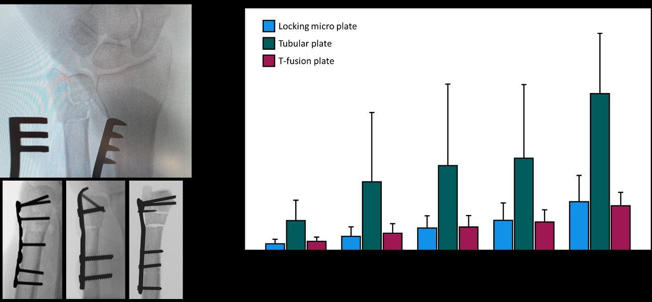
Figure 11.6.7: Top left: Radiograph of a created fracture with an intraarticular and a distal shaft fragment. Bottom left: Radiographs visualizing the three plating techniques using either locking micro plate (left), tubular plate (middle) or T-fusion plate (right). Right: Angular displacement between the distal shaft fragment and ulna shaft over 10000 cycles, presented for separate plating technique in terms of mean value and SD.
Partners:
• Dian Enchev (Prof), University Multiprofile Hospital for Active Treatment and Emergency Medicine ‘N. I. Pirogov’, Bulgaria
• Tatjana Pastor (MD), Department for Plastic and Hand Surgery, Inselspital University Hospital Bern, University of Bern, Bern, Switzerland
The effect of dynamic valgus loading on the forces of medial collateral ligament reconstructions − a biomechanical study (C Peez, I Zderic, B Gueorguiev)
Background: Valgus malalignment has been recognized as an important risk factor for failure of medial collateral ligament reconstructions (MCLR).
Goal: To analyze the forces acting on a MCLR relative to the valgus alignment of the knee.
Results: Eight human cadaveric knees (72.9 ± 9.5 years) were subjected to dynamic valgus loading using a custom-made kinematics rig at 400 N axial loading. After resection of the superficial medial collateral ligament, a single bundle MCLR with a hamstring tendon autograft was performed. The tibial side of the reconstruction was connected to a custom-made tensioning device connected to a force sensor, allowing measurement of the forces acting on the reconstruction. A medial opening wedge distal femoral osteotomy was performed and fixed with an external fixator to gradually adjust the alignment in five degree increments: 0° valgus (force vector passing through the center of the tibial spine), 5° valgus (force vector passing through 75% of the distance between the most medial and lateral aspects of the tibial plateau), and 10° valgus (force vector passing through 100% of the distance between the most medial and lateral aspects of the tibial plateau). For each degree of varus deformity, the resulting forces on the MCLR were captured from 0° to 60° of knee flexion. Valgus malalignment of the knee caused increased forces on the reconstructed MCL. In cases of chronic medial instabilities accompanied by a valgus deformity ≥5°, a realigning osteotomy should be considered concomitantly to a MCLR to protect the graft and potentially reduce graft failures.
86

Figure 11.6.8: Left: Test setup. A servohydraulic materials testing machine was coupled with a custommade rig allowing free dynamic varus/valgus under axial compression. A medial open wedge distal femoral osteotomy was stabilized using an external fixator to gradually adjust the coronal alignment. The tibial end of the MCLR was connected to a custom-made tensioning device to measure the forces acting on the MCLR under dynamic valgus loading. Right: Force changes on the MCLR in response to different degrees of valgus malalignment. Errors bars indicate mean value and SD. * = p<.05, ** = p<.01, *** = p<.001.
Partners:
• Michael Raschke (Prof), Department of Trauma, Hand and Reconstructive Surgery, University Hospital Münster, Germany
• Elmar Herbst (MD), Department of Trauma, Hand and Reconstructive Surgery, University Hospital Münster, Germany
Single versus dual screw and K-wire osteosynthesis of chauffeur fractures – a biomechanical study (N Ion, I Zderic, B Gueorguiev)
Background: Chauffeur fractures, also known as Hutchinson or driver's fractures, represent a distinctive subset of distal radius fractures and typically arise from a high-energy axial load or a direct blow to the volar aspect of the wrist, often caused by an automobile accident. In cases of significant displacement, joint incongruity, or functional impairment, surgical intervention becomes crucial. However, up to date there is no consensus about the best osteosynthesis treatment option.
Goal: To investigate the biomechanical competence of Chauffeur fractures fixed with either one headless cannulated compression screw, two headless cannulated compression screws, or two K-wires.
Results: Eighteen right synthetic radii with a simulated Chauffeur fracture were assigned to three groups for fixation with either one headless cannulated compression screw (3.0 mm x 36 mm), two headless cannulated compression screws (2.2 mm x 30 mm and 2.2 mm x 36 mm), or 2 K-wires (1.8 mm). The specimens underwent non-destructive quasi-static biomechanical testing in neutral position, flexion, and extension, followed by progressively increasing cyclic loading to failure in neutral position with monitoring via motion tracking. Whereas treatment of Chauffeur fractures with one headless cannulated compression screw is not recommended, using 2 K-wires is a valid alternative to the fixation of these fractures with two headless cannulated compression screws.
87

Figure 11.6.9: Top: Setup with a specimen mounted for biomechanical testing in extension (40°) (left), flexion (40°) (middle) and neutral position (right). Bottom: Post-testing images of specimens fixed with either 2 K-wires (left), 2 screws (middle), and 1 screw (right).
Partners:
• Tatjana Pastor (MD), Department for Plastic and Hand Surgery, Inselspital University Hospital Bern, University of Bern, Bern, Switzerland
• Mohor Cosmin Ioan (MD), University Hospital Sibiu, Romania
Biomechanical evaluation of double-stranded knot configurations in high-strength sutures and tapes (M Dhillon, T Pastor, I Zderic, B Gueorguiev)
Background: High-strength sutures are successfully used in orthopaedic trauma surgery to repair ligaments, tendons, and bones. Compared to the simpler surgeons’ knot, doublestranded knots are biomechanically stronger, less bulky, and can better maintain applied tension. Recently, a new dynamic high-strength suture was introduced featuring a salt-infused silicone core attracting water in a fluid environment to better preserve tissue approximation between the repaired structures and reduce the needed numbers of knots.
Goal: To (1) assess the influence of knot number on knot security of two double-stranded knot configurations (Cow-hitch and Nice-knot) tied with either conventional or dynamic sutures and tapes, (2) compare the ultimate force and knot slippage of (a) the two double-stranded knot configurations and of (b) the dynamic suture and tape versus its conventional counterparts.
Results: Seven specimens of a conventional suture, a dynamic suture, a conventional tape, and a dynamic tape were considered to perform a double-stranded Cow-hitch or a Nice-knot. The base of the double-stranded sutures was secured with surgeons’ knots using 1, 2 or 3 alternating throws. With the specimens mounted between two roller bearings and hooked to the transducer of a testing machine, tensile tests were conducted under physiologic conditions to evaluate knot slippage, ultimate force at rupture, and minimum number of knots ensuring 100% knot security. Nice-knots were associated with higher ultimate forces and lower slippage than Cow-hitch knots when used with the dynamic suture and tape. Three knots demonstrated higher ultimate forces and lower slippage versus two knots used with dynamic tape or conventional suture. The dynamic suture and tape reached higher ultimate forces versus conventional suture and tape, respectively.
88

Figure 11.6.10: Setup with a specimen mounted for mechanical testing in fatty-wet conditions. 'F' indicates loading direction. Slippage was evaluated after the end of the test using two markers.
Partner:
• Torsten Pastor (MD), Department of Orthopaedic and Trauma Surgery, Lucerne Cantonal Hospital, Lucerne, Switzerland
Is anesthetic hypothermia immunosuppressive? The impact of peri anesthetic hypothermia on the immune function in laboratory rats (Tempchip) (ongoing) (J TapiaDean, S Zeiter, C Constant)
Background: Prosthetic orthopedic infections and other device-related infections, primarily due to S. aureus or coagulase-negative staphylococci, continue to increase despite modern surgical techniques and often require prolonged antibiotic therapy. They represent a significant clinical and healthcare resource challenge and explain the urgent need for new preventive technologies and treatments targeting ODRI and preclinical in vivo testing. The development of ODRI is the result of interactions between the host immune defence in the vicinity of the implanted orthopedic device and the virulence of the bacteria, including its ability to adhere and form biofilm around the device. Orthopedic device-related infection (ODRI) preclinical models are widely used in translational research and commonly involve rats and mice due to their small size and easy handling. Most ODRI models require induction of general anesthesia, which frequently results in hypothermia in rodents.
Hypothermia is associated with adverse effects on the animals and will have an impact at many levels in preclinical studies. It is a potential factor affecting data variability in biomedical research and increasing animal use and misinterpretation of preclinical study results. In preclinical ODRI studies, the impact of peri-anesthetic hypothermia was clearly shown to not only be frequent in laboratory rodents but also lead to an increase in post-mortem harvested tissues and implant(s) bacterial counts, one of the primary reported outcomes in ODRI preclinical studies targeting new preventive technologies and treatments investigations (Constant et al 2023). Nevertheless, no studies report the impact of perianesthetic hypothermia on rats' immune responses, potentially affecting the outcomes of preclinical infection studies.
Goal: to determine the impact of a core body temperature below the hypothermic threshold of short duration during an anesthetic event on the immune response in rats, more precisely on the white blood cell counts and differential and inflammatory cytokines levels.
89
Results: Thirteen rats were included in this cross-over study. General anesthesia was induced and maintained with sevoflurane inhalation for 40 minutes, and each rat underwent a normothermic and hypothermic (<35°C) event. White blood cell counts and cytokines levels were measured at the start and end of anesthesia and at 1, 3, and 5 days and compared employing one-way repeated measures MANOVA. The study group had a significant impact on these combined outcomes (white blood cell counts and cytokines levels; p=0.001), and an interaction effect was observed between the groups and time points (hypothermia vs. normothermia and timepoints; p=0.011). Particularly, rats experiencing hypothermic events had an increase in the cytokines IL-6 at time points pre-anesthesia (p=0.006) and 5 days (p=0.007), and IFN at 1 day (p=0.002), 3 days (p=0.011) and 5 days (p=0.001). Subsequent analysis, accounting for baseline values, subtraction of pre-anesthesia values showed a significant difference in cytokines IL-6 across all time points after end of anesthesia (p=0.001, 0.005, <0.001 and <0.001) and KC/GRO at the end of anesthesia (p=0.017), 1 day and 3 days (p<0.001), and IL-1b at 5 days (p=0.009).
The data corroborate the concept that rats’ immune response is affected by abnormal body temperature during general anesthesia and underscore the need for temperature management Hypothermia remains an undesirable side effect of anesthesia that brings some alterations to certain parameters relevant to the functionality of the immune system. Maintenance of normothermia during preclinical studies is of paramount importance to prevent data variability and improve experimental reproducibility while avoiding misinterpretation of study results.
White Blood Cells Changes
Figure 11.6.11: White blood cell changes overtime when accounting for baseline values sampled at the start of anesthesia.
Changes Serum Value
End Anesthesia 1day 3days 5days
Changes Normothermic Hypothermic
Figure 11.6.12: IL-b and IL-6 cytokines levels changes overtime when accounting for baseline values sampled at the start of anesthesia. * indicates a p value ≤ 0.05 between the groups for the specific time point.
90
End Anesthesia 1 day 3 days 5 days -4 -2 0 2 4
cells (#*10^9)
End
days 0.0 0.5 1.0 1.5 2.0 2.5 IL-6
* * *
0.0 0.5 1.0 1.5 2.0 2.5 IL-b
*
Normothemic Hypothermic
Anesthesia 1day 3days 5
Normothermic Hypothermic
BiomatErials Aming Neutrophils for healthy inflammation (BEAN) (ongoing) (EI Bektas, M D'Este)
Background: Immune regulation plays a crucial role during regeneration process and determines the fate of inflammation after tissue injury or infection. The interplay between the immune system and bone regeneration is a crucial determinant of the success of healing processes. At the onset of inflammation, neutrophils take the lead as the initial responders, directing the progression of subsequent inflammatory events following material implantation. Chemical composition, topography, surface properties, structure and the fabrication method of materials affect the behavior of neutrophils and other immune cells evoking either the required short-lived immune response for resolution of inflammation and tissue regeneration, or chronic inflammation leading to fibrous tissue formation, degradation of the material and eventually implant failure. Therefore, designing biomaterials to promote appropriate tissue regeneration and material integration is crucial for mimicking the native cascade of events during the tissue repair process.
Goal: The overall aim of this project is to unravel the role of neutrophils in biomaterials immunomodulation, thereby informing us on principles of design biomaterials design to foster bone healing. As part of this project, and to establish appropriate in vitro models, in 2023 we have specifically investigated the role of serum proteins in neutrophils activation, and we have started in investigating the impact of neutrophil-conditioned medium on the differentiation of mesenchymal stromal cells (MSCs), given the critical role played by the recruitment of immune cells and stem cells in initiating the regeneration process.
Results: Neutrophil secretomes were analyzed for inflammatory molecules and metabolites. The presence of FBS, whether in the cell culture media or on the scaffolds, led to a decrease in metabolic activity and an increase in cytotoxic effects on neutrophils. Similarly, FBScontaining groups resulted in an increased production of inflammatory proteins in the neutrophils compared to the FBS-free ones. Furthermore, preliminary findings suggested that the neutrophil response has the potential to impact the osteogenic differentiation of MSCs.

Figure 11.6.13: NE and MMP-9 levels in cell culture media of neutrophils treated with different samples (P: 3D-printed Polycaprolactone (PCL), F 1M: 1% (v/v) FBS containing medium, F1-F10: 1% and 10% (v/v) FBS coated scaffolds, P/C: Collagen coated PCL scaffolds, CP: Tissue Culture plates).
91
Pres:
• Bektas EI, Wesdorp MA, Schwab A, Narcisi R, Eglin D, Stoddart MJ, Mata A, van Osch GJVM, D'Este M. Towards understanding how neutrophil instruct the immune response to biomaterials, EORS 2023 (oral)
• Bektas EI, MiklosicG, WychowaniecJK, D'Este M Evaluating the Role of Protein Coatings in Modulating Neutrophil Activation on 3D Printed PCL Scaffolds, ESB 2023 (oral)
• M D’Este. Hyaluronan derivatives and composites: a round trip from chemistry to biofabrication and immunomodulation, AFPM2023 Conference (keynote lecture).
Partners:
• Swiss Institute for Asthma and Allergy, SIAF, Davos, Switzerland
• The Christine Kühne – Center for Allergy Research and Education (CK-CARE), Davos, Switzerland
IVD with neurovascular network: an in vitro discogenic pain model
Background: Chronic low back pain (LBP) is the world leading cause of disability. Intervertebral disc (IVD) disruption is the most common reason for LBP (discogenic pain). Discogenic pain is associated with the nerve and vessel ingrowth, but little is known about the mechanisms and roles of the neurovascular ingrowth in IVD. To study the communications among multiple tissues/organs in vitro, it is fundamental to reproduce the in vivo-like morphology and anatomical proximities. Sound induced morphogenesis (SIM) is a powerful tool to assemble multi-tissue/organ cellular systems in pre-determined spatial organization, in a contactless, mild, and fast manner. Pilot studies showed that this SIM-orchestrated multicellular morphology of DRG cells and IVD-DRG proximity is necessary to reproduce neuron-neuron and neuron-IVD communications. Nevertheless, to get closer to the in vivo complexity, we need to move a step further and include functional vasculature to the nerveIVD model.
Goal: Our aim is to biofabricate a neurovascular-IVD multicellular system with a spatially organized morphology. We will comprehensively demonstrate the biological role of the guided self-assembled architecture on cellular phenotype. We will use the neurovascular-IVD system to investigate the influence of vessel cells (endothelial cells and pericytes) in IVD-mediated neural plasticity.
Pres:
• Serra T. “Controlling multicellular organization by sound”. Technology Outlook 2023, Vernissage – “How will we live in the future?”, 15.09.2023, ETH Zurich, Switzerland
• Serra T. Swiss Academy of Engineering Sciences 2023
• Serra T. “From idea to market: controlling tissue organization by sound”, EORS 31st Annual Meeting of the European Orthopaedic Research Society, 27-29.09.2023 Porto, Portugal
Pub:
• Gewiess J., et al. (2023). "The influence of intervertebral disc overloading on nociceptor calcium flickering." JOR spine
• Ma J., et al. (2023). "Celecoxib alleviates nociceptor sensitization mediated by interleukin1beta-primed annulus fibrosus cells." European Spine JournalEngineering Sensory Ganglion Multicellular System to Model Tissue Nerve Ingrowth
• Ma J, Eglauf J, Grad S, Alini M, Serra T. Engineering Sensory Ganglion Multicellular System to Model Tissue Nerve Ingrowth. Adv Sci.2023 Dec 19:e2308478. doi: 10.1002/advs.202308478.
92
Cartilage Regeneration with Biomimetic Decellularized Extracellular Matrix Materials (ECMCART) (Started) (Z Li, J Xu, S Grad)
Background: Osteoarthritis is the most common degenerative joint disease and a leading cause of disability worldwide. Its prevalence is strongly increased within the aging population. Current treatments only relieve symptoms without reconstruction of the healthy joint; joint replacement is indicated eventually in most cases. Biofabrication provides a great potential to produce engineered hyaline cartilage tissue mimicking native tissue. Extracellular matrix (ECM) is composed of tissue-specific macromolecules and contains cytokines, growth factors, and signaling molecules, all of which provide microenvironmental cues that regulate cell behavior. The recent development of decellularized ECM (dECM) methodologies has led to advancements in scaffold design by recreation of a tissue-specific 3D microenvironment. Goal: The aim of this project is to develop a biomaterial based on cartilage dECM and to promote chondrocytes redifferentiation and cartilage regeneration with the biomimetic dECM biomaterial. The regeneration of cartilage tissue with dECM materials will be investigated in vitro and ex vivo. The integration between native and neo-cartilage tissue will be assessed. Results: Cartilage dECM particles have been produced from bovine stifle joint cartilage. A DNAase based detergent free method was used to maintain a high percentage of proteoglycans (63%) and collagen (97%) compared with native cartilage tissue, while efficiently removing 99% of DNA (Fig 11.6.14). Different concentrations (0%, 6%, 12%, 20%) of dECM particles were incorporated into hyaluronic acid tyramine derivative (THA) hydrogels. Bovine chondrocytes after 2D expansion were encapsulated within the dECM-THA hydrogel and cultured up to 2 weeks. Addition of 20% dECM particles enhanced redifferentiation of chondrocytes, as indicated by up-regulation of aggrecan and collagen type II gene expression.

Figure 11.6.14: (A) dECM particles production process. (B) The DNA concentration, (C) Collagen and GAG concentration, and (D) retention ratio of collagen and GAG in dECM. Mean + SD, n = 9, * p < 0.05, ** p < 0.01.
Pres:
• Xu J, Vernengo A, Grad S, Alini M, Geurts J, Li Z. 2023. Decellularized extracellular matrix particle-based biomaterials for cartilage repair applications. Annual Meeting SBMS, Bern, Switzerland (oral)
• Xu J, Guo P, Alini M, Vernengo A, Grad S, Geurts J, Li Z. 2023. Decellularized extracellular matrix particle-based biomaterials for cartilage repair applications. ESB, Davos, Switzerland (poster)
• Guo P, Xu J, Vernengo A, Grad S, Alini M, Li Z. 2023. Decellularized Extracellular Matrix Particle-based Biomaterials for Cartilage Repair Applications. TERMIS-AP, Hongkong, China (poster)
93
Pub:
• P. Guo, N. Jiang, C. Mini, G. Miklosic, S. Zhu, A.J. Vernengo, M. D'Este, S. Grad, M. Alini, Z. Li*, Decellularized extracellular matrix particle-based biomaterials for cartilage repair applications, J Mater Sci Technol 160 (2023) 194-203.
• N. Jiang, H. Chen, J. Zhang, P. Cao, P. Wang, Y. Hou, P. Tan, J. Sun, Z. Li*, S. Zhu*, Decellularized-disc based allograft and xenograft prosthesis for the long-term precise reconstruction of temporomandibular joint disc, Acta Biomater 159 (2023) 173-187.
• N. Jiang, J. Zhang, Z. Li*, S.S. Zhu*, Scaffold-based tissue engineering strategies for temporomandibular joint disc regeneration and replacement, eCM 45 (2023) 131-142.
• J. Zhou, R. Ren, Z. Li, S. Zhu, N. Jiang, Temporomandibular joint osteoarthritis: A review of animal models induced by surgical interventions, Oral Dis 29(7) (2023) 2521-2528.
Partner:
• Jeroen Guerts (PD), University of Lausanne, Switzerland
Optimized chondrogenesis in an osteochondral defect model (VariDon2) (Finished) (M Stoddart, E Della Bella, M Schlittler, U Menzel)
Background: Bone marrow derived stem (or stromal) cells (BMSCs) have been proposed a source of cells for autologous cell therapy. While showing promise in vitro, translation into the clinics has proven challenging. One reason for this is the inability to accurately predict cell function and hence, whether cells from a patient will behave in a predictable manner. In a previous AO funded study (Varidon), we defined a TGF-β receptor ratio that was predictive of chondrogenesis. Furthermore, by relatively simple manipulation of the receptor ratio we could convert non-responsive donors and make them responsive to chondrogenic signals.
Goal: Within this study, we aim to develop this technology further to improve chondrogenic differentiation within biomaterials with implant design in mind. Furthermore, we will activate chondrogenesis by way of multiaxial load, in an ex vivo endochondral defect model that more faithfully resembles a cartilage defect.
Results: We investigated how TGF-β1-expanded cells respond to mechanically induced chondrogenesis using a multiaxial load bioreactor in comparison to standard pellet culture. Basal gene expression levels of chondrogenic markers and of TGF-β receptors are altered after expansion of cells with TGF-β1. While chondrogenesis in pellet culture seem to benefit from cell priming, results obtained from constructs subjected to mechanically induced chondrogenesis indicate that priming increases progression to hypertrophy. In situ modification of the receptor profile in 3D gels shows a beneficial change in receptor expression profile. Silica nanoparticles-based nanoflares, developed by partners in Kyoto University, were tested and optimized to detect expression of TGF-β receptors in live cells and sort them accordingly.
Pres:
• Della Bella E, Chen G, Mecchi L, Guex AG, Basoli V, Stoddart MJ, editors. Improving chondrogenic potential of mesenchymal stromal cells by siRNA delivery in hydrogels. 2023 Orthopaedic Research Society Annual Meeting (poster)
• Stoddart M. Stem Cells: What is the real science? ICRS 2023, Sitges, Spain (Keynote)
Pub:
• Armiento AR, Ladner YD, Della Bella E, Stoddart MJ. Isolation and In Vitro Chondrogenic Differentiation of Human Bone Marrow-Derived Mesenchymal Stromal Cells. Methods Mol Biol. 2023;2598:65-73. Book Chapter.
• Brose TZ, Ladner YD, Kubosch EJ, Stoddart MJ, Armiento AR. Co-culture of Human Articular Chondrocytes Seeded in Polyurethane Scaffolds and Human Mesenchymal Stromal Cells Encapsulated in Alginate Beads. Methods Mol Biol. 2023;2598:177-86. Book Chapter.
94
Partners:
• Abe K, Kyoto Institute for Integrated Cell-Material Sciences (WPI-iCeMS), Kyoto University, Japan
• Namasivayam GP, Kyoto Institute for Integrated Cell-Material Sciences (WPI-iCeMS), Kyoto University, Japan
Identification of mechanical conditions promoting hypertrophic endochondral differentiation in vitro (MechEndro) (S Verrier, M Stoddart, †Prof S Perren)
Background: Fracture early mechanical conditions are determinant for the healing path and outcome. Secondary fracture healing is initiated through instability and callus formation depends on the percentage of interfragmentary deformations (Strain theory, Prof Perren). If many in vivo studies have shown the impact of motion amplitude and cyclic loading on the healing outcome, optimal loading parameters have not yet been entirely defined and uncertainties remains concerning the magnitude of strain, its frequency, optimal temporal distribution, and duration. More importantly, the influence of those parameters on the cellular process of hypertrophic cartilage formation and remodeling - critical for bone healing - is still not fully understood.
Goal: This study aims to better understand of the biological effect of strain on the differentiation of naïve human bone marrow derived mesenchymal stem cells (MSC).
Results: Using a custom-made uniaxial bioreactor, in vitro naïve MSC seeded in GelMa hydrogel (5mm Ø x 4mm) were subjected to 5 sec axial loading (10 or 30 % strain) followed by 2 hrs break in presence of chondro-permissive (CP) medium. This cycle was repeated 24 hrs per day for 14 days. Our data showed a strong upregulation of hypertrophic related genes MMP13 and type 10 collagen upon stimulation when compared to chondrogenic related genes SOX9, ACAN, type 2 collagen or to osteoblastic related genes type 1 collagen, Runx2. Cells in CP (with or without stimulation) showed lower glycosaminoglycan production when compared to chondrogenic control medium (C+, containing 10ng / mL TGFβ-1). Likewise, Safranine-O-fast green staining showed stronger proteoglycan synthesis in C+ medium, compared to samples subject to strain and in CP medium. In addition, the cells were significantly larger in 10% and 30% strain conditions compared to control medium with 0% strain. Type 10 collagen immunostaining showed stronger expression in thesamples subjected to strain compared to control. Type 1 collagen expression also appeared to be sensitive strain, while type 2 collagen was not.
Taken together, our in vitro results indicate a hypertrophic-chondrocytes differentiation path of naïve MSC upon 14 days of uniaxial deformation. The influence of the pause between deformation cycles on the cell differentiation is currently under investigation.

11.6.15: MechEndro experimental design.
95
Figure
Pres:
• Füllemann P, Jörimann T, Matthys R, Stoddart M, Verrier S. Effect of strain on naïve MSC differentiation fate, a 3D in vitro study. 2023 ORS (poster)
• Füllemann P, Jörimann T, Stoddart M, Matthys R, Verrier S. Mechanical strain triggers naïve MSCs in vitro differentiation towards hypertrophic chondrocytes”. 2023 eCM (oral)
• Füllemann P, Jörimann T, Stoddart M, Matthys R, Verrier S. In vitro 3D study of the effect of uniaxial loading on naïve MSC differentiation fate, 2023 EORS (oral)
Pub:
• “T-cadherin is a novel regulator of pericyte function during angiogenesis” Boris Dasen, Sebastien Pigeot, Gordian Manfred Born, Sophie Verrier, Olga Rivero, Petra S Dittrich, Ivan Martin, Maria Filippova, American Journal of Physiology- Cell Physiology, (2023) 324(4):C821-C836. DOI: 10.1152/ajpcell.00326.2022.
• “Prognosis of hip osteonecrosis after cell therapy with a calculator and artificial intelligence: ten-year collapse-free survival prediction on three thousand and twenty one hips”. Philippe Hernigou, Sophie Verrier, Yasuhiro Homma, Hélène Rouard, Charles Henri Flouzat Lachaniette & Karadi Hari Sunil Kumar, International Orthopaedics, (2023) 47:1689-1705. DOI: 10.1007/s00264-023-05788-9.
• “Strategies to promote vascularization, survival, and functionality of engineered tissues”. Miriam Filippi, Thomas Später, Marietta Herrmann, Matthias W. Laschke, Arnaud Scherberich, Sophie Verrier, Tissue Engineering (3rd Edition), (2023): 457-489. DOI: 10.1016/b978-0-12-824459-3.00014-7.
Thesis:
• Effect of short time of stimulation on endochondral MSC differentiation. Jessica Keller, MSc, ZHAW.
Partners:
• RISystem AG, Landquart, Switzerland
• Perren N, Perrens 101 GmbH, Davos, Switzerland
Systemic administration of anti-IL-1β to enhance bone healing (HealBone2) (ongoing) (M Stoddart, E Wehrle, M Schröder, L Gens, D Gehweiler, S Zeiter)
Background: Although 90% of fractures typically heal without complications, there remains a small proportion (≤ 10%) of fractures that experience delayed healing or non-union. In patients with such healing complications, there appears to be an important contribution of an inappropriately maintained pro-inflammatory environment to the defective fracture healing process. Interestingly, growth factors e.g. BMP-2, used in bone regenerative approaches have recently been shown to induce pro-inflammatory cytokine release. Thus, immunomodulation of the local fracture microenvironment could be an effective way to enhance fracture healing in troublesome healing environments. The preceding project, HealBone, showed that local administration of IL-1Ra, the receptor antagonist of the pro-inflammatory cytokine IL-1β, can improve BMP-2 induced bone healing in a rat segmental femoral defect. However, the rapid degradation of IL-1Ra in vivo suggests that improved bone healing efficacy may be observed with more effective strategies to inhibit IL-1β activity, such as anti-IL-1β monoclonal antibody therapy. Therefore, the current project focuses on investigating the therapeutic efficacy of systemic anti-IL-1β administration to improve BMP-2 induced bone healing in challenging healing environments.
Goal: To characterize BMP-induced cytokine and proteomic profiles during bone healing, and to test the efficacy of systemic anti-IL-1β administration to improve BMP-2 induced bone healing in challenging healing environments.
Results: A first in vivo study showed pronounced differences in early callus formation and mineralization dependent on treatment (empty, collagen, collagen + BMP-2) of the segmental defect (Fig 11.6.16). Local cytokine profiles demonstrated no excessive and pro-longed
96
cytokine expression by treatment with BMP-2 in the early phase of healing (Fig. 11.6.17). Systemic anti-IL-1β administration in the first two weeks following surgery had a positive effect on late fracture healing with faster cortical bridging and new bone formation, and higher mechanical competence of operated femurs than treatment with BMP-2 alone.

Figure 11.6.16: Representative radiographs of femoral defects in the early phase of healing. (a) empty defect, (b) collagen sponge, (c) collagen+BMP-2.
Pres:

Figure 11.6.17: Local cytokine concentrations in the fracture tissue in the early phases of healing measured with ELISA or V-plex multiplex assay. (a) Interleukin-1ß and (b) interleukin-6. Two-way ANOVA.
• Schröder M, Gens L, Bernhard L, Arens D, Gehweiler D, Zeiter S, Stoddart M, Wehrle E. Effects of low dose BMP-2 on cytokine levels during fracture healing in a femur segmental defect in rats. 2023 eCM (oral)
• Schröder M, Gens L, Bernhard L, Arens D, Gehweiler D, Zeiter S, Stoddart M, Wehrle E. Effects of low dose BMP-2 on fracture healing and cytokine levels in a femur segmental defect in rats. 2023 ESB (poster)
11.7 AO Development Incubator
AO Fracture Monitor (SmartPlate) (ongoing) (M Ernst)
Background: Information on healing progression and load-bearing characteristics in fracture patients is only barely tapped due to the inaccessibility of a confined biological region and the limited value of radiographic methods. A novel approach to continuously measure both, implant load and patient activity, has recently been developed in ARI. The system comprises an implantable data logger that autonomously collects relevant parameters to support surgical decision-making during fracture healing. Wireless synchronization of the assessed implant load data via patient's mobile phone allows for remote monitoring by the treating physician. Proof of concept is obtained from preclinical experiments and from first clinical data collection with prototype devices on external fixation.
Goal: To develop further the AO Fracture Monitor into a commercially applicable system for long-bone bridge plating. Implantable device and accompanying software shall be developed and tested according to the regulatory requirements and undergo clinical evaluation thereafter.
Results: The product development of the AO Fracture Monitor has advanced to the stage where the device can be used in a first-in-human study. A pre-market clinical investigation to gain clinical evidence on the safety and preliminary performance of the device was successfully started in October 2023 at four study centers in Germany after approval from German authorities and local ethics commission. Four patients with distal femur fractures were enrolled and received a Fracture Monitor in addition to the conventional osteosynthesis implant. All patients are recovering well, and implant load and activity parameters are continuously acquired as intended by the Fracture Monitor and transmitted regularly via the accompanying smartphone application to a dedicated cloud application for subsequent data analysis. In parallel, remaining process validation and verification activities are being executed and the technical documentation is being transferred to a legal manufacturer for subsequent CE marking of the product.
97


Figure 11.7.1: Surgical team, led by B. Braun, after first clinical use of the Fracture Monitor at BG Klinik Tübingen. The implant is mounted on a VA-LCP Condylar Plate, DePuy Synthes.
Pres:
• Ernst M, Varjas V, Windolf M. AO Fracture Monitor – Objective and continuous assessment of fracture healing. AO Trauma Switzerland Jahrestagung 2023, Zurich, Switzerland (invited speaker).
• Richards RG. AO Fracture Monitor: Continuous sensor monitoring for personalized fracture care. Keynote lecture, EORS 2023, Porto, Portugal (oral).
• Richards RG. AO Fracture Monitor. 52nd MOA Annual Scientific Meeting 2023, Penang, Malaysia (invited speaker).
• Richards RG. AO Fracture Monitor. 6th International Symposium of Musculoskeletal Regeneration Research Network (MRN) 2023, Perth, Australia (invited speaker).
Partners:
• Braun B (MD), BG Unfallklinik Tübingen, Germany
• Pohlemann T (Prof), University Hospital Saarland, Homburg, Germany
• Raschke M (Prof), University Hospital Münster, Germany
• Schütze K (MD), University Hospital Ulm, Germany
Constant force growth modulation implant (GMI) (ongoing) (J Buschbaum, M Heumann, M Ernst)
Background: Lower limb deformities in children and adolescents are often corrected with temporary (hemi-) epiphysiodesis techniques, where the physis is bridged by an implant to inhibit growth and balance the deformity. Currently utilized implants have their disadvantages. They are not passively safe and require timely surgical removal, as the implant load steadily increases with ongoing growth, potentially leading to devastating complications such as implant-related failures, over-corrections, unwanted secondary deformities, or permanent closure of the growth plate. A novel passively safe implant concept was developed that exerts a predefined, growth-independent constant compression force to the physis to avoid the complications seen when using standard implants. Preclinical experiments have confirmed safe, effective, and controlled treatment when applying this new implant concept.
Goal: To translate the concept into a clinically usable medical device.
Results: Following refinement of the implant's design, its function was biomechanically proven within a full-construct model. A new test setup was developed enabling simulation of clinically relevant scenarios such as tibia plateau deformities and screw bending, which are common complications seen when using standard implants. A comparative analysis to the standard implant demonstrated better biomechanical performance of the growth modulation implant and thus the advantage of the principle of constant compressive force. In addition, various screw configurations were examined to determine their effectiveness when positioning implants within the bone and distributing the compression force generated by them across the physis.
98
This was achieved using a customized test setup designed to mimic realistic bone growth conditions. Together with pediatric surgeons, a practical test was carried out in a wet lab setting, confirming the usability and surgical handling of the implant. Collaborative efforts were initiated with an external development partner to explore regulatory challenges pertinent to the transition towards first-in-human application.

Figure 11.7.2: Surgical wet lab test to confirm handling and usability of the growth modulation implant with a special focus on the feasibility of correct implant and screw placement.
Partners:
• Members of former AOTC Pediatric Expert Group (PAEG)
• 41medical AG, Bettlach, Switzerland
Gel for Delivery of Antibiotics (GEDI) (ongoing) (P Nylund, F Moriarty, M D'Este, A Montali)
Background: Fracture related infections (FRI) are a dreaded complication for orthopaedic trauma patients, leading to longer treatments, poor outcomes, and huge economic burden. FRI persists despite implementation of best clinical practices. In many instances the delivery of the antibiotics is compromised due to damage in the vascular system, or the antibiotics do not reach a therapeutically effective concentration in the affected area. To overcome these issues, an injectable hydrogel has been developed with the intention to control the release of the antibiotic, keeping the local concentration high while avoiding side effects due to high systemic concentrations. The gel was designed to stick to metal and tissues more than to surgical gloves for optimal surgical handling.
Goal: The main goal of this project is to produce all the technical data and documentation necessary for regulatory approval and for future clinical studies and attract the attention of industrial partners capable of bringing this idea to the market. As part of the project, we will show efficacy and safety of the gel in treatment of fracture related infection. Towards this goal we will carry out three separate in-vivo studies on rabbit and sheep.
99

Figure 11.7.3: Illustration of the envisaged components in GEDAI’s package and the perioperative mixing steps prior to application.
Results: Design freeze was achieved in the ARI labs and collaboration was initiated with a contract development and manufacturing organization for technology transfer, achieving the manufacturing of the first batches. Ongoing efforts involve scaling up GMP manufacturing. A joint grant proposal was submitted to the USA Department of Defense for phase I/II clinical study. Although the clinical study was not funded, the feedback has been very encouraging and we engaged in drafting an updated protocol for a phase II clinical study plan, contingent upon obtaining investigational new drug (IND) approval. Work with regulatory consultants has led to improved understanding of the pathway to the IND application in the USA. A pre-IND dossier has been submitted and the scheduled pre-IND meeting is set for February 2024. Based on feedback from USA key opinion leaders our lead formulation is now based on tobramycin sulfate, which we have further tested in vitro.
Pres:
• Richards RG, Matteo D’Este M. An antibiotic-loaded hydrogel for infection prevention and/or treatment, MRS Virtual Workshop (keynote presentation)
• D’Este M. An antibiotic-loaded hydrogel for infection prevention and/or treatment, EORS2023 (keynote presentation)
• Siverino C, Nylund P, Foster AL, Boot W, Zeiter S, Richards RG, D'Este M, Moriarty TF Gentamycin-Vancomycin loaded emulsion-based hydrogel to treat methicillin resistant S. aureus-orthopedic device-related infection in a single stage revision, ESB2023 (poster)
Partners:
• Bruder consulting and Venture Group, NJ, USA
• Ascil Proyectos S.L., Spain
• William T. Obremskey, (MD, MPH, MMHC), Vanderbilt Health Nashville, TN, USA
100
11.8 AO Strategy Fund
Digitally enhanced hands-on surgical training (DEHST) (ongoing) (J Barcik, D Ciric, C Hetreau, J Buschbaum)
Background: The outcomes of orthopedic and trauma surgery are highly determined by the skills and training level of the operating surgeon. Hands-on and tactile exercises are essential pillars of a comprehensive training concept. Conventional hands-on training is typically offered only in course events, limited to basic skills training, and lacks data collection to measure training success. Current digital technologies offer substantial opportunities to augment predominantly mechanical training models with enhanced training scope, user experience, and comprehensive training data assessment. They allow for decentralization of the training – if desired – from course events to home-based training at any time.
Goal: To develop a skills station product line consisting of cost-effective, transportable, and digitally augmented modules for hands-on surgical training targeting the most relevant operational skills in trauma and orthopedics.
Results: In close collaboration with the AO Milestones Program and external engineering companies, the DEHST in-house developed prototype was successfully transferred into a product ready for practical use. The training spectrum includes three modules for exercising freehand distal interlocking of tibia nails, determining the nail entry point, and placing the cephalic component for femoral nailing systems. DEHST was extensively field-tested at the DKOU in Berlin and the AO Davos Courses and received very positive feedback. Production of DEHST’s zero-series is almost complete and thus ready for the skills training events planned for 2024 as part of the AO Milestones Program.
Aside from the productization, further prototype modules for fundamental skills training –pedicle screw placement and distal radius fracture pinning – have been developed. The latter was already in use as part of the Rookie Challenge at DKOU and was able to prove its usefulness. The continuation of this module and the development of further modules, e.g., for the treatment of ankle fractures, is planned for the coming year.


101
Figure 11.8.1: Final DEHST product (left) and new module for pinning distal radius factures in practical use at DKOU (right).
Pres:
• Pastor T, Cattaneo E, Pastor T, Gueorguiev B, Knobe M, Windolf M, Buschbaum J. Training with a novel Digitally Enhanced Hands-on Surgical Training (DEHST) enhances the performance during intramedullary nail distal interlocking. 2023. Swiss College of Surgeons Annual Meeting (oral).
• Pastor T, Cattaneo E, Pastor T, Gueorguiev B, Knobe M, Windolf M, Buschbaum J. Training with a novel Digitally Enhanced Hands-on Surgical Training (DEHST) enhances the performance during intramedullary nail distal interlocking. 2023. Swiss Orthopaedics Annual Meeting (poster).
• Buschbaum J. DEHST – Digitally enhanced hands-on surgical training. AO Trauma Switzerland Jahrestagung 2023 (invited speaker).
• Pastor T, Cattaneo E, Pastor T, Gueorguiev B, Windolf M, Buschbaum J. Training with a novel Digitally Enhanced Hands-on Surgical Training (DEHST) enhances the performance during intramedullary nail distal interlocking. 2023. EORS (oral).
• Buschbaum J. DEHST – Digitally enhanced hands-on surgical training. Seminar: Enhancing Education with Innovative Digital and Practical Solutions. 2023. DKOU (invited speaker).
• Pastor T, Cattaneo E, Pastor T, Gueorguiev B, Knobe M, Windolf M, Buschbaum J. Training with a novel Digitally Enhanced Hands-on Surgical Training (DEHST) enhances the performance during intramedullary nail distal interlocking. 2023. DKOU (oral).
Partners:
• Höntzsch D, (Prof), BG Unfallklinik Tübingen, Germany
• SYNBONE AG, Zizers, Switzerland
• AO Milestones, Davos, Switzerland
OSapp: Virtual osteosynthesis tool for surgical education (OSappSF) (ongoing) (P Varga, D Mischler, L Llano, B Gueorguiev)
Background: Fracture fixation complications not only occur due to suboptimal implants and instruments but are often caused by incorrect surgical techniques. Despite the well taught principles of fracture fixation treatment, less experienced surgeons sometimes fail to understand the underlying biomechanical concepts and thus select an incorrect fixation approach. Especially in trauma surgery standardized procedures are rare and the treatment is highly dependent on the case, which requires a mechanical sense and awareness to correctly interpret the situation and chose the appropriate fixation strategy. To reduce complication rates, it is therefore of utmost importance to not only know the guidelines but also understand the underlying biomechanical principles.
Goal: To (1) foster the understanding of the biomechanical principles of fracture fixation and bone healing via a virtual and interactive osteosynthesis learning platform, and (2) augment and complement existing AO offerings with its unique possibilities of animating and displaying biomechanical simulations.
Results: OSapp (https://osapp.aofoundation.org), a freely accessible interactive platform dedicated to educate basic principles of osteosynthesis, has made notable strides in enhancing the learning experience for medical professionals. A learning assessment study with 30 orthopaedic trauma residents underscored the app's effectiveness, revealing significant benefits for both juniors and seniors in the understanding of biomechanical principles after using OSapp. In 2023, the app attracted over 38,000 unique users worldwide, demonstrating its widespread appeal and utility. It expanded its educational offerings with new content, growing beyond 266 learning units and enriching its repository of basic biomechanical principles. A significant advancement was the seamless integration of OSapp into the Basic Technique section of AO Surgery Reference, facilitating a more cohesive learning environment. The app also introduced two anatomyspecific modules designed for the AO Milestones, further tailoring its


102
SCAN ME
educational content to specific learning goals. Additionally, it launched two new virtual stations for AO Skills Lab, augmenting the practical, hands-on learning with interactive 3D models. Reflecting its quality and impact, OSapp received overwhelmingly positive feedback, with more than 95% of more than 1100 users expressing satisfaction. This suite of achievements highlights OSapp's commitment to advancing osteosynthesis education through innovative, user-friendly, and effective digital solutions.


11.8.2: OSapp integration into AO Surgery Reference allows for seamless interaction with 3D models within the Basic techniques of fracture management collection.
Pres:
Varga P, Mischler D, Gueorguiev B, Windolf M, Babst R, Gebhard F, Jäger M, Schuetz M, Lambert S. OSapp: Digital osteosynthesis learning platform. 2023. DKOU (invited oral).
Partners:
• Lambert S (MD), University College of London Hospital, London, UK
• Babst R (Prof), Lucerne Cantonal Hospital, Lucerne, Switzerland
• Gebhard F (Prof), University Hospital Ulm, Ulm, Germany
• Jäger M (MD), University Hospital Freiburg, Freiburg, Germany
• Schuetz M (Prof), Royal Brisbane Hospital, Brisbane, Australia
103
Figure
11.9 Extramural Projects
A novel highly customizable bone fixation solution (BoneFix) (ongoing) (P Schwarzenberg, P Varga)
Background: Traditional metal osteosynthesis hardware cannot be easily customized for a fracture in the operating theatre and can lead to issues in complex areas such as the hand, leading to require secondary surgery for implant removal. A new osteosynthesis method, BoneFix, has been developed using light-curable polymer composites for highly customizable fixation solutions that have been shown to induce no soft tissue adhesions. This biocompatible platform can be shaped in situ and is designed to use a self-etching primer to adhere directly to the bone surface to be completely bioresorbable, leaving no hardware behind in the body.
Goal: To investigate and validate the biomechanical properties of the current BoneFix platform prototype in multiple loading modes and compare it to traditional metal solutions in ex vivo ovine models, in vivo ovine models, and human cadaveric models.
Results: A cadaveric study was conducted to measure the internal forces acting on the osteosyntheses in the hand during rehabilitation exercises to determine the loading they must sustain. To achieve this, a novel combined computational and experimental approach was developed involving non-contact optical measurements. The results demonstrated that while the BoneFix material is weaker than traditional metal plates in some loading modalities, it could be strong enough to withstand the bending moments of rehabilitation exercises. Bioreactor experiments will analyze how the BoneFix material behaves under cyclic loading to better understand the best application in vivo.

Figure 11.9.1: a) Theory behind the novel measurement technique. By coupling finite element simulations with motion tracking camera measurements, the internal forces acting in the osteosynthesis during rehabilitation exercises can be determined. b) Ex vivo maximum bending moment box plots of BoneFix in an ovine phalanx model. Dashed line represents the measured rehabilitation load from human cadaveric experiments. c) Zoomed in view box plots of weakest BoneFix group from ex vivo experiment with dashed line from cadaveric study showing internal forces are far below the failure limit.
Pres:
• Schwarzenberg P, Colding-Rasmussen T, Hutchinson DJ, San Jacinto Garica J, Granskog V, Pastor T, Weis T, Malkoch M, Wong C, Varga P. Determination of the Internal Loads of the Proximal Phalanx During Rehabilitation Exercises. 2023. ESBiomech (oral).
• Schwarzenberg P, Colding-Rasmussen T, Hutchinson DJ, Mischler D, Horstmann P, Petersen MM, Malkoch M, Wong C, Varga P. Light-curable fixation comparable with plates in torsion. 2022. ESBiomat (poster).
104
Pub:
• Schwarzenberg P, Colding-Rasmussen T, Hutchinson DJ, Mischler D, Horstmann P, Petersen MM, Jacobsen S, Pastor T, Malkoch M, Wong C, Varga P. Biomechanical Performance of a Novel Light-Curable Bone Fixation Technique. Scientific Reports, 2023
• Colding-Rasmussen T, Schwarzenberg P, Hutchinson DJ, Mischler D, Horstmann P, Petersen MM, Jacobsen S, Pastor T, Malkoch M, Wong C, Varga P. Biomechanical Variability and Usability of a Novel Customizable Fracture Fixation Technique. Bioengineering, 2023
Partners:
• Malkoch M (Prof), KTH Royal Institute of Technology, Stockholm, Sweden
• Mustafa K (Prof), University of Bergen, Bergen, Norway
• Wong C (MD), Region Hovedstaden, Copenhagen, Denmark
• Svensson C (Prof), Karolinska Institute, Solna, Sweden
• Eglin D (Prof), Institut Mines-Telecom, St Etienne, France
• Granskog V, Biomedical Bonding AB, Stockholm, Sweden
Modeling of material injection processes into porous structures applied to vertebroplasty (CemFlow) (ongoing) (J Wychowaniec, M D’Este, D Gehweiler, E Zweifel, B Gueorguiev)
Background: Vertebroplasty is a crucial technique for stabilizing osteoporotic vertebral fractures and other weakening lesions like angioma or metastatic tumors. However, the procedure carries a notable risk of cement leakage that can lead to serious complications such as pulmonary embolism or compression of nerve roots or the spinal cord. Simulations of bone cement injection processes can help predict injection rates and pressures, bone cement distribution within the vertebra, and the likelihood of leakage, serving as a valuable risk assessment tool. Realistic simulations of the entire vertebra are essential for accurate risk assessment.
Goal: To collect experimental data through quasi-continuous CT scanning during injection and rheological measurements of bone cement for modeling of material injection processes in vertebroplasty to describe the flow behavior and distribution of bone cement, as well as the biomechanical behavior at the interface between the cement and trabecular structure, and the curing process.
Results: For comprehensive characterization of the PMMA bone cement Vertecem V+, various tests were conducted, including oscillatory amplitude, frequency, and rotational shear rate sweeps, to determine how the properties of the bone cement depend on shear rate and time. Furthermore, injection measurements were carried out using our in-house developed injector setup to compare the rheometer measurements to the actual use-case. Quasicontinuous CT scanning was performed during injection of the bone cement into aluminum and PU foams, as well as human donor vertebrae to assess its injection behavior and perform interface analysis from the image data.


Figure 11.9.2: Injection force measurements on Vertecem V+ with 8-gauge cannula at various flow rates, and regression analysis to obtain power law parameters from the data.
105


Figure 11.9.3: CT images after injections in three human donor vertebrae using Vertevem V+.
Pub:
• Trivedi Z, Gehweiler D, Wychowaniec JK, Ricken T, Gueorguiev B, Wagner A, Röhrle O (2023). A continuum mechanical porous media model for vertebroplasty: Numerical simulations and experimental validation. Biomechanics and Modeling in Mechanobiology, 22(4), 1253–1266. https://doi.org/10.1007/s10237-023-01715-4
• Trivedi Z, Wychowaniec JK, Gehweiler D, Sprecher CM, Boger A, Gueorguiev B, D’Este M, Ricken T, Röhrle O (2023). Rheological Analysis and Evaluation of Measurement Techniques for the Curing Polymethylmethacrylate Bone Cement in Vertebroplasty (arXiv:2312.11426). arXiv. https://doi.org/10.48550/arXiv.2312.11426
Partners:
• Röhrle O (Prof), University of Stuttgart, Germany
• Ricken T (Prof), University of Stuttgart, Germany
• Trivedi Z, University of Stuttgart, Germany
• Völter J, University of Stuttgart, Germany
Smart, multifunctional dental implants (I-SMarD) (ongoing) (A Vautrin, P Varga)
Background: Over 40% of dental implant cases will lead to peri-implantitis, an inflammatory condition caused by bacterial colonization affecting the tissue and bone around the implant. To address this problem, the EU-funded I-SMarD project proposes to develop multi-functional dental implants that can respond to environmental threats such as bacteria by releasing nanoparticles and antibiotics. Moreover, these implants will match the anatomical characteristics of dental tissues and offer the potential to monitor the healing process after surgery. Collectively, the I-SMarD dental implants will offer a personalized approach for preventing bacterial biofilm formation and peri-implantitis. The deposition of these biomaterials requires the presence of porosities in the implant design. The implants will be made of 3Dprinted titanium as conventional manufacturing techniques are not able to produce the desired porous geometries. The presence of porosities decreases the mechanical resistance of an implant. Therefore, investigating and optimizing the mechanical behavior of porous titanium structures via design features is needed to preserve structural integrity throughout its life cycle.
Goal: Design optimization of the stability of porous 3D-printed dental implants via a combined experimental testing and validated finite element (FE) simulation approach.
Results: The experimentally validated FE simulation approach predicting fatigue behavior of porous implants developed previously in this project has been used to design a porous implant by minimizing the loss of endurance limit compared to the solid counterpart. Another FE workflow aiming to predict primary implant stability has been developed and validated in collaboration with the University of Bern. This model is able to capture the quality and distribution of the peri-implant bone based on CT images and has been validated with experimental mechanical testing data on ex vivo human jawbone. This workflow will be applied to predict implant stability in upcoming animal studies of the project.
106

Figure 11.9.4: Experimentally validated FE simulations predict primary stability of dental implants in human jaw bone. The models include CT-based bone material properties (a) and simulate 30° bending according to ISO standards (b). The results (c) demonstrate stronger correlation of the experimental ultimate force (UF Exp.) with the FE-simulated ultimate force (UF FE, right) compared to bone volume fraction (BV/TV, left).
Pres:
• Vautrin A, Aw J, Attenborough E, Varga P. Accuracy of CAD- and CT-based FE modelling to predict the fatigue behavior of porous titanium dental implants. 2023. ESBiomech (poster)
• Vautrin A, Aw J, Attenborough E, Varga P Fatigue life prediction of 3D-printed porous titanium dental implants using validated finite element analysis. 2023. EORS (oral)
Pub:
• Vautrin A, Aw J, Attenborough E and Varga P (2023). "Fatigue life of 3D-printed porous titanium dental implants predicted by validated finite element simulations." Front Bioeng Biotechnol 11: 1240125.
Partners:
• Zysset P (Prof), University of Bern, Bern, Switzerland
• Attenborough E, Attenborough Dental, UK
• Ja A (Prof), University of Leeds, Leeds, UK
• Anastasiou A, University of Manchester, Manchester, UK
• Kontonasaki E, Aristotle University of Thessaloniki, Thessaloniki, Greece
• Amorese C, ICMEA, Corato, Italy
Biomechanical effects of hindfoot alignment in supination external rotation malleolar fractures – a human cadaveric model (I Zderic, B Gueorguiev)
Background: Pressure distribution in the ankle joint is known to be dependent on various factors, including hindfoot alignment.
Goal: To evaluate how hindfoot alignment affects contact pressures in the ankle joint in the setting of supination external rotation (SER) type ankle fractures.
Results: SER fractures were created in 10 human cadaveric lower extremity specimens, simulating progressive stages of injury: withoutfracture (step 0), SER fracture and intact deltoid ligament (step 1), superficial deltoid ligament disruption (step 2), and deep deltoid ligament disruption (step 3). In the last step, an open reduction and internal fixation of the fibula was performed with a 3.5 mm 1/3 tubular plate (step 4). At each step, varus and valgus alignment was simulated by displacing the calcaneal tuberosity 7 mm medial or lateral. Each limb was axially loaded following each osteotomy at a static load of 350 N. Center of force, contact area, and peak contact pressure under loading were measured, and X-rays of the ankle mortise were taken to analyze the medial clear space (MCS) and talar tilt (TT). SER IV fractures with valgus hindfoot alignment demonstrated significant changes in pressure distribution and radiographic parameters when compared to SER IV fractures with varus hindfoot alignment. This should be considered during decision making in treatment of SER fractures in the clinical setting.
107

Figure 11.9.5: Left: Test setup with a specimen mounted for axial loading. A pressure sensor is inserted in the ankle joint for pressure distribution measurements. Middle: Anteroposterior mortise view radiograph of a specimen with simulated SER fracture, used to evaluate MCS TT. Right: Outcome measures for MCS shown separately for varus and valgus alignment in the 5 different treatment stages (steps) in terms of mean value and SD.
Partner:
• Seidel A (MD), Orthopaedic Surgery and Traumatology, Inselspital Bern, Bern, Switzerland
Engineered full-organ 3D intervertebral disc as standardized model for studying disc degeneration and disease (INDEED) (ongoing) (G Miklosic, M D'Este)
Background: Degeneration of the intervertebral disc (IVD) is still insufficiently understood and treated, despite its high prevalence, debilitating effect on patient quality of life, and significant financial burden on the healthcare system. Addressing this requires better models of the IVD, recapitulating its intrinsic properties such as the heterogeneous composition and mechanical function under challenging loading conditions. Conventional in vitro models, such as 2D and 3D cell cultures, are oversimplifications, failing to reproduce its composition and organization, and unable to capture its mechanical properties. The use of explanted human IVDs is rarely an option, owing to their scarcity, comorbidities, and significant donor variability. Animal discs are traditionally employed as more accessible alternatives; however, they too display wide variability and important biological, compositional, and biochemical interspecies differences, limiting their usefulness. Bioprinting, with its precise control over the cell microenvironment, offers a promising avenue for the fabrication of models with better reproducibility and likeness, which could further our understanding of disc degeneration and its treatments.
Goal: The overall aim of the project is the use of biofabrication to create a tissue-engineered, reproducible, and adaptable three-dimensional (3D) IVD model, outperforming state of the art options for the study of IVD disorders. The know-how generated will furthermore be a step towards the biofabrication of IVD tissue replacements.
Results: We formulated an extrudable paste comprising tyramine-functionalized hyaluronic acid (THA), fragmented type I collagen, and gelatin microgels. We observed shear-thinning and elastic recovery in the bioink. We were able to control stiffness in the range of healthy to degenerated human NP. During 28 days of cell culture, the hydrogels exhibited an initial swelling within the first day but remained stable in subsequent weeks. The extruded filaments were uniform and capable of bridging gaps. Stable structures were successfully created through photo-crosslinking of individual deposited layers. Cell viability after printing was excellent in all formulations, with subsequent proliferation and increased metabolic activity. Gene expression analysis indicated that the embedded cells maintained their NP phenotype.
108
Figure 11.9.6: Shear storage modulus tunability of composite bioink with variation in light crosslinking intensity. Achieved by varying sodium persulfate concentration.

Figure 11.9.7: A simple latticebased 3D construct produced with a 22G needle, demonstrating the feasibility of the bioink.

Figure 11.9.8: Live (green) and dead (red) bovine chondrocytes embedded in printed gels. Showing excellent viability at D28 and clustering due to the microporosity introduced by the gelatin microgels.
Funding: SNF 310030E_189310; ARI funding CHF 377'000; Period: 2020 – 2024.
Pres:
• Miklosic G, De Oliveira S, Grastilleur S, Le Visage C, Hélary C, Ferguson SJ, D’Este M. Printing the intervertebral disc: a hyaluronan-collagen bioink analogue of the nucleus pulposus. 33rd Annual Conference of the European Society for Biomaterials, 2023 (oral).
• De Oliveira S, Miklosic G, D’Este M, Grastilleur S, Véziers J, Le Visage C, Hélary C. Anisotropic collagen/hyaluronan 3D printed hydrogels as novel model of Annulus Fibrosus. 33rd Annual Conference of the European Society for Biomaterials, 2023 (oral).
• Miklosic G, De Oliveira S, Grastilleur S, Le Visage C, Hélary C, Ferguson SJ, D’Este M. Printing the intervertebral disc: a hyaluronan-collagen bioink analogue of the nucleus pulposus. 2023 Annual Meeting of the Society for Biomaterials, 2023 (poster).
• Miklosic G, De Oliveira S, Grastilleur S, Le Visage C, Hélary C, Ferguson SJ, D’Este M. Printing the intervertebral disc: a hyaluronan-collagen bioink analogue of the nucleus pulposus. 5th SSB+RM Young Scientists Symposium, 2023 (oral).
Pub:
• De Oliveira S, Miklosic G, Veziers J, Grastilleur S, Coradin T, Le Visage C, Guicheux J, D’Este M, Hélary C. Optimizing the physical properties of collagen/hyaluronan hydrogels by inhibition of polyionic complexes formation at pH close to the collagen isoelectric point. Soft Matter 19, 9027–9035, 2023.
• Okoro PD, Frayssinet A, De Oliveira S, Rouquier L, Miklosic G, D'Este M, Potier E, Hélary C. Combining biomimetic collagen/hyaluronan hydrogels with discogenic growth factors promotes mesenchymal stroma cell differentiation into Nucleus Pulposus like cells. Biomater. Sci. 11, 7768–7783, 2023.
Partners:
• Laboratory for Orthopaedic Technology (Prof Stephen J. Ferguson), ETH Zürich, Zürich, Switzerland
• Le laboratoire de Chimie de la Matière Condensée de Paris (Dr Christophe Hélary), UMR 7574, Sorbonne Université, Paris, France
• Regenerative Medicine and Skeleton (Prof Jerome Guicheux, Dr Catherine Le Visage), INSERM UMRS 1229, Nantes Université, Nantes, France
109
Rising competitiveness of early-stage researchers and research management in Latvia (RISEus2) (ongoing) (M D'Este, M Alini, N Di Luise, N Goudsouzian)
Background: Rising the research performance to level out differences among European countries is of the outmost importance for the European Commission. Due to its recent past and geopolitical economic situation, Latvia has been underperforming in research compared to other countries, and it could increase its research performance by training Early-Stage Researchers and leading staff to give them opportunities to work with top groups in the EU area.
Goal: The aim of the RISEus2 project is to increase the research profile of Early-Stage researchers and strengthen the research management capacity of leading staff at RTU Rudolfs Cimdins Riga Biomaterials Innovation and Development Centre in the area of biomaterials development for bone tissue replacement and regeneration. The project is a close cooperation between the ARI, Institute National Polytechnique de Toulouse CIRIMAT (INPT-CIRIMAT) and
FORM-Lab Frankfurt Orofacial Regenerative Medicine, Goethe University Frankfurt (GUF). Early-stage researchers' mobility visits are foreseen to expand and strengthen their knowledge and professional experience and therefore gain positive impact on the international networking skills, overall research, and innovation potential ofthe RTURBIDC and society of Latvia. These visits are focused on the development of skills and knowledge on new scientific models, facilities, optimization of methods, new directions, multidisciplinary approaches etc.
Results: In 2023, ARI was involved in 4 training and networking events. In one visit to ARI, RTU leading staff was trained on the implementation of quality systems according to ISO standards in research institutions combined with a visit to two local enterprises in the field of biomaterials: Mimix Biotherapeutics (start up from ARI) and RISystem (spin-off from ARI developing standardized implant systems for rodents). Also, ARI contributed to the project by organizing a visit focusing on translational research and exploitation of the research results: start-up, spinoffs, business plan, business canvas, intellectual property strategy development at early-stage biomaterials research, patent search, patenting options, where Roland Herzog - Head Technology Transfer AO Foundation - gave 3-day lectures at RTU partner institution. This visit received very positive feedback and an additional follow up visit was requested by the participants. In addition, the main event organized at ARI for this project was the 2-day thematic workshop “Advanced biofabrication: how to use 3D printing for answering basic research questions and unmet clinical needs”. RTU leading staff together with INPT-CIRMAT early-stage researchers learned both from frontal lectures and hands on in the labs. Many ARI team members contributed to this event by helping in the lab activities and showing their current research activities to participants. Notably, to maximize networking opportunities and future research collaboration, the RISEus2 workshop was organized in combination with other two European projects training events focusing on similar topics: 1) BBCE (GA No. 857287) and 2) PANDORA (Pan-European Educational Platform on Multidrug Resistant Tumors and Personalized Cancer Treatment) funded by COST Innovators Grant, from the success of the COST Action STRATAGEM (CA17104).
110

Group picture with participants and instructors for the workshop “Advanced biofabrication: how to use 3D printing for answering basic research questions and unmet clinical needs”.
Funding: RISEus2 supported by European Union’s Horizon 2020 research and innovation program (GA No 952347), has a duration of 3 years and a total budget of EUR 900'000; ARI's budget is CHF 143'000.
Partners:
• Loca D (Prof), Riga Technical University Rudolfs Cimdins Riga Biomaterials Innovation and Development Centre, Riga, Latvia
• Locs J (Prof), Riga Technical University Rudolfs Cimdins Riga Biomaterials Innovation and Development Centre, Riga, Latvia
• Ghanaati S (Prof), Johann Wolfgang Goethe University, Frankfurt, Germany
• Combes C (Prof), Intp-Cirimat, Toulouse, France
Baltic Biomaterials Centre of Excellence (BBCE) (ongoing) (M D'Este,M Alini, N Di Luise, N Goudsouzian)
Background: According to recent studies, Latvia is the 4th from the bottom in Research excellence performance compared to the other EU countries. Scores of the Research excellence indicators show that currently Latvia is significantly below the EU27 average performance in Science and Technology (S&T) Excellence. The total R&D expenditure in percentage of Gross Domestic Product (GDP) in Latvia, both public and private combined, has been one of the lowest in Europe rating almost 4 times lower than the EU average. In addition, given geopolitical instability, residual funds of public financing will be devoted mostly for defense issues, whereas R&D funding will not be increased significantly. Goal: establish a Baltic Biomaterials Centre of Excellence (BBCE) for advanced biomaterials development based on the long-term strategic cooperation between Riga Technical University, Latvian Institute of Organic Synthesis, Rīga Stradiņš University and Rīga Stradiņš University Institute of Stomatology on the one hand, and the ARI plus Friedrich-Alexander University of ErlangenNuremberg, Germany, on the other.
Results: In 2023 the ARI team organized a series of in-house short-term visits on several topics, hosting researchers from the Latvian partner institutions: 12 early-stage researchers and their supervisors visited ARI for 3 days to learn about the best practices in experimental
111
planning, including the use of the Electronic Laboratory Notebooks; 5 early stage researchers spent 2 weeks drafting their own research paper and getting advices from ARI researchers, and 5 experienced researchers entered the ARI laboratories to learn more about ARI research activities on drug delivery systems. In addition, ARI hosted the bi-annual BIO-GO Higher event in which, 5 high school students and 2 mentors - winners of the BIO-GO-HIGHER science competition in Latvia - visited the ARI labs, attended surgeries in the preclinical facility, and worked in the 3D printing lab, learning the basics of material science and biofabrication. Also, ARI contributed to the BBCE project with the organization of a 2-day thematic workshop “Advanced biofabrication: how to use 3D printing for answering basic research questions and unmet clinical needs”, in connection with PANDORA and RISEus2 projects (see above for more details). Additionally, ARI also hosted 7 researchers for long-term visits, engaging in research project in osteogenesis, antibacterial biomaterials, polymer composites, biomaterials metabolomics, including specific trainings in animal models for musculoskeletal disorders. ARI team members were also involved in visiting the core partners in Riga on the topics of “Implementation of quality systems according to ISO, GMP, GLP standards”, where the ARI Quality Manager Ulrich Bentz shared and exchanged technical aspects of the ARI quality management systems, and “Translational research and intellectual property; patentability, patent search”, where the IP Manager of the AO Innovation Translation Centre Ulf Schaberg held lectures on the fundamentals on patent writing and assisted the participants in a patent claim drafting workshop.

Group picture with participants and instructor (Ulf Schaberg) for the workshop “Translational research and intellectual property; patentability, patent search”.
Lastly, the ESB2023 conference hosted a dedicated BBCE symposium titled “From bone biology to reconstructive biomaterials” with 2 notable speakers, Prof Pamula Elzbieta, ViceDean for Science at the Faculty of Materials Science and Ceramics, AGH University of Science and Technology, Krakov, Poland and Prof John Davies, Institute of Biomedical Engineering, University of Toronto, Canada. The number of visits carried out each year by the ARI members and project partners as part of the BBCE and RISEus2 projects contributes greatly to the establishment of a fruitful collaboration between all the project partners involved.
Funding: EU H2020 grant agreement No 857287; ARI Funding CHF 1.4 M; period: 2020 –2026.
112
Partners:
• Riga Technical University Rudolfs Cimdins Riga Biomaterials innovations and development centre (RTU RBIDC), Riga, Latvia
• Latvian Institute of Organic Synthesis (LIOS), Riga, Latvia
• Riga Stradins University (RSU), Riga, Latvia
• Riga Stradins University Institute of Stomatology (RSU IS), Riga, Latvia
• The Institute of Biomaterials at the Department of Materials Science and Engineering of the University of Erlangen-Nuremberg (FAU), Germany
3D Printed-Matrix Assisted Chemically Modified RNAs Bone Regenerative Therapy for Trauma and Osteoporotic Patients (cmRNAbone) (ongoing) (D van der Heide, M D'Este, M Stoddart)
Background: Mostly bone injuries heal successfully, however, there is an increasing number of cases where bone defects result in delayed healing or non-union. Current treatments such as autografting and bone graft substitutes containing growth factors have limitations, due to donor site morbidity and dose-related safety concerns, respectively. Additionally, current clinically available therapies lack control over spatial architecture to anatomically match defect sites.
Goal: The cmRNAbone project aims to create a 3D-printable bone mimetic composite biomaterial-ink for bone regeneration. This ink combines osteoinductive calcium phosphate particles (CaP) with tyramine modified hyaluronic acid (THA) and collagen type I (Col) for the delivery of chemically modified RNAs (cmRNAs), to induce nerve, vessel, and bone formation to together promote bone regeneration, concept summarized in Fig 11.9.9
Results: A composite biomaterial-ink was created that showed viscoelastic properties suitable for 3D printing. Scaffolds produced by this ink were characterized and showed reduced swelling when including Col and CaP, while compressive moduli increased when incorporating Col and CaP. In vitro indirect cytotoxicity according to ISO guidelines did not show any toxicity from any of the components alone or when combined with different concentrations of CaP up to 30% w/v. In vitro direct cytocompatibility showed higher cell metabolic activity, viability and cell attachment when including Col and CaP compared to the THA alone. Further, in vitro evaluation of osteogenic potential suggests that the lowest concentration of CaP included into the biomaterial-ink, 10% w/v, performs as the best matrix.

Figure 11.9.9: Graphical abstract cmRNAbone project. Composite biomaterial-ink consisting of tyramine modified hyaluronic acid (THA), collagen type I (Col), calcium phosphate particles (CaP), and chemically modified RNA (cmRNA) for bone regeneration.
113
Funding: H2020-SC1-BHC-2018-2020. Total Budget €6.26 million, ARI Budget €710k, Period 2020-2024
Pres:
• van der Heide D, Hatt LP, Della Bella E, Yuan H, De Groot-Barrère F, Stoddart MJ, D'Este M. (2023). "3D-Printable Composite Biomaterial-ink Combining Hyaluronan, Collagen and Osteoinductive Calcium Phosphate Particles for Promoting Bone Regeneration". 33rd Conference and annual meeting of the International Society for Ceramics in Medicine (ISCM) (Bioceramics 33), Solothurn, Switzerland (oral)
• van der Heide D, Hatt LP, Della Bella E, Yuan H, De Groot-Barrère F, Stoddart MJ, D'Este M. (2023). "3D-Printed Composite Scaffolds Combining Hyaluronan, Collagen and Osteoinductive Calcium Phosphate to Promote Bone ". 33rd Annual conference of the European Society for Biomaterials (ESB), Davos, Switzerland (oral)
• van der Heide D, Hatt LP, Della Bella E, Yuan H, De Groot-Barrère F, Stoddart MJ, D'Este M. (2023). "3D Printed Composite Resembling Natural Bone by Combining Hyaluronan, Collagen and Calcium Phosphate to Promote Bone Regeneration". 21st European Cells and Materials (eCM) Conference, Davos, Switzerland (oral)
• van der Heide D, Della Bella E, Yuan H, De Groot-Barrère F, Stoddart MJ, D'Este M. (2023). "3D-Printed Bone Mimicking Scaffolds composed of Hyaluronan, Collagen and Calcium Phosphate Particles for Bone Regeneration". Orthopaedic Research Society (ORS) annual meeting, Dallas, United States (poster)
Partners:
• Stoddart M (Prof), AO Research Institute Davos, Switzerland (Coordinator)
• Banfi A (Prof, PhD), University of Basel, Switzerland
• Plank C (Prof, PhD), ETHRIS GmbH, Germany
• Schepp N, EURICE - European Research and Project Office GmbH, Germany
• Damien D (PhD), CIDETEC, Spain
• De Groot F (PhD), Kuros Biosciences BV, The Netherlands
• Zelphati O, OZ Biosciences SAS, France
• Fernández A (PhD), IDONIAL TECHNOLOGICAL CENTER, Spain
• Van Griensven M (Prof, PhD), Maastricht University, The Netherlands
• Amédée J (Prof, PhD), University of Bordeaux, France
Instructing Immune System to Regenerate Musculoskeletal Tissues via Structurally Programmable Bio-Inks (ImmunoBioInks) (finished) (J Wychowaniec, EI Bektas, A Vernengo, M D'Este)
Background: The musculoskeletal tissue is the framework of our lives. It holds, shapes, and supports freedom of movement of our body and protects the crucial internal organs (brain, heart, and lungs). It is responsible for our body’s immunity by providing source of stem cells (bone marrow) that readily transform to immune system cells fighting pathogens, so any damage it poses significant threat to the individual’s quality of life. The patient’s immune system does not only play crucial role in fighting various pathogens but is also vital in inducing normal healing of damaged tissues. Patients, especially with prolonged diseases, ranging from diabetes to HIV tend to have decreasing capacity for healing after injuries due to their compromised immune system.
Goal: The scope of the EU-funded ImmunoBioInks project was to develop 3D-printed materials to treat musculoskeletal defects in patients with an immune system imbalance. The idea was to combine self-assembling peptides, hyaluronic acid, and nanomaterials into printable scaffolds of defined architecture and with carefully designed mechanical properties that can reprogram the patient’s own immune cells. The interaction of immune cells with this innovative 3D scaffold was expected to trigger the necessary healing response.
114

Figure 11.9.10: TEM images obtained for: (A) EF8, (B) YEF8, (C) EF8Y, (D) YEF8Y, (E) EYF8 diluted hydrogels prepared originally at 20 mM concentration. (F) measured fiber and fiber bundle width measurements. Each point defines 1 measurement across nanofiber or nanofiber bundle width. Median with range is plotted for each peptide. A statistically significant results were considered for p<0.05 (*<0.05, ** - <0.01, *** - <0.005 and **** - <0.001).
Results: To understand the exact influence of the chemical and structural aspects we designed five -sheet forming self-assembling peptides (SAPs) sequences: EF8, YEF8, EF8Y, YEF8Y and EYF8, consisting of F: phenylalanine; E: glutamic acid; K: lysine, and Y: tyrosine). Inclusion of tyrosine was driven by its known immunomodulatory status and capability to influence self-assembly and enable subsequent chemical crosslinking pathways. Selfassembly and gelation of all sequences were evaluated using titrations, oscillatory rheology, FTIR, TEM and SAXS. The position of tyrosine in the peptide sequences dictated the distinct self-assembly into nanostructures, with sequences EF8Y, YEF8Y, EYF8 self-assembling into thin nanofibers d<4 nm, YEF8 self-assembling into rod-like flat ribbons d>20 nm and EF8 (control) consisting of both types of self-assembled structures (Fig 11.9.10 Polarization effects of the SAPs were evaluated with monocytic model THP-1 cells- and peripheral blood mononuclear cells-derived macrophages (MΦs). M1/M2-like polarization modulation was unravelled by immunocytochemistry, flow cytometry, gene expression analysis and ELISA. The observed distinct structural changes influenced the inflammatory profile, with peptides EF8 and EYF8 displaying anti-inflammatory profile, peptides EF8Y, YEF8Y remaining inert and peptide YEF8 displaying a pro-inflammatory state. In summary we demonstrated how minor modification of tyrosine-containing SAPs drives self-assembly into distinct nanostructures, which strongly correlate with the type of inflammatory response of MΦs, opening avenues to future immunomodulatory tissue engineering.
Funding: Horizon 2020, Marie Skłodowska-Curie Individual Fellowship (MSCA-IF); budget: €191’149,44; period: 01/07/2021–30/6/2023; Grant agreement ID: 893099; Project website: https://cordis.europa.eu/project/id/893099
Pres:
• Wychowaniec JK, Bektas EI, Vernengo A, Edwards-Gayle CJC, Mürner M, Teo J, Eglin D, D'Este M. Instructing immune system via structurally programmable tyramine-modified selfassembling β-sheet peptides and hyaluronic acid hydrogels. 2023 ESB (Biomaterials) (oral)
• Wychowaniec JK, Bektas EI, Vernengo A, Edwards-Gayle CJC, Mürner M, Eglin D, D'Este M. Versatile Use of Tyramine-Modified Self-Assembling β-Sheet Peptides and Hyaluronic Acid Hydrogels From Design and 3D Printing to Immunomodulation. 2023 MRS spring meeting (oral)
115
• Wychowaniec JK, Harnessing Peptide Self-assembly: From Design Principles to Instructive Biomaterials. 2023 Engineering Seminar Series at NYU Abu Dhabi (invited lecture).
Pub:
• Chen B, Benavente LP, Chittò M, Wychowaniec JK, Post V, D’Este M, Constant C, Zeiter S, Feng W, González Moreno M, Trampuz A, Wagemans J, Onsea J, Richards, RG, Lavigne R, Moriarty TF, Metsemakers WJ, Alginate Microbeads and Hydrogel delivering meropenem and Bacteriophages for the Treatment of Pseudomonas aeruginosa FractureRelated Infection, J Control Release, 2023, 364, 159-173; https://doi.org/10.1016/j.jconrel.2023.10.029
• Walsh C, Wychowaniec JK, Costello L, Brougham DF, Dooley D, An in vitro and ex vivo analysis of the potential of GelMA hydrogels as a therapeutic platform for preclinical spinal cord injury, Adv. Healthc. Mater. 2023, 230095; https://doi.org/10.1002/adhm.202300951
• Sceglovs A, Wychowaniec JK, Skadins I, Reinis A, Edwards-Gayle CJC, D’Este M, Salma Ancane K, Effect of steam sterilisation on physico-chemical properties of antibacterial covalently cross-linked ε-polylysine/hyaluronic acid hydrogels, Carbohydr. Polym. Technol. Appl., 2023, 6, 100363; https://doi.org/10.1016/j.carpta.2023.100363
• Amirian J, Wychowaniec JK, Zendehdel E, Sharma G, Brangule A, Bandere D, Versatile Potential of Photocrosslinkable Silk Fibroin: Roadmap from Chemical Processing Towards Regenerative Medicine and Biofabrication Applications, Biomacromolecules, 2023, 24, 7, 2957–2981; https://doi.org/10.1021/acs.biomac.3c00098
• Trivedi Z, Gehweiler D, Wychowaniec JK, Ricken T, Gueorguiev-Ruegg B, Wagner A, Rohrle O, A continuum mechanical porous media model for vertebroplasty: Numerical simulations and experimental validation, Biomech Model Mechanobiol, 2023, 22, 1253–1266; https://doi.org/10.1007/s10237-023-01715-4
• Edwards-Gayle CJC and Wychowaniec JK, Characterization of peptide-based nanomaterials, Springer Nature, 2023, Chapter of an open access book entitled: Peptide Bionanomaterials: From Design to Application; https://doi.org/10.1007/978-3-031-293603_8
Partner:
• Eglin D (Prof), Mines Saint-Étienne, Univ Lyon, Univ Jean Monnet, INSERM, U1059
Sainbiose, Saint-Étienne, France
Space ImmunoBioInks (finished) (J Wychowaniec)
Background: Space flights were shown to dysregulate the function of astronauts’ immune system, suppressing both the function of innate and adaptive immune cells including monocytes, T-cells, and natural killer cells. This reduced responses to both dormant as well as potentially external pathogens, providing a need to study immunological phenomena under microgravity (µG) conditions. Biofabrication technologies enable patterning of increasingly complex three-dimensional structures with hierarchical architecture, which can resemble native extracellular matrix (ECM), providing model matrices for studying biological and pathophysiological processes. Due to intrinsically different fluid and soft matter dynamics under µG, new types of bioengineering methods for generating biocompatible constructs using more fluidic channels, self-assembling molecules or using extrinsic fields (e.g. magnetic) are possible. Hence, µG may enable new kinds of biofabrication routes, which could potentially produce complex biomimetic anisotropic tissues and organoids otherwise not possible due to limitations in Earth’s gravity.
Goal: In Space ImmunoBioInks project we proposed to study behavior of self-assembling peptide (SAPs)-based bioinks, used for modulation of immune system cells, under µG with an ultimate future goal of providing novel biofabrication tools for studies in space. The integration of supramolecular self-assembling peptides into new biofabrication pipeline omitting Earthbound extrusion-based rheological requirements is expected to enable formation of materials
116
with new kinds of dynamicity and molecularly designed shapes. The envisaged technology will contribute towards space bioengineering research.

Figure 11.9.11: Geometric mean fluorescence intensity (gMFI) for the expression of the (A) CD105 (antiinflammatory), (B) CD163 (anti-inflammatory) and (C) HLADR receptors (pro-inflammatory) on THP-1 derived macrophages, obtained for multiple peptide-based hydrogels prepared at 20 mM and either cultured on earth conditions (1g) or at microgravity conditions for 24 hours (µG). A statistically significant results were considered for p<0.05 (* - <0.05, ** - <0.01, *** - <0.005 and **** - <0.001).
Results: In this work we explored the immunomodulatory potential of tyramine-modified SAPs to serve as macrophage polarization biomaterial in space, hence named: Space ImmunoBioInks. For this, we used a selection of SAPs sequences similar to the ImmunoBioinks project (vide supra). The SAPs underwent simulated µG for 24/48 hours using a random positioning machine, and their structure and immunomodulatory potential were reassessed. M1/M2-like polarization modulation was unravelled by immunocytochemistry, flow cytometry, gene expression analysis and ELISA. From ImmunoBioInks we identified that sequence EF8 is anti-inflammatory and sequence YEF8 pro-inflammatory (Fig. 11.9.11), when evaluated in standard gravity conditions, related to structural differences. We noticed that for these two sequences forming hydrogels, the extend of polarization capacity against THP-1 cells-derived macrophages was weaker after simulated µG treatment (Fig. 11.9.11) potentially indicating re-structuring of the formed hydrogel networks. Still, our Space ImmunoBioinks are injectable and structuring hydrogels that enable the physical entrapment of immune cells and subsequent modulation of their polarization in a peptide design-driven manner, providing basis for future immunomodulatory tissue engineering in space.
Funding: Leading House for the Middle East and North Africa for Research Partnership Grant 2022; budget: 15,000 CHF; period: 01/01/2023–31/12/2023; Grant agreement ID: RPG-202238; Project website: https://www.hes-so.ch/en/hes-so/about-us/international/leading-housemena/research-partnership-grant-2021-1-2
Pres:
• Wychowaniec JK Harnessing Peptide Self-assembly: From Design Principles to Instructive Biomaterials. 2023 Engineering Seminar Series at NYU Abu Dhabi (invited lecture)
Partner:
• Teo J (Prof), New York University Abu Dhabi, Abu Dhabi, United Arab Emirates
117
Skin microbiome related immune tolerance in fracture related infections (SNMouse) (ongoing) (P Fehrenbach, EMA Kuhn, L Gens, TF Moriarty)
Background: Fracture-related infection (FRI) is one of the most serious and common complications associated with the surgical fixation of bone fractures. Staphylococcus epidermidis is a pathogen commonly found in FRI. However, this bacterium is also part of the human skin microbiota. Literature suggests a lifelong increased immune tolerance to S. epidermidis if exposed to it on the skin in early life (neonatal).
In this project we want to investigate the influence of this immune tolerance on a FRI with the same pathogen. The skin of neonatal mice was colonized with a genetically modified S. epidermidis, which produce a fluorophore to make them recognizable and an antigen to check for specific immune cell response. The same bacterium was used in a FRI model using an inoculated titanium pin in the tibia of the now adult mice. Three weeks after surgery, the mice were euthanised and the immune cell profile was analyzed
The study is still ongoing and therefore there is no data yet. But it will be completed mid 2024.


Figure 11.9.12: At euthanasia samples were taken to investigate bacterial numbers in the infection. First, they were plated on an unselective medium (left) to see all bacteria. The colonies formed were restreaked on mannitol salt agar plates (middle), where only Staphylococci can grow and the media changes color depending on the species. S. aureus is seen in yellow, S. epidermidis in pink. To check for the specific genetically modified S. epidermidis, they were again re-streaked on a chloramphenicol containing plate, as the plasmid provides them with a resistance to this antibiotic.
FLAMIN-GO, H2020-NMBP-TR-IND, "From pathobiology to synovia on chip: driving rheumatoid arthritis to the precision medicine goal" (running) (T Serra, M Alini)
Background: Rheumatoid arthritis (RA) is an autoimmune inflammatory disorder, primarily characterized by synovial joint inflammation, affecting ~0.5 to 1% of the overall population (~2900000 patients in the EU) and is more common in women than men (3:1). RA is a huge public health problem as it leads over time to permanent disability. There is no cure for RA but remission of symptoms is more likely when treatment begins early. However, approximately 40% of RA patients fail to achieve even 20% improvement in disease activity, with significant disability remaining in about a third of patients, and major work-related and social costs for patients and society. In addition, 10-20% of patients do not respond to any current medication, pointing to considerable disease heterogeneity and the need for testing and developing new drugs. A further point related to RA heterogeneity, is that there are no biomarkers of treatment response to individual drugs. Thus, a number of unmet needs still persist particularly related to response/non-response to powerful but expensive drugs. Conventional randomized clinical trials (RCT) may address some of these challenges, but they are time-consuming, expensive and are ethically doubtful, since many patients (currently ~40% regardless of the modality of action) fail to achieve disease benefit, while being exposed to potentially toxic drugs. Thus, the rheumatology community has a need for developing an alternative strategy to deliver innovative trials.
118
Goal: FLAMIN-GO’s goal is to develop a personalized next-generation synovia-on-chip (SoC), that by effectively mimicking the complexity of a rheumatoid arthritic joint, will permit patientspecific clinical trials-on-chip (CToC). This includes i) selecting the best on-market drug for each patient’s treatment, to obtain maximum benefits, reducing risk of side effects, and ii) enable rapid discovery and testing of new therapeutic targets, contributing to determine a new drug development path.
Results: A process and protocol for generation of a 3D printed personalized RA osteochondral unit on a chip (OC-U) tissue has been produced.
Pub:
• Jahangir S, Vecstaudza J, Augurio A, Canciani E, Stipniece L, Locs J, Alini M, Serra T. CellLaden 3D Printed GelMA/HAp and THA Hydrogel Bioinks: Development of Osteochondral Tissue-like Bioinks. Materials 2023, 16(22), 7214; doi.org/10.3390/ma16227214
Significance: The idea is to use these models for optimal drug identification in less than two months, offering a faster, more cost-effective, and ethically sound approach for drug screening.
Funding: H2020; ARI Funding EUR 500’000.
Period: 2021-2024.
Partners:
• Ineb-Instituto Nacional de Engenharia Biomedica, Portugal
• Consiglio Nazionale delle Ricerche, Italy
• Queen Mary University of London, United Kingdom
• Associazione per la Ricerca che Cura Organizzazione non Lucrativa di Utilita Sociale, Italy
• Rigas Tehniska Universitate, Latvia
• Enginsoft Turkey Muhendislik Yazilim Ticaret Limited Sirketi, Türkiye
• Standard Biotools France SARL, France
• Trustech SRL, Italy
• Max-Planck-Gesellschaft zur Förderung der Wissenschaften EV, Germany
• Znanost Na Cesti, Zavod Za Promocijo Znanosti, Ljubljana, Slovenia
• Regenhu SA, Switzerland Development of bioprinted osteochondral tissue: an in-vitro model for drug discovery
• EU Core Consulting SRL, Italy
Sustained local ionic homeostatic imbalance to trigger ectopic bone formation and boost orthotopic bone formation (SLIHI4BONE) (ongoing) (E Wehrle, N Giger, M Schröder, D Gehweiler, J Tapia-Dean, S Zeiter)
Background: Heterotopic ossification (HO) - the formation of mature lamellar bone outside of bone - occurs in many millions of patients worldwide. This undesirable formation of bone can lead to considerable functional limitations and pain. On the other hand, large bone defects and bone loss are a significant clinical problem in orthopaedic surgery. Large bone defects occur in (non-healing) fractures, infected fractures, and tumour resections. Currently, autologous (derived from the same individual) bone transplantation is the treatment of choice for large bone defects - however, the removal of these bone grafts is limited in quantity, painful and associated with complications.
Some bone graft substitutes (BGS) have been shown to induce bone formation within their pores after implantation in soft tissues, which is the demonstration of an osteoinductive potential. Unfortunately, this osteoinductive potency is limited and the underlying mechanism is still debated. As these materials were shown to mineralize prior to ossification, M. Bohner and coworkers proposed that the local consumption of calcium and phosphate levels during calcification may provoke a Sustained Local Ionic Homeostatic Imbalance (SLIHI), and that this SLIHI modulates inflammation to trigger an osteoinductive response.
119
Goal: The goal of the project is to demonstrate experimentally that SLIHI is critical to the formation of material-induced HO and that the mechanism can be used to enhance bone graft substitutes. To investigate SLIHI, we will establishan in vivo model that allows for simultaneous assessment of osteoinductive and osteoconductive properties of a material. Additionally, we aim to use a multi-Omics approach combined with Histology to CT registration to tackle the complexity of the system.
Results: Surgical approaches for orthotopic and ectopic material application have been established in mice. A first pilot study (n=4/application site) has been carried including longitudinal micro-CT and gait measurements during the 8-week postoperative observation time. Spatial transcriptomics approaches are currently being adapted from the MechOmics project to allow for application of the method to tissue samples containing bone graft substitutes. To investigate systemic effects of the bone graft substitutes serum proteomics will be performed.
Fund: SNF Sinergia grant number: 213520, total budget CHF 1,8 Mio, ARI budget CHF 660k, Period 2023-2027.
Partners:
• Bohner M, RMS Foundation, Bettlach, Switzerland
• Müller R, Laboratory for Bone Biomechanics, ETHZ, Zurich, Switzerland
Life-changing therapy for Osteo-Arthritis patients: a biomarker lead approach (OA_BIO) (ongoing) (Z Li, E Ciftci, M Alini)
Background: Osteoarthritis (OA) is the most common degenerative joint disease and a leading cause of disability worldwide, affecting >40 million people in Europe. With the aging population, OA is predicted to affect 170 million people globally by 2030. Current treatments only relieve OA symptoms. Liraglutide is well known as an anti-diabetic medication that is used to treat type 2 diabetes and obesity, and to support chronic overweight management. Liraglutide has shown a unique triple effect (anti-inflammation, pain relief, and cartilage regeneration) in inflammatory and post-traumatic OA animal models.
Goal: The aim of this project is to determine and validate the anti-inflammatory and regenerative effect of liraglutide on human OA chondrocytes.
Results: The experiment was performed with 5 groups (control, OA model, OA model+ Dexamethasone, OA model + 0.5 µM liraglutide, and OA model+ 10 µM liraglutide), and sample analyses at day 1 and day 14. Pellets formed with human chondrocytes were cultured with IL-1β to mimic an inflammatory OA condition. The gene and protein expression levels of regenerative and inflammatory biomarkers were evaluated, and histological analysis was performed. Our results show that liraglutide downregulated IL6 and IL8 release from inflammatory chondrocytes (Fig 11.9.13). The RT-qPCR results showed that the anabolism markers (ACAN and PRG4) were higher in the liraglutide groups compared to OA model, which was also supported by the histological analysis. Our results indicate that liraglutide has anabolic and anti-inflammatory effects on human OA chondrocyte pellets cultured in vitro
120


Figure 11.9.13: Cumulative release of inflammatory mediators IL6 and IL8 during 14 days of culture.
Funding: Eurostars Grant, Innosuisse, ARI Funding EUR 250'000; Period: 2021-2024.
Pres:
• Ciftci Dede E, Li K, Grad S, Alini M, Li Z. Anti-inflammatory and anabolic effects of liraglutide on inflammatory human osteoarthritic chondrocytes ON BEHALF OF THE OA-BIO CONSORTIUM. EORS2023, Porto September 27-29, 2023 (oral).
Partners:
• Berenbaum F (Prof), 4Moving Biotech, Saint-Antoine Hospital, Sorbonne University, Paris, France
• Tryfonidou M (Prof), Utrecht University, Utrecht, the Netherlands
• Eckstein F (Prof), Chondrometrics GmbH, Ainring, Germany
A game changer for the treatment of osteoarthritis: a cost effective combined advanced therapy to treat knee osteoarthritis (SINPAIN) (Ongoing) (Z Li, H Meng, S Grad, S Verrier)
Background: Osteoarthritis (OA) is one major cause of years lived with disability in the elderly and considered a high burden disease, which makes it a research priority in Europe. There is no cure for OA and anti-OA treatments need to be reconsidered. Current pharmacological interventions consist of analgesic, anti-inflammatory drugs as well as intraarticular steroids and hyaluronic acid (IA-HA) with moderate efficacy and associated long-term side effects. New medications are thus needed both to alleviate pain and slow down disease progression. Goal: Taking advantage of the explosion of RNA technologies in recent years, SINPAIN aims to develop a pipeline of siRNA-based therapy built on the combination of current technologies (dynamic IA-HA and nanocarriers) that will be designed step-by-step in order to reach a
121
successful management of inflammation and innervation therapy for the treatment of early (grade 0-1) and later stages (grade 3-4) of knee OA. The goal for ARI is the development and characterization of a 3D in vitro cell culture model consisting of a cartilage layer and a bone layer.
Results: For the development of the cartilage layer, chondrons were reproduced by promoting pericellular matrix production of primary human OA chondrocytes in alginate gel in chondrogenic media. Then the reproduced chondrons were harvested and seeded into fibrin gel-polyurethane (PU) scaffolds. The cell morphology, expression of chondrogenic genes (COL2, ACAN) and inflammatory genes (IL8, COX2) in response to IL1β of isolated chondrocytes and/or reproduced chondrons cultured in PU scaffolds were tested by qPCR and histology (Fig. 11.9.14).
For the development of the subchondral bone layer, primary human umbilical vein endothelial cells (HUVECs) were cultured in fibrin-polyurethane scaffolds +/- pericytes for 2 weeks in Endothelial Growth Media 2 (EGM2), then angiogenesis was analyzed by histology.
In the cartilage layer, the reproduced chondrons in 3D scaffolds had elevated matrix production activity and upregulated inflammatory response compared to isolated chondrocytes, reflecting the physiological relevance of the pericellular matrix. In the subchondral bone layer, coculture of pericytes significantly promoted angiogenesis of hUVECs (Fig. 11.9.15). Coculture of both layers did not show compromised cell viability.

Figure 11.9.14: Development of the cartilage layer (A) Schematics of reproducing chondrons (B) Reproduced chondrons stained with collagen VI. (C) Cell morphology and (D) anabolic and inflammatory response of chondrocytes in PU scaffolds and reproduced chondrons in PU scaffolds to IL1β.
122

Figure 11.9.15: Coculture with pericytes promoted angiogenesis of HUVECs (A) Toluidine-blue staining of pericytes, HUVECs and pericytes-HUVECs coculture in scaffolds. (B) Semi-quantitative analysis of tubular formation.
Funding: Horizon Europe Grant, ARI Funding CHF 640’000, Period: 2022-2026.
Pres:
• Meng H, Verrier S, Grad S, Li Z. The role of pericytes in regulating cartilage degradation and fibrosis. EORS 2023, Porto, September 2023 (oral)
Partners:
• Damien Dupin, Foundation CIDETEC (CID), San Sebastián, Spain
• Dinseh Dhumal, OZ Biosciences SAS (OZB), Marseille, France
• Henning Madry, Saarland University (USAAR), Saarbrücken, Germany
• Nuria Coderch, ASPHALION (ASPH), Barcelona Spain
• Meriem Lamghari, Instituto de Investigação e Inovação em Saúde da Universidade do Porto (i3S), Porto, Portugal
• Annalisa Chiocchetti, Università degli Studi del Piemonte Orientale “Amedeo Avogadro” (UPO), Novara, Italy
• Neill Liptrott, University of Liverpool (UOL), Liverpool, UK
• Janine Jost, European Research and Project Office GmbH (EURICE), Ingbert, Germany
• Paolo Gargiulo, Reykjavík University (RU), Reykjavík, Iceland
• Bruno Peault, University of California (UCLA), California, USA
• Olivier Chassande, L'Institut national de la santé et de la recherche médicale (INSERM), Bordeaux, Fance
123
Induced pluripotent stem cell-based therapy for spinal regeneration (iPSpine) (ongoing) (S Grad, A Vernengo)
Background: This multicentre project aims to develop and demonstrate the Proof-of-Concept for a novel induced pluripotent stem cell (iPSC)-based therapeutic strategy as a regenerative therapy. iPSpine is targeting a societal challenge affecting millions of people, i.e., low back pain caused by intervertebral disc degeneration. The iPSpine team will: 1) differentiate iPSCs towards notochordal-like cells which are specialized tissue specific progenitor cells with a critical role in rejuvenating the intervertebral disc; 2) develop smart biomaterials as a conductive microenvironment to prime iPSCs towards notochordal-like cells and instruct intervertebral disc regeneration, and 3) demonstrate the safety and efficacy of the iPSpine advanced therapy in clinically relevant pre-clinical models.
Aims: The aims of the ARI investigators are (i) to create a suitable organ culture model using bovine intervertebral discs (IVDs) and (ii) to test biomaterial and notochordal cell-based therapies in this preclinical ex-vivo setting.
Results: ARI developed and characterized an organ culture model of intervertebral disc (IVD) degeneration induced by the enzyme collagenase. In this study, we use it as a testing platform to explore the regenerative effects of notochordal cells (NCs) encapsulated in the consortium's NPgel over 21 days of culture (Fig. 11.9.16). Injection of NPgel, with and without NCs, helped to slow down IVD height loss. The trends in gene expression in the nucleus pulposus with NCs+NPgel treatment point towards possible therapeutic potential. Notably, the NPgelinjected discs, but not NCs+NPgel, showed tissue integration over the culture period. This ex vivo IVD degeneration model will serve as a cost-effective and translational platform for elucidating and optimizing the therapeutic effects of the iPSpine developed ATMP in the degenerated IVD environment.

Figure 11.9.16: Workflow of the study. Collagenase-digested bovine IVDs were treated with NPgel or notochordal cells (NCs) + NPgel and analyzed for cell viability, histological staining, and gene expression.
Funding: EU H2020-SC1-BHC-2018-2020 RIA- Grant; ARI Funding EUR 491,250; Period: 2019-2024
124
Pub:
• Vernengo AJ, Bumann H, Kluser N, Soubrier, A, Šećerović A, Gewiess J, Jansen JU, Neidlinger-Wilke C, Wilke HJ, Grad S. Chemonucleolysis combined with dynamic loading for inducing degeneration in bovine caudal intervertebral discs. Front. Bioeng. Biotechnol 11:1178938 (2023). doi: 10.3389/fbioe.2023.1178938.
• Basatvat S, Bach FC, Barcellona MN, Binch AL, Buckley CT, Bueno B, Chahine NO, Chee A, Creemers LB, Dudli S, Fearing B, Ferguson SJ, Gansau J, Gantenbein B, Gawri R, Glaeser JD, Grad S, Guerrero J, Haglund L, Hernandez PA, Hoyland JA, Huang C, Iatridis JC, Illien-Junger S, Jing L, Kraus P, Laagland LT, Lang G, Leung V, Li Z, Lufkin T, van Maanen JC, McDonnell EE, Panebianco CJ, Presciutti SM, Rao S, Richardson SM, Romereim S, Schmitz T C, Schol J, Setton L, Sheyn D, Snuggs J, Sun Y, Tan X, Tryfonidou MA, Vo N, Wang D, Williams B, Williams R, Yoon ST, Le Maitre CL. Harmonization and Standardization of Nucleus Pulposus Cell Extraction and Culture Methods. JOR Spine. 2023 Jan 10;6(1):e1238. doi: 10.1002/jsp2.1238.
• Salzer E, Schmitz TC, Mouser V, Vernengo A, Gantenbein B, Jansen J, Neidlinger-Wilke C, Wilke HJ, Grad S, Le Maitre C, Tryfonidou M, Ito K. Ex vivo intervertebral disc cultures: degeneration-induction methods and their implications for clinical translation. Eur Cell Mater. 2023 Mar 29;45:88-112. doi: 10.22203/eCM.v045a07.
Partners:
• Tryfonidou M (Prof), University of Utrecht, NL
• Creemers L (PhD), University Medical Centre Utrecht, NL
• Ito K (Prof), Technical University of Eindhoven, NL
• Guicheux J (Prof), University of Nantes, FR
• Pandit A (Prof), National University of Galway, IE
• Wilke H-J (Prof), University of Ulm, DE
• Gantenbein B (Prof), University of Bern, CH
• Jorgensen C (Prof), Institute National de la Sante, FR
• Templin M (Dr), Naturwissenschaftliches und medizinisches Institut, DE
• Le Maitre C (Prof), Sheffield Hallam University, UK
• Vadala G (Prof), University Campus Biomedico, Rom, IT
• De Boer M (Dr), Ntrans Technologies, NL
• Noel D (Prof), University of Montpellier, FR
• Isasi R (Prof), University of Miami, US
• Kienle A, Spineserv Gmbh, DE
• Chan D (Prof), The University of Hong Kong, HK
• Buljovcic Z (Dr), Pharmalex Gmbh, DE
• Lether I, National Reumafonds, NL
Advanced in vitro organ degeneration models for musculoskeletal research (Multireact) (finished) (S Grad, M Alini, A Secerovic, A Ristaniemi)
Background: Currently, the translation of research from the lab to the clinic is not reliable due to an oversimplification of the in vitro models and limitations of animal testing. Most in vitro models provide static or oversimplified dynamic (e.g., only compression) environments over short-term tissue culture periods.
Goal: The overall objective was to develop a multi-axis dynamic in vitro system to mimic movement, with a focus on intervertebral discs (IVD), for long-term musculoskeletal tissue culture. This interdisciplinary project was a collaboration between CSEM (6-DOF bioreactor), ETH Zurich (biomechanics) and ARI (in vitro organ models).
Results: A fully functional system was developed consisting of sample holders for bovine IVDs of different sizes and corresponding bio-chambers and interfaces (Fig. 11.9.17). In addition, a finite element model was developed and utilized for prediction of the effect of different load combinations (see Pub.). Case studies were performed simulating sports activities and daily
125
movements, and the impact of different combinations of compression, bending and torsion on IVD biology were reported (see Pres.). The development of this advanced in vitro platform for long-term tissue culture under 6-DOF loading was impactful by paving the way for a reduction of the gap between state-of-the-art fundamental research on IVD conditions and clinical research on regeneration therapies. This new platform can allow researchers to better understand the interplay between mechanical, structural, and biological impacts, primarily for future therapies against IVD degeneration by providing a more representative environment to the sample. Furthermore, this development is in accordance with the reduction of animal use, as it is now used with ex vivo tissues (slaughterhouse) and could be adapted to human samples in the long-term.

11.9.17: Illustration of the loading system with bio-chamber within the hexapod device. The transparent box serves as an incubator to maintain the temperature at 37°C.
Funding: SNF Sinergia; ARI Funding CHF 670,410; 2020-2023.
Pres:
• Šećerović A, Ristaniemi A, Cui S, Li Z, Alini M, Crivelli F, Heub S, Weder G, Ferguson SJ, Ledroit D, Grad S. Development and validation of the new generation of bioreactors for long-term intervertebral disc organ culture under multiaxial loading. ORS Annual Meeting, Dallas, 2023 (poster)
• Šećerović A, Mürner M, Crivelli F, Heub S, Weder G, Ferguson SJ, Ledroit D, Grad S. A biofildelic platform for preclinical assessment of hydrogel efficacy in multiaxially loaded intervertebral discs. ESB Annual Meeting, Davos, 2023 (rapid fire poster)
• Šećerović A, Ristaniemi A, Crivelli F, Heub S, Weder G, Ferguson SJ, Ledroit D, Grad S. Advanced bioreactor studies of region-specific response in the intervertebral disc to compression, flexion/extension and torsion. EORS Annual Meeting, Porto, 2023 (oral)
Pub:
• Ristaniemi A, Šećerović A, Dischl V, Crivelli F, Heub S, Ledroit D, Weder G, Grad S, Ferguson SJ. Physiological and degenerative loading of bovine intervertebral disc in a bioreactor: A finite element study of complex motions. J Mech Behav Biomed Mater. 2023 May 11;143:105900. doi: 10.1016/j.jmbbm.2023.105900.
• Ristaniemi A, Secerovic A, Grad S, Ferguson S. A Novel Fiber-Reinforced Poroviscoelastic Bovine Intervertebral Disc Finite Element Model For Organ Culture Experiment Simulations. J Biomech Eng. 2023 Sep 29:1-34. doi: 10.1115/1.4063557.
126
Figure
Partners:
• Ferguson SJ (Prof), ETH Zürich, Switzerland
• Weder G (Dr), CSEM Neuchâtel, Switzerland
• Heub S (Dr), CSEM Neuchâtel, Switzerland
Cartilaginous tissue regeneration by non-viral gene therapy; taking the hurdles towards efficient delivery (Carthago) (ongoing) (S Grad, M Stoddart, L Wen, D Zuncheddu)
Background: Chronic low back pain due to intervertebral disc (IVD) degeneration and osteoarthritis (OA) worldwide impact human health and well-being due to pain and impaired mobility. Non-viral gene therapy has great promise as safe and precision treatment to restore IVD and joint tissue health. "Carthago" will fulfil the promise of non-viral gene therapy in these diseases. We will do this through educating 15 young researchers in 10 different countries in physics, quality by design, nucleic acid chemistry, nanomedicine, cartilage and IVD biology, ethics, entrepreneurship, and academic transferable skills.
Goal: This multidisciplinary team will exploit the potential of gene therapy in IVD and joint disease by taking a multi-faceted approach towards the delivery and activity of oligonucleotides and encoding nucleic acids (NA). The role of the ARI team is to test the newly developed NA delivery systems in our cell and organ culture models using bioreactor systems for cartilage and IVD. Two PhD candidates (Early-Stage Researchers) are performing the in vitro / ex vivo studies, while being trained in interdisciplinary fields.
Funding: EU H2020-MSCA-ITN-2020; ARI Funding EUR 562’55 ; Period: 2020-2024.
Partners:
• Creemers L (Prof), University Medical Center Utrecht, Netherlands
• Oommen V (Prof), University of Uppsala, Sweden
• Tomuta I (Prof), Medical and Pharmaceutical University Cluj-Napoca, Romania
• Howard K (Prof), Aarhus University, Denmark
• Nieminen H (Prof), Aalto University, Finland
• Pego A (Dr), INEB, Porto, Portugal
• Waligora M (Dr), University Krakow, Poland
• Chan A (Dr), Percuros BV, Leiden, Netherlands
• Cameron J (Dr), Albumedix, Nottingham, United Kingdom
• Engbersen J (Prof), 20Med Therapeutics BV, Hengelo, Netherlands
• Kralisch D (Dr), Jenacell, Jena, Germany
Pres:
• Stoddart M. Regulating MSC fate under complex mechanical load. TERMIS EU2023, Manchester, UK.
• Stoddart M. Recapitulating articulating joint motion ex vivo to better understand chondrogenic differentiation. EORS 2023 Annual meeting, Porto, Portugal. Keynote
• Stoddart M. Improving ex vivo cartilage kinematic models. TERMIS AP 2023. Hong Kong, Keynote
127
Injectable spheroid-loaded microscaffolds for IVD repair (DiskedInj) (started) (S Grad, M Mürner)
Background: Numerous attempts for alleviating low back pain (LBP) have been presented by stimulating the damaged intervertebral disc (IVD) to repair. Main approaches under investigation have consisted in either injecting single cell suspensions (cell-based therapy) or delivering biomaterial matrices (scaffold-based therapy) to regain some of the IVD´s functionality. Still, LBP remains a redundant healthcare burden for our society, and searching for new and more ambitious therapeutic modalities to stimulate IVD repair is of high relevance. DiskedInj proposes to tackle the issue through a novel strategy merging the advantage of both cell-based and scaffold-based options: the “third tissue engineering strategy” (Fig. 11.9.18).
Aim: The main objective of DiskedInj is to fabricate cellularized units based on human bone marrow stromal cells (hBMSCs) combined with polymeric biodegradable microscaffolds, to be used as building blocks, with an optimal design in terms of size and architecture, to maintain high cellular activities.
The proposed combination will not only protect the cells from external damages due to the presence of the scaffolds, but those small units will also exhibit high fusiogenic property and consequently, can self-assemble in situ shortly after their injection into the damaged or degenerated IVD. To enhance the regenerative potential of our technology, we propose additionally to control the differentiation of the hBMSC-spheroids by functionalizing the polymeric microscaffolds carrying them with defined growth factors.
Mathematical modelling will be involved early in the project to guide decision making of key factors, such as the optimal cellular density and the most suitable microscaffold design. As IVD degeneration is mostly caused by either pro-inflammatory condition or traumatological events, we will evaluate both the paracrine and autocrine activities of the hBMSCs spheroid-laden microscaffolds. To assess the healing potential of our technology and to ensure that the outcomes will have a translational value, we selected suitable preclinical models, starting from in vitro cell culture to ex vivo organ models.

Partners:
• Ovsianikov A (Prof), TU Vienna, Austria
• Hellmich C (Prof), TU Vienna, Austria
• Razansky D (Prof), ETH Zürich, CH
128
Figure 11.9.18: The concept of the strategy used in DiskedInj.
3D Printed-Matrix Assisted Chemically Modified RNAs Bone Regenerative Therapy for Trauma and Osteoporotic Patients (cmRNAbone) (ongoing) (D van der Heide, M D'Este, M Stoddart)
Background: Mostly bone injuries heal successfully, however, there is an increasing number of cases where bone defects result in delayed healing or non-union. Current treatments such as autografting and bone graft substitutes containing growth factors have limitations, due to donor site morbidity and dose-related safety concerns, respectively. Additionally, current clinically available therapies lack control over spatial architecture to anatomically match defect sites.
Goal: The cmRNAbone project aims to create a 3D-printable bone mimetic composite biomaterial-ink for bone regeneration. This ink combines osteoinductive calcium phosphate particles (CaP) with tyramine modified hyaluronic acid (THA) and collagen type I (Col) for the delivery of chemically modified RNAs (cmRNAs), to induce nerve, vessel, and bone formation to together promote bone regeneration, Fig. 11.9.19
Results: A composite biomaterial-ink was created that showed viscoelastic properties suitable for 3D printing. Scaffolds produced by this ink were characterized and showed reduced swelling when including Col and CaP, while compressive moduli increased when incorporating Col and CaP. In vitro indirect cytotoxicity according to ISO guidelines did not show any toxicity from any of the components alone or when combined with different concentrations of CaP up to 30% w/v. In vitro direct cytocompatibility showed higher cell metabolic activity, viability and cell attachment when including Col and CaP compared to the THA alone. Further, in vitro evaluation of osteogenic potential suggests that the lowest concentration of CaP included into the biomaterial-ink, 10% w/v, performs as the best matrix.
Figure 11.9.19: Graphical abstract cmRNAbone project. Composite biomaterial-ink consisting of tyramine modified hyaluronic acid (THA), collagen type I (Col), calcium phosphate particles (CaP), and chemically modified RNA (cmRNA) for bone regeneration.
Fund: H2020-SC1-BHC-2018-2020. Total Budget €6.26 million, ARI Budget €710k, Period 2020-2024 97
Pres:
• van der Heide D, Hatt LP, Della Bella E, Yuan H, De Groot-Barrère F, Stoddart M J, D'Este M. (2023). "3D-Printable Composite Biomaterial-ink Combining Hyaluronan, Collagen and Osteoinductive Calcium Phosphate Particles for Promoting Bone Regeneration". 33rd Conference and annual meeting of the International Society for Ceramics in Medicine (ISCM) (Bioceramics 33), Solothurn, Switzerland (oral)
129
• van der Heide D, Hat, LP, Della Bella E, Yuan H, De Groot-Barrère F, Stoddart MJ, D'Este M. (2023). "3D-Printed Composite Scaffolds Combining Hyaluronan, Collagen and Osteoinductive Calcium Phosphate to Promote Bone ". 33rd Annual conference of the European Society for Biomaterials (ESB), Davos, Switzerland (oral)
• van der Heide D, Hatt LP, Della Bella E, Yuan H, De Groot-Barrère F, Stoddart MJ, D'Este M. (2023). "3D Printed Composite Resembling Natural Bone by Combining Hyaluronan, Collagen and Calcium Phosphate to Promote Bone Regeneration". 21st European Cells and Materials (eCM) Conference, Davos, Switzerland (oral)
• van der Heide D, Della Bella E, Yuan H, De Groot-Barrère F, Stoddart MJ, D'Este M. (2023). "3D-Printed Bone Mimicking Scaffolds composed of Hyaluronan, Collagen and Calcium Phosphate Particles for Bone Regeneration". Orthopaedic Research Society (ORS) annual meeting, Dallas, United States (poster)
Partners:
• Stoddart M (Prof), AO Research Institute Davos, Switzerland (Coordinator)
• Banfi A (Prof, PhD), University of Basel, Switzerland
• Plank C (Prof, PhD), ETHRIS GmbH, Germany
• Schepp N, EURICE - European Research and Project Office GmbH, Germany
• Damien D (PhD), CIDETEC, Spain
• De Groot F (PhD), Kuros Biosciences BV, The Netherlands
• Zelphati O, OZ Biosciences SAS, France
• Fernández A (PhD), IDONIAL TECHNOLOGICAL CENTER, Spain
• Van Griensven M (Prof, PhD), Maastricht University, The Netherlands
• Amédée J (Prof, PhD), University of Bordeaux, France
Holistic training of next generation Osteoarthritis researchers (OSTASKILLS) (started) (M Stoddart, L Mecchi, C Cordeiro, G Guex)
Background: Osteoarthritis (OA) is the single most common cause of disability in older adults. The 2010 Global Burden Disease Study reports that the burden of musculoskeletal disorders is much larger than estimated in previous assessments and accounts for 6.8% of DALYs (Disability Adjusted Life Years) worldwide. The prevalence of OA is increasing due to population ageing and an increase in related factors such as obesity. Dutch Arthritis Society (ReumaNL), as leading Dutch charity foundation on rheumatic diseases, invests approximately €15Mio yearly in research via an extensive national and international research network. In a recent evaluation of the research projects financed by ReumaNL, it was noted that many promising scientific achievements with potential impact for patients, die at the lab bench and never make it to clinical translation. Obviously, the reason for this observation is multifactorial with an important factor being that current training programs for doctoral candidates are mainly aimed at training the next generation of basic scientists or clinicians but not at training the next generation of entrepreneurial scientists capable of translating basic research in clinical applications and products for the health care market.
Goal: To implement the next step in OA treatments there is a strong need to engage this worldwide epidemic disease in a holistic and multidisciplinary way, and hence train the next generation of entrepreneurial scientist & MD’s on translational research in an innovative approach.The OSTASKILLS doctoral program provides this unique training experience for Early Stage Researchers (ESR’s) to engage in a holistic approach bringing innovations in medical devices, ATMPs and pharmaceutical products aimed at treating OA, to patients and the health care markets.
130
Pres:
• Mecchi L, Guex AG, Stoddart M. 3D Printed Gyroid Scaffolds to Control Mechanical Activation of Latent TGF-β1 under Compression and Shear. TERMIS EU 2023, Manchester UK
• Cordeiro MC, Mecchi L, Guex AG, Stoddart MJ. Dynamic compression and shear promote morphologic changes and YAP nuclear/cytoplasmic translocation differences in human bone marrow-derived stromal cells. 6th Young Scientist Symposium poster & Rapid-Fire presentation
Funding: EU H2020 H2020-MSCA-COFUND-2020 ARI Funding €441,000; Period: 20212026
Partners:
• Stichting Nationaal Reumafonds, The Netherlands
• Maastricht University, UNIMAAS, The Netherlands
• University Hospital Basel, UNIBAS, Switzerland
• Twente University, UT, The Netherlands
• University Hospital of Regensburg, UHREG, Germany
• Lund University, LU, Sweden
• Orthros Medical, OTR, The Netherlands
• Artialis, ART, Belgium
• Hy2Care Hy2, The Netherlands
• CO.DON AG CDON, Germany
• Tetec Tissue Engineering Technologies AG, Tetec Germany
• Chondropeptix
131
12 Team Members
132
Director Richards R Geoff Prof, Prof, PhD 01.10.91 Vice Director Alini Mauro Prof, PhD 01.07.99 - 30.09.23 Gueorguiev Boyko Prof, PhD (01.03.03 – 30.09.09) 01.10.23 ARI Management Alini Mauro Prof, PhD (30%) (01.07.99 – 30.09.23) 01.10.23 Barblan Claudia Manager Admin. Services (80%) 01.10.23 Bentz Ulrich Dipl Ing HTL Mikrotechnik 01.08.07 Büscher Philipp Dipl. Ing. 01.06.21 Gueorguiev Boyko Prof, PhD (01.03.03 – 30.09.09) 01.07.10 Stoddart Martin Prof, PhD (01.08.95– 30.09.96) 01.07.05 Wahl Sonia Dipl DH Ökonomin HFP 01.12.95 - 30.09.23 Zeiter Stephan Dr med vet, PhD (01.02.00 – 12.05.02) 01.06.03 ARI Management Plus (Focus Area Leaders) Buschbaum Jan Dr rer med 01.08.15 D'Este Matteo PhD 01.04.11 Gehweiler Dominic Dr med 01.03.16 Goudsouzian Nora BSc 01.02.02 Grad Sibylle PD, Dr sc nat, PhD 03.08.00 Lanker Urban Animal Care (Eidg FA1) 16.06.86 Moriarty Fintan PhD 19.03.07 Serra Tiziano Assistant Prof, PhD 01.10.16 Varga Peter PD, PhD 04.08.14 Wehrle Esther Dr med vet, Dr rer nat 14.04.22
Technical Staff Arens Daniel Dr med vet 01.11.07 Anthon Anita Sn. Administrative Assistant AO NPR (20%)01.06.21 Badrutt Isabella Sn. Executive Assistant 16.07.12 Banzer Gordian PhD Student, MSc 01.10.23 Barblan Claudia Sn. Administrative Assistant (70%) 15.11.10 - 30.09.23 Barcik Jan PhD 01.04.17 Bektas Tas Ezgi Irem PhD 01.08.21 Bluvol Mauro Chemielaborant (Eidg FA1) 01.06.03 Bosque Tanja Sn Assistant AO Network 01.06.21 Brazerol Carmen Animal Care (Eidg FA1) 01.03.18 Caspar Jan Poly mechanics 01.01.09 Casutt Simona BSc 01.03.23 Chittò Marco Dr rer, PhD nat 01.08.21 Ciftci-Dede Eda PhD 01.04.22 Ciric Daniel MSc (Engineering) 01.07.20 Ciriello Simona PhD, Journal Production Editor 12.09.16 Constant Caroline Dr med vet, MSc (Engineering) 01.08.19 Cordeiro Carolina Maria PhD Student, MSc 08.08.22 Della Bella Elena PhD 01.01.18 Devantay Nicolas MSc (Nanosciences) 02.12.19 Di Luise Nunzia PhD 15.06.17 Dönz Anna Administrative Assistant 23.08.21 Erb Peter Animal Care (Eidg FA1) 03.05.93
Scientific &
Fehrenbach
133 Ernst Manuela MSc, Human Movement Science 01.10.11 Escher Carla Sn. Administrative Assistant (50%) 01.01.95 Faoro Lorena Animal Care (temporary) 01.11.23 Faoro Loris Animal Care (Eidg FA1) 01.11.16
Pierina Arztgehilfin, Animal Care (Eidg FA1) (70%) 01.12.07
Faoro
Pia PhD Student, MSc 01.04.22
Alicia MSc (Engineering) 01.04.23 Furlong-Jäggi Pamela Chemikerin FH, BSc (40%) 01.02.04 Furter Andrea Animal Care (Eidg FA1) 24.04.06 Gens Lena Dr med vet 01.06.21 Giger Nico MSc 01.05.23
Nilo Animal Care 01.08.23
Alisa MSc (Engineering) 01.07.23
Carla MSc (Engineering) 01.08.23
Maximillian PhD Student, MSc 01.06.21
Maria MSc (Immunology) 01.01.18 Jahangir Shahrbanoo PhD (18.04.18 – 28.09.18) 01.04.21 Keller-Stoddart Iris MTL Technician (60%) 21.10.09 Krüger Thomas BSc 01.06.22 Kuhn Eliane PhD Student, MSc 01.05.22 Li Zhen Assistant Prof, PhD 01.08.11 Ma Junxuan Dr med, PhD 02.03.17 Mecchi Laura PhD Student, MSc 01.03.22 Meng Huan PhD 01.11.22 Menzel Ursula PhD, Dipl Biol 01.07.11 Miklosic Gregor PhD Student, MSc 01.02.20 Mischler Dominic MSc, Medical Technology (06.09.17 - 28.02.18)01.10.18 Mollet Leonie Animal Care 01.09.22 - 31.07.23 Müller Gregor Lic phil, Librarian (50%) 17.01.05 Müller Reto Animal Care (Eidg FA1) 13.11.01 Nehrbass Dirk Dr med vet, FTA Pathol/Toxicopathology 01.10.10 Nylund Pamela PhD 01.03.22 Perren Dominic Animal Care 01.02.83 Peter Robert Dipl Laborant HFP 15.09.84 Post Virginia PhD (60%) 20.09.10 Randriantsilefisoa Roots PhD 01.07.21 Safari Fatemeh PhD 01.01.23 Schlittler Maja BSc 03.07.23 Schneider Monika Sn Administrative Assistant (60%) 06.02.06 Schröder Maria PhD 01.02.23 Schwarzenberg Peter PhD 01.09.21 Secerovic Amra PhD 01.09.20 Siverino Claudia PhD 01.11.19 Soubrier Astrid PhD Student, MSc 05.08.19 Spiller Flurin Polymechaniker EFZ (Eidg FA1) 01.08.15 Sprecher Christoph PhD, Dipl Ing FH 01.02.00 Tapia-Dean James Med vet 01.07.22 van der Heide Daphne PhD Student, MSc 01.09.20 Vautrin Antoine PhD Student, MSc 15.04.21 Verrier Sophie Dr sces sc nat 01.08.04 Vivalda Marisa Sn Administrative Assistant 01.05.03
Sonia Dipl DH Ökonomin HFP(50%)(01.12.95 – 30.09.23)01.10.23
Larissa PM Digital Process Automation and 01.05.23 Data Reporting & Grants (80%) Wen Liru PhD Student, MSc 06.07.21 Wychowaniec Jacek PhD 01.07.21
Feist
Hämmerl
Hangartner
Hetreau
Heumann
Hildebrand
Wahl
Welti
134 Xu Jiangyao Guest PhD Student, MSc 20.12.22 Zderic Ivan PhD 01.02.11 Zindl Claudia Dr med vet 01.06.23 Zuncheddu Daniele PhD Student, MSc 01.02.20 Zweifel Erich European Industrial Engineer EIE 30.11.92
Apprentice Ambühl David Apprentice 01.08.20 Hämmerl Nilo Apprentice Animal Care 01.04.19 - 31.07.23 Kurz Marina Apprentice 01.08.23 Mollet Leonie Apprentice Animal Care 01.08.23 Vonlanthen Nadja Apprentice 01.08.21 Medical Research Fellows Bocea Bogdan Research Fellow (Romania) 01.06.23 - 31.08.23 Dhillon Mehar Research Fellow (USA) 01.03.23 - 31.08.23 Ganchev Konstantin Research Fellow (Bulgaria) 01.09.23 - 30.11.23 Ion Nicolas Research Fellow (Romania) 01.06.23 - 31.08.23 Jacob Alina Research Fellow (Germany) 01.01.23 - 22.12.23 Llano Lionel Research Fellow (Argentina) 01.06.23 Mechkarska Rayna Research Fellow (Bulgaria) 01.09.22 - 28.02.23 Pastor Tatjana Research Fellow (Germany) 01.11.22 Peez Christian Research Fellow (Germany) 01.01.23 - 22.12.23 Reimann Lotta VET Research Fellow (Germany) 01.02.23 Salvatore André VET Research Fellow (Argentina) 18.02.22 - 28.02.23 Unterguggenberger Clemens Guest Research Fellow (Germany) 01.09.22 - 10.08.23 Vanvelk Nils Research Fellow (Belgium) 01.04.22 - 31.03.23 Non-Medical Research Fellows Cameron Paula Research Fellow (French - Canadian) 01.11.23 Internships Airoldi Marielle Internship (Switzerland) 01.04.23 - 31.08.23 Al Saify Ivan Internship (Netherlands) 01.08.23 Banzer Gordian Masterstudent (Liechtenstein) 01.03.23 - 31.08.23 Beer Zoé Internship (Switzerland) 01.06.23 - 31.08.23 Berger Silvia Internship (Switzerland) 23.01.23 - 22.04.23 Bernhard Laura Internship/Masterstudent (Switzerland) 01.04.23 - 22.12.23 Camichel Cherilyn Internship/Masterstudent (Switzerland) 01.10.23 Cocchi Greta Internship (Italy) 01.09.23 Hangartner Alisa Internship/Masterstudent (Switzerland) 01.10.22 - 30.06.23 Hetreau Carla Internship/Masterstudent (France, Germany) 01.07.22 - 31.07.23 Klaus Aline Internship (Switzerland) 01.10.22 - 20.03.23 Lorenzetti Chiara Internship/Masterstudent (Switzerland) 01.10.23 Mürner Marcia Internship/Masterstudent (Switzerland) 01.10.22 Natta Micaela Internship (Italy) 01.07.23 Parolini Romedi Internship (Switzerland) 01.12.22 - 31.05.23 Sommer Simone Internship/Masterstudent (Switzerland) 01.10.23 Schlatter Jérôme Internship/Masterstudent (Switzerland) 01.10.22
Alica Internship (Germany) 01.10.23
1 Eidg FA = Eidg Fähigkeitsausweis
Stegmaier
Employees
135 VET Student Datoussaid Anas VET Student (Belgium) 01.01.23 - 31.03.23 Petermann Christoph VET Student (Switzerland) 28.08.23 - 02.12.23 Thielen Marie-Therese VET Student (Germany) 01.06.23 - 31.08.23
Abe Katsuhiko Guest PhD Student (Japan) 20.12.22 - 15.06.23 Banicevic Ivana Guest PhD Student (Montenegro) 30.05.23 - 22.07.23 Barzdina Ance Guest Student (Latvian) 01.08.23 - 29.10.23 Blackman Samuel Guest PhD Student (USA) 01.06.23 - 20.08.23 Brühl Katja Guest Research Fellow (Germany) 01.10.23 Chen Baixing Guest Student (China) 01.06.23 - 15.12.23 Demir Oznur Guest Researcher (Turkey) 05.08.23 - 01.12.23 Feng Wenli Guest Student (China) 01.03.22 - 23.02.23 Gansau Jennifer Guest Scientist (USA) 01.03.23 - 30.04.23 Gao Wei Guest Scientist (China) 18.04.22 Gobbo Virginia Guest PhD Student (Italy) 02.02.23 - 31.03.23 Gonzales Edin Ivan Guest Student (Mexico) 01.05.23 - 31.10.23 He Dacheng Guest Student (China) 01.07.23 Markovic Maja Guest Researcher (Serbia) 01.08.23 - 27.09.23 Menghini Danilo Guest PhD Student (Switzerland) 12.06.23 Milosevic Mia Guest PhD Student (Serbia) 30.05.23 - 13.07.23 Natta Micaela Guest PhD Student (Italy) 01.04.23 - 31.05.23 Nonhoff Melanie Guest Researcher (Germany) 01.02.23 - 31.03.23 Palarie Victor Guest Researcher (Romania) 10.07.23 - 27.09.23 Patt-Lafitte Guillaume Guest Researcher (France) 01.04.23 - 30.06.23 Petrovic Predrag Guest Student (Serbia) 24.06.23 - 29.07.23 Pylostomou Athanasia Guest Researcher BBCE (Greece) 05.10.22 - 19.12.23 Resnick Jennifer Guest VET Student (USA) 19.05.23 - 04.08.23 Schiemer Theresa Guest Student (Austria) 22.05.23 - 16.06.23 Schulze Martin Guest Researcher (Germany) 01.02.23 - 31.03.23 Tavakoli Shima Guest Researcher (Iran) 01.09.23 - 31.10.23 Trivedi Zubin Guest PhD Student (India) 08.01.23 - 24.02.23 Vahid Jahed Guest Scientist BBCE 19.02.23 - 16.05.23 Zarzo Sara Guest Researcher 01.09.23 - 31.10.23 Zvicer Jovana Guest Researcher 06.08.23 - 28.09.23
Guest Scientists / Students
left
Augurio Adriana PhD 01.05.22 - 31.07.23 Bagnol Romain PhD Student, MSc 01.10.19 - 31.12.23 Basoli Valentina PhD 01.04.17 - 31.01.23 Di Marzio Nicola PhD Student, MSc 01.01.20 - 31.12.23 Guex Geraldine PhD 01.03.20 - 28.02.23 Hatt Phelipe PhD Student, MSc 01.01.20 - 31.12.23 Lauterborn Sascha Junior Project Leader, MSc 01.01.23 - 31.12.23 Rösch Melanie Administrative Assistant 01.11.22 - 30.09.23 Schweizer Tiziano PostDoc 01.07.23 - 18.09.23 Tognato Riccardo PhD 01.04.22 - 31.12.23 Vernengo Andrea PostDoc 01.09.19 - 31.08.23
2023
Guest Presentations at AO Center
January 30, 2023 Prof Ganesh Pandian Namasivayam from Kyoto University, Institute for Integrated Cell-Material Sciences (iCeMS) gave a guest presentation with the title: Transcription therapy for cell fate control.
February 22, 2023 Dr Jeroen Geurts from Lausanne University Hospital, Laboratory of Rheumatology Unit, Lausanne, Switzerland gave a guest presentation with the title: Subchondral bone and marrow adipose tissue – The underlying cause of osteoarthritis?
April 21, 2023 Prof Bojana Obradovic from University of Belgrade, Faculty of Technology and Metallurgy, Belgrade, Serbia with the title: Introduction to the ExcellMater project and biomaterials engineering at FTM.
April 21, 2023 Dr Zeljko Radovanovic from University of Belgrade, Faculty of Technology and Metallurgy, Belgrade, Serbia with the title: Dense and macroporous sintered bioceramic and biocomposite materials, bioactive cements, and coatings (ExcellMater Site Expert Visit)
April 21, 2023 Dr Jasmina Stojkovska from University of Belgrade, Faculty of Technology and Metallurgy, Belgrade, Serbia with the title: Development of biomimetic in vitro environments for skeletal tissue and tumor engineering (ExcellMater Site Expert Visit)
October 18, 2023 Dr Melina Kalagasidis Krušić from University of Belgrade and PolymLab group, Vice Dean of FTM Belgrade, Serbia. Presentation of Faculty of Technology and Metallurgy Belgrade.
October 18, 2023 Dr Vesna Radojevic, Faculty of Technology and Metallurgy Belgrade, Serbia. Presentation of Advanced Biocomposite Materials Group.
13 ARI Patents
Cannula
• First Application: PCT/CH2008/000238 filed 2008-05-27
• Case: 10.2283
• Developer / Inventors: AOR&D, A Gisep, V Boner, N Suhm
Cannula and Device for Liquid Jet Irrigation of Bone
• First Application: PCT/CH2008/000019 filed 2008-01-15
• Case: 10.2356
• Developer / Inventors: AOR&D, A Gisep, P Kuhn
Bone Fixation Device with Cover
• First Application: PCT/CH2009/000095 filed 2009-03-18
• Case: 10.2406
• Developer / Inventors: AOR&D, RG Richards, C Nötzli
Bone Fixation Device
• First Application: PCT/CH2008/000349 filed 2008-08-15
• Case: 10.2470
• Developer / Inventor: ARI, M Windolf
136
Device for Processing and Transmitting Measured Signals for Monitoring and/or Controlling Medical Implants, Diagnostic Devices or Biological Processes
• First Application: PCT/CH2009/000198 filed 2009-06-11
• Case: 10.2555
• Developer / Inventor: ARI, M Windolf
Cannula and Kit for Bone Cement Injection
• First Application: PCT/CH2011/000007 filed 2011-04-19
• Case: 10.2567
• Developer / Inventor: ARI, M Windolf
Method for Designing and/or Optimizing a Surgical Device
• First Application: PCT/CH2010/000046 filed 2010-02-25
• Case: 10.2607
• Developer / Inventors: AOR&D, S Brianza, D Schuima, A Tami
Surgical Instrument
• First Application: PCT/CH2010/000330 filed 2010-12-24
• Case: 10.2676
• Developer / Inventors: AOR&D, S Brianza, R Schwyn
Identification and Selection of Functionally Committed Mesenchymal Stem Cells Subpopulations
• First Application: PCT/CH2006/000425 filed 2006-08-11
• Case: 22.2277
• Developer / Inventors: ARI, M Alini, M Stoddart
Method and Device for Measuring the Local Mechanical Resistance of a Porous Body
• First Application: PCT/CH2006/000611 filed 2006-10-31
• Case: 10.2281
• Developer / Inventors: AOR&D, R Schwyn, M Hänni, N Suhm
Thermosensitive Hyaluronic Acid Conjugates and Methods for the Preparation thereof
• First Application: IP 5003 PCT E filed 2013-10-02
• Case: 10.F5003
• Developer / Inventors: AOR&D, M D'Este, D Eglin
Method for manufacturing an auxiliary device suitable for the manufacture of a patient customized implant
• First Application: PCT/CH2015/000001 filed 2015-01-13
• Case: 10.3180
• Developer / Inventors: L Kamer, D Eglin
Kit for assembling a medical device provided with data acquisition means
• First Application: PCT/CH2015/000062 filed 2015-04-29
• Case: 10.3211
• Developer / Inventors: M Windolf
Bone plate
• First Application: PCT/ CH2015/000117 filed 2015-08-07
• Case: 10.3302
• Developer / Inventors: M Windolf, D Epari, M Schütz, T Pohlemann, C Nötzli
137
Bone Implant for Correcting Unbalanced Growth Plate Activity
• First Application: CH2016/01338 filed 2016-10-06
• Case: 10.3487
• Developer / Inventors: M Windolf, M Schütz
Surface Acoustic Wave (SAW) 3D Printing Method
• First Application: CH01058/17 filed 2017-08-25
• Case: 10.F5004
• Developer / Inventors: T Serra, D Eglin, M Alini
Device and Method for Real-Time Tracking, Navigation and Manipulation of Bone Fragment, Surgical Instruments, Tools or Implants in Computer-Assisted Surgery ("X-in-1 GO")
• First Application: CH00145/18 filed 2018-02-07
• Case: 10.3567
• Developer / Inventor: J Buschbaum, M Windolf
Identification and isolation of osteoprogenitor cells (TGFb Receptor)
• First Application: EP19184241.8 filed 2019-07-03
• Case: F5969
• Developer / Inventors: M Stoddart
Patterning device for the preparation of three-dimensional structures (3D SIM Device)
• First Application: EP20190203370 filed 2019-10-15
• Case: BFHTI-4-EP
• Developer / Inventors: T Serra, M Thurner
Device for measuring, processing and transmitting implant parameters (Fracture Monitor III)
• First Application: CH01335/19 filed 2019-10-22
• Case: 10.3988
• Developer / Inventors: M Windolf
Biphasic Plate (Biphasic Plate II)
• First Application: CH 01515/19 filed 2019-11-29
• Case: 10.4024
• Developer / Inventors: M Windolf, D Epari
None-stick antibiotics gels (GEDAI gel)
• First Application: CH 01628/19 filed 2019-12-16
• Case: F6183
• Developer / Inventors: M D'Este
138
14 Publications & Presentations
14.1 2019-2023 Five-year ARI Key Performance Indicators

The five-year key performance indicators of extramural funds and average publication Impact Factor show steady growth. The extramural funds have risen since 2007 from 1.16 million CHF to 5 21 million. The acquisition of 5’210 k CHF of extramural funds in 2023 shows our excellent scientific reputation and is the highest amount ever, despite the difficult situation of ongoing non-association of Switzerland for EU projects. The fact that Switzerland became a nonassociated third country in the Horizon Europe research program in June 2022 did not hinder the actual extramural funding
The number of publications has steadily grown, which were 53 in 2007 with an extreme blip during covid times of over 100, being 99 in 2023. The average Impact Factor has been steadily increasing which was 1.85 in 2007 and has been above 3 since 2014 and above 4 since 2019, which we aim to keep.
The AO funding has remained steady since 2008 after having merged with AO Development Institute at that time. The extra AO funds in 2021 were due to 2 million of AO Development Incubator grants for specific valorization projects.
139
14.2 2023 Published peer reviewed papers (epub & in print)
Alini M, Diwan AD, Erwin WM, Little CB, Melrose J. An update on animal models of intervertebral disc degeneration and low back pain: Exploring the potential of artificial intelligence to improve research analysis and development of prospective therapeutics. JOR Spine. 2023;6:e1230
Amirian J, Wychowaniec JK, Amel Zendehdel E, Sharma G, Brangule A, Bandere D. Versatile potential of photo-cross-linkable silk fibroin: Roadmap from chemical processing toward regenerative medicine and biofabrication applications. Biomacromolecules. 2023;24(7):2957–81
Armiento AR, Ladner YD, Della Bella E, Stoddart MJ. Isolation and In Vitro Chondrogenic Differentiation of Human Bone Marrow-Derived Mesenchymal Stromal Cells. Methods Mol Biol. 2023;2598:65-73
Baertl S, Gens L, Nehrbass D, Sumrall ET, Zeiter S, Mannala GK, Rupp M, Walter N, Richards RG, Moriarty TF, Alt V. Staphylococcus aureus from an acutefracture-related infection displays important bacteriological and histopathologic differences from a chronic equivalent in a murine bone infection model. Clin Orthop Relat Res. 2023;480:2044-2060
Barcik J, Ernst M, Buchholz T, Constant C, Mys K, Epari D, Zeiter S, Windolf M. The absence of immediate stimulation delays bone healing. Bone. 2023;175:116834
Barra A, Wychowaniec JK, Winning D, Cruz MM, Ferreira LP, Rodríguez BJ, Oliveira H, Ruiz-Hitzky E, Nunes C, Brougham DF, Ferreira P. Magnetic chitosan bionanocomposite films as a versatile platform for biomedical hyperthermia. Adv Healthc Mater. 2023;epub Dec 2
Basatvat S, Bach FC, Barcellona MN, Binch AL, Buckley CT, Bueno B, Cahine NO, Creemers LB, Dudli S, Fearing B, Ferguson SJ, Gansau J, Gantenbein B, Gawri R, Glaeser JD, Grad S, Guerrero J, Haglund L, Hernandez PA, Hoyland JA, Huang C, Iatridis JC, Illien-Junger S, Jing L, Kraus P, Laagland LT, Lang G, Leung V, Li Z, Lufkin T, van Maanen JC, McDonnell EE, Panebiancho CJ, Presciutti SM, Rao S, Richardson SM, Romerein S, Schmitz TC, Schol J, Setton L, Sheyn D, Snuggs JW, Sun Y, Tan X, Tryfonidou MA, Vo N, Wang D, Williams B, Williams R, Yoon ST, Le Maitre CL. Harmonization and standardization of nucleus pulposus cell extraction and culture methods. JOR Spine. 2023;6:e1238
Berk T, Zderic I, Schwarzenberg P, Pastor T, Lesche F, Halvachizadeh S, Richards RG, Gueorguiev B, Pape H-C. Evaluation of cannulated compression headless screws as an alternative implant for superior pubic ramus fracture fixation: a biomechanical study. Int Orthop. 2023;47:1079-1087
Berk T, Zderic I, Varga P, Schwarzenberg P, Lesche F, Halvachizadeh S, Richards G, Gueorguiev B, Pape H-C. Evaluation of Cannulated Compression Headless Screw (CCHS) as an alternative implant in comparison to standard S1-S2 screw fixation of the posterior pelvis ring: a biomechanical study. BMC Musculoskelet Disord. 2023;24(1):215
Berk T, Zderic I, Caspar J, Schwarzenberg P, Pastor T, Halvachizadeh S, Makelov B, Richards G, Pape H-C, Gueorguiev B. A novel implant for superior pubic ramus fracture fixation – development and a biomechanical feasibility study. Medicina. 2023;59(4):740
Berk T, Zderic I, Schwarzenberg P, Pastor T, Pfeifer R, Halvachizadeh S, Richards G, Gueorguiev B, Pape HC. Simulated full weight bearing following posterior column acetabular fracture fixation: a biomechanical comparability study. J Orthop Surg Res. 2023;18(1):401
Berk T, Zderic I, Schwarzenberg P, Halvachizadeh S, Teuben M, Richards G, Gueorguiev B, Pape HC. Cerclage augmentation of S1-S2 transsacral screw fixation in osteoporotic posterior pelvis ring injuries: A biomechanical feasibility study. J Orthop Res. 2023;41:2740-2748
140
Berk T, Zderic I, Schwarzenberg P, Drenchev L, Kostov Skulev H, Pfeifer R, Pastor T, Richards G, Gueorguiev B, Pape H-C. Antegrade posterior column acetabulum fracture screw fixation via posterior approach: A biomechanical comparative study. Medicina. 2023;59(7):1214
Berk T, Zderic I, Schwarzenberg P, Pastor T, Halvachizadeh S, Richards G, Gueorguiev B, Pape H-C. Is a washer a mandatory component in young trauma patients with S1-S2 iliosacral screw fixation of posterior pelvis ring injuries? A biomechanical study. Medicina. 2023;59(8):1379
Berk T, Zderic I, Varga P, Schwarzenberg P, Berk K, Grüneweller N, Pastor T, Halvachizadeh S, Richards G, Gueorguiev B, Pape HC. Substitutional semi-rigid osteosynthesis technique for treatment of unstable pubic symphysis injuries: a biomechanical study. Eur J Trauma Emerg Surg. 2023;49:2569-2578
Berk T, Zderic I, Schwarzenberg P, Pfeifer R, Pastor T, Halvachizadeh S, Richards RG, Gueorguiev B, Pape HC. Anterior column acetabulum fracturefixation with a screw-augmented acetabular cup-a biomechanical feasibility study. Clin Biomech (Bristol, Avon). 2023;109:106095
Boaretti D, Marques FC, Ledoux C, Singh A, Kendall JJ, Wehrle E, Kuhn GA, Bansod YD, Schulte FA, Müller R. Trabecular bone remodeling in the aging mouse: A micro-multiphysics agent-based in silico model using single-cell mechanomics. Front Bioeng Biotechnol. 2023;11:1091294
Braun BJ, Histing T, Menger MM, Platte J, Grimm B, Hanflik AM, Richter PH, Sivananthan S, Yarboro SR, Gueorguiev B, Pokhvashchev D, Marmor MT. "Bring Your Own Device" - A new approach to wearable outcome assessment in trauma. Medicina. 2023;59(2):403
Breulmann FL, Hatt LP, Schmitz B, Wehrle E, Richards RG, Della Bella E, Stoddart MJ. Prognostic and therapeutic potential of microRNAs for fracture healing processes and nonunion fractures: A systematic review. Clin Transl Med. 2023;13(1):e1161
Brose TZ, Ladner YD, Kubosch EJ, Stoddart MJ, Armiento AR. Co-culture of Human Articular Chondrocytes Seeded in Polyurethane Scaffolds and Human Mesenchymal Stromal Cells Encapsulated in Alginate Beads. Methods Mol Biol. 2023;2598:177-86
Chen B, Benavente LP, Chitto M, Wychowaniec JK, Post V, D'Este M, Constant C, Zeiter S, Feng W, González Moreno M, Trampuz A, Wagemans J, Onsea J, Richards RG, Lavigne R, Moriarty TF, Metsemakers W-J. Alginate microbeads and hydrogels delivering meropenem and bacteriophages to treat Pseudomonas aeruginosa fracture-related infections. J Control Release 2023;364:159-73
Cintean R, Fritzsche C, Zderic I, Gueorguiev-Rüegg B, Gebhard F, Schütze K. Sacroiliac versus transiliac-transsacral screw osteosynthesis in osteoporotic pelvic fractures: a biomechanical comparison. Eur J Trauma Emerg Surg. 2023;49:2553-2560
Colding-Rasmussen T, Schwarzenberg P, Horstmann PF, Smedegaard Ottesen CB, San Jacinto Garcia J, Hutchinson DJ, Malkoch M, Mørk Petersen M, Varga P, En Tierp-Wong CN. Biomechanical variability and usability of a novel customizable fracture fixation technique. Bioengineering. 2023;10(10):1146
Constant C, Aubin CE, Kremers HM, Garcia DVV, Wyles CC, Rouzrokh P, Larson AN. The use of deep learning in medical imaging to improve spine care: A scoping review of current literature and clinical applications. N Am Spine Soc J. 2023;15:100236
Dasen B, Pigeot S, Born GM, Verrier S, Rivero O, Dittrich PS, Martin I, Filippova M. T-cadherin is a novel regulator of pericyte function during angiogenesis. Am J Physiol Cell Physiol. 2023;324(4):C821-C36
Dhillon MS, Hooda A, Moriarty TF, Sharma S. Biofilms – What should the orthopedic surgeon know? Indian J Orthop. 2023;57(1):44-51
141
Dietrich LG, Vögelin E, Deml MJ, Pastor T, Gueorguiev B, Pastor T. Quality of Life and Working Conditions of Hand Surgeons-A National Survey. Medicina. 2023;59(8)
Fuente R, Pastor-Arroyo EM, Gehring N, Oro Carbajosa P, Alonso-Duran L, Zderic I, Tapia-Dean J, Hamid AK, Bettoni C, Santos F, Wagner CA, Rubio-Aliaga I. Blocking FGF23 signaling improves the growth plate of mice with X-linked hypophosphatemia. J Endocrinol. 2023;259:e230025
Gao M, Liu X, Guo P, Wang J, Li J, Wang W, Stoddart MJ, Grad S, Li Z, Wu H, Li B, He Z, Zhou G, Liu S, Zhu W, Chen D, Zou X, Zhou Z. Deciphering postnatal limb development at single-cell resolution. iScience. 2023;26(1):105808
Gewiess J, Eglauf J, Soubrier A, Grad S, Alini M, Peroglio M, Ma J. The influence of intervertebral disc overloading on nociceptor calcium flickering. JOR Spine. 2023;6:e1267
Guo P, Jiang N, Mini C, Miklosic G, Zhu S, Vernengo AJ, D'Este M, Grad S, Alini M, Li Z. Decellularized extracellular matrix particle-based biomaterials for cartilage repair applications. J Mater Sci Technol. 2023;160:194-203
Halvachizadeh S, Martin DP, Pfeifer R, Jukema GN, Gueorguiev B, Pape HC, Berk T. Which non-infection related risk factors are associated with impaired proximal femur fracture healing in patients under the age of 70 years? BMC Musculoskelet Disord. 2023;24(1):405
Hatt LP, van der Heide D, Armiento AR, Stoddart MJ. β-TCP from 3Dprinted composite scaffolds acts as an effective phosphate source during osteogenic differentiation of human mesenchymal stromal cells. Front. Cell Dev. Biol. 2023:11:1258161
Hernigou P, Verrier S, Homma Y, Rouard H, Lachaniette CHF, Sunil Kumar KH. Prognosis of hip osteonecrosis after cell therapy with a calculator and artificial intelligence: ten year collapse-free survival prediction on three thousand and twenty one hips. Int Orthop. 2023;47:1689-1705
Herteleer M, Runer A, Remppis M, Brouwers J, Schneider F, Panagiotopoulou VC, Grimm B, Hengg C, Arora R, Nijs S, Varga P. Continuous shoulder activity tracking after open reduction and internal fixation of proximal humerus fractures. Bioengineering. 2023;10(2):128
Hickok NJ, Li B, Oral E, Zaat SAJ, Armbruster DA, Atkins GJ, Chen AF, Coraca-Huber DC, Dai T, Greenfield EM, Kasinath R, Libera M, Marques CNH, Moriarty TF, Scott Phillips K, Raghuraman K, Ren D, Rimondini L, Saeed K, Schaer TP, Schwarz EM, Spiegel C, Stoodley P, Truong VK, Tsang SJ, Wildemann B, Zelmer AR, Zinkernagel AS. The 2023 Orthopedic Research Society’s international consensus meeting on musculoskeletal infection: Summary from the in vitro section: J Orthop Res. 2023;epub Dec 25
Huber S, Ladner Y, Stoddart MJ, Leunig M, Ferguson SJ. The acetabular labrum tissue shows unique transcriptome signatures compared to cartilage and responds to combined cyclic compression and surface shearing. Gene. 2023;856:147140
Isenmann M, Stoddart MJ, Schmelzeisen H, Gross C, Della Bella E, Rothweiler RM. Basic principles of RNA interference: Nucleic acid types and in vitro intracellular delivery methods. Micromachines. 2023;14(7)
Jennings JA, Arts JJ, Abuhussein E, Alt V, Ashton N, Baertl S, Bhattacharyya A, Cain JD, Dintakurthi Y, Ducheyne P, Duffy H, Falconer R, Gautreaux M, Gianotti S, Hamilton JL, Hylen A, van Hoogstraaten S, Libos A, Markovics A, Mdingi V, Montgomery EC, Morgenstern M, Obremskey W, Priddy LB, Tate J, Ren Y, Ricciardi B, Tucker LJ, Weeks J, Vanwelk N, Williams D, Xie C, Hickok N, Schwarz EM, Moriarty TF. 2023 International Consensus Meeting on musculoskeletal infection: Summary from the treatment workgroup and consensus on treatment in preclinical models. J Orthop Res. 2023;epub Dec 09
Jiang N, Chen H, Zhang J, Cao P, Wang P, Hou Y, Tan P, Sun J, Li Z, Zhu S. Decellularizeddisc based allograft and xenograft prosthesis for the long-term precise reconstruction of temporomandibular joint disc. Acta Biomater. 2023;159:173-187
142
Jiang N, Zhang J, Li Z, Zhu SS. Scaffold-based tissue engineering strategies for temporomandibular joint disc regeneration and replacement. Eur Cell Mater. 2023;45:131-42
Kastner P, Zderic I, Gueorguiev B, Pastor T, Luger J, Gotterbarm T, Schopper C. The effect of cerclage banding distally to a clamshell fracture pattern in total hip arthroplasty – A biomechanical study. Bioengineering. 2023;10(12):1397
Ladner YD, Alini M, Armiento AR. The Dimethylmethylene Blue Assay (DMMB) for the Quantification of Sulfated Glycosaminoglycans. Methods Mol Biol. 2023;2598:115-21
Ladner YD, Kasper H, Armiento AR, Stoddart MJ. A multi-well bioreactor for cartilage tissue engineering experiments. iScience. 2023;26(7):107092
Leite Pereira C, Grad S, Gonçalves RM. Biomarkers for intervertebral disc and associated back pain: From diagnosis to disease prognosis and personalized treatment. JOR Spine. 2023;6:e1280
Li K, Zhu Y, Zhang P, Alini M, Grad S, Li Z. Anti-inflammatory and pro-anabolic effects of 5aminosalicylic acid on human inflammatory osteoarthritis models. J Orthop Translat. 2023;38:106-16
Litowczenko J, Wychowaniec JK, Załęski K, Marczak Ł, Edwards-Gayle CJC, Tadyszak K, Maciejewska BM. Micro/nano-patterns for enhancing differentiation of human neural stem cells and fabrication of nerve conduits via soft lithography and 3D printing. Biomater Adv. 2023;154:213653
Ma J, Häne S, Eglauf J, Pfannkuche J, Soubrier A, Li Z, Peroglio M, Hoppe S, Benneker L, Lang G, Wangler S, Alini M, Creemers LB, Grad S, Häckel S. Celecoxib alleviates nociceptor sensitization mediated by interleukin-1beta-primed annulus fibrosus cells. Eur Spine J. 2023;32:2048-2058
Ma J, Eglauf J, Grad S, Alini M, Serra T. Engineering sensory ganglion multicellular system to model tissue nerve ingrowth. Adv Sci (Weinh). 2023;epub Dec 19:e2308478
Makelov B, Mischler D, Varga P, Apivatthakakul T, Fletcher JWA, Veselinov D, Berk T, Raykov D, Gueorguiev B. Single-stage externalized locked plating for treatment of unstable meta-diaphyseal tibial fractures. J Clin Med. 2023;12(4):1600
Meng X, Li L, Huang C, Shi K, Zeng Q, Wen C, Grad S, Alini M, Qin L, Wang X. Anti-inflammatory and anabolic biphasic scaffold facilitates osteochondral tissue regeneration in osteoarthritic joints. J Mater Sci Technol. 2023;156:20-31
Metsemakers W-J, Onsea J, Moriarty TF, Pruidze N, Nadareishvili L, Dadiani M, Kutateladze M. Bacteriophage therapy for human musculoskeletal and skin/soft tissue infections. Clin Microbiol Infect. 2023;29:695-701
Mischler D, Gueorguiev B, Windolf M, Varga P. On the importance of accurate elasto-plastic material properties in simulating plate osteosynthesis failure. Front Bioeng Biotechnol. 2023;11:1268787
Moriarty TF, Hickok NJ, Saeed K, Schaer TP, Chen AF, Schwarz EM. The 2023 Orthopaedic Research Society International Consensus Meeting on musculoskeletal infection. J Orthop Res. 2023;epub Oct 12
Mosina M, Siverino C, Stipniece L, Sceglovs A, Vasiljevs R, Moriarty TF, Locs J. Galliumdoped hydroxyapatite shows antibacterial activity against pseudomonas aeruginosa without affecting cell metabolic activity. J Funct Biomater. 2023;14:51
Mys K, Visscher L, van Knegsel KP, Gehweiler D, Pastor T, Bashardoust A, Knill AS, Danker C, Dauwe J, Mechkarska R, Raykov G, Karwacki GM, Knobe M, Gueorguiev B, Windolf M, Lambert S, Nijs S, Varga P. Statistical morphology and fragment mapping of complex proximal humeral fractures. Medicina. 2023;59(2):370
143
Okoro PD, Frayssinet A, De Oliveira S, Rouquier L, Miklosic G, D'Este M, Potier E, Helary C. Combining biomimetic collagen/hyaluronan hydrogels with discogenic growth factors promotes mesenchymal stroma cell differentiation into Nucleus Pulposus like cells Biomater Sci. 2023;11:7768-7783
Panebianco CJ, Constant C, Vernengo AJ, Nehrbass D, Gehweiler D, DiStefano TJ, Martin J, Alpert DJ, Chaudhary SB, Hecht AC, Seifert AC, Nicoll SB, Grad S, Zeiter S, Iatridis JC. Combining adhesive and nonadhesive injectable hydrogels for intervertebral disc repair in an ovine discectomy model. JOR Spine. 2023;6:e1239
Pastor T, Zderic I, van Knegsel KP, Beeres FJP, Migliorini F, Babst R, Nebelung S, Ganse B, Schoeneberg C, Gueorguiev B, Knobe M. Biomechanical analysis of helical versus straight plating of proximal third humeral shaft fractures. Arch Orthop Trauma Surg. 2023;143:49834991
Pastor T, Kastner P, Souleiman F, Gehweiler D, Migliorini F, Link BC, Beeres FJP, Babst R, Nebelung S, Ganse B, Schoeneberg C, Gueorguiev B, Knobe M. Anatomical analysis of different helical plate designs for proximal humeral shaft fracture fixation. Eur J Trauma Emerg Surg. 2023;49(1):411-8
Pastor T, Zderic I, Berk T, Souleiman F, Vögelin E, Beeres FJ, Gueorguiev B, Pastor T. New generation of superior single plating vs low-profile dual mini-fragment plating in diaphyseal clavicle fractures. A biomechanical comparative study. J Shoulder Elbow Surg. 2023;epub Sep 23
Pastor T, Zderic I, Pastor T, Drenchev L, Kostov Skulev H, van Knegsel K, Lenz M, Link B-C, Gueorguiev B, Beeres FJP. Helical plating compared with straight plating and nailing for treatment of proximal third humeral shaft fractures a biomechanical study. Medicina. 2023;59(11):59112043
Pastor T, Zderic I, Souleiman F, Drenchev L, Skulev HK, Berk T, Gueorguiev B, Knobe M. Medial helical versus straight lateral plating of distal femoral fractures-a biomechanical comparative study. Clin Biomech (Bristol, Avon). 2023;110:106119
Pastor T, Zderic I, Drenchev L, Skulev HK, Berk T, Beeres FJP, Link BC, Gueorguiev B, Stoffel K, Knobe M. Is augmented femoral lateral plating with helically shaped medial plates biomechanically advantageous over straight medial plates? J Orthop Res. 2023;epub Nov 17
Pukalski Y, Barcik J, Zderic I, Yanev P, Baltov A, Malouchev O, Stoffel K, Richards G, Gueorguiev B, Enchev D. Superior fixation strength of coronoid process replacement using individually designed 3D printed prosthesis with curved cemented intramedullary stem. J Shoulder Elbow Surg. 2023;32(5):1079-87
Richter J, Ciric D, Kalchschmidt K, D'Aurelio C, Kabir K, Dauwe J, Gueorguiev B. Advances in fixation strength of reorienting rectangular triple pelvic innominate osteotomiesA biomechanical investigation of two screw fixation techniques. Clin Biomech. 2023;108:106065.
Ristaniemi A, Šećerović A, Dischl V, Crivelli F, Heub S, Ledroit D, Weder G, Grad S, Ferguson SJ. Physiological and degenerative loading of bovine intervertebral disc in a bioreactor: A finite element study of complex motions. J Mech Behav Biomed Mater. 2023;143:105900
Ristaniemi A, Šećerović A, Grad S, Ferguson SJ. A novel fiber-reinforced poroviscoelastic bovine intervertebral disc finite element model for organ culture experiment simulations. J Biomech Eng. 2023;145(12)
Rotman S, Post V, Foster AL, Lavigne R, Wagemans J, Trampuz A, Gonzalez Moreno M, Metsemakers W-J, Grijpma DW, Richards RG, Eglin D, Moriarty TF. Alginate chitosan microbeads and thermos-responsive hyaluronic acid hydrogel for phage delivery. J Drug Deliv Sci Technol. 2023;79:103991
144
Safali S, Berk T, Makelov B, Acar MA, Gueorguiev B, Pape H-C. The possibilities of personalized 3D printed implants - a case series study. Medicina. 2023;59(2):249
Salzer E, Schmitz TC, Mouser VH, Vernengo A, Gantenbein B, Jansen JU, Neidlinger-Wilke C, Wilke HJ, Grad S, Le Maitre CL, Tryfonidou MA, Ito K. Ex vivo intervertebral disc cultures: degeneration-induction methods and their implications for clinical translation. Eur Cell Mater. 2023;45:88-112
Sands A, Zderic I, Swords M, Gehweiler Dr, Ciric D, Roth C, Nötzli C, Gueorguiev B. First tarsometatarsal joint fusion in foot a biomechanical human anatomical specimen analysis with use of low-profile nitinol staples acting as continuous compression implants. Medicina. 2023;59(7):1310
Sceglovs A, Wychowaniec JK, Skadins I, Reinis A, Edwards-Gayle CJC, D' Este M, SalmaAncane K. Effect of steam sterilisation on physico-chemical properties of antibacterial covalently cross-linked ε-polylysine/hyaluronic acid hydrogels. Carbohydrate polymer technologies and applications. 2023;6:100363
Schader JF, Zderic I, Dauwe J, Sommer C, Gueorguiev B, Stoffel K. Biomechanical comparison of intramedullary versus extramedullary implants for fixation of simple pertrochanteric fractures. J Orthop Trauma. 2023;37(5):243-8
Schröter S, Klink J, Ihle C, Gueorguiev B, Herbst M, Maiotti M, Histing T, Ahrend MD. Long-term outcomes after medial open wedge high tibial osteotomy - a retrospective study of 69 patients. Z Orthop Unfall. 2023;epub Aug 30
Schwarzenberg P, Colding-Rasmussen T, Hutchinson DJ, Mischler D, Horstmann P, Petersen MM, Jacobsen S, Pastor T, Malkoch M, Wong C, Varga P. Biomechanical performance of a novel light-curable bone fixation technique. Sci Rep. 2023;13(1):9339
Siverino C, Vanvelk N, Nehrbass D, Mischler D, Richards RG, Morgenstern M, Zeiter S, Arens D, Moriarty TF. Comparative bone healing with induced membrane technique (IMT) versus empty defects in septic and aseptic conditions in a novel rabbit humerus model. BMC Musculoskelet Disord. 2023;24(1):886
Stoddart MJ, Della Bella E, Armiento AR. Cartilage Tissue Engineering: An Introduction. Methods Mol Biol. 2023;2598:1-7
Stoffel K, Zderic I, Pastor T, Woodburn W, Castle R, Penman J, Saura-Sanchez E, Gueorguiev B, Sommer C. Anterior variable-angle locked plating versus tension band wiring of simple and complex patella fractures - a biomechanical investigation. BMC Musculoskelet Disord. 2023;24(1):279
Stojceski F, Buetti-Dinh A, Stoddart MJ, Danani A, Della Bella E, Grasso G. Influence of dexamethasone on the interaction between glucocorticoid receptor and SOX9: A molecular dynamics study. J Mol Graph Model. 2023;125:108587
Tepic S, Arens D, Buchholz T, Nehrbass D, Cvetkovic O, Stoddart MJ, Richards RG, Zeiter S. Arginine concentration in arterial vs venous blood in a bleomycin-induced lung inflammation model in mice. PLoS One. 2023;18(5):e0285770
Tognato R, Parolini R, Jahangir S, Ma J, Florczak S, Richards RG, Levato R, Alini M, Serra T. Sound-based assembly of three-dimensional cellularized and acellularized constructs. Mater Today Bio. 2023;22:100775
Trivedi Z, Gehweiler D, Wychowaniec JK, Ricken T, Gueorguiev B, Wagner A, Röhrle O. A continuum mechanical porous media model for vertebroplasty: Numerical simulations and experimental validation. Biomech Model Mechanobiol. 2023;22:1253-1266
Troeltzsch M, Zeiter S, Arens D, Nehrbass D, Probst FA, Liokatis P, Ehrenfeld M, Otto S. Chronic periodontal infection and not iatrogenic interference is the trigger of medication-related osteonecrosis of the jaw: insights from a large animal study (PerioBRONJ Pig Model). Medicina 2023;59(5)
145
Vainieri ML, Grad S. Osteochondral Explant Isolation and Culture Under a Compression and Shear Bioreactor. Methods Mol Biol. 2023;2598:325-36
van Knegsel KP, Hsu CE, Huang KC, Benca E, Pastor T, Ganse B, Varga P, Gueorguiev B, Knobe M. Relative lateral wall thickness is an improved predictor for postoperative lateral wall fracture after trochanteric femoral fracture osteosynthesis. Sci Rep. 2023;13(1):17750
van Knegsel KP, Zderic I, Kastner P, Varga P, Knobe M, Berk T, Gueorguiev B, Pastor T. Knot holding capacity of two different high-strength sutures - a biomechanical analysis. Int Orthop. 2023;epub Nov 28
Vautrin A, Aw J, Attenborough E, Varga P. Fatigue life of 3D-printed porous titanium dental implants predicted by validated finite element simulations. Front Bioeng Biotechnol. 2023;11:1240125
Vernengo A, Bumann H, Kluser N, Soubrier A, Šećerović A, Gewiess J, Jansen JU, NeidlingerWilke C, Wilke H-J, Grad S. Chemonucleolysis combined with dynamic loading for inducing degeneration in bovine caudal intervertebral discs. Front Bioeng Biotechnol. 2023;11:1178938.
Veselinov D, Skulev H, Yankova R, Ivanov S, Moriarty TF, Gueorguiev B. Enhancing antibacterial performance: structure, phase composition, and microroughness of selective plated copper coatings on medical-grade stainless steel 1.4021 and nickel alloy Ni200. Appl Sci. 2023;13(17):9840
Walsh CM, Wychowaniec JK, Costello L, Brougham DF, Dooley D. An in vitro and ex vivo analysis of the potential of GelMA hydrogels as a therapeutic platform for preclinical spinal cord injury. Adv Healthc Mater. 2023;12:e2300951
Windolf M, Knierzinger D, Nijs S, Sermon A, Blauth M, Richards RG, Buschbaum J. In vitro testing and clinical handling of a novel implant positioning technology for proximal humeral plating. Medicina. 2023;59(3):450
Wittmann C, Vanvelk N, Fürst AE, Moriarty TF, Zeiter S. Development and characterization of a subcutaneous implant-related infection model in mice to test novel antimicrobial treatment strategies. Biomedicines. 2023;11(1):40
Yuan H, Schmutz B, Varga P, Gueorguiev B, Zderic I, Windolf M, Acklin Y. Extraction force, energy and nail deformation for 1.5 m versus 1.0 m intramedullary femoral nail bow design: A biomechanical investigation. Clin Biomech. 2023;103:105906
Zderic I, Varga P, Styger U, Drenchev L, Gueorguiev B, Asimus E, Saunders WB, Kowaleski M, Boudrieau RJ, Déjardin LM. Mechanical assessment of two hybrid plate designs for pancarpal canine arthrodesis under cyclic loading. Front Bioeng Biotechnol. 2023;11:1170977
Zhelev D, Hristov S, Zderic I, Ivanov S, Visscher L, Baltov A, Ribagin S, Stoffel K, Kralinger F, Winkler J, Richards RG, Varga P, Gueorguiev B. Treatment of metaphyseal defects in plated proximal humerus fractures with a new augmentation technique a biomechanical cadaveric study. Medicina. 2023;59(9):1604
Zhou Z, Wang W, Wang J, Wang H, Wang W, Lai Y, Lin X, Huang Y, Zou X, Stoddart M, Li Z, Tian W, Liu S, Wu X, Gao M, Li J, Yang L, Chen D. Function-oriented design: A novel strategy for advanced biomedical materials. J Mat Sci Technol. 2023;145:197-209
Ziegler P, Bahrs C, Konrads C, Hemmann P, Ahrend M.-D. Ankle fractures of the geriatric patient: a narrative review. EFORT Open Rev. 2023;8(1):1-10
146
14.3 2022 epub, 2023 in print
Albers CE, Zderic I, Kastner P, Gueorguiev B, Tosounidis TH, Keel MJB, Bastian JD. The ideal site of cement application in cement augmented sacroiliac screw fixation: the biomechanical perspective. Eur J Trauma Emerg Surg. 2023;49(3):1449-58 (epub 2022; Dec12)
Chen JX, Li YH, Wen J, Li Z, Yu BS, Huang YC. Annular defects impair the mechanical stability of the intervertebral disc. Global Spine J. 2023;13(3):724-9 (epub 2021; Mar 30)
Constant C, Desrochers A, Gagnon CA, Provost C, Nichols S, Marchionatti E, Gara-Boivin C. Single-step production of autologous bovine platelet concentrate for clinical applications in cattle. J Dairy Sci. 2023;106(1):565-75 (epub 2022; Nov 21)
Constant C, Marchionatti E, Desrochers A, Babkine M, Nichols S. Admission lactate concentration has predictive value for death or severe complications within 30 days after admission in cattle with long-bone fractures. J Am Vet Med Assoc. 2023;261:273-280 (epub 2022; Nov 24)
Constant C, Moriarty TF, Arens D, Pugliese B, Zeiter S. Peri-anesthetic hypothermia in rodents: A factor to consider for accurate and reproducible outcomes in orthopedic devicerelated infection studies. J Orthop Res. 2023;41:619-628 (epub 2022; Jun 18)
Díaz-Payno PJ, Kalogeropoulou M, Muntz I, Kingma E, Kops N, D'Este M, Koenderink GH, Fratila-Apachitei LE, van Osch GJVM, Zadpoor AA. Swelling-Dependent Shape-Based Transformation of a Human Mesenchymal Stromal Cells-Laden 4D Bioprinted Construct for Cartilage Tissue Engineering. Adv Healthc Mater. 2023 Jan;12(2):e2201891 (epub 2022; Oct 29)
Feng W, Chittò M, Moriarty TF, Li G, Wang X. Targeted drug delivery systems for eliminating intracellular bacteria. Macromol Biosci. 2023;23:e2200311 (epub 2022; Oct 3)
Gens L, Marchionatti E, Steiner A, Stoddart MJ, Thompson K, Mys K, Zeiter S, Constant C. Surgical technique and comparison of autologous cancellous bone grafts from various donor sites in rats. J Orthop Res. 2023;41:834-844 (epub 2022; Aug 11)
Germscheid N, Cheung JPY, Neva MH, Oner FC, Kwon BK, Valacco M, Awwad W, Sciubba DM, Lewis SJ, Rhines LD, Yoon ST, Alini M, Grad S, Fisher CG, Samartzis D. Research practices and needs among spine surgeons worldwide. Global Spine J. 2023;13:1894-908 (epub 2021; Dec 6)
Gewiess J, Sprecher C, Milz S, Gleich J, Helfen T. Osseous microarchitecture in frequent fracture zones of the distal clavicle. JSES Int. 2023;7:98-103 (epub 2022; Oct 20)
Hatt LP, Armiento AR, Mys K, Thompson K, Hildebrand M, Nehrbass D, Müller WEG, Zeiter S, Eglin D, Stoddart MJ. Standard in vitro evaluations of engineered bone substitutes are not sufficient to predict in vivo preclinical model outcomes. Acta Biomater. 2023;156:177-189 (epub 2022; Aug 18)
Ihle C, Dorn J, Ateschrang A, Baumgartner H, Herbst M, Döbele S, Histing T, Schröter S, Ahrend MD. Work intensity and quality of life can be restored following double-level osteotomy in varus knee osteoarthritis. Knee Surg Sports Traumatol Arthrosc. 2023;31:1546-1555 (epub 2022; Mar 10)
Kastner P, Zderic I, Gueorguiev B, Richards G, Schauer B, Hipmair G, Gotterbarm T, Schopper C. Cementless femoral stem revision in total hip arthroplasty: The periprosthetic clamshell fracture. A biomechanical investigation. J Orthop Res. 2023;41:641-648 (epub 2022; Jun 23)
Miclau K, Hambright WS, Huard J, Stoddart MJ, Bahney CS. Cellular expansion of MSCs: Shifting the regenerative potential. Aging Cell. 2023 Jan;22(1):e13759 (epub 2022; Dec 19)
147
Pastor T, Hermann P, Haug L, Gueorguiev B, Pastor T, Vögelin E. Semi-occlusive dressing therapy versus surgical treatment in fingertip amputation injuries: a clinical study. Eur J Trauma Emerg Surg. 2023;49(3):1441-7 (epub 2022; Dec 10)
Schol J, Sakai D, Warita T, Nukaga T, Sako K, Wangler S, Tamagawa S, Zeiter S, Alini M, Grad S. Homing of vertebral-delivered mesenchymal stromal cells for degenerative intervertebral discs repair – an in vivo proof-of-concept study. JOR Spine. 2023;6:e1228 (epub 2022; Nov 14)
Wesdorp MA, Schwab A, Bektas EI, Narcisi R, Eglin D, Stoddart MJ, Van Osch GJVM, D'Este M. A culture model to analyze the acute biomaterial-dependent reaction of human primary neutrophils in vitro. Bioact Mater. 2023;20:627-637 (epub 2022; Jul 2)
Zhou J, Ren R, Li Z, Zhu S, Jiang N. Temporomandibular joint osteoarthritis: A review of animal models induced by surgical interventions. Oral Dis. 2023;29(7):2521-8 (epub 2022; May 25)
14.4 Conference paper
Trivedi Z, Gehweiler D, Wychowaniec JK, Ricken T, Gueorguiev B, Wagner A, Röhrle O. Analysing the bone cement flow in the injection apparatus during vertebroplasty. Proc Appl Math Mech. 2023;23(1): e202200295
14.5 Books, Book chapters, Theses
Stoddart M, Della Bella E, Armiento AR (Eds). Cartilage Tissue Engineering. Methods in Molecular Biology. Springer Protocols. 2023; New York: Springer. 392 p.
Filippi M, Später T, Herrmann M, Laschke MW, Scherberich A, Verrier S. Strategies to promote vascularization, survival, and functionality of engineered tissues. in: de Boer J, van Blitterswijk CA, Uquillas JA, Malik N (Eds). Tissue Engineering. Third Edition. 2023; London. San Diego. Cambridge MA. Oxford: AP / Elsevier, p. 457-89
Windolf M, Hofmann-Fliri L, Epari D. Basic mechanobiology of bone healing and biomechanics of fracture fixation. in: Bavonratanavech S, Babst R, Oh CW (Eds.). Minimally Invasive Plate Osteosynthesis. Third Edition. 2023; AO Trauma / Thieme, p. 24-43
Li K, Basoli V, Li Z, Grad S. The importance of mechanical stimulation in cartilage formation: applications of bioreactors. in: Eslaminejad MB, Hosseini S, editors. Cartilage: From Biology to Biofabrication,2023; Springer Singapore, p. 97-123
Edwards-Gayle CJC, Wychowaniec JK. Characterization of Peptide-Based Nanomaterials. in: Elsawy MA, editor. Peptide Bionanomaterials. From Design to Application. 2023 Springer Cham, p. 255-308
Zeiter S, Seebeck P. Anesthesia delivery systems. in Dyson MC, Jirkof P, Lofgren J, Nunamaker EA, Pang D, editors. American College of Laboratory Animal Medicine: Anesthesia and Analgesia in Laboratory Animals. Third Edition 2023; Academic Press, p. 205-228
Hatt LP. Biomaterials for cranio-maxillofacial bone repair: The need for a better validation process. 2023 ETH Zürich (PhD – M. Zenobi-Wong, M.J. Stoddart)
Li K. Establishment of ex vivo osteoarthritis models and their application in the evaluation of the effects of 5-aminosalicylic acid. 2023 Central South University (Z. Li) – PhD
Wittmann C. Characterisation of a preclinical model for infection. 2023 University of Zurich (Fürst A, Zeiter S) – Dr med vet
Hangartner A. Printability and cytocompatibility of a radical-free photopolymerizable hyaluronangelatin-based bioink. 2023 ETH Zürich (Ferguson S., D’Este M., Miklosic G., Bektas E.) – MSc ETH
148
Mürner M. Flexion, shear, and dynamic compression-induced IVD degeneration models for the evaluation of injectable biomaterial efficacy. 2023 ETH Zürich (S.J. Ferguson, S. Grad, A. Šećerović – MSc)
14.6 Abstracts published in journals
Constant C, Zeiter S, Zderic I, Gueorguiev-Rüegg B, Marchionatti E. Biomechanical Investigation of an Interlocking Nail Combined with Locking Compression Plate Fixation in Equine Humerus Fracture Model. Vet Surg. 2023;52(7):O13. (2023 ACVS / oral)
Di Marzio N, Alini M, Serra T. Acoustic patterning of microcapillary networks for 3D vascularized in vitro models. Tissue Eng Part A. 2023;29(13-14):340 (TERMIS EU / oral)
Hélary C, Frayssinet A, Okoro PD, D'Este M, Potier E. Combining physical properties of collagen/hyaluronan hydrogels and chemical stimulation with growth factors promotes mesenchymal stem cells differentiation into NP cells. Tissue Eng Part A. 2023;29(13-14):202 (TERMIS EU / oral)
Hohmann E, Paschos N, Keough N, Molepo M, Oberholster A, Erbulut D, Tetsworth K, Glat V, Gueorguiev B. Quality assessment of laboratory cadaveric and biomechanical studies: the development and validation of a new basic science quality score. Orthop Procs. 2023;105B(Suppl 15):68 (2023 SAOA / oral)
Mecchi L, Guex AG, Stoddart M. 3D printed gyroid scaffolds to control mechanical activation of latent TGF-β1 under compression and shear. Tissue Eng Part A. 2023;29(13-14):307 (TERMIS EU / oral)
Pastor T, Cattaneo E, Gueorguiev B, Windolf M, Buschbaum J. Training with a novel digitally enhanced hands-on surgical training (DEHST) enhances the performance during intramedullary nail distal interlocking. Br J Surg. 2023;110(Suppl 5):v15-6 (SCS / oral)
Pramanik B, Bu W, D'Este M, Dawso J, Oreffo R, Sun H, Mata A. Progressively self-stiffening hydrogels for tissue engineering. Tissue Eng Part A. 2023;29(13-14):764 (TERMIS EU / poster)
Tankus EB, Miklosic G, Mainardi A, Sharma N, D'Este M, Barbero A, Thieringer FM. 3D bioprinting of a hypoxia-gradient for generating heterogenous cartilage scaffolds. Tissue Eng Part A. 2023;29(13-14):58 (TERMIS EU / oral)
Yilmaz D, Parajasingam T, Gregorio L, Gehre C, Marques FC, Mathavan N, Qin X-H, Wehrle E, Kuhn GA, Müller R. Prematurely aging PolgA mice exhibit sex-specific hallmarks of musculoskeletal aging. J Bone Miner Res. 2023;38(S2):1039-40 / 89-90 (ASBMR / oral)
14.7 Abstracts (conference presentations)
Abernethy M, Zeiter S, Richards G. One health experiences in research: useful convenience or essential collaboration? 2023 ESB (Biomaterials) (oral)
Albers CE, Zderic I, Kastner P, Gueorguiev B, Tosounidis TH, Keel MJ, Bastian JD. The ideal site of cement application in cement augmented sacroiliac screw fixation: the biomechanical perspective. 2023 EFORT (oral)
Al-Shehab Z, Li J, Vazquez M, Kim O, Vernengo A, Weiser J. Experimental design for biomaterials education: 3D bioprinting. 2023 AIChE (oral)
Amirian J, Wychowaniec JK, D'Este M, Vernengo A, Brangule A, Bandere D. Preparation and printability of a composite silk fibroin-hyaluronic acid-PLGA biomaterial ink as co-delivery carrier. 2023 ESB (Biomaterials) (poster)
149
Bagnol R, Siverino C, Barnier V, O'Mahony L, Grijpma D, Eglin D, Moriarty TF. Physicochemical characterization an immunomodulatory bacterial exopolysaccharide polyelectrolyte coating. 2023 SfB (poster)
Bagnol R, Siverino C, Barnier V, O'Mahony L, Grijpma D, Eglin D, Moriarty TF. Physicochemical and biological characterization of polyelectrolyte coatings with immunoregulatory properties derived from a Bifidobacterium longum exopolysaccharide. 2023 ESB (Biomaterials) (oral)
Barcik J, Schröder M, Arens D, Gens L, Mischler D, Gehweiler D, Zeiter S, Varga P, Stoddart M, Wehrle E. Longitudinal monitoring of healing progression via in vivo stiffness measurements in a mouse femur defect model. 2023 eCM (oral)
Barcik J, Ernst M, Buchholz T, Constant C, Zeiter S, Verrier S. Short-term response of bone related markers over stimulatory and resting periods – case series conducted on sheep. 2023 eCM (poster)
Barcik J, Ernst M, Buchholz T, Constant C, Mys K, Epari D, Zeiter S, Gueorguiev B, Windolf M. The effect of immediate and delayed mechanical stimulation on secondary bone healing. 2023 EORS (oral)
Basoli V, Traweger A, Plank C, Rip J, Alini M, Grad S. Therapeutic mRNA for the treatment of osteoarthritis. Utopia or future strategy? 2023 ORS (poster)
Bektas EI, Miklosic G, Wychowaniec JK, D'Este M. Evaluating the role of protein coatings in modulating neutrophil activation on 3D printed PCL scaffolds. 2023 ESB (Biomaterials) (oral)
Bektas EI, Wesdorp MA, Schwab A, Stoddart MJ, Mata A, van Osch GJVM, D'Este M. Towards understanding how neutrophil instruct the immune response to biomaterials. 2023 EORS (oral)
Berk T, Zderic I, Varga P, Schwarzenberg P, Berk K, Grüneweller N, Pastor T, Halvachizadeh S, Richards G, Gueorguiev B, Pape HC. Substitutional semi-rigid osteosynthesis technique for unstable pubic symphysis injuries: a biomechanical study. 2023 DKOU (oral)
Breulmann FL, Ramasamy S, Herzog M, Pandian GN, Della Bella E, Stoddart MJ. Differentially expressed microRNAs during early endochondral differentiation of human mesenchymal stromal cells as biomarkers for non-union fractures. 2023 ORS (poster)
Buetti-Dinh A, Pianta E, Lanzilotti C, Grasso G, Stojceski F, Stoddart MJ, Della Bella E. Upregulation of pro-inflammatory cytokines during dexamethasone-induced osteogenesis in human BMSCs. 2023 ORS (poster)
Chen X, Myers CA, Clary CW, Varga P, Coombs D, DeWall RJ, Fritz B, Rullkoetter PJ. Impact of bone health on the mechanics of plate fixation for the Vancouver Type B1 periprosthetic femoral fracture. 2023 ORS (poster)
Hangartner A, Miklosic G, Pavan M, Galesso D, Bektas E, D'Este M. Radical-free photopolymerizable hyaluronan bioink for articular cartilage tissue engineering. 2023 YSS SSB+RM (poster + rfp)
Chen S, Croft AS, Bigdon S, Li Z, Oberli AE, Crump KB, Gantenbein B. Conditioned medium from intervertebral disc inhibits autologous mesenchymal stromal cells and osteoblasts. 2023 SSBE (poster)
Ciftci E, Grad S, Alini M, Li Z. Liraglutide's in vitro anti-inflammatory and regenerative properties on inflammatory human osteoarthritic chondrocytes- on behalf of the OA-BIO Consortium. 2023 EORS (oral)
Constant C, Larson AN, Aubin C-E. Intelligence artificielle utilisant l'apprentissage multitâche basé sur un réseau neuronal pour la planification chirurgicale de l’instrumentation et fusion postérieure de la scoliose idiopathique de l'adolescent. Artificial intelligence using neural network-based multi-task learning for surgical planning of posterior instrumentation and fusion in adolescent idiopathic scoliosis. 2023 Société de la Scoliose du Quebec (oral)
150
Cordeiro MC, Mecchi L, Guex AG, Stoddart M. Short, intensive mechanical loading reduces the viability of hBMSCs seeded in GelMA scaffolds. 2023 YSS SSB+RM (poster + rfp)
Della Bella E, Chen G, Mecchi L, Guex AG, Basoli V, Stoddart MJ. Improving chondrogenic potential of mesenchymal stromal cells by siRNA delivery in hydrogels. 2023 ORS (poster)
De Oliveira S, Miklosic G, D'Este M, Grastilleur S, Véziers J, Le Visage C, Hélary C. Anisotropic collagen/hyaluronan 3D printed hydrogels as novel model of Annulus Fibrosus. 2023 ESB (Biomaterials) (oral)
Di Marzio N, Alini M, Serra T. Acoustic patterning of microcapillary networks in a fluidic chamber for 3D vascularized in vitro models. 2023 YSS SSB+RM (poster)
Di Marzio N, Neto E, Alini M, Lamghari M, Serra T. Sound patterning of microcapillary networks for establishing a guided peripheral neurovascular system model. 2023 ESB (Biomaterials) (oral)
Fehrenbach P, Hofstee MI, Siverino C, Zaat SAJ, de Jong E, Moriarty TF. Staphylococcus aureus abscess community specific host pathogen interaction in bone infection. 2023 YSS SSB+RM (poster)
Fleischhacker E, Sprecher CM, Milz S, Wirz R, Zboray R, Parilli A, Saller M, Helfen T, Böcker W, Ockert B. Particle load and immune response in peri-implant soft tissue over osteosynthesis plates made of CFR-PEEK and titanium - A study at the proximal humerus. 2023 ESB (Biomaterials) (poster)
Füllemann P, Jörimann T, Matthys R, Stoddart M, Verrier S. Effect of strain on naïve MSC differentiation fate, a 3D in vitro study. 2023 ORS (poster)
Füllemann P, Jörimann T, Stoddart M, Matthys R, Verrier S. Mechanical strain triggers naïve MSCs in vitro differentiation towards hypertrophic chondrocytes. 2023 eCM (oral)
Füllemann P, Jörimann T, Stoddart M, Matthys R, Verrier S. In vitro 3D study of the effect of uniaxial loading on naïve MSC differentiation fate. 2023 EORS (oral)
Gao W, Tognato R, Serra T. Cell-laden soft robot fabricated with extrinsic fields. 2023 ESB (Biomaterials) (poster)
Grad S. Simulation of physiological and detrimental loading in whole intervertebral disc organ models. 2023 EORS (oral)
Gueorguiev B, Zhelev D, Zderic I, Baltov A, Ribagin S, Richards G, Varga P, Hristov S. Plated proximal humerus fractures: A novel technique for treatment of metaphyseal voids. 2023 DKOU (oral)
Guo P, Xu J, Vernengo A, Grad S, Alini M, Li Z. Decellularized extracellular matrix particlebased biomaterials for cartilage repair applications. 2023 TERMIS AP (poster)
Hangartner A, Miklosic G, Pavan M, Galesso D, Beninato R, Bektas E, D'Este M. Radical-free photopolymerizable hyaluronan bioink for articular cartilage tissue engineering. 2023 ESB (Biomaterials) (poster)
Hatt LP, Armiento AR, Pirera ME, Stoddart MJ. β-TCP from 3D-printed scaffold can act as an effective phosphate source during the osteogenic differentiation of human mesenchymal stromal cell. 2023 YSS SSB+RM (poster)
Hatt LP, Armiento AR, Stoddart MJ. β-TCP from 3D-printed scaffold can act as an effective phosphate source during the osteogenic differentiation of human mesenchymal stromal cells. 2023 ORS (poster)
Hatt LP, Armiento AR, Stoddart MJ. β-TCP from 3D-printed scaffold can act as an effective phosphate source during the osteogenic differentiation of human mesenchymal stromal cell. 2023 eCM (oral)
151
Hatt LP, Wirth S, Ristaniemi A, Ciric DJ, Thompson K, Eglin D, Stoddart M, Armiento AR. Micro-porous PLGA/β-TCP/TPU scaffolds prepared by solvent-based 3D printing for bone tissue engineering purposes. 2023 eCM (poster)
Hatt LP, Armiento AR, van der Heide D, Pirera ME, Stoddart MJ. β-TCP from 3D-printed scaffold can act as an effective phosphate source during the osteogenic differentiation of human mesenchymal stromal cell. 2023 ESB (Biomaterials) (poster)
Heumann M, Benneker LM, Constant C, Ernst M, Richards RG, Wilke H-J, Windolf M. Decreasing spinal implant load indicates progression of posterolateral fusion when measured continuously – in vivo proof of concept in sheep. 2023 Swiss Orthopaedics (oral)
Heumann M, Benneker LM, Constant C, Ernst M, Richards RG, Wilke H-J, Windolf M. Decreasing spinal implant load indicates progression of posterolateral fusion when measured continuously – in vivo proof of concept in sheep. 2023 Spine Loading and Deformation (oral)
Iaquinta MR, Lanzilotti C, Tognon M, Martini F, Stoddart MJ, Della Bella E. Effect of PPARG inhibition on human BMSC cell fate. 2023 ORS (oral)
Klaus A, Enzmann S, Matthys R, Stoddart M, Verrier S. Effect of resting period and number of deformations in mechanically driven mesenchymal stromal cell differentiation. 2023 YSS SSB+RM (poster)
Kendall JJ, Baumann V, Mathavan N, Wehrle E, Müller R. Mechanoregulation of bone formation during non-unions in prematurely ageing mice. 2023 ESB (Biomechanics) (oral)
Kuhn E, Hofstee MI, Chittò M, Moriarty TF. Antibiotic evasion by Staphylococcus aureus abscess communities in vitro. 2023 YSS SSB+RM (poster)
Li K, Zhu Y, Alini M, Stoddart MJ, Grad S, Li Z. Establishment of a coculture system with bovine osteochondral and synovial explants as an ex vivo inflammatory osteoarthritis model. 2023 TERMIS AP (poster)
Litowczenko-Cybulska J, Wychowaniec JK, Załęski K, Marczak Ł, Tadyszak K, Fiedorowicz K, Jennifer C, Edwards-Gayle C, Maciejewska BM. Tuning responsivity of neural maturation via topographical and biochemical cues. 2023 ESB (Biomaterials) (poster)
Ma J, Eglauf J, Tognato R, Grad S, Alini M, Serra T. In vitro bovine disc nerve ingrowth model assembled using hydrodynamic forces. 2023 ORS (poster)
Ma J, Tognato R, Eglauf J, Grad S, Alini M, Serra T. In vitro large animal disc nerve ingrowth model assembled using hydrodynamic forces. 2023 TERMIS-EU (oral)
Ma J, Tognato R, Eglauf J, Grad S, Alini M, Serra T. Multicellular dorsal root ganglion system assembled using hydrodynamic forces to study disc nerve ingrowth. 2023 ESB (Biomaterials) (oral)
Ma J, Tognato R, Eglauf J, Grad S, Alini M, Serra T. In vitro bovine intervertebral disc nerve ingrowth model to study discogenic pain associated pathology. 2023 EFIC (poster)
Marques FC, Yilmaz D, Çakmak GR, Wehrle E, Kuhn GA, Müller R. Osteocytes identified with 2D high-resolution confocal images can be positioned in their 3D local mechanical environment and associated with tissue-level remodelling events. 2023 ECTS (oral)
Marques FC, Singh A, Mathavan N, Günther D, Wehrle E, Müller R. A correlative multimodal imaging approach for spatial transcriptomics mechanoregulation analysis. 2023 ESB (Biomechanics) (oral)
Marques FC, Wehrle E, Müller R. Validation of a multimodal 2d-3d registration algorithm using unimodal synthetic experiments. 2023 ESB (Biomechanics) (poster)
Mathavan N, Paul G, Yilmaz D, Kuhn GA, Müller R, Wehrle E. Assessing the use of the PolgA mouse model of premature aging to investigate the effects of aging on bone fracture healing. 2023 ECTS (oral)
152
Mathavan N, McLennan C, Yilmaz D, Kuhn GA, Müller R, Wehrle E. Investigating the influence of age on gait adaptations to surgically-induced femoral fractures. 2023 ECTS (poster)
Mecchi L, Guex AG, Stoddart M. 3D printed scaffold for mechanical activation of latent TGFβ1. 2023 YSS SSB+RM (poster)
Mecchi L, Guex AG, Stoddart MJ. Role of compression and shear in latent TGF-β1 mechanical activation. 2023 ESB (Biomaterials) (oral)
Miklosic G, De Oliveira S, Grastilleur S, Le Visage C, Hélary C, Ferguson SJ, D'Este M. Printing the intervertebraldisc: a hyaluronan-collagen bioink analogue of the nucleus pulposus. 2023 YSS SSB+RM (oral)
Miklosic G, De Oliveira S, Grastilleur S, Le Visage C, Hélary C, Ferguson SJ, D'Este M. Printing the intervertebraldisc: a hyaluronan-collagen bioink analogue of the nucleus pulposus. 2023 SfB (poster)
Miklosic G, De Oliveira S, Grastilleur S, Le Visage C, Hélary C, Ferguson SJ, D'Este M. Printing the intervertebraldisc: a hyaluronan-collagen bioink analogue of the nucleus pulposus. 2023 ESB (Biomaterials) (oral)
Mischler D, Valenti A, Varga P. Prediction of overloading failure of osteosynthesis plates using validated finite element simulations. 2023 ESB (Biomechanics) (oral)
Mischler D, Windolf M, Gueorguiev B, Varga P. Validated finite element simulations predict overloading failure of osteosynthesis plates. 2023 EORS (oral)
Mürner M, Bektas EI, Vernengo AJ, Edwards-Gayle CJC, Eglin D, D'Este M, Wychowaniec JK. Effect of tyrosine-including sequence on the physicochemical properties of β-sheet peptide hydrogels. 2023 YSS SSB+RM (poster)
Panebiancho CJ, Constant C, Vernengo AJ, DiStefano TJ, Hom WW, Martin J, Alpert D, Chaudhary SB, Hecht AC, Seifert AC, Nicoll SB, Grad S, Zeiter S, Iatridis JC. Adhesive and non-adhesive hydrogels for intervertebral disc repair in an ovine discectomy model. 2023 ORS (poster)
Panebiancho CJ, Constant C, Vernengo AJ, Nehrbass D, Gehweiler D, DiStefano TJ, Martin J, Alpert D, Chaudhary SB, Hecht AC, Seifert AC, Nicoll SB, Grad S, Zeiter S, Iatridis JC. Intervertebral disc repair using adhesive and non-adhesive injectable hydrogels in an ovine discectomy model. 2023 PSRS (oral)
Pastor T, Zderic I, Schopper C, Haefeli P, Kastner P, Souleiman F, Gueorguiev B, Knobe M. Impact of anterior malposition and bone cement augmentation on the fixation strength of cephalic intramedullary nail head elements. 2023 EFORT (oral)
Pastor T, Zderic I, Berk T, Souleiman F, Vögelin E, Beeres FJP, Gueorguiev B, Pastor T. New generation superior single plating versus low-profile dual mini-fragment plating of diaphyseal clavicle fractures – a biomechanical study. 2023 EORS (oral)
Pastor T, Cattaneo E, Pastor T, Gueorguiev B, Windolf M, Buschbaum J. Training with a novel digitally enhanced hands-on surgical training (DEHST) enhances the performance during intramedullary nail distal interlocking. 2023 DKOU (oral)
Pastor T, Zderic I, Dhillon M, Gueorguiev B, Pastor T, Vögelin E. New suture materials in tendon transfer surgeries. A biomechanical comparative analysis. 2023 SGH-/SGHR (oral)
Pylostomou A, Tognato R, Wychowaniec JK, D'Este M, Weiser JR, Serra T, Loca D, Vernengo A. Thermosensitive support baths promoting aligned anisotropic stem cell clustering for endochondral bone tissue engineering. 2023 eCM (oral)
Pylostomou A, Weiser JR, D'Este M, Wychowaniec JK, Ma J, Tognato R, Serra T, Loca D, Vernengo AJ. Cell-paterned temperature-sensitive hydrogels for endochondral ossification. 2023 ESB (Biomaterials) (poster)
153
Pylostomou A, Tognato R, Kim O, Edwards-Gayle CJC, Ma J, D'Este M, Wychowaniec J, SerraT, Loca D, Weiser J, Vernengo A. Patterning and controlled condensation of cells embedded within thermosensitive support baths for anisotropic tissue engineering. 2023 AIChE (oral)
Randriantsilefisoa R, Miklosic G, Bektas EI, D'Este M. Filling the gaps: Dynamic alginatehydroxyapatite based composite containing human mesenchymal stromal cells for critical sized bone defects. 2023 SfB (oral)
Randriantsilefisoa R, Miklosic G, Bektas EI, D'Este M. Filling the gaps: Dynamic alginatehydroxyapatite composite for bone tissue regeneration. 2023 eCM (oral)
Randriantsilefisoa R, Miklosic G, Bektas EI, D'Este M. Dynamic alginate-hydroxyapatite based composite containing human mesenchymal stromal cells for critical sized bone defects. 2023 ESB (Biomaterials) (oral)
Ren Y, Weeks J, Xue T, Rainbolt J, de Mesy Bentley KL, Shu Y, Galloway CA, Liu Y, Cherian P, Neighbors J, Ebetino FH, Moriarty TF, Sun S, Schwarz EM, Xie C. Efficacy of bisphosphonate-conjugated sitafloxacin in a murine 1-stage revision model of S. aureus osteomyelitis. 2023 ORS (oral)
Schröder M, Gens L, Bernhard L, Arens D, Gehweiler D, Zeiter S, Stoddart M, Wehrle E. Effects of low dose BMP-2 on cytokine levels during fracture healing in a femur segmental defect in rats. 2023 eCM (oral)
Schröder M, Gens L, Bernhard L, Arens D, Gehweiler D, Zeiter S, Stoddart M, Wehrle E. Effects of low dose BMP-2 on fracture healing and cytokine levels in a femur segmental defect in rats. 2023 ESB (Biomaterials) (poster)
Schwarzenberg P, Colding-Rasmussen T, Hutchinson DJ, San Jacinto Garcia J, Jacobsen S, Horstmann P, Petersen MM, Mischler D, Granskog V, Pastor T, Weis T, Malkoch M, Wong C, Varga P. Biomechanical analysis of a novel osteosynthesis device with standard and physiological testing. 2023 ESB (Biomaterials) (poster)
Šećerović A, Ristaniemi A, Cui S, Li Z, Alini M, Crivelli F, Heub S, Weder G, Ferguson SJ, Ledroit D, Grad S. Development and validation of the new generation of bioreactors for longterm intervertebral disc organ culture under multiaxial loading. 2023 ORS (poster)
Šećerović A, Mürner M, Crivelli F, Heub S, Weder G, Ferguson SJ, Ledroit D, Grad S. A biofidelic platform for preclinical assessment of hydrogel efficacy in multiaxially loaded intervertebral discs. 2023 ESB (Biomaterials) (poster)
Sieberath A, Dalagarno K, Eglin D, Della Bella E, Sprecher C, Salber J. Manufacturing and assessment of a multi-well osteoblast-osteoclast co-culture in vitro model. 2023 ESB (Biomaterials) (poster)
Siverino C, Gens L, Ernst M, Buchholz T, Windolf M, Zeiter S, Richards RG, Moriarty TF. Debridement, antibiotics, irrigation, and implant retention in a sheep fracture-related infection model. 2023 ORS (oral)
Siverino C, Arens D, Zeiter S, Richards RG, Moriarty TF. Bone defect healing using the induced-membrane technique after chronically infected non-union in a novel rabbit model. 2023 ORS (poster)
Siverino C, Weisemann F, Trenkwalder K, Heider A, Moriarty TF, Hackl S. Towards preoperative diagnosis of infected nonunion of femur or tibia with targeted proteomics in blood plasma. 2023 SVGO / SBMS (oral)
Siverino C, Nylund P, Foster AL, Boot W, Zeiter S, Richards RG, D'Este M, Moriarty TF. Gentamycin-vancomycin loaded emulsion-based hydrogel to treat methicillin resistant S. aureus-orthopedic device-related infection in a single stage revision. 2023 ESB (Biomaterials) (poster)
154
Souleiman F, Hennings R, Zderic I, Pastor T, Gehweiler D, Gueorguiev B, Galie J, Tomlinson M, Schepers T, Swords M. "Flexible Nature of fixation"- ein CT-Vergleich zwischen Suturebutton und neuartigem Screw-Suture System. 2023 DKOU (oral)
Souleiman F, Heilemann M, Hennings R, Hepp P, Richards G, Gueorguiev B, Varga P, Gehweiler D, Osterhoff G. Künstliche Intelligenz zur Prozessoptimierung - eine 3D-Analyse am Beispiel des distalen Tibiofibulargelenks. 2023 DKOU (oral)
Sprecher CM, Wolf S, Milz S, Engelhardt-Woelfler H, Gahlert M, Janner S, Meng B, Cochran DL, Röhling S. Mechanically loaded Zirconia and Titanium implants in artificially induced periimplant inflammation. 2023 ESB (Biomaterials) (poster)
Stoddart M. Regulating MSC fate under complex mechanical load. 2023 TERMIS-EU (oral)
Tankus EB, Miklosic G, Basoli V, Sharma N, D'Este M, Barbero A, Thieringer FM. Advancing cartilage formation: hypoxic culture and 3D‐bioprinting for osteochondral defect treatment. 2023 3D Printing for Life Sciences (oral)
Tankus EB, Miklosic G, Basoli V, Mainardi A, D'Este M, Barbero A, Thieringer FM. Guiding nasal chondrocytes through 3D bioprinted design to generate an osteochondral tissue. 2023 ESB (Biomaterials) (oral)
Tankus EB, Miklosic G, Basoli V, Sharma N, D'Este M, Barbero A, Thieringer FM. From nasal chondrocytes to a 3D bioprinted osteochondral tissue. 2023 SSBE (poster)
Tognato R, Ma J, Alini M, Jahangir S, Serra T. Fabricating 3D osteo-inductive construct through hydrodynamic forces. 2023 TERMIS-EU (oral)
Tognato R, Ma J, Jahangir S, Stoddart M, Alini M, Richards G, Levato R, Parolini R, Florczak S, Serra T. Acoustic patterning of three dimensional osteo-inductive constructs. 2023 ESB (Biomaterials) (oral)
van der Heide D, Della Bella E, Yuan H, de Groot-Barrère F, Stoddart MJ, D'Este M. 3D-printed bone mimicking scaffolds composed of hyaluronan, collagen and calcium phosphate particles for bone regeneration. 2023 ORS (poster)
van der Heide D, Hatt LP, Della Bella E, Yuan H, De Groot-Barrère F, Stoddart MJ, D'Este M. 3D printed composite resembling natural bone by combining hyaluronan, collagen and calcium phosphate to promote bone regeneration. 2023 eCM (oral)
van der Heide D, Hatt LP, Della Bella E, Yuan H, de Groot-Barrère F, Stoddart MJ, D'Este M. 3D-printed composite scaffolds combining hyaluronan, collagen and osteoinductive calcium phosphate to promote bone regeneration. 2023 ESB (Biomaterials) (oral)
van Knegsel K, Zderic I, Pastor T, Benca E, Gueorguiev B, Varga P, Knobe M. Relative lateral wall thickness is a biomechanically valid measure of pertrochanteric femur fracture stability. 2023 DKOU (oral)
Vautrin A, Aw J, Attenborough E, Varga P. Fatigue life prediction of 3D-printed porous titanium implants using validated finite element analyses. 2023 EORS (oral)
Vecstaudza J, Sprecher CM, D'Este M, Locs J, Della Bella E. Osteoclastic resorption of calcium phosphate-based materials is influenced by the crystallization pathway. 2023 eCM (oral)
Vecstaudza J, Sprecher CM, D'Este M, Locs J, Della Bella E. Comparing the effects of amorphous calcium phosphate crystallization on human osteoclast resorptive activity in vitro 2023 ESB (Biomaterials) (poster)
Vernengo A, Bumann H, Kluser N, Soubrier A, Secerovic A, Zuncheddu D, Williams R, Snuggs J, Janai R, Sammon C, Jansen JU, Neidlinger-Wilke C, Wilke HJ, Le Maitre C, Grad S. Injectable hydrogel-encapsulated porcine notochordal cells implanted into an ex vivo model of intervertebral disc degeneration. 2023 ORS (poster)
155
Wehrle E, Günther D, Mathavan N, Correia Marques F, Singh A, Müller R. Spatial transcriptomics of musculoskeletal tissues during bone healing in mice. 2023 ORS (poster)
Wehrle E. Individualized omics-based preclinical models for bone healing. 2023 eCM (oral)
Weisemann F, Siverino C, Trenkwalder K, Heider A, Moriarty TF, Hackl S. Towards a preoperative diagnosis of infected nonunion with targeted proteomics from human blood plasma. 2023 ORS (poster)
Windolf M, Hofmann-Fliri L, Gurung R, Epari D. From basic research to patient benefit – The journey of the Biphasic Plate. 2023 eCM (oral)
Wychowaniec JK, Bektas EI, Vernengo AJ, Edwards-Gayle CJC, Mürner M, Eglin D, D'Este M. Versatile use of tyramine-modified self-assembling β-sheet peptides and hyaluronic acid hydrogels: from design via 3D printing to immunomodulation. 2023 MRS Spring Meeting (oral)
Wychowaniec JK, Bektas EI, Vernengo AJ, Edwards-Gayle CJC, Mürner M, Teo J, Eglin D, D'Este M. Instructing immune system via structurally programmable tyramine-modified selfassembling β-sheet peptides and hyaluronic acid hydrogels. 2023 ESB (Biomaterials) (oral)
Xu J, Vernengo A, Grad S, Alini M, Geurts J, Li Z. Decellularized extracellular matrix particlebased biomaterials for cartilage repair applications. 2023 SVGO / SBMS (oral)
Xu J, Guo P, Alini M, Vernengo A, Grad S, Geurts J, Li Z. Decellularized extracellular matrix particle-based biomaterials for cartilage repair applications. 2023 ESB (Biomaterials) (poster)
Yilmaz D, Schlatter J, Marques FC, Gehre C, Qin X-H, Wehrle E, Kuhn GA, Müller R. Degenerated 3D osteocyte lacuno-canalicular network in a mouse model of premature aging. 2023 ECTS (oral)
Yilmaz D, Parajasingam T, Gregorio L, Gehre C, Marques FC, Qin X-H, Wehrle E, Kuhn GA, Müller R. Prematurely aged polga mice exhibit degenerated osteocyte network and mechanosensation. 2023 ESB (Biomechanics) (oral)
Zderic I, Zhelev D, Hristov S, Ribagin S, Ivanov S, Richards G, Gueorguiev B. Biomechanical comparison of two 2-mm headless cannulated screws versus a single 3-mm screw in capitellar humerus fracture fixation. 2023 ESB (Biomechanics) (poster)
Zderic I, Zhelev D, Hristov S, Baltov A, Ribagin S, Richards G, Varga P, Gueorguiev B. Metaphyseal voids in plated proximal humerus fractures treated with a novel technique – a biomechanical study. 2023 ESB (Biomechanics) (poster)
Zderic I, Warner S, Stoffel K, Woodburn W, Castle R, Penman J, Saura-Sanchez E, Helfet DL, Gueorguiev B, Sommer C. Lateral rim variable angle locked plating versus tension band wiring of simple and complex patella fractures – a biomechanical study. 2023 EORS (oral)
Zderic I, Berk T, Schwarzenberg P, Pastor T, Pfeifer R, Halvachizadeh S, Richards RG, Gueorguiev B, Pape H.-C. Load stable posterior column acetabulum fracture fixation: A biomechanical comparability study. 2023 DKOU (oral)
Zhelev D, Hristov S, Ribagin S, Ivanov S, Richards G, Gueorguiev B. Is fixation of capitellar humerus fractures with two 2 mm headless cannulated screws advantageous versus a single 3 mm screw? A biomechanical study. 2023 EFORT (oral)
Zhelev D, Hristov S, Zderic I, Baltov A, Ribagin S, Richards G, Varga P, Gueorguiev B. Biomechanical assessment of a novel technique for treatment of metaphyseal voids in plated proximal humerus fractures. 2023 EFORT (oral)
156
14.8 Presentations (not in conference proceedings)
14.04.2023
16.06.2023
17.06.2023
28.06.2023
13.09.2023
27.09.-29.09.2023
Richards Geoff: "Introduction AO Research Institute Davos (ARI) and translational research", Block course: Skeletal Repair for ETHZ and ZHAW Students, Davos, Switzerland (Speaker)
Richards Geoff: “AO Fracture Monitor”, 52nd Malaysian Orthopaedic Association Annual Scientific Meeting, Penang, Malaysia (Invited Speaker)
Richards Geoff: “Latest Research in Bone Infection”, 52nd Malaysian Orthopaedic Association Annual Scientific Meeting, Penang, Malaysia (Invited Speaker)
Richards Geoff: “AO Fracture Monitor”, 6th International Symposium of Musculoskeletal Regeneration Research Network (MRN), Perth, Australia (Invited Speaker)
Richards Geoff: “An Antibiotic-Loaded Hydrogel for Infection Prevention and/or Treatment”, Biomaterials for Orthopedics Workshop, Materials Research Society Virtual Workshop (Online Lecture)
Richards Geoff: “AO Fracture Monitor: Continuous sensor monitoring for personalized fracture care”, European Orthopaedic Research Society EORS 2023 Annual Meeting, Porto, Portugal (Plenary Lecture)
11.12.2023
Richards Geoff: “From the present to the future of fracture treatment”, AO Trauma Course Basic Principles of Fracture Management for Swiss Surgeons, Davos, Switzerland (Invited Speaker)
08.03.-10.03.2023 Alini Mauro: "How to write Hypotheses, Aims and Objectives", Training Visit, BBCE, Riga, Latvia (Invited Speaker)
29.03.2023 Alini Mauro: “Looking back at my last 40 years”, TERMIS EU Career Award Achievement, Manchester, UK (Invited Speaker)
17.05.2023 Alini Mauro: “How bioreactors can be used for biomaterial development?”, 4th Premurosa Summer School, Riga, Latvia (Invited Speaker)
10.09.2023 Alini Mauro: “Development of 3D Osteochondral Bio-Implants by SIM”, International Cartilage Regeneration & Joint Preservation Society ICRS 2023, Sitges, Spain (Invited Speaker)
06.10.2023 Alini Mauro: “How bioreactors can be used for biomaterial development?”, RiseUS Winter School, Riga, Latvia (Invited Speaker)
18.10.2023 Alini Mauro: “Sound-based assembly of three-dimensional cellularized and acellularized constructs”, TERMIS AP, Hong Kong (Invited Speaker)
157
03.02.2023 Gueorguiev Boyko: "Biomechanics and design of intramedullary nails", AO Trauma Research Symposium – Complex Trauma Management, Cairo, Egypt (Invited Speaker)
05.05.-06.05.2023 Gueorguiev Boyko: "Research and Development at AO Research Institute Davos", 51. Jahrestagung – AO Trauma Deutschland, Ulm, Germany (Invited Speaker)
12.05.2023 Gueorguiev Boyko: "Periprothetische Fraktur – Entwicklung eines neuen Systems", AO Trauma Switzerland Jahrestagung – Neues aus der Alterstraumatologie, Zurich, Switzerland (Invited Speaker)
12.09.2023 Gueorguiev Boyko: "AO Research: Digital trends in the field of traumatology and orthopaedics", 5th Swiss Orthogeriatrics day, Bern, Switzerland (Invited Speaker)
29.03.2023 Stoddart Martin: "Regulating MSC fate under complex mechanical load", TERMIS EU 2023, Manchester, UK (Invited Speaker)
04.04.2023 Stoddart Martin: 4th Annual Mayo Clinic Symposium on Regenerative Medicine and Surgery. Round table discussion participant. Scottsdale, Arizona, USA
28.06.2023 Stoddart Martin: “Mechanically Regulating Chondrogenesis”, 6th International Symposium of Musculoskeletal Regeneration Research Network (MRN), Perth, Australia (Invited Speaker)
20.07.2023 Stoddart Martin: “Bone Grafting: The Basic Science Perspective”, AOCMF Online Panel Discussion – Bone grafting across specialties CMF/Trauma/Spine (Online)
11.09.2023 Stoddart Martin: “Stem Cells: What is the real science?”, International Cartilage Regeneration & Joint Preservation Society ICRS 2023, Sitges, Spain (Invited Speaker)
12.09.2023 Stoddart Martin: “ICRS Minibattle: Mother Nature Always Wins with Endogenous Repair”, International Cartilage Regeneration & Joint Preservation Society ICRS 2023, Sitges, Spain (Invited Speaker)
22.09.2023 Stoddart Martin: “Regulating healing by mechanics”, AOTRC Research Commission and Latin America Research Support Group, Rio de Janeiro, Brazil.
28.09.2023 Stoddart Martin: “Recapitulating articulating joint motion ex vivo to better understand chondrogenic differentiation”, European Orthopaedic Research Society EORS 2023 Annual Meeting, Porto, Portugal (Keynote Lecture)
17.10.2023 Stoddart Martin: "Improving ex vivo cartilage kinematic models", TERMIS AP 2023, Hong Kong (Invited Speaker)
158
13.01.2023
07.02.2023
15.06.2023
11.-12.09.2023
14.09.2023
08.11.2023
09.11 2023
29.11.2023
13.-14.12.2023
Zeiter Stephan: “Training of rodent surgeons – current practice and available guidelines”, Swiss Experimental Surgery Symposium, Fribourg, Switzerland
Zeiter Stephan: “First class science can’t be done with second class preclinical surgical practice and models”, 3R Kompetenzkolloquium Nordrhein-Westfalen, Germany
Zeiter Stephan: “Good surgical practice during rodent surgery”, 3R Netzwerk Baden-Württemberg, Heidelberg, Germany (Keynote Lecture)
Zeiter Stephan: “Good surgical practice for rodent surgery”, Course on aseptic technique in rodents, Zurich, Switzerland
Zeiter Stephan: “Surgery is more than just cutting and suturing”, Institut für Labortierkunde, Zurich, Switzerland (online)
Zeiter Stephan: “SchRitt füR SchRitt – 3R – eine Reise: Praxisbezogene Beispiele zur Umsetzung der 3R“, Freiburger 3R Kolloquium (online)
Zeiter Stephan: „Continuing education for animal caretaker”, Institut für Labortierkunde, Zurich, Switzerland (online)
Zeiter Stephan: “Aseptic surgeries; a systematic review”, Swiss Laboratory Animal Science Association (SGV), Zurich, Switzerland
Zeiter Stephan: “Good surgical practice for rodent surgery”, Course on aseptic technique in rodents, Zurich, Switzerland
24.04.2023 D’Este Matteo: “cmRNAbone project: 3D Printed-Matrix-Assisted Chemically Modified RNAs Bone Regenerative Therapy for Trauma and Osteoporotic Patients”, European Additive Manufacturing Skills, Leuven, Belgium (Invited Speaker)
18.-20.05.2023
07.-09.06.2023
13.09.2023
D’Este Matteo: “An antibiotic-loaded hydrogel for infection prevention and/or treatment”, Alliance for Advanced Therapies in Orthopaedics 2023 Conference, Berlin, Germany (Invited Speaker)
D’Este Matteo: “Hyaluronan derivatives and composites: a round trip from chemistry to biofabrication and immunomodulation”, Advanced Functional Polymers for Medicine Annual Conference, Barcelona, Spain (Invited Speaker)
D’Este Matteo: “An Antibiotic-Loaded Hydrogel for Infection Prevention and/or Treatment”, Biomaterials for Orthopedics Workshop, Materials Research Society Virtual Workshop (Online Lecture)
28.09.2023 D'Este Matteo: "An antibiotic-loaded hydrogel for infection prevention and/or treatment", European Orthopaedic Research Society EORS 2023 Annual Meeting, Porto, Portugal (Keynote Lecture)
159
13.01.2023
12.02.2023
26.06.2023
29.09.2023
02.10.2023
Grad Sibylle: “Recent research and potential new treatment options for intervertebral disc pathologies”, Swiss Society of Spinal Surgery (SGS) Winter Meeting, Bern, Switzerland (Invited Speaker)
Grad Sibylle: “Biomaterial carriers for intradiscal cell delivery – Evaluation in vitro and in organ models”, Orthopaedic Research Society (ORS) Symposium on “New biomaterials and technologies in spine surgery”, Dallas, TX, USA (Invited Speaker)
Grad Sibylle: “Biology of the Lumbar Intervertebral Disc” and “Cellular and Molecular Research”, Eurospine EduWeek, Strasbourg, France (Invited Speaker)
Grad Sibylle: “Simulation of physiological and detrimental loading in whole intervertebral disc organ models”, European Orthopaedic Research Society (EORS) ISSLS Translational Symposium on Low Back Pain Research, Porto, Portugal (Invited Speaker)
Grad Sibylle: “Lessons from Bioreactor-Loaded ex vivo Models”, LoaD Consortium Meeting, Rotterdam, Netherlands (Invited Speaker)
28.02.-31.03.2023 Serra Tiziano: “Controlling multicellular organization by sound”, TERMIS EU Meeting, Manchester, UK (Invited Speaker)
03.07.2023 Serra Tiziano: “Contactless biofabrication technologies”, MERLN Institute for Technology-Inspired Regenerative Medicine, Maastricht University, Netherlands (Invited Speaker)
15.09.2023
Serra Tiziano: Controlling multicellular organization by sound”, Technology Outlook 2023, Vernissage – “How will we live in the future?”, ETH Zurich, Switzerland (Invited Speaker)
27.-29.09.2023 Serra Tiziano: “From idea to market: controlling tissue organization by sound”, European Orthopaedic Research Society EORS 2023 Annual Meeting, Porto, Portugal (Invited Speaker)
16.11.2023 Serra Tiziano: “Musica per la biologia: controllare l’organizzazone di cellule e tessuti con il suono”, Technology Outlook 2023, Vernissage – “Come vivremo nel futuro”, Lugano, Switzerland (Invited Speaker)
04.05.2023 Varga Peter: “Computational biomechanics of bone fractures and their fixations”, School of Dentistry, Faculty of Health Sciences, Aristotle University of Thessaloniki, Thessaloniki, Greece (online lecture, invited by Prof Eleana Kontonasaki)
27.09.2023
25.10.2023
Varga Peter: “In silico optimization of fracture fixations exemplified on the proximal humerus”, European Orthopaedic Research Society EORS 2023 Annual Meeting, Porto, Portugal (Invited Speaker)
Varga Peter: “OSapp (Osteosynthesis app)”, German Congress of Orthopaedics and Traumatology, DKOU, Berlin, Germany (Invited Speaker)
160

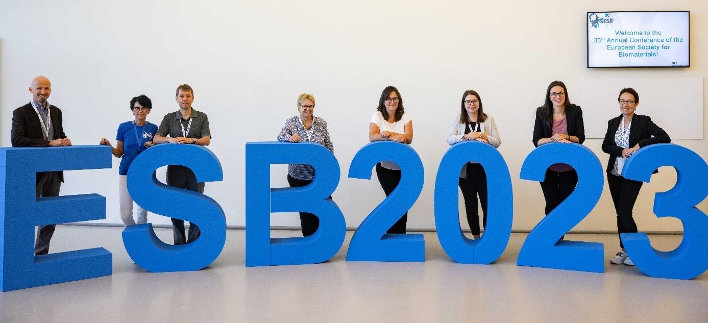
161

162