

ARI Activity Report 2024


Switzerland was Guest Nation at the 70th anniversary meeting of the Orthopedic Research Society in 2024. AO Research Institute Davos had the honor to coordinate activities carried out by Prof Geoff Richards together with Prof Martin Stoddart. Activities included Swiss Scientific symposia, Swiss booth and various “Swissness and Grisons” activities at the meeting.
Top: The “Swiss” delegation attending the Swiss symposia, below the chairs and speakers of the Swiss Symposia from AO, Swiss universities and Swiss hospitals.
1 Introduction
2024 was full of great scientific work. Many of the team have received awards from leading scientific societies and several have made the next steps in their careers either internally on our transparent ARI Career path or externally through their collaborations with universities. To note just a few: Dr Sibylle Grad became Professor in the Department of Health Sciences and Technology, Federal Institute of Technology (ETHZ) and she was awarded the 2024 Women’s Leadership Forum Award from the ORS to recognize a woman scientist who, throughout her professional lifetime, has made significant contributions to the understanding of the musculoskeletal system and musculoskeletal diseases and injuries. Dominic Gehweiler received his habilitation from the University of Münster's Faculty of Medicine. This qualification is a recognition of Dominic's outstanding contributions to preclinical research and teaching applying medical imaging techniques in the field of bone healing and biomechanics. Fintan Moriarty became a visiting professor at the Beijing University of Chemical Technology (BUCT) in Beijing China. Mauro Alini has been honored with the prestigious 2024 PSRS Lifetime Research Award. This award honors Dr Alini’s sustained and long-lasting contributions to spine research recognizing his invaluable work over the years. Stephan Zeiter together with Petra Seebeck from the University of Zürich received the Culture of Care Award from the Swiss 3R competence center for their initiative for good surgical practice for animals used in research. Boyko Gueorguiev was also elected to be President of the European Orthopaedic Research Society
Seven scientists from the ARI remain ranked among the most-cited researchers in the world across all scientific fields, according to researchers at Stanford University which is a remarkable achievement for a non-university institute and these seven represent the amazing overall teamwork that goes behind every publication from ARI. With this great achievement I wish to thank all our ARI staff for their commitment to the Mission of the AO. ARI is the main contributor to AO’s evidence-based changes in healthcare working hand in hand with the other AO Institutes and importantly our dedicated AO surgeon network. Together we improve patient care and we can be extremely proud of what we do!
Again (as in 2023) ARI acquired over 4 million CHF of extramural funds and an additional 1.5 million from grants from AO internal sources. The scientific output in number of publications with 98 peer-reviewed papers (with an average impact factor of 4.48) is on a very high level. 125 abstracts and presentations were made from the projects within ARI. Employee fluctuation rate was ~3% (3 of 94 permanent headcount) and several employees celebrated over 20 years work jubilees, and with a very good employee satisfaction survey from ARI employees, displays that ARI is a great place to work.
Thank you to all our dedicated and motivated team, who enjoy working here at ARI and AONPR (AO Network Preclinical Research) advancing innovation in orthopedics through translational research and development to improve patient care. Thank you to all who collaborate and work with us from internal AO operational teams, institutes and clinical specialties to all our external scientific and clinical partners. Finally thank you to the AO network who keep us focused to improving patient care, especially to the research commissions and ARI Advisory Committee who you can read about in the report. Do not hesitate to reach out to the relevant team members on their projects or leaders on their programs or me.
Sincerely

Prof Dr R Geoff Richards FLSW, FBSE, FIOR, FORS, FTERM Executive Director AO Research & Development, Director AO Research Institute Davos (ARI)
2 ARI Purpose / Goals / Outlook
Purpose
To further the AO's mission, ARI advances innovation in orthopedics through translational R&D Orthopedics concerns musculoskeletal, spine, and craniomaxillofacial trauma, degenerative musculoskeletal diseases, infections, and congenital disorders.
Overall goals
• Contribute high-quality applied preclinical research and development (exploratory and translational) focused on clinical solutions and applications.
• Investigate and improve the performance of materials, biologics, and devices for surgical procedures and treatments.
• Foster a close relationship with the AO network, academic societies, and universities.
• Provide a supportive, inclusive, and diverse research environment and mentorship for our employees, scientists, and the AO network.
ARI goals, 2023-2025
• Valorize AO Fracture Monitor together with AO ITC.
• Implement the specific-pathogen-free sheep flock in studies.
• Valorize the biphasic plate together with AO ITC
• Strengthen and advance research activities in patient diagnostics and personalized medicine.
• Develop training technologies to support education within the AO network.
• Continue developing 3D (bio)printing and SIM technologies.
ARI principles
• Maintain world-class research and nurture in-house talents for long-term innovation.
• Support the AO network with cutting-edge research and development for clinical problems.
• Continue developing ARI technology portfolio. Translate and valorize ARI innovations together with the AO ITC's Technology Transfer team.
• Maintain our world-class certificates (ISO, AAALAC, GLP).
• Engage with scientific networks and consortia: global (e.g., ORS, TERMIS, ICORS) and European societies (e.g., DKOU, ECLAM, ESB-Biomaterials, ESB-Biomechanics, EORS, TERMIS-EU).
Outlook
The AO Foundation's contract with Synthes was replaced with a new collaboration agreement with Depuy Synthes (DPS) which started new in January 2016. The agreement was renewed in spring 2020 for another 5 years. The ARI is not mentioned within the agreement. The ARI budget is taken from the AO Foundation's endowment funding stream, giving the ARI freedom to operate without direct obligations to the AO Foundation's industrial partners.
2021 marked the start of the new HORIZON EUROPE program. Switzerland's status was reverted from 'To Be Associated' to ' a non-associated third country in the Horizon Europe research program. The endorsement of the Common Understanding in December 2023 by both the European Commission and Switzerland are positive steps towards an association to Horizon Europe. Third Country status continues to apply. The financial guarantee from the State Secretariat for Education, Research, and Innovation (SERI) covers the costs of successful Swiss-based applicants in Horizon Europe projects. Complemented by the national transitional measures, this support limits the erosion of Switzerland’s competitiveness and partially maintains its integration in the European research community. The greater loss for Swiss researchers (including ARI researchers) is not being able to work seamlessly in research projects with peers across Europe. This did not change in 2024.
3 Funding Summary
Comments:
Overall, the ‘AO Research Institute’ (ARI) closed the year with a net result of CHF -11,948 K, CHF 403 K below budget. However, for the unit 'Network Preclinical Research' (NPR), a rollover of CHF 296 K from 2024 to 2025 was foreseen in the 2025 budget. Considering this rollover, ARI shows an underspend of CHF 107 K or less than 1% compared to the budgeted net result. This underspend was mainly driven by additional lower expenses than planned in NPR caused by ongoing contract negotiations for the Clinical Priority Programs (CPP), delayed milestone activities, and the critical assessment of individual projects.
Income:
The ‘Management & Overhead ARI’ (M&O) could achieve a higher income for the ARI Orthopaedics Conference in Davos. 'Regenerative Orthopaedics' slightly exceeded the targets, and the marked decrease in income compared to the current figures for 2023 was caused by the expiration of various third-party funded projects from the public sector, of which a number could not be renewed. This development is also expected in 2025. Project delays in Development Incubator projects in 'Biomedical Development' and the cancelation of two Development Incubator projects in 'Preclinical Services' led to a loss of intercompany income, which could only be partially compensated by additional acquired third-party income.
Expenses:
The higher expenses shown in M&O were mainly driven by increased personnel costs (an additional FTE for the organization of the 2025 ‘AO Orthopaedic Research Summit’ and salary adaptions), higher rental costs for the accommodation of non-permanent staff, price increases for electricity, and higher costs for traveling. Savings in ‘Regenerative Orthopaedics’ were realized by lower material costs, unrealized investments, and delayed recruiting (for PostDocs and PhDs). Both 'Biomedical Development' and 'Preclinical Services' were also affected by higher personnel costs due to the salary review. 'Preclinical Services' managed to reduce personnel costs, but the aging buildings and the age and complexity of the existing machinery were the main drivers for higher costs. 'Biomedical Development' was able to save costs for external services due to project delays but could not fully compensate for the loss of income. The underspend in 'Fellowships' was caused by fewer hired interns (several fellowships were rescheduled for another time) and the one-year postponement of two apprenticeships due to unqualified candidates.
Cost category:
The main cost categories were 'Personnel Expenses' with 60% of the total, followed by 'Material Expenses' with 11%, and 'Scientific & Regional
with 6%.
4 Research Structure & Advisory Committees
4.1 AO Research Institute Davos (ARI) Organigram

(December 2024)
4.2 AO Foundation Executive Committee (AOEC)
The AO’s Executive Committee reports directly to the AO Foundation Board, and includes the CEO, CFO/COO and executive directors representing key areas of AO activity.

(December 2024)
4.3 AO Foundation R&D Platform
The AO R&D Platform supports the active exchange and mutual discussion about strategies of the AO units with respect to their related goals in R&D. It supports the AO Foundation Board (AO FB) in defining general strategic areas and their implementation in an advisory function. It ensures that relevant activities are in line with the AO Mission and strategies as defined by the AO FB. All research stakeholders are finally accountable to the AO FB. The AO R&D Platform further develop the strategies and their implementation on behalf of the AO FB in an advisory capacity. It has no funding and decision authority. The R&D Platform is represented on the AO EC by the AO Executive Director of Research and Development. The R&D expert of the AO FB is the Chair of the R&D Platform, currently Prof Anita Ignatius, Director and Chair of the Trauma Research Center Ulm (ZTF), University Hospital Ulm, Germany.

4.4 AO Research Institute Davos Advisory Committee (ARI AC)
The ARI AC provides operational and strategic scientific advice to the ARI on behalf of the AO FB. The ARI AC acts as both a sounding board and sparring partner for the ARI Director and mentor group to the Program Leaders, Focus Area Leaders, and ARI scientists. The tasks and responsibilities include advising ARI on:
Portfolio of competences (skills of personnel and type of equipment).
Strategy and priority setting for direct funds of ARI.
Business development and initial advice on technology transfer. Regulatory issues, use of ARI funds, advancement of the ARI capabilities, to assure the efficient use of the infrastructure.
The ARI AC comprises the following external members:
Prof Brian Johnstone, Oregon Health and Science University, USA (Chair). Represents ARI AC on the AO R&D Platform and Innovation Platform (2nd right).
Prof Chris Evans, Mayo Clinic, USA (front right)
Prof Gerjo Van Osch, Vice dean of Research Erasmus, Rotterdam, NL (front left)
Prof Hamish Simpson, George Harrison Law Professor of Orthopaedics & Trauma, University of Edinburgh (right)
Dr Juerg Gasser, Independent R&DConsultant for Regenerative Therapies in Bone, Joint and Tendon (previously career until retirement, Novartis) (back left).

4.5 AO CMF Research Commission (AO CRC)
The AO CRC is the international coordination body for all activities of the AO CMF clinical division for research and development of the AO. Its mission is to promote excellence in patient care and treatment outcomes in trauma and musculoskeletal disorders.
In October 2024 for the first time AO CRC has consisted of five members, each representing a region and thus serving a dual role as a member of the international research commission and as a member of the respective regional board. Among others, the advantage of this composition includes a direct link to the regions, which enables a transfer of knowledge about the Commission's activities to the regions and, in turn, facilitates feedback to the Commission about regional research needs.
AO CRC main activities:
Activities Overview:
- Coordinates international research across divisions (similar to AO Trauma, Spine, and VET) as a central body for CMF research within the AO.
- Collaborates with external partners (consortia) and runs large-scale projects such as clinical priority programs (CPP), startup grants, and other global initiatives.
- Members, as regional representatives, are also members of regional boards and ensure that the flow of information is bidirectional.
Key Research Areas:
a. Scientific Knowledge Development:
- Enhances academic credibility and leadership, focusing on the clinical quality program.
- Projects involve external and internal bodies (AO ITC and ARI), with ongoing projects like the AO CMF CPP and the Knowledge Forum in AO Spine.
- Long-term projects, typically 5-year terms, may be renewed. Additional activities include studies and projects by AO ITC and ARI.
b. Individual Research Career Development:
- Provides individual support through small grants (e.g., startup grants, seed grants).
- Supports research fellowships (AO ITC, ARI, research symposia, AO PEER offerings (f2f, online courses), etc.
The AO CRC comprises the following members, permanent guests and AO representatives:
Dr Thomas B. Dodson, AO CMF Research Commission (RC) chair, Seattle, WA, USA
Dr Rodrigo Pereira, AO CMF RC member (representative AO CMF LAT), Rio de Janeiro, Brazil
Dr Lamont Jones, AO CMF RC member (representative AO CMF NA), Detroit, MI, USA
Dr Patricia Stoor, AO CMF RC member (representative AO CMF ESA), Helsinki, Finnland
Dr Khalid Abdelgalil, AO CMF RC member (representative AO CMF MENA), Abu Dhabi, UAE
Dr Takahiro Kanno, AO CMF RC member (representative AO CMF AP), Izumo, Japan
Dr Chelsea Bahney, AO CMF Research Commission permanent guest, Vail, CO, USA
Dr Catherine Chaussain, AO CMF Research Commission permanent guest, Paris, France
Philipp Buescher, Head AO Network Preclinical Research (NPR)
Prof Martin Stoddart, ARI Program Leader Regenerative Orthopaedics, Davos, Switzerland


4.6 AO Spine Research Commission (AO SRC)
From left to right: Takahiro Kanno, Philipp Büscher, Khalid Abdelgalil, Rodrigo Pereira, Lamont R Jones, Patricia Stoor, Chelsea Bahney, Aleksandra Hodor, Thomas B Dodson
AO Spine's preclinical research activities are led by Principal Scientist, Dr Sibylle Grad from the ARI. The focus is on intervertebral disc (IVD) degeneration and postoperative spine infection, with a specialization in organ models and biomarkers. The preclinical outcomes are brought to the AO Spine Knowledge Forums, which are expert-driven global clinical study groups, for clinical evaluation. In 2024, the following preclinical activities are being performed:
1. Whole spine model bioreactor: proof-of-concept studies using the first-ever whole spine model bioreactor, which simulates 6DOF loading, characterized the IVD’s mechanical changes, end plate alterations, cell response, and regulation of collagenase genes under multiaxial loading.
2. Neural cell sensitization: the evaluation of the effect of different IVD loading scenarios on neural cell sensitization and implementation of neural cell responses as an outcome parameter to determine IVD health and disease
3. IVD infection organ model: in collaboration with Balgrist University Hospital, an IVD infection organ model for proof-of-concept testing of antibiotic release is being developed.
4. Biomarkers: candidate biomarkers for IVD degeneration are being explored.
5. Infection and serum biomarkers: in collaboration with the Swiss Institute of Allergy and Asthma Research (SIAF) Davos, Schulthess Clinic Zürich, and University of Regensburg, we aim to understand the impact of the immune status on the susceptibility to postoperative spine infection. Data collection is now complete for a matched-cohort study comparing serum biomarkers from patients with and without postoperative surgical site infections.
The AO SRC consists of the following members:
Dr Klaus Schnake, Chairperson, Erlangen, Germany
Dr Charles Fisher, Past Chairperson, Vancouver, Canada
Dr Shekar Kurpad, AO Spine Knowledge Forum Spinal Cord Injury Representative, Milwaukee, WI, USA
Dr Stephen Lewis, AO Spine Knowledge Forum Deformity Representative, Toronto, Canada
Dr S. Tim Yoon, AO Spine Knowledge Forum Degenerative Representative, Atlanta, GA, USA
Dr Ilya Laufer, AO Spine Knowledge Forum Tumor Representative, New York, NY, USA
Dr Gregory Schroeder, AO Spine Knowledge Forum Trauma & Infection Representative, Pittsburgh, PA, USA
Dr Alfredo Guiroy, AO Spine Latin America Regional Research Officer, Mendoza, Argentina
Dr Jefferson Wilson, AO Spine North America Regional Research Officer, Toronto, Canada
Dr Daisuke Sakai, AO Spine Asia Pacific Regional Research Officer, Tokyo, Japan
Dr Kabir Abubakar, AO Spine Middle East and Northern Africa Regional Research Officer, Kano, Nigeria
Dr Aron Lazary, AO Spine Europe and Southern Africa Regional Research Officer, Budapest, Hungary
Dr Sibylle Grad, ARI Representative, Davos, Switzerland


The AO Spine Research Commission
Congress
Bangkok, Thailand.
right: Yabin Wu, Ramona Ritzmann, Marije de Jong, Sibylle Grad, Nelson Astur, Jefferson Wilson, Brian Kwon, Ilya Laufer, Kabir Abubakar, Niccole Germscheid, Charles Fisher, Marta Morawska, Klaus Schnake, Nicola Di Marzio, S. Tim Yoon, Stephen Lewis, Janneke Loomans, Aron Lazary, Olesja Hazenbiller, Marie-Laure Vial, Daisuke Sakai
4.7 AO Trauma Research Commission (AO TRC)
The AO TRC is the international coordination body for all activities of the AO Trauma clinical division for research and development of the AO Foundation. The AO TRC partners with external institutes and funds research projects and clinical studies in collaboration with external institutes as part of consortia within clinical priority programs (CPP).
AO TRC strategy focuses on two fields:
1) To be a knowledge leader, performing large research projects (CPPs) as a consortia with external opinion leaders, experienced clinicians and researchers in collaboration with ARI and AO ITC that help AO Trauma gain scientific knowledge and enhance academic recognition and credibility. Gaining state-of-the-art knowledge serves to promote AO Trauma to maintain its leadership position. To this aim, AO Trauma conducts two CPPs that focus on clinically highly relevant topics. 1) AO Trauma CPP Patient Outcome lead by Dr Marylin Heng (Miami, USA). 2) In 2024, a new CPP focus was created, and a new consortium is being established with a focus on bone non-union
The approval process for these projects includes the AO RRTF (Research Review Task Force) process without exception.
2) AO TRC provides individual support to young clinicians to increase awareness of research and provide training in the fundamentals of research processes. Within this framework, the AO TRC offers funding programs for smaller projects. These grants follow the AO FB guidelines in terms of target group (young clinicians < 40 years), access (open to all Clinical specialties). Out of this pool of young clinicians, new talents are identified. AO TRC also coordinates research symposiums and offers research fellowship programs.
AOTRC comprises the following members and AO representatives:
Prof Peter Giannoudis, AO TRC chairperson, Leeds, UK
Prof Dhaval Desai, AO TRC member (representative AO TAP), Surat, India
Dr Leah Gitajn, AO TRC member (representative AO TNA), Lebanon, USA
Dr An Sermon, AO TRC member (representative AO TESA), Leuven, Belgium
Dr Vincenzo Giordano, AO TRC member (representative AO TLAT), Rio de Janeiro, Brazil
Prof Ahmed Kholeif, AO TRC member (representative AO TMENA), Cairo, Egypt
Philipp Buescher, Head AO Network Preclinical Research (NPR), Davos, Switzerland
Dr Alex Joeris, AO ITC Head of Clinical Science, Dübendorf, Switzerland
Prof Geoff Richards, AO Executive Director Research & Development, Davos, Switzerland
at the Global Spine
2024 in
From left to


From left to right: Philipp Büscher, Leah Gitajn, Peter Giannoudis, Alexander Joeris, Dhaval Desai, Vincenzo Giordano, An Sermon, Ahmed Kholeif, Geoff Richards
4.8 AO Vet Research Commission (AO VET RC)
AO VET RC pursues two main goals with its research activities. First one is to perform research activities that help to gain scientific knowledge and enhance academic recognition and credibility. Gaining state-of-the-art knowledge serves to promote the AO to maintain its leadership position. AO VET RC also provides individual support to young clinicians to increase awareness of research and provide training in the fundamentals of research processes as well as identifying new talents. The preclinical research activities of AO VET are coordinated at ARI by Dr med vet Stephan Zeiter, Program manager Preclinical Services.
AO VET RC also supports the other AO Clinical Divisions as an advisory body (Animal Welfare Advisory Committee (AWAC) and AAALAC).
The AO VET Research Commission comprises the following members and AO representatives:
Prof Kenneth Johnson, AO VET RC chair, Sidney, Australia
Dr Yukihiro Fujita, AO VET RC member (representative AP), Tokyo, Japan
Ass Prof Kyla Ortved, AO VET RC member (representative NA), Pennsylvania, MI, USA
Dr Kevin Parsons, AO VET RC member (representative ESA), Bristol, UK
Dr Diego Quinteros, AO VET RC member (representative LAT), Buenos Aires, Argentina
Philipp Buescher, Head AO Network Preclinical Research (NPR), Davos, Switzerland
Dr med vet Stephan Zeiter, ARI Preclinical Services Program Manager, Davos, Switzerland


From left to right: Tania Bosque, Yukihiro Fujita, Diego Quinteros, Maria Clara Sardoy (wife), Joshua Stacy (husband), Kyla Ortved, Caroline Constant (Preclinical Services, Project Leader), Jayne Symon (wife), Kenneth Johnson, Philipp Büscher
4.9 AO Research Review Task Force (AO RRTF)
The AO RR TF is an independent peer review body valid for all AO decision-making bodies for grants to all external applicants for AO research funding. The AO RRTF is assigned jurisdiction over many external AO peer review process, while other internal AO Peer Review Policies and expectations govern specific AO Institute research programs, partnering, internal research contracting, and some limited external research funding processes. Decision-making bodies are defined as bodies that have funding allocation roles within the AO Foundation, including AO Trauma, AO Spine, AO CMF, AO VET, and their respective Research Commissions (RCs). For ARI projects, the decision-making body is ARI together with the ARI Advisory Committee (ARI AC). For each Clinical Division research grant, the decision-making body is the respective research commission
The chairperson of the AO RR TF is Jaimo Ahn, Ann Arbor, USA
4.10 AO Network Preclinical Research (AO NPR)
The goal of the AO Network Preclinical Research (AO NPR) is to gain in efficiency and effectiveness with one central team for all external preclinical research. AO NPR is the international coordination group for all external preclinical research activities of the AO. AO NPR manages and supports the global research commissions of the AO Trauma, AO CMF, and AO VET to establish a cohesive global research vision and strategy for AO worldwide. AO NPR supports coordination between external partner institutes and AO Institutes and works closely with ARI and the AO Innovation Translation Center (AO ITC.
AO NPR is the entry point for all external research partners for preclinical research. AO NPR promotes excellent research of all AO partners, which are directly or indirectly related with clinical needs in patient care. It helps to strengthen networking among AO clinicians and researchers worldwide, making clinically relevant research attractive for the young generation of AO surgeons
AO NPR Manages the Clinical Priority Programs (CPP’s) of Clinical Divisions and the Research activities of Clinical Divisions AO Trauma, AO CMF, and AO VET together with the AO GN regions. AO NPR manages the research governance of the Research Commissions of the Clinical Divisions AO Trauma, AO CMF and AO VET, the AO R&D Platform, and the AO Research Review Commission (AO RRC)
AO NPR is headed by Philipp Büscher. Team members are Tania Bosque, Anna Dönz, Larissa Welti and Anita Anthon.
5 ARI Teams / Personnel
5.1
Biomedical Development
Program Leader: Boyko Gueorguiev-Rüegg, Deputy: Peter Varga
Team Members: David Ambühl, Gordian Banzer, Denise Bentivoglio, Jan Barcik, Jan Buschbaum, Paula Cameron, Cherilyn Camichel, Jan Caspar, Daniel Ciric, Manuela Ernst, Alicia Feist, Dominic Gehweiler, Alisa Hangartner, Carla Hetreau, Maximilian Heumann, Moritz Kraus, Lionel Llano, Giulia Minikus, Dominic Mischler, Tatjana Pastor, Fabian Pretz, Luise Puls, Luke van Rossenberg, Peter Schwarzenberg, Simone Sommer, Jérôme Schlatter, Flurin Spiller, Viktor Topuzyan, Antoine Vautrin, Ivan Zderic, Erich Zweifel
Supporting the in-house processes for development and design of medical devices according to EN ISO 13485 and running advanced projects in close collaboration with clinical, scientific, and industrial partners, as well as with the AO specialties, the AO ITC and the AO Education Institute (AO EI), the Biomedical Development Program offers extensive know-how, expertise and experience in the fields of biomechanical testing and computational analyses to improve patient care.
A variety of clinical problems are addressed by development of new concepts, approaches, tools and novel implant systems for surgical applications and research in traumatology and orthopedics. Moreover, digital and hands-on technologies for surgical training and education are developed.
The process of finding optimal solutions to clinical questions is enhanced by capabilities ranging from in silico methods to anatomical labs for quick and effective hands-on work when an anatomical environment is required. Specifically, tailored test procedures with implementation of supplemental radiographs, video and motion tracking systems are applied in diverse experiments on fracture fixation and joint reconstruction. Advancing with state-ofthe-art technologies, powerful numerical methods and comprehensive tools for virtual simulations are integrated to answer various questions with special reference to biomechanical performance of bone-implant constructs and fracture healing. Modalities for medical imaging, processing, and analysis, including CT scanners with a wide range of resolutions and scanned volumes, are interlinked to account for increasingly sophisticated demands for morphological investigations, extract statistical and individual information from medical image data, and extend the knowledge on variations of biomechanical bone characteristics and their role in persisting clinical problems. The capabilities of the Program are completed by the Prototype Workshop offering rapid and high-quality manufacturing of devices, tools, and implants.
5.2 Preclinical Services
Program Leader: Stephan Zeiter, Deputy: Nora Goudsouzian
Team Members: Florence Albrecht, Daniel Arens, Julija Bulatova, Barbara Brändle, Carmen Brazerol, Caroline Constant, Anas Datoussaid, Peter Erb, Lorena Faoro, Loris Faoro, Pierina Faoro, Lisa Fell, Andrea Furter, Lena Gens, Jeremia Giger, Nilo Hämmerl, Maria Hildebrand, Urban Lanker, Salome Leuthold, Leonie Mollet, Reto Müller, Dirk Nehrbass, Dominic Perren, Lotta Reimann, Monika Schneider, Zdenka Slavikova, James Tapia-Dean, Carmen Volz, Fabienne Wynne, Claudia Zindl
The Preclinical Services team at ARI, encompassing Preclinical Surgery and Tissue Morphology, has reaffirmed its dedication to both animal welfare and preclinical research this year. Our team provides comprehensive support, guiding projects from initial planning and surgical procedures to data analysis and publication. In 2024, we successfully conducted over 20 studies and carried out more than 450 surgical procedures, utilizing various animal models, including mice, rats, rabbits, and sheep.
A key focus of our work has been improving surgical techniques and outcomes for researchers. In collaboration with the University of Zurich, we once again delivered the "Good Surgical Practice for Rodent Surgery" course at multiple locations, emphasizing high standards in surgical care and ethical treatment of animals. In recognition of these efforts, we were honoured with the “Culture of Care Award” from the Swiss 3R Competence Center (3RCC). This initiative, along with a series of online lectures, reflects our strong commitment to education and training. Expanding on this mission, we introduced a new veterinary student scholarship program in partnership with Aberystwyth School of Veterinary Science, Aberystwyth University, Wales. Through this and other global university collaborations, we provide students with practical training and a solid foundation in preclinical research. Beyond research and education, our team remains actively engaged in professional organizations such as the American College of Veterinary Surgeons (ACVS), the European College of Veterinary Surgeons (ECVS), and the European College of Laboratory Animal Medicine (ECLAM). These affiliations ensure we stay at the forefront of advancements in veterinary science and preclinical research.
Maintaining the highest standards, our research operates under rigorous quality management systems, including GLP and AAALAC accreditation. We are committed to fostering transparency, upholding ethical research practices, and educating the public on the significance of our work. As we look ahead, we remain dedicated to advancing preclinical science while maintaining the integrity and values that have long defined our approach.
5.3 Regenerative Orthopaedics
Program Leader: Martin Stoddart, Deputy: Sibylle Grad
Team Members: Mauro Alini, Ivan Al Saify, Ezgi Irem Bektas Tas, Corinne Bischofberger, Samuel Blackman, Maruo Bluvol, Katja Brühl, Simona Casutt, Claire Chabot, Marco Chittò, Alessandro Cianciosi, Eda Ciftci-Dede, Greta Cocchi, Carolina Maria Cordeiro, Darine D’Adam, Elena Della Bella, Matteo D’Este, Nicolas Devantay, Pia Fehrenbach, Chencheng Feng, Severine Flück, Pamela Furlong-Jäggi, Wei Gao, Nico Giger, Dacheng He, Leopold Henssler, Marina Holdener, Maria Rosa Iaquinta, Shahrbanoo Jahangir, Lauma Jevina, Ilse Jonkers, Anita Jose, Iris Keller-Stoddart, Livia Kiener, Thomas Krüger, Eliane Kuhn, Marina Kurz, Puk Kwant, Zhen Li, Yuqi Liu, Chiara Lorenzetti, Junxuan Ma, Pai Mansi, Laura Mecchi, Huan Meng, Danilo Menghini, Ksenia Menshikh, Ursula Menzel, Gregor Miklosic, Fintan Moriarty, Marcia Mürner, Micaela Natta, Pamela Nylund, Robert Peter, Virginia Post, Clara Presciutti, Athanasia Pylostomou, Roots Randriantsilefisoa, Noémi Reinert, Nicoletta Restione, Fatemeh Safari, Amra Secerovic, Theresa Schiemer, Maja Schlittler, Maria Schröder, Tiziano Serra, Claudia Siverino, Astrid Soubrier, Christoph Sprecher, Alica Stegmaier, Jorge Úbeda Garrido, Daphne van der Heide, Sophie Verrier, Nadja Vonlanthen, Svenja Wacker, Esther Wehrle, Liru Wen, Katrin Wendrich, Lucille Wespi, Jacek Wychowaniec, Jiangyao Xu, Leonardo Zonca, Daniele Zuncheddu
Biomedical Materials Focus Area
The Biomedical Materials Focus Area is committed to the design of advanced biomaterials and the development of (bio)manufacturing technologies to achieve improved patient care and outcomes in musculoskeletal disorders. Using a variety of chemical approaches, we create responsive biomaterials that react to environmental stimuli and actively interact with cells and tissues. We design biomaterial surfaces and antibacterial delivery systems for prevention and treatment of infections, and we are investigating how materials "talk" to the body at the cellular level by harnessing the inflammatory processes to trigger a healing response and prevent chronic inflammation. We also develop bio-processing technologies for translating tissue engineering approaches to regenerative, patient-tailored precision medicine.
By deepening our understanding on how materials interact with/in the body, and how additive manufacturing and bioprocessing modulate these interactions, we aim to advance orthopaedic patient care.
Bone Biology Focus Area
Bone healing depends on biological factors and the mechanical conditions in the defect region. Despite the advances in fracture fixation, there remains a subset of patients that suffer from healing complications, resulting in delayed healing and non-unions. Currently it is not possible to reliably identify healing complications at an early stage when treatments may be more effective. We study biological factors involved in the different phases of bone healing with a major focus on early immunological, angiogenic and mechano-molecular components. The immune system is involved in guiding and directing the healing response. We are investigating how modulation of inflammation may be used to enhance the bone healing process, as well as assessing the potential of immune cell characterization to be used as a predictive biomarker of the individual healing potential.
Mechano-molecular mechanisms are important for successful bone healing. Via our novel technology we aim to precisely study how mechanics influence molecular mechanisms during bone healing in vivo (femur defect loading model in mice) and in vitro (bone bioreactor). In combination with emerging molecular omics techniques, we want to comprehensively characterize the local and systemic mechano-molecular regulation of bone healing. Via this combined in vivo and in vitro approach, we aim for identification of novel therapeutic targets, systemic biomarkers, and mechanical intervention therapies relevant towards translation of personalized medicine approaches for impaired healing conditions.
Disc and Cartilage Biology Focus Area
Traumatic and degenerative damage to the articular joint and intervertebral disc (IVD) are major causes of pain and functional impairment. However, the factors that contribute to the loss of function and the underlying pathophysiology are still poorly understood. In addition, current medical and surgical approaches barely address the underlying pathology and are often unsatisfactory. We investigate mechanical and molecular mechanisms leading to cartilage and IVD damage and identify tissue and systemic biomarkers, which may serve as diagnostic and therapeutic targets. Collaboration with clinical partners provides access to patients’ samples, data and clinical context.
We have established whole IVD organ culture systems with the ability to maintain entire discs for several weeks under controlled nutrient and mechanical loading conditions. Our unique multiaxial bioreactors enable us to apply load and motion in six degrees of freedom onto the spinal segment, reproducing physiological conditions.
Our ex-vivo IVD defect and degeneration models allow us to design and evaluate new biological treatment strategies, including the delivery of therapeutic cell populations, anabolic, anti-catabolic or anti-inflammatory molecules, biomaterials or combinations thereof. The goal is to develop functional therapies which will restore the mechanical properties of the IVD, enhance endogenous regenerative processes, or inhibit paradox nerve or vessel growth and activation.
To study the potential of new therapies for articular cartilage repair, we have implemented jointspecific bioreactor systems applying multiaxial load to tissue-engineered constructs or osteochondral explants. Co-culture models, osteoarthritis-mimicking proinflammatory conditions and a physiological oxygen environment are employed to investigate disease mechanisms and test tailored treatments.
Infection Biology Focus Area
Fracture-related infection (FRI) remains one of the most challenging complications in orthopedic and musculoskeletal trauma surgery. FRI has been convincingly shown to delay healing, worsen functional outcome, and incur significant socio-economic costs. Antibiotic prophylaxis, wound debridement, and postsurgical care can reduce, but not prevent, the incidence of these infections and so novel interventional strategies are required. We work on in vitro, in vivo and ex vivo studies to better understand, prevent, and treat FRI.
A significant portion of the work involves collaboration with the preclinical services team in ARI to model FRI in a complex living system and provide robust evaluation of the new interventional technologies under development such as antibiotic loaded hydrogels. This expertise also extends to extramural studies performed with industrial partners to evaluate external innovations in the prevention and treatment of FRI prior to clinical implementation. In parallel to the preclinical in vivo evaluations, greater focus has been applied to the opportunities of working with human materials, either in vitro through basic cell culture studies or also in clinical studies with patients experiencing FRI. Through partnerships with clinician scientists in the AO network, we have gained access to biological materials from patients with FRI in an effort to more accurately study host pathogen interactions.
Progenitor Cell Biology and Mechanoregulation Focus Area
Work is dedicated to advancing stem cell therapies for bone and cartilage, with the goal of clinical application. We have identified predictive markers of donor variation to assess the potency of cells from individual donors. In our search for biomarkersthat can determine patientspecific healing potential, extracellular vesicles and non-coding RNA sequences, such as miRNA, are increasingly being utilized as diagnostic and therapeutic tools. Developing a serum-based biomarker approach could significantly enhance patient-specific clinical decisionmaking.
Additionally, we aim to investigate the role of mechanical and soluble factors in the activation of mesenchymal stem cells, as well as in promoting differentiation and tissue repair. Mechanical forces, applied through rehabilitation protocols, can modify the function of both stem cells and immune cells. These studies are contributing to the emerging field of regenerative rehabilitation. Beyond direct differentiation, it is known that biomechanical stimulation can also influence the cell secretome. Investigating these changes may uncover new targets present during articulation, opening up potential avenues for clinical therapies.
Sound Guided Tissue Regeneration (SGTR) Focus Area
SGTR team, focuses on advancing the engineering of living systems using extrinsic fieldsbased biofabrication technologies, such as hydrodynamic waves, magnetic fields, light, and stimuli-responsive materials for modelling and regeneration.
Our team leverages contactless biofabrication approaches to spatially pattern and assemble cells, aggregates, organoids, and extracellular matrices in a controlled and programmed manner, with the goal of creating advanced human in vitro models, in alignment with the 3Rs philosophy.
Our overarching goal is the translation of innovative biofabrication technologies for the repair, regeneration and modelling of musculoskeletal tissues. One of the key innovations developed by our team is Sound Induced Morphogenesis (SIM), a novel technology that enables contactless bioassembly, under fast and mild culture conditions, by hydrodynamic waves, opening new frontiers for functional and morphologically relevant tissue generation. In 2020, SIM was licensed to mimiX Biotherapeutics, a successful Swiss startup that is now advancing in clinical translation. SIM was selected as part of the “Technology Outlook 2023” by the Swiss Academy of Engineering Sciences, SATW. Implants from a loudspeaker: Technology Outlook
5.4 ARI Administrative Services
Manager Admin Services: Claudia Barblan
Manager Purchasing: Ulrich Bentz
Team Members: Isabella Badrutt, Simona Ciriello, Nunzia Di Luise, Carla Escher, Scarlett Kollipka, Gregor Müller, Shannon Smit, Marisa Vivalda, Sonia Wahl
Administrative support services are essential to the operation of any organization. It includes the tasks performed on a day-to-day basis that keep the institute running smoothly and efficiently.
The main goal of the ARI Administrative Services team is to provide an excellent service in all administration and organization fields of the ARI and to numerous AO partners.


Personnel Management & Organize ARI Fellowships


Organize the ARI Directors Office Support all ARI Programs and Focus Areas
Leading Gender Equality Initiative


Meetings

5.5 Operations standards and safety
Quality Manager: Ulrich Bentz
Successful 2023 routine audit of AO Research Institute Davos:

From April 3 to 4, 2024, a new external auditor from the SQS (Swiss Association for Quality and Management Systems) inspected ARI for two days for the recertification audit of the institute. ARI has passed the routine audit with six minor con-conformities. The entire ARI is certified according to the international standard ISO 9001:2015. Parts of the Biomedical Development Program are additionally certified to develop medical devices according to EN ISO 13485:2016. ARI is one of the very few academic research organizations to have achieved this certification. ARI is a GLP (Good Laboratory Practice) compliant test facility since February 2016.
The fourth inspection by Swissmedic took place in April / October 2024 and ARI has received the renewed statement of GLP compliance on January 30th, 2025, from the Swiss Federal Office of Public Health for the next 3 years. We can offer contract research services to all interested customers under GLP, especially if they want to get their medical devices approved by the FDA. Since the achievement of the GLP certification all major commercial studies have been conducted under GLP (without pilot studies).
AAALAC international accreditation of Preclinical facility:

The Preclinical Facility was first accredited by AAALAC International in early 2013. The Association for Assessment and Accreditation of Laboratory Animal Care International (AAALAC) is a private, nonprofit organization that promotes the humane treatment of animals in science through voluntary accreditation and assessment programs. ARI is one of only 4 accredited institutions in Switzerland, and the only accredited academic Research Institute in Switzerland. In November 2025 we had the fifth AAALAC international site visit and got some great comments on our facility and team. The final confirmation for the renewal of the accreditation was received March 24th, 2025. The next reaccreditation site visit is due in 2027
6 Gender Equality Initiative
Gender Equality (GE) is a fundamental value of the European Union (EU). In 2021, Switzerland adopted the first national strategy for Gender Equality 2030. GE Is considered to benefit Research and Innovation (R&I), attracting and retaining more talents, and ensuring that everyone can maximize their potential. In January 2022, ARI appointed an internal Gender Equality working group (GEWG) with the aim of establishing a set of commitments and actions. The GEWG has been composed in line with the recommendations of the Horizon Europe Guidance on Gender Equality plans and includes representatives of all major position groups, hierarchy levels, educational backgrounds, and genders from the institute. An initial assessment of the gender equality status quo of ARI was conducted in 2022. The specific methodological approaches used to carry out the assessment were: 1) identification and review of existing measures promoting gender equality at ARI; 2) collection of sexdisaggregated data about ARI employees; 3) ARI employee survey. The results of the initial assessment allowed to identify the strengths and weaknesses concerning gender equality at ARI and were used as baseline to set up clear objectives and prioritized set of measures.
A summary of the Gender Equality Plan (GEP) 2023-2025 is shown below:
Area 1: Work life balance and organizational culture. Objective 1.1: Promoting reconciliation of career and family life. Objective 1.2: Continuing promoting alternative and flexible working arrangements. Objective 1.3: Promoting use of inclusive language around the organization.
Area 2: Gender balance in leadership and decision making. Objective 2.1 Supporting and promoting women in leadership positions.
Area 3: Gender equality in recruitment and career progression. Objective 3.1: Raising awareness on gender issues at different levels. Objective 3.2: Updating the ARI career path.
Area 4: Measures against gender-based violence, including sexual harassment. Objective 4.1. Preventing chance of gender-based violence, including sexual harassment.
Area 5: Integrating sex dimension into research content. Objective 5.1: Raising awareness of including sex aspect in research content. Objective 5.2: Setting up standard procedures integrating sex aspect into research content.
Regular meetings are organized: 3-4 meetings per year with the whole GEWG and task forces meetings in between to discuss specific objectives and measures. There is data collection, followed by analysis, reviewing, and reporting.
Dedicated resources allocated by ARI for the GEP include:
1. A dedicated ARI gender equality function composed by one gender equality officer, a team with different expertise, including one human resource representative, and an executive leadership member (director of the institute), publicly supporting the whole function.
2. Earmarked staff time for the whole ARI gender equality function to work throughout the whole GEP cycle.
Earmarked budget to supporting specific measures and areas of the GEP, such as work-life balance and parental leave, as well as staff training, and development will be evaluated and potentially allocated in the next few years.
In 2024, the second year of implementation of the GEP, the working group was reorganized due to some staff changes. In addition, a new external task force was established to work on area 1 “Work life balance and organizational culture”, consisting of 6 volunteer ARI members.
Furthermore, a second data collection was conducted, including data provided by Human Resources (HR) and a survey of ARI employees. Key findings include: 1) general awareness on Diversity, Equity Inclusivity and Accessibility-related topics (DEI&A) has increased among ARI staff; 2) part-time work has become more accepted and, consequently, slightly more common among men.
ARI also organized two workshop events: a first event – Unconscious bias workshop, held by Tatjana Topalovic, Senior Program Manager Diversity, Inclusion and Mentorship at AO took place for selected ARI staff, and a second event – Including sex aspect into research workshop, held by Daniele Zuncheddu, PhD student Disc and Cartilage – where an ARI manuscript including sex aspect was presented.
Also, in the second edition of"Inspiring Female Scientists from ARI's Network," Professor Gerjo van Osch, principal investigator at the Department of Orthopaedics & Sports Medicine & Otorhinolaryngology, Erasmus University Medical Center, The Netherlands, shared her successful career path as a female scientist. She talked about the opportunities she encountered along her career journey and what and who motivated and guided her through these choices. More than sixty researchers, including twelve visiting students from the University of Bern, attended the event and actively participated in the discussion. This event contributed to fostering a more gender-diverse culture among careers in science for the next generations.
Furthermore, ARI proposed to organize an Academia Raetica event, “Researchers Beer” on diversity in research and technology at the Kulturplatz Davos. David Schmid, Regional Development & Relations Manager Eastern Switzerland and Samantha Paoletti, Head Research and Business Development Life Science Technologies both work for Swiss Center for Electronics and Microtechnology (CSEM), presented the Diversity-gr project, which promotes diversity and equality in the IT and technology sector.
Lastly, inspired by the ARI experience, the AO Executive Committee (AO EC) mandated HR to establish a similar initiative on the DE&IA-related topics called Inclusive Excellence best practices and policies. HR leads a voluntary, multidisciplinary group of employees from different departments, the Inclusive Excellence Focus Group. This group is responsible for initiating, managing, and implementing DEIA-related projects and initiatives. The established goals entail the following three key pillars: increase awareness, education, and policies and practices.


A selection of achievements in 2024. HR Dat a
7 ARI Abstracts periodical / ARI conferences
7.1 ARI Abstracts periodical
ARI Abstracts Periodical is a non-profit online platform dedicated to publishing supplements from the ARI Orthopaedics Conference and various third-party events. It operates as an openaccess resource, featuring collections of congress abstracts in PDF format. These abstracts have been peer-reviewed by the respective conference organizers. The platform is managed by ARI, a non-profit foundation based in Switzerland and is designed by scientists for scientists. While the abstract collections do not have a DOI and are not searchable on PubMed, they may be cited, depending on the policies of the relevant journal. ARI Abstracts also includes all eCM official society meeting abstracts up until July 2023, when the platform was rebranded as the ARI Orthopaedics Conference, in addition to abstracts from other congresses. All content is permanently recorded in the ISSN Register, ISSN: 2522-235X, by the ISSN International Centre. For more information, visit ARI Abstracts
7.2 ARI Orthopaedics annual conference
The 2024 ARI Orthopaedics Conference, focused on Orthopaedic Infections, brought together leading experts from around the world specializing in orthopaedic infections, bone health, antibacterial biomaterials, spinal infections, and bacterial biofilms. The conference featured presentations on a range of important topics, including the challenges of non-union, advancements in biomaterials, and innovative strategies for eliminating implantassociated infections. These presentations highlighted the latest scientific advancements and provided a foundation for in-depth discussions on how to tackle these persistent clinical problems.
In addition to the invited talks, the conference included 17 presentations selected from submitted abstracts, offering a diverse cluster of perspectives on orthopaedic infections. One of the conference's highlights was the awarding of the Berton Rahn Research Award to Prof Edward Schwarz, in recognition of his groundbreaking research project in the field of bone infection This prestigious award underscored the significance of his contributions to advancing our understanding of infection-related challenges in orthopaedics.

Professor Edward Schwarz (left) from The University of Rochester receiving his Berton Rahn Research Award from ARI Director, Prof Geoff Richards (right).
To encourage and recognize the contributions of emerging researchers in the field, the conference awarded both a podium and a poster presentation award to the most outstanding student presenters. Andrea Nüesch (Sheffield Hallam University, United Kingdom) the award for the best oral presentation, while Julia van Agtmaal (Maastricht University, The Netherlands) was awarded with the best poster presentation award. These awards highlight the conference's commitment to encourage the next generation of experts in orthopaedic infections.

The best student prize winners at the 2024 ARI Orthopaedics conference. From left to right: Fintan Moriarty (ARI), Claudia Siverino (ARI), Andrea Nüesch (UK), Julia Agtmaal (NL), Edward Schwarz (USA), Marco Chitto (ARI).
8 Institutional and Professional Relations
Director, Program Leaders & Managers and Focus Area Leaders
R. Geoff Richards has been Director of the ARI since 2009 (having been at ARI since 1991). He is a full Professor at the Medical Faculty of Albert-Ludwigs University, Freiburg, Germany (since 2015). He currently holds honorary Professorships at Cardiff University, Wales, GB (since 2007) Aberystwyth School of Veterinary Science, Aberystwyth University, Wales, UK since 2022. He has Doctor Honoris Causa from the Technical University of Varna, Bulgaria. He is an elected Fellow of the Learned Society of Wales (FLSW) since 2020 (the national academy for arts and sciences of Wales). He is also a Fellow of: Biomaterials Science and Engineering (FBSE) since 2012, International Orthopaedic Research Societies (FIOR) since 2016, Orthopaedic Research Society (FORS) since 2021, Tissue Engineering and Regenerative Medicine International (FTERM) since 2021 He was also awarded honorary Fellow in 2019 of his alma mater at Aberystwyth University in Wales. In 2017 Geoff co-founded of the International College of Fellows for Orthopaedic Research at the International Combined Orthopaedic Research Societies (ICORS), where he represents AO Foundation as an executive committee member. Geoff is a cofounder of arguably the first ever open access journal worldwide, the Not-for-Profit open access eCM Journal. He is a past president of TERMIS Global (Tissue Engineering & Regenerative Medicine International Society) and past Chair of International Fellows of Tissue Engineering and Regenerative Medicine (2022-2024). He is also Past Chair of the International College of Fellows for Orthopaedic Research (2022-2025) and is a member of the ICORS executive committee. He is a guest lecturer of the MSc Course Skeletal Repair at the Department of Health Sciences and Technology (D-HEST) of the ETH Zurich. He is the ARI representative tothe AO Trauma R&D Commission. Locally, Geoff is Past President of Science City Davos (since 2021, member since 2013). He was elected to the "Stiftungsrat" (Board of Trustees), Stiftung Sport Gymnasium Davos (Sport Foundation, Gymnasium high School Davos), Swiss Olympic Sport School, Davos in 2022. He is a member of numerous Davos and Graubünden committees including Davos Regional Development Digital Advisory Council
Mauro Alini was the Vice Director of the ARI since 2009 (having been at ARI since 1998) until his partial retirement in October 2023. He remains in 2024 at 30% as an Emeritus Research Advisor for ARI. He is an adjunct Professor at the Division of Orthopaedic Surgery of the McGill University, Montreal, Canada. He is a Fellow of: International Orthopaedic Research (FIOR) since 2016, Orthopaedic Research Society (FORS) since 2021, Tissue Engineering and Regenerative Medicine International (FTERM) since 2018. He is coEditor in Chief of the Journal Orthopaedic Research, Spine until the end of 2024. He is on the Assistant Editorial Board of the European Spine Journal. He is a member of the Scientific Editorial Board of the eCM Journal. He is also on the international Editorial Board of the Journal of Orthopaedic Translation and Journal Orthopaedic Research.


Boyko Gueorguiev-Rüegg was the Vice Director of the ARI since September 2023 until the end of 2024. He was program leader of Biomedical Development at the ARI since 2010 (having been at ARI originally in 2003) also until the end of 2024. He is an Honorary Professor at the Technical University of Varna, Bulgaria in the fields of biomedical engineering and biotechnology (since 2016). He is President of the European Orthopaedic Research Society (EORS) and in the board since 2018. He is Honorary Member of the Bulgarian Orthopedic and Traumatology Association and of the Serbian Trauma Association (2019). He is a Member of the Academic Council at the University Multiprofile Hospital for Active Treatment and Emergency Medicine 'N I Pirogov', Bulgaria (2017). He is Honorable Research Fellow of the Institute of Metal Science, Equipment and Technologies with Hydro- and Aerodynamics Centre "Acad A Balevski" at the Bulgarian Academy of Sciences (2022). He is appointed as Associate Editor and Editorial Board Member of the Journal of Orthopaedic Trauma, BMC Musculoskeletal Disorders, Bone & Joint Research, and Medicina, Section Editor for Orthopaedic Biomechanics at the Indian Journal of Orthopaedics, Academic Editor at the Editorial Board of Medicine, and Editorial Board Member of International Journal of Orthopaedics. He is the ARI representative of the AO TC System.

Martin Stoddart is a Principal Scientist and Program Leader of Regenerative Orthopaedics at the ARI since 2020 (having been at ARI since 2005). He is a full Professor at the Medical Faculty of Albert-Ludwigs University of Freiburg, Germany (since 2015). He is honorary Professor at the Institute for Science and Technology in Medicine, University of Keele, UK (since 2016). In 2016 he was elected Fellow of the Royal Society of Biology (FRSB) and an ICRS Fellow member. Since 2022 he is a Fellow of the International Combined Orthopaedic Research Societies (FIOR). He lectures on the Skeletal Repair MSc module at the Department of Health Sciences and Technology (D-HEST) of ETH Zurich. He is a member of the ICORS steering Committee and Member at large on the TERMIS EU Council, Global Membership Committee and Global Governing Board. He is a member of the International Consortium for Regenerative Rehabilitation Leadership Council. He is Editor-in-Chief of eCM Journal. He is an editor of BioMed Research International Orthopedics, an editor of Journal of Functional Morphology and Kinesiology, an Associate editor for Frontiers in Bioengineering and Biotechnology, and a member of the Review Editorial Board of Frontiers in Craniofacial Biology. He is the Cocoordinator and organizer of the yearly ARI Orthopaedics Conferences and a web editor of ARI Abstracts periodical (formerly eCM periodical). He is the ARI representative to the AO CMF Research Commission (AO CRC).
Stephan Zeiter is a program manager of the Preclinical Services at the ARI since 2014 (having been at ARI since 2003). He is the past president of the European College of Laboratory Animal Medicine (ECLAM). He is a member of the scientific committee of the Swiss Laboratory Animal Science Association. In Davos, he is past president and a member of the board of the Society for Natural Sciences (NGD). Stephan is a guest lecturer in the MSc Course Skeletal Repair at the Department of Health Sciences and Technology (D-HEST) of ETH Zurich. He is ARI`s radiation safety and animal welfare officer. He has been co-founder of the Preclinical Model Section of Orthopaedic Research Society (ORS) and the European Academy of Laboratory Animal Surgery (EALAS).

Matteo D'Este is Principal Scientist and Focus Area Leader for Biomedical Materials at the ARI. He is Adjunct Professor at the Département de génie des mines, de la métallurgie et des matériaux of the Laval University, Québec City, Canada. He is a member of the European Society for Biomaterials Council and of the Executive Committee of the Swiss Society for Biomaterials and Regenerative Medicine (SSB+RM). He is a lecturer at the Department of Health Sciences and Technology (D-HEST) of ETH Zurich, teaching Biomaterials for the Skeletal Repair and Advanced Hydrogels for the Practical methods in tissue engineering course. Matteo served as Chair of the 33rd conference of the European Society for Biomaterials ESB2023 in Davos, he is Scientific Editor of the eCM Journal and coorganizer of the annual ARI Orthopaedics Conferences (formerly eCM conferences) on the topics of biomaterials and biofabrication.


Sibylle Grad is a Principal Scientist and Focus Area Leader for Disc and Cartilage Biology at the ARI. She is Professor in biomedical engineering at the Department of Health Sciences and Technology (D-HEST) of the ETH Zurich, organizer, and lecturer of the ETH MSc Course Skeletal Repair and co-organizer of the course Practical Methods in Tissue Engineering. She is a fellow of Orthopaedic Research Society (FORS) since 2024. She is a scientific editor for the eCM Journal and a co-organizer of the annual ARI Orthopaedics Conferences (formerly eCM conferences) on the topics disc and cartilage. She is a member of the International Review Board of JOR Spine and associate editor for Frontiers in Bioengineering and Biotechnology. Sibylle is a EUROSPINE EduWeek Faculty member, ICRS Fellow member, ORS Career Development Committee member. She is ARI representative to the AO Spine Research Commission (AO SRC). Locally she is a Board member of Academia Raetica.
Fintan Moriarty is a Principal Scientist and Focus Area Leader for Infection Biology at the ARI. He is lecturer in infection biology at the Center for Muskuloskeletal Infections (ZMSI), University Hospital Basel, Basel, Switzerland (since 2022). He is a guest lecturer at the Bern University of Applied Sciences, MSc program in Medical Technology. Fintan Moriarty is a lecturer in the MSc Course Skeletal Repair at the Department of Health Sciences and Technology (DHEST) of ETH Zurich. He is a scientific editor for the eCM Journal and a co-organizer of the annual ARI Orthopaedics Conferences (formerly eCM conferences) on the topic infection. He is also a member of the Editorial Board of Journal of Orthopaedic Trauma (JOT). He has a visiting professorship at the Beijing University of Chemical Technology in Beijing China.
Tiziano Serra is a Research Scientist and Focus Area Leader of Sound Guided Tissue Regeneration at ARI. He is Assistant Professor at the Complex Tissue Regeneration Department, MERLN Institute for Technology-Inspired Regenerative Medicine (Maastricht University, NL) and Adjunct Professor at the University of Eastern Piedmont "Amedeo Avogadro", UPO (Novara, Italy) where he held a course of Bioengineering within the Master Degree in Medical Biotechnology. He is Visiting Professor at the Charles Perkins Institute, School of Medicine (University of Sydney, AU). He is coorganizer of the annual ARI Orthopaedics Conferences (formerly eCM conferences) on the topics of biomaterials and biofabrication.


Peter Varga is a Focus Area Leader for Biomechanics and Modeling and Deputy Program Leader for Biomedical Development at the ARI. He has habilitation in Biomedical Engineering at the Medical Faculty of the University of Bern and is the lecturer of the virtual Tissue Biomechanics Laboratory course. He is a guest lecturer in the MSc Course Skeletal Repair at the Department of Health Sciences and Technology (D-HEST) of ETH Zurich. Peter is a member of the European Society of Biomechanics council.
Esther Wehrle is the Focus Area Leader for Bone Biology at the ARI. She is a lecturer at the Department of Health Sciences and Technology (D-HEST) of ETH Zurich where she lectures on the MSc Courses “Skeletal Repair” and “Bone Biology and Consequences for Human Health”. She is a scientific editor for Scientific Reports, senior advisor of the International Society for Bone Morphometry (ISBM) working group on "Spatial transcriptomics in skeletal tissues" (2024-2026) and a co-organizer of the annual eCM conference (now rebranded ARI Orthopaedics Conferences) on the topic Bone and Fracture Repair.
Other Professional Relations of ARI Team


Daniel Arens is a member of the credential committee of Specialized Veterinarians in Laboratory Animal Science (SVLAS).
Caroline Constant is a member of the Diversity Equity and Inclusion Committee of the American College of Veterinary Surgery (ACVS). She is the representative to the AO VET research Commission from ARI.
Elena Della Bella is a member of the Teaching Board of the PhD course in Biomedical Sciences and Biotechnology, University of Ferrara (Italy) (Cycle XXXVII). Elena achieved the national scientific qualification as Associate Professor in the Italian higher education system for the disciplinary field of 05/F1 - Experimental biology (2023-2034). She is a member of the Scientific Communications Committee of Orthopaedic Research Society (ORS) and member of the ORS International Section of Fracture Repair (ISFR) Membership Committee. Elena is Deputy Editor in Basic Science & Molecular Biology for Craniomaxillofacial Trauma & Reconstruction Open Journal (AO CMF journal) and member of the eCM Journal International Review Panel.
Dominic Gehweiler is the Focus Area Leader for Imaging & Prototyping at the ARI and has habilitation at the Faculty of Medicine of the University of Münster, Germany.
Zhen Li is a Principal Scientist and Deputy Focus Area Leader for Disc and Cartilage Biology at the ARI (having been at ARI since 2004). Zhen Li is a Visiting Professor at the Fuzhou Second General Hospital, Fuzhou, China. She is the Co-Chair of European Orthopaedics Society (EORS) Equal Representation Committee since 2024, and Osteoarthritis Research Society International (OARSI) Finance Committee Member for the term of 2024-2027. She is the European Development Committee Member of International Chinese Musculoskeletal Research Society (ICMRS). Zhen Li is the Executive Editor-in-chief of Advanced Orthopaedics journal, the International Editorial Board Member of Journal of Orthopaedic Translation, a member of the JOR Spine Advisory Review Board and eCM Journal International Review Panel. She is also a co-organizer of the annual ARI Orthopaedics conferences (formerly eCM conferences) on the topic of Cartilage and Disc Biology.
Peter Schwarzenberg is Member of the Orthopaedic Research Society International Section of Fracture Repair (ORS ISFR) Membership Committee (2-year term). The aim of the Committee is to promote the section worldwide.
Claudia Siverino is a Research Scientist and Deputy Focus Area Leader for Infection Biology at the ARI. Claudia is a Communications Committee Member of the Preclinical model section at the ORS since 2023 and part of the ORS Basic Science Tip team. She is the Chair of the social media at EORS and a member of the Webinars Committee since 2024. She is also a co-organizer of the ARI Orthopaedics conference on the topic of Infection Biology.
Christoph Sprecher is lecturer at the block course for ETHZ/ZHAW students at ARI; additionally, he contributed to teaching activities for high school students from the Schweizerische Alpine Mittelschule Davos and for the Zukunftstag (Future Day) of school children in the age of 11-13 years.
Daphne Van der Heide is a member of the Swiss Society for Biomaterials and Regenerative Medicine (SSBM+RM). She contributed to organizing numerous symposia and networking events for this section of the society.
Sophie Verrier is Principal Investigator in the Regenerative Orthopaedics Program, Bone Biology Focus Area. She is a board member of the Swiss Bone and Mineral Society (SBMS). She is part of the eCM International Review Panel (eCM Journal) and co-organizer of the annual ARI Orthopaedics Conferences (formerly eCM conferences) on the bone topic.
9 Good News
9.1 New noncommercial extramural funding
Innosuisse: Osteotrack - feasibility phase. Overall Budget CHF 224K, 2025-2026. ARI personnel: Manuela Ernst, Max Heumann.
Z Bone: “Bioengineering native osteochondral architecture: from in vitro models to biological joint resurfacing“ (SNF: 227429, Marcus Mumme, Marcy Zenobi- Wong, Stephan Zeiter, Hala Zreiqat). Overall budget CHF 2,097’9349 CHF, ARI budget CHF 273’604, 2024-2028. ARI personnel: James Tapia-Dean, Stephan Zeiter.
BIOCOOPERATION: for vascular network engineering combining smart hydrogels, light- and sound-based biofabrication. Scientific Exchanges Grant, Swiss National Science Foundation GRANT_NUMBER: 229689. Budget: 30K CHF. ARI personnel: Tiziano Serra
ORS International Section of Fracture Repair (ISFR) Interdisciplinary Academic Exchange Award Grant (to Maria Rosa Iaquinta to spend a 3-month research visit to ARI – Hosts Elena Della Bella and Martin Stoddart)
SI-WHIM - Space ImmunoBioInks for Wound Healing In Microgravity- COG-2023-35
Funding body: HES-SO, Leading House for the Middle East and North Africa. Total fund of CHF 40,000. Dr Jacek Wychowaniec and Prof Jeremy Teo from New York University in Abu Dhabi (NYUAD) co-PIs. Duration: May 2024 to November 2025
SNF approved project FAITH: Self-tunnelling antibiofilm hydrogel with dual antibacterial and matrix-degrading enzyme. 427K CHF 3 years. ARI applicant: Fintan Moriarty
Eurostars approved by EU (EUREKA): SHIELD: Saving ortHopedic Implants: bactEriaL infection Defeat. 190K CHF 3 years. ARI applicant: Fintan Moriarty
9.2 AO Foundation intramural funding (grants beyond ARI retainer & Clinical Division Research Commission grants)
AO Development Incubator (AO DI): AO Fracture Monitor – development phase. Overall Budget CHF 5.3 Mio, 2019-2025. ARI personnel: Manuela Ernst.
AO Strategy Fund (AO SF): OSApp – virtual osteosynthesis tool for surgical education. ARI budget CHF 522K, 2020-2024. ARI personnel: Dominic Mischler, Alicia Feist, Peter Varga.
AO Development Incubator (AO DI): Growth modulation implant. Overall budget CHF 1.6 Mio, 2021-2024. ARI personnel: Jan Buschbaum, Manuela Ernst, Max Heumann.
AO Milestones: Digitally enhanced hand-on surgical training – development of DEHST extensions and field testing. Budget 2025 CHF 320K. ARI personnel: Jan Buschbaum, Daniel Ciric, Carla Hetreau.
AO Education Institute (AO EI): OSapp integration into AO Surgery Reference. Budget 2025 CHF 60K. ARI personnel: Alicia Feist, Peter Varga.
9.3 Professorship / Habilitation
Sibylle Grad awarded title of “Professor” at ETH Zurich
Sibylle Grad, who was a Private Lecturer at ETH Zurich, has been announced by her alma mater as an Adjunct Professor in the Department of Health Sciences and Technology Grad obtained her degree in Pharmacy and her PhD in Natural Sciences from the Department of Cell Biology of ETH Zurich (Federal Institute of Technology). After completing her first post-doctoral training she joined ARI in 2000. She has since then acquired extensive research expertise in the field of tissue engineering and regenerative medicine with focus on articular cartilage and intervertebral disc repair and regeneration. In 2018, she obtained her habilitation in Biomedical Engineering from the Department of Health Sciences and Technology at ETH Zurich. The newly appointed Prof Grad said: “I’m very honored to receive this professorship. It is an important recognition of my contributions to research and education at a translational academic level. It is also great motivation for me to continue teaching at ETH and to continue with my translational research activities at ARI. This is a big step for me because it means better integration in the Department of Health Sciences and Technology, closer collaborations with the professors of the department, better visibility, and better access to the facilities of ETH. I want to thank my colleagues at ARI, especially Geoff Richards, Mauro Alini, and Martin Stoddart, for their support, and I would like to thank my mentor at ETH, Prof Viola Vogel, and all professors at the Institute for Biomechanics for the long-lasting collaborations. I am very much looking forward to this new role.”
Dominic Gehweiler received Habilitation
PD Dr med Dominic Gehweiler received his habilitation from the University of Münster's Faculty of Medicine. This qualification is a recognition of Dominic's outstanding contributions to preclinical research and teaching applying medical imaging techniques in the field of bone healing and biomechanics.
Dominic's habilitation is fostering ARI’s excellent research collaboration with the University of Münster under the lead of Prof Michael Raschke - Director of the Department of Trauma, Hand and Reconstructive Surgery at Münster University Hospital, and AO Technical Commission Executive Board’s (AO TCEB) chairperson - who acted as Dominic's principal habilitation promotor.


Dominic’s career in ARI started in 2015 with a medical research fellowship and focused on advancing the implementation of medical imaging techniques with becoming the Focus Area Leader in 2019. Since then, he is significantly advancing the team’s capabilities by implementing novel technologies and fostering interdisciplinary collaborations with internal and external partners. With his profound expertise connecting human medicine, computer science and electrical engineering, Dominic is an invaluable contributor to cutting-edge applied preclinical research and development.
We are proud to have PD Dominic Gehweiler as part of the ARI team and look forward to his continued contributions and success.
9.4 Awards
Sibylle Grad receives Women’s Leadership Award Switzerland was Guest Nation at the 70th anniversary meeting of the Orthopedic Research Society, which took place in Long Beach, CA, United States, from February 2 to 6, 2024.

ARI had the honor to coordinate the guest nation activities, a task carried out by ARI’s Geoff Richards and Martin Stoddart. The ARI scientists organized two research interest groups, “Infection” and “Bone Injury and Regeneration”, and a guest nation workshop. Several other ARI researchers stood out during the meeting: Sibylle Grad was awarded the 2024 Women’s Leadership Forum Award from the ORS. This award is given every year to recognize a woman biologist, clinician, or engineer who, throughout her professional lifetime, has made significant contributions to the understanding of the musculoskeletal system and musculoskeletal diseases and injuries. She will have also demonstrated outstanding leadership through service to the professional community and mentorship of colleagues and trainees.
Junxuan Ma received the JOR Spine Early Career Award, and Stephan Zeiter gave a spotlight talk, “Demanding Models for a Challenging Clinical Problem: Preclinical Models for Orthopedic Infection”.
Martin Stoddart introduced Switzerland as a guest nation during the opening ceremony and participated in the ORS Open Door Outreach event for 70 school kids aged 10 to 13 where ORS scientists discussed their career paths, current career opportunities, and held hands on workshops. For Stoddart, this year’s meeting was a unique opportunity to showcase the work of ARI and the AO Foundation: “ARI has been closely connected to the ORS for many years. That made this year special with organizing the Guest Nation and ARI receiving a number of awards”.
2024 PSRS Lifetime Research Achievement Award
The PSRS Lifetime Research Achievement Award was created in 2013 by the Philadelphia Spine Research Society (PSRS) to honor an investigator who has established him/herself with sustained and longlasting contributions to spine research. The award is given at the ORS PSRS International Research Symposium and is made possible by generous support from Irving M. Shapiro, BDS, PhD, Thomas Jefferson University Spine Research Award Fund. Dr Mauro Alini has been honored with the prestigious 2024 PSRS Lifetime Research Award. This award honors Dr Alini’s sustained and long-lasting contributions to spine research recognizing his invaluable work over the years.

Best New Investigators Oral Presentation Award
Heumann M, Jacob A, Gueorguiev B, Richards G, Benneker L. The potential of strain sensors on a posterior instrumentation to assess healing of transosseous fractures in a lumbar vertebra: a cadaveric study. 32nd Annual Meeting of the European Orthopaedic Research Society (EORS), Aalborg, Denmark, 19 September 2024.
Best Oral Presentation Award
Penev P, Ganchev K, Raykov D, Gueorguiev B. Intramedullary fixation of lateral malleolus fractures. 27th conference “Days of the Bulgarian orthopaedics and traumatology” and 1st national conference on sport physiotherapy, Bulgarian Orthopedic and Traumatology Association (BOTA), Borovets, Bulgaria, 26-28 September 2024.
ORS ISFR Poster Award
The ORS ISFR 18th Biennial Meeting on The Future of Fracture Repair was held in conjunction with the OTA Basic Science Focus Forum was held at the Montreal Convention Centre, Montréal, Canada from October 21 – 23, 2024. The cross-disciplinary meeting brought together scientists, clinicians, engineers, and trainees to discuss the latest discoveries and foster growth and innovation in the field of fracture repair, bone regeneration and orthopaedic trauma. Maria Schröder received a 2024 ORS ISFR Poster Award for her abstract “Low dose BMP-2 promotes fracture healing in a femur segmental defect model in rats without inducing excessive and prolonged inflammation”. ARI coauthors are Lena Gens, Daniel Arens, Nico Giger, Laura Bernhard, Dominic Gehweiler, Ivan Zderic, Stephan Zeiter, Martin Stoddart and Esther Wehrle.

Graubünden Forscht Award
On November 8 and 9, 2024, the Davos Congress Centre served as a hub for scientific exchange, hosting 57 young researchers from the member and partner institutions of Academia Raetica who showcased their latest projects. Academia Raetica, based in Davos, is the organization dedicated to promoting science, research, and education in the canton of Graubünden and its surroundings. Throughout the event, attendees had the opportunity to vote for their favourite projects, determining the recipients of seven scientific awards across three categories: “Education and Humanities,” “Medicine and Life Sciences,” and “Natural Sciences.” In the “Medicine and Life Sciences” category, Laura Mecchi, PhD student ARI was among the honourees.

Best Poster Award
The ORS-PSRS 7th International Spine Research Symposium was held in Skytop, Pennsylvania, USA, on November 10-14, 2024. The event brought together almost 200 attendees to discuss the latest discoveries and medical innovations in spine research. Marcia Mürner received the best poster award in Development and Pathobiology for her abstract “A Comprehensive Intervertebral Disc Degeneration Model: Integrating Inflammation, Structural Disruption, and Neural Sensitization”. Coauthors are Junxuan Ma, Chencheng Feng, and Sibylle Grad from the ARI, and Rathina Vel Balasubramanian, Julia Fernández-Pérez, and Aleksandr Ovsianikov from the Technical University of Vienna, Austria.
SPARK Grant

Dr Fatemeh Safari, a dedicated postdoctoral researcher at the ARI Institute, has been awarded the prestigious SPARK grant from the Swiss National Science Foundation SNSF for her project titled “Deciphering osteoclast-chondrocyte interactions in a physioxic organoid model: implications for inflammatory responses and sexual dimorphism.” With this support, Fatemeh will dive deep into the complex world of chondrocyte-osteoclast interactions, aiming to unveil new and vital insights into how the immune system communicates with cartilage tissue in both healthy and pathological conditions.
Culture of Care Award

Stephan Zeiter together with Petra Seebeck from the University of Zürich received the Culture of Care Award from the Swiss 3R competence center for their initiative for good surgical practice for animals used in research. In their course, over 250 researchers across Switzerland have been trained, improving both animal welfare and research quality. The award recognizes initiatives that go beyond the legal requirements and ensure compassion and respect for laboratory animals and the staff working with them.
9.5 Top scientists
ARI researchers among the world’s top two percent of scientists
Seven scientists from the ARI rank among the most-cited researchers in the world across all scientific fields, according to researchers at Stanford University.

ARI Director Prof R Geoff Richards, Prof Mauro Alini, Emeritus Research Advisor, and Prof Martin Stoddart, Program Leader Regenerative Orthopaedics and Focus Area Leader Progenitor Cell Biology and Mechanoregulation, have been ranked among the most-cited scientists in the world regardless of scientific discipline. They were included in a list compiled by a group of researchers at Stanford University, which measures the impact of scientists' research publications throughout their active careers.
The three also appear in a separate list of most-cited authors in the year 2023, along with three other current ARI scientists: Dr Sibylle Grad, Deputy Program Leader Regenerative Orthopaedics and Focus Area Leader Disc and Cartilage Biology, Dr Matteo D’Este, Focus Area Leader Biomedical Materials, as well as Dr Fintan Moriarty, Focus Area Leader Infection Biology. This list also includes the late Prof Stephan M Perren (ARI Director from 1967-1996), who passed away in 2019.
"At ARI and the AO Foundation, we are committed to being at the forefront of innovation,” said Stoddart. "The external recognition of our scientists as leaders in their fields validates our achievements and inspires us to push further. The insights from our clinical partners are vital, helping us to ask the essential questions that drive our mission forward."
Stoddart added: "It can also serve as further confirmation that the research conducted at ARI is of a very high quality. As such, it can help us bring in third-party funding for new research projects from institutions such as the European Union or the Swiss National Science Foundation (SNSF)."
Led by Prof John PA Ioannidis, the Stanford University research group first published its database of around 250,000 most-cited authors worldwide across all scientific fields in 2020. Their rankings are based on a composite indicator drawing on six citation metrics from Scopus, an abstract and citation database maintained by the Dutch science publisher Elsevier
Apart from the composite indicator, the database provides standardized information on citations, h-index, co-authorship-adjusted hm-index, and citations to papers in different authorship positions.
The Stanford researchers maintain two versions of their list: one ranking is compiled using citation data beginning in 1996 to measure the impact of scientists over the course of their entire careers, while the other list focuses on a single calendar year.
The seven ARI representatives appear in the list's latest iteration, published on September 16, 2024.
From left to right: Geoff Richards, Sibylle Grad, Mauro Alini, Martin Stoddart, Fintan Moriarty, Matteo D’Este.
9.6 New Board Positions
Prof Boyko Gueorguiev is the new EORS President Boyko Gueorguiev was elected to be President of the European Orthopaedic Research Society (EORS) for the period 2024–26 at the general assembly of the Society during its 32nd annual meeting in Aalborg, Denmark, where he introduced two new initiatives: Tendon Regeneration Network (TENET) fostering scientific and industrial capacities to develop advanced regenerative therapies, and Go East aiming at expanding the Society activities in this part of the world. The presidential position keeps a strong connection of the AO Foundation with the EORS.

EORS Executive Committee 2024. From left to right: Holger Jahr, Stijn Bolink, Gabriela Graziani, Boyko Gueorguiev (with the Presidential Medal), Eduardo Garcia Rey, Gianluca Vadala.

International Combined Orthopaedic Research Societies (ICORS) Steering Committee. AO Foundation is an Associate Scientific Member. ARI is represented by Geoff Richards (also member of ICORS Executive Committee) representing AO and APORS (Asia Pacific), Martin Stodart representing AO, Boyko Gueorguiev representing EORS.
9.7 ARI new MOU's (Memorandums of Understanding)
ARI and two universities in Bulgaria – Medical University Sofia and Trakia University Stara Zagora – have agreed to begin a new strategic cooperation to develop synergies in research, innovation, and education. The initiatives are thought to aim on the common goal to further improve patient care and treatment outcomes for trauma and musculoskeletal disorders. The signed MoU’s are in line with the extension of the traditional collaborations with the Bulgarian institutions Medical University Varna, Technical University Varna, Institute of Metal Science at the Bulgarian Academy of Sciences, and University Burgas.
From left to right: Boyko Gueorguiev, Prof Dimitar Raykov, Rector Medical University Varna, Assoc Prof Biser Makelov, Trakia University Stara Zagora, Geoff Richards.

9.8 Collaborations
Medical students from University Hospital Basel
The ARI is continuing its collaboration with the Center for Musculoskeletal Infection at University Hospital Basel on both a research and educational level. Once again in 2024, the ARI joined forces to organize a winter school (February 12-16) and a summer school (June 17-21) for medical students from University Hospital Basel. During these intensive weeks, the students attend the operating room and outpatient clinic in Basel to see first-hand the management of fracturerelated infection. In the latter half of the week, they attend a two-day research seminar and laboratory workshop in ARI to learn about preclinical research on this topic led by the Infection Biology team at ARI. The world-renowned experts on bone infection from Basel share their expertise with the ARI’s medical research fellows who also attend the lectures.

Medical students from University Hospital Basel attending a winter school on bone infection co-organized by Fintan Moriarty from ARI and Mario Morgenstern of the University Hospital Basel.
Osteosynthesis training and workshops for students from ETHZ and ZHAW Hands-on training is an essential part in the education of students pursuing Health Sciences and Biomedical Engineering tracks. On April 5 and 6, 2024, around 40 students from ETH Zurich (ETHZ) and 15 students from the University of Applied Sciences Winterthur (ZHAW) joined a practical course in “Skeletal Repair” at the ARI

Osteosynthesis exercises with artificial bone models were guided by a team of clinicians from Kantonsspital Graubünden in Chur who provided expert insight into surgical treatment of long bone fractures. ARI scientists and faculty from ZHAW and Chur furthermore organized a broad range of practical workshops. Workshop themes included laboratory experiments such as rheological measurements, infection studies, joint and spine bioreactors. Rationale and limitations of in vivo studies, biomechanical aspects of osteosynthesis, and clinical imaging were other topics covered by the workshop instructors.
This course is an excellent opportunity for trainees to strengthen their understanding of the interdisciplinary approaches to patient care and fosters the long-lasting collaboration between ARI, ETHZ, ZHAW, and Kantonsspital Graubünden.


Students from ETH (upper image) and ZHAW (lower image) in front of the AO Center Davos.
Final CARTHAGO Consortium Meeting
From June 17-19, 2024, the ARI hosted the final consortium meeting of the ITN project Carthago that is funded by the Horizon2020 research and innovation program of the European Commission. Twelve Early-Stage Researchers and 10 Principal Investigators from 10 partner universities, companies and research organizations joined the meeting to present and discuss the extensive research output as well as administrative matters. The consortium meeting was followed by different training sessions for the Early-Stage Researchers. These sessions included workshops on entrepreneurship and innovation, drug delivery, industrial product development, and Intellectual Property. The convention was concluded by an interactive discussion about the importance of networking and the perspectives for future careers of the young researchers.

cmRNAbone - Carthago Public outreach Event
On the 18th June 2025 ARI held a public outreach event highlighting some of the work being performed in the cmRNAbone and the Carthago EU projects. Both projects are aiming to develop nucleic acid based therapies for bone (cmRANbone) and cartilage/ intervertebral disc (Carthago). To better serve the Davos population all presentations and Q&A sessions were in German. The event was well attended and enjoyied by all participants.

Members of the Carthago project attending the consoritum meeting at the ARI.
Sibylle Grad presenting to the public audience.
cmRNAbone Final Meeting –June 19, 2024
cmRNAbone is a 4.5-year EU funded project H2020-SC1-BHC-2018-2020, with a project budget of €6.26 million (ARI Budget €710k). The project aimed to deliver messenger RNA encoding proteins that are beneficial for bone regeneration. The 10 project partners completed all project aims and received an extremely favorable response to the final project report from the EU. The project will continue as part of an AO Trauma funded CPP project that will start in summer 2025 in collaboration with ARI.
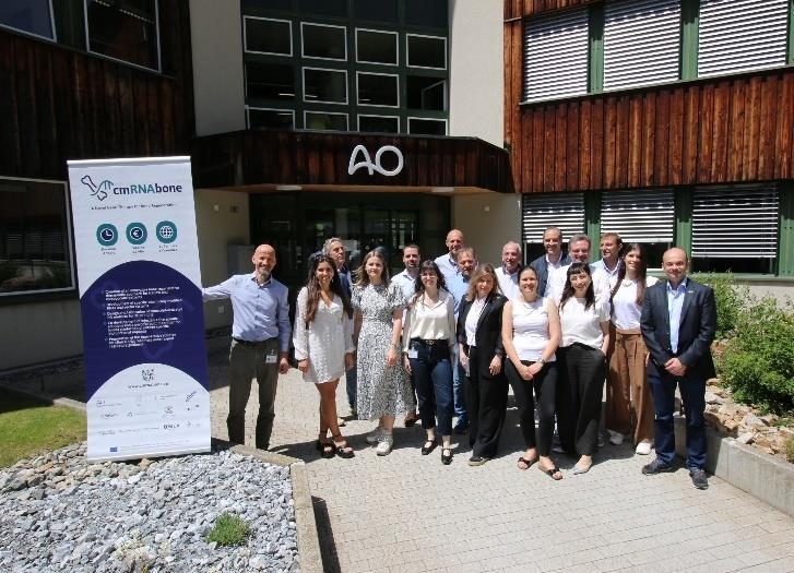
DEHST: Integration into the AO Davos Courses and AO Approval
The AO’s Digitally Enhanced Hands-On Surgical Training (DEHST) achieved two significant milestones this year. DEHST has been integrated into various educational programs of the AO Davos Courses. DEHST enhanced the AO Trauma Basic Principles Course by introducing a new hands-on exercise focused on intra-operative imaging and further expanded the practical exercise on intramedullary nailing with the freehand distal interlocking training module. The latter was also incorporated into the Basic Principles Course for Swiss Surgeons. Furthermore, the tri-plane fracture exercise of the pediatric fracture management course was augmented with DEHST and a new module for veterinary surgery was featured. The feedback was very positive, and the integration of additional modules into the AO Davos Courses 2025 is already underway. Another important achievement was DEHST’s certification with the “AO Approved” label by the AO Technical Commission (AO TC) Trauma Board. This certification validates the relevance and effectiveness of DEHST as an educational tool.


Two important achievements with DHEST in 2024: Integration into the AO Davos Courses 2024 (left) and receiving the “AO Approved” label from the AO Technical Commission Trauma Board.
OSapp hand-over to the AO Education Institute
The end of 2024 marked an important achievement in the history of OSapp (https://osapp.aofoundation.org/), the digital tool and knowledge hub of biomechanical principles for surgical education. Following an initial ARI-funded pilot phase, the tool has been developed based on AO Strategy Fund support and became an important part of AO’s digital portfolio. With the completion of the AO Strategy Fund project at the end of the year, ARI has handed over the strategic ownership of OSapp to the AO Education Institute and will remain involved by developing new content for extending the integration in AO Surgery Reference.

9.9 Visits
Senior politicians of the Aargau and Graubünden government
During the visit of the Aargau government to the Graubünden government, senior civil servants from both cantons were welcomed to the AO Center by the AO’s ITC Director Claas Albers and ARI Director, Prof R. Geoff Richards. After an introduction to the AO and its history, senior scientists presented the AO’s latest innovations and research projects. We are honoured to have had the opportunity to provide such an interested group of senior politicians with insight into the work of the AO.
From left: Prof R Geoff Richards, Daniel Spadin (Kanzleidirektor, GR), Dieter Egli (Landstatthalter, AG), Joana Filippi (Staatsschreiberin, AG), Alex Hürzeler (Regierungsrat, AG), Claas Albers, Jean-Pierre Gallati (Regierungsrat, AG), Jon Domenic Parolini (Regierungspräsident, GR), Martin Bühler (Regierungsrat, GR), Markus Dieth (Landamann, AG), Stephan Attiger (Regierungsrat, AG), Peter Peyer (Regierungsrat, GR), Marcus Caduff (Regierungsrat, GR), Carmelia Maissen (Regierungsrätin, GR).
AO Clinical Fellows
From all over the world to Davos!

On September 23, 2024 we welcomed a group of AO clinical fellows to the AO Center. Part Bansal from India (AO Trauma, fellowship at University Hospital Zurich), Jon Havard Sommernes from Norway (AO Spine, fellowship at University Hospital Basel), Manisha Kumar Javaji from India (AO Trauma, fellowship at University Hospital Basel), Elena Marina Maidol from Argentina (AO CMF, fellowship at University Hospital Basel), and Suraj Deshmukh from India (AO Spine, fellowship at Clinic Schulthess). They were given a tour of the ARI by Peter Varga and Prof Boyko Gueorguiev and got first-hand information about the AO’s latest research projects and innovations.
Visiting Professor
Fintan Moriarty has agreed to a visiting professorship at the Beijing University of Chemical Technology (BUCT) in Beijing China. This collaboration builds on the MOU signed between ARI and BUCT in 2023, and the exchange of students between both institutes in the past few years.

Fintan Moriarty and the Academic partners of the Biomedical Materials department of the Beijing University of Chemical Technology in December 2024.
10 ARI Medical Research Fellows
The ARI's Research Fellowship program again attracted resident and senior surgeons from around the world. Some of the many benefits to a surgeon are:
• Creation of tangible research results
• Possibility of a research publication as a co-author.
• Knowledge about how to approach research challenges.
• Inspiration from being part of a world-renowned international multidisciplinary R&D team.
• Inside knowledge of the AO.
• Enlargement of personal network for future R&D and AO Foundation activities.
• Chance to have a research friend/mentor that is always easy to contact.
Research Fellows

Chencheng Feng: Department of Orthopaedics, Xinqiao Hospital, Army Medical University, China
ARI Projects: 1. Bioreactor loaded whole intervertebral disc organ culture as models for ex vivo testing of biomaterials and biologics. 2. The biomechanics of spinal fusion with AO fracture monitor. I was involved in the one-year medical research fellow program in 2024 to investigate the pathogenesis of intervertebral disc degeneration based on the bioreactor. This ex vivo model provided a novel perspective to conduct research about intervertebral discs, which inspires me a lot. Moreover, as a spine surgeon, I participated in a biomechanical study of AO fracture monitor in transforaminal lumbar interbody fusion. This smart implant will be applied to evaluate the state of spinal fusion after surgery in the future. I truly enjoyed the innovative work in ARI. As a Chinese, I experienced a totally different life in Davos. I learned a lot from the culture of western countries. The excellent natural environment is unforgettable in my life. I will definitely come back to ARI to visit my friends.

Leopold Henssler: Department of Trauma Surgery, University Hospital Regensburg, Germany
ARI Project: Analysis of kinetics, neutralization, and efficacy of different administration routes of bacteriophage treatment in a sheep FRI model.
I joined the ARI as a Medical Research Fellow for four months in spring 2024 and worked with the team of infection biology. During my clinical work in Regensburg, I already started an investigation about macroscopic detection of necrotic bone using autofluorescence of the bone. After already receiving financial support by the AO for my research, I was given the opportunity to continue the experiments in Davos with this experienced team. Besides my own research I contributed to an ongoing project within the focus area of infection biology about kinetics and efficacy of treatment with bacteriophages in a sheep model of a fracture-related infection comparing intravenous and local phage application. Working with people from different teams and focus areas was very pleasant, as I was able to experience a supportive and helpful working atmosphere. Additionally, life in Davos from January to April was amazing, especially for activities in the snow like ski touring and free riding, which made my stay an overall unforgettable experience.

Fabian Pretz: Department of Orthopedic and Trauma Surgery, Lucerne Cantonal Hospital and University of Lucerne, Switzerland
ARI Project: Biomechanical comparison of augmented MIPO versus ORIF locking plate fixation in proximal humerus fractures with low bone mineral density; Biomechanical comparison properties and elongation of four different high-strength sutures and cerclages; Primary stability of nailing versus low profile dual plating of midclavicular fractures – a biomechanical study.
During my five-month research fellowship at the ARI, I had the opportunity to work on four self-conceived projects as part of my PhD studies in Health Sciences at the University of Lucerne. My focus at ARI was to design and execute biomechanical studies that explored innovative techniques in trauma surgery, aiming to enhance patient outcomes and advance the field of traumatology. The collaborative and inspiring environment allowed me to build valuable professional networks and form lasting friendships. I greatly appreciated the supportive atmosphere, which fostered both personal and professional growth. Beyond my work, I thoroughly enjoyed exploring the beautiful surroundings of Davos, creating unforgettable memories. I am deeply grateful for this enriching experience and would be delighted to return to ARI in the future.

Noémie Reinert: University Hospital, Basel, Switzerland
ARI Project: Establishment of a polymicrobial infection model in rabbit and evaluation of an injectable hydrogel for delivery of antibiotics as prophylactic treatment.
During my 12-month fellowship, I focused on establishing an in vivo polymicrobial infection model (with P. aeruginosa and S. aureus) in the setting of a humerus osteotomy and plate fixation. Once established, the efficacy of a Tobramycin-loaded hydrogel (GEDAI-T) is evaluated as a prophylaxis against infection in this polymicrobial contamination model. My internship involved the investigation of the in vitro and in vivo interactions of P. aeruginosa and S. aureus. In a third phase of my project, a phage cocktail will be evaluated as a wound lavage as novel antibacterial treatment strategy in the context of the increasing incidence of antibiotic resistance. During my time at the ARI infection biology group, I gained precious insights into bacterial growth, polymicrobial in vivo and in vitro interactions, building up invaluable knowledge and expertise as an orthopedic surgeon in the management of implant-related infections.

Luke van Rossenberg: University of Lucerne, Switzerland
ARI Project: Biomechanical comparison of fixation methods for forearm fractures as well as the evaluation of steel wire and suture cerclages.
During my four-month internship with the biomedical development team at ARI, I focused on conducting biomechanical studies of forearm fractures, specifically investigating different plate configurations, and cerclage fixation techniques in general. My role involved experimental testing and data analysis to assess fixation methods, contributing to the development of improved surgical techniques. With a background as a medical doctor with an aptitude for (trauma) surgery, I joined ARI's research team driven by a passion for enhancing future surgical care. Throughout my internship I developed an in depth understanding of biomechanics in fracture treatment, collaborated closely with experienced researchers and built lasting friendships. Outside of work, I enjoyed skiing with colleagues, swimming, and hiking through the stunning alpine surroundings, making my time in Davos both professionally and personally rewarding.
Guest Students

Julija Bulatova: Latvian Institute of Organic Synthesis, Riga, Latvia
ARI Project: Baltic Biomaterials Centre of Excellence (BBCE).
During my two-month stay at ARI, I dedicated my time to research aimed at testing the ability of biomaterials to aid in the healing of bone defects. While working on the BBCE project, I sought to broaden my skills and acquire new histological techniques. Throughout my trip, I became acquainted with processing and preparing of rat head samples for microscopic analysis and had the excellent opportunity to observe surgical procedures on rats and sheep, which provided me with valuable insights into other areas of the research process. My time in Davos was one of the most fulfilling experiences of my life. I was surrounded by a welcoming and friendly atmosphere, along with the breathtaking natural mountain scenery. These moments not only enhanced my professional expertise but also created lasting memories.

Darine D’Adam: Swiss Federal Institute of Technology (ETH), Zurich, Switzerland
ARI Project: Exploring the genetic drivers of staphylococcal abscess communities and biofilms in infection
During my six-month research project at ARI, I investigated the genetic and morphological factors influencing Staphylococcus aureus biofilms and staphylococcal abscess communities (SAC). By analyzing S. aureus mutants, I studied differences in biofilm formation on metal discs and SAC development on collagen layers. Using microscopy and quantitative assays, I assessed bacterial viability, fibrin deposition, and structural organization. This research aims to improve our understanding of S. aureus persistence in infections and its resistance to treatment. Coming from ETH Zurich, where I studied Medical Technology, this project allowed me to apply and expand my expertise in microbiology and biomedical research. Working in the collaborative and dynamic environment of Davos, alongside an inspiring and supportive team, has made this experience both scientifically enriching and personally rewarding.

Maria Rosa Iaquinta: University of Ferrara, Ferrara, Italy
ARI Project: Functional roles of fracture-related miRNA during endochondral differentiation.
I had the opportunity to spend three months (September-December 2024) at the Regenerative Orthopaedics Program of the ARI, thanks to the Interdisciplinary Academic Exchange Grant from the International Section of Fracture Repair (ISFR), Orthopaedic Research Society (ORS). This grant supported my research on the functional role of fracture-related microRNAs in endochondral differentiation. My time at ARI was an invaluable experience, allowing me to deepen my knowledge while collaborating with a multidisciplinary team of international scientists. Beyond the research, Davos provided a beautiful environment and the opportunity to connect with fascinating people from around the world. I am truly grateful for this enriching experience.

Livia Shanice Kiener: Zurich University of Applied Sciences (ZHAW), Winterthur, Switzerland
ARI Project: Staphylococcus aureus growth phenotypes: The impact of external conditions on biofilm and SAC formation.
I first joined the ARI for a two-month internship during my bachelor's degree, where I explored biofilm and staphylococcal abscess communities (SAC). During this time, I learned how to design in vitro experiments under various growth conditions, including the use of different antibiotics and sugars. I was truly fascinated by this work and had exceptional guidance throughout the internship, which motivated me to return for a small research project through my university. For this project, I investigated the factors that influence specific growth modes in S. aureus, with a particular focus on biofilm versus SAC formation. My time at ARI was incredibly valuable, not only did it provide me with my first real insight into research, but it also introduced me to a wonderful group of people. Beyond the lab, I also enjoyed the experiences outside of work, from hiking to skiing. I’m grateful for the opportunity and the many great memories I made along the way.

Melanie Rebecca Kuhn: Department of Orthopaedic Trauma, University Medical Center, Ulm, Germany
ARI Project: Bone biology and fracture healing
I recently finished my medical studies at the University of Ulm and started my work as a medical doctor in the Department of Orthopedic Trauma at the University Medical Center Ulm in Germany. Since I really enjoy research, I would like to continue doing research in the future. I am very grateful that I had the opportunity to get to know the AO Research Institute in Davos. During my internship in December 2024, I was allowed to join the Bone Biology group of Esther Wehrle. I gained valuable insights into musculoskeletal research focusing on fracture healing in rodents I was actively involved in the whole team and learned many laboratory techniques and much about data analysis. Beyond the professional aspects, I was able to build meaningful relationships with colleagues from all over the world, which made the experience even more enriching. This internship not only expanded my knowledge but also confirmedmy interest in pursuing a career in scientific research. I am grateful for this unique opportunity, and I will carry the lessons learned with me into the future. Thank you to the whole team!

Puk Kwant: Biology and medical laboratory research, Leiden, The Netherlands
ARI Project: Impact of the immune status on the susceptibility to surgical site infection around the spine.
During my nine-month internship at the Infection Biology Department at ARI, I had the opportunity to investigate the relationship between immune status and susceptibility to surgical site infections around the spine. This internship was part of my bachelor’s studies in biology and medical laboratory research. I worked alongside a team of researchers to examine how variations in immune function can influence the likelihood of infections in patients undergoing spinal surgery, contributing to a deeper understanding of infection biology and postoperative complications. The internship allowed me to apply my academic knowledge to real-world research, where I gained valuable laboratory and analytical skills. Outside of the lab, I made the most of the Davos surroundings, enjoying hiking in the mountains and skiing during the winter months, which made for a truly memorable experience.

Lauma Levina: Riga Technical University, Riga, Latvia
ARI Project: Experimental data analysis/focus on research topics and use of ex vivo models in biomaterials research.
As part of my doctoral studies, I had a great opportunity to visit the ARI for 3 months. During this time, I had the opportunity to learn to work with osteoclast cell cultures, learn new aspects of cell imaging and gene expression. Doing ELISAS became second nature to me. With these methods, I could study how platelet-rich fibrin and material synergies impact cell culture activity from samples prepared in Latvia. Also, I had an opportunity to be involved in an additional project that was based in ARI. This was all done under the supervision of wonderful Dr Elena Della Bella which I am grateful about. Outside of the lab I either went to the mountains or spent time with other students from ARI and nearby research institutes. Time spent in Davos was wonderful because of the people, research opportunities and nature.

Yuqi Liu: Xiangya Medical School, Central South University, China
ARI Project: Optimizing the role of GPR68 in human MSC chondrogenic and osteogenic differentiation.
As a guest PhD student at ARI, I investigated GPR68’s role in directing human MSC differentiation into chondrogenic and osteogenic lineages. My work involved pellet culture, qPCR, and pharmacological modulation of GPR68 to assess its impact on lineage commitment. This experience enhanced my expertise in cell-based differentiation assays, while the collaborative environment at ARI deepened my understanding of regenerative medicine. My time in Davos was both academically enriching and personally rewarding. I look forward to applying these insights to my PhD research.

Ksenia Menshikh: Università del Piemonte Orientale, Novara, Italy
ARI Project: PREMUROSA
I joined the ARI as a postdoctoral fellow for 6 weeks in the framework of the EU Horizon 2020 MSCA ITN project PREMUROSA (GA No 860462). During that short yet intense secondment I collaborated with the group of Dr Tiziano Serra and mastered skills of sound-guided tissue engineering. I successfully applied this non-invasive manipulating method to study how my bone cancer-like engineered scaffold may affect the process of vascularization. Specifically, I patterned the components of my systemparticles of a calcium phosphate scaffold and osteosarcoma cell spheroid - using acoustic waves and then monitored how fast and efficiently endothelial cells are migrating towards them and whether they are forming a characteristic network. The obtained results helped me in understanding whether I can advance the in vitro model of osteosarcoma (primary bone cancer) I am working on - in particular, whether I could have it vasculard. A manuscript on this work has been submitted and, hopefully, will soon serve as an input in addressing the in vitro-in vivo gap in preclinical drug screening and help us understand osteosarcoma better. This secondment to ARI was an invaluable experience for me from all points of view. As a young researcher, I had an opportunity to learn new techniques and work in a very efficiently built and friendly research environment with top scientists in the field. As a nature lover, I did not miss a single chance to go for a hike or a run in the breathtaking landscapes of Davos.

Giulia Minikus: Swiss Federal Institute of Technology (ETH), Zurich, Switzerland
ARI Project: Efficient quantitative bone fracture healing tools towards clinical application.
I had the opportunity to write my Master's thesis in the Biomechanics and Modeling group at the ARI, where I investigated the computational efficiency of different modelling approaches in healing simulations after bone fractures. My work was embedded in a larger research project, which allowed me to work closely with other students and researchers. As the final project of my Biomedical Engineering study program at ETH Zurich, I appreciated the opportunity to complete my degree in such an inspiring and supportive working environment. In addition to the enriching work experience, I enjoyed the beautiful landscape of Davos while running and cross-country skiing.

Luise Puls: University Hospital, Basel, Switzerland
ARI project: Impact of end screw configuration on peri-implant fracture risk in distal femoral plating.
In January 2024, I had the incredible opportunity to join the ARI as a medical fellow for a four-month period. My research focused on the biomechanical investigation of the end screw in femoral plating. As someone completely new to this field, I was amazed by how much I was able to learn in such a short time. This was made possible by the exceptional support, patience, and guidance of the team, who welcomed me warmly. My time at ARI was not only a valuable academic and professional opportunity but also a truly enriching personal journey. The chance to collaborate with experienced researchers in such a stimulating and inspiring environment strengthened my fascination with biomechanics and has motivated me to continue exploring this field in the future. I am deeply grateful for this experience and the wonderful people I had the privilege of working with.
Nicoletta Restione: Polytechnic, Milan, Italy
During my three-month research stay at the ARI from October to December, I worked on my master's thesis as part of my biomedical engineering studies at Politecnico di Milano, specializing in cell, tissue, and biotechnology engineering. My project focused on developing injectable hydrogels based on gelatin and silk fibroin for nucleus pulposus regeneration. This experience provided me with the invaluable opportunity to work in an international research environment, collaborating with colleagues from diverse backgrounds while gaining hands-on experience with advanced biomedical technologies. In particular, I conducted experiments using a uniaxial bioreactor, which was directly aligned with my research goals and academic background. My time at AO was both professionally and personally enriching, allowing me to deepen my expertise in biomaterials while fully immersing myself in a dynamic and stimulating scientific community.

Svenja Wacker: Albert-Ludwig University Freiburg, Freiburg, Germany
ARI Project: Impact of irrigation fluid acidity on articular cartilage: an ex vivo injury model.
During my 10 months as a medical research fellow at the ARI, I had the chance to explore the pH of irrigation solutions in arthroscopy due to a collaboration with the University Medical Center Freiburg. It not only allowed me to get an insight into scientific research and lab work but also to dig deeper into one of my major interests in medicine, orthopedics and trauma surgery. My time at ARI laid the foundation for my doctoral thesis and gave me the opportunity to enhance my scientific knowledge and skills In addition to the academic and professional development, this period was also a time of personal growth. I formed lasting friendships and created unforgettable memories through various adventures - whether it was skiing, hiking in the mountains (both day and night), or enjoying memorable nights out with colleagues. Overall, these 10 months were an exciting and enriching experience, filled with both academic achievements and personal milestones. It was a period I wouldn't want to miss, and I am grateful for the opportunity to have been part of such a dynamic and inspiring environment.

Lucille Wespi: Swiss Federal Institute of Technology, Zurich, Switzerland
ARI Project: Investigating mineralocorticoid receptor activation during glucocorticoid-induced in vitro osteogenesis
As part of my Master’s studies in Biomedical Engineering at ETH Zurich, I had the privilege of conducting my thesis in the Cell Biology and Mechanoregulation group at ARI. My project focused on investigating mineralocorticoid receptor activation during glucocorticoid-induced osteogenesis in human bone marrow mesenchymal stromal cells. During my time at ARI, I had the opportunity to deepen my expertise in cell culture and molecular biology techniques, while also strengthening my skills in experimental design and planning. A particular highlight was presenting my research at my first scientific conference, where I engaged with researchers from around the world and received invaluable feedback. Working in ARI's dynamic and collaborative environment, surrounded by passionate scientists, was both inspiring and rewarding, and the supportive atmosphere allowed me to grow both professionally and personally. Moreover, the breathtaking natural setting of Davos not only provided an ideal balance but also offered plenty of opportunities to hike and recharge after work, further contributing to my positive experience at ARI.

Fabienne Wynne: Aberystwyth University Penglais & The Royal Veterinary College, Aberystwyth, UK
ARI Project: ARI Veterinary Externship.
During my one-month externship at ARI, I focused on anaesthesia across various species, assisted in surgeries and diagnostic imaging procedures, and participated in pre- and post-operative patient evaluations. As my externship concluded, I was eager to continue my clinical training, building on the skills and knowledge I developed with the AO VET team at the Preclinical Facility. Working alongside supportive professionals, I gained invaluable insights into the ethical and animal welfare considerations of clinical research. The AO Veterinary externship sparked my interest in anaesthesiology and solidified my commitment to pursuing a career as an equine veterinary surgeon. Beyondthe clinical experience, I had thechance to explore the breathtaking mountain ranges with new friends. This opportunity was not only professionally enriching but also profoundly transformative, leaving me with unforgettable memories.

Leonardo Zonca: Polytechnic, Milan, Italy
ARI Project: Characterization of an injectable hydrogel for nucleus pulposus regeneration.
During my three-month stay at the ARI, I had the opportunity to work on the project characterization of an injectable hydrogel for nucleus pulposus regeneration. This research was part of my master’s thesis in biomedical engineering at Politecnico di Milano and focused on evaluating the hydrogel developed in Milan using a bovine intervertebral disc model in a uniaxial bioreactor. At ARI, I gained hands-on experience in biomechanical testing and tissue engineering while collaborating with an international team of scientists and clinicians. This experience deepened my understanding of translational biomedical research and strengthened my technical and analytical skills. Beyond work, I thoroughly enjoyed my time in Davos, exploring its beautiful surroundings and engaging in outdoor activities that made my stay even more memorable. I am grateful for this enriching experience and for the support of the ARI team.
Internships

Florence Albrecht: University of Bern, VetSuisse Faculty, Bern, Switzerland
I completed a 2-month internship at the ARI for my veterinary studies and had the opportunity to work in several ongoing projects. The focus of my internship was to help during surgeries, ranging from inducing and monitoring anaesthesia to postoperative care and occasionally even scrubbing in. During this time, I was able to practice my veterinary skills and learn about working with and taking care of laboratory animals from experienced veterinarians. Since the first day of the internship, I felt very welcomed into the team and was grateful to be able to work and learn in such a familial environment. While off work, I was lucky to catch a few hockey matches and discovered I liked hockey. I enjoyed my time in Davos and hope to return some time in the future.

Denise Bentivoglio: Polytechnic of Turin, Italy
ARI Project: Development of a custom 2D-3D registration code for pre- and post-operative clinical images.
During my six-month internship at the ARI, I was part of the Biomedical Development program within the Biomechanics and Modelling focus area. My work involved contributing to the development of a custom code for performing 2D-3D registration of pre- and post-operative clinical images. This experience allowed me to enhance my coding skills while collaborating with colleagues from diverse cultural backgrounds, fostering both technical and interpersonal growth. Outside of work, my six months in Davos gave me the opportunity to enjoy the Swiss summer, explore the surrounding mountains, and connect with new friends from around the world.

Corinne Bischofberger: Swiss Federal Institute of Technology (ETH), Zurich, Switzerland
ARI Project: Investigating titanium allergy and non-union development.
During my three-month internship at ARI, I explored the impact of titanium allergy on septic and aseptic non-union. As a master's student in Biomedical Engineering at ETH Zurich, I aimed to deepen my understanding of orthopaedic complications related to fracture healing. My work focused on cytokine activity in patient PBMCs to determine whether titanium hypersensitivity contributes to non-union. Results showed no significant differences in cytokine activity between patient groups, but control samples exhibited consistently higher activity, likely due to sample handling. A single titanium allergy patient showed elevated cytokine activity, but further studies are needed for confirmation. Optimization of stimulation conditions could improve future research. This study found no clear link between titanium allergy and non-union, emphasizing the need for larger, well-controlled studies. My time at ARI provided invaluable insights into orthopaedic research, while Davos' natural beauty offered a refreshing balance to my work.

Lisa Fell: University of Veterinary Medicine, Hannover, Germany
ARI Project: Preclinical Research
During my two-month internship at the ARI, I focused on working in different projects with the vets at the Preclinical Services. I learned how to handle the laboratory animals and how the planning of an animal experiment works. As a part of a motivated and professional team I really enjoyed my time there. I got to know all the activities that are necessary during an animal experiment. This started with the preparation of the animals for the surgeries, continued with the anaesthetic monitoring during them and the medical aftercare and ended with the recording of post-operative monitoring of the animals as well as regular scoring of their health status and also imaging diagnostics. During my week with the animal caretakers, I learned how valuable and important their work is. Especially in the field of animal testing, close cooperation is essential for the welfare of the animals. I liked the respectful treatment of them. Always perceived as a colleague at eye level and encouraged in my actions, I was given the confidence to work independently. The team at the Preclinical Services made my time at ARI unforgettable and I am grateful to have had the opportunity to work with them.

Jeremia Giger: University of Zurich, VetSuisse Faculty, Switzerland
During my two-month stay at the ARI as a veterinary intern in the preclinical facility, I was involved in supporting the team during surgical procedures, anaesthesia, and the daily care and nursing of the animals. This internship was part of my veterinary medicine studies, which I will complete in spring 2025. Throughout my time at ARI, I was able to expand my practical skills and gain valuable hands-on experience with animal species that are not commonly covered in the university curriculum. I was warmly welcomed by the team, which made it easy to integrate into the daily workflow and contribute effectively. One of the highlights of the internship was the chance to work alongside an international team of professionals from diverse backgrounds, which provided a unique perspective on collaborative research and veterinary care. The experience deepened my understanding of the preclinical field and allowed me to apply my academic knowledge in a practical setting.
11 Project Abstracts by Sponsors
11.1 AO CMF
AO CMF Clinical Priority Program (CPP) Consortium: Instructive bone regenerating hydrogel for translational bone repair (AO CMF BOOST) (started) (ARI consortium personnel: M Stoddart, E Della Bella, T Serra, M D'Este, E Bektas)
Background: As part of a strategy to better utilize funding streams, AO CMF made an open call for collaborative clinical priority program (CPP), with the instruction to ideally to include both ARI with external partners. After an open call eligible consortia were independently evaluated by the AO RRC and the highest ranked consortia was selected.
Goal: Due to the lack of sufficient autograft volume, large bone defects commonly require additional material, both as a void filler and as a source of osteogenic material. This project aims to develop a novel bone forming substitute comprising of a self-assembling peptide system, combined with a bone allograft. The unique aspect of the material is that it can be used to regulate exposure to endogenously produced growth factors, thus improving osteogenesis while at the same time controlling the immune response and inflammation. This is achieved by the incorporation of peptides that can selectively bind, organize, and present specific growth factors (Interleukin-1, vascular endothelial growth factor, bone morphogenetic protein 2). The binding efficiency can be fine-tuned, thus regulating the presentation to cells and subsequent downstream signaling. The graft will be prepared intraoperatively and in addition can also be 3D printed intraoperatively using soundwaves to produce defined patterned sheets that can be sutured into calvarial defects. Furthermore, the artificial bone graft is mixed with bone marrow aspirate concentrate (BMAC) to form a rich intraoperative cellbased implant material that is precellularized. A further challenge in the development of novel bone biomaterials are the methodologies commonly used to test their functionality in vitro. A significant number of materials, if not most, have been tested in vitro with promising results, yet they commonly go on to fail in vivo. This suggests there is a fundamental flaw in the process used to test materials in vitro. With this in mind, a second arm of this study will specifically address how materials are tested in vitro and ex vivo, with in vivo data being reverse correlated to in vitro results in order to establish more predictive early outcome measures. This will be achieved by requiring a detailed analysis of immune regulation, inflammation, and osteogenic differentiation.
Pres:
• Serra T. “Controlling multicellular organization by extrinsic fields”. 1st Alpine Winter School for Biofabrication, 12.01.2024, Radstadt, Austria
• Serra T. “Engineering of multicellular systems by hydrodynamic waves”. Keynote speaker at the Innovative Biomaterials for Novel Medical Devices, ExcellMater Conference, 10.04.2024, Belgrade, Serbia
• Serra T. “Biofabrication and way to market”, Lecture at University of Eastern Piedmont "Amedeo Avogadro", UPO, 02.05.2024, Novara, Italy
• Serra T. “Controlling multicellular organization by sound”. Invited Seminar at CHU de Nantes, 23.05.2024, Nantes, France
• Serra T. “Engineering multicellular systems by Sound-Induced Morphogenesis”, Charles Perkins Center, 12.10.2024, University of Sydney, Australia
• Serra T. “Advanced bioassembly technologies”, AusBioprinting Workshop 2024, 02.12.2024, UTS, Sydney, Australia
Pub:
• Di Marzio N, Tognato R, Della Bella E, De Giorgis V, Manfredi M, Cochis A, Alini M, Serra T. Differential proteomics profile of microcapillary networks in response to sound patterndriven local cell density enhancement. Biomater Biosyst. 2024 Mar 29:14:100094. doi: 10.1016/j.bbiosy.2024.100094. eCollection 2024 Jun.
Partners:
• Mata A (D. Eng), University of Nottingham, United Kingdom
• Akdis C (MD) and Akdis Mübeccel (MD, PhD), Swiss Institute of Allergy and Asthma Research, University Zurich, Davos, Switzerland
• Zhiyu Z (MD, PhD) and Yingying Lu (MD, PhD), The Seventh Affiliated Hospital, Orthopaedics Department, Scientific Research Center Sun Yat-sen University, China
11.2 AO Spine
Evaluation of anti-degenerative therapies and diagnostic targets for the intervertebral disc (Theranostic follow-on; Printdisc follow-on) (ongoing) (S Grad, A Soubrier, D Menghini)
Background: Disorders of the intervertebral disc (IVD) are multifactorial and require targeted approaches. (1) In early stages of IVD degeneration, physical therapy has shown promising effects in terms of back pain relief. Specifically, traction therapy was demonstrated to improve symptoms and induce beneficial effects on imaging parameters in clinical and preclinical studies. However, effects of traction load on IVD cell phenotype, matrix and water content have not been systematically investigated. (2) Another targeted approach consists in the application of antibiotics for prevention of bacteria invasion after a nucleotomy procedure; it is hypothesized that bacterial infection may increase the risk of developing Modic changes of the endplate.
Goal: The goals of our research are to advance our in vitro cell and organ culture models and then use them to investigate (1) the influence of traction loading on non-degenerative and induced-degenerative bovine IVDs maintained in organ culture; and (2) the feasibility of an antibiotic-releasing hydrogel in a bovine IVD organ culture nucleotomy model.
Results: (1) A new organ model was established consisting of a holding system and biochamber that allow the application of traction forces or unloading to bovine IVDs. The biological and biomechanical outcome parameters showed consistency. Namely, the biological readouts indicate higher water content and an anti-catabolic response of the IVDs after active dynamic unloading, while the biomechanical outcomes suggest a higher water content and improved mechanical resistance (Fig. 11.2.1). Further study will investigate the effect of active dynamic unloading in an induced degenerative IVD model. (2) The study demonstrates that mechanics can be partially restored in a nucleotomized IVD using a mechanically interlocked patch and HA-Tyramine hydrogel injection ex-vivo. Hydrogels with different viscosity show a different mechanical response after injection in the bovine IVD. Samples combining an interlocked patch repair with either low or medium viscosity hydrogel successfully withstood physiological stresses (2.3 MPa) and could restore partial biomechanical parameters of the nucleus pulposus. We conclude that combining a HA-Tyramine hydrogel and interlocking patch holds potential for further study, with the hydrogel serving as viable carrier for drugs while offering load protection

Figure 11.2.1: Sample preparation and bioreactor setup for dynamic axial loading and unloading of bovine caudal intervertebral discs under organ culture conditions.
Pres:
• Menghini D, Ongini E, D’Este M, Moriarty F, Grad S, Snedeker JG, Dudli S. Combination of an Annulus Fibrosus Repair with a Mechanically Interlocked Patch and a Nucleus Pulposus Augmentation with Acid-Tyramine Hydrogel in an Ex vivo Model. ORS 2024 (poster)
• Menghini D, Ongini E, D’Este M, Moriarty F, Grad S, Snedeker JG, Dudli S. Hydrogel injection and mechanically interlocking patch partially recover intervertebral disc mechanics while withstanding physiological load. ISSLS 2024 (poster)
• Soubrier A, Kasper H, Alini M, Jonkers I, Grad S. Dynamic unloading of healthy bovine tail discs: biomechanics and biology suggest facilitated water uptake. EORS 2024 (podium)
Pub:
• Schulze F, Määttä J, Grad S, Heggli I, Brunner F, Farshad M, Distler O, Karppinen J, Lotz J, Dudli S. Proteomic analysis of serum in a population-based cohort did not reveal a biomarker for Modic changes. JOR Spine. 2024 Jul 15;7(3):e1337. doi: 10.1002/jsp2.1337
• Jansen JU, Teixiera GQ, Vernengo A, Grad S, Neidlinger-Wilke C, Wilke H-J. Papain Injection Creates a Nucleotomy-like Cavity for Testing Gels in Intervertebral Discs. Gels 2024, 10, 571. https://doi.org/10.3390/gels10090571
Partners:
• Jonkers I (Prof), KU Leuven, Belgium
• Dudli S (Prof), Balgrist University Hospital, University of Zürich, Switzerland
• Snedeker J (Prof), ETH Zürich, Switzerland
11.3 AO Trauma
Systematic assessment of the impact of postoperative activity on fracture healing by controlled mechanical stimulation (ActiveFix III) (ongoing) (J Barcik, M Ernst)
Background: It is widely accepted that mechanical stimulus (interfragmentary motion) is integral to the callus formation process during secondary bone healing. While certain aspects of mechanical stimulation e.g. the magnitude of interfragmentary motion, loading mode or interfragmentary strain, have been studied repeatedly, the impact of temporal factors such as the number and distribution of loading cycles on healing progression has been widely disregarded. However, these factors directly relate to the clinical rehabilitation of fracture patients, but previous experiments often lacked appropriate models to investigate their effect on fracture healing with clinically relevant stimulation protocols.
Goal: The ActiveFix III project aims to further investigate the role of patients' activity in fracture healing. We intend to investigate how the number of loading cycles applied per day impacts the formation of fracture callus and healing time. In the frame of this project, the same tiltingwedge active fixator and control unit used and developed during the previous ActiveFix II project is applied.
Results: A total of 12 sheep were operated and subdivided into four groups based on the number of cycles applied per day, i.e., 10, 100, 1,000, and 10 000 cycles. Healing progression was monitored through weekly radiographs and through measurement of the repair tissue stiffness during stimulation with the active fixator. The final healing stage was assessed by post-mortem high-resolution computed tomography (CT) scans after sacrificing the animals five weeks post-operation. Radiographically, callus formation was observed in all groups starting from the third week post-operation, indicating that as little as 10 cycles per day can be sufficient to induce callus formation. However, the largest callus formation was observed in the group that received 10 000 cycles per day (Fig. 11.3.1). Evaluation of the repair tissue stiffness and CT scans for the final assessment are currently ongoing.

Figure 11.3.1: Contact radiographs from exemplary animals that received 10 and 10 000 stimulation cycles/day respectively. The bone wedge is tilted in clockwise direction, hence exerting compressive strain on the partial osteotomy on the right side of the wedge.
Pres:
• Barcik J. In vivo models for studying the mechanobiology of fracture healing. 2024 EORS (oral)
• Barcik J, Constant C, Buschbaum J, Zeiter S, Ernst M. The impact of postoperative mechanical stimulation on callus formation - a case series conducted on sheep. 2024 DKOU (oral)
• Barcik J, Ernst M, Buchholz T, Constant C, Zeiter S, Verrier S. Linking local fracture mechanics with systemic biological response. 2024 ORS (poster)
Patient-specific rehabilitation planning for plated long bone fractures (RehabFE) (ongoing) (P Varga, D Mischler, A Valenti, B Gueorguiev)
Background: Plate osteosyntheses failures, like plate bending, persist in patients. Finite element (FE) models simulate bone-plate mechanics but lack in vivo validation due to unknown loads. Implantable sensors monitoring plate strain during healing offer a chance to validate these models, though the connection between sensor signals and implant failure is unclear.
Goal: To validate FE models by comparing sensor signals from implantable sensors on plates in cadaveric ovine tibia fractures to virtual signals from corresponding FE models, particularly at the onset of plate bending, using a calibration factor from isolated sensor tests.
Results: Seven cadaveric ovine tibia fractures with plate fixations were tested. A four-point bending test on the isolated sensor showed high correlation (R2>0.99) between experimental and virtual signals, yielding a calibration factor. After applying this factor, FE models strongly correlated with experimental sensor signals at yield (concordance correlation coefficient = 0.89, standard error of estimate = 187.0, relative standard error = 11.9%). This indicates FE models can predict the sensor signal at plate bending onset, enabling retrospective in vivo validation and supporting tailored rehabilitation protocols to lower patient complications.

Figure 11.3.2: Experimental test setup including the AO Fracture Monitor (A), the corresponding samplespecific FE model (B), and the FE results (C). The yellow arrows indicate the AO Fracture Monitor mounted on the plate. Experimental sensor signal at yield load was well predicted using the calibrated virtual sensor signal from the FE models (D). Implant material is indicated in the figure legend (S = Stainless steel, T = Titanium).
Pres:
• Mischler D, Valenti A, Ernst M, Varga P. Predicting in-vivo plate bending using a continuous implant load monitor. 2024 GCB Symposium (poster)
Validated simulations of bone fracture healing (SimBo) (ongoing) (P Schwarzenberg, B Gueorguiev, M Ernst, P Varga)
Background: Bone fracture healing is a complex process that relies on both mechanical and biological cues at the fracture site. The mechanical stability is crucial, and any issues can have detrimental effects on healing and lead to delayed or nonunion of the fracture. While our understanding of the mechanical stimuli that guide bone healing has advanced and subjectspecific computer simulations are more accessible, we still do not have a validated healing simulation model that can predict the structural time-course of healing.
Goals: SimBo aims to develop a mechanoregulatory modeling platform and validate it against unique preclinical datasets established at ARI. This is the first time these types of models are validated against an in vivo ground truth measurement. The healing simulations could predict nonunion risk, determine rehabilitation protocols, and assist with implant design and selection.
Results: A mechanoregulatory modeling platform developed at ARI has shown promising early results. It demonstrates the ability to predict the healing process over time in response to mechanical stimuli in specimen-specific ovine osteotomy models. The platform accurately predicted morphometric changes during healing and successfully matched in vivo sensor measurements with simulations. Additionally, it identified delayed unions and nonunions from a cohort using only post-operative CT scan data. These findings are laying the groundwork for optimizing and validating the platform to predict a wide range of healing outcomes.

Figure 11.3.3: a) Cross section views of healing simulations of a normal healer, delayed healer, and nonunion at the 30-day timepoint. Cartilage is shown in red and mineralized tissue in tan. b) Healing curves for the three cases, showing the fastest healing in the normal healer, slower healing in the delayed union, and incomplete healing in the nonunion.
Pres:
• Schwarzenberg P, Schlatter J, Ernst M, Dailey H, Varga P. Predictive fracture healing simulations correlate with in vivo sensors in an ovine tibia model. 2024 ESBiomech (oral)
• Hetreau C, Feist A, Ernst M, Varga P, Schwarzenberg P. In silico diagnostic and prognostic evaluation of bone fracture healing. 2024 EORS (oral)
• Varga P, Schlatter J, Ernst M, Dailey H, Schwarzenberg P. Prognostic fracture healing simulations agree with in vivo sensors in an ovine tibia model. 2024 DKOU (oral)
Pub:
• Schwarzenberg P, Schlatter J, Ernst M, Windolf M, Dailey H, Varga P (2024). Prognostic bone healing simulations in an ovine tibia model validated with in vivo sensors. J Orthop Res 43(2):370-378. doi: 10.1002/jor.26007
Theses:
• Schlatter J. Predictive finite element-based fracture healing simulations validated against continuous in vivo sensor data in an ovine tibia model. Master Thesis, ETH Zurich, 2024
• Feist A. Preclinical validation of mechanoregulatory fracture healing simulations. Master Thesis, University of Bern, 2024
• Sommer S. Towards optimized predictive bone fracture healing simulations. Master Thesis, ETH Zurich, 2024
• Camichel C. Development of an early-stage healing measure using µCT image-based finite element simulations in a mouse femur defect model. Master Thesis, ETH Zurich, 2024
• Minikus G. Efficient quantitative bone fracture healing tools towards clinical application. Master Thesis, ETH Zurich, 2024
Multiphasic bone putty with dynamic porosity for cell invasion (MEDICI) (ongoing) (R Randriantsilefisoa, M D'Este)
Background: Most currently available synthetic bone graft substitutes function as bone fillers lacking appropriate biological competence. Producing a vascularized bone graft substitute that promotes cell migration and differentiation is crucial to restore native bone structures on large defects.
Goal: Introduce a material designed to improve cell and vasculature invasion, and preserving mechanical competence, for the repair of bone defects.
Table: Comparison of the fabrication methos
Criteria
Scalability
Reproducibility
Interconnectivity of Pores
Perioperative use
Inconveniences of Fabrication
Fugitive particles
Scalable with extra cell encapsulation step Preparation needed at the surgical site, extending OR time.
Morphology reliant on particle distribution. Shape is moldable to defect size and geometry.
Negative molding
Higher scalability: the design is done via CAD. Ready for implantation without on-the-spot preparation.
Controlled morphology. Shape precisely adjusted during printing for better defect fit.
Needs further development. Easily achieved through design.
Easily applicable, similar to bone putties
Cells are encapsulated presurgery needing infrastructures.
Needs planning an pre-production
Pre-production requires time
Results: We developed two approaches for fabricating dynamic scaffolds for bone regeneration, and characterized their porosity, morphology, chemical composition, and biological remodeling. The aim was to explore optimal porosities in a bioceramic material to facilitate cell and fluid invasion, thereby supporting the differentiation of human mesenchymal stromal cells (hMSCs) into osteoblasts at critical bone defect sites. The first approach consists in making a composite based on hydroxyapatite and fugitive alginate beads of varying sizes. This method employs simple aqueous synthesis at room temperature from hydroxyapatite precursors. The second approach uses PVA as a negative molding template and sacrificial component. Drawing inspiration from trabecular bone and coral mineralization, our negative molding hyperboloid structure achieved 55% porosity – typical of trabecular bone – using a stable hydroxyapatite composite without sintering. The biological characterization indicates that the geometrical features left behind from the fugitive elements determine different expressions of osteogenic markers. The gyroid geometry showed a trend for higher RUNX2 at early time points, and higher collagen expression at week 3. The main characteristics of the 2 methods are summarized in Table 1. Next steps of the project will investigate cell and vasculature invasion.
Thesis:
• Al Saify I. Dynamically porous calcium phosphate composites as bone graft substitutes. Master Thesis Faculty of Life & Health Sciences, Maastricht University
Partners:
• Niloofar Tahmasebi Birgani (Prof), Maastricht University, NL
• Loca D (Prof), Riga Technical University, Riga, Latvia
Linking mechanics and omics to improve early bone healing (MechOmics) (ongoing) (E Wehrle, M Stoddart, S Zeiter, S Verrier, M Schröder, N Giger, J Barcik, D Arens, D Gehweiler)
Background: Mechanical loading is a key factor for normal progression of the fracture healing process. Despitethe advances in fracture fixation, there remains a subset of patients that suffer from healing complications, resulting in delayed healing and non-unions. Currently it is not possible to reliably identify healing complications at an early stage when treatments, e.g. mechanical intervention therapies may be more effective. Understanding of the underlying mechanically induced molecular mechanisms on an individual basis could enable wider-scale harnessing of the mechano-sensitivity of the regenerative process in clinical applications. Novel multimodal approaches in small animals have the potential to precisely capture and understand these mechanical-induced biological changes during fracture healing on an individual basis. Within this project we will use and adapt well-established equipment for precisely controlled local application of cyclic mechanical loading in mouse femur defect models.
Goal: To identify systemic biomarkers indicating early deviations from normal healing progression also allowing for initiation and targeted adjustments of individualized mechanical intervention therapies.
Results: Within the project the previously developed displacement-controlled loading mode was successfully applied to longitudinally monitor healing progression via in vivo stiffness measurements in individual animals. Spatial transcriptomics analyses (Fig 11.3.4) were complemented with additional timepoints capturing healing-phase associated spatiotemporal molecular patterns for union and non-union defects.

Figure 11.3.4: Spatial transcriptomics of a non-union (bottom) and union (top) femur fracture in mice. Destained H&E section prior to transcriptomic probe transfer (A), Gene clustering (B).
Pres:
• Schröder M, Giger N, Barcik J, Gens L, Arens D, Zeiter S, Varga P, Stoddart M, Wehrle E. Spatial transcriptomics reveal distinct gene expression patterns in non-union and union bone fractures in mice. 2024 ECTS (oral)
• Barcik J, Schröder M, Giger N, Gens L, Arens D, Camichel C, Schwarzenberg P, Gehweiler D, Stoddart M, Zeiter S, Varga P, Wehrle E. In vivo stiffness measurement enables discrimination between impaired and normal fracture healing in mice. 2024 ESB (oral)
• Giger N, Schröder M, Gens L, Arens D, Zeiter S, Stoddart M, Wehrle E. Spatial transcriptomics workflow enables distinct tissue-specific molecular characterization of nonunion and union bone fractures in mice. 2024 EORS (oral)
• Giger N, Schröder M, Gens L, Arens D, Zeiter S, Stoddart M, Wehrle E. Probe transferbased spatial transcriptomics to study fracture healing in a femur defect model in mice. SVGO / SBMS 2024; Bern (oral)
• Giger N, Schröder M, Arens D, Gens L, Zeiter S, Stoddart M, Wehrle E. Spatial transcriptomics to study fracture healing in mouse models. 2024 Graubünden Forscht (oral)
• Schröder M, Giger N, Barcik J, Gens L, Arens D, Gehweiler D, Varga P, Zeiter S, Stoddart M, Wehrle E. Spatial transcriptomics reveal distinct gene expression patterns and treatment targets during fracture healing in (non)-union models in mice. 2024 ISFR (oral)
• Wehrle E. Omics-based mouse models to study mechanobiology and tissue crosstalk during bone healing. 2024 ORS (oral)
• Wehrle E Spatial transcriptomics of bone healing. 2024 DGfZ (oral)
• Wehrle E Enhancing healing with mechanical loading. 2024 ISFR (oral)
Mechanistic understanding of non-unions in vivo (MeNU) (ongoing) (M Moriarty, E Wehrle, C Siverino, C Chabot, J Tapia-Dean, S Zeiter, Alicia Feist, Peter Schwarzenberg)
Background: Fracture non-union is a severe and common clinical complication where a bone fracture fails to heal within an expected time frame typically 6 months despite appropriate treatment. Non-unions are caused by multiple factors, primarily mechanical instability at the fracture site and infection. Failure of maintaining the required mechanical stability within the fracture site can prevent healing, resulting in incomplete recovery and non-unions. Fracturerelated-infection (FRI) can complicate this process by introducing a persistent inflammatory response that impedes bone regeneration. A comprehensive understanding of how mechanical instability and infection contribute to non-union, allowing for the development of targeted interventions that can improve healing outcomes, reduce the need for repeated surgeries and ultimately improve patient quality of life.
Goals: To develop mouse models of mechanically-associated non-union in order to investigate the impact of fixation stiffness and infection on fracture healing and non-union progression (see Fig 11.3.5 for study design)
Results: Mechanical testing and finite element (FE) modeling were used to characterize the different types of external fixators (RISystem, Landquart). These methods revealed significant differences in stiffness among the various external fixators, with the more flexible fixator exhibiting the lowest stiffness. Additionally, mechanical stiffness values showed a strong correlation with FE predictions, confirming the robustness of the external fixator model. These results validate the design of the fixator and its ability to predict stiffness in experimental models, offering a more accurate tool for studying fracture healing and non-unions. This predictive capability is valuable for investigating mechanical conditions that promote healing and those that contribute to non-unions, particularly when combined with infection. The optimized external fixator set-ups will be used in in vivo studies aiming to determine optimal mechanical conditions for studying healing and non-union.

Figure 11.3.5: MeNU Project Overview
Partner:
• Geris L, Laboratory for Biomechanics and Computational Tissue Engineering, KU Leuven, Leuven, Belgium
Influence of the GEl for delivery of antibiotics on bone healing and infection eradication in a sheep model (dontDAIR) (ongoing) (C Siverino, TF Moriarty, M D’Este, S Zeiter)
Background: Fracture-related infection (FRI) is one of the most challenging complications in orthopedic trauma surgery. Management of FRI relies on the debridement of necrotic bone and soft tissue. However, it is difficult to guarantee the complete eradication of the infection. Therefore, the use of local antimicrobial treatments may be a useful adjunctive therapy. In recent years, the GEl for Delivery of Antibiotics (GEDAI) has been developed and has already demonstrated great potential in eradicating methicillin-resistant Staphylococcus aureus (MRSA) infections in a sheep nail model. Goal: The aim of this study is to test if the tobramycin loaded hydrogel (GEDAI-T) can also be used in case of a Debridement, Antibiotics, Irrigation, and implant Retention (DAIR) approach in a large animal FRI model with a tibia defect and plating osteosynthesis.
Results: The use of the GEDAI-T on the plate following the DAIR approach resulted in a decrease in bacterial numbers compared to the control animals that only received systemic antibiotic treatment (Fig. 11.3.6). However, the reduction was not statistically significant, and it did not achieve complete eradication of the infection. The antibiotic tolerance of bacteria within the biofilm may be responsible for this finding, reflecting the clinical experience that mature biofilm is not treatable, and must be surgically removed.
Figure 11.3.6: Quantitative bacteriology analyses of the different tissue retrieved at euthanasia. The two groups received either only systemic antibiotic treatment or systemic antibiotic and the local application of the GEDAI-T. Culture-negative samples were arbitrarily assigned a value of 1 for the purposes of displaying on a log10 axis. Data show mean with SD. Statistical analysis was performed using two-way ANOVA Šídák's multiple comparisons test.
Pub:
• Clinical management and innovation in fracture non-union. Siverino C, Metsemakers WJ, Sutter R, Della Bella E, Morgenstern M, Barcik J, Ernst M, D'Este M, Joeris A, Chittò M, Schwarzenberg P, Stoddart M, Vanvelk N, Richards G, Wehrle E, Weisemann F, Zeiter S, Zalavras C, Varga P, Moriarty TF. Expert opinion in biological therapy. 2024
Immune cell profiling of PBMCs of fracture non-union patients (SACTAK) (ongoing) (P Fehrenbach, C Siverino, TF Moriarty)
Background: Fracture non-union is a challenging complication that occurs in a minority of patients with bone fractures, and results from an incompletely understood interplay between mechanical and biological factors. Goal: The goal of this study was to characterize peripheral blood mononuclear cells (PBMCs) from patients with non-union (NU, n=44) and matched control patients with uneventful fracture healing (H, n=18). Peripheral blood mononuclear cells (PBMCs) were immunophenotyped using high-dimensional mass cytometry, analyzing immune cell types such as T cells, B cells, and dendritic cells, as well as activation and exhaustion markers (Fig. 11.3.7).
Results: NU patients displayed elevated T regulatory and Th1 cells compared to healed patients. NU also led to a decrease in activation marker CD38 in CD4+ T cells and Tregs. Moreover, T regulatory and Th2 cells were elevated in aseptic NU compared to H patients. These findings implicate Th1 in fracture non-union. These data reveal the broad peripheral immune response underlying NU and point towards potential future immunotherapies or diagnostics to improve the care of patients with NU.

Figure 11.3.7: Method overview. Blood samples were taken from patients with non-union and PBMCs were isolated. PBMCS were stained with metal-tagged antibodies and measured with CyTOF Helios.
Pres:
• 18th World Immune Regulation Meeting, 13-16 March 2024, Davos, Switzerland (poster and oral)
• ARI Othopaedics 2024, Orthopaedic Infection, 24-26 June 2024 Davos, Switzerland (oral)
• 7th European congress of immunology, 1-4 September 2024, Dublin, Ireland (poster)
• Graubünden forscht, 8 and 9 November 2024, Davos, Switzerland (oral)
• 7th Young Scientist Symposium, 17 January 2024, Zürich, Switzerland (oral)
• 19th World Immune Regulation Meeting, 12-15 March 2025, Davos (poster)
Partners:
• Akdis C, Swiss Institute of Asthma and Allergy Research (SIAF), Davos, Switzerland
• de Jong E, Amsterdam UMC, The Netherlands
• Zaat B, Amsterdam UMC, The Netherlands
Establishment of a polymicrobial infection model in rabbit and evaluation of an injectable hydrogel for delivery of antibiotics as prophylactic treatment (Polybac) (ongoing) (C Siverino, TF Moriarty, M D’Este, S Zeiter)
Background: Fracture-related infections (FRI) are a feared complication after fracture fixation in trauma surgery. Up to one-third of these infections are polymicrobial and associated with a higher risk for treatment failure, with Pseudomonas aeruginosa (PA) and Staphylococcus aureus (SA) amongst the most common pathogens. Goals: The main aim of this project is to establish an in vivo polymicrobial infection model with clinical isolate of PA and of SA in the setting of a humerus osteotomy and plate fixation. Once established, the efficacy of a tobramycin-loaded hydrogel is evaluated as a prophylaxis against infection in this polymicrobial contamination model (Fig. 11.3.8).

Figure 11.3.8: In vivo rabbit model and study outline highlighting the in vivo model and test groups.
Results: The preliminary results show that in vivo, PA outcompetes SA with a 1-log reduction for SA compared to PA. In the tobramycin-loaded hydrogel group (n=3 to date), all collected samples (100%) were culture-negative, confirming successful prophylaxis treatment through the intraoperative application of the antibiotic-loaded hydrogel, compared to a 75% infection rate in the control group with the same inoculum.
Partners:
• Kuehl R, University Hospital Basel, Switzerland
• Di Luca M, University of Pisa, Italy
Fracture-related infection: testing an antimicrobial and osteoinductive functionalized scaffold in a rabbit model (SugarFRI) (ongoing) (C Siverino, TF Moriarty, D Arens, S Zeiter)
Background: Fracture-related infection (FRI) is a clinically challenging complication. The surgical management of FRI involves eradicating the infection through debridement of the affected tissues, managing dead space, and administering systemic antimicrobial therapy. However, incorporating local antimicrobials alongside systemic therapy may enhance therapeutic effectiveness. Furthermore, debridement often results in significant bone defects. Therefore, using scaffolds that combine local antimicrobials with bone enhancers, such as Bone Morphogenetic Protein 2 (BMP2) may be a promising therapy with clinical potential.
Goal: This study aims to develop a scaffold with antimicrobial and osteogenic properties and validate its antibacterial and bone healing properties in a rabbit FRI humerus model.
Results: Rabbits were infected with 3x10⁶ CFU for 4 weeks. After six weeks, the collagen scaffold loaded with BMP2 and Tobramycin led to a complete eradication of the infection in the treated animals compared to the control groups that only received systemic antibiotic treatment (Fig. 11.3.9).
Debridementtissue
BoneDefect
BoneProx/Distal
BoneMarrow
Implant
Figure 11.3.9: Quantitative bacteriology analyses of the different tissue retrieved at euthanasia. The two groups received either only systemic antibiotic treatment or systemic antibiotic and the local application of the collagen scaffold loaded with BMP2 and Tobramycin. Culture-negative samples were arbitrarily assigned a value of 1 for the purposes of displaying on a log10 axis. Data show mean with SD. Statistical analysis was performed using two-way ANOVA Šídák's multiple comparisons test.
Pub:
• Clinical management and innovation in fracture non-union. Siverino C, Metsemakers WJ, Sutter R, Della Bella E, Morgenstern M, Barcik J, Ernst M, D'Este M, Joeris A, Chittò M, Schwarzenberg P, Stoddart M, Vanvelk N, Richards G, Wehrle E, Weisemann F, Zeiter S, Zalavras C, Varga P, Moriarty TF. Expert opinion in biological therapy. 2024
Poly (D-amino acid) based hydrogel loaded with antibiotics for staphylococcal biofilm eradication (DANNO) (ongoing), (M Chittò, TF Moriarty, S Zeiter)
Background: Orthopaedic device-related infections (ODRIs), primarily caused by Staphylococcus aureus biofilms, pose significant challenges in clinical settings due to the difficulty of eradicating biofilm-associated bacteria. The biofilm protects bacteria from both the immune system and antimicrobial treatments, leading to persistent infections and device failure. Poly-d-amino acid (PAA)-based hydrogels, with their potential for controlled drug delivery, have emerged as promising candidates for combating these infections.
Goal: The aim of this study was to evaluate the antibiofilm effectiveness of PAA hydrogels, with and without vancomycin, both in vitro and in vivo
Results: In vitro results showed that the PAA hydrogel loaded with vancomycin significantly reduced biofilm formation, with the strongest biofilm reduction observed after 72 hours of
treatment (p<0.0001), as assessed by colony-forming unit (CFU) quantification (Fig. 11.3.10a). Confocal laser scanning microscopy (CLSM) also revealed a significantly lower biofilm area in the vancomycin-loaded PAA hydrogel group compared to untreated controls (p=0.0194). In vivo testing in a mouse model showed that vancomycin treatment, PAA treatment, and PAA hydrogel with vancomycin all reduced CFU counts, with the vancomycin-loaded hydrogel demonstrating the best antibiofilm activity (Fig. 11.3.10b-c). Quantification from the implants collected after euthanasia after Live/Dead staining showed indeed a reduction of the biofilm area on the PAA hydrogel loaded with vancomycin group (p<0.05) and an increase of dead cells in the vancomycin treatment group (p<0.05) compared to the control group respectively (Fig 11.3.10d, e, f).

Figure 11.3.10: (a) Treatment of Staphylococcal biofilm with Vancomycin (50mg/ml), PDap (200mg/ml), and PDap + Vancomycin over a total period of 72 hours, with treatments refreshed every 24 hours (bc) quantitative bacteriological evaluation of soft tissue and implant at euthanasia (Day 4). (d-e) Biofilm area quantification and (f) representative CLSM images of biofilm from the retrieved disks at euthanasia. Each symbol represents data from a single image. The scale bars (red line) represent 500 μm.
Conclusion: These findings suggest that the PAA-based hydrogel, particularly when combined with vancomycin, holds promise as a therapeutic strategy for treating biofilmassociated ODRIs.
Pres:
• Targeted poly (D-amino acids) nanoparticles loaded with sitafloxacin for staphylococcal biofilm eradication. Chittò M, Feng W, Wang X and Moriarty TF. ARI Orthopedics, June 2024, Davos, Switzerland (oral)
Pub:
• Poly(d-amino acid) nanoparticles target staphylococcal growth and biofilm disassembly by interfering with peptidoglycan synthesis ACS Nano. 2024 Mar 19;18(11):8017-8028. Feng, Chitto, Xie, Ren, Liu, Kang, Li, Moriarty, Wang.
Partner:
• Wang Xing, Beijing University of Chemical Technology, Beijing, China
Targeting stationary Staphylococcus aureus using a sitafloxacin-sugar carrier-free nanodrug (Nanolysin) (ongoing) (EMA Kuhn, M Chittò, TF Moriarty)
Background: Staphylococcus aureus possesses several survival strategies such as forming biofilm, Staphylococcalabscess communities (SAC) or hiding inside host cells. But within every bacterial population some have reduced metabolic activity, which makes them more tolerant to antibiotics.
Goal: In this study we tested different metabolites to reactivate inactive S. aureus
Results: In combination with the fluoroquinolone antibiotic sitafloxacin (sita) the sugars glucose (glu), fructose (fru) and mannose (man), the killing efficiency of S: aureus is increased in vitro in planktonic culture but also in biofilm and SAC (Fig 11.3.11). We further combined sita together with one of these sugars in a carrier-free nanodrug. This part was done in collaboration with the group of Prof Xing Wang from the Beijing University of Chemical Technology. The synthesis and stability testing show stable nanoparticles.
Conclusion: By specifically targeting the inactive bacteria, we hope to completely clear the infection and avoid later recurrence.

Figure11.3.11: Fluorescence imaging of live/dead stained Staphylococcus aureus abscess communities (SAC) S. aureus USA300 grown between two layers of collagen gel supplemented with human plasma formed mature SAC after 16 hours. Then, they were either untreated (untr) or treated with 30 µg/mL sitafloxacin (sita) in modified M9 medium with 0.2% of one of the sugars glucose (glu), fructose (fru) or mannose (man) for 24 hours at 37°C with 5 % CO2 without shaking. The SAC were stained with live/dead stain for 30 Minutes followed by washing and imaging using fluorescence microscopy. The images represent the individual channels of a z-stack for alive (green) and dead (magenta) bacteria, and the merged image. Scale bar = 100 µm.
Pres:
• Combining sitafloxacin and sugars to targeting stationary phase Staphylococcus aureus Young Scientist Symposium in Zürich, February 2024 (rapid fire)
• Conference “New approaches to combat antibiotic-resistant bacteria”, May 2024, Ascona, Switzerland (poster and rapid fire)
• Combining sitafloxacin and sugars to targeting stationary phase Staphylococcus aureus, ARI Orthopedics, June 2024, Davos, Switzerland (poster)
Pub:
• Antibacterial mechanisms and clinical impact of sitafloxacin. pharmaceuticals (Basel). 2024 Nov 16;17(11):1537 Kuhn EMA, Sominsky LA, Chittò M, Schwarz EM, Moriarty TF.
Partners:
• Bumann D (Prof), Biozentrum, University of Basel, Basel, Switzerland
• Wang X (Prof), Beijing University of Chemical Technology, Beijing, China
A combination of enzybiotic and antivirulence approach for the treatment of S. aureus fracture-related infections (Enzybiotic2) (ongoing) (M Chittò, TF Moriarty, S Zeiter, L Gens)
Background: Fracture-related infections (FRIs) caused by Staphylococcus aureus are particularly challenging to treat due to the formation of fibrin-embedded bacterial communities, known as staphylococcal abscess communities (SAC), which protect the bacteria from both immune responses and antibiotic treatment. Goal: This study explores a novel enzymatic approach to target these infections by combining two enzymes staphylokinase and an endolysin to locally induce fibrinolysis and bacterial cell wall degradation without interfering with the fracture healing process.
Results: In vitro experiments showed that the conjugate had stronger antimicrobial effects than the individual enzymes against SAC and biofilms (Fig 11.3.12). Animal models treated with FL-028 demonstrated significant reductions in bacterial infection in the bone compared to antibiotic-only treatments (Fig. 11.3.12A). Histological analysis revealed fewer SAC in the enzyme-treated groups (Fig 11.3.12B). Additionally, mechanical testing showed no delays in bone healing, suggesting the treatment does not hinder fracture recovery.

Figure 11.3.12: Treatment of mature SACs with the combination enzyme leads to a significant reduction in bacterial counts (p = 0.0040). The fusion protein eradicates more bacteria when compared to the fusion partners being applied as individuals in a combined treatment (p = 0.0001). (A) 48-hour old biofilms were treated with the combination over a two-day period, with a concentration of 10 µM per each dose. The combination treatment resulted in a 2-log10 reduction in CFU compared to the control group and a 1-log10 reduction compared to treatment with individual enzymes (B).
Conclusion: These findings suggest that enzyme-based therapies, particularly this conjugate, could be a promising strategy for treating deep, difficult-to-eradicate infections while minimizing the risk of antibiotic resistance.
Pub:
• Patent: Treatment of Staphylococcal Abscesses, WO2025/017211A1. Lavigne, Chitto, Moriarty.
Partner:
• Rob Lavigne, Laboratory of Gene technology, KU Leuven, Belgium
Application of phage display to optimize bacteriophage therapy for fracture related infection (PLAY) (ongoing) (M Chittò, TF Moriarty)
Background: Fracture related infections (FRI) pose a significant challenge with significant treatment failure rates. Treatment failure can be largely attributed to antibiotic resistance and biofilm formation by infecting pathogens, at least from the bacteriological perspective. Bacteriophages are the natural enemies of bacteria and have received increasing attention in recent years as innovative antimicrobial therapeutic agents due to their inherent advantages, including activity against antibiotic resistant isolates. Phage efficacy may be limited in certain deep tissues i.e., in the case of FRI, due to physical barriers like biofilm matrix, extracellular polymers such as polysaccharides, and fibrin pseudo capsule that surround Staphylococcus aureus Goal: This project aims to employ genetic manipulation of bacteriophages, to equip them with enhanced capabilities for treating FRI increasing their antimicrobial efficacy as well as ability to penetrate through macromolecular barriers that may limit them in vivo
Results: CRISPR/Cas and homologous recombination techniques are being employed to engineer the bacteriophages, facilitating the integration of the desired DNA sequences. A 34 bp region from the staphylokinase (SAK2) gene was amplified with Phusion PCR using ISP phage as template with primers containing a BsaI restriction sequence. Subsequently, the PCR product and the plasmid pCas3cRh were digested with BsaI. The linearized plasmid, and the digested PCR product were then combined in a ligation buffer at a ratio of 5:1 and ligated using T4 DNA ligase. The ligated plasmid, pCas3cRh_SAK2, was transformed into E. coli MG1655 and selected through plating on gentamicin plates.

Pub:

11.3.13: Schematic representation of the construction of
• Phage therapy: A primer for orthopaedic trauma surgeons. Injury2024 Nov:55 Suppl 6:111847, Baixing Chen , T Fintan Moriarty, Willem-Jan Metsemakers, Marco Chittò
Partner:
• Lavigne R, Laboratory of Gene Technology, KU Leuven, Belgium.
Identification and functional validation of nonunion related miRNA markers circulating in fracture patient serum (MiFunk) (STARTED) (M Stoddart, E Della Bella, W Obremskey)
Background: Despite increased understanding of the factors underlying bone nonunion, there are only a few prospective methods available to the clinician that can aid in predicting patient outcomes. New molecular understanding of cellular regulation has been gained by studies into the regulation of gene expression by noncoding RNA species. In particular, miRNA holds promise and previous ARI studies have linked circulating miRNAs from fracture patients to cellular mechanisms involved in fracture repair. However, further work is required to assess the prognostic value of the markers identified. Furthermore, as there are a multitude of reasons that lead to nonunion, it is likely a panel of markers will be required to assess a variety of mechanisms in parallel.
Figure
pCas3cRh_SAK2
34 bp SAK2
Goal: This study aims to correlate candidate miRNA markers with known patient outcomes using samples with full patient history. Additional mechanisms, such as angiogenesis, will be investigated to further expand the marker panel. Markers associated with patient nonunion will be further studied in in vitro studies to establish the functional significance of the miRNA marker. This knowledge can then be used to correct dysregulated miRNA expression, providing a potential therapeutic for early intervention in high-risk nonunion patients.
Results: Previous studies conducted at ARI have linked circulating miRNAs from fracture patients to cellular mechanisms involved in fracture repair. Among these, miR-335-5p was identified in fracture patients and its functional role in direct human bone marrow mesenchymal stromal cell (BMSC) osteogenic differentiation was assessed. The results showed no influence of the downregulation of this miRNA during in vitro osteogenesis (Della Bella et al. 2024). However, its inhibition showed a potential role in chondrogenic and endochondral differentiation (Breulmann et al., manuscript under revision). In order to further validate these data, we have performed functional analysis employing miR-335-5p mimic during in vitro osteogenesis. Our results confirmed that miR-335-5p does not have a functional role in direct osteogenesis. Furthermore, we conducted experiments to assess the role of miR-335-5p mimic during mechanically induced chondrogenesis and endochondral differentiation using a multiaxial load bioreactor, which is becoming more widespread and offer advantages over more simple loading devices.
Pres:
• Stoddart M, Breulmann FL, Berger S, Della Bella E. miR-335-5p regulates endochondral differentiation in human bone marrow mesenchymal stromal cells. ORS ISFR 18th Biennial Meeting Montreal, Canada 2024. Poster.
Pub:
• Della Bella E, Menzel U, Naros A, Kubosch EJ, Alini M, Stoddart MJ. Identification of circulating miRNAs as fracture-related biomarkers. PLoS One. 2024 May 31;19(5):e0303035. doi: 10.1371/journal.pone.0303035.
Partner:
• Iaquinta M R (Dr), University of Ferrara, Italy (awardee of the 2024 ORS-ISFR Interdisciplinary Academic Exchange Award Grant)
11.4 AOTC System
Feasibility of the AO Fracture Monitor for measuring spinal fusion (SmartFusion) (completed) (M Heumann, J Buschbaum, M Ernst)
Background: CT-based monitoring of spinal fusion cases has multiple limitations. Beside the radiation exposure to the patient, the interpretation of CT images is highly subjective. Furthermore, CT only provides a coarse visual overview (snapshots) of the fusion process as no continuous data is available. The AO Fracture Monitor is an implantable sensor system allowing for continuous and wireless implant load monitoring in plate osteosynthesis, which may also provide an objective means to monitor the progress of spinal fusion to rapidly react to complications such as implant loosening.
Goal: To investigate the feasibility of applying the AO Fracture Monitor measurement principle to spinal fusion assessment.
Results: Two biomechanical studies were conducted to explore potential applications for the Spine Monitor and provide scientific proof of its concept. The first biomechanical study measured loads on posterior instrumentation after a TLIF procedure at L4-L5, using the AO Fracture Monitor and strain gauges. These measurements were compared to a simulated interbody fusion, achieved by additional hardware to stiffen the operated segment. The results demonstrated a decline in implant load during lateral bending in the simulation of fusion, thereby validating the feasibility of interbody fusion monitoring via rod load. A follow-up study examined the effect of adjacent level fusion on rod load (Fig. 11.5.1). Two segments (L4-S1) were operated, and the study found that adjacent level fusion (L5-S1) did not affect rod load at the adjacent level (L4-L5) when separated by a pedicle screw. This study helps to determine the area of application of the Monitor for later clinical use.
In addition to the research activities, the spinal fusion monitor prototype was further refined to meet the requirements for rod load monitoring in the spine, addressing current design challenges with input from leading spine surgeons. The functionality was validated through biomechanical testing. Within the scope of this project, the feasibility of the AO Fracture Monitor for measuring spinal fusion was proven. The outcome of the research and development conducted in this project provides a significant foundation for subsequent projects.

Figure 11.5.1: Posterior view of a two-level TLIF construct (L4-S1) stabilized with pedicle screws and rods. The left rod is equipped with the modified version of the AO Fracture Monitor, while the right rod is equipped with strain gauges to pick up strain on the rod caused by different loading scenarios.
Pres:
• Heumann M, Feng C, Benneker LM, Spruit M, Mazel C, Buschbaum J, Gueorguiev B, Ernst M. Impact of transforaminal lumbar interbody fusion on rod load: a comparative biomechanical analysis be-tween a cadaveric instrumentation and simulated bone fusion. 2024 EORS (oral)
• Heumann M, Jacob A, Gueorguiev B, Richards RG, Benneker, LM. The potential of strain sensors on a posterior instrumentation to assess healing of transosseous fractures in a lumbar vertebra: a cadaveric study. 2024 EORS (oral, best new investigators oral presentation award)
Pub:
• Heumann M, Benneker LM, Constant C, Ernst M, Richards RG, Wilke H-J, Gueorguiev B, Windolf M (2024). Decreasing implant load indicates spinal fusion when measured continuously. J Biomech 163:111929. doi: 10.1016/j.jbiomech.2024.111929
• Heumann M, Jacob A, Gueorguiev B, Richards RG, Benneker, LM (2024). Load changes on a short-segment posterior instrumentation after transosseous disruption of L3 vertebra – a biomechanical human cadaveric study. Global Spine J, Epub ahead of print.
Partners:
• Benneker LM (Prof MD), Sonnenhof Spital Bern, Bern, Switzerland
• Spruit M (PhD MD), Sint Maartenskliniek, Nijmegen, Netherlands
• Mazel C (Prof MD), Institute Mutualiste Montsouris, Paris, France
• Bransford R (MD), UW Medicine-Harborview Medical Center, Seattle, USA
• Derman P (MD), Texas Back Institute, Dallas, USA
• Ouellet JA (Prof MD), McGill University Health Center, Montreal, Canada
• Watanabe K (PhD MD), Keio University School of Medicine, Tokyo, Japan
• Wong CC (Prof MD), ALTY Orthopaedic Hospital, Kuala Lumpur, Malaysia
• Zigler J (MD), Texas Back Institute, Plano, USA
• Wilke HJ (Prof), University Ulm, Ulm, Germany
Biomechanical comparison of distal femoral plate, transfixation pin casting and standard casting on cadaver specimens with highly comminuted fracture of the proximal phalanx (ongoing) (I Zderic, B Gueorguiev)
Background: Comminuted fractures of the proximal phalanx are common injuries in horses. They usually don’tallowaccuratereconstructionand stabilization of allfragments. Horseshave only a guarded prognosis for survival whatever the treatment used. On the other hand, bridge plating techniques are applicable to all long-bone fractures with complex fragmentation and where intramedullary nailing, or conventional plate fixation is not suitable.
Goal: To evaluate the biomechanical competence of equine comminuted proximal phalanx fracture fixation using the LCP distal femoral plate (DFP) applied without a cast, and to compare it with transfixation pin cast (TFPC) or standard Cast (SC).
Results: Comminuted proximal phalanx fractures were simulated in nine paired equine lower forelimbs by means of four osteotomies and central fragments removal. Specimens were pairwise assigned for treatment with either an LCP-DFP or TFPC. Biomechanical testing was performed with the cannon bone aligned vertically and included initial non-destructive quasistatic loading in axial compression to 1000N, as well as internal and external torsion to 20Nm, followed by cyclic loading to failure. Interfragmentary movements and onset of implant failure were monitored by means of triggered and calibrated x-rays. From a biomechanical perspective, DFP construct without a cast may be recommended as a bridging plate for biological fracture fixation of highly comminuted proximal phalanx fracture. Reduced stiffness at initial load is a significant potential advantage as it may stimulate secondary bone callus via micromotion at the comminuted part of the fracture.

Figure 11.5.2: Anteroposterior and lateral X-rays of a specimen instrumented with LCP-DFP (left) and TFPC (right). Cycles to implant failure in terms of mean and standard deviation values shown for each group separately, with asterisks denoting significant differences.
Pres:
• Farfan M, Zderic I, Gueorguiev B, Rossignol F. Biomechanical comparison of distal femoral plate, transfixation pin casting and standard casting on cadaver specimens with highly comminuted fracture of the proximal phalanx. 2025 ACVS (oral)
Partners:
• Rossignol F (Dr), Clinique vétérinaire de Grosbois, Boissy-Saint-Léger, France
• Farfan M (Dr), Clinique vétérinaire de Grosbois, Boissy-Saint-Léger, France
Biomechanical comparison of reamers in intramedullary nailing (completed) (A Hangartner, D Ciric, I Zderic, J Caspar, B Gueorguiev)
Background: Reaming is a crucial step in intramedullary nailing for bone fracture fixation, promoting stability and healing. However, it can cause complications like elevated intramedullary pressure, leading to fat embolism or thermal damage due to friction. The Monobloc reamer was developed to address these issues by reducing pressure and improving cutting efficiency, featuring a flexible shaft and deeply fluted head to minimize pressure buildup and enhance marrow flow.
Goal: This study aimed to evaluate the biomechanical performance of the Monobloc reamer compared to the conventional Synream system. Key parameters such as intramedullary pressure, temperature increase, axial load, torque, and energy consumption were assessed during the reaming to determine if the Monobloc reamer offers a safer, more efficient alternative, potentially improving surgical outcomes in orthopedic trauma.
Results: Biomechanical testing was performed on eight pairs of fresh frozen porcine humeri, each instrumented with thermocouples and pressure sensors. Specimens were pairwise assigned to either the Synream or Monobloc reamer group and compared through five sequential reaming stages. The Monobloc reamer required lower axial loads at stages 1 (p = 0.0041), 4 (p = 0.0490), and 5 (p = 0.0232), indicating better cutting efficiency and reduced mechanical stress. While torque was similar, the Monobloc reamer consumed less energy, especially in later stages, indicating greater operational efficiency. It also showed a slight reduction in intramedullary pressure, though not statistically significant. Temperature increases were higher at certain locations for the Monobloc, but below thresholds for thermal damage. The Monobloc reamer demonstrated potential to reduce surgical effort, fatigue, and complications compared to the Synream.


Figure 11.5.3: Left: Specimen instrumented with thermocouples and a pressure sensor. Right: Maximum axial load in the 2 groups (Synream and Monobloc) at each reaming stage with same reaming diameter, presented in terms of mean values and standard deviations.
Partner:
• Sands A (MD), New York Presbyterian-Lower Manhattan Hospital, New York, USA
11.5 ARI AC (AOF Direct Funds)
Impact of end screw configuration on peri-implant fracture risk in distal femoral plating (ongoing) (L Puls, D Mischler, L Llano, P Varga, I Zderic, RG Richards, B Gueorguiev)
Background: Locked plating for distal femoral fractures often leads to secondary fractures around the plate's proximal end due to stress concentration. This study explores whether modifying the end screw type can strengthen healed femoral bone constructs and lower the risk of such fractures.
Goal: To determine if changing the end screw type (locking, cortical, unicortical locking, or 15° angulated cortical) in a locking compression plate system increases the strength of healed femoral constructs and reduces peri-implant fracture risks.
Results: Testing 24 artificial femurs showed all failures occurred at the proximal plate screw. The 15° angulated cortical end screw had the highest failure load (289.8 N ± 26.8 N) compared to locking end screws (238.3 N ± 19.3 N), with a significant difference (p = 0.032). Strain measurements also favored the angulated cortical screw, suggesting it enhances construct strength and lowers fracture risk, especially for high-risk patients like those with severe osteoporosis.
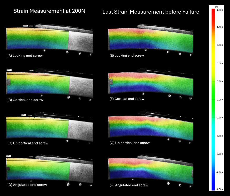
Figure 11.6.1: Strain measurements of the four end screw configurations. Picture at 200 N (A – D) and last captured frame before failure (E – H).
Partner:
• Stoffel K (Prof MD), University Hospital Basel, Basel, Switzerland
Reducing femoral peri-implant fracture risk through optimized plate length and screw configuration – a biomechanical study (finished) (L Puls, L Llano, I Zderic, M Kraus, P Varga, D Mischler, RG Richards, B Gueorguiev)
Background: Locked plating of femoral fractures is linked to secondary peri-implant fractures, possibly due to stress at the proximal plate end. This study examines if altering screw configurations and plate length can improve the strength of healed femoral bone constructs and decrease peri-implant fracture risks.
Goal: To assess whether using a variable angle condylar locking compression plate with different configurations short plates with cortical or locking screws 50 mm below the lesser trochanter, or long plates with screws in the femoral neck – enhances construct strength and reduces fracture risks after healing.
Results: Testing 24 synthetic femurs showed long plates with cortical neck screws had the highest failure load (1091 N ± 142 N), significantly outperforming long plates with locking neck screws (888 N ± 80 N), and both short plate setups (cortical: 471 N ± 42 N; locking: 450 N ± 19 N). Long plate failures involved neck screw pull-out, while short plates failed via peri-implant fractures at the proximal screw. No femoral neck fractures occurred. Long plates, especially with cortical neck screws, increased strength and reduced fracture risk, supporting their use in high-risk patients like those with severe osteoporosis.

Partner:
• Stoffel K (Prof MD), University Hospital Basel, Basel, Switzerland
Figure 11.6.2: Failure mechanism for short plate with locking screws (A), short plate with cortical screws (B), long plate with locking screws (C), and long plate with cortical screws (D, E).
Evaluating the biomechanical efficacy of 2.5+2.0 double plating against 3.5 single plating in ulna shaft fracture fixation: A cadaveric study (ongoing) (M Kraus, I Zderic, B Gueorguiev)
Background: The main postoperative complications in fixation of ulna shaft fractures are nonunion and implant irritation. Current standards recommend 3.5 mm locking compression plates, that can cause soft tissue irritation in patients, which may necessitate the secondary removal of implants. An alternative approach using a combination of two smaller plates in orthogonal configuration, has been proposed to overcome the shortcomings of the 3.5 mm plates.
Goal: To compare the biomechanical properties of a single 3.5 mm locking compression plate versus double-plating using one 2.5 mm and one 2.0 mm matrix mandible plate in a human ulna shaft fracture model.
Results: Eight pairs of human ulnar specimens with a standardized 10 mm fracture gap were pairwise assigned for instrumentation with double plating using one 2.5 mm and one 2.0 mm matrix mandible plate, or with a single 3.5 mm locking compression plate. Both groups underwent biomechanical testing for axial, torsional, bending stiffness and torsional cyclic load to failure using a servo-hydraulic testing machine and interfragmentary motion tracking. The double-plating system using 2.5 mm and 2.0 mm mandible plates offers a biomechanical stability that is comparable to the single 3.5 mm plate in ulna shaft fracture fixation, with no significant disadvantages in terms of stiffness and load to failure.

11.6.3: Representative mediolateral and anteroposterior X-rays of one double plated and one single plated specimen (left); Test setup with a specimen mounted for biomechanical testing for bending (middle); Boxplot showing outcome measure cycles to failure for each group separately, with no significant differences between the groups.
Pres:
• Zderic I, Kraus M, van Rossenberg L, Gueorguiev B, Richards G, Pape HC, Pastor T, Pastor T. Evaluating the biomechanical efficacy of 2.5-mm and 2.0-mm double plating against 3.5-mm single plating in ulna shaft fracture fixation: a cadaveric study. 2024 EORS (oral)
Partners:
• Pastor To (PD PhD MD), Cantonal Hospital Lucerne, Lucerne, Switzerland
• Pastor Ta (MD), Inselspital Bern, Bern, Switzerland
• Pape HC (Prof MD), University Hospital Zurich, Zurich, Switzerland
• Burkhart K (PD MD), University of Cologne, Germany; Diakonie-Klinikum Stuttgart, Germany
Figure
Primary stability of nailing versus low-profile dual plating of mid-clavicular fractures –a biomechanical cadaveric study. (ongoing) (F Pretz, I Zderic, B Gueorguiev)
Background: Low-profile dual plating techniques have gained popularity for diaphyseal clavicula fractures due to their potential to reduce soft tissue irritation. Intramedullary nailing is also an established surgical option for diaphyseal clavicle fractures, yet it remains unclear whether a 2x2.0 mm dual plating construct provides biomechanical stability comparable to intramedullary nailing.
Goal: To compare the biomechanical properties of the 2x2.0 mm low-profile dual plating with the 2.5 mm TEN in a human cadaver model.
Results: Twelve paired human cadaveric clavicles with simulated unstable diaphyseal shaft fractures AO/OTA 15.2C were stabilized via elastic nailing (Group 1) or dual plating using a superior and an anterior 2.0 mm matrix mandible plate (Group 2). Specimens underwent biomechanical testing with initial quasistatic superior-inferior and anterior-posterior bending, followed by cyclic superior-inferior loading to failure. Interfragmentary movements were monitored by optical motion tracking. Dual plating demonstrated significantly higher initial construct stiffness and reduced displacement amplitudes compared to intramedullary nailing, indicating superior early stability for midshaft clavicle fractures. These findings suggest that low-profile dual plating may offer improved initial stability without compromising long-term performance, making it a promising alternative particularly for less common, unstable midshaft clavicle fractures.

Figure 11.6.4: Example X-ray of a left clavicle instrumented with TEN and photograph of a left clavicle instrumented 2x2.0 mm dual plating with the planned osteotomy marked with black ink (left); Test setup with a specimen mounted for biomechanical testing (middle); Fracture displacement amplitudes shown at intermittent time points every 2500 cycles over the first 20000 cycles in terms of mean and standard deviation for each group separately (right).
Partners:
• Beeres FJP (Prof MD), Cantonal Hospital Lucerne, Lucerne, Switzerland
• Link BC (PD MD), Cantonal Hospital Lucerne, Switzerland
• van de Wall BJM (PD MD), Cantonal Hospital Lucerne, Switzerland
• Pastor T (PD PhD MD), Cantonal Hospital Lucerne, Lucerne, Switzerland
• Babst R (Prof MD), University of Lucerne, Lucerne, Switzerland
Biomechanical comparison of augmented MIPO versus ORIF locking plate fixation in unstable low-density proximal humerus fractures (ongoing) (F Pretz, I Zderic, B Gueorguiev)
Background: Proximal humerus fractures are common in patients with low bone mineral density. Locking plate osteosynthesis using either Minimally Invasive Plate Osteosynthesis (MIPO) or Open Reduction and Internal Fixation (ORIF) technique is widely performed. However, it has not yet been biomechanically investigated whether augmented MIPO achieves stabilization comparable to ORIF screw configuration for instable proximal humerus fracture fixation in osteoporotic bones.
Goal: To compare the biomechanical stability of four proximal humeral head screws with cement augmentation in PHILOS plates using a MIPO configuration versus six non-cemented head screws in an ORIF configuration.
Results: Fourteen paired human cadaveric humeri with simulated unstable three-part proximal humerus fractures (AO 11-B1) were stabilized using PHILOS plates with four proximal head screws in both groups (row A and B). In the ORIF group, two additional calcar screws were used, while in the MIPO group, the four screw tips were augmented with bone cement. Cyclic axial loading tests were conducted until failure. Cyclic axial loading tests were conducted until failure. Interfragmentary movements were monitored using optical motion tracking and anteroposterior x-ray imaging and compared between the groups. From a biomechanical perspective, PHILOS plates with four augmented screws as used in the MIPO technique demonstrated comparable initial construct stability and cycles to failure as compared to the commonly used PHILOS plates with additional calcar screws as used in the ORIF technique.

Figure 11.6.5: Anteroposterior radiograph of the MIPO-augmented group demonstrating visible bone cement at all four screw tips and of an instrumented specimen from the ORIF group (left); Biomechanical testing setup with a mounted specimen from the MIPO group (middle); Cycles to clinically relevant 5° varus deformation shown for each group separately in terms of mean and standard deviation (right).
Partners:
• Beeres FJP (Prof MD), Cantonal Hospital Lucerne, Lucerne, Switzerland
• Link BC (PD MD), Cantonal Hospital Lucerne, Switzerland
• van de Wall BJM (PD MD), Cantonal Hospital Lucerne, Switzerland
• Pastor T (PD PhD MD), Cantonal Hospital Lucerne, Lucerne, Switzerland
• Babst R (Prof MD), University of Lucerne, Lucerne, Switzerland
Novel U-plate vs posterior locking plate fixation in intraarticular olecranon fractures - A biomechanical analysis (ongoing) (V Topuzyan, I Zderic, B Gueorguiev)
Background: The treatment of displaced olecranon fractures typically involves various open reduction and internal fixation techniques, tailored to the fracture type and pattern. While posterior locking plates (PLPs) are known to provide superior biomechanical strength compared to other methods, they are associated with a high rate of postoperative hardwarerelated complications like pullout of the olecranon tip.
Goal: To biomechanically evaluate a prototype U-plate and compare its performance to that of the PLP in the treatment of intra-articular olecranon fractures.
Results: Seven pairs of fresh-frozen human arms were used. A simple two-part transverse articular fracture AO/OTA 2U1B1 was simulated by means of osteotomies and the specimen were pairwise assigned for fixation with either with a U-plate or a PLP. Elbows were subjected to cyclic loading until implant failure. Interfragmentary movements were monitored by optical motion tracking. No specimen failed during the cyclic loading test. The main failure mechanism for the PLP was split of the proximal fragment in the sagittal plane (n=5). U-plate cadavers failed through an avulsion tear of the triceps tendon with a significant fracture gap increase (n=5). The mean peak load before failure of the U-plate was 698 N (SD, 135 N) and 603 N (SD, 96 N) for PLP and was comparable. From a biomechanical perspective, the U-plate is a valid alternative to the PLP offering similar construct stability.

11.6.6:
a
a
or a PLP;
the mediolateral axis shown for each group separately over the first 10000 test cycles in terms of mean and standard deviation.
Partners:
• Hristov S (PhD MD), Multiprofile University Hospital Burgas, Burgas, Bulgaria
• Pastor T (PD PhD MD), Cantonal Hospital Lucerne, Lucerne, Switzerland
Comparative biomechanical analysis of radial neck plate versustripod fixationin mason type III radial fractures: A human cadaveric study (ongoing) (M Kraus, I Zderic, B Gueorguiev)
Background: Treatment of Mason Type III proximal radius fractures with the conventional radial neck plate is associated with soft tissue irritation and restricted range of motion. A novel tripod fixation approach, using three crossed screws, should minimize such complications and preserve range of motion.
Goal: To compare the biomechanical competence of conventional radial neck plating versus tripod fixation in a mason Type III fracture model.
Results: Sixteen paired human cadaveric radii were used to simulate Mason Type III fractures through a standardized osteotomy, creating a transverse neck fracture with an additional 50:50 head split component. The specimens were divided into two groups, one fixed using a conventional radial neck plate with two additional cannulated headless compression screws (HCS) and the other stabilized with tripod fixation using three crossed 40 mm HCS and two
Figure
Left: Mediolateral X-rays of
specimen instrumented with
U-plate
Right: Angular displacement around
additional HCS. Biomechanical testing assessed stiffness in various directions, load to failure, and cycles to failure under axial compression. From a biomechanical perspective, tripod fixation performs comparably to plate fixation in terms of stiffness and resistance to failure. Given its potential to reduce soft tissue irritation and minimize secondary hardware removal, the tripod technique may offer a viable alternative to conventional plating.

Figure 11.6.7: Top: X-rays of a specimen instrumented with plate or tripod screw configuration (left) and test setup with a specimen mounted for biomechanical testing in torsion, axial compression, and bending (right). Bottom: Boxplots for stiffness in anteroposterior bending, axial compression, and mediolateral bending, shown for each group separately.
Partners:
• Pape HC (Prof MD), University Hospital Zurich, Zurich, Switzerland
• Pastor To (PD PhD MD), Cantonal Hospital Lucerne, Lucerne, Switzerland
• Pastor Ta (MD), Inselspital Bern, Bern, Switzerland
• Burkhart K (PD MD), University of Cologne, Germany; Diakonie-Klinikum Stuttgart, Germany
Biomechanical assessment of novel dynamic versus conventional high-strength sutures in distal biceps tendon repair (ongoing) (M Kraus, I Zderic, B Gueorguiev)
Background: Tendon ruptures are a common injury and often require surgical intervention to heal. A refixation is commonly performed with high-strength suture material. However, slipping of the thread is unavoidable even at 7 knots potentially leading to reduced compression of the sutured tendon at its footprint.
Goal: This study aimed to evaluate the biomechanical properties and effectiveness of a novel dynamic high-strength suture, featuring self-tightening properties.
Results: Distal biceps tendon rupture tenotomies and subsequent repairs were performed in sixteen paired human forearms using either conventional or the novel dynamic high-strength sutures in a paired design. Eachtendon repair utilized an intramedullary biceps button for radial fixation. Biomechanical testing aimed to simulate an aggressive postoperative rehabilitation protocol stressing the repaired constructs. For that purpose, each specimen underwent in nine sequential days a daily mobilization over 300 cycles under 0-50 N loading, followed by a final destructive test. From a biomechanical perspective, the novel dynamic high-strength suture demonstrated higher resistance against gap formation at the bone tendon interface compared to the conventional suture, which may contribute to better postoperative tendon integrity and potentially quicker functional recovery in the clinical setting.

Figure 11.6.8: Test setup (left) showing a human arm mounted on the test machine extended and flexed position during biomechanical testing; Distal repair site measurement over the 9 test day cycles in terms of mean and standard deviations for each group separately denoting significantly higher tendon elongation for conventional versus dynamic sutures (p<0.002).
Pres:
• Zderic I, Kraus M, van Rossenberg L, Gueorguiev L, Richards L, Pape HC, Pastor T, Pastor T. Biomechanical assessment of novel dynamic versus conventional highstrength sutures in distal biceps tendon repair. 2024 EORS (oral)
Partners:
• Pape HC (Prof MD), University Hospital Zurich, Zurich, Switzerland
• Pastor Ta (MD), Inselspital Bern, Bern, Switzerland
• Pastor To (PD PhD MD), Cantonal Hospital Lucerne, Lucerne, Switzerland
Biomechanical assessment of novel dynamic high-strength suture tape in distal triceps tendon repair (ongoing) (M Kraus, I Zderic, B Gueorguiev)
Background: Distal triceps tendon rupture is related to high complication rates with up to 25% failures. Elbow stiffness is another severe complication, as the traditional approach considers prolonged immobilization to ensure tendon healing. Recently a dynamic high-strength suture tape was designed, implementing a silicone-infused core for braid shortening and preventing repair elongation during mobilization, thus maintaining constant tissue approximation.
Goal: To biomechanically compare the novel dynamic tape versus a conventional highstrength suture tape in a human cadaveric distal triceps tendon rupture repair model.
Results: Distal triceps tendon rupture tenotomies and repairs were performed via the crossed transosseous locking Krackow stitch technique for anatomic footprint repair using either conventional (ST) or novel dynamic (DT) tape. A postoperative protocol mimicking intense early rehabilitation was simulated, by a 9-day, 300-cycle daily mobilization under 120N pulling force followed by a final destructive test. From a biomechanical perspective, DT revealed lower tendon displacement and greater resistance in load to failure over ST.

Figure 11.6.9: Top left: Setup with an instrumented arm mounted for biomechanical testing; Top right: Distal triceps repair with caliper denoting the measurement site; Bottom: Longitudinal displacement measurements for ST and DT before (left) and after (right) testing shown in terms of mean and standard deviation values over the repetitive 9 testing days.
Pres:
• Kraus M, Boca B, Ion N, Dhillon M, Zderic I, Puls L, Gueorguiev B, Richards G, Pape HC, Pastor T, Pastor T. Assessment of a novel dynamic high-strength suture tape in distal triceps tendon repair - a biomechanical comparative study. 2024 ESBiomech (oral)
• Zderic I, Kraus M, Axente B, Dhillon M, Puls L, Gueorguiev B, Richards G, Pape HC, Pastor T, Pastor T. Biomechanical superiority of a novel dynamic suture tape over conventional tape in distal triceps tendon repair: a cadaveric study on an intense early rehabilitation protocol. 2024 EORS (oral)
Partners:
• Pape HC (Prof MD), University Hospital Zurich, Zurich, Switzerland
• Pastor T (MD), Inselspital Bern, Bern, Switzerland
NEU-DISC, IVD with neurovascular network: an in vitro discogenic pain model
Background: Chronic low back pain (LBP) is the world leading cause of disability. Intervertebral disc (IVD) disruption is the most common reason for LBP (discogenic pain). Discogenic pain is associated with the nerve and vessel ingrowth, but little is known about the mechanisms and roles of the neurovascular ingrowth in IVD. To study the communications among multiple tissues/organs in vitro, it is fundamental to reproduce the in vivo-like morphology and anatomical proximities. Sound induced morphogenesis (SIM) is a powerful tool to assemble multi-tissue/organ cellular systems in pre-determined spatial organization, in a contactless, mild, and fast manner. Pilot studies showed that this SIM-orchestrated multicellular morphology of DRG cells and IVD-DRG proximity is necessary to reproduce neuron-neuron and neuron-IVD communications. Nevertheless, to get closer to the in vivo complexity, we need to move a step further and include functional vasculature to the nerveIVD model.
Goal: Our aim is to biofabricate a neurovascular-IVD multicellular system with a spatially organized morphology. We will comprehensively demonstrate the biological role of the guided self-assembled architecture on cellular phenotype. We will use the neurovascular-IVD system to investigate the influence of vessel cells (endothelial cells and pericytes) in IVD-mediated neural plasticity.
Pub:
• Ma J, Eglauf J, Grad S, Alini M, Serra T. Engineering sensory ganglion multicellular system to model tissue nerve ingrowth adv sci (Weinh). 2024 Mar 20;11(11):2470062. doi: 10.1002/advs.202470062
Bioink platfoRm fOr invAsion anD adhEsioN (BROADEN) (ongoing) (J Wychowaniec, M D'Este)
Background: Despite recent advances in the field of 3D bioprinting and implantable biomaterials, there still is a clear need of versatile materials for a large spectrum of musculoskeletal applications. Implanted biomaterials available in the field could be improved by simultaneous: i) appropriate adhesion to tissues in vivo, ii) controlled degradation matching the desired tissue regeneration outcome; and iii) allowing tissue deposition and replacement by enabling controlled cellular invasion from neighbouring tissues. The main complexity in the development of functional inks is that the materials, usually biopolymeric hydrogels need a narrow and somewhat opposite set of physical and biological properties which allow for the degradation, adhesion as well as cellular invasion, whilst maintaining accurate printing (e.g. shape fidelity) and cellular viability and proliferation.
Goal: This project aims to develop multifunctional ink formulations for the fabrication of 3D constructs in a series of systemic and rational studies to generate rules for decoupling individual properties and to allow future biomaterial designs with complete control over all three important aspects (adhesion, degradation, and cellular invasion). To achieve this, we propose a rational design of composite biomaterials based on chemically modified oxidized tyramine-functionalized hyaluronan (o-THA) and a combination of bioactive polymers that include collagen, gelatin, and/or bioactive peptides. The three properties depend on the feeding ratio of biopolymers and the crosslinking method used, hence rational variations of the chemical composition and degree of functionalization of the main component (oTHA), as well as appropriate choice of secondary biopolymer will enable design of materials that can possess enhanced adherence to tissues but at the same time retain the necessary degradation time in vivo as well as porosity/crosslinking density to enable cellular infiltration.

Figure 11.6.10: (A) Oxidation degree of selection of 6 synthesized oxidized THA (o-THA) variations measured by absorbance at λ=550 nm using Purpald reagent assay with propionaldehyde as a standard curve. (B) Storage modulus measured for the selection of THA and o-THA hydrogels at 2 w/v% fabricated using: 0.1 mM ruthenium, 5 mM sodium persulfate, photo-crosslinked at 0.3 mW/cm2 power source.
Results: THA was synthesized from hyaluronan of two molecular weights: Mw=280 kDa (LMW THA) and Mw=1640 kDa (HMW THA). Proton nuclear magnetic resonance spectroscopy (1H NMR) and UV absorbance measurements at 275 nm on Infinite® 200 Pro plate reader (Tecan) were performed to confirm the degree of substitution (DoS) of tyramine on HA, which was 6.0 ± 0.6 %. Then selection of o-THA molecules was synthesized by a reaction with sodium periodate (NaIO4). By changing amount of NaIO4,the following degrees of oxidation 5.2 ± 0.4 %, 11.4 ± 2.1 %, 18.1 ± 2.4 % were obtained for LMW, and 6.8 ± 0.8 %, 13.7 ± 1.3 %, 19.2 ±
3.1 % for HMW o-THAs, respectively (Fig. 11.6.10A). Utilizing selection of Ruthenium (0.1- 1 mM) and Sodium persulfate (1-5 mM) concentrations, we prepared an array of hydrogels, whose viscoelastic properties were evaluated by oscillatory rheology (Fig. 11.6.10B) and adhesion by lap-shear tests using commercially available Chamonix leather as a model. Storage modulus of o-THA decreased with oxidation level, largely unaffected by molecular weight in photo-cross-linked hydrogels (Fig. 11.6.10B). Higher oxidation also yielded faster degradation (<24 hours). We thus present a family of o-THA biomaterials with tunable physicochemical and biological properties for the use in musculoskeletal tissue engineering.
Pres:
• Wychowaniec JK, Bektas EI, Vernengo A, Mürner M, Airoldi M, Eglin D, D'Este M. Effect of molecular weight of tyramine-modified hyaluronan on polarization state of peripheral blood mononuclear cells-derived macrophages, CESB 2024 (oral)
NeutrophIl iMMunomodulation fOr Resolution of inflammaTion and bone hEALling (IMMORTAL) (Ongoing), (E
Irem Bektas, M D'Este)
Background: The interplay between the immune system and bone regeneration is a crucial determinant of the success of healing processes. Neutrophils, through apoptosis, can contribute to the resolution of inflammation, creating an environment conducive to tissue repair This modulation holds the potential not only for regulating the immune response but also for influencing the behavior of other cells involved in bone healing, such as macrophages and mesenchymal stromal cells. Understanding and harnessing the osteoimmunomodulatory effects of neutrophil apoptosis offers promising avenues for therapeutic interventions, aiming to optimize the bone healing process and enhance clinical outcomes in conditions such as fractures or bone defects.
Goal: In this project, our focus will be on investigating the impact of neutrophil apoptosis on polarization of macrophages derived from peripheral blood monocytes. Our aim is to elucidate its role in the resolution of inflammation, and the subsequent healing response.
Results: Neutrophil response was detected to the apoptotic agents chlorogenic acid, PF04217903 and Roscovitine with various concentrations. Myeloperoxidase (MPO) is one of the markers for neutrophil extracellular traps and degranulation. It was noted that MPO release was decreased in neutrophils exposed to a 30 µM concentration of apoptotic agents, likely due to the neutrophils undergoing apoptosis, which helps preserve their intracellular molecules from being released into the external environment. Additional tests are being carried out to evaluate the mechanisms underlying the reduced MPO release and the impact of apoptosis on neutrophil function.
Chlorogenicacid100nM
Chlorogenicacid500nM
Chlorogenicacid5µM
Chlorogenicacid30µM
PF-04217903100nM
PF-04217903500nM
PF-042179035µm
PF-0421790330µm
Roscovitine100nM
Roscovitine500nM
Roscovitine5µM
Roscovitine30µM
TCP
PMA
Figure 11.6.11: MPO levels in cell culture media of neutrophils treated with various apoptotic agents. (Control groups; TCP: Tissue culture plastic, PMA (Phorbol 12-myristate 13-acetate))
Thesis:
• Chiara Lorenzetti, ETH Zürich, Study of the Modulatory Role of Neutrophil-Biomaterial Interactions in MSC Osteogenesis, July 2024.
Pres:
• D’Este M. Towards understanding neutrophils role in biomaterials-mediated immunomodulation; World Biomaterials Conference, Daegu, Korea, May 2024 (oral)
• D’Este M. Soft biomaterials and composites: a journey from biofabrication to immunomodulation; BIOMATSAN24/BIOMAT (French Biomaterials Societies) Super Besse, France, January 2024 (Plenary)
Cartilage regeneration with biomimetic decellularized extracellular matrix materials (ECMCART) (ongoing) (Z Li, J Xu, S Grad)
Background: Osteoarthritis is the most common degenerative joint disease and a leading cause of disability worldwide. Its prevalence is strongly increased within the aging population. Current treatments only relieve symptoms without reconstruction of the healthy joint; joint replacement is indicated eventually in most cases. Biofabrication provides a great potential to produce engineered hyaline cartilage tissue mimicking native tissue. Extracellular matrix (ECM) is composed of tissue-specific macromolecules and contains cytokines, growth factors, and signaling molecules, all of which provide microenvironmental cues that regulate cell behavior. The recent development of decellularized ECM (dECM) methodologies has led to advancements in scaffold design by recreation of a tissue-specific 3D microenvironment. Goal: The aim of this project is to develop a biomaterial based on cartilage dECM and to promote chondrocytes redifferentiation and cartilage regeneration with the biomimetic dECM biomaterial. The regeneration of cartilage tissue with dECM materials will be investigated in vitro and ex vivo.
Results: Cartilage dECM particles were produced from bovine stifle joint cartilage tissue. A DNAase based detergent free method was used to maintain a high percentage of proteoglycans (63%) and collagen (97%) compared with native cartilage tissue, while efficiently removing 99% of DNA. Different concentrations (0%, 6%, 12%, 20%) of dECM particles were incorporated into hyaluronic acid tyramine derivative (THA) hydrogels. Bovine chondrocytes after 2D expansion were encapsulated within the THA-dECM hydrogel and cultured up to 2 weeks. Addition of 20% dECM particles enhanced redifferentiation of chondrocytes, as indicated by up-regulation of aggrecan, collagen type II, and Sox9 gene expression. The inclusion of dECM particles significantly enhanced the compressive stiffness of the hydrogels. Specifically, on day 1, hydrogels with 12% and 20% dECM concentration exhibited a remarkable increase in Young's modulus compared with THA only. Despite a reduction in Young's modulus after 14 days of culture, a discernible difference persisted between the 20% dECM group and the THA-only group, highlighting the reinforcing effect of dECM particles on the hydrogel's mechanical properties (Fig. 11.6.12).

Figure 11.6.12: (left) dECM particles production process and characterization. (right) dECM particles at high dose (20%) promote chondrocyte redifferentiation after 2 weeks culture in vitro.
Pres:
• Jiangyao Xu, Mauro Alini, Sibylle Grad, Jeroen Geurts, Zhen Li. Decellularized extracellular matrix based hydrogel promotes chondrocyte redifferentiation. OARSI 2024, Vienna, Austria (poster)
• Jiangyao Xu, Mauro Alini, Sibylle Grad, Jeroen Geurts, Zhen Li. Decellularized extracellular matrix-based hydrogel promotes chondrocyte redifferentiation in vitro and ex vivo SBMS 2024, Bern, Switzerland (oral presentation)
Pub:
• Xu J, Jiang N, Zhu S, Alini M, Grad S, Geurts J, Li Z Decellularized extracellular matrixbased hydrogels for cartilage repair and regeneration. Advanced Orthopaedics epub December 2024.
• Jiang N, Tan P, Sun Y, Zhou J, Ren R, Li Z, Zhu S. Microstructural, micromechanical atlas of the temporomandibular joint disc. journal of dental research 103 (5), 555-564 (2024).
• Bordbar S, Li Z, Lotfibakhshaiesh N, Ai J, Tavassoli A, Beheshtizadeh N, Vainieri L, Khanmohammadi M, Sayahpour FA, Baghaban Eslaminejad M, Azami M, Grad S, Alini M. Cartilage tissue engineering using decellularized biomatrix hydrogel containing TGF-βloaded alginate microspheres in mechanically loaded bioreactor. Sci Rep. 2024 May 25;14(1):11991. doi: 10.1038/s41598-024-62474-5
Partners:
• Guerts J (PD), University of Lausanne, Switzerland
• Zhu S (Prof), West China School of Stomatology, Sichuan University, Chengdu, China
• Jiang N (Prof), West China School of Stomatology, Sichuan University, Chengdu, China
Development of an in vitro micro-vascular invasion model of hypertrophic cartilage for the study of cellular and molecular interplay during endochondral ossification in healthy and compromised conditions (VascEndoC) (ongoing) (S Verrier, E Wehrle, M Stoddart, A Jose)
Background: Bone is a highly vascularized tissue presenting remarkable regenerative capacity. Successful healing of bone depends on various interconnected pathways, with revascularization and differentiation of recruited cells at the fracture site playing important roles. Yet, while bone injuries mostly heal effectively, approximately 10% of cases experience delayed healing or non-union of the fracture mainly due to impaired callus formation or failed cartilage-to-bone transition and subsequent remodeling. In about 50% of these cases, the initial injury was associated with significant vascular damage. Thus, extensive research focuses on developing strategies to enhance vascularization and facilitate bone healing. However, although angiogenesis is key in bone healing, non-union fracture gaps can also be well-vascularized. The lack of vascularization does not provide a complete answer, and a deeper understanding of the bone formation – vascularization coupling during endochondral bone healing is needed
Goal: In this project, we propose to develop an in vitro micro-vascular invasion model of hypertrophic cartilage. This precise and well controlled platform will complement preclinical studies (MechOmics) by enabling detailed examination of molecular factors and mechanical conditions relevant for hypertrophic-cartilage-to-bone transition. By comprehensively studying the impact of healthy or compromised micro-environmental cues (biological/local matrix stiffness) on this interplay, new therapeutic approaches can be explored to prevent or rescue compromised healing cases.
Results: In a feasibility study, we showed the micro-vascular invasion of MSCs pellets that were pre-treated for 7 days in chondrogenic medium (containing TGFβ1) (Fig. 11.6.13). Pellets characterization further showed increased type X collagen and MMP13 hypertrophy-related gene expression along with type II collagen chondrogenic marker when compared to day 0 control.

Figure 11.6.13: z-stack confocal images of TGFβ1 pre-treated MSC pellets culture for 17 days in presence of GFP-HUVEC (bars = 200 µm).
Pres:
• Verrier S. Cells in bone (neo) vascularization. SBMS Summer School, 2024. (oral, invited)
• Füllemann P, Jörimann T, Matthys R, Stoddart M, Verrier S. Effect of strain on naïve MSC differentiation fate, a 3D in vitro study. ORS 2024 (poster)
Pub:
• Jörimann T, Füllemann P, Jose A, Matthys R, Wehrle E, Stoddart MJ, Verrier S. In vitro induction of hypertrophic chondrocyte differentiation of naïve MSCs by strain. Cells. 2024 Dec 30;14(1):25. doi: 10.3390/cells14010025
• Hernigou P, Homma Y, Hernigou J, Flouzat Lachaniette CH, Rouard H, Verrier S. Mesenchymal stem cell therapy for bone repair of human hip osteonecrosis with bilateral match-control evaluation: impact of tissue source, cell count, disease stage, and volume size on 908 hips. Cells. 2024 May 1;13(9):776. doi: 10.3390/cells13090776.
Partner:
• Farrell E (Prof, PhD), Bone and Tissue Engineering, Department of Oral and Maxillofacial Surgery, Erasmus MC, Rotterdam, Netherlands
Systemic administration of anti-IL-1β to enhance bone healing (HealBone2) (ongoing) (M Stoddart, E Wehrle, M Schröder, L Gens, D Gehweiler, S Zeiter)
Background: Although 90% of fractures typically heal without complications, there remains a small proportion (≤ 10%) of fractures that experience delayed healing or non-union. In patients with such healing complications, there appears to be an important contribution of an inappropriately maintained pro-inflammatory environment to the defective fracture healing process. Interestingly, growth factors e.g. BMP-2, used in bone regenerative approaches have recently been shown to induce pro-inflammatory cytokine release. Thus, immunomodulation of the local fracture microenvironment could be an effective way to enhance fracture healing in troublesome healing environments. The preceding project, HealBone, showed that local administration of IL-1Ra, the receptor antagonist of the pro-inflammatory cytokine IL-1β, can improve BMP-2 induced bone healing in a rat segmental femoral defect. However, the rapid degradation of IL-1Ra in vivo suggests that improved bone healing efficacy may be observed with more effective strategies to inhibit IL-1β activity, such as anti-IL-1β monoclonal antibody therapy. Therefore, the current project focuses on investigating the therapeutic efficacy of systemic anti-IL-1β administration to improve BMP-2 induced bone healing in challenging healing environments.
Goal: To characterize BMP-induced cytokine profiles during bone healing, and to test the efficacy of systemic anti-IL-1β administration to improve BMP-2 induced bone healing in challenging healing environments.
Results: The first in vivo study showed pronounced differences in early callus formation and mineralization dependent on treatment (empty, collagen, collagen + BMP-2) of the segmental defect (Fig 11.6.14). Local cytokine profiles demonstrated no excessive and pro-longed cytokine expression by treatment with BMP-2 in the early phase of healing. Systemic anti-IL1β administration in the first two weeks following surgery tendentially improved functional healing outcome of operated femurs compared to treatment with BMP-2 alone as assessed by 3-point bending 14 weeks post-operatively. Overall, the results obtained so far point towards a potential for low dose BMP-2 application to promote impaired fracture healing without requiring simultaneous immunomodulation treatment.

Figure 11.6.14: Representative radiographs of femoral defects in the early phase of healing. (a) empty defect, (b) collagen sponge, (c) collagen+1 µg BMP-2.
Pres:
• Schröder M, Gens L, Bernhard L, Arens D, Tapia-Dean J, Giger N, Gehweiler D, Zeiter S, Stoddart M, Wehrle E. Effects of low dose BMP-2 and immunomodulation targeting IL-1ß on fracture healing in a femur defect model in rats. 2024 EORS (oral)
• Schröder M, Gens L, Arens D, Giger N, Bernhard L. Gehweiler D, Zeiter S, Stoddart M, Wehrle E. Low dose BMP-2 promotes fracture healing in a femur segmental defect model in rats without inducing excessive and prolonged inflammation. 2024 Annual Meeting SVGO/SBMS (oral)
• Schröder M, Gens L, Bernhard L, Arens D, Tapia-Dean J, Giger N, Gehweiler D, Zeiter S, Stoddart M, Wehrle E. Effects of low dose BMP-2 and immunomodulation targeting IL-1ß on fracture healing in a femur defect model in rats. 2024 ISFR (best poster award)
• Schröder M, Gens L, Bernhard L, Arens D, Tapia-Dean J, Giger N, Gehweiler D, Zeiter S, Stoddart M, Wehrle E. Effects of low dose BMP-2 and immunomodulation targeting IL-1ß on fracture healing in a femur defect model in rats. 2024 ORS (poster)
Study of glucocorticoid-induced effects on MSC differentiation (CORTISONE)
(STARTED) (E Della Bella, M Stoddart, J Úbeda Garrido)
Background: The widespread use of dexamethasone to study in vitro human osteogenic differentiation is raising multiple questions about its validity. There are multiple discrepancies in vitro compared to the physiological process of osteoblast development and to the common clinical experience, indicating that hypercortisolism is detrimental to bone health, whether from endogenous or iatrogenic causes.
Goal: This project aims to increase the understanding of glucocorticoid stimulation on human bone marrow mesenchymal stromal cells (BMSCs), with the hypothesis that the current protocols for in vitro osteogenesis, based on dexamethasone, recapitulate the development of pathological calcification, rather than that of physiological bone formation.
Results: While dexamethasone generally results in a higher mineral deposition, hydrocortisone sustains osteogenic differentiation, but without PPARG upregulation nor the formation of adipocyte-like cells when used at 10 nM. The use of hydrocortisone moreover results in improved expression of genes encoding for key osteogenic transcription factors, such as DLX5 and SP7, but it does not stimulate the expression of BGLAP. The expression of glucocorticoid-metabolizing enzyme genes, namely HSD11B1 and HSD11B2, are differently regulated by dexamethasone and hydrocortisone, with dexamethasone specifically increasing the expression of HSD11B1. The action of HSD11B1 gene product increases the availability of active glucocorticoids not only to the GR, but also to the MR. Preliminary results showed that inhibition of MR with spironolactone can increase the expression of late osteogenic markers SPP1 and IBSP, while downregulating the early marker ALPL.
Pres:
• Wespi L, Stoddart MJ, Della Bella E. Role of non-canonical pathways and side effects of glucocorticoids in the regulation of osteogenic differentiation. SSB+RM 28th Annual Meeting St. Gallen 2024 (poster)
• Vonlanthen N, Stoddart MJ, Della Bella E. Are different glucocorticoids equivalent in inducing in vitro osteogenesis? Orthopaedic Research Society Annual Meeting, Long Beach CA 2024 (poster)
Thesis:
• Wespi L. Investigating mineralocorticoid receptor activation during glucocorticoid-induced in vitro osteogenesis. ETH Zurich MSc, ETH Zurich, Biomedical Engineering MSc 2024.
• Stegmaier A. Optimization of glucocorticoid use for in vitro osteogenic differentiation of human bone marrow mesenchymal stromal cells. University of Reutlingen BSc 2024.
Partners:
• Annapaola Parrilli (Dr), Center for X-ray Analytics, EMPA, Dübendorf, Switzerland
• Kristaps Kļaviņš (Prof), Riga Technical University, Riga, Latvia
RolE oF tIming iN mEchanically induced Cartilage Formation (RefineCF) (started) (M
Stoddart, K Wendrich)
Background: The ability to modify mesenchymal stromal cell (MSC) differentiation by mechanical forces alone offers the opportunity to improve patient outcomes by optimizing rehabilitation protocols after surgery. Using a custom designed and built, multiaxial load bioreactor, we have previously shown that bone marrow MSCs can be induced towards a cartilage phenotype by combining compression and shear. This process is driven by the mechanical activation of endogenously produced TGF-β protein, a key chondrogenic growth factor. Despite the proof of concept obtained, the effect of load duration and initiation of load, key features of rehabilitation protocols, are unexplored. Furthermore, there is increasing evidence that circadian rhythm plays a major role in both tissue homeostasis and healing. Therefore, we will also study the role and effect of circadian rhythm on mechanically induced chondrogenesis. The overarching aim of this study is to provide experimental data on the timing and duration of load on cell differentiation and tissue maturation. This could form the basis of evidence based rehabilitation protocols after articular cartilage repair surgery. Regenerative rehabilitation is an increasing field whereby the final clinical outcome of the initial treatment or surgery is enhanced by the rehabilitation protocol later applied. This hand-in-hand approach of surgery and rehabilitation has been largely overlooked, with multiple aspects of rehabilitation still being eminence based rather than applying evidence-based practice. Various proposals for rehabilitation after marrow stimulation for cartilage repair have been suggested, yet there is little experimental evidence to validate any of them. However, with the further development of complex bioreactor systems, key questions such as the initiation and duration of load can be further investigated using human cells in a preclinical environment. Simple questions such as how many hours per day mechanical stimulation should be applied are still largely unexplored.

Figure 11.6.15: The Skeletal organization of the circadian clocks and the molecular oscillator. Depicted are the molecular clocks in the musculoskeletal system, tendon, bone, muscle and cartilage. Enlarged image shows key components of the molecular clock. Positive limb; BMAL1 and CLOCK; negative limb; PER and CRY. PER and CRY inhibit BMAL1:CLOCK binding to E-box promoter regions. BMAL1:CLOCK bind to E-boxes of target genes, including Pers and Crys, as well as a spectrum of clockcontrolled genes (CCGs), which are largely specific to the specific tissue physiology. PER and CRY are periodically degraded by kinases including CK1, GSK3β and AMPK, this determines the period of the cycle. Nuclear hormone receptors RORs and REV-ERBs compete to modulate the transcription of Bmal1. (Adapted from Rogers and Meng, 2023, Tick tock, the cartilage clock. Osteoarthritis Cartilage 31, 1425-1436. 10.1016/j.joca.2023.05.010.)
Partner:
• Qing-Jun Meng (Prof), Faculty of Biology, Medicine and Health, University of Manchester, UK
11.6 AO Development Incubator
AO Fracture Monitor (SmartPlate) (ongoing) (M Ernst)
Background: Information on healing progression and load-bearing characteristics in fracture patients is only barely tapped due to the inaccessibility of a confined biological region and the limited value of radiographic methods. A novel approach to continuously measure both, implant load and patient activity has recently been developed in the ARI. The system comprises an implantable data logger, which autonomously collects relevant parameters to support surgical decision-making during fracture healing. Wireless synchronization of the assessed implant load data via the patient's mobile phone allows for remote monitoring by the treating physician. Proof of concept is obtained from preclinical experiments and from first clinical data collection with prototype devices on external fixation.
Goal: The AO Fracture Monitor shall be further developed into a commercially applicable system for long-bone bridge plating. Implantable device and accompanying software shall be developed and tested according to the regulatory requirements and undergo clinical evaluation thereafter.
Results: Started in Q4 2024, clinical data collection in the frame of a first-in-human prospective clinical investigation is currently ongoing, with 14 patients enrolled across the four participating sites to date. The primary objective of the study is to demonstrate safety of the novel active implant. Additionally, device performance is analyzed from the continuously acquired data and post-processing algorithms are reviewed and refined. There were no device-related adverse events reported so far, and all devices are acquiring data as expected. In parallel, regulatory approval is prepared for by completing and transferring the technical documentation to a legal manufacturer. Manufacturing processes are refined, and remaining verification and validation activities are being completed. The web application for data visualization and storage are currently migrated to a medical grade cloud environment to fulfil regulatory requirements.

Figure 11.7.1: a) Plate with Fracture Monitor mounted on an artificial femur; b) Preliminary data from the clinical investigation - average daily number of loading events recorded by the AO Fracture Monitor for all patients with a minimum monitoring period of 16 weeks (blue bars represent standard deviation). The data illustrates the gradual increase of patient activity after surgery until it stabilizes at around 60 days post-op.
Pres:
• Richards RG. AO Fracture Monitor: Digitalization of the bone healing episode of care. 23rd APOA Congress, Dubai, UAE (Keynote Lecture)
• Ernst M. AO Fracture Monitor – Improving Fracture Aftercare. AtiO Conference 2024, Berlin, Germany (Early-Stage Pitch Contest, Finalist)
• Richards RG. Continuous monitoring of fracture healing – from a research tool to an active implantable medical device Orthopaedic Trauma Association (OTA) Annual Meeting 2024, Montreal, Canada (Invited Speaker)
• Richards RG. Continuous sensor monitoring for personalized fracture care. Uniting innovation and collaboration for breakthroughs in musculoskeletal regeneration. Joint Symposium of Pre-MRN & AoE Program. The Chinese University of Hong Kong (CUHK), Shatin, N.T. Hong Kong (Invited Speaker)
• Richards RG. AO fracture monitor for digitally measuring of bone healing. 7th International Symposium of Musculoskeletal Regeneration Research Network (MRN 2024), Nov 24-25, 2024, Shenzhen Campus, Sun-Yat sen University, Shenzhen, China (Invited Speaker)
Partners:
• Braun B (PD MD), BG Unfallklinik Tübingen, Tübingen, Germany
• Pohlemann T (Prof MD), University Hospital Saarland, Homburg, Germany
• Raschke M (Prof MD), University Hospital Münster, Münster, Germany
• Schütze K (PD MD), University Hospital Ulm, Ulm, Germany
Constant force growth modulation implant (GMI) (ongoing) (J Buschbaum, M Heumann, M Ernst)
Background: Lower limb deformities in children and adolescents are often corrected with temporary (hemi-) epiphysiodesis technique, in which the physis is bridged by an implant to inhibit growth and balance the deformity. Currently utilized implants have their disadvantages. They are not "passively" safe and require timely surgical removal, as the implant load steadily increases with ongoing growth potentially leading to devastating complications such as implant-related failures, over-corrections, unwanted secondary deformities, or permanent physeal closure of the growth plate. A novel "passively" safe implant concept was developed that exerts a predefined, growth-independent constant compression force to the physis to avoid the complications of standard implants. Preclinical experiments have confirmed safe, effective and controlled treatment with this new implant concept.
Goal: The goal is to translate the concept into a clinically usable medical device.
Results: Following the completion of the prototype development phase last year, the formal development process for medical devices has commenced in collaboration with our external partner. The implant and screw design were optimized and validated trough mechanical tests. A new insertion instrument was developed to enhance implant handling during implantation. The usability and surgical handling of the implant was confirmed by leading pediatric surgeons in a practical wet-lab test. The implant's manufacturing process was developed, the supply chain was defined, and the technical documentation was created in accordance with the medical device development process. In addition, a comprehensive regulatory strategy has been developed to outline the path for the first clinical application of the implant.


Figure 11.7.2: The developed growth modulation implant, mounted on a transparent tibia model (left) and during careful inspection by surgeons in a usability test (right).
Patent:
• Buschbaum J. A device for modulating growth plate activity and/or for orthodontic applications, EP24174069.5
Partners:
• Slongo T (MD), Inselspital Bern, Bern, Switzerland
• Sepulveda M (MD), Universidad Austral de Chile, Valdivia, Chile
• Narayanan U (Prof MD), University of Toronto, Toronto, Canada
• Dwyer J (MD), University Hospital of North Staffordshire NHS Trust, Stoke-on-Trent, UK
• Mukhopadhaya J (MD), Paras HMRI Hospital, Patna, India
• 41medical AG, Bettlach, Switzerland
Gel for Delivery of Antibiotics
(GEDI) (ongoing) (P Nylund, F Moriarty, M D'Este, A Montali)
Background: Fracture related infections (FRI) are a dreaded complication for orthopaedic trauma patients, leading to longer treatments, poor outcomes, and huge economic burden. FRI persists despite implementation of best clinical practices. In many instances the delivery of the antibiotics is compromised due to damage in the vascular system, or the antibiotics do not reach a therapeutically effective concentration in the affected area. To overcome these issues, an injectable hydrogel GEl for Delivery of AntIbiotics (GEDAI) has been developed with the intention to control the release of the antibiotic, keeping the local concentration high while avoiding side effects due to high systemic concentrations. The gel was designed to stick to metal and tissues more than to surgical gloves for optimal surgical handling.
Goal: The main goal of this project is to produce all the technical data and documentation necessary for regulatory approval and for future clinical studies and attract the attention of industrial partners capable of bringing this idea to the market. As part of the project, we will show efficacy and safety of the gel in treatment of fracture related infection. Towards this goal we will carry out three separate in-vivo studies on rat, rabbit and sheep with the formulation GEDAI-T which is intended to locally deliver tobramycin sulphate.

Results: A method for in vivo measurement of local antibiotic concentrations has been established, along with the completion of stability and drug release studies. At our contract and development manufacturing organization (CDMO) Ascil technical batches have been produced, and the manufacturing of GLP-like materials has begun. Efforts are currently focused on scaling up GMP manufacturing. Two pre-application approvals have been obtained, and a full application for each have been submitted to the U.S. Department of
Figure 11.7.3: Application of GEDAI-T on a titanium plate.
Defense for a Phase I/II clinical study. A pre-Investigational New Drug (IND) meeting was held in February, resulting in promising and constructive feedback. In response to a suggestion from the FDA, three GLP animal study outlines were developed and submitted. Two of those outlines included new models that were developed in the ARI PCF. These studies are scheduled to take place at ARI PCF in spring/summer 2025. Additionally, in collaboration with our regulatory consultants we commenced to prepare documents for the electronic Common Technical Document (eCTD) required for the IND application.
Pres:
• Siverino C, Nylund P, Foster AL, Boot W, Zeiter S, Richards RG, D'Este M, Moriarty TF In vitro and in vivo evaluation of a gentamycin-vancomycin loaded emulsion-based hydrogel for orthopedic device-related infection. Oral, World Biomaterials Conference, Daegu, KR, May 2024.
• Nylund P. In vitro and in vivo evaluation of a gentamycin-vancomycin loaded emulsionbased hydrogel for orthopedic device-related infection SSB-RM Confence, Switzerland, September 2024 (oral)
• Siverino C, Nylund P, Foster AL, Boot W, Zeiter S, Richards RG, Montali A, D'Este M, Moriarty TF. In vitro and in vivo evaluation of a gentamycin-vancomycin loaded emulsionbased hydrogel for orthopedic device-related infection ARI Orthopaedics conference, June 2024, Switzerland (poster)
Partners:
• Bruder consulting and Venture Group, NJ, USA
• Ascil Proyectos S.L., Spain
• William T. Obremskey, (MD, MPH, MMHC), Vanderbilt Health Nashville, TN, USA
11.7 AO Strategy Fund
Digitally enhanced hands-on surgical training (DEHST) (ongoing) (D Ciric, C Hetreau, J Buschbaum)
Background: The outcomes of orthopedic and trauma surgery are highly determined by the skills and training level of the operating surgeon. Hands-on and tactile exercises are essential pillars of a comprehensive training concept. Conventional hands-on training is typically offered only in course events, limited to basic skill training, and lacks data collection to measure training success. Current digital technologies offer substantial opportunities to augment predominantly mechanical training models with enhanced training scope, user experience and comprehensive training data assessment. They allow for decentralization of the training, if desired, from course events to home-based training at any time.
Goal: It is envisioned to develop a skill station product line consisting of cost-effective, transportable, and digitally augmented modules for hands-on surgical training targeting the most relevant operational skills in trauma and orthopedics.
Results: DEHST’s training portfolio, initially consisting of three modules for intramedullary nailing, has now been expanded to include four new modules. These cover basic skills training, pinning and plating of distal radius fractures, and a novel concept for intraoperative imaging training. In addition to these new developments, the focus this year was on integrating DEHST into AO's educational offerings. As part of the AO Milestones program, DEHST was used in two pilot events in North America, where it demonstrated its practicality. A significant milestone was the integration of DEHST into the AO Davos Courses. During the pediatric fracture management course, the tri-plane fracture exercise was enhanced with DEHST, and a new module for veterinary surgery was featured. DEHST also augmented AO’s Trauma Basic Principles Course by adding a new hands-on exercise on intraoperative imaging and further expanded the intramedullary nailing practical exercise with a freehand distal interlocking training module which was also incorporated into the Basic Principles Course for Swiss Surgeons. Feedback was very positive, and the integration of additional modules into next year’s AO Davos Courses is already underway.
Another highlight this year was DEHST’s certification with the “AO Approved” label by the AO Technical Commission (AO TC) Trauma Board. This certification represents a significant endorsement, recognizing the value of DEHST and its relevance as an educational tool.

Pres:
Figure 11.8.1: DEHST in action at the AO Davos Courses 2024 with the new hands-on exercise on intraoperative imaging training.
• Buschbaum J, Cattaneo E, Pastor T, Gueorguiev B, Pastor T. Digitally Enhanced Handson Surgical Training (DEHST) enhances surgical performance during intramedullary nail distal interlocking. 2024 AMEE (e-poster)
• Gueorguiev, B. Digitally Enhanced Hands-on Surgical Training. 2024. APORS-ICORS-AO Foundation session in APOA (oral)
• Buschbaum J. Digitally enhanced hands-on surgical training (DEHST). A Faculty Development Workshop for Simulator-Based Teaching. 2024 AMEE (invited speaker)
Pub:
• Pastor T, Cattaneo E, Pastor T, Gueorguiev B, Beeres FJP, Link BC, Windolf M, Buschbaum J. Digitally enhanced hands-on surgical training (DEHST) enhances the performance during freehand nail distal interlocking. Arch Orthop Trauma Surg, 2024
Partners:
• Höntzsch D (PD PhD MD), BG Unfallklinik Tübingen, Germany
• AO Milestones, Davos, Switzerland
OSapp: Virtual osteosynthesis tool for surgical education (OSappSF) (completed) (P Varga, D Mischler, A Feist, L Llano, B Gueorguiev)
Background: Fracture fixation complications not only occur due to suboptimal implants and instruments but are often caused by incorrect surgical techniques. Despite the well taught principles of fracture fixation treatment, less experienced surgeons sometimes fail to understand the underlying biomechanical concepts and thus select the incorrect fixation approach. Especially in trauma surgery standardized procedures are rare and the treatment is highly dependent on the case, which requires a mechanical sense and awareness to correctly interpret the situation and choose the appropriate fixation strategy. To reduce complication rates, it is therefore of utmost importance to not only know the guidelines but also understand the underlying biomechanical principles.
Goal: Foster the understanding of the biomechanical principles of fracture fixation and bone healing via a virtual and interactive osteosynthesis learning platform. Augment and complement existing AO offerings with its unique possibilities of animating and displaying biomechanical simulations.
Results: OSapp (https://osapp.aofoundation.org), a freely accessible interactive platform dedicated to educating basic principles of osteosynthesis, continued to grow in content, credibility and visibility. With ~2.7 million page views by more than 100.000 users from 200 countries in 2024 alone, OSapp has become a strong component of AO’s online offerings. The 90% positive feedback by 1600 responders indicates that the tool is highly valued by the surgeon community. Credibility and effectiveness of the learning tool has been demonstrated in a recently published learning assessment study reporting significant improvement in residents’ biomechanical knowledge after self-learning with OSapp. The implementation of OSapp models into AO Surgery Reference has been continued and extended. OSapp integration has been featured as an important milestone in the “20 years of AO Surgery Reference” history. OSapp was implemented in the Basic Principles Course during the AO Davos Courses with a Biomechanical Challenge lecture, with complementing faculty presentations, and as a digital resource for Skills Lab stations. At the end of 2024, OSapp has been successfully handed over to the AO Education Institute, concluding the AO Strategy Fund project. ARI keeps its involvement in OSapp by continuing content creation for the AO Surgery Reference integration.


SCAN ME

Figure 11.8.2: Timeline of the OSapp project from the idea creation to the successful delivery to the AO Education Institute (AO EI).
Pres:
• Llano L, Mischler D, Chatterjee S, Nousiainen M, Lambert S, Varga P. OSapp: Beyond textbooks: transformative learning with an online interactive biomechanics tool for orthopaedic residents. 2024 AMEE (oral)
• Peter V, Mischler D, Taype D, Chatterjee S, Ghidinelli M, Nousiainen M, Lambert S, Llano L. Transformative learning of the biomechanical principles of osteosynthesis with an online interactive tool. 2024 DKOU (oral)
Pub:
• Llano L, Mischler D, Taype D, Chatterjee S, Ghidinelli M, Nousiainen M, Lambert S, Varga P. Beyond Textbooks: Interactive Learning of Biomechanical Principles of Osteosynthesis with an Online Tool for Orthopaedic Residents. Journal of Surgical Education, 2024.
Partners:
• Lambert S (MD), University College of London Hospital, UK
• Babst R (Prof MD), Luzerner Kantonsspital, Switzerland
• Gebhard F (Prof MD), Universitätsklinikum Ulm, Germany
• Jäger M (MD), Universitätsklinikum Freiburg, Germany
• Schuetz M (Prof MD), Royal Brisbane Hospital, Brisbane, Australia
11.8 Extramural Projects
A novel highly customizable bone fixation solution (BoneFix) (ongoing) (P Schwarzenberg, P Varga)
Background: Traditional metal osteosynthesis hardware cannot be easily customized for a fracture in the operating theatre and can lead to issues in complex areas such as the hand, leading to require secondary surgery to remove the implant. A new osteosynthesis method, BoneFix, has been developed using light-curable polymer composites for highly customizable fixation solutions that have been shown to induce no soft tissue adhesions. This biocompatible platform can be shaped in situ and is designed to use a self-etching primer to adhere directly to the bone surface to be completely bioresorbable, leaving no hardware behind in the body.
Goal: To investigate and validate the biomechanical properties of the current BoneFix platform prototype in multiple loading modes and compare it to the traditional metal solutions in ex vivo ovine models, in vivo ovine models, and human cadaveric models.
Results: A cadaver study was conducted to measure the internal forces acting on osteosyntheses in the hand during rehabilitation exercises, aiming to determine the loading they must endure. Based on newly identified clinically relevant loading parameters, cyclic testing in a bioreactor was performed to predict the performance of BoneFix osteosynthesis in real-world scenarios. The bioreactor study showed that BoneFix could withstand the predicted rehabilitation period with a safety factor approximately 10 times greater than the physiological loads. However, further testing is needed to understand the impact of overloading events on the osteosynthesis.

Figure 11.9.1: a) An ovine phalanx with an osteotomy cut using a 3D printed cutting guide. b) The mechanical testing apparatus outside of the bioreactor with an osteotomized ovine phalanx fixed with BoneFix. c) A 3D rendering of the testing apparatus submerged in the bioreactor.
Pres:
• Varga P, Cameron PMN, Hutchinson DJ, Malkoch M, Schwarzenberg P. Fatigue strength assessment of a novel light-curable bone fixation technique. 2024 ESBiomech (oral)
• Pastor T, Banzer G, Pat-Lafitte G, Schlatter J, Dhillon M, Hutchinson DJ, Malkoch M, Varga P, Schwarzenberg P. Biomechanical test of a novel osteosynthesis in metacarpal fractures during rehabilitation exercises. 2024 Swiss Society for Hand Surgery (oral)
Pub:
• Schwarzenberg P, Colding-Rasmussen T, Hutchinson DJ, San Jacinto Garcia J, Granskog V, Petersen MM, Pastor T, Weis T, Malkoch M, Wong C, Varga P. Determination of the internal loads experienced by proximal phalanx fracture fixations during rehabilitation exercises. Frontiers in Bioengineering and Biotechnology, 2024
Partners:
• Malkoch M (Prof PhD), KTH Royal Institute of Technology, Sweden (Coordinator)
• Mustafa K (Prof DDS PhD), University of Bergen, Norway
• Wong C (PhD MD), Region Hovedstaden, Denmark
• Svensson C (Prof PhD), Karolinska Institute, Sweden
• Eglin D (Prof PhD), Institut Mines-Telecom, France
• Granskog V (PhD), Biomedical Bonding AB, Sweden
Smart, multifunctional dental implants (I-SMarD) (ongoing) (A Vautrin, P Varga)
Background: Over 40% of dental implant cases will lead to peri-implantitis, an inflammatory condition caused by bacterial colonization that affects the tissue and bone around the implant. To address this problem, the EU-funded I-SMarD project (grant agreement No: 953128) proposes to develop multi-functional dental implants that can respond to environmental threats such as bacteria by releasing nanoparticles and antibiotics. Collectively, the I-SMarD dental implants will offer a personalized approach for preventing bacterial biofilm formation and periimplantitis. The deposition of these biomaterials requires the presence of porosities in the implant design. In previous steps of the project, a 3D-printed titanium implant was designed according to these requirements. Sufficient implant strength was ensured with mechanical testing and simulations. Implants biomechanics depend not only on implant strength, but also on the stability of the bone-implant interface. Thus, another aim of the project is the development of computational tools that can evaluate the stability of the implants used in the project’s animal study that started in September 2024.
Goal: Implant stability can be evaluated with subject-specific finite element (FE) simulations that model the peri-implant bone distribution based on computed tomography (CT) images. In order to be applicable to animal studies, these simulations need to be based on clinically available images such as cone-bean CT (CBCT), which is widely used in the dental field
Results: In this project, a homogenized FE (hFE) able to evaluate implant primary stability was already validated with micro-CT images (Fig 11.9.2A). The same experimental dataset has been used to re-validate the hFE model with lower resolution CBCT images. Results showed that CBCT-based hFE is a worst predictor than µCT-based hFE, but it correlates better than µCT-based peri-implant bone density, which showed the added value brought by hFE modeling. This CBCT-based computational tool could not only be used in animal studies, but also as a preoperative planning tool to estimate primary stability.

Figure 11.9.2: Slices of the same bone biopsy with µCT and CBCT (A); 30° off-axis loading applied in the hFE models, with the bone volume fraction (BV/TV) mapped to each element (B); the ultimate loads predicted by the hFE models (FultCBCT and FultµCT) were correlated to the experimental ultimate force (Fult) to assess their predictive capability (C).
Pres:
• Vautrin A, Thierrin R, Wili P, Voumard B, Klingler S, Chappuis V, Varga P, Zysset P. Homogenized finite element simulations can estimate the primary stability of dental implants in human jawbones. 2024 ESBiomech (oral)
• Vautrin A, Thierrin R, Wili P, Voumard B, Rauber C, Klingler S, Chappuis V, Varga P, Zysset P. Bone-implant primarystability prediction by CBCT-based finite element simulations. 2024 EORS (oral)
Pub:
• Vautrin A, Thierrin R, Wili P, Voumard B, Klingler S, Chappuis V, Varga P, Zysset P. (2024). “Homogenized finite element simulations can predict the primary stability of dental implants in human jawbone.” J Mech Behav Biomed Mater, 158:106688
Partners:
• Zysset P (Prof), University of Bern, Switzerland
• Attenborough E, Attenborough Dental, UK
• Ja A (Prof), University of Leeds, UK
• Anastasiou A (PhD), University of Manchester, UK
• Kontonasaki E (Prof), Aristotle University of Thessaloniki, Greece
• Amorese C, ICMEA, Italy
Extended trochanteric osteotomy (ETO) for revision total hip arthroplasty and reconstruction with a standard vs revision stem - A biomechanical analysis (ongoing) (I Zderic, B Gueorguiev)
Background: ETO is a standard approach for complex femoral revisions, providing excellent exposure to the medullary canal and acetabulum while protecting soft tissue and neurovascular structures. A short ETO may enable reconstruction with standard implants, avoiding modular stems.
Goal: To analyze the biomechanical stability of standard (STD) versus revision (REV) CORAIL® stems after ETO and assesses clinical outcomes.
Results: An in-vitro SYNBONE®-model compared stability of STD (n=12) and REV (n=12) collared hip stems (size 14, 135°) with an 60mm open ETO. Axial stability was tested with 500 cyclic loads (20° adduction, 1Hz, 500N) and quasi-static loading tofailure (n=6 each). Torsional stability was tested with 500 cycles (10Nm internal rotation, 1Hz) and quasi-static loading to failure (n=6 each). Stiffness (axial N/mm; torsional Nm/°) and peak load (N)/torque (Nm) at failure were recorded. REV showed higher axial stiffness (297.5 ± 39.7 N/mm vs. STD 245.9 ± 33.2 N/mm, p=0.037) and greater peak axial force (1228.1 ± 86.8 N vs. STD 1002.5 ± 114.1 N, p=0.001). Torsional stiffness (STD: 2.11 ± 0.25 Nm/°; REV: 2.38 ± 0.28 Nm/°, p=0.372) and peak torque at failure (STD: 20.1 ± 5.9 Nm; REV: 20.3 ± 3.7 Nm, p>0.999) showed no significant differences. Axial failure with REV occurred at the stem tip, while STD fractures occurred either at the stem tip or through the ETO. Both stems showed spiral fractures at the ETO’s distal end under torsion. Standard stems are suitable for primary and revision surgeries with short ETO, offering a safe, cost-effective alternative to long revision stems or modularity.

Figure 11.9.3: Left: Test setup for axial and torsional testing showing a specimen mounted on the test machine; Peak torque outcome measures for each group separately shown in terms of mean and standard deviation with asterisk denoting statistical significance.
Partners
• Rüdiger HA (Prof MD), Schulthess Klinik, Zürich, Switzerland
• Hax J (MD), Schulthess Klinik, Zürich, Switzerland
• Pape HC (Prof MD), University Hospital Zürich, Zürich, Switzerland
Stabilization of the anterior pelvic ring increases overall stability in FFP fractures – a biomechanical study (ongoing) (I Zderic, B Gueorguiev)
Background: Surgical treatment of anterior pelvic ring injuries is gaining importance due to increasing osteoporotic pelvic fractures caused by low-energy trauma. Although different fixation techniques for pelvic fragility fractures (FFP) type IIc exist, their effectiveness remains unknown.
Goal: To compare the biomechanical competence of different fixation methods for the anterior pelvic ring in combination with posterior ring stabilization under physiological loading.
Results: Twelve female cadaveric pelvises (mean age 86.9 ± 10.5 years) were used. After simulating an FFP typeIIc fracture, the posterior ring was stabilized with a transsacral-transiliac screw. The anterior ring was treated in four groups: (1) no fixation, (2) external fixator, (3) 4.5 mm retrograde screw, (4) 7.3 mm antegrade screw. The biomechanical testing was carried out by simulating single leg standing conditions with cyclic loading until failure. Bone mineral density was measured, and the preparations were randomized. High-resolution optical cameras and x-rays were used to precisely measure displacements and angular changes. Treating the anterior pelvic ring leads to increased stability in the posterior pelvic ring. This highlights the importance of holistic treatment of pelvic fractures that address both the anterior and posterior rings. Antegrade and retrograde screw fixation techniques for anterior pelvic ring fractures in FFP type IIc injuries provide improved stability and longevity compared to external fixation or no fixation.

Figure 11.9.4: Anteroposterior X-rays of FFP IIc fixation with transsacral-transiliac screw combined with anterior ring fixation with antegrade 7.3mm (top left) or 4.5 mm retrograde screw (top right); Cycles to 5mm and 10mm ramus fracture displacement shown in terms of mean and standard deviation for each group separately (bottom).
Partners:
• Gebhard F (Prof MD), Universitätsklinikum Ulm, Ulm, Germany
• Schütze K (Prof MD), Universitätsklinikum Ulm, Ulm, Germany
• Kress F (MD), Universitätsklinikum Ulm, Ulm, Germany
Varus stem alignment increases biomechanical stability of a cementless metadiaphyseal anchoring hip stem (ongoing) (I Zderic, B Gueorguiev)
Background: The pitfalls of the minimally invasive procedure in total hip arthroplasty can mainly be found in the reduced intraoperative view over the surgical site for the surgeon and its consequences. This issue can lead to malposition of both cup and stem implant components. Whereas malposition of the cup component mainly leads to stability issues because of offset irregularities, aberrant cup inclination and cup version, malposition of the stem component primary leads to dimension problems in terms of undersizing and consecutive suspected vertical instability.
Goal: To biomechanically evaluate the stability of a cementless metadiaphyseal anchoring hip stem in 8° varus versus a neutral position.
Results: Twenty paired human cadaveric femora were pairwise assigned either to a varus group, featuring an 8° varus-aligned cementless hip stem, or a control group with neutral implant position. The specimens were biomechanically tested under progressively increasing cyclic axial loading until catastrophic failure. Loosening was determined at 1 mm axial displacement of the implant. Load and cycles to implant loosening were measured for each parameter. An 8° varus aligned cementless metadiaphyseal anchoring hip demonstrated superior biomechanical axial stability compared to the same implant in neutrally aligned position. In synopsis with clinical results reporting intra- and postoperative evidence, such an aligned stem not only should not be revised but can even be considered as advantageous.

Figure 11.9.5: Left: Anteroposterior xrays of a femoral pair denoting the study groups with stem implanted in neutral position (left) or with 8° varus offset (right); Right: Load at 1mm stem subsidence under cyclic incremental loading denoting significantly higher values for varus misaligned stem group.
Partners:
• Shopper C (PD MD), Johannes Kepler University Hospital Linz, Linz, Austria
• Kastner P (MD), Johannes Kepler University Hospital Linz, Linz, Austria
International Team for Implantology (ITI) Grant 1799-2023: An in vitro platform to decipher the correlation between macrophage polarisation and bone remodelling. Main applicant: Prof Géraldine Guex (University Center for Dental Medicine, Basel, Switzerland). Co-Applicant: Dr Elena Della Bella. (STARTED)
Background: Successful long-term outcomes were reported to be strongly patient dependent with- at times- implant loss after years of function. Without signs for peri-implantitis, the underlying cause is often unknown, mainly owed to the shear complexity of multiple cell types acting in interdependent processes during osseointegration. In recent years, knowledge about osseointegration has broadened to include the immune response, leading to a paradigm shift that challenged the field, now termed as osteoimmunity. The importance of macrophage
polarisation in response to implant materials and/or bacteria has been appreciated, but further research is needed to elucidate concomitant downstream events in bone remodelling.
Goal: We aim to establish in vitro model systems of osseointegration that allow us to evaluate different steps within this complex cascade: immune response to dental implants in a model of macrophage culture and the effect of macrophage secretome on osteoclast activity and osteogenic differentiation.
Results: Protocols to isolate monocytes from buffy coats and subsequent differentiation into osteoclasts were established and optimized. Cells from 4 human healthy donors (2 males, 2 females) were isolated and screened for their osteoclast formation potential and resorptive ability. TRAP-positive, multi-nucleated osteoclasts were successfully cultured. On a calciumbased surface, their characteristic pits were visualized, and they overlap with the areas with more intense TRAP positivity. The osteoclast sealing zones were visualized by actin staining: an annular-shaped staining indicates a resorption pit (stationary resorption) while a crescentshaped staining indicates a resorption trench (mobile resorption).
Funding: International Team for Implantology (ITI) Grant 1799-2023: “An in vitro platform to decipher the correlation between macrophage polarisation and bone remodelling.” Main applicant: Prof Géraldine Guex (University Center for Dental Medicine, Basel, Switzerland).
ARI Co-Applicant: Dr Elena Della Bella. Overall budget CHF 45’564. Period: January 2024June 2025.
SI-WHIM - space immunobioinks for wound healing in microgravity (ongoing) (J Wychowaniec)
Background: Effective wound healing requires both an immediate protective seal and the modulation of immune cells. The initial seal helps prevent the infiltration of external pathogens, while immune cells must be regulated to support healthy healing processes. Based on data from the previous Research Partnership Grant Space ImmunoBioinks, we hypothesize that our designed materials - Space ImmunoBioinks, which are based on self-assembling peptides (SAPs) - can achieve these goals. First, the supramolecular self-assembling nature of the material eliminates the need for external stimuli to trigger crosslinking or polymerization, making it ideal for quickly dressing wounds, especially in the unique environment of space. The material can be applied directly through extrusion to immediately protect and stabilize the wound site. Second, Space ImmunoBioink functions as a hydrogel that can physically trap biochemical signals or immune cells. Our preliminary findings suggest that macrophages (MΦs) exposed to the SAPs within the hydrogel are likely to be modulated in a way that supports wound healing. These immune cells can be recruited to the wound site either through chemotaxis, via diffusion of released biochemical signals, or by being delivered directly as part of the Space ImmunoBioink hydrogel.
Goal: In SI-WHIM - Space ImmunoBioInks for Wound Healing In Microgravity project we propose to generate a small lab-on-a-chip bioreactor onboard the random positioning machine that provides microgravity (µG) conditions, to study efficacy and stability of Space ImmunoBioInks for healing simulated ‘wounds’ under μG. This will be achieved by studying MΦ-mediated myofibrotic activity of fibroblastic cells into simulated ‘wounds’ under μG. The envisaged technology will contribute towards space bioengineering research.

Figure 11.9.6: (A) Primary human fibroblast cell number cultured with 2 mM SAPs (selection of EF8, YEF8, EF8Y and EYF8) supplemented in medium (B) Number of αSMA+ human fibroblast cells cultured with 2 mM SAPs supplemented in medium. Controls are cells with or without addition of 10 ng mL-1 amount TGFβ1 growth factor for 3 days.
Results: Our Space ImmunoBioinks are injectable and structuring SAP hydrogels with a wide range of physical properties, inducing a spectrum of biological responses. They enable modulation of MΦ polarization in a peptide design-driven manner. We showed that our physically assembled biomaterials retain their structural features and potential for MΦ polarization after simulated µG, providing basis for future immunomodulatory tissue engineering and regenerative medicine in space. We firstly supplemented 2 mM SAPs in cell culture medium to primary human fibroblasts and evaluated their cytotoxicity as compared to tissue culture plastic (TCP) with or without TGFβ1 supplementation (Fig. 11.9.6). All supplemented SAPs led to the same cell number of cells as in the TCP-TGFβ1 control group (Fig. 11.9.6A), indicating their lack of cytotoxic effects. We then measured the possible influence of 2 mM SAPs on differentiation of fibroblasts into myofibroblasts using αSMA+ positive cell counting and found no significant differences between TCP-TGFβ1 and SAPs groups (Fig. 11.9.6B). As such, we briefly conclude that SAPs do not induce any cytotoxic effects to stromal cells, allowing their possible use as vehicle for MΦ-mediated myofibrotic activity of fibroblastic cells into simulated ‘wounds’ under μG in future studies.
Funding: Leading House for the Middle East and North Africa for Consolidation Grant 2023; budget: 40,000 CHF; period: 01/05/2024–31/11/2025; Grant agreement ID: COG-2023-35; Project website: https://www.hes-so.ch/en/la-hes-so/international/leading-housemena/projets/detail-projet/si-whim
Pres:
• Wychowaniec JK, Sapudom J, Bektas EI, Tipay PS, Airoldi M, Mürner M, Vernengo A, Edwards-Gayle CJC, Eglin D, Teo J, D'Este M. Space ImmunoBioInks: guiding inflammatory response of macrophages by self-assembling peptides under standard and simulated micro-gravity ISNSC2024 (invited speaker)
• Wychowaniec JK, Sapudom J, Bektas EI, Vernengo A, Tipay PS, Mürner M, Airoldi M, Edwards-Gayle CJC, Eglin D, Teo J, D'Este M. Space ImmunoBioInks: macrophage polarization by self-assembled tyrosine-containing injectable peptide hydrogels with distinct nanostructures is retained in microgravity CESB 2024 (oral)
• Wychowaniec JK, Sapudom J, Bektas EI, Vernengo A, Tipay PS, Mürner M, Airoldi M, Edwards-Gayle CJC, Eglin D, Teo J, D'Este M. Space ImmunoBioInks: the effects of microgravity on macrophage polarization induced by self-assembled tyrosine-containing injectable peptide hydrogels with distinct nanostructures SSBRM 2024 (poster)
• Wychowaniec JK, Bektas EI, Vernengo A, Mürner M, Airoldi M, Edwards-Gayle CJC, Tipay PS, Sapudom J, Teo J, Eglin D, D'Este M. Self-assembly of tyrosine-containing peptides
into distinct nanostructures is key in determining inflammatory response of macrophages. WBC 2024 (oral)
Partner:
• Teo J (Prof), New York University Abu Dhabi, Abu Dhabi, United Arab Emirates
Decoding cell-instructive properties of biomaterials through sequential exposure to innate immune cells for modulating mesenchymal stromal cell differentiation. SNF Spark, Ezgi Irem Bektas, ongoing.
Background: Immune regulation is essential during the regeneration process, influencing the resolution of inflammation following tissue injury or infection. Neutrophils are the first immune cells recruited to inflammation sites, acting as primary responders that coordinate subsequent inflammatory events after implantation. This early activation of the inflammatory response is considered crucial for determining future outcomes, although it has not been extensively studied.
Goal: The aim of the project is investigating the effect of different surface topographies on the behaviour of neutrophils and peripheral blood monocytes derived macrophages, and to establish how sequential exposure of neutrophils and macrophages influences the differentiation of mesenchymal stromal cells (MSCs) and healing response.
Results: SEM images demonstratedthat the desired topographies were successfully achieved through stereolithography. Cell titer blue (CTB) and Lactate dehydrogenase (LDH) assays confirmed that neutrophils incubated on the fabricated topographies remained metabolically active and exhibited no signs of toxicity between 1 and 5 hours. An array of topographies is being investigated to determine their effect on neutrophil activation.


Figure 11.9.7: A) Experimental design of the project, B) Metabolic activity (CTB) and cytotoxicity (LDH) of neutrophils incubated on pillar (P 50/100/200), grooves (G50/100/200) and flat (FT) topographies at 1h, 5h, and 24h time-points.
Pres:
• Bektas EI, Lorenzetti C, Miklosic M, Wychowaniec JK, D'Este M. Investigating the role of neutrophils in biomaterials-driven immunomodulation and MSCs osteogenesis in vitro. WIRM 2024 (poster)
• Bektas EI, Miklosic M, Wychowaniec JK, D'Este M. Neutrophil-material interactions: exploring inflammatory responses and coating effects. WBC 2024 (poster)
• Bektas EI, Lorenzetti C, Miklosic M, Wychowaniec JK, D'Este M. The role of neutrophilbiomaterial interplay in the modulation of MSC osteogenesis. SSBRM 2024 (poster)
• Presciutti C, Bektas EI & D'Este M. Introducing a model to investigate the effect of surface topography on neutrophil activation. SSBRM 2024 (poster)
• Bektas EI, Lorenzetti C, Miklosic M, WychowaniecJK, D'Este M. Evaluating the neutrophil response to protein coatings on PCL and the Impact of neutrophil-conditioned medium on MSC differentiation. CESB 2024 (oral)
3D printed-matrix assisted chemically modified RNAs bone regenerative therapy for trauma and osteoporotic patients (cmRNAbone) (finished) (D van der Heide, M D'Este, M Stoddart)
Background: Mostly bone injuries heal successfully, however, there is an increasing number of cases where bone defects result in delayed healing or non-union. Current treatments such as autografting and bone graft substitutes containing growth factors have limitations, due to donor site morbidity and dose-related safety concerns, respectively. Additionally, current clinically available therapies lack control over spatial architecture to anatomically match defect sites.
Goal: The cmRNAbone project aims to create a 3D-printable bone mimetic composite biomaterial-ink for bone regeneration. This ink combines osteoinductive calcium phosphate particles (CaP) with tyramine modified hyaluronic acid (THA) and collagen type I (Col) for the delivery of chemically modified RNAs (cmRNAs), to induce nerve, vessel, and bone formation to together promote bone regeneration (Fig 11.9.8).
Results: A composite biomaterial-ink was created that showed viscoelastic properties suitable for 3D printing. Scaffolds produced by this ink were characterized and showed reduced swelling when including Col and CaP, while compressive moduli increased when incorporating Col and CaP. In vitro indirect cytotoxicity according to ISO guidelines did not show any toxicity from any of the components alone or when combined with different concentrations of CaP up to 30% w/v. In vitro direct cytocompatibility showed higher cell metabolic activity, viability and cell attachment when including Col and CaP compared to the THA alone. Further, in vitro evaluation of osteogenic potential suggests that the lowest concentration of CaP included into the biomaterial-ink, 10% w/v, performs as the best matrix.

Figure 11.9.8: Graphical abstract cmRNAbone project. Composite biomaterial-ink consisting of tyramine modified hyaluronic acid (THA), collagen type I (Col), calcium phosphate particles (CaP), and chemically modified RNA (cmRNA) for bone regeneration.
Fund: H2020-SC1-BHC-2018-2020. Total Budget €6.26 million, ARI Budget €710k, Period 2020-2024
PhD Thesis: D van der Heide, ETH Zürich, Supervisors: Matteo D’Este, Promoter: Marcy Zenobi-Wong. Title: Biomaterials for bone tissue engineering: from 3D printing to cmRNA delivery
Pres:
• van der Heide D, Hatt LP, Della Bella E, Yuan H, De Groot-Barrère F, Stoddart MJ, D'Este M. Effect of calcium phosphate granules on osteogenic differentiation of hMSCs in vitro. 27th Annual Meeting Swiss Society for Biomaterials + Regenerative Medicine (SSB+RM), St. Gallen, Switzerland, 2024. (oral)
• van der Heide D, Della Bella E, Yuan H, De Groot-Barrère F, Stoddart MJ, D'Este M. Can biphasic calcium phosphate serve as phosphate source during osteogenic differentiation of human mesenchymal stromal cells in vitro?". 12th World Biomaterials Congress (WBC), Daegu, KR, 2024. (poster)
• van der Heide D, Hatt LP, Della Bella E, Yuan H, De Groot-Barrère F, Stoddart MJ, D'Este M. 3D printing and osteogenic properties of a composite ink consisting of collagen, hyaluronic acid and calcium phosphate. 12th World Biomaterials Congress (WBC), Daegu, KR, 2024. (poster)
Partners:
• Stoddart M (Prof), ARI, Switzerland (Coordinator)
• Banfi A (Prof, PhD), University of Basel, Switzerland
• Plank C (Prof, PhD), ETHRIS GmbH, Germany
• Schepp N, EURICE - European Research and Project Office GmbH, Germany
• Damien D (PhD), CIDETEC, Spain
• De Groot F (PhD), Kuros Biosciences BV, The Netherlands
• Zelphati O, OZ Biosciences SAS, France
• Fernández A (PhD), IDONIAL TECHNOLOGICAL CENTER, Spain
• Van Griensven M (Prof, PhD), Maastricht University, The Netherlands
• Amédée J (Prof, PhD), University of Bordeaux, France
Baltic Biomaterials Centre of Excellence (BBCE) (ongoing) (M D'Este,M Alini, N Di Luise, N Goudsouzian)
Background: Due to recent history and geopolitical factors, Latvia’s R&D funding has been well below EU average. The goal of this project is to establish a Baltic Biomaterials Centre of Excellence (BBCE) for advanced biomaterials development based on the long-term strategic cooperation between Riga Technical University, Latvian Institute of Organic Synthesis, Rīga Stradiņš University and Rīga Stradiņš University Institute of Stomatology on the one hand, and the ARI plus Friedrich-Alexander University of Erlangen-Nuremberg, Germany, on the other. Results: In 2024 the ARI team provided a series of training courses covering a wide range of topics. ARI hosted BBCE Latvian researchers in Davos and delivered trainings at the core partners institutions in Latvia. The short-term trainings delivered in 2024 in Latvia were:
• “Soft Biomaterials synthesis, characterization and properties", April 8th to 12th 2024, where Dr Jacek Wychowaniec from ARI delivered theoretical and hands-on training on viscoelastic characterization of biomaterials.
• "Drug delivery systems: global technical, regulatory and quality challenges”, May 13th to 17th, 2024, where Dr Pamela Nylund shared her experience on development of a hydrogel for delivery of antibiotics for orthopedic infections. Additionally, there was one short term training course at ARI for advanced staff on the topics of "Translational research and intellectual property: patentability, patent search, and patenting options ", April 2024. This visit was kindly supported by AO ITC and Ulf Schaberg sharing insights on patenting strategies and valorization.


The number of visits carried out each year by the ARI members and project partners as part of BBCE projects contributes greatly to the establishment of a fruitful collaboration between all the project partners involved.
Funding: EU H2020 grant agreement No 857287; ARI Funding CHF 1.4 M; period: 2020 –2026.
Partners:
• Riga Technical University Rudolfs Cimdins Riga Biomaterials innovations and development centre (RTU RBIDC), Riga, Latvia
• Latvian Institute of Organic Synthesis (LIOS), Riga, Latvia
• Riga Stradins University (RSU), Riga, Latvia
• Riga Stradins University Institute of Stomatology (RSU IS), Riga, Latvia
• The Institute of Biomaterials at the Department of Materials Science and Engineering of the University of Erlangen-Nuremberg (FAU), Germany
Impressions of trainings delivered by ARI staff in the context of BBCE
Pub:
• A chemical toolkit for the fabrication of antibacterial wet adhesives using one pot synthesis; Rotsiniaina Randriantsilefisoa, Matteo D’Este; Materials Today Chemistry 43 (2025) 102504
• Preparation and characterization of photo-cross-linkable methacrylated silk fibroin and methacrylated hyaluronic acid composite hydrogels; Jhaleh Amirian, Jacek K. Wychowaniec, Matteo D′Este, Andrea J. Vernengo, Anastasija Metlova, Antons Sizovs, Agnese Brangule, Dace Bandere Biomacromolecules 2024, 25, 7078−7097
Engineered full-organ 3D intervertebral disc as standardized model for studying disc degeneration and disease (INDEED) (Completed) (G Miklosic, M D'Este)
Background: Degeneration of the intervertebral disc (IVD) is still insufficiently understood and treated, despite its high prevalence, debilitating effect on patient quality of life, and significant financial burden on the healthcare system. Addressing this requires better models of the IVD, recapitulating its intrinsic properties such as the heterogeneous composition and mechanical function under challenging loading conditions. Conventional in vitro models, such as 2D and 3D cell cultures, are oversimplifications, failing to reproduce its composition and organization, and unable to capture its mechanical properties. The use of explanted human IVDs is rarely an option, owing to their scarcity, comorbidities, and significant donor variability. Animal discs are traditionally employed as more accessible alternatives; however, they too display wide variability and important biological, compositional, and biochemical interspecies differences, limiting their usefulness. Bioprinting, with its precise control over the cell microenvironment, offers a promising avenue for the fabrication of models with better reproducibility and likeness, which could further our understanding of disc degeneration and its treatments.
Goal: The overall aim of the project is the use of biofabrication to create a tissue-engineered, reproducible, and adaptable three-dimensional (3D) IVD model, outperforming state of the art options for the study of IVD disorders. The know-how generated will furthermore be a step towards the biofabrication of IVD tissue replacements.
Results: During the last phase of the project, we achieved a composite bioink tailored for the biofabrication of nucleus pulposus constructs, formulated with 3 % hyaluronan and 2 % collagen, to approximate the composition of native NP tissue. By varying the degree of cross linking, we achieved a wide range of swelling behaviors, compressive moduli, and shear moduli, encompassing both healthy and degenerated human IVD. The bioink formulations support NP phenotype without supplementation with growth factors. Furthermore, the varying crosslinking degrees enabled us to modulate cell morphologies, nascent matrix production, and gene expression profiles. Interestingly and somewhat paradoxically, softer substrates allowed increased cell spreading, with a tendency to de-differentiate into a fibroblast-like phenotype, whereas in stiffer substrates, cells maintained a rounder morphology, which better corresponds to NP. These observations were possible thanks to the absence of growth factors supplementation, providing valuable insight on how material cues influence biological properties, which could inform future model design and IVD research. Based on the successful results ARI team and French collaborators are planning a follow-up grant application with the same funding scheme.

PhD graduation of Gregor Miklosic
Funding: SNF 310030E_189310; ARI funding CHF 377'000; Period: 2020 – 2024.
PhD Thesis: Gregor Miklosic, ETH Zürich, Supervisors: Matteo D’Este, Promoter: Stephen Ferguson, title: Biomaterials and biofabrication for intervertebral disc modeling and repair, defense on 27.09.2024
Pres:
• CESB2024, Composite hydrogel bioink for in vitro modelling of the intervertebral disc, oral presentation, presenter Dr Matteo D’Este, https://www.cesb2024.org/
Pub:
• Miklosic G, Ferguson SJ, D’Este M. Engineering complex tissue-like microenvironments with biomaterials and biofabrication, Trends in Biotechnology 42(10) (2024) 1241-1257.
Partners:
• Laboratory for Orthopaedic Technology (Prof Stephen J. Ferguson), ETH Zürich, Zürich, Switzerland
• Le laboratoire de Chimie de la Matière Condensée de Paris (Dr Christophe Hélary), UMR 7574, Sorbonne Université, Paris, France
• Regenerative Medicine and Skeleton (Prof Jerome Guicheux, Dr Catherine Le Visage), INSERM UMRS 1229, Nantes Université, Nantes, France
Life-changing therapy for osteo-arthritis patients: a biomarker lead approach (OA_BIO) (completed) (Z Li, E Ciftci, S Grad, M Alini)
Background: Osteoarthritis (OA) is the most common degenerative joint disease and a leading cause of disability worldwide, affecting >40 million people in Europe. With the aging population, OA is predicted to affect 170 million people globally by 2030. Current treatments only relieve OA symptoms. Liraglutide is well known as an anti-diabetic medication that is used to treat type 2 diabetes and obesity, and to support chronic overweight management. Liraglutide has shown a unique triple effect (anti-inflammation, pain relief, and cartilage regeneration) in inflammatory and post-traumatic OA animal models.
Goal: The aim of this project is to determine and validate the anti-inflammatory and regenerative effect of liraglutide on human OA chondrocytes.
Results: Efficacy of the glucagon-like peptide 1 receptor agonist (GLP-1RA) liraglutide was tested in an in vitro 3D OA model formed with human chondrocytes. Results showed that liraglutide has anti-inflammatory and anabolic effects on IL-1β-induced human chondrocyte pellets cultured in vitro (Fig. 11.9.9). IL-1β induces inflammatory responses in human chondrocytes cultured in scaffolds under mechanical loading condition. Liraglutide showed
donor-dependent and dose-dependent anti-inflammatory and anabolic effects on human chondrocytes cultured in scaffolds under mechanical loading condition.

Figure 11.9.9: Liraglutide preserved cartilage matrix expression in OA chondrocytes pellets cultured for 14 days. (A) Aggrecan immunostaining of pellets after liraglutide treatment for 1 and 14 days and semiquantitative analyses of aggrecan positively stained area on day 1 and 14 (n=6). (B) Toluidine blue staining of pellets for short and long periods of treatment and their semi-quantitative analyses of toluidine blue positively stained area (n=6). Scale bars 200 µm. Data were presented as mean ± SD. *p < 0.05, ***p < 0.001, ****p< 0.0001.
Funding: Eurostars Grant, Innosuisse, ARI Funding EUR 250'000; Period: 2021-2024.
Pres:
• Ciftci, E., Grad, S., Berenbaum, F., Alini, M., Li, Z. Anti-inflammatory and anabolic effects of liraglutide on 3D inflammatory osteoarthritic pellets of human chondrocytes. OARSI 2024, Vienna, Austria (poster)
• Ciftci, E., Berenbaum, F., Alini, M., Grad, S., Li, Z. Glucagon-like peptide-1(GLP-1) receptor agonist as disease modifying osteoarthritis drug – a study on human articular chondrocytes pellet and 3D scaffold models. September 4-5, 2024. SSBRM, Basel, Switzerland (poster and rapid fire)
• Ciftci, E., Berenbaum, F., Alini, M., Grad, S., Li, Z. Exploring glucagon-like peptide-1 (GLP1) receptor agonist liraglutide for disease-modifying osteoarthritis treatment: a study using human chondrocytes in 3D pellet and scaffold systems. September 23, 2024. NetwOArk COST Action Symposium, Thessaloniki, Greece (oral)
Partners:
• Berenbaum F (Prof), 4Moving Biotech, Saint-Antoine Hospital, Sorbonne University, Paris, France
• Tryfonidou M (Prof), Utrecht University, Utrecht, the Netherlands
• Eckstein F (Prof), Chondrometrics GmbH, Ainring, Germany
A game changer for the treatment of osteoarthritis: a cost effective combined advanced therapy to treat knee osteoarthritis (SINPAIN) (ongoing) (Z Li, H Meng, S Grad, S Verrier)
Background: According to the WHO Osteoarthritis (OA) is one major cause of years lived with disability in the elderly and considered a high burden disease, which makes it a research priority in Europe. There is no cure for OA and anti-OA treatments need to be reconsidered. Current pharmacological interventions consist of analgesic, anti-inflammatory drugs as well as intraarticular steroids and hyaluronic acid (IA-HA) with moderate efficacy and associated longterm side effects. New medications are thus needed both to alleviate pain and slow down disease progression.
Goal: Taking advantage of the advancements of RNA technologies in the last years, SINPAIN aims to develop a pipeline of siRNA-based therapy built on the combination of current technologies (dynamic intraarticular hyaluronic acid and nanocarriers) that will be designed step-by-step in order to reach a successful management of inflammation and innervation for the treatment of early (grade 0-1) and later stages (grade 3-4) of knee OA.
Results: Within this project, ARI has established an in vitro osteochondral OA model with the cartilage layer developed by 3D culture of reproduced chondrons, and the subchondral layer developed with endothelial cells and pericytes coculture. Then the two layers were combined and cocultured for 3 days, for the maturation of the osteochondral scaffolds. The constructs were then subjected to physiological mechanical loading and inflammatory stimulation (Fig.11.9.10).

In the absence of mechanical loading, the IL1β treatment significantly increased the gene expression of VEGF in the subchondral layer. However, in the presence of mechanical loading, there was increased VEGF expression, but the IL1β was not able to promote the VEGF expression, thus there was no difference of VEGF expression ± IL1β. Similar trends were observed in PDGF expression, that the PDGF response to IL1β was significantly reduced by mechanical loading. The NGF expression showed a contradictory pattern to VEGF and PDGF, hence the mechanical loading further promoted the NGF expression induced by IL1β (Fig. 11.9.11). This bilayer cartilage – subchondral OA model will be used for testing the effect of targeted nano-therapy on OA inflammation and angiogenesis of subchondral bone.
Figure 11.9.10: Schematics of development of cartilage – subchondral angiogenesis OA model

Figure 11.9.11: Mechanical loading inhibits the expression of angiogenesis factors but promotes NGF. Gene expression analysis of (A) VEGF (B) PDGF (C) CTGF (D) NGF from subchondral layer. N=2 donors, n=6 technical replicates.
Funding: Horizon Europe Grant, ARI Funding CHF 640’000, Period: 2022-2026.
Pres:
• Meng H, Verrier S, Häckel S, Grad S, & Li Z. (2024). An in vitro 3D osteoartritis model focusing on angiogenesis of subchondral bone and cartilage degradation OARSI 2024 (poster)
• Meng H, Verrier S, Häckel S, Grad S, & Li Z. (2024). A mechanical stimulated in vitro 3D osteoarthritis model targeting on subchondral vascularisation and cartilage degradation SBMS 2024 (oral)
• Meng H, Verrier S, Häckel S, Grad S, & Li Z. (2024). Human joint-in-lab: a coculture 3D model to investigate mechanisms and therapeutics of osteoarthritis Graubünden Forscht 2024 (oral)
Partners:
• Damien Dupin, Foundation CIDETEC (CID), San Sebastián, Spain
• Dinseh Dhumal, OZ Biosciences SAS (OZB), Marseille, France
• Henning Madry, Saarland University (USAAR), Saarbrücken, Germany
• Nuria Coderch, ASPHALION (ASPH), Barcelona Spain
• Meriem Lamghari, Instituto de Investigação e Inovação em Saúde da Universidade do Porto (i3S), Porto, Portugal
• Annalisa Chiocchetti, Università degli Studi del Piemonte Orientale “Amedeo Avogadro” (UPO), Novara, Italy
• Neill Liptrott, University of Liverpool (UOL), Liverpool, UK
• Janine Jost, European Research and Project Office GmbH (EURICE), Ingbert, Germany
• Paolo Gargiulo, Reykjavík University (RU), Reykjavík, Iceland
• Bruno Peault, University of California (UCLA), California, USA
• Olivier Chassande, L'Institut national de la santé et de la recherche médicale (INSERM), Bordeaux, Fance
Cartilaginous tissue regeneration by non-viral gene therapy; taking the hurdles towards efficient delivery (Carthago) (ongoing) (S Grad, M Stoddart, L Wen, D Zuncheddu)
Background: Chronic low back pain due to intervertebral disc (IVD) degeneration and osteoarthritis (OA) worldwide impact human health and well-being due to pain and impaired mobility. Non-viral gene therapy has great promise as safe and precision treatment to restore IVD and joint tissue health. "Carthago" is investigating the potential of non-viral gene therapy in these diseases. This is addressed through educating 15 young researchers in 10 different countries in physics, quality by design, nucleic acid chemistry, nanomedicine, cartilage and IVD biology, ethics, entrepreneurship, and academic transferable skills.
Goal: This multidisciplinary team is exploiting the potential of gene therapy in IVD and joint disease by taking a multi-faceted approach towards the delivery and activity of oligonucleotides and encoding nucleic acids (NA). The role of the ARI team is to test the newly developed NA delivery systems in our cell and organ culture models using bioreactor systems for cartilage and IVD. Two PhD candidates (Early-Stage Researchers) are performing the in vitro / ex vivo studies, while being trained in interdisciplinary fields.
Funding: EU H2020-MSCA-ITN-2020; ARI Funding EUR 562’55; Period: 2020-2024.
Pres:
• Zuncheddu D, Buedo P, Stoddart MJ, Creemers LB, Grad S and Waligóra M. Sex bias in preclinical research on intervertebral disc degeneration and osteoarthritis: a cross-sectional analysis EORS 2024 (poster)
• Zuncheddu D, Buedo P, Stoddart MJ, Creemers LB, Grad S and Waligora M Underreporting of biological sex in preclinical research on intervertebral disc degeneration and osteoarthritis. Graubünden Forscht (podium)
• Wen L, Grad S, Creemers L, Stoddart MJ. YAP siRNA Knock down reduces IL-1β-induced chondrocyte inflammation EORS 2024 (poster)
• Wen L, Grad S, Creemers L, Stoddart MJ Establishment of an ex vivo Cartilage Degeneration Model by combined Collagenase treatment and Mechanical Loading. TERMIS EU 2024 (poster)
Pub:
• Rudnik-Jansen I, Du J, Karssemakers-Degen N, Tellegen AR, Wadhwani P, Zuncheddu D, Meij BP, Thies J, Emans P, Öner FC, Mihov G, Garcia JP, Ulrich AS, Grad S, Tryfonidou MA, Ingen HV, Creemers LB. Drug retention after intradiscal administration. Drug Deliv. 2024 Dec;31(1):2415579. doi: 10.1080/10717544.2024.2415579.
• Wen L, Armiento AR, Guex AG, Grad S, Creemers L, Stoddart MJ. Noggin inhibits TGF-β1 OR TGF-β3 induced chondrogenesis of mesenchymal stromal cells. European Cells and Materials Vol.47 2024 (pages 142–151) Doi: 10.22203/eCM.v047a10.
Partners:
• Creemers L (Prof), University Medical Center Utrecht, Netherlands
• Oommen V (Prof), University of Uppsala, Sweden
• Tomuta I (Prof), Medical and Pharmaceutical University Cluj-Napoca, Romania
• Howard K (Prof), Aarhus University, Denmark
• Nieminen H (Prof), Aalto University, Finland
• Pego A (Prof), INEB, Porto, Portugal
• Waligora M (Prof), University Krakow, Poland
• Cameron J (Dr), Albumedix, Nottingham, United Kingdom
• Rip J (Dr), 20Med Therapeutics BV, Hengelo, Netherlands
• Kralisch D (Dr), Jenacell, Jena, Germany
Injectable spheroid-loaded microscaffolds for IVD repair (DiskedInj) (ongoing) (S Grad, M Mürner)
Background: Numerous approaches for alleviating low back pain (LBP) have been presented by stimulating the regeneration of the damaged intervertebral disc (IVD). Investigations have considered either injecting single cell suspensions (cell-based therapy) or delivering biomaterial matrices (scaffold-based therapy) to regain some of the IVD´s functionality. Still, the regenerative potential and the therapeutic success have been limited. DiskedInj proposes to tackle the issue through a novel strategy merging the advantage of both cell-based and scaffold-based options: the “third tissue engineering strategy”.
Aim: The main objective of DiskedInj is to fabricate cellularized units based on human bone marrow stromal cells combined with polymeric biodegradable microscaffolds, to be used as building blocks, with an optimal design in terms of size and architecture, to maintain high cellular activities. The first aim for the ARI team is to develop a suitable organ model for ex vivo testing of the new treatment.
Results: Combining two disease inducers, namely a matrix degrading enzyme and a proinflammatory cytokine, resulted in additive, synergistic or antagonistic responses in the bovine IVD organ model. Overall, responses of the degradation and inflammation models seemed to be largely additive in the combinatory group. The largest variety of degenerative and proinflammatory markers was induced in the combinatory group. Therefore, this model will improve the evaluation of multi-modal therapy such as bone marrow stromal cells combined with polymeric biodegradable microscaffolds.
Funding: SNF WEAVE; ARI Funding CHF 427,340; Period: 2023-2027
Pres:
• Marcia Mürner, Rathina Vel Balasubramanian, Feng Chencheng, Julia Fernández-Pérez, Aleksandr Ovsianikov, Sibylle Grad. A holistic disease model for ex vivo testing of biological treatment strategies for intervertebral disc regeneration. SSB+RM 2024 (poster)
• Marcia Mürner, Junxuan Ma, Rathina Vel Balasubramanian, Chencheng Feng, Julia Fernández-Pérez, Aleksandr Ovsianikov, Sibylle Grad. A Comprehensive Intervertebral Disc Degeneration Model: Integrating Inflammation, Structural Disruption, and Neural Sensitization. ORS-PSRS 2024 (poster)
Partners:
• Ovsianikov A (Prof), TU Vienna, Austria
• Hellmich C (Prof), TU Vienna, Austria
• Razansky D (Prof), ETH Zürich, CH
Sustained local ionic homeostatic imbalance to trigger ectopic bone formation and boost orthotopic bone formation (SLIHI4BONE) (ongoing) (E Wehrle, N Giger, D Gehweiler, J Tapia-Dean, S Zeiter)
Background: Heterotopic ossification (HO) - the formation of mature lamellar bone outside of bone - occurs in many millions of patients worldwide. This undesirable formation of bone can lead to considerable functional limitations and pain. On the other hand, large bone defects and bone loss are a significant clinical problem in orthopaedic surgery. Large bone defects occur in (non-healing) fractures, infected fractures, and tumour resections. Currently, autologous (derived from the same individual) bone transplantation is the treatment of choice for large bone defects - however, the removal of these bone grafts is limited in quantity, painful and associated with complications. Some materials have been shown to induce bone formation within their pores after implantation in soft tissues, which is the demonstration of an osteoinductive potential. Unfortunately, this osteoinductive potency is limited and the underlying mechanism is still debated. As these materials were shown to mineralize prior to ossification, it has been proposed that the local
consumption of calcium and phosphate levels during calcification may provoke a Sustained Local Ionic Homeostatic Imbalance (SLIHI), and that this SLIHI modulates inflammation to trigger an osteoinductive response.
Goal: The goal of the project is to demonstrate experimentally that SLIHI is critical to the formation of material-induced HO and that the mechanism can be used to enhance bone graft substitutes. To investigate SLIHI, we first establish a mouse model that allows for simultaneous assessment of osteoinductive and osteoconductive properties of a material. We then optimize the pore size and ion-loadings of the implants. After material optimization, we will investigate the immune reaction that leads to bone formation. To tackle the complexity of the underlying mechanism, we aim to use a multi-omics approach
Results: A series of pilot studies have been conducted in mice to investigate the application of calcium phosphate granules intermuscularly (Fig 11.9.12). The model was shown to be compatible with in vivo micro-CT. This intermuscular model is now being combined with material application in an established critical size defect model (CSD; MechOmics). This setup will enable the differentiation between osteoconduction and osteoinduction

Figure 11.9.12: Histology section of intermuscular application site eight weeks after implantation of calcium phosphate granules. Representative image showing newly formed bone by H&E staining: A. Bright-field image, B. Polarized light, scale bar 100 µm.
Pres:
• Giger N, Schroeder M, Arens D, Gens L, Zeiter S, Stoddart M, Wehrle E. Spatial transcriptomics to study fracture healing in mouse models. 2024 Graubünden forscht.
• Giger N, Schröder M, Arens D, Gens L, Zeiter S, Stoddart M, Wehrle E. Probe transferbased spatial transcriptomics to assess immunomodulation induced by calcium phosphate scaffolds in a femur defect model in mice. 2024 Students on Stage (FGCZ).
Fund: SNF Sinergia grant number: 213520, total budget CHF 1,8 Mio, ARI budget CHF 660k, Period 2023-2027.
Partners:
• Bohner M, RMS Foundation, Bettlach, Switzerland
• Müller R, Laboratory for Bone Biomechanics, ETHZ, Zurich, Switzerland
Bioaction - A revolutionary approach to implant-associated infections (ongoing) (C Siverino, M Chitto, F Moriarty)
Background: Bioaction is an EU-funded project that seeks to address implant-associated infections from a completely new perspective. Rather than simply fighting against pathogenic bacteria, Bioaction aims to turn these bacteria into valuable allies in the promotion of tissue regeneration. The approach is an alternative to traditional antibiotic therapies, which often exacerbate the growing problem of antimicrobial resistance. Goal: Bioaction innovative geneloaded bio-hydrogels convert harmful bacteria into a programmable bio-factory for tissue regeneration, turning a clinical threat into a potential opportunity for patient benefit. When applied to infection sites, the hydrogel carriers interact with local bacteria and inject the genetic material. These genetic sequences are incorporated into bacterial chromosomes, transforming the bacteria into a source for producing beneficial proteins.

Figure 11.9.13: Schematic Overview of the concept underpinning the BIOACTION project.
Results: The gene-loaded hydrogels are now being tested in vitro to assess the production of proteins necessary for tissue regeneration and promote bone growth, ultimately accelerating the healing process.
Partners:
• Consiglio Nazionale delle Ricerche (CNR), Italy
• Università del Piemonte Orientale (UPO), Italy
• Ferentis, Latvia
• The Institute for Bioengineering of Catalonia (IBEC)
• University of Liège, Belgium
• INsociety SRL, Italy
SNMouse - Skin microbiome related immune tolerance in fracture related infections (ongoing) (P Fehrenbach, EMA Kuhn, L Gens, TF Moriarty)
Background: Fracture-related infection (FRI) is one of the most serious and common complications associated with the surgical fixation of bone fractures. Staphylococcus epidermidis is a pathogen commonly found in FRI. However, this bacterium is also part of the human skin microbiota. Literature suggests a lifelong increased immune tolerance to S. epidermidis skin infection if exposed to it on the skin in early life (neonatal). Goal: In this project we want to investigate the influence of this immune tolerance on a deep bone infection with the same pathogen.
Results: The skin of neonatal mice was colonized with a genetically modified S. epidermidis, which produce a fluorophore to make them recognisable and an antigen to check for specific immune cell response. The same bacterium was used in a FRI model using an inoculated titanium pin in the tibia of the now adult mice. Three weeks after surgery, the mice were euthanised and the immune cell profile was analysed. As seen in fig. 11.9.14, there are already many differences in cluster distribution between colonized and non-colonized mice for all tested immune cells in the bone marrow. Conclusion: Results to date suggest that skin colonization has indeed an effect on the immune cells within the bone marrow and potentially on the reaction to a deep bone infection.

Fig 11.9.14: UMAP of bone marrow immune cells
UMAP representative of all bone marrow innate immune cells from the non-colonized (NC) group on the left compared to the colonized (C) group colored by different clusters (color code according to graph legend to the right).
Pub:
• Staphylococcus aureus Panton-Valentine Leukocidin worsens acute implant-associated osteomyelitis in humanized BRGSF mice. JBMR Plus. 2024 Jan 4;8(2). Hofstee, Siverino, Saito, Meghwani, Tapia-Dean, Arveladze, Hildebrand, Riool, Zeiter, Zaat, Moriarty.
Partner:
• Gowrishankar Muthukrishnan, University of Rochester, USA
FLAMIN-GO, H2020-NMBP-TR-IND, "From pathobiology to synovia on chip: driving rheumatoid arthritis to the precision medicine goal" (running) (T Serra, M Alini)
Background: Rheumatoid arthritis (RA) is an autoimmune inflammatory disorder, primarily characterized by synovial joint inflammation, affecting ~0.5 to 1% of the overall population (~2900000 patients in the EU) and is more common in women than men (3:1). RA is a huge public health problem as it leads over time to permanent disability. There is no cure for RA, but remission of symptoms is more likely when treatment begins early. However, approximately 40% of RA patients fail to achieve even 20% improvement in disease activity, with significant disability remaining in about a third of patients, and major work-related and social costs for patients and society. In addition, 10-20% of patients do not respond to any current medication, pointing to considerable disease heterogeneity and the need for testing and developing new drugs. A further point related to RA heterogeneity, is that there are no biomarkers of treatment response to individual drugs. Thus, a number of unmet needs still persist particularly related to response/non-response to powerful but expensive drugs. Conventional randomized clinical trials (RCT) may address some of these challenges, but they are time-consuming, expensive and are ethically doubtful, since many patients (currently ~40% regardless of the modality of action) fail to achieve disease benefit, while being exposed to potentially toxic drugs. Thus, the rheumatology community has a need to develop an alternative strategy to deliver innovative trials.
Goal: FLAMIN-GO’s goal is to develop a personalized next-generation synovia-on-chip (SoC), that by effectively mimicking the complexity of a rheumatoid arthritic joint, will permit patientspecific clinical trials-on-chip (CToC). This includes i) selecting the best on-market drug for each patient’s treatment, to obtain maximum benefits, reducing risk of side effects, and ii) enable rapid discovery and testing of new therapeutic targets, contributing to determine a new drug development path.
Results: A process and protocol for generation of a 3D printed personalized RA osteochondral unit on a chip (OC-U) tissue has been produced.
Significance: The idea is to use these models for optimal drug identification in less than two months, offering a faster, more cost-effective, and ethically sound approach for drug screening.
Funding: H2020; ARI Funding EUR 500’000. Period: 2021-2024.
Partners:
• Ineb-Instituto Nacional de Engenharia Biomedica, Portugal
• Consiglio Nazionale delle Ricerche, Italy
• Queen Mary University of London, United Kingdom
• Associazione per la Ricerca che Cura Organizzazione non Lucrativa di Utilita Sociale, Italy
• Rigas Tehniska Universitate, Latvia
• Enginsoft Turkey Muhendislik Yazilim Ticaret Limited Sirketi, Turkey
• Standard Biotools France SARL, France
• Trustech SRL, Italy
• Max-Planck-Gesellschaft zur Förderung der Wissenschaften EV, Germany
• Znanost Na Cesti, Zavod Za Promocijo Znanosti, Ljubljana, Slovenia
• Regenhu SA, Switzerland Development of bioprinted osteochondral tissue: an in-vitro model for drug discovery
• EU Core Consulting SRL, Italy
12 Team Members
Scientific
Employees left 2024
Guest Presentations at AO Center
January 18, 2024 Prof Markus Wimmer from Rush Medical Center, Chicago, US gave a guest presentation with the title: The ionic phase of cartilage and its influence on tissue properties
January 22, 2024 Prof Marc Bohner from RMS Foundation, Bettlach, Switzerland gave a guest presentation with the title: Sustained local ionic homeostatic imbalance to trigger ectopic bone formation and boost orthotopic bone formation (SLIHI4BONE).
February 13, 2024 Prof Norbert Suhm from University Hospital Basel, Orthopaedic and Traumatology Department, Basel, Switzerland gave a guest presentation with the title: From CPP fracture fixation in osteoporotic bone to health services research in fracture.
March 6, 2024 Prof Per Wretenberg from Örebro University Hospital, Department of Orthopaedics, Sweden gave a guest presentation with the title: Tissue engineering for cartilage repair.
March 25, 2024 Dr Uwe Freudenberg from Leibniz Institute of Polymer Research Dresden, Germany gave a guest presentation with the title: Cell-instructive hydrogel materials.

June 21, 2024 Prof Gerjo van Osch from Erasmus University Medical Center, Department Orthopaedics & Sports Medicine & Otorhinolaryngology, Netherlands gave a guest presentation with the title: Inspiring female scientists from ARI network.
November 15, 2024 Prof Melanie Haffner-Luntzer from Institute of Orthopaedic Research and Biomechanics, Trauma Research Center Ulm, Germany gave a guest presentation with the title: Interactions between psychological and physical trauma - New paths for translational research.
November 25, 2024 Prof Silvia Farè from Polytechnic University of Milan, Italy gave a guest presentation with the title: Design of new natural polymer formulations for regenerative medicine applications.
November 25, 2024 Dr Gabriela Graziani from Polytechnic University of Milan, Italy gave a guest presentation with the title: Antimicrobial functionalization of biomedical devices and surfaces.
December 4, 2024 Christina Busmalis, Member AO Foundation Board, gave a guest presentation with the title: A glimpse into the AI-driven future of healthcare.
December 9, 2024 Prof Géraldine Guex from University Center for Dental Medicine Basel, UZB, University of Basel, Switzerland gave a guest presentation with the title: Why would anyone like going to the dentist? The exciting world of oral implantology.
December 16, 2024 Prof Qing-Jun Meng from University of Manchester, UK gave a guest presentation with the title: Circadian rhythms, skeletal ageing, and exercise timing.
13 ARI Patents
Cannula
• First Application: PCT/CH2008/000238 filed 2008-05-27
• Case: 10.2283
• Developer / Inventors: AOR&D, A Gisep, V Boner, N Suhm
Cannula and Device for Liquid Jet Irrigation of Bone
• First Application: PCT/CH2008/000019 filed 2008-01-15
• Case: 10.2356
• Developer / Inventors: AOR&D, A Gisep, P Kuhn
Bone Fixation Device with Cover
• First Application: PCT/CH2009/000095 filed 2009-03-18
• Case: 10.2406
• Developer / Inventors: AOR&D, RG Richards, C Nötzli
Bone Fixation Device
• First Application: PCT/CH2008/000349 filed 2008-08-15
• Case: 10.2470
• Developer / Inventor: ARI, M Windolf
Device for Processing and Transmitting Measured Signals for Monitoring and/or Controlling Medical Implants, Diagnostic Devices or Biological Processes
• First Application: PCT/CH2009/000198 filed 2009-06-11
• Case: 10.2555
• Developer / Inventor: ARI, M Windolf
Cannula and Kit for Bone Cement Injection
• First Application: PCT/CH2011/000007 filed 2011-04-19
• Case: 10.2567
• Developer / Inventor: ARI, M Windolf
Method for Designing and/or Optimizing a Surgical Device
• First Application: PCT/CH2010/000046 filed 2010-02-25
• Case: 10.2607
• Developer / Inventors: AOR&D, S Brianza, D Schuima, A Tami
Surgical Instrument
• First Application: PCT/CH2010/000330 filed 2010-12-24
• Case: 10.2676
• Developer / Inventors: AOR&D, S Brianza, R Schwyn
Identification and Selection of Functionally Committed Mesenchymal Stem Cells Subpopulations
• First Application: PCT/CH2006/000425 filed 2006-08-11
• Case: 22.2277
• Developer / Inventors: ARI, M Alini, M Stoddart
Method and Device for Measuring the Local Mechanical Resistance of a Porous Body
• First Application: PCT/CH2006/000611 filed 2006-10-31
• Case: 10.2281
• Developer / Inventors: AOR&D, R Schwyn, M Hänni, N Suhm
Thermosensitive Hyaluronic Acid Conjugates and Methods for the Preparation thereof
• First Application: IP 5003 PCT E filed 2013-10-02
• Case: 10.F5003
• Developer / Inventors: AOR&D, M D'Este, D Eglin
Method for manufacturing an auxiliary device suitable for the manufacture of a patient customized implant
• First Application: PCT/CH2015/000001 filed 2015-01-13
• Case: 10.3180
• Developer / Inventors: L Kamer, D Eglin
Kit for assembling a medical device provided with data acquisition means
• First Application: PCT/CH2015/000062 filed 2015-04-29
• Case: 10.3211
• Developer / Inventors: M Windolf
Bone plate
• First Application: PCT/ CH2015/000117 filed 2015-08-07
• Case: 10.3302
• Developer / Inventors: M Windolf, D Epari, M Schütz, T Pohlemann, C Nötzli
Bone Implant for Correcting Unbalanced Growth Plate Activity (GoForce I)
• First Application: CH2016/01338 filed 2016-10-06
• Case: 10.3487
• Developer / Inventors: M Windolf, M Schütz
Surface Acoustic Wave (SAW) 3D Printing Method
• First Application: CH01058/17 filed 2017-08-25
• Case: 10.F5004
• Developer / Inventors: T Serra, D Eglin, M Alini
Device and Method for Real-Time Tracking, Navigation and Manipulation of Bone Fragment, Surgical Instruments, Tools or Implants in Computer-Assisted Surgery ("X-in-1 GO")
• First Application: CH00145/18 filed 2018-02-07
• Case: 10.3567
• Developer / Inventor: J Buschbaum, M Windolf
Identification and isolation of osteoprogenitor cells (TGFb Receptor)
• First Application: EP19184241.8 filed 2019-07-03
• Case: F5969
• Developer / Inventors: M Stoddart
Patterning device for the preparation of three-dimensional structures (3D SIM Device)
• First Application: EP20190203370 filed 2019-10-15
• Case: BFHTI-4-EP
• Developer / Inventors: T Serra, M Thurner
Device for measuring, processing and transmitting implant parameters (Fracture Monitor III)
• First Application: CH01335/19 filed 2019-10-22
• Case: 10.3988
• Developer / Inventors: M Windolf
Biphasic Plate (Biphasic Plate II)
• First Application: CH 01515/19 filed 2019-11-29
• Case: 10.4024
• Developer / Inventors: M Windolf, D Epari
None-stick antibiotics gels (GEDAI gel)
• First Application: CH 01628/19 filed 2019-12-16
• Case: F6183
• Developer / Inventors: M D'Este
Treatment of staphylococcal abscesses (FibriLysins)
• First Application: WO2025/017211 filed 2024-05-03 Filed together with KU Leuven
• Case: (handled by KU Leuven)
• Developer / Inventors: M Chitto MARCO, F Moriarty et al.
Device for modulating growth plate activity (GoForce II)
• First Application: EP24174069 filed 2024-05-03
• Case: 120332P1441EP_WALL
• Developer / Inventors: J Buschbaum
14 Publications & Presentations
14.1 2020-2024 Five-year ARI Key Performance Indicators

The five-year key performance indicators of extramural funds and average publication Impact Factor show steady growth. The extramural funds have risen since 2007 from 1.16 million CHF to 4 million.
The number of publications has steadily grown, which were 53 in 2007 to 98 in 2024. The average Impact Factor has been steadily increasing which was 1.85 in 2007 and has been above 3 since 2014 and above 4 since 2019, which we aim to keep, being 4.48 in 2024.
The AO funding has remained overall steady since 2008 after having merged with AO Development Institute at that time, though inflation was never applied and many services such as desk charge IT charge etc. have been adsorbed into this funding.
14.2 2024 Published peer reviewed papers (epub & in print)
Amirian J, Wychowaniec JK, M DE, Vernengo AJ, Metlova A, Sizovs A, et al. Preparation and characterization of photo-cross-linkable methacrylated silk fibroin and methacrylated hyaluronic acid composite hydrogels.Biomacromolecules. 2024;25(11):7078-7097.
Bakhtiar, H, Mousavi MR, Rajabi S, Pezeshki-Modaress M, Ayati A, Ashoori A, Ellini MR, M. Baaji K, Kamali A, Abediasl Z, Azarpazhooh A, Kishen A. Fabrication and characterization of a novel injectable human amniotic membrane hydrogel for dentin-pulp complex regeneration. Dent Mater. 2023;39(8):718.
Berk T, Thalmann M, Jensen KO, Schwarzenberg P, Jukema GN, Pape HC, Halvachizadeh S. Implementation of a novel nursing assessment tool in geriatric trauma patients with proximal femur fractures. PLoS One. 2023;18(6):e0284320.
Berk T, Zderic I, Schwarzenberg P, Pastor T, Drenchev L, Skulev HK, et al. Antegrade anterior column acetabulum fracture fixation with cannulated compression headless screws-A biomechanical study on standardized osteoporotic artificial bone. PLoS One. 2024;19(6):e0300256.
Bordbar S, Li Z, Lotfibakhshaiesh N, Ai J, Tavassoli A, Beheshtizadeh N, Vainieri L, Khanmohammadi M, Sayahpour FA, Baghaban Eslaminejad M, Azami M, Grad S, Alini M. Cartilage tissue engineering using decellularized biomatrix hydrogel containing TGF-β-loaded alginate microspheres in mechanically loaded bioreactor Sci Rep. 2024;14(1):11991.
Burch MA, Keshishian A, Wittmann C, Nehrbass D, Thompson K, Arens D, Richards RG, Mdingi V, Chitto M, Morgenstern M, Moriarty TF, Eijer H. Impact of perioperative dexamethasone administration on infection and implant osseointegration in a preclinical model of Orthopedic Device-Related Infection. Microorganisms. 2024;12(6).
Chen B, Chitto M, Tao S, Wagemans J, Lavigne R, Richards RG, Metsemakers WJ, Moriarty TF. Isolation and antibiofilm activity of bacteriophages against cutibacterium acnes from patients with periprosthetic joint infection. Viruses. 2024,16(10).
Chen B, Moriarty TF, Metsemakers WJ, Chitto M. Phage therapy: A primer for orthopaedic trauma surgeons. Injury. 2024;55 Suppl 6:111847.
Chen B, Moriarty TF, Steenackers H, Vles GF, Onsea J, Vackier T, Spriet I, Lavigne R, Richards RG, Metsemakers WJ. Exploring the potential of naturally occurring antimicrobials for managing orthopedic-device-related infections. J Bone Jt Infect. 2024;9(5):249-260.
Chen B, Ponce Benavente L, Chitto M, Post V, Constant C, Zeiter S, Nylund P, D'Este M, Gonzalez Moreno M, Trampuz A, Wagemans J, Lavigne R, Onsea J, Richards RG, Metsemakers W. J, Moriarty TF. Combination of bacteriophages and vancomycin in a codelivery hydrogel for localized treatment of fracture-related infections. NPJ Biofilms Microbiomes. 2024;10(1):77.
Chen S, Bigdon S, Riether C, Ma X, Niu X, Hackel S, Li Z, Gantenbein B. The role of the bone morphogenetic protein antagonist noggin in nucleus pulposus intervertebral disc cells. Int J Mol Sci. 2024;25,11803.
Chen S, Croft AS, Bigdon S, Albers CE, Li Z, Gantenbein B. Conditioned medium of intervertebral disc cells inhibits osteo-genesis on autologous bone-marrow-derived mesenchymal stromal cells and osteoblasts. Biomedicines. 2024;12(2):376.
Csakany T, Varga P, Gueorguiev B, Lakatos E, Kurutz M. Biomechanical behavior of injected cement spacers versus traditional cages in low-density lumbar spine under compression loading. Medicina (Kaunas). 2024;60(7).
Deichsel A, Peez C, Raschke MJ, Richards RG, Gueorguiev B, Zderic I, Herbst E, Kittl C. Arthroscopic centralization of the medial meniscus reduces load on a posterior root repair under dynamic varus loading: A biomechanical investigation. Am J Sports Med. 2024;52(12):3030-3038.
Della Bella E, Menzel U, Naros A, Kubosch EJ, Alini M, Stoddart MJ. Identification of circulating miRNAs as fracture-related biomarkers. PloS ONE. 2024;19(5):e0303035.
De Oliveira S, Miklosic G, Veziers J, Grastilleur S, Coradin T, Le Visage Cl, Guicheux J, D'Este M, Helary C. Optimizing the physical properties of collagen/hyaluronan hydrogels by inhibition of polyionic complexes formation at pH close to the collagen isoelectric point. Soft Matter. 2023;19(46):9027-35.
Dhillon M, Pastor T, Zderic I, Hebsacker S, Link BC, Fletcher J, Richards RG, Gueorguiev B, Pastor T. Biomechanical evaluation of double-stranded knot configurations in high-strength sutures and tapes. Eur J Trauma Emerg Surg. 2024;50(3).
Di Marzio N, Tognato R, Della Bella E, De Giorgis V, Manfredi M, Cochis A, Alini M, Serra T. Differential proteomics profile of microcapillary networks in response to sound pattern-driven local cell density enhancement. Biomater Biosyst. 2024;14:100094.
di Summa PG, Di Marzio N, Jafari P, Jaconi ME, Nesic D. FastSkin((R)) Concept: A novel treatment for complex acute and chronic wound management. J Clin Med. 2023;12(20).
Feng W, Chitto M, Xie W, Ren Q, Liu F, Kang X, Zhao D, Li G, Moriarty TF, Wang X. Poly(damino acid) nanoparticles target staphylococcal growth and biofilm disassembly by interfering with peptidoglycan synthesis. ACS Nano. 2024;18(11):8017-28.
Ferry T, Onsea J, Roussel-Gaillard T, Batailler C, Moriarty TF, Metsemakers WJ. Bacteriophage therapy in musculoskeletal infections: From basic science to clinical application. EFORT Open Rev. 2024;9(5):339-348.
Fleischhacker E, Sprecher CM, Milz S, Saller MM, Wirz R, Zboray R, Parrilli A, Gleich J, Siebenbürger G, Böcker W, Ockert B, Helfen T. Inflammatory tissue response in human soft tissue is caused by a higher particle load near carbon fiber-reinforced PEEK compared to titanium plates. Acta Biomater. 2024;180:128-139.
Gao W, Tognato R, Natta M, Della Bella E, Cianciosi A, Serra T. Sound-Based assembly of magnetically actuated soft robots toward enhanced release of extracellular vesicles. Adv Intell Syst. 2024;epub:202400437
Grüneweller N, Leunig M, Zderic I, Gueorguiev B, Wähnert D, Vordemvenne T. Stabilization of traumatic iIliosacral instability using innovative implants: a biomechanical comparison. J Clin Med. 2024;13(1):194.
Grüneweller N, Leunig J, Zderic I, Gueorguiev B, Colcuc C, Wahnert D, Vordemvenne T. Lumbopelvic stabilization with two methods of triangular osteosynthesis: A biomechanical study. J Clin Med. 2024;13(16).
Hernigou P, Homma Y, Hernigou J, Flouzat Lachaniette CH, Rouard H, Verrier S. Mesenchymal stem cell therapy for bone repair of human hip osteonecrosis with bilateral match-control evaluation: Impact of tissue source, cell count, disease stage, and volume size on 908 hips. Cells. 2024;13(9).
Hetreau C, Mischler D, Schlatter J, Valenti A, Ernst M, Varga P, Schwarzenberg P. Longitudinal CT-based finite element analyses provide objective fracture healing measures in an ovine tibia model. J Orthop Res. 2024;42(8):1762-1770.
Heumann M, Benneker LM, Constant C, Ernst M, Richards RG, Wilke HJ, Gueorguiev B, Windolf M. Decreasing implant load indicates spinal fusion when measured continuously. J Biomech. 2024;163:111929.
Heumann M, Jacob A, Gueorguiev B, Richards RG, Benneker LM. Load changes on a shortsegment posterior instrumentation after transosseous disruption of L3 vertebra - A biomechanical human cadaveric study Global Spine J. 2024 Aug 30;21925682241282276. Online ahead of print
Hofstee M, Siverino C, Saito M, Meghwani H, Tapia-Dean J, Arveladze S, Hildebrand M, Rangel-Moreno J, Riool M, Zeiter S, Zaat SAJ, Moriarty TF, Muthukrishnan G. Staphylococcus aureus Panton-Valentine Leukocidin worsens acute implant-associated osteomyelitis in humanized BRGSF mice. JBMR Plus. 2024;8(2):ziad005.
Huang X, Yu J, Gou S, Qin H, Lu WW, Li Z, Tong L, Chen D. CRISPR/CasRx-mediated RNA knockdown targeting β-catenin and Ihh signaling alleviates osteoarthritis. j gendis. 2024;101468. epub Nov 16
Isik M, Okesola BO, Eylem CC, Kocak E, Nemutlu E, D'Este M, Mata A, Derkus B. Bioactive and chemically defined hydrogels with tunable stiffness guide cerebral organoid formation and modulate multi-omics plasticity in cerebral organoids. Acta Biomater. 2023;171:223-238.
Jacob A, Heumann M, Zderic I, Varga P, Ion N, Bocea B, Haschtmann D, Fekete T, Wirtz CR, Richards RG, Gueorguiev B, Loibl M. Cyclic testing of standalone ALIF versus TLIF in lumbosacral spines of low bone mineral density: an ex vivo biomechanical study. Eur Spine J. 2024;33(8).
Jacob A, Loibl M, Kruger S, Blankson BH, Zderic I, Wirtz CR, Dunn RN, Kruger NA. Civilian ballistic spinal cord injuries versus blunt trauma: comparative analysis of clinical characteristics, management, and outcomes. Spine J. 2024 Nov 8:S1529-9430(24)01107-0. Online ahead of print
Jäger CF, Spiegel C, Kohler FC, Kielstein H, Zderic I, Gueorguiev-Rüegg B, Hofmann G, Lenz M, Weschenfelder W. Biomechanical comparison of radiopalmar double plating with conventional palmar plating in comminuted distal radius fractures Research Square. 2024; preprint 20 May.
Jahangir S, Vecstaudza J, Augurio A, Canciani E, Stipniece L, Locs J, Alini M, Serra T. CellLaden 3D printed GelMA/HAp and THA hydrogel bioinks: Development of osteochondral tissue-like bioinks. Materials (Basel). 2023;16(22).
Jiang N, Tan P, Sun Y, Zhou J, Ren R, Li Z, Zhu S. Microstructural, micromechanical atlas of the temporomandibular joint disc. J Dent Res. 2024;103(5):555-564.
Jansen JU, Teixeira GQ, Vernengo A, Grad S, Neidlinger-Wilke C, Wilke HJ. Papain injection creates a nucleotomy-like cavity for testing gels in intervertebral discs. Gels. 2024;10(9):571.
Juergensen L, Rischen R, Toennemann M, Gosheger G, Gehweiler D, Schulze M. Accuracy of pelvic bone segmentation for 3d printing: a study of segmentation accuracy based on anatomic landmarks to evaluate the influence of the observer. 3D Print Med. 2024;10(1):33.
Krupa I, Treacy NJ, Clerkin S, Davis JL, Miller AF, Saiani A, Wychowaniec JK, Reynaud EG, Brougham DF, Crean J. Protocol for the growth and maturation of hiPSC-derived kidney organoids using mechanically defined hydrogels. Curr Protoc. 2024;4(7):e1096.
Kuhn EMA, Sominsky LA, Chittò M, Schwarz EM, Moriarty TF. Antibacterial mechanisms and clinical impact of sitafloxacin. Pharmaceuticals. 2024;17(11):1537
Llano L, Mischler D, Taype D, Chatterjee S, Ghidinelli M, Nousiainen M, Lambert S, Varga P. Beyond textbooks: Interactive learning of biomechanical principles of osteosynthesis with an online tool for orthopaedic residents. J Surg Educ 2024,82(1):103350.
Ladner YD, Stoddart MJ. Tribological loading of cartilage and chondrogenic cells. Biomater Biosyst. 2024;13:100088
Li K, Zhu Y, Alini M, Stoddart MJ, Grad S, Li Z. Establishment of a coculture system with osteochondral and synovial explants as an ex vivo inflammatory osteoarthritis model. Eur Cell Mater. 2024;47:15-29
Li Z, Smith LJ, Li B. Editorial: Cell-based therapeutics for intervertebral disc degeneration. Front Bioeng Biotechnol. 2023;11:1250266
Lichte P, Blasius FM, Ganse B, Gueorguiev B, Pastor T, Nebelung S, Migliorini F, Klos K, Modabber A, Scaglioni MF, Schopper C, Hildebrand F, Knobe M. Intraoperative pneumatic tourniquet application reduces soft-tissue microcirculation, but without affecting wound healing in calcaneal fractures. Eur J Med Res. 2024;29:462
Marais LC, Zalavras CG, Moriarty FT, Kühl R, Metsemakers WJ, Morgenstern M. The surgical management of fracture-related infection. Surgical strategy selection and the need for early surgical intervention. J Orthop. 2024;50:36-41
Marchionatti E, Constant C, Steiner A. Preoperative skin asepsis in bovine surgery: an outcome-blinded 3-arm randomized clinical trial under non-sterile operating room conditions Frontiers in Veterinary Science. 2024;11:1446649
Metsemakers WJ, Moriarty TF, Morgenstern M, Marais L, Onsea J, O'Toole RV, Depypere M, Obremskey WT, Verhofstad MHJ, McNally M, Morshed S, Wouthuyzen-Bakker M, Zalavras C. The global burden of fracture-related infection: can we do better? Lancet Infect Dis. 2024;24(6):E386-E393.
Miklosic G, Ferguson SJ, D'Este M. Engineering complex tissue-like microenvironments with biomaterials and biofabrication. Trends Biotechnol. 2024;42(10):1241-1257
Mdingi VS, Gens L, Mys K, Varga P, Zeiter S, Marais LC, Richards RG, Moriarty FT, Chitto M. Short-term celecoxib promotes bone formation without compromising cefazolin efficacy in an early orthopaedic device-related infection: Evidence from a rat model. Antibiotics (Basel). 2024;13(8).
Orfanos G, Zderic I, Gueorguiev B, Nylund P, D'Este M, Varga P, Okoro T. The impact of adjuvant antibiotic hydrogel application on the primary stability of uncemented hip stems. BMJ Surg Interv Health Technol. 2024;6:e000307.
Ossendorff R, Grad S, Tertel T, Wirtz DC, Giebel B, Borger V, Schildberg FA. Immunomodulatory potential of mesenchymal stromal cell-derived extracellular vesicles in chondrocyte inflammation. Front Immunol. 2023;14:1198198.
Palladino S, Schwab A, Copes F, D'Este M, Candiani G, Mantovani D. Development of a hyaluronic acid-collagen bioink for shear-induced fibers and cells alignment. Biomed Mater. 2023;18(6).
Pape HC, Starr AJ, Gueorguiev B, Wanner GA. The role of big data management, data registries, and machine learning algorithms for optimizing safe definitive surgery in trauma: a review. Patient Saf Surg. 2024;18(1):22.
Pastor T, Meier R, Merky D, Haug L, Pastor T, Zubler C, Vögelin E. Mid-term clinical outcome of microvascular gracilis muscle flaps for defects of the hand. Arch Orthop Trauma Surg. 2024;144:1865-1873.
Pastor T, Zderic I, Dhillon M, Gueorguiev B, Richards RG, Pastor T, Vögelin E. New dynamic suture material for tendon transfer surgeries in the upper extremity – a biomechanical comparative analysis. Arch Orthop Trauma Surg. 2024;144:2905-2914.
Pastor T, Cattaneo E, Pastor T, Gueorguiev B, Beeres FJP, Link BC, Windolf M, Buschbaum J. Digitally enhanced hands-on surgical training (DEHST) enhances the performance during freehand nail distal interlocking. Arch Orthop Trauma Surg. 2024;144:1611-1619.
Pastor T, Zderic I, Beeres FJP, Helmy N, Richards RG, Kriechling P, Drenchev L, Skulev HK, Gueorguiev B, Pastor T. 45 degrees helical plates are a valid alternative to straight plates for treatment of proximal humeral shaft fractures. J Orthop Res. 2024; epub Nov 25
Peez C, Deichsel A, Briese T, Gueorguiev B, Richards RG, Zderic I, Glasbrenner J, Kittl C, Raschke MJ, Herbst E. Exposure of Hoffa fractures is improved by posterolateral and posteromedial extensile approaches: A qualitative and quantitative anatomical study. J Bone Joint Surg AM. 2024;106(9):809-816
Peez C, Deichsel A, Zderic I, Richards RG, Gueorguiev B, Kittl C, Raschke MJ, Herbst E. Valgus malalignment causes increased forces on a medial collateral ligament reconstruction under dynamic valgus loading: A biomechanical study. Knee Surg Sports Traumatol Arthrosc. 2024;32(4):864-871.
Peez C, Zderic I, Deichsel A, Lodde M, Richards RG, Gueorguiev B, Kittl C, Raschke MJ, Herbst E. Fragment size of lateral Hoffafracturesdetermines screw fixation trajectory: a human cadaveric cohort study. Acta Orthop. 2024;95(8):290-7.
Peez C, Deichsel A, Zderic I, Richards RG, Drenchev L, Skulev HK, Gueorguiev B, Raschke MJ, Kittl C, Herbst E. Fixation of Takeuchi Type II/III lateral hinge fractures provides favourable stability of a medial open wedge high tibial osteotomy-A biomechanical study. Knee Surg Sports Traumatol Arthrosc. 2024;epub Dec 15. Online ahead of print
Puetzler J, Vallejo Diaz A, Gosheger G, Schulze M, Arens D, Zeiter S, Siverino C, Richards RG, Moriarty TF. Implant retention in a rabbit model of fracture-related infection. Bone Joint Res. 2024;13(3):127-135.
Raykov G, Ivanov S, Gueorguiev B, Pastor T, Berk T, Pastor T, Zderic I. Optimizing subtalar arthrodesis: A human cadaveric evaluation of a novel partially-threaded screw combination in the delta configuration. Medicina (Knaus). 2024;60(6):844
Rudnik-Jansen I, Du J, Karssemakers-Degen N, Tellegen AR, Wadhwani P, Zuncheddu D, Meij BP, Thies J, Emans P, Oner FC, Mihov G, Garcia JP, Ulrich AS, Grad S, Tryfonidou MA, Ingen HV, Creemers LB. Drug retention after intradiscal administration. Drug Deliv. 2024;31(1)2415579.
Rusimov L, Baltov A, Enchev D, Gueorguiev B, Prodanova K, Hadzhinikolova M, Rusimov V, Rashkov M. Open reduction and internal fixation versus minimally invasive plate osteosynthesis of unstable proximal humerus fractures treated with locking plate and intramedullary allograft: A retrospective study. Shoulder Elbow. 2024;epub April 16.
Safari F, Yeoh WJ, Perret-Gentil S, Klenke F, Dolder S, Hofstetter W, Krebs P. SHIP1 deficiency causes inflammation-dependent retardation in skeletal growth. Life Sci Alliance. 2024;7(5):e202302297
Schader JF, Helfen T, Braunstein V, Ockert B, Haasters F, Hertel R, et al. Experimental guide wire placement for total shoulder arthroplasty in glenoid models: higher precision for patientspecific aiming guides compared to standard technique without learning curve. BMC Musculoskelet Disord. 2024;25(1):449.
Schulze F, Maatta J, Grad S, Heggli I, Brunner F, Farshad M, Distler O, Karppinen J, Lotz J, Dudli S. Proteomic analysis of serum in a population-based cohort did not reveal a biomarker for Modic changes JOR Spine. 2024;7(3):e1337.
Schwarzenberg P, Colding-Rasmussen T, Hutchinson DJ, San Jacinto Garcia J, Granskog V, Mork Petersen M, Pastor T, Weis T, Malkoch M, Nai En Tierp-Wong C, Varga P. Determination of the internal loads experienced by proximal phalanx fracture fixations during rehabilitation exercises. Front Bioeng Biotechnol. 2024;12:1388399.
Schwarzenberg P, Schlatter J, Ernst M, Windolf M, Dailey HL, Varga P. Prognostic bone fracture healing simulations in an ovine tibia model validated with in vivo sensors. J Orthop Res. 2024;Nov 9. Online ahead of print.
Seidel A, Chidda A, Perez V, Krause F, Zderic I, Gueorguiev B, Lalonde KA, Meulenkamp B. Biomechanical effects of hindfoot alignment in supination external rotation malleolar fractures: A human cadaveric model. Foot Ankle Int. 2024;45(7):764-772.
Sheehy EJ, von Diemling C, Ryan E, Widaa A, O' Donnell P, Ryan A, Chen G, Brady RT, Lopez-Noriega A, Zeiter S, Moriarty TF, O'Brien FJ. Antibiotic-eluting scaffolds with responsive dual-release kinetics facilitate bone healing and eliminate S, aureus infection. Biomaterials. 2024, 313:e122774.
Simon M, Indermaur M, Schenk D, Voumard B, Zderic I, Mischler D, Pretterklieber M, Zysset P. Homogenized finite element analysis of distal tibia sections: Achievements and limitations. Bone Rep. 2024;21:101752.
Singh K, Wychowaniec JK, Edwards-Gayle CJC, Reynaud EG, Rodriguez BJ, Brougham DF. Structure-dynamics correlations in composite PF127-PEG-based hydrogels; cohesive / hydrophobic interactions determine phase and rheology and identify the role of micelle concentration in controlling 3D extrusion printability. J Colloid Interface Sci. 2024;660:302-313.
Singh A, Yilmaz D, Wehrle E, Kuhn GA, Muller R. Daily rhythms in metabolic and locomotor behaviour of prematurely ageing PolgA mice. FEBS Open Bio. 2024;291(14).
Siverino C, Metsemakers WJ, Sutter R, Della Bella E, MorgensternM, Barcik J, ErnstM, D'Este M, Joeris A, Chitto M, Schwarzenberg P, Stoddart M, Vanvelk N, Richards RG, Wehrle E, Weisemann F, Zeiter S, Zalavras C, Varga P, Moriarty TF. Clinical management and innovation in fracture non-union. Expert Opin Biol Ther. 2024;24(8):1-19.
Siverino C, Tirkkonen-Rajasalo L, Freitag L, Gunther C, Thompson K, Styger U, Zeiter S, Eglin D, Stadelmann VA. Restoring implant fixation strength in osteoporotic bone with a hydrogel locally delivering zoledronic acid and bone morphogenetic protein 2. A longitudinal in vivo microCT study in rats. Bone. 2024;180:117011.
Souleiman F, Heilemann M, Osterhoff G, Hepp P, Gueorguiev B, Richards RG, Gehweiler D, Hennings R. The most responsive foot position for non-invasive detection of isolated unstable syndesmotic injuries - a 3D analysis. J Orthop Surg Res. 2024;19:715
Trivedi Z, Wychowaniec JK, Gehweiler D, Sprecher CM, Boger A, Gueorguiev B, et al. Rheological analysis and evaluation of measurement techniques for curing Poly(Methyl Methacrylate) Bone Cement in vertebroplasty ACS Biomater Sci Eng. 2024;10(7):4575-4586.
Unterguggenberger C, Jahangir S, Salzmann GM, Stoddart MJ, Grad S, Schmal H, Kubosch EJ. Response of articular cartilage to hyperosmolar stress: Report of an ex vivo injury model. Am J Sports Med. 2024;52(6):1596-1607.
van der Heide D, Hatt LP, Della Bella E, Hangartner A, Lackington WA, Yuan H, De GrootBarrère F, Stoddart MJ, D'Este M. Characterization and biological evaluation of 3D printed composite ink consisting of collagen, hyaluronic acid and calcium phosphate for bone regeneration. Carbohydrate polymer technologies and applications. 2024;7:100518.
van der Heide D, Hatt LP, With S, Pirera ME, Armiento AR, Stoddart MJ. In vitro osteogenesis of hMSCs on collagen membranes embedded within LEGO®-inspired 3D printed PCL constructs for mandibular bone repair. Biofabrication. 2024;12-16(4)
Vanvelk N, de Mesy Bentley KL, Verhofstad MHJ, Metsemakers WJ, Moriarty TF, Siverino C. Development of an ex vivo model to study Staphylococcus aureus invasion of the osteocyte lacuno‐canalicular network. J Orthop Res. epub 2024;1–11. Online a head of print.
Vautrin A, Thierrin R, Wili P, Voumard B, Klingler S, Chappuis V, Varga P, Zysset P. Homogenized finite element simulations can predict the primary stability of dental implants in human jawbone. J Mech Behav Biomed Mater. 2024;158:106688.
Wallimann A, Achermann Y, Ferris C, Morgenstern M, Clauss M, Stadelmann V, Rudiger HA, O'Mahony L, Moriarty TF. Emergence of rifampicin-resistant staphylococci on the skin and nose of rifampicin-treated patients with an orthopaedic-device-related infection. J Bone Jt Infect, 2024,9(4):191-196.
Wang F, Chen H, He Z, Li J, Zhu Z, Tang T, Li J, Zhou J, Tan Q, Li Z, Stoddart MJ, Liu X, Gao M, Zhou Z, Liu S. Cellular response to short-time mechanical stimuli: Mediating Ca2+ Influx via Piezo1. European Cells and Materials. 2024;48.
Warner S, Sommer C, Zderic I, Woodburn W, Castle R, Penman J, Saura-Sanchez E, Helfet DL, Gueorguiev B, Stoffel K. Lateral rim variable angle locked plating versus tension band wiring of simple and complex patella fractures: a biomechanical study. Arch Orthop Trauma Surg. 2024;144(5):2131-2140.
Wehrle E, Gunther D, Mathavan N, Singh A, Muller R. Protocol for preparing formalin-fixed paraffin-embedded musculoskeletal tissue samples from mice for spatial transcriptomics STAR Protoc. 2024;5(2):102986.
Wen L, Armiento A, Creemers L, Stoddart MJ, Grad S. Noggin inhibits TGF-β1 OR TGF-β3 induced chondrogenesis of mesenchymal stromal cells. eCM Cells & Materials. 2024;47(9):142-151.
Wickert K, Roland M, Andres A, Diebels S, Ganse B, Kerner D, Frenzel F, Tschernig T, Ernst M, Windolf M, Müller M, Pohlemann T, Orth M. Experimental and virtual testing of bone-implant systems equipped with the AO Fracture Monitor with regard to interfragmentary movement. Front Bioeng Biotechnol. 2024;12:1370837.
Winning D, Wychowaniec JK, Wu B, Heise A, Rodriguez BJ, Brougham DF. Thermoresponsiveness across the physiologically accessible range: Effect of surfactant, cross-linker, and initiator content on size, structure, and transition temperature of Poly(Nisopropylmethacrylamide) microgels. ACS Omega. 2024;9(34):36185-36197.
Wychowaniec JK, Srejber M, Zeng N, Smith AM, Miller AF, Otyepka M, Saiani A. Effects of proline substitution/inclusion on the nanostructure of a self-assembling β-sheet-forming peptide. RSC Adv. 2024, 14(50):37419-37430.
Yilmaz D, Marques FC, Fischer Y, Zimmermann S, Hwang G, Atkins PR, Mathavan N, Singh A, de Souza PPC, Kuhn GA, Wehrle E, Muller R. Elucidating the mechano-molecular dynamics of TRAP activity using CRISPR/Cas9 mediated fluorescent reporter mice. Heliyon. 2024;10(12):e32949.
Yilmaz D, Mathavan N, Wehrle E, Kuhn GA, Müller R. Mouse models of accelerated aging in musculoskeletal research for assessing frailty, sarcopenia, and osteoporosis - A review. Ageing Res Rev. 2024;93:102118.
Yilmaz D, Singh A, Wehrle E, Kuhn GA, Mathavan N, Muller R. Unveiling frailty: comprehensive and sex-specific characterization in prematurely aging PolgA mice. Front Aging. 2024;5:1365716
Zhou J, Wang J, Li J, Zhu Z, He Z, Li J, Tang T, Chen H, Du Y, Li Z, Gao M, Zhou Z, Xi Y. Repetitive strikes loading organ culture model to investigate the biological and biomechanical responses of the intervertebral disc. JOR Spine. 2024;7(1):e1314
Zhou L, Ho KK, Zheng L, Xu J, Chen Z, Ye X, Zou L, Li Y, Chang L, Shao H, Li X, Long J, Nie Y, Stoddart MJ, Lai Y, Qin L. A rabbit osteochondral defect (OCD) model for evaluation of tissue engineered implants on their biosafety and efficacy in osteochondral repair Front. Bioeng. Biotechnol. 2024;12
14.3 2023 epub, 2024 in print
Barra A, Wychowaniec JK, Winning D, Cruz MM, Ferreira LP, Rodriguez BJ, Oliveira H, RuizHitzky E, Nunes C, Brougham DF, Ferreira P. Magnetic chitosan bionanocomposite films as a versatile platform for biomedical hyperthermia. Adv Healthc Mater. 2024;13(11):e2303861 (epub 2023, Dec 02)
Hickok NJ, Li B, Oral E, Zaat SAJ, Armbruster DA, Atkins GJ, Chen AF, Coraca-Huber DC, Dai T, Greenfield EM, Kasinath R, Libera M, Marques CNH, Moriarty TF, Scott Phillips K, Raghuraman K, Ren D, Rimondini L, Saeed K, Schaer TP, Schwarz EM, Spiegel C, Stoodley P, Truong VK, Tsang SJ, Wildemann B, Zelmer AR, Zinkernagel AS. The 2023 Orthopedic Research Society's international consensus meeting on musculoskeletal infection: Summary from the in vitro section. J Orthop Res. 2024;42(3):512-7 (epub 2023; Dec 25)
Jennings JA, Arts JJ, Abuhussein E, Alt V, Ashton N, Baertl S, Bhattacharyya S, Cain JD, Dintakurthi Y, Ducheyne P, Duffy H, Falconer R, Gautreaux M, Gianotti S, Hamilton JL, Hylen A, van Hoogstraten S, Libos A, Markovics A, Mdingi V, Montgomery EC, Morgenstern M, Obremskey W, Priddy LB, Tate J, Ren Y, Ricciardi B, Tucker LJ, Weeks J, Vanvelk N, Williams D, Xie C, Hickok N, Schwarz EM, Fintan Moriarty T. 2023 International Consensus Meeting on musculoskeletal infection: Summary from the treatment workgroup and consensus on treatment in preclinical models. J Orthop Res. 2024;42(3):500-11 (epub 2023; Dec 9)
Ma J, Eglauf J, Grad S, Alini M, Serra T. Engineering sensory ganglion multicellular system to model tissue nerve ingrowth. Adv Sci (Weinh). 2024;11: e2308478 (epub 2023; Dec 19)
Moriarty TF, Hickok NJ, Saeed K, Schaer TP, Chen AF, Schwarz EM. The 2023 Orthopaedic Research Society International Consensus Meeting on musculoskeletal infection. J Orthop Res. 2024;42(3):497-9 (epub 2023; Oct 12)
Pastor T, Zderic I, Berk T, Souleiman F, Vögelin E, Beeres FJP, Gueorguiev B, Pastor T. New generation of superior single plating vs. low-profile dual minifragment plating in diaphyseal clavicle fractures: a biomechanical comparative study. J Shoulder Elbow Surg. 2024;33(2):409-16 (epub 2023; Sep 23)
Pastor T, Zderic I, Drenchev L, Skulev HK, Berk T, Beeres FJP, Link BC, Gueorguiev B, Stoffel K, Knobe M. Is augmented femoral lateral plating with helically shaped medial plates biomechanically advantageous over straight medial plates? J Orthop Res. 2024;42(4):886-93 (epub 2023; Nov 17)
van Knegsel KP, Zderic I, Kastner P, Varga P, Knobe M, Berk T, Gueorguiev B, Pastor T. Knot holding capacity of two different high-strength sutures-a biomechanical analysis. Int Orthop. 2024 Mar;48(3):643-649 (epub 2023; Nov 28)
14.4 Theses
Bagnol R. Biomaterial approaches for improved bond regeneration, Bacterial exopolysaccharides, immunomodulation and coaxial printing. 2024 University of Twente (Veldkamp A., Moriarty F.) – PhD
Berk T. Residual stress analysis of different screw types used for pubic ramus and acetabular column fracture treatment. 2024 The Bulgarian Academy of Sciences (Drenchev LB, Skulev HK, Gueorguiev B) – PhD
Camichel C. Development of an early-stage healing measure using μCT image-based finite element simulations in a mouse femur defect model. 2024 ETH Zurich (Müller R, Varga P, Schwarzenberg P, Wehrle E) – MSc ETH HST
Chen B. Safety and efficacy of bacteriophage in the treatment of musculoskeletal infection. 2024 Faculty of Medicine, Leuven, Belgium (Metsemaker WJ, Lavigne R, Richards RG) – PhD
Cocchi G. Sound based assembly of a spatially organised vascular network. 2024 Maastricht University (Moroni L, Serra T) – MSc
Datoussaid A. Characterisation of an ovine model for large bone defects: Biomechanical Evaluation. 2024 Institute Polytechnique de Paris, University in Palaiseau (Zeiter S, Varga P) – MSc
Di Marzio N. Acoustic patterning of microcapillary networks for 3D vascularized in vitro models. 2024 Health Sciences, Università degli Studi del Piemonte Orientale “Amedeo Avogadro” (Cochis A, Serra T) – PhD
Feist A. Preclinical validation of mechanoregulatory fracture healing simulations. 2024 Faculty of Medicine Biomedical Engineering, University of Bern (Varga P, Schwarzenberg P) – MSc
Gehweiler D. Implementierung hochauflösender bildgebender Verfahren in die biomechanische Forschung – von der Visualisierung zur Simulation; Implementation of highresolution imaging techniques in biomechanical research – from visualization to simulation. 2024 University Münster (Raschke M, Gueorguiev B) - PD
Hadzhinikolova M. Shoulder arthroplasty - analysis of the results and complications. 2024 University Multiprofile Hospital for Active Treatment and Emergency Medicine “N. I. Pirogov” , Sofia, Bulgaria (Rashkov M, Gueorguiev B) - PhD
Ladner YD. Tackling the role of multi-axial load in mesenchymal stromal cell chondrogenesis
2024 ETH Zurich (Stoddart M) - PhD
Lorenzetti C. Study of the modulatory role of neutrophil-biomaterial interactions in MSC osteogenesis. 2024 ETH Zurich and AO Research Institute (Grad S, D’Este M, Bektas E) –MSc ETH HST
Miklosic G. Biomaterials and biofabrication for intervertebral disc modeling and repair. 2024 ETH Zurich (D’Este M) – PhD
Minikus G. Efficient quantitative bone fracture healing tools towards clinical application. 2024 ETH Zurich (Ferguson SJ, Varga P, Schwarzenberg P) – MSc ETH BME
Pastor T. Biomechanical analysis of different helical plate designs for femoral and humeral fracture fixation. 2024 The Bulgarian Academy of Science (Drenchev LB, Skulev HK, Gueorguiev B) – PhD
Pfannkuche J. Investigating the anti-inflammatory potential of losartan and its pathway on human and bovine nucleus pulposus cells. 2024 Albert-Ludwigs-Universität Freiburg i.Br. (Hein L) – Dr med
Saify IA. Dynamically porous calcium phosphate / Composites as bone graft substitutes. 2024 Maastricht University and ARI (Birgani NT, Randriantsilefisoa R, D’Este M) – MSc
Schlatter J. Predictive finite element-based fracture healing simulations validated against continuous in vivo sensor data in an ovine tibia model. 2024 ETH Zurich (Ferguson SJ, Varga P, Schwarzenberg P) – MSc ETH ME
Sommer S. Towards optimised predictive bone fracture healing simulations. 2024 ETH Zurich (Taylor WR, Varga P, Schwarzenberg P) – MSc ETH HST
Stegmaier A. Optimization of glucocorticoid use for in vitro osteogenic differentiation of human bone marrow mesenchymal stromal cells. 2024 Faculty of Life Sciences, University of Reutlingen (Kemkemer R, Stoddart M, Della Bella E) – BSc
Trivedi Z. Simulating vertebroplasty using a multiphase continuum-mechanical approach: rheological characterization, numerical simulations, and experimental validation. 2024 University of Stuttgart (Röhrle O, Gueorguiev B) - PhD
Unterguggenberger C. Einfluss der Zusammensetzung der Spülflüssigkeit auf den Gelenkknorpel – Der Einfluss von hyperosmolarem Stress in einem ex vivo Verletzungsmodell. 2024 Albert-Ludwigs-Universität Freiburg i.Br. (Hein L) – Dr med
van der Heide D. Biomaterials for bone tissue engineering: From 3D printing to CMRNA Delivery. 2024 ETH Zürich and ARI (Zenobi-Wong M, D’Este M, Stoddart MJ) – PhD
Wespi L. Investigating mineralocorticoid receptor activation during glucocorticoid-induced in vitro osteogenesis. 2024 ETH Zurich (Zenobi-Wong M, Stoddart MJ, Della Bella E) – MSc
14.5 Abstracts published in journals
Barcik J, Ernst M, Buchholz T, Constant C, Mys K, Epari D, Zeiter S, Gueorguiev B, Windolf M. The effect of immediate and delayed mechanical stimulation on secondary bone healing. Orthop Procs. 2024;106-B(S2):82 (2023 EORS / oral)
Bektas EI, Wesdorp MA, Schwab A, Stoddart MJ, Mata A, van Osch GJVM, D'Este M. Towards understanding how neutrophil instruct the immune response to biomaterials. Orthop Procs. 2024;106-B(S1):117 (2023 EORS / oral)
Ciftci E, Grad S, Alini M, Li Z. Liraglutide's in vitro anti-inflammatory and regenerative properties on inflammatory human osteoarthritic chondrocytes- on behalf of the OA-BIO Consortium. Orthop Procs. 2024;106-B(S2):44 (2023 EORS / oral)
Ciftci E, Grad S, Berenbaum F, Alini M, Li Z. Anti-inflammatory and anabolic effects of liraglutide on 3D inflammatory osteoarthritic pellets of human chondrocytes. Osteoarthritis and Cartilage 2024,32(S1):S113-S114 (OARSI / poster)
Fehrenbach P, Weisemann F, Siverino C, Trenkwalder K, Bürgi L, Hackl S, Zaat SAJ, de Jong E, Moriarty TF. Increased number of Th1 cells and monocytes is associated with infected nonunion in patients with long bone fracture. 2024 ECI (poster)
Füllemann P, Jörimann T, Stoddart M, Matthys R, Verrier S. In vitro 3D study of the effect of uniaxial loading on naïve MSC differentiation fate. Orthop Procs. 2024;106-B(S2):135 (2023 EORS / oral)
Grad S. Simulation of physiological and detrimental loading in whole intervertebral disc organ models. Orthop Procs. 2024;106-B(S2):51 (2023 EORS / oral)
Jacob A, Heumann M, Zderic I, Varga P, Caspar J, Lauterborn S, Haschtmann D, Fekete T, Gueorguiev B, Loibl M. Biomechanical and radiological assessment of an ALIF stand-alone device with integrated screws and angular-stable locking plate at l5/S1 and its association with lumbar bone mineral density. Orthop Procs. 2024;106-B(S1):26 (2023 EORS / oral)
Meng H, Verrier S, Häckel S, Grad S, Li, Z. An in vitro 3D osteoarthritis model focusing on angiogenesis of subchondral bone and cartilage degradation. Osteoarthritis and Cartilage 2024,32(S1):S414-S415 (OARSI / poster)
Mischler D, Windolf M, Gueorguiev B, Varga P. Validated finite element simulations predict overloading failure of osteosynthesis plates. Orthop Procs. 2024;106-B (S1):80 (EORS 2023 / oral)
Pastor T, Cattaneo E, Pastor T, Gueorguiev B, Windolf M, Buschbaum J. Training with a novel digitally enhanced hands-on surgical training (DEHST) enhances the performance during intramedullary nail distal interlocking. Orthop Procs. 2024;106-B(S1):39 (2023 EORS / oral)
Pastor T, Zderic I, Berk T, Souleiman F, Vögelin E, Beeres FJP, Gueorguiev B, Pastor T. New generation superior single plating versus low-profile dual mini-fragment plating of diaphyseal clavicle fractures – a biomechanical study. Orthop Procs. 2024;106-B(S2):107 (2023 EORS / oral)
Pastor T, Zderic I, Varga P, Gueorguiev B, Pastor T. Influence of knot number on holding capacity of two high-strength suture tapes: a biomechanical analysis. Orthop Procs. 2024;106B(S2):25 (2023 EORS / oral)
Safari F, Grad S, Stoddart MJ, Li Z. Establishment of a joint-in-lab model using ex vivo osteochondral and synovium explant co-culture system. Osteoarthritis and Cartilage 2024,32(S1):S288 (OARSI / poster)
Šećerović A, Ristaniemi A, Crivelli F, Heub S, Weder G, Ferguson SJ, Ledroit D, Grad, S. Advanced bioreactor studies of region-specific response in the intervertebral disc to compression, flexion/extension and torsion. Orthop Procs. 2024;106-B(S2):116 (2023 EORS / oral)
Vautrin A, Aw J, Attenborough E, Varga P. Fatigue life prediction of 3D-printed porous titanium implants using validated finite element analyses. Orthop Procs. 2024;106-B(S1):81 (2023 EORS / oral)
Zderic I, Warner S, Stoffel K, Woodburn W, Castle R, Penman J, Saura-Sanchez E, Helfet DL, Gueorguiev B, Sommer C. Lateral rim variable angle locked plating versus tension band wiring of simple and complex patella fractures – a biomechanical study. Orthop Procs. 2024;106B(S1):56 (2023 EORS / oral)
Xu J, Alini M, Grad S, Geurts J, Li Z. Decellularized extracellular matrix-based hydrogel promotes chondrocyte redifferentiation. Osteoarthritis and Cartilage 2024,32(S1):S293-S294 (OARSI / poster)
14.6 Abstracts (conference presentations)
Amicone A, Miklosic G, D`Este M, Ferguson SJ. Biomechanical characterization of multi-scale triphasic PCL melt electro written scaffolds with PVA gel infiltration for articular cartilage repair. 2024 Biofabrication (poster)
Amirian J, Wychwaniec JK, D’Este M, Vernengo A, Sizovs A, Metiova A, Brangule A, Bandere D. Dual drug delivery viMethacrylated Silk Fibroin (SFMA) – Methacylated Hyaluronic Acid (HAMA) ink for bone tissue engineering. 2024 CESB (oral)
Ardicli S, Wawrocki S, Babayev H, Bektas EI, Ardicli O, YaziciD, Pat Y, Beha C, Rückert B, Heider A, Zhou Z, D'Este M, Della Bella E, Serra T, Ligorio C, Mata A, Akdis M, Stoddart MJ, Akdis CA. Innovative bone materials promote bone formation and simultaneously reduce the inflammatory response. 2024 Graubünden forscht (oral/ poster)
Barcik J, Schröder M, Giger N, Gens L, Arens D, Camichel C, Schwarzenberg P, Gehweiler D, Stoddart M, Zeiter S, Varga P, Wehrle E. In vivo stiffness measurement enables discrimination between impaired and normal fracture healing in mice. 2024 ESB (oral)
Barcik J. In vivo models for studying the mechanobiology of fracture healing. 2024 EORS (oral)
Barcik J, Ernst M, Buchholz T, Constant C, Zeiter S, Verrier S. Linking local fracture mechanics with systemic biological response. 2024 ORS (poster)
Barcik J, Constant C, Buschbaum J, Zeiter S, Ernst M. The impact of postoperative mechanical stimulation on callus formation - a case series conducted on sheep. 2024 DKOU (oral)
Bektas EI, Miklosic G, Wychowaniec JK, D'Este M. Neutrophil-material interactions: Exploring inflammatory responses and coating effects. WBC (poster)
Bektas EI, Miklosic G, Wychowaniec JK, D’Este M. Investigating the role of neutrophils in biomaterials-driven immunomodulation and MSCs osteogenesis in vitro. 2024 WIRM (poster)
Bektas EI, Lorenzetti C, Miklosic G, Wychowaniec JK, D’Este M. Evaluating the neutrophil response to protein coatings on PCL and the impact of neutrophil-conditioned medium on MSC differentiation. 2024 CESB (oral / Keynote)
Benca E, van Knegsel K, Pestel M, IZderic I, Caspar J, Hirtler L, Strassl A, Gehweiler G, Zehetmayer S, Gueorguiev B, Widhalm H, Windhager R, Varga P. Odontoid process type II and III fracture fixation using bone allograft screws versus cannulated screws. 2024 ESB (oral)
Boot W, Post V, Moriarty TF, Wahl P. Gentamicin fails to eradicate Staphylococcus aureus biofilm in vitro, even in combination with rifampin. 2024 ARI Orthopaedics (poster)
Buschbaum J, Cattaneo E, Pastor T, Gueorguiev B, Pastor T. Digitally enhanced hands-on surgical training (DEHEST) enhances surgical performance during intramedullary nail distal interlocking. 2024 AMEE (e-poster)
Buschbaum J. Digitally enhanced hands-on surgical training (DEHEST). 2024 AMEE (oral / invited)
Cameron PMN, Hutchinson D, Malkoch M, Varga P, Schwarzenberg P. Fatigue strength assessment of a novel light-curable bone fixation technique. 2024 EORS (oral)
Chen B, Benavente LP, Chittò M, Post V, Constant C, Zeiter S, Nylund P, D’Este M, González Moreno M, Trampuz A, Wagemans J, Lavigne R, Onsea J, Richards RG, Metsemakers WJ, Moriarty TF. Carboxymethyl cellulose hydrogel containing phage cocktail and vancomycin for topical delivery against fracture-related infections. 2024 ARI Orthopaedics (oral)
Chen B, Peez C, Chittò M, Post V, Arens D, Moriarty TF. Evaluating the Safety and Pharmacokinetics of systemic versus local phage therapy in health sheep. 2024 ARI Orthopaedics (poster)
Chidda A, Seidel A, Perez V, Krause F, Zderic I, Gueorguiev B, Lalonde KA, Meulenkamp B. Biomechanische Studie zur Analyse des Einflusses der Rückfussachse auf die Stabilität von Supinations-Aussenrotationsverletzungen des Sprunggelenkes. 2024 DKOU (oral)
Chidda A, Perez V, Krause F, Zderic I, Gueorguiev B, Lalonde KA, Meulenkamp B, Seidel A. Les effets biomécaniques de l’alignement de l’arrière-pied sur les fractures malléolaires type supination rotation externe: un modèle cadavérique humain. 2024 SOFCOT (oral)
Ciftci E, Berenbaum F, Alini M, Grad S, Li Z. Exploring glucagon-like peptide-1 (GLP-1) receptor agonist liraglutide for disease-modifying osteoarthritis treatment: A study using human chondrocytes in 3D pellet and scaffold systems. 2024 NetwOArk COST Action (oral)
Ciftci E, Berenbaum F, Alini M, Grad S, Li Z. Glucagon-like peptide-1(GLP-1) receptor agonist as disease modifying osteoarthritis drug – a study on human articular chondrocytes pellet and 3D scaffold models. 2024 SBB+RM (poster)
Chittò M, Feng W, Wang X, Moriarty TF. Targeted poly (D-amino acids) nanoparticles loaded with sitafloxacin for staphylococcal biofilm eradication. 2024 ARI Orthopaedics (poster)
Constant C, Moriarty TF, Vanvelk N, Zeiter S. Cutting and breaking bones are not equivalent fracture-related infection models: In vivo comparison of traumatic fractures mimicking features of the clinical condition with simple osteotomies. 2024 ORS (poster)
Contant C. Artificial intelligence and emerging technologies to improve surgical outcomes. 2024 ACVS (oral)
Cordeiro MC, Mecchi L, Guex AG, Stoddart M. Dynamic compression and shear induces morphologic and YAP nuclear/cytoplasmic translocation changes in human bone marrow mesenchymal stem cells (hBMSCs) 2024 WBC (poster)
Cordeiro MC, Stoddart M. Visualization of spatial gene expression in a stem cell-biomaterial model using multiplex fluorescence in situ hybridization by RNAscope. 2024 School of RNAscope event (oral)
Datoussaid A, Zderic I, Zindl C, Panagiotopoulos E, Zeiter S. Characterisation of an ovine model for large bone defects: Biomechanical evaluation. 2024 ICMMB (oral)
D'Este M, Lorenzetti C, Miklosic G, Wychowaniec J, Bektas EI. Towards understanding neutrophils role in biomaterials-mediated immunomodulation. WBC (oral inv.)
D’Este M. From biopolymers to musculoskeletal tissues through biofabrication and bioinks. 2024 BioMaH (oral)
De Oliveira S, Miklosic G, Guicheux J, Le Visage C, D'Este M, Helary C. Anisotropic collagen/hyaluronan 3d printed hydrogels as novel in vitro model of annulus fibrosus 2024 EORS (oral)
Dhillon M, Pastor T, Zderic I, Gueorguiev B, Pastor T. Biomechanical evaluation of doublestranded knot configurations in high-strength sutures and tapes. How many knots are necessary to achieve knot security? 2024 EFORT (poster)
Dhillon M, Klos K, Lenz M, Zderic I, Gueorguiev B. Nail versus plate in tibiocalcaneal arthrodesis: a biomechanical study 2024 EORS (oral)
Dhillon M, Pastor T, Zderic I, Pastor T, Gueorguiev B. Biomechanical evaluation of doublestranded knot configurations in high-strength sutures and tapes. How many knots are necessary to achieve knot security? 2024 EORS (oral)
Elharouni F, Lindenmann S, Maazoouz Y, Wehrle E, Müller R, Bohner M. Production of controlled macroporous calcium phosphate model scaffolds for osteoinduction. 2024 SSB+RM YSS (poster)
Fehrenbach P, Weisemann F, Siverino C, Trenkwalder K, Bürgi L, Hackl S, Zaat SAJ, de Jong EC, Moriarty TF. Increased number of Th1 cells and monocytes is associated with infected non-union in patients with long bone fracture. 2024 Graubünden forscht (oral/ poster)
Fehrenbach P, Weisemann F, Siverino C, Trenkwalder K, Bürgi L, Hackl S, Zaat SAJ, de Jong EC, Moriarty TF. Increased number of monocytes is associated with infected non-union in patients with long bone fracture. 2024 WIRM (oral)
Fehrenbach P, Weisemann F, Siverino C, Trenkwalder K, Bürgi L, Hackl S, Zaat SAJ, de Jong E, Moriarty TF. Increased number of Th1 cells and monocytes is associated with infected nonunion in patients with long bone fracture. 2024 ARI Orthopaedics (oral)
Fu Y, Feng W, Moriarty TF, Wang X. Antimicrobial chiral polymers and carry-free nanodrugs. 2024 ARI Orthopaedics (oral)
Fu Y, Moriarty TF, Wang X. Carrier-free nanodrugs for the treatment of multidrug-resistant infection. 2024 ARI Orthopaedics (poster)
Giger N, Schroeder M, Arens D, Gens L, Zeiter S, Stoddart M, Wehrle E. Spatial transcriptomics to study fracture healing in mouse models. 2024 Graubünden forscht (oral/ poster)
Giger N, Schroeder M, Gens L, Arens D, Zeiter S, Stoddart MJ, Wehrle E. Spatial transcriptomics workflow enables distinct tissue-specific molecular characterization of nonunion and union bone fractures in mice. 2024 EORS (poster)
Giger N, Schröder M, Gens L, Arens D, Zeiter S, Stoddart MJ, Wehrle E. Probe transfer-based spatial transcriptomics to study fracture healing in a femur defect model in mice. 2024 SVGO / SBMS (oral)
Grad S. Ex vivo degeneration models - from multiaxial to multiscale. 2024 ORS PSRS (oral, invited)
He D, Grad S, Secerovic A The effect of complex spinal loading on cartilaginous endplates and bone: Multiaxial bioreactor studies. 2024 SVGO / SBMS (poster)
Heumann M, Feng C, Benneker L, Spruit M, Mazel C, Buschbaum J, GueorguievB, Ernst M. Impact of transforaminal lumbar interbody fusion on rod load: a comparative biomechanical analysis between a cadaveric instrumentation and simulated bone fusion 2024 EORS (oral)
Heumann M, Jacob A, Gueorguiev B, Richards G, Benneker L. The potential of strain sensors on a posterior instrumentation to assess healing of transosseous fractures in a lumbar vertebra: a cadaveric study 2024 EORS (oral)
Heumann M, Benneker L, Constant C, Richards RG, Wilke HJ, Windolf M, Ernst M. Smart fusion spine: a novel method to in-vivo measure spinal implant loads for the assessment of posterolateral fusion - Proof of concept in an in-vivo sheep model. 2024 Global Spine Congress (oral)
Kraus M, Boca B, Ion N, Dhillon M, Zderic I, Puls L, Gueorguiev B, Richards RG, Pape HC, Pastor T, Pastor T. Biomechanical superiority of a novel dynamic suture tape over conventional tape in distal triceps tendon repair: a cadaveric study on an intense early rehabilitation protocol. 2024 ESB (oral)
Kuhn EMA, Chittò M, Wang X, Moriarty TF. Sugar-antibiotic nanodrugs to targeting stationary phase Staphylococcus aureus. 2024 New approaches to combat antibiotic-resistant bacteria (poster)
Kuhn EMA, Chen X, Chittò M, Wang X, Moriarty TF. Combining sitafloxacin and sugars to targeting stationary phase Staphylococcus aureus. 2024 ARI Orthopaedics (poster)
Llano L, Zderic I, Peez C, Richards G, Barla J, Gueorguiev B. Intercalary fragments at the posterior malleolus change the ankle joint pressure distribution – A biomechanical cadaveric study. 2024 EFORT (oral)
Llano L, Mischler D, Chatterjee S, Nousiainen M, Lambert S, Varga P. Transformative Learning with an Online Interactive Biomechanics Tool for Orthopaedic Residents. 2024 AMEE (oral)
Marchionatti E, Constant C, Steiner A. Preoperative skin asepsis in bovine surgery: An outcome-blinded 3-arm randomized clinical trial. 2024 ACVS (poster)
Marques FC, Singh A, Mathavan N, Yilmaz D, Günther D, Kuhn GA, Wehrle E, Müller R. Multiscale mechanoregulation analysis using super-resolution spatial transcriptomics data and multimodal imaging. 2024 ASBMR (poster)
Marques FC, Singh A, Mathavan N, Günther D, Wehrle E, Müller R. Spatial μprobe: unveiling multiscale mechanoregulation in bone fracture healing through correlative imaging. 2024 ESB (oral)
Marques FMC, Singh A, Mathavan N, Yilmaz D, Günther D, Kuhn GA, Wehrle E, Mueller R. Multiscale mechanoregulation analysis using super-resolution spatial transcriptomics data and multimodal imaging. 2024 ISBM (oral)
Marques FC, Yilmaz D, Schlatter J, Gehre C, Qin XH, Wehrle E, Kuhn GA, Müller R. An automated image pre-processing pipeline to identify lacunar-canalicular networks from FITCstained confocal microscopy images. 2024 ECTS (poster)
Mathavan N, Paul G, Lindenmann S, Wissmann S, Marques FC, Yilmaz D, Kuhn GA, Wehrle E, Müller R. Sexual dimorphism in the mechanosensitivity of bone regeneration with aging. 2024 ASBMR (poster)
Mathavan, N, Singh A, Correia Marques F, Günther D, Kuhn G, Wehrle E, Müller R. Spatial transcriptomics in mechanomics: new horizons in exploring the mechano-regulation of bone regeneration. 2024 ORS (oral)
Mecchi L, Schwarzenberg P, Varga P, Stoddart M. Finite element model predicting mechanical latent TGF-β1 activation in a regenerative cartilage model. 2024 ORS (poster)
Mecchi L, Schwarzenberg P, Varga P, Stoddart MJ. Finite element model predicting mechanical latent TGF-β1 activation in a regenerative cartilage model. 2024 YSS SSB+RM (oral)
Mecchi L, Schwarzenberg P, Varga P, Stoddart MJ. Exploring the relationship between mechanical forces and biological outcome in a cartilage model. Original abstract title: Exploring the relationship between protein activation in a cartilage model and the maximum principal strain calculated through a finite element model. 2024 Graubünden forscht (oral/ poster)
Meng H, Verrier S, Häckel S, Grad S, Li Z. A mechanical stimulated 3D in vitro model for treating osteoarthritis. Original abstract title: Human joint-in-lab: a coculture 3D model to investigate mechanisms and therapeutics of osteoarthritis. 2024 Graubünden forscht (oral/ poster)
Meng H, Verrier S, Häckel S, Grad S, Li Z. A mechanical stimulated in vitro 3D osteoarthritis model targeting on subchondral vascularisation and cartilage degradation. 2024 SVGO / SBMS (poster)
Menghini D, Ongini E, D'Este M, Moriarty TF, Grad S, Snedeker JG, Dudli S. Combination of an annulus fibrosus repair with a mechanically interlocked patch and a nucleus pulposus augmentation with acid-tyramine hydrogel in an ex vivo model. 2024 ORS (poster)
Menghini D, Ongini E, D'Este M, Moriarty TF, Grad S, Snedeker JG, Dudli S. Hydrogel injection and mechanically interlocking patch partially recover intervertebral disc mechanics while withstanding physiological load. 2024 ISSLS (poster)
Miklosic G, De Oliveira S, Le Visage C, Hélary C, Ferguson SJ, D'Este M. Composite hydrogel bioink for in vitro modelling of the intervertebral disc. 2024 CESB (oral)
Miklosic G, De Oliveira S, Grastilleur S, Le Visage C, Hélary C, Ferguson SJ, D'Este M. Composite hydrogel bioink for in vitro modelling of the intervertebral disc. 2024 WBC (oral)
Mischler D, Kessler F, Zysset P, Varga P. Validated finite element simulations can predict pedicle screw loosening. 2024 ESB (oral)
Mischler D, Kessler F, Zysset P, Varga P. Accurate prediction of pedicle screw loosening using homogenized non-linear finite element models. 2024 ORS (oral)
Mischler D, Kessler F, Zysset P, Varga P. Accurate prediction of pedicle screw loosening using homogenized non-linear finite element models 2024 EORS (oral)
Moriarty TF. Targeted poly (D-amino acids) nanoparticles loaded with sitafloxacin for staphylococcal biofilm eradication. 2024 Thesinge Biofim Meeting (oral)
Mürner M, Ma J, Balasubramanian RV, Feng C, Fernández-Pérez J, Ovsianikov A, Grad S. A comprehensive intervertebral disc degeneration model: Integrating inflammation, structural disruption, and neural sensitization. 2024 ORS PSRS (poster)
MürnerM,CrivelliF,Heub S,WederG,FergusonSJ, LedroitD,GradS, Šećerović A. Multiaxial loading-induced intervertebral disc degeneration models to test novel injectable biomaterials. 2024 SSB+RM YSS (poster)
Okoro P, Frayssinet A, De Oliveira S, Miklosic G, D'Este M, Potier E, Helary C. Combining biomimetic collagen/hyaluronan hydrogels with discogenic growth factors promotes mesenchymal stroma cell differentiation into nucleus pulposus like cells. 2024 EORS (poster)
Orfanos G, Zderic I, Varga P, Gueorguiev-Ruegg B, Nylund P, D’Este M, Okoro T. The impact of adjuvant antibiotic hydrogel application around an uncemented femoral stem - a biomechanical study 2024 EFORT (oral)
Orfanos G, Zderic I, Varga P, Gueorguiev B, Nylund P, D'Este M, Okoro T. The impact of adjuvant antibiotic hydrogel application around an uncemented femoral stem - a biomechanical study. 2024 British Hip Society (poster)
Pylostomou A, Tognato R, Wychowaniec JK, Weiser JR, D'Este M, Ma J, Serra T, Loca D, Vernengo A. Condensed orchestration of mesenchymal stromal cells via dynamic temperature-actuated hydrogels. 2024 ScSB (oral)
Pylostomou A, Wychowaniec JK, Tognato R, Edwards-Gayle CJ, Weiser JR, Loca D, D’Este M. Serra T, Vernengo AJ. Patterning and controlled compaction of cells embedded within thermosensitive hydrogels for anisotropic tissue engineering. 2024 TERMIS-WC (poster)
Randriantsilefisoa R, IAl Saify I, Miklosic G, Bektas EI, Della Bella E, D'Este M. Bioceramic composite with dynamic porosity for critical-sized bone defects. 2024 WBC (poster)
Reimann L, Zeiter S, Marchionatti E, Steiner A, Constant C. The choice of control groups in preclinical bone defect models in rats - A systematic literature review of current literature. 2024 ORS (poster)
Richards RG. AO Fracture Monitor: Digitalization of the bone healing episode of care. 2024 APOA (oral / Keynote)
Safari F, Grad S, Stoddart MJ, Li Z. Establishment of ex vivo osteochondral explant and synovium co-culture system. 2024 ORS (poster)
Safari F, Grad S, Stoddart MJ, Li Z. Establishment of a joint-in-lab model using ex vivo osteochondral and synovium explant co-culture system. 2024 OARSI (poster)
Safari F, Grad S, Stoddart MJ, Li Z. Effect of oxygen and glucose level on osteochondral and synovium explant co-culture. 2024 SVGO / SBMS (poster)
Schiemer T, Siverino C, Moriarty FT, Zeiter S, Klavins K. Serum metabolite changes in a fracture-related infection model. 2024 ARI Orthopaedics (poster)
Schröder M, Gens L, Arens A, Giger N, Bernhard L, Gehweiler D, Zeiter S, Stoddart M, Wehrle E. Low dose BMP-2 promotes fracture healing in a femur segmental defect model in rats without inducing excessive and prolonged inflammation. 2024 SVGO / SBMS (poster)
Schröder M, Giger N, Barcik J, Gens L, Arens D, Zeiter S, Varga P, Stoddart M, Wehrle E. Spatial transcriptomics reveal distinct gene expression patterns in non-union and union bone fractures in mice. 2024 ECTS (oral)
Schröder M, Gens L, Bernhard L, Arens D, Tapia-Dean J, Gehweiler D, Zeiter S, Stoddart MJ, Wehrle E. Effects of low dose BMP-2 and immunomodulation targeting IL-1ß on fracture healing in a femur defect model in rats. 2024 ORS (poster)
Schröder M, GensL, ArensD, Giger N, Gehweiler D, Nehrbass D, Zderic I, Zeiter S, Stoddart M, Wehrle E. Effects of low dose BMP-2 and immunomodulation targeting IL-1ß on fracture healing in a femur defect model in rats. 2024 EORS (oral)
Schröder M, Giger, Barcik J, Gens L, Arens D, Gehweiler D, Varga P, Zeiter S, Stoddart M, Wehrle E. Spatial transcriptomics reveal distinct gene expression patterns and treatment targets during fracture healing in (Non)-union models in mice. 2024 ISFR (oral)
Schröder M, Gens L, Arens D, Giger N, Bernhard L, Gehweiler D, Zderic I, Zeiter S, Stoddart M, Wehrle E. Low dose BMP-2 promotes fracture healing in a femur segmental defect model in rats without inducing excessive and prolonged inflammation 2024 ISFR (poster)
Schwarzenberg P, Schlatter J, Ernst M, Dailey H, Varga P. Predictive fracture healing simulations correlate with in vivo sensors in an ovine tibia model. 2024 ESB (oral)
Schwarzenberg P. In silico diagnostic and prognostic evaluation of bone fracture healing. 2024 EORS (oral)
Schwarzenberg P, Hetreau C, Mischler D, Schlatter J, Valenti A, Ernst M, Varga P. Objective fracture healing measures with sensors and finite element simulations in an ovine tibia model. 2024 ESB (oral)
Šećerović A, Ristaniemi A, Alini M, Crivelli F, Heub S, Weder G, Ledroit D, Ferguson SJ, Grad S. Combined rotations exert a detrimental stress on nucleus pulposus cells ex vivo simulation of sport overloading on the spine in multiaxial bioreactors. 2024 ORS (poster)
Seidel A, Chidda A, Perez V, Krause F, Zderic I, Gueorguiev B, Lalonde KA, Meulenkamp B. Biomechanical effects of hindfoot alignment in supination external rotation malleolar fractures: a human cadaveric model. 2024 SGO (oral)
Siverino C, Gens L, Ernst M, Bucholz T, Windolf M, Zeiter S, Richards RG, Morgenstern M, Moriarty TF. Debridement, antibiotics, irrigation, and implant retention in a preclinical model of FRI in sheep. 2024 ARI Orthopaedics (oral)
Siverino C, Nylund P, Foster AL, Boot W, Zeiter S. In vitro and in vivo evaluation of a gentamycin-vancomycin loaded emulsion-based hydrogel for orthopedic device-related infection. 2024 ARI Orthopaedics (poster)
Siverino C, Nylund P, Foster AL, Boot W, Zeiter S, Richards RG, D'Este M, Moriarty TF. In vitro and in vivo evaluation of a gentamycin-vancomycin loaded emulsion-based hydrogel for orthopedic devicerelated infection. 2024 WBC (oral)
Siverino C, Della Bella E, Moriarty F. Identification of microRNA biomarkers associated with staphylococcal fracture-related infection. 2024 ORS (poster)
Siverino C, Fan J, Schiemer T, Matusevica NG, Moriarty F, Klavins K. Detecting differences in serum metabolites to diagnose fracture related infection (FRI) in a sheep model. 2024 ORS (poster)
Siverino C, Kyllönen L, Freitag L, Günther C, Thompson K, Styger U, Zeiter S, Eglin D, Stadelmann VA. Restoring implant fixation strength in osteoporotic bone with a hydrogel locally delivering zoledronic acid and Bone Morphogenetic Protein 2. A longitudinal in vivo microCT study in rats. 2024 ORS (poster)
Siverino C, Weisemann F, Fehrenbach P, Trenkwalder K, Heider A, Atkins G, Moriarty TF, Hackl S. Diagnosis of infected and aseptic non-union correlating local gene expression and systemic proteomics, miRNA, and immune cells profiles. 2024 ORS (oral)
Siverino C, Sun Q, Yang D, Solomon B, Moriarty F, Atkins G. Establishing a human osteocytestaphylococcus epidermidis infection model to reveal mechanisms of chronic bone and joint infections. 2024 EORS (oral)
Siverino C, Nylund P, Foster AL, Boot W, Zeiter S, Richards RG, Montali A, D'Este M, Moriarty TF. In vitro and in vivo evaluation of a gentamycin-vancomycin loaded emulsion-based hydrogel for orthopedic device-related infection. 2024 SBB+RM (oral)
Soubrier A, Kasper H, Alini M, Jonkers I, Grad S. Dynamic unloading of healthy bovine tails discs: biomechanics and biology suggest facilitated water uptake. 2024 EORS (oral)
Stoddart M, Breulmann F, Berger S, Della Bella E. miR-335-5p regulates endochondral differentiation in Human bone marrow mesenchymal stromal cells. 2024 ISFR (oral)
Tapia Dean J,Zeiter S, Arens D, Fürst A, Constant C. The immune modulating effects of hypothermia in rats. 2024 ESLAV EECLAM AAALAC (poster)
van der Heide D, Hatt P, Della Bella E, Yuan H, de Groot-Barrère F, Stoddart MJ, D'Este M. 3D Printing and osteogenic properties of a composite ink consisting of collagen, hyaluronic acid and calcium phosphate. 2024 WBC (poster)
van der Heide D, Della Bella E, Yuan H, de Groot-Barrère F, Stoddart MJ, D'Este M. Can biphasic calcium phosphate serve as phosphate source during osteogenic differentiation of human mesenchymal stromal cells in vitro? 2024 WBC (poster)
van der Heide D, Della Bella, E, Stähli, C, Maazouz Y, Bohner M, Yuan H, de Groot-Barrère F, Stoddart MJ, D'Este M. Effect of calcium phosphate granules on osteogenic differentiation of hMSCs in vitro. 2024 SBB+RM (oral)
Varga P Continuous Real-Life Shoulder Activity Monitoring using a dual-sensor set-up to derive clinical relevant digital mobility outcomes. 2024 EORS (oral)
Varga P, Gueorguiev B, Mischler B. Improving proximal humerus fracture fixations - insights from in silico analyses. 2024 VPH Conference (oral)
Varga P, Hetreau C, Mischler D, Schlatter J, Valenti A, Ernst M, Schwarzenberg P. Diagnostic fracture healing measurements with sensors and simulations in an ovine tibia model. 2024 DKOU (poster)
Varga P, Mischler D, Taype D, Chatterjee S, Ghidinelli M, Nousiainen M, Lambert S, Llano L. Transformative learning of the biomechanical principles of osteosynthesis with an online interactive tool. 2024 DKOU (oral)
Varga P, Schlatter J, Ernst M, Dailey H, Schwarzenberg P. Prognostic fracture healing simulations agree with in vivo sensors in an ovine tibia model. 2024 DKOU (oral)
Vautrin A, Thierrin R, Wili P, Voumard B, Rauber C, Klingler S, Chappuis V, Varga P, Zysset P. Homogenized finite element simulations can estimate the primary stability of dental implants in human jawbones. 2024 ESB (oral)
Vautrin A, Thierrin R, Wili P, Voumard B, Rauber C, Klingler S, Chapuis V, VargaP, Zysset P. Bone-implant primary stability prediction by CBCT-based finite element simulations. 2024 EORS (oral)
Vescovi A. Roque M, Bonvin E, Plank C, de Groot F, Stoddart M, van Griensven M, Balmayor ER, Amédée J, Banfi A, Di Maggio N. VEGF chemically modified mRNA-loaded matrix for vascularized bone. 2024 SBB+RM (oral)
Vonlanthen N, Stoddart M, Della Bella E. Are different glucocorticoids equivalent in inducing in vitro osteogenesis? 2024 ORS (poster)
Wehrle E. Enhancing healing with mechanical loading 2024 ISFR (oral/invited)
Wen L, Grad S, Creemers LB, Stoddart MJ. Yap siRNA knock down reduces il-1β-induced chondrocyte inflammation. 2024 EORS (poster)
Wen L, Grad S, Creemers L, Stoddart MJ. Establishment of an ex vivo cartilage degeneration model by combined collagenase treatment and mechanical loading. 2024 TERMIS-WC (poster)
Wespi L, Stegmaier A, Stoddart MJ, Della Bella E. Role of non-canonical pathways and side effects of glucocorticoids in the regulation of osteogenic differentiation. 2024 SSB+RM (rapid fire /poster)
Weisemann F, Siverino C, Fehrenbach P, Trenkwalder K, Heider A, Atkins G, MoriartynTF, Hackl S. Diagnosis of infected and aseptic non-union correlating local gene expression and systemic proteomics, miRNA, and immune cells profiles. 2024 ARI Orthopaedics (poster)
Wychowaniec J, Bektas EI, Vernengo AJ, Mürner M, Airoldi M, Edwards-Gayle CJC, Tipay PS, Sapudom J, Teo J, Eglin D, D'Este M. Self-assembly of tyrosine-containing peptides into distinct nanostructures is key in determining inflammatory response of macrophages. 2024 WBC (oral)
Wychowaniec JK, Bektas E, Vernengo AJ, Mürner M, Airoldi M, Eglin D, D'Este M Effect of molecular weight of tyramine-modified hyaluronan on polarization state of peripheral blood mononuclear cells-derived macrophages. 2024 CESB (oral)
Wychowaniec JK, Sapudom J, Bektas E, Vernengo AJ, Tipay PS, Mürner M, Airoldi M, Edwards-Gayle CJC, Eglin D, Teo J, D'Este M. Space ImmunoBioInks: Macrophage polarization by self-assembled tyrosine-containing injectable peptide hydrogels with distinct nanostructures is retained in microgravity. 2024 CESB (oral)
Wychowaniec JK, Sapudom J, Bektas E, Vernengo AJ, Tipay PS, Mürner M, Airoldi M, Edwards-Gayle CJC, Eglin D, Teo J, D'Este M. Space ImmunoBioInks: The effects of microgravity on macrophage polarization induced by self-assembled tyrosine-containing injectable peptide hydrogels with distinct nanostructures. 2024 SSB+RM (poster)
Wychowaniec JK, Sapudom J, Bektas E, Vernengo AJ, Tipay PS, Mürner M, Airoldi M, Edwards-Gayle CJC, Eglin D, Teo J, D'Este M. Space ImmunoBioInks: Guiding inflammatory response of macrophages by self-assembling peptides under standard and simulated microgravity. 2024 ISNSC (oral)
Xu J, Alini M, Grad S, Geurts J, Li Z. Decellularized extracellular matrix-based hydrogel enhances chondrocyte redifferentiation in vitro and ex vivo 2024 Graubünden forscht (oral)
Yilmaz D, Mathavan N, Marques FC, Ledoux C, Boaretti B, Whittier D, Wehrle E, Schädli GN, Singh A, Kuhn GA, Müller R. Spatially resolved age- and sex-specific alterations in bone mechanomics and mechanoregulation in prematurely aging PolgA mice. 2024 ASBMR (poster)
Yilmaz D, Mathavan N, Marques FC, Ledoux C, Boaretti B, Whittier D, Wehrle E, Schädli GN, Singh A, Kuhn GA, Müller R. Spatially resolved age- and sex-specific alterations in bone mechanomics and mechanoregulation in prematurely aging PolgA mice. 2024 ASBMR (poster)
Yilmaz D, Mathavan N, Marques FMC, Ledoux C, Boaretti D, Whittier D, Wehrle E, Schädli GN, Singh A, Kuhn GA, Mueller R. Spatially resolved age- and sex-specific alterations in bone mechanomics and mechanoregulation in prematurely aging PolgA mice. 2024 ISBM (oral)
Yilmaz D, Marques FC, Boaretti D, Schädli GN, Whittier D, Mathavan N, Singh A, Wehrle E, Kuhn GA, Müller R. Unveiling age- and sex-specific impairments in mechanical adaptation of prematurely aged PolgA mice. 2024 ECTS (oral)
Zderic I, Klos K, Dhillon M, Gueorguiev B, Lenz M. Nail versus plate in tibiocalcaneal arthrodesis a biomechanical study. 2024 ESB (oral)
Zderic I, Ion I, Bocea Axente B, Mihai R, Ioan MC, Richards RG, Lenz M, Gueorguiev B, Pastor T. Single versus dual screw and K-wire osteosynthesis of chauffeur fractures – A biomechanical study. 2024 EFORT (oral)
Zderic I, Mechkarska R, Pastor T,Llano L, Baltov A, Richards RG, Gueorguiev B, Enchev, D. Osteosynthesis of intraarticular distal ulna fractures – A biomechanical investigation of three plating techniques. 2024 EFORT (poster)
Zderic I, Ganchev K, Llano L, Richards RG, Penev P, Raikov D, Gueorguiev B. Perkutane Fixierung komplexer Knöchelfrakturen im Vergleich zur klassischen PlattenosteosyntheseEine biomechanische Studie. 2024 DKOU (oral)
Zderic I, Kraus M, Puls L, Gueorguiev B, Richards RG, Pape HC, Pastor T, Dhillon M, Bocea Axente B. Assessment of a novel dynamic high-strength suture tape in distal triceps tendon repair-a biomechanical comparative study. 2024 EORS (oral)
Zderic I, Kraus M, Van Rossenberg L, Puls L, Gueorguiev B, Richards RG, Pape HC, Pastor T, Pastor T. Evaluating the biomechanical efficacy of 2.5-mm and 2.0-mm double plating against 3.5-mm single plating in ulna shaft fracture fixation: a cadaveric study 2024 EORS (oral)
Zderic I, Kraus M, Van Rossenberg L, Gueorguiev B, Richards RG, Pape HC, Pastor T, Pastor T. Biomechanical assessment of novel dynamic versus conventional high-strength sutures in distal biceps tendon repair 2024 EORS (oral)
Zeiter S. Meeting a different need: Farm animal surgeons in research. 2024 ACVS (oral)
Zuncheddu D, Buedo P, Stoddart M, Creemers LB, Grad S, Waligóra M. Sex bias in preclinical research on intervertebral disc degeneration and osteoarthritis: a cross-sectional analysis. 2024 EORS (poster)
Zuncheddu D, Buedo P, Stoddart MJ, Creemers LB, Grad S, Waligora M. Sex bias in intervertrebal disc degeneration (IDD) and osteoarthritis (OA) research. Original abstract title: Under-reporting of biological sex in preclinical research on intervertebral disc degeneration and osteoarthritis. 2024 Graubünden forscht (oral/ poster)
14.7 Presentations (not in conference proceedings)
01.03.2024
05.04.2024
Richards Geoff: "AO Fracture Monitor: Digitalization of the bone healing episode of care", Asia Pacific Orthopaedic Association (APOA) Congress 2024, Dubai (Keynote Lecture)
Richards Geoff: “Introduction ARI and preclinical translation”, Block course: Skeletal Repair for ETHZ and ZHAW Students, Davos, Switzerland (Speaker)
24.06.2024
10.09.2024
22.10.2024
22.11.-25.11.2024
Richards Geoff: “Session 1: State of the art in clinical management of orthopaedic infection”, ARI Orthopaedics Conference 2024, Davos, Switzerland (Session Moderator)
Richards Geoff: “AO Fracture Monitor: Continuous sensor monitoring for personalized fracture care”, Medical University Varna, Bulgaria (Invited Speaker)
Richards Geoff: “Continuous monitoring of fracture healing – from a research tool to an active implantable medical device”, Orthopaedic Trauma Association (OTA) Annual Meeting 2024, Montreal, Canada (Invited Speaker)
Richards Geoff: “AO Fracture Monitor for digitally measuring of bone healing”, 7th International Symposium of Musculoskeletal Regeneration Research Network (MRN 2024), Shenzhen Campus, Sun-Yat sen University, Shenzhen, China (Invited Speaker)
22.03.2024 Alini Mauro: “Contactless bioprinting for cells assembly”, State Research Institute Centre for Innovative Medicine, Vilnius, Lithuania (Invited Speaker)
29.08.2024 Alini Mauro: “Precision medicine: How far are we?” PREMUROSA (ITN EUconsortium), Round Table, Ljubljana, Slovenia (Invited Speaker)
11.11.2024
Alini Mauro: “Lifetime achievement award talk”, Philadelphia Spine Research Symposia, Skytop Lodge in Skytop, Pennsylvania, USA (Invited Speaker)
23.11.2024 Alini Mauro: “Development of three-dimensional osteochondral implants by Sound Induced Morphogenesis (SIM). Pre-MRN Symposium, Department of Orthopaedics and Traumatology, Chinese University of Hong Kong (Invited Speaker)
28.02.-02.03.2024 Gueorguiev Boyko: “Digitally Enhanced Hands-On Surgical Training (DEHST) and Digital Osteosynthesis Learning Platform (OSapp)”, Asia Pacific Orthopaedic Research Society (APORS) at 23rd Asia Pacific Orthopaedic Association Congress, Dubai, UAE (Invited Speaker)
02.04.2024 Gueorguiev Boyko: “Research and development at AO Research Institute Davos”, School of Medicine, Kyungpook National University, Kyungpook National University Hospital, Daegu, Korea (Invited Speaker)
03.04.2024 Gueorguiev Boyko: “Why and how do locking plates fail?” Korea University Guro Hospital, Seoul, Korea (Invited Speaker)
03.04.2024 Gueorguiev Boyko: “Biomechanics and design of intramedullary nails”, Korea University Guro Hospital, Seoul, Korea (Invited Speaker)
04.07.-06.07.2024 Gueorguiev Boyko: “AI-based detection of trauma fractures in the thoracolumbar spine”, 3rd International Symposium on Bioinformatics and Biomedicine (BioInfoMed’2024), Burgas, Bulgaria (Invited Speaker)
12.09.2024 Gueorguiev Boyko: “AO Foundation – Bulgaria: collaborations and developments”, Bulgarian Academy of Sciences, Sofia, Bulgaria (Invited Speaker)
26.09.-28.09.2024 Gueorguiev Boyko: “New dynamic suture and tape materials for use in orthopaedic trauma surgery”, 27th conference “Days of the Bulgarian Orthopaedics and Traumatology” and 1st National Conference on Sport Physiotherapy, Bulgarian Orthopedic and Traumatology Association (BOTA), Borovets, Bulgaria (Invited Speaker)
26.09.-28.09.2024 Gueorguiev Boyko: “Fracture healing status evaluation using in vivo implant sensors and validated CT-based finite element analyses”, 27th conference “Days of the Bulgarian Orthopaedics and Traumatology” and 1st National Conference on Sport Physiotherapy, Bulgarian Orthopedic and Traumatology Association (BOTA), Borovets, Bulgaria (Invited Speaker)
10.10-13.10.2024 Gueorguiev Boyko: “Analysis of new concepts for development of specific anatomical orthopaedic plate systems”, XXII International Symposium on Clinical Anatomy, Bulgarian Anatomical Society, Varna, Bulgaria (Invited Speaker)
29.10.2024
Gueorguiev Boyko: “Recent advances in biomedical device development”, Technical University Sofia, Sofia, Bulgaria (Invited Speaker)
23.01.2024 Stoddart Martin: “Inducing chondrogenesis by mechanics”, ETH Mechanobiology Seminar, ETH Zurich, Switzerland (Invited Speaker)
08.03.2024
15.04.2024
18.06.2024
18.07.2024
19.08.2024
19.09.2024
Stoddart Martin: “Mechanical activation of TGF: Driving MSC chondrogenesis by tribology”, 10th International Symposium on Regenerative Rehabilitation, Boston, MA, USA (Invited Speaker)
Stoddart Martin: “Tissue regeneration”, Ulm Strategy Retreat CRC 1149 (Invited Speaker)
Stoddart Martin: “Delivery of nucleic acids to musculoskeletal tissues”, cmRNAbone EU Public Outreach, Davos, Switzerland
Stoddart Martin: “Recreating the knee joint in the laboratory”, Academia Raetica Public Outreach “Researchers Beer”, Davos, Switzerland (Invited Speaker)
Stoddart Martin: “The role of donor variation and mechanics on MSC chondrogenesis”, 9th VSS – Steadman Philippon Research Institute (SPRI), Vail, Colorado, USA (Invited Speaker)
Stoddart Martin: “Cartilage degeneration model as a therapy testing platform”, European Orthopaedic Research Society (EORS), Aalborg, Denmark (Invited Speaker)
22.10.2024 Stoddart Martin “Mechanical activation of TGFβ – A player in musculoskeletal healing?” Orthopaedic Trauma Association (OTA) Annual Meeting 2024, Montreal, Canada (Keynote Speaker)
17.01.2024 Zeiter Stephan: “First class science can’t be done with second class preclinical surgical practice and models”, Universitätsklinikum Hamburg, Germany (online lecture)
04.02.2024 Zeiter Stephan: “Demanding models for a challenging clinical problem: Preclinical models for orthopaedic infection”, Orthopaedic Research Society (ORS), Long Beach, USA (Keynote Speaker)
05.10.2024 Zeiter Stephan: “Meeting a different need: farm animal surgeons in research”, American College of Veterinary Surgeons (ACVS) annual meeting, Phoenix, USA (Invited Speaker)
18.01.2024 D’Este Matteo: “Soft biomaterials and composites: a journey from biofabrication to immunomodulation”, BIOMATSAN24/BIOMAT (French Biomaterials Societies), Super Besse, France (Plenary Lecture)
09.04.2024 D’Este Matteo: “Bioinks and biofabrication strategies for musculoskeletal tissues”, Seminar Series Spring 2024 Biozentrum University of Basel, Switzerland (Invited Speaker)
30.05.2024 D’Este Matteo: “From biopolymers to musculoskeletal tissues through biofabrication and bioinks”, The 3rd International Conference on Recent Advances in Musculoskeletal Tissue Regeneration, Kyungpook National University Hospital in Korea (Invited Speaker)
26.05.-31.05.2024
26.05.-31.05.2024
June 2024
June 2024
15.09.-18.09.2024
D’Este Matteo: Organized symposium: “Engineering regenerative biomaterials through bioinspired and biocooperative approaches”, World Biomaterials Congress 2024 (WBC2024), Chair: M. D’Este, Keynote speaker: Alvaro Mata, “Biocooperative approaches to engineer biomaterials with enhanced complexity and functionality”, Daegu, Korea
D’Este Matteo: “Towards understanding neutrophils role in biomaterialsmediated immunomodulation”, World Biomaterials Congress 2024 (WBC2024), Daegu, Korea (Invited Speaker)
D’Este Matteo: “Bioinks and biofabrication strategies for musculoskeletal tissues”, Doctoral School Università Piemonte Orientale, Italy (Invited Speaker)
D’Este Matteo: “Gel for delivery of antibiotics”, Biorad Medisys Innovation Symposium, Islikon ZH, Switzerland (Invited Speaker)
D’Este Matteo: “Evaluating the neutrophil response to protein coatings on PCL and the impact of neutrophil-conditioned medium on MSC differentiation”, CESB2024, 8th China-Europe Symposium on Biomaterials in Regenerative Medicine, Nuremberg, Germany (Invited Speaker)
15.09.-18.09.2024
D’Este Matteo: “Composite hydrogel bioink for in vitro modelling of the intervertebral disc”, CESB2024, 8th China-Europe Symposium on Biomaterials in Regenerative Medicine, Nuremberg, Germany (Invited Speaker)
04.10.-05.10.2024 D’Este Matteo: “Rheological properties and new materials for injectability”, The Injectable Summit, Marbella, France (Invited Speaker)
15.10.-18.10.2024 D’Este Matteo: “From biopolymers to musculoskeletal tissues through biofabrication and bioinks”, BioMaH 2024 Conference, Rome, Italy (Invited Speaker)
03.02.2024 Grad Sibylle: “Complex in vitro/ex vivo models for cartilage and disc”, Orthopaedic Research Society (ORS), Symposium on “Tissue Degeneration and Regeneration”, Long Beach, CA, USA (Invited Speaker)
10.04.-12.04.2024
09.09.-10.09.2024
24.10.2024
Grad Sibylle: “Ex vivo testing of biomaterials for intervertebral disc repair using organ culture bioreactors”, ExcellMater Conference 2024 “Innovative Biomaterials for Novel Medical Devices”, Belgrade, Serbia (Invited Speaker)
Grad Sibylle: “Bioreactor loaded ex-vivo models and co-culture systems for osteoarthritis research”, British ORS Annual Meeting, Sheffield, UK (Invited Speaker)
Grad Sibylle: “Mechanically loaded whole organ models for preclinical orthopaedic research”, UPM Biomaterials Workshop, Basel, Switzerland (Invited Speaker)
11.11.-14.11.2024 Grad Sibylle: “Ex vivo degeneration models – from multiaxial to multiscale”, ORS Philadelphia Spine Research Symposium, Skytop, PA, USA (Invited Speaker)
01.03.2024 Moriarty Fintan:“An antibiotic-loaded hydrogel for infection prevention and/or treatment”, Asia Pacific Orthopaedic Association (APOA) Congress 2024, Dubai (Invited Speaker)
12.01.2024
Serra Tiziano: “Controlling multicellular organization by extrinsic fields”, 1st Alpine Winter School for Biofabrication, Radstadt, Austria (Invited Speaker)
10.04.2024 Serra Tiziano: “Engineering of multicellular systems by hydrodynamic waves”, Innovative Biomaterials for Novel Medical Devices, ExcellMater Conference 2024, Belgrade, Serbia (Keynote speaker)
02.05.2024
Serra Tiziano: “Biofabrication and way to market”, University of Eastern Piedmont “Amedeo Avogadro”, UPO, Novara, Italy (Invited Speaker)
23.05.2024 Serra Tiziano: “Controlling multicellular organization by sound”, Centre Hospitalier Universitaire de Nantes, Nantes, France (Invited Seminar)
12.10.2024
Serra Tiziano: “Engineering multicellular systems by Sound-InducedMorphogenesis”, Charles Perkins Center, University of Sydney, Australia (Invited Speaker)
07.03-09.03.2024 Varga Peter: “On the relevance of patient-specific implants for proximal humerus fracture fixation – insights from in silico analysis”, Advances in patient specific 3rd printed metal implants, Grindelwald, Switzerland (Invited Speaker)
04.07.-06.07.2024 Varga Peter: “Healing status evaluation using in vivo sensors and validated CT-based finite element analyses”, 3rd International Symposium on BioinformaticsandBiomedicine (BioInfoMed’2024),Burgas, Bulgaria(Invited Speaker)
27.08.2024 Buschbaum Jan: “Digitally enhanced hands-on surgical training (DEHST)”. 2024 AMEE Conference Workshop - A Faculty Development Workshop for Simulator-Based Teaching (Invited Speaker)
10.04.-12.04.2024 Ernst Manuela: “AO Fracture Monitor – Improving Fracture Aftercare” AtiO Conference 2024, Berlin, Germany. (Early-Stage Pitch Contest, Finalist)
21.02.2024 Li Zhen: “Anti-inflammatory therapy for osteoarthritis treatment”, AO Research Institute – Sun Yat-Sen University Symposium, Davos, Switzerland
23.09.2024 Li Zhen: “Exploring Glucagon-like Peptide-1 (GLP-1) Receptor Agonist Liraglutide for Disease-Modifying Osteoarthritis Treatment: A Study Using Human Chondrocytes in 3D Pellet and Scaffold Systems”, NetwOArk COST Action Symposium, Thessaloniki, Greece (Invited Speaker)
24.11.2024 Li Zhen: “Osteoarthritis models and therapies“, Musculoskeletal Regeneration Networking Meeting, Shenzhen, China (Invited Speaker)
