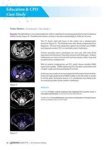
Education & CPD Case Study Urine Matters (Continued) - Case Study 3 Discussion: The left kidney is very hydronephrotic with no significant remaining parenchyma and no obvious PSMA activity (Figure 4). Therefore the ureteric activity is not due to physiological “hold-up� of urine. The CT shows solid soft tissue in the ureter, not a tubular/cystic structure (Figure 5). The findings have also slowly progressed since diagnosis. The two main diagnostic options to consider are a PSMAavid separate ureteric TCC or a prostate cancer metastasis. Ureteric prostate cancer metastases are very rare, with only 40-50 reported in the literature. They often present with flank pain. Ureteric metastases are more common with primary breast, colon, lung and lymphomatous malignancies. 90% of ureteric malignancies are TCC, which shows variable PSMA expression/uptake. PSMA-expressing TCC has been associated with a higher stage and decreased patient survival. A decision was made not to investigate the left ureteric lesion further. Given the high-grade level of PSMA avidity in the left ureter is similar to all the other metastatic lesions, it is considered more likely to be an unusual prostate cancer ureteric metastasis. Figure 4 Reference: Liu P et al, A hidden ureteral metastasis that originated from prostate cancer: a case report and literature. Transl Cancer Res 2017;6(3):650-655 Schallier D et al, Ureteral Metastasis: Uncommon manifestation in Prostate Cancer. AntiCancer Research 2015; 35: 6317-6320
Figure 5
36 | gamma GAZETTE | 2019 Summer Edition