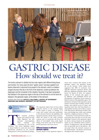
VETERINARY A complete examination of the entire equine stomach requires a 3m endoscope, and feed must be withheld for at least 12 hours
GASTRIC DISEASE How should we treat it?
The horse’s stomach is divided into two main regions with different lining tissue and function. For many years the term “gastric ulcers” has been applied to all lesions observed or assumed to be present in the stomach, which is a balloonshaped structure that sits in the front of the abdomen, tucked up between the diaphragm and the remainder of the intestine. Recently, it has been recognised that disease in the squamous region at the top of the stomach is not the same as disease in the glandular region, at the bottom of the stomach. WORDS AND PHOTOS: GAYLE D. HALLOWELL, SCHOOL OF VETERINARY MEDICINE AND SCIENCE, UNIVERSITY OF NOTTINGHAM
T
HE stomach produces gastric acid, which not only helps digestion but also prevents microorganisms from reaching and colonising the rest of the intestines. The lower, glandular region is always bathed in acid, and mucus and bicarbonate produced by the stomach lining protect it from acid-induced damage.
26
The squamous region does not have these protective factors, so the stomach lining in this region is prone to damage if it comes into contact with acid, particularly at the junction between the glandular and squamous portions, aka the lesser curvature, which sits closest to the acidic stomach contents and is at particular risk from the effects of ‘acid splash’ when acidic liquid sitting at the lower end of the stomach is pushed upwards as the horse moves.
Different lesions in different stomach regions
Overall, disease of the squamous region is
much more common than disease of the glandular region, but glandular lesions are on the increase. Latest studies have suggested that 80% of racehorses have clinically important squamous disease and 60% have significant glandular disease. Risk factors for disease in these two separate regions of the stomach also appear to be different, and established factors for development of squamous ulcers cannot be linked to disease of the glandular region. The risk factors associated with squamous ulcers include increased grain feeding, periods of fasting, and reduced access to water, and squamous ulcers have also been linked to a metropolitan location, lack of direct contact with other horses, talk instead of radio in the barn, and feeding straw while some training yards have a higher prevalence of others. Examination of stomach lesions under the microscope show that they are not the same pathology: Lesions in the squamous region are ulcers, whereas lesions in the glandular region are not. Glandular lesions contain a mixture of inflammatory cells and although they are generally seen at the pylorus – the exit point where the stomach connects to the small intestine – the disease can be found throughout this stomach region.
TRAINERMAGAZINE.COM ISSUE 52
EUROPEAN TRAINER ISSUE 52 GASTRIC DISEASE V2 .indd 26
18/12/2015 01:32