International Journal of Environment, Agriculture and Biotechnology


Vol-8, Issue-1; Jan-Feb, 2023
Journal Home Page Available: https://ijeab.com/ Journal DOI: 10.22161/ijeab
Peer Reviewed
International Journal of Environment, Agriculture and Biotechnology
Vol-8, Issue-1; Jan-Feb, 2023
Journal Home Page Available: https://ijeab.com/ Journal DOI: 10.22161/ijeab
Peer Reviewed
Department of Plant Biotechnology, Centre for Plant Molecular Biology & Biotechnology, Tamil Nadu Agricultural University, Coimbatore, Tamil Nadu, India
Received: 25 Dec 2022; Received in revised form: 17 Jan 2023; Accepted: 25 Jan 2023; Available online: 06 Feb 2023
©2023 The Author(s). Published by Infogain Publication. This is an open access article under the CC BY license (https://creativecommons.org/licenses/by/4.0/).
Abstract A phyA was cloned from Aspergillus niger by reverse transcription polymerase chain reaction. The amplified 1404 bp fragment was cloned in a T/A cloning vector and confirmed by sequencing. The isolated phytase gene showed 99.0 % sequence identity at nucleotide and protein level with Aspergillus niger CBS 513.88 phytase. The amino acid sequence from the phyA cDNA contained the consensus motifs, RHGXRXP and HD which are conserved among histidine acid phosphatases. The phytase cDNA was subcloned in pET28a(+) expression vector and expressed in E. coli. The expression of the gene was confirmed through SDS-PAGE analysis. A 52 kDa protein as per the calculated molecular mass of the translational product of phytase gene was observed in crude lysate of E. coli culture induced with IPTG. The protein was purified using nickel based His-bind resin column and checked on SDS-PAGE. The purified recombinant enzyme showed a single band of 52 kDa protein. The phytase activity of pET-phyA IPTG induced E. coli culture and purified phytase was 383.5 U/ml and 826.33 U/ml, respectively.
Keywords Aspergillus niger, phytase, cloning, expression, E. coli
Phosphorus is one of the most limiting nutrients for animals because most of the phosphorus in the plant seeds including the feeding plants is in the form of phytic acid (De Boland et al., 1975), most of which cannot be digested by monogastric animals and acts as an antinutritional factor hindering the uptake of a range of minerals. Furthermore, high phytic acid content in animal manure by excretion results in elevated level phosphorus level in soil and water and accompanying environmental concerns (Lambrechts et al., 1992). Phytate also chelates trace elements of iron and zinc between phosphate groups within a single phytate molecule or between two phytate molecules. The enzyme phytase is able to release the bioavailable phosphorus from phytic acid, consequently improving the phosphorus bioavailability and the uptake of minerals. On the other hand, the content of phytase in plant itself is too limited to release sufficient inorganic phosphorus (Brinch-Pederson et al., 2002).
Phytases are a special class of phosphatases that catalyze the sequential hydrolysis of myo-inositol-(1,2,3,4,5,6)hexakisphosphate or phytic acid (Ins P6) to less phosphorylated myo-inositol derivatives and inorganic phosphate (Haros et al., 2007). Phytase is the only known enzyme that can initiate the phosphate hydrolysis at carbon 1, 3 or 6 in the inositol ring of phytate. The removal of phosphate group by phytase results in releasing of calcium, iron, zinc, and other metals. Phytate degrading activity has been detected in plants, microorganisms, and in some animal tissues and phytases from several plant and microbial species (Hill et al., 2007) have been purified and characterized. In plants, phytase activity is found in many plant seeds (Laboure et al., 1993). Hence, although currently phytases are used mainly as animal feed additives in diets of monogastric animals, there is a great potential for the use of this class of enzymes in processing and manufacturing of food for human consumption (Jorquera et al., 2008).
Phytase activity has been found most frequently in fungi, such as Aspergillus ficuum (Gibson, 1987), and Aspergillus
https://dx.doi.org/10.22161/ijeab.81.5
Geetha et al.
Cloning of phytase gene from Aspergillus niger 563 and its expression in E.coli system
niger (Han et al., 2018). Phytase is also produced by grampositive bacteria, such as Bacillus (Farhat-Khemakhen et al., 2012 and Saadi et al., 2021), and gram negative bacteria, like Aerobacter aerogenes (Greaves et al., 1967), Pseudomonas sp. (Singh et al., 2017), Escherichia coli (Greiner et al., 1993), and Klebsiella (Jareonkitmongkol et al., 1997). Generally, phytases from gram-negative bacteria are intracellular proteins, while phytases from gram-positive bacteria and fungi are extracellular enzymes (Kim et al., 1998; Choi et al., 2001). In this study, we cloned and sequenced the phytase gene of Aspergillus niger and recombinantly expressed the phytase in E.coli
Sodium phytate, Isopropyl thiogalactoside (IPTG), deoxynucleoside triphosphates (dNTPs) and βmercaptoethanol were purchased from Sigma Aldrich Chemical Company, U.S.A. All the other chemicals used were of analytical grade manufactured in India.
A. niger 563 obtained from National Chemical Laboratory (NCL), Pune was used to isolate a phytase gene. E.coli DH5α and Bl21 (DE3) were used as hosts for genetic manipulation and expression, respectively. The vector pTZ52R/T (MBI Fermentas) was used for TA cloning and nucleotide sequencing, and pET28a(+) (Novagen, Madison, WI) was used for expression of phytase in E.coli E.coli strains were grown at 37°C in Luria-Bertani (LB) medium.
The hyphae of Aspergillus niger was inoculated into PDA medium (20% potato extract and 2% sucrose) and shaken at 100 rpm for 2-3 days at 28°C and mycelia was collected. The total RNA was extracted by method of TRIZOL. One hundred milligram of Aspergillus niger mycelial mat was ground with 0.8 ml of trizol reagent followed by the addition of 0.2 ml (1/4th volume) of chloroform. After grinding, sap was transferred to a 1.5 ml microfuge tube, mixed by gentle inversion and kept as such for 3 min. The contents were centrifuged at 12,000 rpm for 20 min and an equal volume of isopropanol was added to the supernatant. After an incubation of 5 min, the mixture was centrifuged at 12,000 rpm for 10 min. Supernatant was discarded and the pellet washed with 70% ethanol. The pellet was resuspendedin100μlofDEPC treated water (pre-warmed to 65°C) and 13 μl of2.5 Msodium acetate(pH5.5). The mixture was incubated at -70°C for 1 h after the addition of 3 volume of absolute alcohol. The contents were centrifuged at 12,000 rpm for 10 min and the pellet washed
with 75% ethanol. The pellet was air-dried and dissolved in 20 μl of DEPC treated water. Isolated total RNA was used as template for amplification of the phytase cDNA using RT-PCR.
Cloning of the phytase gene and sequencing
RT-PCR was performed with total RNA isolated from Aspergillus niger with a view to obtaining the cDNA corresponding to phytase gene. First strand cDNA was synthesized according to the manufacturer’s instructions (RevertAiD™H Minus First Strand cDNA Synthesis Kit # K1631, MBI Fermentas, Germany). PCR reactions were carried out in 20 µl reaction volume containing 2.0 µl of cDNA, 2.0 µl of 10X PCR buffer (10 mM Tris-HCl pH 9.0, 50 mM KCl, 1.5 mM MgCl2), 0.5 µl of 200 mM dNTPs, 0.5 µl of 10 µM of respective forward and reverse primers (PHYF - 5’ ATGGGCGTCTCTGCTGTTCTACTTC 3’ and PHYR5’ CTAAGCAAAACACTCCGCCCAATC 3’), 0.5 µl of 1.5 U Taq DNA polymerase (Bangalore Genei Pvt. Ltd., Bangalore, India) and 14.0 µl of sterile double distilled water. Amplification was performed using the temperature profile of the pre-incubation at 94°C for 5 min leading to 35 cycles of melting at 94°C for 1 min, annealing at 54°C for 1 min and synthesis at 72°C for 1 min followed by an extension of 72°C for 10 min.
The PCR product was recovered and purified from a 1.0% agarose gel using a DNA Gel Extraction kit (MBI Fermentas) according to the manufacturer's protocols, then subcloned into pTZ57R/T vector. The resulting plasmid was used for sequence analysis. The methods for E.coli competent cell preparation and transformation were according to the methods of sambrook et al. (1989). The recombinants were selected on the LB agar plate containing ampicillin (50 mg/L). The presence of the gene in the recombinant clones was confirmed by sequencing using M13 primers.
The nucleotide sequencing of the phytase (phy) gene was done by out sourcing at Bangalore Genie (India). Nucleotide and amino acid sequence homology searches were performed on the NCBI database by BLAST search and multiple alignments were done using the CLUSTAL W program. For phylogenetic analysis, gene sequences obtained in this study and phy gene sequences from different Aspergillus sp. available in NCBI database were used.
For the expression of mature protein, the entire ORF of the phytase gene without native signal peptide sequence was amplified with the specific primers, ECOF2 (Forward 5’ TCCGAATTCCTGGCAGTCCCCGCCTCGAGA 3’) and HINR2 (Reverse 5’ CGCAAGCTTAGCTAAGCAAAACACTCCGCC 3’) with insertion of EcoRI and HindIII sites at the 5’ and 3’ ends respectively The PCR product was digested with EcoRI and HindIII and ligated into the respective sites of pET28a(+) expression vector to generate plasmid pET28a(+)-phyA
The pET28a(+)-phyA plasmid was transformed into E. coli DH5αandsubsequentlyintotheexpressionstrain E. coli BL21. The transformants cells were grown at 30°C in LB medium containing 50mg/L kanamycin at the midexponential phase and their phytase expressions induced by adding isoprophyl β-D-thiogalactopyranoside (IPTG) to the culturesatafinal concentrationof1mM.
The E. coli strain BL21 harbouring the plasmid pET28a(+)-phyA was streaked separately on LB agar plate with kanamycin 50 mg/l and incubated at 37°C for 12-16 h so as to get individual colonies. Single colony was inoculated in 3 ml LB broth containing kanamycin 50 mg/l and grown on a rotary shaker (220 rpm) at 37°C for 16 h. From this, 500 µl of overnight grown culture was inoculated in 50 ml of LB broth and grown at 37°C and 220 rpm till the OD reached 0.3-0.5 at 600 nm. One millilitre of the aliquot was taken in an eppendorf tube and centrifuged at 10,000 rpm for 5 min and the pellet was resuspended in 50 µl of the Laemmli buffer (0.06 M TrisHCl, pH 6.8, 10% glycerol, 5% -Mercaptoethanol and 2% SDS) and stored at -20°C for later use as an uninduced culture. The remaining culture was induced by the addition of IPTG (isopropyl--D- thiogalactopyranoside) to a final concentration of 1 mM. The culture was allowed to grow at 30°C and the aliquot was collected at 1, 2, 3, 4, 5, 6 and 7 h after induction and the pellet was resuspended in Laemmli buffer The supernatant were checked for protein expression and purity by Coomassie-stained Sodium dodecyl sulfate (SDS)-polyacrylamide gel (PAGE) 12% electrophoresis.
Recombinant fusion phytase protein was purified using BugBuster® His•Bind Purification Kit (catalogue # 70793-3, Novagen, Germany) For purification, 100 ml of bacterial culture was collected by centrifugation at 10400 rpm for 10 min and protein was extracted with BugBuster™ Reagent. The cells were disrupted by sonication (5 times for 30 seconds). After centrifugation at 11400 rpm for 30 min, the supernatant containing the
targetproteinwaspurifiedusingHis•BindResinaccording to the manufacturer instruction (# TB054, Novagen, Germany).
Phytase assay
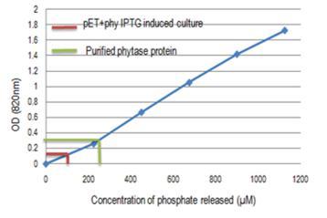
Phytase assay was carried out by taking200μlof crude sample of phytase into 10 ml test tubes and incubated at 37°C in water bath for 5 min. Two hundred microlitre of 1.25% (w/v) sodium phytate in 200 mM sodium acetate buffer (pH 5.0) was added for enzymatic hydrolysis of phytate, and incubated for 30 min at 37°C. The reaction was terminated by adding 400 μl of 15% trichloroacetic acid. The mixture was centrifuged at 4600 rpm for10minand200μlofsupernatantwasaddedto1.8 ml of double distilled water. Two millilitre of fresh colour reagent (3 volume of 1 M H2SO4 + 1 volume of 2.5% ammonium molybdate + 1 volume of 10% ascorbic acid) was added and mixed well. The mixture was incubated at 50°C for 15 min and left at room temperature for 2-3 min. The absorbance was read at 820 nm, using water as the blank and the serially diluted potassium phosphate solutions as standards. Phytase activity was calculated per ml of culture and expressed as U/ml. One unit of phytase is defined as the amount of enzyme required to release 1 µmol of inorganic phosphate per min from sodium phytate at 37°C.
df = Dilution factor

30 = Time (in minutes) of assay per the Unit definition
0.200 = Volume (in milliliter) of sample used
III.
Cloning and sequence analysis of phytase gene from Aspergillus niger
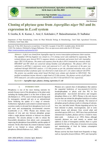
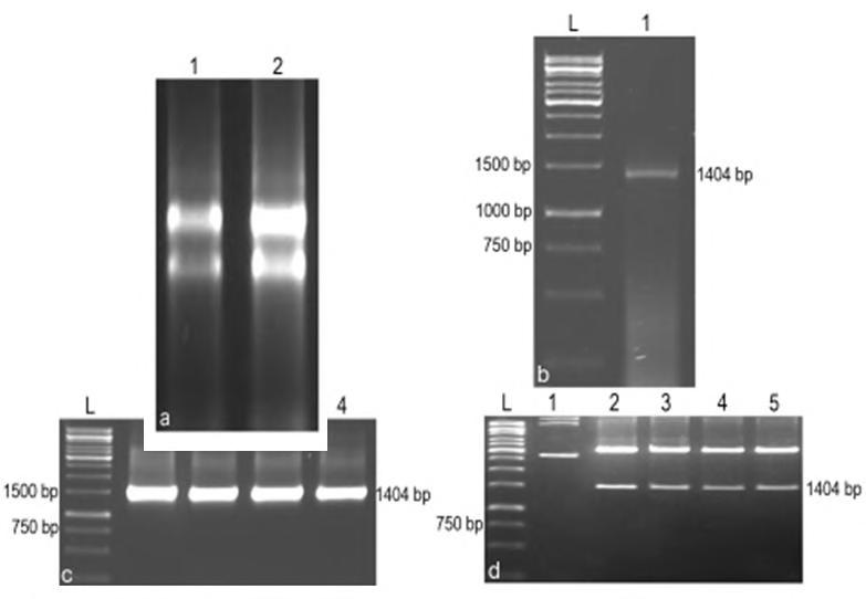
RT-PCR analysis with total RNA isolated from Aspergillus niger using phytase gene specific primers showed amplification of a 1404 bp fragment. The 1404 bp band was eluted and ligated with a T-tailed T/A cloning vector. The ligated product was transformed into E. coli competent cells. Colony PCR analysis of randomly selected colonies showed the amplification of 1404 bp fragment and restriction digestion of the plasmids isolated from these colonies with the restriction enzymes, HindIII and EcoRI released a fragment of the expected size (Fig.1). Four clones (CPHY10, CPHY13, CPHY15 and CPHY20) were sequenced using automated DNA sequencer and aligned using CLUSTAL X bioinformatics tool (Fig.2).
Geetha et al.
Cloning of phytase gene from Aspergillus niger 563 and its expression in E.coli system
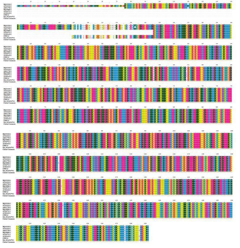
Among the clones CPHY13 showed a maximum of 99.0% sequence identity at nucleotide and protein level with Aspergillus niger CBS 513.88 phytase (Accession # XM_001401676). The CPHY13 cDNA shared 99.0% sequence identity with the Aspergillus species (Accession # DQ192035, AB022700, AY013315) at nucleotide levels and 98.0% sequence similarity with Aspergillus species (Accession # AAF25481, AAR08366, ACE79229) at amino acid level. It showed 100% homology at protein level with Aspergillus awamori phytase (Accession # ABA29207) and followed by 99.0% with phyA - Aspergillus niger (Accession # XP_001401713, AAG40885). The cloned fragment contained a single open reading frame of 1404 bp long (from ATG start codon to TAG stop codon), which could potentially encode an approximately 52 kDa protein having 467 amino acids (Fig.3).
The amino acid sequence from the phyA cDNA contained the consensus motifs RHGXRXP and HD which are conserved among histidine acid phosphatases. The aligned sequences formed two different clusters. Cluster dendrogram results revealed that the cloned phytase gene from the Aspergillus niger formed a separate sub cluster along with the phytase gene sequences of other Aspergillus sp. at nucleotide and protein level (Fig.4). The genebank accession number for the Aspergillus niger phyA sequence is JQ241266.
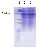
For the expression of phytase in E. coli, the cDNA was cloned in EcoRI and HindIII site of the multiple cloning site of the E. coli expression vector pET28a(+). The recombinant colonies were confirmed by colony PCR analysis and restriction digestion (Fig.5). The expression of the phytase gene in E. coli was confirmed by SDS-PAGE analysis of crude lysate of E. coli BL21 cells harbouring the pET28a(+) vector alone as control and the recombinant pET28a(+) with phytase gene (both uninduced and induced with IPTG). An expected size of protein band (approximately 52 kDa) was observed in the crude lysate of induced culture, which was absent in uninduced culture and in cells with pET28a(+) vector without insert. There was an increase in the amount of induced protein with increase in time. When E. coli strain BL21 with pET28a(+)-phyA was grown at 37°C, there was no difference in the protein banding pattern of total cell extracts before and after induction with IPTG. In contrast, when the cells were grown at 30°C, an intense 52 kDa band was visible specifically in the extract of induced cells containing the recombinant plasmid (Fig.6). Phytase was purified from the 100 ml culture after 6 h induction with 50 mM IPTG using nickel based His bind resin column according to the recommendation of the manufacturer
(BugBuster® His•Bind Purification Kit, Novagen, Germany). On SDS-PAGE, the purified recombinant enzyme showed a single band of 52 kDa. The phytase activity was calculated as 826.33 U/ml for purified phytase and 383.5 U/ml for pET-phyA IPTG induced culture
In this study, a phytase gene (phyA) was isolated from the cDNA of A. niger. This gene contains a single open reading frame of 1404 bp long (from ATG start codon to TAG stop codon), which encodes an approximately 52 kDa protein having 467 amino acids. The phytase from A. niger phyA is well characterised by several earlier workers. It is encoded by a 1.4 kb DNA fragment and has a molecular mass of 80 kDa, with 10 Nglycosylation sites (Han and Lei, 1999). Average molecular masses of bacterial phytases are smaller than those of fungal phytases (40–55 vs. 80–120 kDa), mainly due to glycosylation differences (Choi et al., 2001; Golovan et al., 2000).
From the sequence analysis it was confirmed that the amino acid sequence from the phyA cDNA contained the consensus motifs RHGXRXP and HD which are conserved among histidine acid phosphatases. The clone CPHY13 showed a maximum of 99.0% sequence identity at nucleotide and protein level with Aspergillus niger CBS 513.88 phytase (Accession # XM_001401676). These motifs play an important role in the phosphorylation (Kostrewa et al., 1997 and Oh et al., 2001). A highly conserved sequence motif RHGXRXP (Ullah et al., 1991), involved in the catabolic reactions, is found at the active sites of phytase. Furthermore, phyA contains a remote Cterminal His-Asp motif (HD motif) that is also likely to take part in the catalysis. Together, it is therefore suggested that the phyA belongs to a member of the phytase subfamily of histidine acid phosphatases (Mitchell et al., 1997). The conserved sequence RHGXRXP in the substrate-binding site interacts with the phosphate groups in the substrate to form a complex of enzyme-substrate. The HD elements in the catalyzation domain further function to release the phosphate group from the substrate (Loewus and Murthy, 2000; Mullaney et al., 2000).
Due to the enormous potential for application of phytase in the animal feed industry, several researchers have attempted to produce this enzyme cost-effectively. A. niger phyA phytase has been cloned and over expressed in several microbial hosts, including S. cerevisiae (Han et al., 1999), P. pastoris (Han and Lei, 1999), A. niger (Van Dijck, 1999) and E. coli (Singh et al., 2018). The pET prokaryotic expression system is one of the most effective expression systems, possesses high performance and specific
Geetha et al.
interaction between bacteriophage T7 promoter and T7 RNA polymerase to increase the expression efficacy of exogenous gene in bacteria. In this study, the recombinant pET-phyA construct expressed the phytase enzyme efficiently in the host E. coli cells. The expressed protein was purified by nickel based column affinity chromatograph conveniently because of the six histidines tag fused to the E. coli protein.
For the expression in E. coli, phytase cDNA was cloned in pET vector devoid of N-terminal signal peptide. As E. coli cells are not able to cleave the signal peptide of secreted proteins, the cDNA had to be modified to remove the signal peptide. An expected size of protein (approximately 52 kDa) was observed in the crude lysate of induced culture, which was absent in uninduced culture and in cells with pET28a(+) vector without insert. There was an increase in the amount of induced protein with increase in time. This demonstrated that the recombinant DNA had indeed expressed in E. coli cells (BL21 strain). When E. coli BL21 pET28a(+)-phyA was grown at 37°C, there was no difference in the protein electrophoretic
patterns of total cell extracts before and after induction with IPTG. In contrast, when the cells were grown at 30°C, an intense 52 kDa band was visible specifically in the induced extract from the subsequent time interval. This report was supported by Phillippy and Mullaney (1997) who reported maximum activity of 1.5 µmol/mg protein. The phytase activity of pET-phyA IPTG induced E. coli culture was 383.5 U/ml while it was 826.33 U/ml in purified phytase.
Similar results were observed in the methylotrophic yeast P. pastoris by the heterologous expression of Debaryomyces castellii CBS 2923 phytase and maximum production level obtained was 476 U/ml (Ragon et al., 2008). Chen et al. (2004) reported the successful expression of E. coli appA gene in P. pastoris with the maximum phytase activity after an induction period of 96 h, of 118-204 IU/ml at the flask scale and 1880-4946 IU/ml at high cell-density fermentation. Xiong et al. (2005) and Bei et al. (2001) also reported 865 U/ml and 165 U/ml of enzymatic activity, respectively with A. niger phytase in P. pastoris
Fig.1. PCR amplification and cloning of phytase gene in T/A cloning vector. a. Total RNA isolated from Aspergillus niger, 1 to 2: Total RNA of Aspergillus niger. b. PCR amplification of phytase gene, L: 1 kbp ladder, 1: Amplification of phytase gene from cDNA of A. niger c. Colony PCR analysis, L: 1 kbp ladder, 1 to 4: Phytase gene amplfication from selected colonies. d. Restriction analysis, L: 1 kbp ladder, 1: Undigested pTA-phyA plasmid, 2 to 5: Plasmid (from positive clones) digested with HindIII and EcoRI
Cloning of phytase gene from Aspergillus niger 563 and its expression in E.coli system ISSN:
Geetha et al.
Cloning of phytase gene from Aspergillus niger 563 and its expression in E.coli system
Geetha et al.
Consensus motifs of histidine acid phosphatase . Indicates stop codon
Fig.3. Nucleotide and amino acid sequence of phytase gene (phyA)
Fig.4.Cluster dendrogram illustrating the phylogenetic relationship based on the multiple sequence alignment of the nucleotide sequences of known phytase gene and the phytase gene from Aspergillus niger
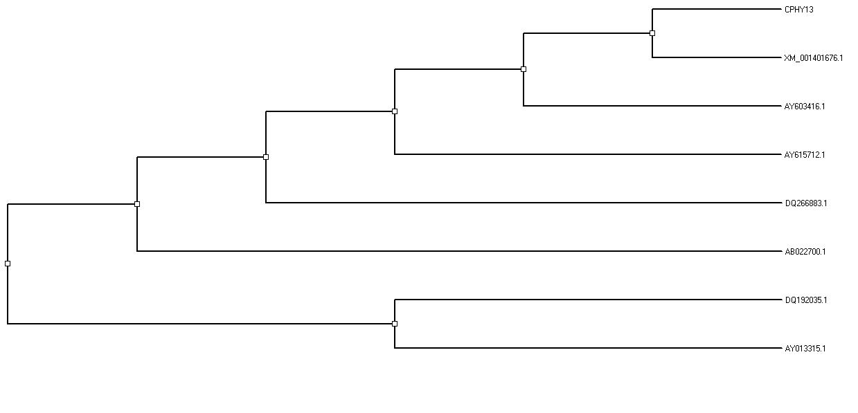
Fig.5. Cloning of phytase gene in E. coli expression vector pET28a(+).
a. Colony PCR analysis, L: 1 kbp ladder, 1 to 4: Amplification of phytase gene from E. coli colonies. b. Restriction analysis, L: 1 kbp ladder, 1: Undigested pET28a(+)-phyA plasmid, 2 to 4: Plasmids (from selected colonies) digested with HindIII and EcoRI
Cloning of phytase gene from Aspergillus niger 563 and its expression in E.coli system ISSN: 2456-1878 (Int.
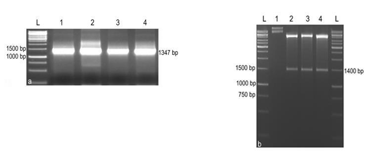
Fig.6. SDS-PAGE analysis of crude lysate of E. coli cells harbouring pET28a(+)-phyA 1: Protein marker, 2&3: Phytase expression after IPTG induction of 6 and 7 h.
[6] De Boland, A.R., G.B. Garner and B.L. Odell. 1975: Identification and properties of "phytate" in cereal grains and oilseed products. J. Agricult. Food Chem., 23: 11861189.
[7] Farhat-Khemakhem, A., M.B. Farhat, I. Boukhris, W. Bejar, K. Bouchaala and R. Kammoun. 2012. Heterologous expression and optimization using experimental designs allowed highly efficient production of the PHY US417 phytase in Bacillus subtilis 168. AMB Express., 2:10.
[8] Gibson, D. 1987. Production of extracellular phytase from Aspergillus ficuum on starch media. Biotechnol. Lett., 9: 305
310.
[9] Golovan, S., G.R. Wang, J. Zhang and C.W. Forsberg. 2000. Characterization and overproduction of the Escherichia coli appA encoded bifunctional enzyme that exhibits both phytase and acid phosphatase activities. Can. J. Microbiol., 46: 59-71.
[10] Greaves, M.P., G. Anderson and D.M .Webley. 1967. The hydrolysis of inositol phosphates by Aerobacter aerogenes. Biochim. Biophys. Acta., 132: 412–418.
[11] Han, Y.M. and X.G. Lei. 1999. Role of glycosylation in the functional expression of an Aspergillus niger phytase (phyA) in Pichia pastoris Arch. Biochem. Biophys., 364: 83–90.
[12] Han, N., H. Miao, T. Yu, B. Xu, Y. Yang and Q. Wu. 2018. Enhancing thermal tolerance of Aspergillus niger PhyA phytase directed by structural comparison and computational simulation. BMC Biotechnol.,18:36.
[13] Haros, M., M. Bielecka, J. Honke and Y.Sanz. 2007. Myoinositol hexakisphosphate degradation by Bifidobacterium infantis ATCC 15697. Inter. J. Food Microbiol., 117: 7684.
[14] Hill, J.E., D. Kysela and M. Elimelech. 2007. Isolation and assessment of phytate-hydrolysing bacteria from the DelMarVa Peninsula. Env. Microbiol., 9: 3100-3107.
Fig.8. Standard curve for the phosphorus concentration measurement
[1] Bei, J.L., Z. Chen, L. Yang, L. Liao, X.Z. Wang, and Z.Y. Jiang. 2001. Overexpression of artificial synthetic gene of Aspergillus niger NRRL3135 phytase in Pichia pastoris Sheng Wu Gong Cheng Xue Bao., 17: 254–258.
[2] Brinch-Pedersen, H., L.D. Sorensen and P.B. Holm. 2002. Engineering crop plants: getting a handle on phosphate. Trends Plant Sci., 7: 118-125.
[3] Chen, C.C., P.H. Wu, C.T. Huang, and K.J. Cheng. 2004. A Pichia pastoris fermentation strategy for enhancing the heterologous expression of an Escherichia coli phytase. Enzyme Microbial. Technol., 35: 315-320.
[4] Choi, Y.M., H.J. Suh and J.M. Kim. 2001. Purification and properties of extracellular phytase from Bacillus sp. KHU10. J. Protein Chem., 20: 287–292.
[5] Cosgrove, D.J. 1970. Inositol phosphate phosphatases of microbiological origin. Inositol phosphate intermediates in the dephosphorylation of the hexaphosphates of myo-inositol, scylloinositol, and D-chiro-inositol by a bacterial (Pseudomonas sp.) phytase. Aust. J. Biol. Sci., 23: 1207–1220.
[15] Jareonkitmongkol, S., M. Ohya, R. Watanabe, H. Takagi and S. Nakamori. 1997. Partial purification of phytase from a soil isolate bacterium, Klebsiella oxytoca MO-3. J. Ferment. Bioeng., 83: 393–394.
[16] Jorquera, M., O. Martinez, F. Maruyama, P. Marschiner and M.D.L.L. Mora. 2008. Current and future biotechnology applications of bacterial phytases and phytase-producing bacteria. Microbes Environ., 23: 182-191.
[17] Kim, Y.O., H.K. Kim, K.S. Bae, J.H. Yu and T.K. Oh. 1998 Purification and properties of a thermostable phytase from Bacillus sp DS11. Enzyme Microb. Technol., 22: 2–7.
[18] Kostrewa, D., F.G. Leitch, A. D’Arcy, C. Broger, D. Mitchell and A.P.G.M. Loon. 1997. Crystal structure of phytase from Aspergillus ficuum at 2.5 Å resolution. Nature Struct. Biol., 4: 185–190.
[19] Laboure, A.M., J. Gagnon and A.M. Lescure. 1993. Purification and characterization of a phytase (myo-inositolhexakisphosphate phosphohydrolase) accumulated in maize (Zea mays) seedlings during germination. Biochem. J., 295: 413–419.
[20] Lambrechts, C., H. Boze, G. Moulin and P. Galzv. 1992. Utilization of phytate by some yeast. Biotechnol. Lett., 14: 61-66.
Cloning of phytase gene from Aspergillus niger 563 and its expression in E.coli system ISSN:
Geetha et al.
Cloning of phytase gene from Aspergillus niger 563 and its expression in E.coli system
[21] Loewus, F.A. and P.P.N. Murthy. 2000. Myo-Inositol metabolism in plants Plant Sci., 150: 1–19.
[22] Mitchell, D.B., K. Vogel, B.J. Weimann, L. Pasamontes and A.P. van Loon. 1997. The phytase subfamily of histidine acid phosphatases: isolation of two genes for two novel phytases from the fungi Aspergillus terrus and Myceoliophthora thermophila Microbiology, 143: 245252.
[23] Mullaney, E.J., C.B. Daly, and A.H.J. Ullah. 2000. Advances in phytase research. Adv. Appl. Microbiol., 47: 157–199.
[24] Oh, B.C., B.S. Chang, K.H. Park, N.C. Ha, H.K. Kim, B.H. Oh and T.K. Oh. 2001. Calcium-dependent catalytic activity of a novel phytase from Bacillus amyloliquefaciens DS11. Biochemistry, 40: 9669–9676.
[25] Phillippy, B.Q., and E.J. Mullaney. 1997. Expression of an Aspergillus niger phytase (phyA) in E. coli J. Agric. Food Chem., 45: 3337–3342.
[26] Ragon, M., V.N. Roux, P. Chemardin, G. Moulin and H.H. Boze. 2008. Molecular gene cloning and overexpression of the phytase from Debaryomyces castellii CBS 2923. Protein Express. Purif., 58: 275–283.
[27] Saadi, M.I., A. Doosti , H. Jalali, E. N. Abdolyousefi, M. Hooshiyar, R. Tabrizi, and E. Noshadi. 2021. Cloning of Bacillus subtilis phytase gene construct in Escherichia coli Iran. J. Microbiol., 13(5): 664-670.
[28] Singh, N., S. Kuhar, K. Priya, R. Jaryal and R. Yadav. 2018. Phytase: The feed enzyme, an overview. Adv. Ani. Biotechnol. Appl., 269- 327.
[29] Singh, S , S. Singh, P.K. Sharma, D. Sharma. 2017 Isolation, identification and molecular characterization of phytase producing bacteria, Pseudomonas sp. aazad. J. Pure Appl Microbiol., 11:1845-1850.
[30] Ullah, A.H.J., B.J. Cummins and H.C.J. Dischinger. 1991. Cyclohexanedione modification of arginine at the active site of Aspergillus ficuum phytase. Biochem. Biophys. Res. Commun., 178: 45-53.
[31] Van Dijck, P.W.M. 1999. Chymosin and phytase: made by genetic engineering. J. Biotechnol., 67: 77–80.
[32] Xiong, A.S., Q.H. Yao, R.H. Peng, P.L. Han, Z.M. Cheng and Y. Li. 2005. High level expression of a recombinant acid phytase gene in Pichia pastoris. J. Appl. Microbiol., 98: 418–428.
ISSN: 2456-1878 (Int. J. Environ. Agric. Biotech.)
https://dx.doi.org/10.22161/ijeab.81.5