Peer Reviewed
1Department of Chemistry, College of Natural and Computational Science, Bonga University, Bonga, Ethiopia
2*Department of Plant Science, College of Agriculture and Natural Resource, Bonga University, Bonga, Ethiopia
Correspondence should be addressed to Tamirat Wato Wana; tamiratwato1@gmail.com
Evaluation of the Phytochemical and Antibacterial Activity of Four Selected Plant Extracts against Some Pathogenic Bacteria
International Journal of Environment, Agriculture and Biotechnology Vol-7, Issue-4; Jul-Aug, 2022 Journal Home Page Available: https://ijeab.com/ Journal DOI: 10.22161/ijeab

ISSN: 2456 1878 (Int. J. Environ. Agric. https://dx.doi.org/10.22161/ijeab.74.2Biotech.)8 265
Orcid: https://orcid.org/0000 0001 5509 5033
Abstract This study aimed to evaluate the phytochemical and antibacterial activity of Acanthus eminens, Celosia trigyna, Drymaria cordata, and Phytolacca dodecandra against the selected pathogenic bacteria; Two strains of Gram positive (Staphylococcus aureus and Bacillus cereus) and three strains of Gram negative (Escherichia coli, Salmonella typhi, and Pseudomonas aeruginosa) bacteria The presences of phytochemicals were analyzed using the standard methods of phytochemical analysis, while the antibacterial activities were analyzed using the disc diffusion method. The results indicated the presence of terpenoids, cardiac glycosides, saponins, flavonoids, and alkaloids in the extracts of A. eminens and C. trigyna Alkaloids, flavonoids, and phenols are present in the extract of D. cordata and P. dodecandra. Methanolic extracts of Acanthus eminens, Celosia trigyna, Drymaria cordata, and Phytolacca dodecandra were potentially effective with variable efficiency against the tested bacterial strains at a concentration of 4 mg/ml while Celosia trigyna extract was found to be the most effective with a concentration of against all tested bacterial strains. On the other hand, Phytolacca dodecandra extract was found to be effective with a concentration of against B. cereus, S. aureus, S. typhi, and P. Aeruginosa suppressing their growth with inhibition zones of 10.3, 16.7, 11.6, and 11.1 mm, respectively. Celosia trigyna and Phytolacca dodecandra methanolic extracts were the most effective plant extracts and showed bacteriostatic and bactericidal activities against the highly susceptible strains of pathogenic bacteria (Bacillus cereus, Staphylococcus aureus, and Pseudomonas aeruginosa) with MIC’s ranging from 20 to 0.8 mg/ml and MBC of 4.0 and 0.16 mg/ml, respectively. These plant extracts have high potential antibacterial effects on bacterial strains tested, especially Bacillus cereus, Staphylococcus aureus, and Pseudomonas aeruginosa. They have been highly effective to be used as a natural alternative treatment to control pathogenic bacteria.
Birhanu Bekele Gosa1 , Tamirat Wato Wana2*
Received: 27 Jul 2022; Received in revised form: 20 Aug 2022; Accepted: 25 Aug 2022; Available online: 31 Aug 2022 ©2022 The Author(s). Published by Infogain Publication. This is an open access article under the CC BY license (https://creativecommons.org/licenses/by/4.0/).
Keywords A. eminens, C. trigyna, D. cordata, P. dodecandra, phytochemicals, antibacterial activity, HPLC-UV.
I. INTRODUCTION In the modern world, multiple drug resistance has developed against many microbial infections due to the indiscriminate use of commercial antimicrobial drugs commonly used in the treatment of infectious diseases. In addition to this problem, antibiotics are sometimes associated with adverse effects on the host including hypersensitivity, immunosuppression, and allergic reactions. Therefore, there is a need to develop alternative antimicrobial drugs for the treatment of infectious diseases from medicinal plants. Plants are one of the most important sources of medicines for treating illnesses since the beginning of human civilization [1 3]. Plants in general
al. International Journal of Environment, Agriculture and Biotechnology, 7(4)
Some researchers reported that ethanolic clove extract was potentially active against S. aureus, Vibrio parahaemolyticus, and P. aeruginosa while it was inactive against E. coli and Salmonella enteritidis [12]. Other researchers ascertained the activity of clove oil against all tested pathogenic bacteria while Vibrio cholera, S. typhi, and Klebsiella pneumonia were found to be resistant to aqueous clove extract [13, 14]. Moreover, the methanolic clove extract was reported to be potentially effective against S. aureus, P. aeruginosa, and E. coli with MIC ranging from 0.1 to 2.31 mg/ml [15]. The evaluation of plants for their potential application based on their medicinal properties is important for modern day medicine as the widespread and long term use of antibiotics has led to the emergence of multi drug resistant strains, besides several side effects. The adverse effects of these synthetic drugs can be overcome by using traditional or herbal formulations which are safe, efficacious, and multifunctional. Further, the development of herbal medicines based on ethnomedical leads is relatively easier in comparison to synthetic drugs [16 18]. In the present scenario of the emergence of multiple drug resistance to human pathogenic organisms, this has necessitated a search for new antimicrobial substances from natural sources including plants. Plant and plant products play a wide range of antimicrobial properties.
2.2. Extracts preparation 100 g of the fine powder of plant materials were soaked in 300 ml of methanol with stirring for 72 hours, filtered through double layers of muslin, centrifuged at 9000 rpm for 10 min, and finally filtered again filtered through Whatman No1 filter paper and concentrated using a rotary evaporator at 40˚C. The resulting crude extracts were weighed and stored in the refrigerator until phytochemical screening and antimicrobial activity were carried out. The extract yields were weighted, stored in small bottles in Fridge at 5°C and their yield percentages were calculated using the following formula: Extract yield % = Q/T x 100 (where Q; the weight of extracted plants residues and T; the weight of plant powder).
ISSN: 2456 1878 (Int. J. Environ. Agric. https://dx.doi.org/10.22161/ijeab.74.2Biotech.)8
The study on medicinal plants is essential to promote the proper use of herbal medicine to determine their potential as a source for the new drugs [9]. Antimicrobials of plant origin have enormous therapeutic potential. They are effective in the treatment of infectious diseases while simultaneously mitigating many of the side effects that are often associated with synthetic antimicrobials [10]. Drug resistant bacteria and fungal pathogens have further complicated the treatment of infectious diseases. In recent years, drug resistance to human pathogenic bacteria has been commonly reported from all over the world. However, the situation is alarming in developing as well as developed countries due to the indiscriminate use of antibiotics [11].
2.3. Antibacterial activities of the selected plant extracts: Bacterial strains. The antibacterial potency of each plant extract was evaluated using five bacterial strains. Bacterial strains: Staphylococcus aureus and Bacillus cereus were Gram positive and Escherichia coli, Salmonella typhi, and Pseudomonas aeruginosa were Gram negative bacteria. The bacterial strains were provided from the culture collection of the Mizan Aman research center.
266 and high valued medicinal plants specifically, have a long history of use as a source of cheap and effective remedy for various ailments [4]. The use of plants and herb extract in the treatment of human ailments is a very ancient art, a practice that has been passed on for generations and Scientists in Africa and other developing countries are researching local plants abundant in the continent for their possible use in traditional medicine. Plants are the richest repository of drugs for traditional medicines, modern medicines, folk medicines, pharmaceutical intermediates, and chemical entities [5 7]. It is important to mention that traditional medicinal systems are at a transitional stage in the development of modern medicines in developing countries Therefore, the use of neglected and little known medicinal and aromatic plants must be promoted and encouraged at regional as well as global levels for the betterment of mankind [8].
2.1. Plant materials Collection and Authentication. The raw material of medicinal plants Acanthus eminens (stems), Celosia trigyna (leaves), Drymaria cordata (leaves), Phytolacca dodecandra (roots) was collected from the medicinal plant's farm of Bonga University. The plant materials were washed, disinfected, rinsed with distilled water, and spread out and dried in the chemistry laboratory at room temperature for about thirty days. Dried samples of plants materials were milled into a fine powder using a high capacity grinding machine and subsequently stored separately in sterilized polythene bags in the refrigerator at the temperature of 4ºC until required for use. The plants are deposited and voulcher numbers were given by at the National Herbarium of Addis Ababa, Ethiopia The four selected medicinal plants were authenticated by Botanist Mr. Seyoum Robo at Bonga University.
II. MATERIALS AND METHODS
Boloche et 2022
PartgroundAbove The aboveground parts of the plant are fumigated to heal and alleviate the severe headache or migraine Phytolacca dodecandra MG 2004S4 S Leaves Infusion from leaves is used to control external parasites in livestock in general by washing their whole bodies.
ISSN: 2456 1878 (Int. J. Environ. Agric. https://dx.doi.org/10.22161/ijeab.74.2Biotech.)8
2.5. Antibacterial activity of plant extract
Roots The whole parts are chopped and then mixed with water to treat Gonorrhea, rabies, anthrax.
Boloche et al. International Journal of Environment, Agriculture and Biotechnology, 7(4) 2022
The disk diffusion method is used to evaluate the antimicrobial activity of each plants extract. The plant extracts residues (100 mg) were re dissolved in 5 ml of methanol, sterilized through a Millipore filter (0.22 mm) then loaded over sterile filter paper discs (8 mm in diameters) to obtain a final concentration of 10 mg/disc. 20 ml of an agar plate medium was poured into sterile petri dishes followed with 30 ml of seeded medium previously inoculated with bacterial suspension (100 ml of medium/1 ml of 107 MPN) to attain 105 MPN/ml of medium. Sterile filter paper discs loaded with plant extract concentration of (10 mg/ml) were placed on the top of an agar plate. Filter paper discs loaded with 5 mg of Gentamycin were used as a positive control. The plates were kept in the fridge at 5 0C for 2 hrs to permit plant extracts diffusion then incubated at 35 0C for 24 hrs. The existences of inhibition zones were measured with the help of a template and the diameter of the zone of inhibition was determining the effectiveness of the antibiotic. The large diameter indicated the sensitivity of the bacterium to the antibiotic. The zone sizes were compared to a standardized chart to determine the
2.4. Inoculums preparation Each bacterial strain was sub cultured overnight at 35 0C in an agar plate slant for 24 hrs Individual microorganisms placed on the plate were grown into individual colonies, each a clone genetically identical to the individual ancestor organism. After the incubation, the colony of the organisms was taken and each was inoculated into 7 ml of peptone water in a bijou bottle and shook vigorously to obtain the solution homogeneity. The turbidity produced by these organisms was adjusted and used to match the turbidity standard prepared as described by [19].
Table 1.1: Medicinal plants with mode of preparation used by local people from Kafa Zone, Southwest, Ethiopia. Habit: Tree (T), Herb (H), Shrub (S), Climber (C). Synthetic Name nameVoucher Habit Parts Used Disease and Mode of application
267 Table 1: The ethnobotanical data of selected medicinal plant species and their extract percentage yield Plant species Family Local name Commonname Plantusedpart yieldExtract(%) A. eminens Acanthaceae Phecho Acanthus Stems 4.63 C. trigyna Amaranthaceae Degicho Woolflower Leaves 3.27 D. cordata Caryophyllaceae Hakeato Drymaria Leaves 6.54 P. dodecandra Phytolaccaceae Yengamo Endod Roots 8.74
Acanthus eminens 16191 S LeavesStems+ Infusions of leaves of used for backache, skin diseases, cough, eye infections, wounds, pneumonia, anti diarrhea and edema. Celosia trigyna MG 2005S67 H All parts The whole parts are chopped and the sap is used for Arthritis, Diarrhea, Dysentery Drymaria cordata MG 2005S30 H/C Leaves The sap is used for treating respiratory chest ailments, colds and bronchitis.
III. RESULTS AND DISCUSSION
3.1. Plants extraction yield: The ethnobotanical data of the employed plants and their extract percentage yield are illustrated in Table 1. The extract of 100 g of each dried plant material with methanol yielded plant extracts residues ranging from 2.29 to 6.12 g. The highest yield of plant extract was obtained from Phytolacca dodecandra (6.12 g) followed by Drymaria cordota (4.58 g) while Celosia tigyna (2.29 g) give the lowest extract yield, Extractrespectively.yield
3.2. Phytochemical Screening: The two extracts were screened for the presence of major phytochemicals using standard qualitative methods as described previously [20 22]. Each plant extracts were screened for the presence of terpenoids, flavonoids, saponins, tannins, alkaloids, fatty acids, steroids, phenols, cardiac glycosides, anthraquinones, and phlobatannis as outlined below:
Test for Alkaloids: To 2 ml of the extract, 2 ml of 10% hydrochloric acid was added. To the acidic medium, 1 ml Hager’s reagent (saturated picric acid solution) was added. The presence of alkaloids is confirmed by the formation of a yellow colored precipitate
ISSN: 2456 1878 (Int. J. Environ. Agric. https://dx.doi.org/10.22161/ijeab.74.2Biotech.)8 268 bacterium sensitivity, resistance, and intermediate sensitivity to that of antibiotics
2.6. Determination of minimum inhibitory concentrations (MIC’s) of the effective plant extract Minimum inhibitory concentrations (MICs) are the lowest concentration of an antimicrobial that inhibited the visible growth of a microorganism after overnight incubation. The MIC of the selected plant extracts was carried out using a disc diffusion method and evaluating the resistivity of bacterial strains. Different concentrations of the effective plant extracts (100, 20, 4, 0.8, 0.16, and 0.32 mg/ml) were prepared separately by dissolving 200 mg in 100 ml of methanol, sterilized. The most effective extracts of plants that exhibited a strong antibacterial activity at 10 mg/ml were manipulated to determine their minimum inhibitory concentrations. 1 ml of the standardized inoculums from peptone water was then inoculated into the solution in the test tubes. These were all incubated at 37˚C for 24 hrs and observed for turbidity of growth. The lowest concentrations which showed no turbidity in the test tubes were recorded as the MIC. 2.7. Determination of minimum bactericidal concentrations (MBC’s) of the effective plant extract. The minimum bactericidal concentration is the lowest concentration of a substance that prevents the growth of an organism after subculture onto antibiotic free media or the concentration of plant extract that did not exhibit any bacterial growth on the freshly inoculated agar plates. Agar plates were incubated at the temperature of 37 0C for 24 hours then examined for bacterial growth corresponding to plant extracts concentration.
Boloche et al. 2022
Test for Glycosides: 2 ml of acetic acid was added to 2 ml of the extract. The mixture was cooled in a cold water bath and 2 ml of concentrated H2SO4 was then added, color development from blue to bluish green indicates the presence of glycosides
% = Q/T x 100 (where Q; the weight of extracted plants residues and T; the weight of plant powder).
Test for Phenols: 0.5 ml of the extract, 5 ml of Folin Ciocalteu reagent, and 4 ml of aqueous sodium carbonate were added. The appearance of blue color indicates the presence of phenols
Test for Anthraquinones: 2 ml of the extract was boiled with 5ml of 10% hydrochloric acid for 3 minutes and 5 ml of chloroform was added. 5 drops of 10% ammonia were further added. A rose pink coloration indicates the presence of anthraquinones or a positive result.
International Journal of Environment, Agriculture and Biotechnology, 7(4)
Test for Phlobatannins: 2 ml of the extract were boiled with 1% aqueous hydrochloric acid. The formation of a red precipitate indicates the presence of Phlobatannins.
Test for Terpenoids: 5 ml of the extract was mixed in 2 ml of chloroform and 3 ml of concentrated sulphuric acid was carefully added to form a layer. A reddish brown coloration at the interface indicates the presence of terpenoids
Test for Saponins: To 2 ml of the extract, 2 ml of distilled water was added and it was agitated in a test tube for 5 minutes. The formation of foams indicates the presence of saponins Test for Tannins: 4 drops of 0.1% ferric chloride were added to 2 ml of the extract, a brownish green or blue black coloration indicated the presence of tannins.
Test for Steroids: 2 ml of extract were dissolved in 10 ml of chloroform and then 10 ml of concentrated sulphuric acid was added by the side of the test tube. The upper layer turned red whereas the sulphuric acid layer turned yellow with green fluorescence. This indicates the presence of Thesteroids.result for the phytochemicals screening tests (analysis) of the methanolic extracts of Acanthus eminens, Celosia
Test for Flavonoids: 2 ml of 10% Sodium hydroxide was added to 2 ml of the extract in a test tube. An intense yellow color was formed which turned colorless upon the addition of 2 ml of dilute hydrochloric acid indicating the presence of flavonoids.
Phytochemicals such as saponins, alkaloids, steroids, and terpenoids are highly present, flavonoids and phenols are moderately present, and cardiac glycoside, anthraquinones, and tannins are slightly present in the extracts of leaves of Celosia trigyna. In the leaves extract of Drymaria cordata, phytochemicals such as flavonoids and alkaloids are highly present, saponins, tannins, and phenols are slightly present, and cardiac glycoside, steroids, terpenoids, and anthraquinones are not detected. Finally, alkaloids are highly present, saponins, flavonoids, and terpenoids are moderately present, steroids phenols and tannins slightly present, whereas cardiac glycoside and anthraquinones are not detected in the root extract of Phytolacca dodecandra
Table 2: Phytochemical presents in the methanol extracts of Acanthus eminens, Celosia trigyna, Drymaria cordata, and Phytolacca dodecandra.
269 trigyna, Drymaria cordata, and Phytolacca dodecandra is shown in Table 2. While Table 3 represents the results for the antibacterial activity of the extracts of the above four selected medicinal plants against the test bacteria.
Acanthus eminens TCelosiarigyna Drymaria Phytolacca cordata dodecandra
Note: +: slightly present; ++: moderately present; +++: highly present; : not detected
Phytochemicals
The result for the phytochemical analysis is presented in table 2. The result revealed the presence of different phytochemicals in the extract of methanol solvent. The results show that terpenoids, saponins, and flavonoids are highly present, cardiac glycoside, alkaloids, and phenols moderately present, and anthraquinones and tannins not detected in the extracts of stem bark of Acanthus eminens
ISSN: 2456 1878 (Int. J. Environ. Agric. https://dx.doi.org/10.22161/ijeab.74.2Biotech.)8
In line with the present study, [23] reported that phytochemicals such as tannins, saponins, flavonoids, and alkaloids are bioactive compounds that have an extensive range of beneficial pharmacological effects like; antimicrobial, antihypertensive, antioxidant, anti inflammatory, anticancer, and anti diabetic activities, in addition to alleviating hypercholesterolemia.
3.3. Antibacterial activity of plant extract Four plant species were investigated to evaluate the antibacterial activity of extracts against pathogenic bacteria including two strains of Gram positive bacteria (Bacillus cereus and Staphylococcus aureus) and three strains of Gram negative bacteria (Escherichia coli, Salmonella typhi, and Pseudomonas aeruginosa) using the disc diffusion method.
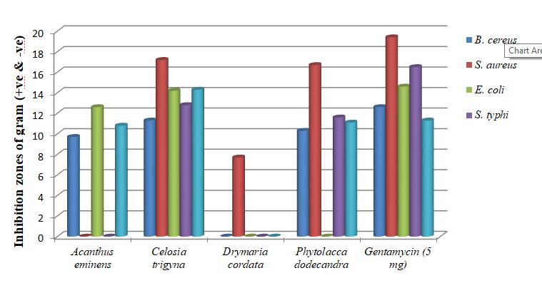
Evaluation of the antibacterial activity of these plant extracts was recorded in Table 3 and Figure 1. The results revealed that all plant extracts were potentially effective in suppressing microbial growth of pathogenic bacteria with variable potency. Celosia trigyna was the most effective extract retarding microbial growth of all Gram positive and Gram negative bacteria tested pathogenic bacteria at concentration of 4 mg/ml while extract of Drymaria cordata was effective only against Staphylococcus aureus. Phytolacca dodecandra exhibited an inhibitory effect against four of the pathogenic strains (Bacillus cereus, Staphylococcus aureus, Salmonella typhi, and Pseudomonas aeruginosa) whereas Acanthus eminens was effective against three of the pathogenic bacteria (Bacillus cereus, Escherichia coli, and Pseudomonas aeruginosa).
Boloche et al. International Journal of Environment, Agriculture and Biotechnology, 7(4) 2022
Cardiac glycoside ++ + Saponins +++ +++ + ++ Flavonoids +++ ++ +++ ++ Alkaloids ++ +++ +++ +++ Steroids + +++ + Terpenoids +++ +++ ++ Phenols ++ ++ + + Anthraquinones + Tannins + + +
Results of antibacterial activity of the four plant extracts can be suggested that both Celosia trigyna and Phytolacca dodecandra plant extracts were the most effective extracts and showed strong antibacterial activity against pathogenic bacteria. The two Gram negative bacteria (Escherichia coli and Salmonella typhi) were relatively the most resistant strain to plant extracts whereas two Gram positive bacteria (Bacillus cereus and Staphylococcus aureus) and one Gram negative bacteria (Pseudomonas aeruginosa) were the most susceptible strains to the extracted plants. Hence, experiments were conducted to determine their minimal inhibitory concentration (MIC) and minimal bactericidal concentration (MBC) against the most susceptible bacterial
PPlant extract Conc. mg/ml Inhibition zones Gram(mm)(+ve) bacteria Gram ( ve) bacteria B cereus S aureus P aeruginosa C. trigyna 100 0.0 ± 0.0 0.0 ± 0.0 0.0 ± 0.0 20 8.9 ± 0.31 10.1 ± 0.95 7.7 ± 0.46
Fig 1: Methanolic plants extract against pathogenic bacteria and positive control
Boloche et al. of Environment, Agriculture Biotechnology, 7(4) 2022
Table 3: Antibacterial screening test of methanolic plants extract (4 mg/ml) against some pathogenic bacteria. Plant species Inhibition zones (mm) Gram (+ve) pathogenic bacteria Gram ( ve) pathogenic bacteria B. cereus S. aureus E. coli S. typhi P. aeruginosa A. eminens 9.7 ± 0.43 0.0 ± 0.0 12.5 ± 0.54 0.0 ± 0.0 10.7 ± 0.65 C. trigyna 11.2 ± 0.53 17.2± 0.12 14.2 ± 0.34 12.8 ± 0.15 14 3 ± 0.37 D. cordata 0.0 ± 0.0 7.7 ± 0.27 0.0 ± 0.0 0.0 ± 0.0 0.0 ± 0.0 P. dodecandra 10.3 ± 0.25 16.7± 0.42 0.0 ± 0.0 11.6 ± 0.13 11.1 ± 0.21 Gentamycin(5mg) 12.6 ± 0.18 19.4 ± 0.21 14.6 ± 0.44 16.5 ± 0.37 11.3 ± 0.29
Data are means of three replicates (n = 3) ± standard error.
and
International Journal
ISSN: 2456 1878 (Int. J. Environ. Agric. https://dx.doi.org/10.22161/ijeab.74.2Biotech.)8 270 strains (Bacillus cereus, Staphylococcus aureus, and Pseudomonas aeruginosa). In coherent with this finding, [24] reported that significant anti bacterial activity of C. longa extract against two pathogenic bacterial strains. The results of MIC and MBC of C. longa extract demonstrated promising antibacterial activity of C. longa rhizome.
An extract of plant species and positive control
3.4. Minimum inhibitory concentrations (MIC’s) of the effective plant extract The MIC and MBC of the most effective plant extracts (Celosia trigyna and Phytolacca dodecandra) were employed by the disc diffusion method to evaluate their bacteriostatic and bactericidal properties. The concentration effect of the effective plant extracts was reported in Table 4 and illustrated in Figure 2 An inhibitory effect of C. trigyna extract started at 20 mg/ml with inhibition zones of 8.9, 10.1, and 7.7 mm against Bacillus cereus, Staphylococcus aureus, and Pseudomonas aeruginosa while extract of P. dodecandra suppressed bacterial growth of these strains at concentration of 0.8 mg/ml with inhibition zones of 16.8, 13.9 and 13.4 mm, respectively. This findings were in accordance with those reported in a work by Jawhari et al. that the inhibition zone diameters of extracts studied ranged from 5.5 to 15.65 mm, and the highest inhibition zone values against pathogens of medical importance such as Pseudomonas aeruginosa, Staphylococcus aureus, and Klebsiella pneumonia were 15.65, 15, and 15.3 mm, respectively [25]
Table 4: MIC’s of the most effective plant extract against Bacillus cereus, Staphylococcus aureus and Pseudomonas aeruginosa
International Journal of Environment, Agriculture and Biotechnology, 7(4)
Boloche et al. 2022
3.5. Minimum bactericidal concentrations (MBC’s) of the effective plant extract The minimum bactericidal concentration was confirmed by the absence of bacterial growth of the tested strains streaked from the inhibition zone corresponding to their lowest minimum inhibitory concentrations. C. trigyna extract showed potentially bactericidal activity against the tested pathogenic bacteria (Bacillus cereus, Staphylococcus aureus, and Pseudomonas aeruginosa) with MBC of 4 mg/ml while MBC of P. dodecandra extracts reached 0.16 mg/ml except for Pseudomonas aeruginosa which was less sensitive and its minimal bactericidal concentration reached to 0.032 mg/ml. The results of MIC and MBC of the effective plant extracts suggested that Celosia trigyna and Phytolacca dodecandra can be used to control and prevent pathogenic bacteria. Celosia trigyna extract suppresses microbial growth of all tested bacterial strains followed by an extract of Phytolacca dodecandra which appear to be potentially effective against three bacterial strains or pathogenic bacteria (Bacillus cereus, Staphylococcus aureus, and Pseudomonas aeruginosa) and less effective against two of them (Escherichia coli, Salmonella typhi). A great variation in MIC of Celosia trigyna extract demonstrated in several investigations may be due to considerable variation in their method of extraction, constituents as well as bacterial strains used. The difference value in minimum inhibitory concentrations of the plant extracts has happened from the variation of secondary metabolites and volatile nature of their constituents. In line with this study, [24, 26], reported that various biological activities of plant extracts are believed to be due to the presence of bioactive compounds. They explained that these plant secondary metabolites are nutritional constituents which are present in very tiny amounts in plants and have the potential for influencing the physiological and cellular activities after consuming them. Celosia trigyna extract was found to be the most effective with a concentration of (4 mg/ml) against all tested bacterial strains. On the other hand, Phytolacca dodecandra extract was found to be effective with a concentration of (4 mg/ml) against B. cereus, S. aureus, S. typhi, and P. Aeruginosa suppressing their growth with inhibition zones of 10.3, 16.7, 11.6, and 11.1 mm, respectively. These results are in accordance with that of [12, 23]. Some researchers have suggested that antimicrobial components of the plant extracts (terpenoid, alkaloid, and phenolic compounds) interact with enzymes and proteins of the microbial cell membrane causing its disruption to disperse a flux of protons towards the cell exterior which induces cell death or may inhibit enzymes necessary for amino acids biosynthesis [24, 25]. Other researchers attributed the inhibitory effect of these plant extracts to hydrophobicity characters of these plants extract which enable them to react with protein of microbial cell membrane and mitochondria disturbing their structures and changing their permeability. It has been reported that the relationship between a zone of inhibition and MIC values may be greatly affected by the composition of crude extracts that are a mixture of phytoconstituents which may influence the diffusion power of the active constituents, and the different levels of intrinsic tolerance of test strains to antimicrobials which can differ MIC values from one isolate to another [26 28].
ISSN: 2456 1878 (Int. J. Environ. Agric. https://dx.doi.org/10.22161/ijeab.74.2Biotech.)8 271 4 11.6 ± 0.44 17.5 ± 0.23 11.7 ± 0.79 0.8 19.4 ± 0.86 18.3 ± 0.67 17.1 ± 0.12 0.16 22.7 ± 0.39 20.5 ± 0.36 19.3 ± 0.23 0.032 25.9 ± 0.12 24.3 ± 0.91 21.7 ± 0.65 P. dodecandra 100 0.0 ± 0.0 0.0 ± 0.0 0.0 ± 0.0 20 0.0 ± 0.0 0.0 ± 0.0 0.0 ± 0.0 4 0.0 ± 0.0 0.0 ± 0.0 0.0 ± 0.0 0.8 16.7 ± 0.85 13.9 ± 043 13.6 ± 0.79 0.16 18.9 ± 0.13 16.5 ± 0.74 16.3 ± 021 0.032 20.5 ± 0.46 18.7 ± 0.21 17.4 ± 0.37
Phytochemical Analysis: The HPLC UV chromatogram of four selected plants Me. Ext is shown in Figure 3. Seven phytochemicals were identified from C. trigyna leave; four phytochemicals from P. dodecandra root, five phytochemicals from D. cordata leave and four phytochemicals from A. eminens stem methanol extract when compared to the standard chromatogram. The identified compounds from the HPLC chromatogram as shown in Figure 3 were pheophytin, chondrillasterol acetate, chondrillasterol, carotenoid, lutein, ethinyl estradiol and drospirenone isolated from C. trigyna leave methanol extract. Citronellal, cardinene, nerolidol and neryl acetate were isolated from P. dodecandra root methanol Extract. Stigmasterol, cerebroside, glucocerebroside, monogalactosyldiacylglycerol and digalactosyldiacglycerol were isolated from D. cordata leave methanol Extract. Isopulegol, borneol, caryophyllene and linalool were isolated from A. eminens stem methanol extract. The respective peak position, retention time and concentration of identified phytochemicals are given in Table 5.
Boloche et al. International Journal of Environment, Agriculture and Biotechnology, 7(4) 2022

Fig.2: MIC of the most effective plant extract against B. cereus, S. aureus, and P. aeruginosa In the present study, extracts of Celosia trigyna leaves and Phytolacca dodecandra roots have the most effective against four pathogenic bacterial strains like B. cereus, S. aureus, S. typhi, and P. aeruginosa whereas extracts of Drymaria cordata leaves and Acanthus eminens stems have indicated practically low activities against pathogenic bacteria. The observed activities of these extracts were relatively similar to other works. Thus, n butanol leaves extracts of Cassia angustifolia exhibited maximum zone of inhibition against Staphylococcus aureus (17.0 mm), Salmonella typhi (12.0 mm), and Klebsiella pneumoniae (10.0 mm); while, methanol extracts have not shown any activity against both the isolates. MICs values of leaf methanol extract of C. angustifolia exhibited stronger activity against K. pneumonia and E. coli (0.62 and 1.25 mg/mL, respectively) [29, 30].
272
ISSN: 2456 1878 (Int. J. Environ. Agric. https://dx.doi.org/10.22161/ijeab.74.2Biotech.)8
273 Fig 3: HPLC

Me. Ext
Boloche et al. International Journal of Environment, Agriculture and Biotechnology, 7(4) 2022
ISSN: 2456 1878 (Int. J. Environ. Agric. https://dx.doi.org/10.22161/ijeab.74.2Biotech.)8 UV chromatogram of C. trigyna leave, P. dodecandra root, D. cordata leave and A. eminens stem

Boloche et
274 Table 5: Identified phytochemicals in C. trigyna leave, P. dodecandra root, D. cordata leave and A. eminens stem Me. Ext Peak Retention Time (min) Phytochemicals areaPeak Concentration (μg/ml) C. trigyna leave Me. Ext 1 13.54 Pheophytin 7.23 0.5427 2 32.65 Chondrillasterol acetate 2.21 0.0034 3 18.77 Chondrillasterol 0.45 4.3134 4 25.82 Carotenoid 0.68 0.0231 5 10.37 Lutein 0.97 0.7254 6 19.58 Ethinyl estradiol 0.32 1.3125 7 11.54 Drospirenone 0.77 2.7326 P. dodecandra root Me. Ext 1 11.98 Citronellal 85.06 3.3412 2 24.83 Cardinene 0.78 0.5754 3 25.27 Nerolidol 0.54 0.0043 4 19.21 Neryl acetate 0.75 2.1432 D. cordata leave Me. Ext 1 16.43 Stigmasterol 5.67 0.7489 2 26.52 Cerebroside 0.69 0.1227 3 12.93 Glucocerebroside 0.34 2.1539 4 11.57 Monogalactosyldiacylglycerol 0.72 0.0856 5 21.34 Digalactosyldiacglycerol 0.89 1.5328 A. eminens stem Me. Ext 1 11.53 Isopulegol 4.56 5.5321 2 13.25 Borneol 0.48 0.9234 3 20.62 Caryophyllene 0.79 0.0069 4 11.46 Linalool 0.63 1.2954
The data are available from the corresponding author upon request.
The findings of this study indicate about methanolic extracts of four selected medicinal plants have high potential antibacterial activity against the different pathogenic bacterial strains. This activity supports their use in the treatment of infections caused by such resistant bacteria. The plant extracts which proved to be potentially effective are Celosia trigyna and Phytolacca dodecandra those can be used as a natural alternative for the treatment of pathogenic microbes, this has led to the search for new antimicrobial agents mainly among plant extracts to discover new chemical structures according to modern phytochemistry. The extract of those two plants has potential antibacterial effects on bacterial strains tested, especially Bacillus cereus, Staphylococcus aureus, and Pseudomonas aeruginosa. Their antibacterial activity was confirmed by evaluation of both diameters of inhibition zones and minimal inhibitory concentrations.
The authors declare that there is no conflicting interest.
ACKNOWLEDGMENTS
IV. CONCLUSION
DECLARATION OF CONFLICT OF INTEREST
ISSN: 2456 1878 (Int. J. Environ. Agric. https://dx.doi.org/10.22161/ijeab.74.2Biotech.)8
al. International Journal of Environment, Agriculture and Biotechnology, 7(4) 2022
The authors acknowledge all the technical support offered by microbiology staff from the Department of Biology and Plant Science. We authors also thank Bonga University for financial support.
DATA AVAILABILITY
[1] P. Agrawal, V. Rai, and R. B. Singh, “Randomized placebo controlled, single blind trial of holy basil leaves in patients with noninsulin dependent diabetes mellitus,” International Journal of Clinical Pharmacology Therapeutics, vol. 34, no. 9, pp. 406 409, 1996 PMID: 8880292.
[15] A Pandey, and P. Singh, “Antibacterial activity of Syzygiumaromaticum (Clove) with metal ion effect against food borne pathogens,”Asian Journal of Plant Science & Research, Vol. 1, no. 2, pp. 69 80, 2011.
[21] G. E. Trease, and W.C. Evans, “Pharmacology. 11th Edition,” Bailliere Tindall Ltd., London. Pp. 60 75, 1989.
Boloche et al. International Journal of Environment, Agriculture and Biotechnology, 7(4) 2022
[8] A Q Rizwana, M Ahmad, Z Yousaf, and M. Arshad, “Taxonomic study and medicinal importance of three species of the genus Artimisia,” The Pakistan Journal of Forestry, vol. 52, no. 1, pp. 23 31, 2002.
[5] A. I. Nneamaka, “Screening of Some Nigerian Medicinal Plants for Bacterial Activity. Amadu Bello University, Zairia,”Pp.108, 1991.
[23] R. Ullah, M. S. Alsaid, A.S. Alqahtani, A. A. Shahat, A. A. Naser, H. M. Mahmood, S. R. Ahamad, A. A. Al Mishari, S. Ahmad “Anti inflammatory, antipyretic, analgesic, and antioxidant activities of Haloxylon salicornicum aqueous fraction,” Open Chemistry, vol. 17, pp. 1034 1042, 2019. https://doi.org/10.1515/chem 2019 0113
[6] K. A. Hammer, C F Carson, and T V. Riley, “Antimicrobial activity of essential oils and other plant extracts,” JournalofApplied Microbiology, vol. 86, no. 6, pp. 985, 1999.
[4] N. Nazir, A. Rahman, F. Uddin, A. A. K. Khalil, M. Zahoor, M. Nisar, S. Ullah, R. Ullah, E. Ezzeldin, G. A. E. Mostafa, et al. “Quantitative Ethnomedicinal Status and Phytochemical Analysis of Berberis lyceum Royle,” Agronomy, vol. 11, pp. 1 13, 2021 https://doi.org/10.3390/agronomy11010130
[9] S Gajalakshmi, S, Vijayalakshmi, and V. Rajeswari, “Phytochemical and Pharmacological Properties of Annona muricata: A Review,” International Journal of Pharmacy and Pharmaceutical Sciences, vol. 4, pp. 3 6, 2012.
[10] S Ramasamy, and C. Manoharan, “Antibacterial effect of volatile components of selected medicinal plants against human pathogens,” Asian Journal of Microbiology, Biotechnology&EnvironmentalSciences, vol. 6, pp. 209 210, 2004.
[19] C. Monica, “Medical Laboratory Manual for Tropical Countries,” Microbiology. Butterworth and Co (Publishers) Ltd., Borough Green, Sevenoaks, Kent TN, vol. 2, pp. 479, 1984. [20] A. Sofowara, “Screening for bioactive agents. In: Medicinal Plants and Traditional Medicine in Africa,” Sofowara, A. (Ed.), 2nd Ed., Spectrum Books Limited: Ibadan, Nigeria, pp. 134 156, 1993.
[11] P Saranraj, and D. Stella, “Antibiogram of nosocomial infection and its antimicrobial drug resistance,” International Journal of PharmaceuticalandBiologicalScience Archive, Vol. 2, no. 6: pp. 1598 1610, 2011.
[22] J.B. Harborne, “Phytochemical methods. A Guide to Modern Techniques of Plant Analysis, 3rd ed,” Chapman and Hall publishing: London, United Kingdom, pp. 67, 1998.
[24] N. Suwal, R. K. Subba, P. Paudyal, D. P. Khanal, M. Panthi, N. Suwal, M. A. Nassan, M. Alqarni, G. El Saber Batiha, and N. Koirala, “Antimicrobial and antibiofilm potential of Curcumalonga Linn. Rhizome extract against biofilm producing Staphylococcus aureus and Pseudomonas aeruginosa isolates,” Cellular and Molecular Biology, vol. 67, no. 1, pp. 17 23, 2021. Doi: http://dx.doi.org/10.14715/cmb/2021.67.1.3
[16] M. G. Jinukuti, and A. Giri, Antimicrobial activity of phytopharmaceuticals for prevention and cure of diseases. AnnalsofPhytomedicine, vol. 2, no. 2, pp. 28 46, 2013.
[12] H. Mahfuzul, M. Bari, M. L. Juneja, and V. K. Kawamoto, “Antimicrobial activity of cloves and cinnamon extracts against food borne pathogens and spoilage bacteria and inactivation of Listeria monocytogenes in ground chicken meat with their essential oils,” JournalofFoodScience& Technololgy Vol. 72, pp. 9 21, 2007.
[25] F. Z. Jawhari , A.E. L. Moussaoui, M. Bourhia, H. Imtara, H. Saghrouchni, K. Ammor, H. Ouassou, Y. Elamine, R. Ullah, E. Ezzeldin, G. A. E. Mostafa, and A. Bari, “Anacyclus pyrethrum var. pyrethrum (L.) and Anacyclus pyrethrum var. depressus (Ball) Maire: Correlation between Total Phenolic and Flavonoid Contents with
275 REFERENCES
ISSN: 2456 1878 (Int. J. Environ. Agric. https://dx.doi.org/10.22161/ijeab.74.2Biotech.)8
[3] A Yothi, K. Venkatesh, P. Chakrapani, and R. Roja, Phytochemical and Pharmacological Potential of Annona cherimola A Review,” International Journal of Phytomedicine, vol. 3, pp. 439 447, 2011.
[13] S Saeed, and P. Tariq, In vitro Antibacterial activity of clove against Gram negative bacteria. Pakistan Journal of Botany, vol. 40, no. 5, pp. 2157 2160, 2008. [14] M. Saeed, M. Nadeem, M. R. Khan, M. A. Shabbir, A. Shehzad, and R. M. Amir, “Antimicrobial activity of Syzygium aromaticum extracts against food spoilage bacteria,” AfricanJournalofMicrobiololgyResearch, vol. 7, no. 41, pp. 4848 4856, 2013.
[7] A. Kashayap, “Characterization and evaluation of antimicrobial potential of biologically synthesized silver nanoparticles from Berberis aristata,” M. Phil. Thesis, Himachal Pradesh University, Shimla, 2017.
[17] V. A. Badar, S. Kumar, and B. Navale, “Study of prescribing pattern of antimicrobial agents in medicine intensive care unit of a teaching hospital in Central India,” Journal of the Association ofPhysiciansofIndia, vol. 60, pp. 20 22, 2012. [18] M. G. Jinukuti, and A. Giri,“Anticancer activity of acetone and methanol extracts of Terminalia chebula Retz and Withaniasomnifera (Linn.) Dunal on HeLa cell line,” AnnalsofPhytomedicine, vol. 4, no. 2, pp. 88 92, 2015.
[2] J Parekh, D Jadeja, and S. Chanda, “Efficacy of Aqueous and Methanol Extracts of Some Medicinal Plants for Potential Antibacterial Activity,” Turkish Journal of Biology, Vol. 9, pp. 203 210, 2005.
276 Antioxidant and Antimicrobial Activities of Chemically Characterized Extracts,” Plants, vol. 10, pp. 1 19, 2021 https://doi.org/ 10.3390/plants10010149 [26] M. Panthi, R. K. Subba, B. Raut, D. Khanal, N. Koirala, “Bioactivity evaluations of leaf extract fractions from young barley grass and correlation with their phytochemical profiles,” BMC Complementary Medicine and Therapies, vol. 20, No. 1, pp. 64, 2020. Doi: 10.1186/s12906 020 2862 4. [27] A Pandey, and P. Singh, “Antibacterial activity of Syzygiumaromaticum (Clove) with metal ion effect against food borne pathogens,” Asian Journalof PlantScience& Research, Vol. 1, no. 2, pp. 69 80, 2011. [28] S, Burt, “Essential oils: their antibacterial properties and potential application in foods: a review,” International JournalofFoodMicrobiology, vol. 94, pp. 223 253, 2004. [29] A O Gill, and R. A. Holley, “Disruption of Escherichia coli, Listeria monocytogenes and Lactobacillus sakei cellular membranes by plant oil aromatics,” International JournalofFoodMicrobiology, vol. 108, pp. 1 9, 2006. [30] M Friedman, P R Henika, C E Levin and R. E. Mandrell, “Antibacterial activities of plant essential oils and their components against Escherichia coli O157:H7 and Salmonella enterica in apple juice,” Journal ofAgriculturalandFood Chemistry, vol. 52, pp. 6042 6048, 2004.
Boloche et al.
ISSN: 2456 1878 (Int. J. Environ. Agric. https://dx.doi.org/10.22161/ijeab.74.2Biotech.)8
International Journal of Environment, Agriculture and Biotechnology, 7(4) 2022