Vol-8, Issue-2; Mar-Apr, 2023
Journal Home Page Available: https://ijeab.com/ Journal DOI: 10.22161/ijeab Peer Reviewed

Vol-8, Issue-2; Mar-Apr, 2023
Journal Home Page Available: https://ijeab.com/ Journal DOI: 10.22161/ijeab Peer Reviewed
1DishaLifeSciencesPvt.Ltd.,Ahmedabad,Gujarat,India.
2SwarrnimStartupandInnovationUniversity,Gandhinagar,Gujarat,India.
*Correspondingauthor:nisha_nayak272@yahoo.co.in
Received:26Feb2023;Receivedinrevisedform:25Mar2023;Accepted:03Apr2023;Availableonline:13Apr2023
©2023TheAuthor(s).PublishedbyInfogainPublication.ThisisanopenaccessarticleundertheCCBYlicense (https://creativecommons.org/licenses/by/4.0/).
Abstract Biosurfactants are a group of heterogenous metabolites synthesized by a variety of microorganisms. They exhibit the properties of the surface tension reduction, emulsion stabilization, promote foaming, and specific activity at extreme temperatures, pH, and salinity. A bacterial strain was screened for its biosurfactant production in 250 ml MSM broth with crude oil as an inducer for 5 days. The screening activity performed by (i) drop collapse test, (ii) oil displacement test, (iii) emulsification index proved the presence of biosurfactant TLC and FTIR analysis confirmed that the biosurfactant produced by the selected bacterial isolate is a rhamnolipid. The potential isolate was identified by 16S rRNA gene sequencing analysis and it was identified as Pseudomonas aeruginosa.
Keywords crude oil, emulsification index, rhamnolipid, TLC, FTIR, PCR
Biosurfactants are biologically derived surface-active substances that are primarily produced as secondary metabolites by filamentous fungus, yeast, and bacteria. Due to their special amphiphilic composition, which combines hydrophobic and hydrophilic parts, which increases the bioavailability of water and lowers the surface tension. This provides emulsification activity (Nayarisseri et al., 2018; Meliani and Bensoltane, 2014). The diverse structure of biosurfactants results from their different microbial origin, the substrate on which they are grown and cultivation conditions used (Santos et al., 2016). In recent years, there has been a substantial increase in the production of biosurfactants and their commercialization (Henkel and Hausmann, 2019).
Someproperties ofbiosurfactantsthat make them unique from chemical surfactants include: reduction of surface and interfacial tension, low toxicity, high biodegradability, emulsification, selective performance, specific activity, possibility of production from cheap raw materials,
ISSN: 2456-1878 (Int. J. Environ. Agric. Biotech.)

https://dx.doi.org/10.22161/ijeab.82.13
antimicrobial properties, easier production and morevariety (De Giani et al., 2021; Bagheri et al., 2013).
There are basically five classes of biosurfactants: (i) glycolipids, (ii) phospholipids and fatty acids, (iii) lipopeptides and lipoproteins, (iv) polymeric surfactants and (v) specific biosurfactants (Desai and Banat, 1997; Varjani and Upasani, 2017). The biosurfactantproduction is an important survival strategy by different microorganisms as it helps in uptake of hydrophobic substrates for surfaceassociated modes of motility (Chrzanowski et al., 2012).
Pseudomonas aeruginosa is the preferred microorganism for the production of rhamnolipid type of biosurfactant utilizing glycerol, mannitol, fructose, glucose, and vegetable oils (Koch et al., 1991; Santos et al., 2002).
Rhamnolipids are one of the most important glycolipid biosurfactants, which are produced by two bacterial species of Pseudomonas aeruginosa and Burkholderia (Fracchia et al., 2012).
Rhamnolipid has a high emulsion capacity and is often used in the pharmaceutical and environmental industries such as increasing oil recovery and bioremediation
(Suhandono et al., 2021). Pseudomonas species are the largest producers of rhamnolipids. They produce two different types of rhamnolipids that differ in the number of rhamnose sugars. Mono-rhamnolipids and di-rhamnolipids are the main rhamnolipids. These molecules have high surface activity and are used in various medical fields as antifungal, antibacterial and antiviral materials (Kaskatepe et al., 2015). Microorganisms especially bacteria represent an excellent source of biosurfactants, so that the isolation and characterization of the emulsifying capacity of biosurfactant molecules represents an important step for their future application in the areas of biotechnology (Singh et al., 2019; Volkering et al., 1997).
The present study involves: Screening, production and characterization of biosurfactant from P. aeruginosa BS1 using crude oil.
A bacterial strain belonging to Pseudomonas aeruginosa was procured from the preserved culture collection of Disha Life Sciences Pvt. Ltd. It was then screened for biosurfactant production in Minimal Salt Medium (MSM) (g/L) as described by Ohadi et al., (2017) with some modifications: MgSO4, 0.1; KH2PO4, 0.5; NH4Cl, 0.01; FeSO4.7H2O, 0.001; NaHCO3, 1; and K2HPO4, 0.5, pH 7.0 with 0.1% crude oil (as an inducer) by the following methods.
Drop collapse test, as described by Jain et al., (1991), was performed to screen the biosurfactant production. Crude oil was applied to the solid glass surface of a microscope glass slide and 250 μL of the supernatant was placed on the oilcoated surface and drop size was observed after 1 min with the help of a magnifying glass. The result was considered to be positive when the diameter of the drop was increased by 1 mm from that which was produced by distilled water that was taken as the negative control (Youssef et al., 2004).
In oil displacement test (Safary et al., 2010), 40 μl of crude oil was placed to the surface of 40 ml of distilled water in a petri dish forming thin oil layer on it. After that, 10 μl of culture supernatant was gently placed on the centre of the oil layer. Clear zone formation by displacing oil indicates the presenceofbiosurfactant.The diameterofthe clearzone on the oil surface was visualized under visible light and measured after 30 seconds, which was correlated to the surfactant activity, also known as an oil displacement activity.
Emulsion activityof the culture supernatantwas detectedby addition of 2 mL of crude oil to the equal volume of cellfree supernatant, mixed with a vortex for 2 min and allowed to stand for 24 hours at 35 ± 2 °C. The emulsification activity was observed after 24 h and it was calculated using the following formula (Khan et al., 2017):
EI24(%)=
The production and extraction of biosurfactant was carried out according to Abbasi et al., (2012) with some modifications. The culture was inoculated in 300 ml Luria Bertini(LB)brothtowhich2%(v/v)ofcrudeoilwasadded. The culture was incubated at 37 °C for 5 days at 120 rpm with shaking conditions. After incubation, cells were removed from the culture broth by centrifugation at 10,000×g for 15 min at 4 °C. The cell free supernatant was acidified with 6 N HCL to pH 2 and stored overnight at 4 °C to enhance the precipitation of biosurfactant. The resulted precipitate was separated by centrifugation (15,000×g, 15 min, 4 °C) and extracted several times with ethyl acetate at room temperature. The solvent was completely evaporated by drying at room temperature. The crude biosurfactant was obtained as a viscous browncoloured substance.
The rhamnolipids extracted were analysed by thin-layer chromatography(TLC) according to(Bhat et al., 2015) with few modifications. The TLC was carried out on silica 60 gel aluminium sheets (Loba, Mumbai, India) using the solvent system CHCl3/CH3OH/CH3COOH (81:17:2). When the solvent reached the top, the plate was removed and allowed to air dry. The plate was then kept in iodine vapour chamber for development of yellow spot to check the presence of lipids in biosurfactant.
The partially purified biosurfactant was characterized by Bruker Alpha II Fourier Transform Infrared spectrophotometer (FTIR) spectroscopy to find out functionalgroups Therangeof spectrausedwas4,000 cm 1
400 cm 1
Phenol sulfuric acid method is used for detection of rhamnose-sugar(carbohydrate) in biosurfactant. The concentration of carbohydrates was determined by comparing it with D-glucose as a standard. The basic protocol of DuBois et al , (1956) was followed, with the modifications indicated below. The biosurfactant (10 μL) and phenol solution (80 μl) were taken and then 1 ml of
concentrated sulfuric acid was added slowly down the side of the tube. The tube was then incubated for 30 s at room temperature. The absorbance was read at 490 nm using distilled water as blank in a Double Beam Spectrophotometer (Systronics, Ahmedabad, India).
2.5 Identification of biosurfactant producing bacteria
Strain BS1 was identified by 16S rRNA gene sequence analysis. The molecular identification was carried out by amplification, sequencing and analysis of conserved 16S rRNA region. Genomic DNA was isolated from the pure culture and the quality of DNA was checked by gel electrophoresis on a 1% agarose gel. The PCR was performed using universal primers. The PCR products were sequenced and the obtained sequences were compared with the known ones in the National Centre for Biological Information (NCBI) database using Basic Local Alignment Search Tool (BLAST) and accession number was obtained by submission to NCBI GenBank.
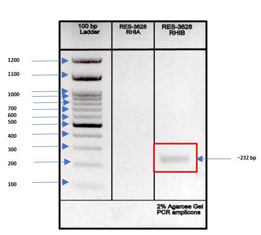
The PCR was carried out according to Pacwa-Płociniczak et al., (2014) with a few changes. The primers rhlA and rhlB were used to detect potential rhamnolipid synthesis by the BS1 strain. The PCR was run with a mixture containing 1 μl of the DNA template, 0.2 μM of each primer, 10× reaction buffer, 1.5 mM of MgCl2, 200 μM of dNTP and 1 U of Taq DNA polymerase in a Thermal Cycler. PCR amplification was carried out as follows- denaturation at 95 °C for 3 min, and 30 cycles of 60 s, followed by annealing for 1 min at 56 °C and an extension step of 1.5 min at 72 °C and a final extension step of 10 min at 72 °C. The experiment included a control reaction mixture without added DNA.
Sample (5 ml) was withdrawn at regular intervals (24 h) from the flask containing 250 ml MSM broth for 5 days to perform the screening tests. The results obtained after the screening tests are as follows:
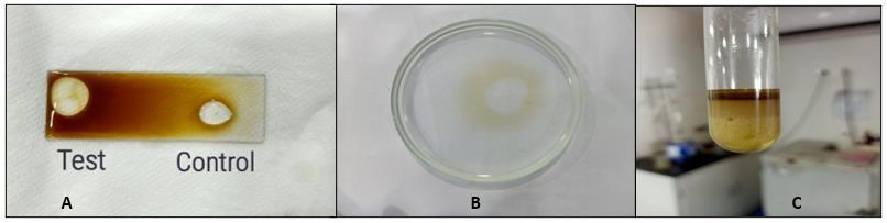
From the results obtained, it can be seen that the bacterial isolate gave positive results for the drop collapse test. Production of biosurfactant decreases the surface tension of the supernatant and as a result, the shape of the test droplet was larger as compared to the control (distilled water) as shown in Fig 1(a). Oil displacement test is considered positive when a clear zone is formed as oil gets displaced by the presence of biosurfactant as depicted in Fig 1(b). However, the extent of oil displacement differed considerably. There was a gradual increase in oil displacement activity from 20 mm to 30 mm at the end of 5 days. Pseudomonas aeruginosa ATCC-10145 a highly positive strain showed 8.0 cm of oil displacement (ElSheshtawy and Doheim, 2014). Thavasi et al., (2011) reported positive biosurfactant producing bacteria using drop collapsed and oil displacement test, among which many isolates belonged to Pseudomonas sp.
https://dx.doi.org/10.22161/ijeab.82.13
Higher biosurfactant concentration in the culture medium was related to high emulsion capacity. The emulsification activity of biosurfactant also increased during incubation period. It was 40% after 24 hours and it reached 72% at the end of 120 hours. In the current study, crude oil was used but according to the previous works, Pseudomonas rhamnolipids can effectively emulsify and stabilize emulsions with various types of hydrocarbons and oils such as linseed oil, almond oil, mineral oil (Benincasa et al., 2004), diesel (Haba et al., 2003; Wei et al., 2005), kerosene, n-alkanes, aromatic compounds, coconut oil, and olive oil (Patel and Desai, 1997). Rahman et al., (2002) reported results for two P. aeruginosa strains. The strains showed emulsification index from 25% to 90% for oily phases diesel and kerosene.

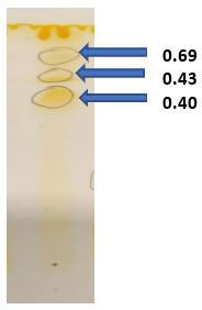
ResultsofTLCshowedyellowspotsindicatingthepresence of lipids in the structure of produced biosurfactant. TLC of partially purified rhamnolipids recovered from cell-free supernatant of P. aeruginosa revealed three different spots with different Rf (retention factor) values. The first major spot (Rf = 0.69) was a mono-rhamnolipid whereas the second spot (Rf = 0.43) and third spot (Rf = 0.40) confirmed the presence of di-rhamnolipids. These results are as per the findings of Abdel-Mawgoud et al., (2007), where crude biosurfactant extract of P. aeruginosa BS20 showed two main spots with Rf values 0.4 and 0.68 representing dirhamnolipidsandmono-rhamnolipidsrespectively.Another study done by Thio et al., (2022) on Pseudomonas sp showed two spots with Rf values of 0.68 and 0.38 during TLC which confirmed the lipid nature of biosurfactant.
spectra of biosurfactants from P. aeruginosa BS1 also revealed the presence of lipid moiety in the purified glycolipids showing peaks at 1014 cm-1 (Moussa et al., 2014).Similarly,thebiosurfactantshowedanintensityband at 1636 cm-1 indicated bending of the hydroxyl (-OH) group, which reflects the presence of the carboxylic acid functional group in the compound (Figure 3).
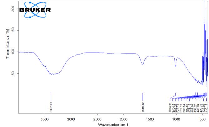
The partially purified sample biosurfactant was checked for functional group by using FTIR. The prominent peak was found at 3382 cm-1 vibrations which indicates stretching for
The concentration of carbohydrate was 5.52 mg/ml as per the carbohydrate standard curve and O.D. taken at 490 nm. Abbasi et al., (2013) followed the same procedure and the biosurfactant obtained had 32% (w/w) carbohydrate content.
After gene sequencing analysis, the obtained sequence was found to be Pseudomonas aeruginosa BS1 and accession no OQ568205.1 was obtained by submission of sequence to NCBI GenBank
CH and
CH2. The strong stretching of C=O of the carbonyl group was observed at 1636 cm-1. The FTIR
The whole DNA was screened for presence of biosurfactant producing gene RhlA and RhlB by using PCR technique. The results of Gel electrophoresis showed the presence of
RhlBnear232bphaving inthegenomewhichalsoindicates biosurfactant production property in bacteria.
aeruginosa MR01 using vegetable oil refinery wastes. New CellularandMolecularBiotechnologyJournal, 3(9),91-99.
[5] Benincasa, M., Abalos, A., Oliveira, I., & Manresa, A. (2004).Chemicalstructure,surfacepropertiesandbiological activities of the biosurfactant produced by Pseudomonas aeruginosa LBI from soapstock. Antonie Van Leeuwenhoek, 85,1-8.
[6] Bhat, R., Dayamani, K. J., Hathwar, S., Hegde, R., & Kush, A. (2015). Exploration on production of rhamnolipid biosurfactants using native Pseudomonas aeruginosa strains. JournalofBioScience&Biotechnology, 4(2).
[7] Chrzanowski, Ł., Dziadas, M., Ławniczak, Ł., Cyplik, P., Białas, W., Szulc, A., ... & Jeleń, H. (2012). Biodegradation of rhamnolipids in liquid cultures: effect of biosurfactant dissipation on diesel fuel/B20 blend biodegradation efficiency and bacterial community composition. Bioresourcetechnology, 111,328-335.
[8] De Giani, A., Zampolli, J., & DiGennaro, P. (2021). Recent trendsonbiosurfactantswithantimicrobialactivityproduced by bacteria associated with human health: Different perspectives on their properties, challenges, and potential applications. Frontiersinmicrobiology, 12,655150.
The present study was aimed at screening, production and characterization of a biosurfactant producing bacterial strain. Biosurfactant producing bacteria Pseudomonas aeruginosa was a potent producer which was confirmed by all major screening tests. Characterization of produced biosurfactant showed rhamnolipid nature based on TLC and FTIR results. More targeted studies to develop methods to scale up production of rhamnolipid biosurfactant and also explore new renewable resources as substrate for the bacterial strain for higher and efficient rhamnolipid production can be a way forward.
[1] Abbasi, H., Hamedi, M. M., Lotfabad, T. B., Zahiri, H. S., Sharafi, H., Masoomi, F., ... & Noghabi, K. A. (2012). Biosurfactant-producing bacterium, Pseudomonas aeruginosa MA01 isolated from spoiled apples: physicochemical and structural characteristics of isolated biosurfactant. Journal of bioscience and bioengineering, 113(2),211-219.
[2] Abbasi,H.,Sharafi,H.,Alidost,L.,Bodagh,A.,Zahiri,H.S., & Noghabi, K. A. (2013). Response surface optimization of biosurfactant produced by Pseudomonas aeruginosa MA01 isolated from spoiled apples. Preparative biochemistry & biotechnology, 43(4),398–414.
[3] Abdel-Mawgoud,A.M.,Aboulwafa,M.M.,&Hassouna,N. A. H. (2007). Microbial production of surfactants: screening and identification of two promising isolates and their biosurfactants. EgyptianJournalofBiotechnology, 27
[4] Bagheri Lofabad, T., Partovi, M., & Bahmaei, M. (2013). Rhamnolipid biosurfactant production by Pseudomonas
ISSN: 2456-1878 (Int. J. Environ. Agric. Biotech.)
https://dx.doi.org/10.22161/ijeab.82.13
[9] Desai, J. D., & Banat, I. M. (1997). Microbial production of surfactantsandtheircommercialpotential. Microbiologyand Molecularbiologyreviews, 61(1),47-64.
[10] DuBois, M., Gilles, K. A., Hamilton, J. K., Rebers, P. T., & Smith, F. (1956). Colorimetric method for determination of sugars and related substances. Analytical chemistry, 28(3), 350-356.
[11] El-Sheshtawy, H. S., & Doheim, M. M. (2014). Selection of Pseudomonas aeruginosa for biosurfactant production and studies of its antimicrobial activity. Egyptian journal of petroleum, 23(1),1-6.
[12] Fracchia, L., Cavallo, M., Martinotti, M. G., & Banat, I. M. (2012). Biosurfactants and bioemulsifiers biomedical and related applications–present status and future potentials. Biomedical science, engineering and technology, 14(1),1-49.
[13] Haba,E.,Abalos,A.,Jauregui,O.,Espuny,M.J.,&Manresa, A. (2003). Use of liquid chromatography-mass spectroscopy for studying the composition and properties of rhamnolipids produced by different strains of Pseudomonas aeruginosa. Journal of Surfactants and Detergents, 6, 155161.
[14] Henkel, M., & Hausmann, R. (2019). Diversity and classification of microbial surfactants. In Biobased surfactants (pp.41-63).AOCSPress.
[15] Jain, D. K., Collins-Thompson, D. L., Lee, H., & Trevors, J. T. (1991). A drop-collapsing test for screening surfactantproducing microorganisms. Journal of Microbiological Methods, 13(4),271-279.
[16] Kaskatepe, B., Yildiz, S., Gumustas, M., & Ozkan, S. A. (2015). Biosurfactant production by Pseudomonas aeruginosain kefir and fish meal. Brazilian Journal of Microbiology, 46,855-859.
[17] Khan, A.H. A., Tanveer, S., Alia, S., Anees, M., Sultan, A., Iqbal,M.,&Yousaf, S.(2017). Roleofnutrientsinbacterial biosurfactant production and effect of biosurfactant
production on petroleum hydrocarbon biodegradation. EcologicalEngineering, 104,158-164.
[18] Koch, A. K., Käppeli, O., Fiechter, A., & Reiser, J. (1991). Hydrocarbon assimilation and biosurfactant production in Pseudomonas aeruginosa mutants. Journal of Bacteriology, 173(13),4212-4219.
[19] Meliani, A., & Bensoltane, A. (2014). The ability of some Pseudomonas strains to produce biosurfactants. Poult. Fish. Wildl.Sci, 2,112.
[20] Moussa, T. A. A., Mohamed, M. S., & Samak, N. (2014). Production and characterization of di-rhamnolipid produced by Pseudomonas aeruginosa TMN. Brazilian Journal of ChemicalEngineering, 31,867-880.
[21] Nayarisseri, A., Singh,P.,&Singh,S.K.(2018). Screening, isolation and characterization of biosurfactant producing BacillussubtilisstrainANSKLAB03. Bioinformation, 14(6), 304.
[22] Ohadi, M.,Dehghannoudeh,G.,Shakibaie, M.,Banat, I.M., Pournamdari, M., & Forootanfar, H. (2017). Isolation, characterization, and optimization of biosurfactant production by an oil-degrading Acinetobacter junii B6 isolated from an Iranian oil excavation site. Biocatalysisand AgriculturalBiotechnology, 12,1-9.
[23] Pacwa-Płociniczak, M., Płaza, G. A., Poliwoda, A., & Piotrowska-Seget, Z. (2014). Characterization of hydrocarbon-degrading and biosurfactant-producing Pseudomonas sp. P-1 strain as a potential tool for bioremediation of petroleum-contaminated soil. Environmental Science and Pollution Research, 21, 9385-9395.
[24] Patel,R.M.,&Desai,A.J.(1997).Biosurfactantproduction by Pseudomonas aeruginosaGS3 from molasses. Letters in AppliedMicrobiology, 25(2),91-94.
[25] Rahman,K.S.M.,Rahman,T.J.,McClean,S.,Marchant,R., &Banat,I.M.(2002).Rhamnolipidbiosurfactantproduction by strains of Pseudomonas aeruginosa using low‐cost raw materials. Biotechnologyprogress, 18(6),1277-1281.
[26] Safary,A.,Ardakani,M.R.,Suraki, A.A.,Khiavi,M.A.,& Motamedi, H. (2010). Isolation and characterization of biosurfactant producing bacteria from Caspian Sea. Biotechnology, 9(3),378-382.
[27] Santos, A. S., Sampaio, A. P. W., Vasquez, G. S., Anna, L. M. S., Pereira, N., & Freire, D. M. (2002). Evaluation of different carbon and nitrogen sources in production of rhamnolipids by a strain of Pseudomonas aeruginosa. In Biotechnology for Fuels and Chemicals: The Twenty–ThirdSymposium (pp.1025-1035).HumanaPress.
[28] Santos,D.K.F.,Rufino,R.D.,Luna,J.M.,Santos,V.A.,& Sarubbo, L. A. (2016). Biosurfactants: multifunctional biomolecules of the 21st century. International journal of molecularsciences, 17(3),401.
[29] Singh, P., Patil, Y., & Rale, V. (2019). Biosurfactant production: emerging trends and promising strategies. Journalofappliedmicrobiology, 126(1),2-13.
[30] Suhandono, S., Kusuma, S. H., & Meitha, K. (2021). Characterizationandproductionofrhamnolipidbiosurfactant in recombinant escherichia coli using autoinduction medium
ISSN: 2456-1878 (Int. J. Environ. Agric. Biotech.)
https://dx.doi.org/10.22161/ijeab.82.13
andpalmoilmill effluent. BrazilianArchivesofBiologyand Technology, 64
[31] Thavasi, R., Sharma, S., & Jayalakshmi, S. (2011). Evaluation of screening methods for the isolation of biosurfactant producing marine bacteria. J Pet Environ BiotechnolS, 1(2),1-7.
[32] Thio, C. W., Lim, W. H., Md. Shah, U. K., & Phang, L. Y. (2022). Palm kernel fatty acid distillate as substrate for rhamnolipids production using Pseudomonas sp. LM19. GreenChemistryLettersandReviews, 15(1),83-92.
[33] Varjani, S. J., & Upasani, V. N. (2017). Critical review on biosurfactantanalysis,purificationandcharacterizationusing rhamnolipid as a model biosurfactant. Bioresource technology, 232,389-397.
[34] Volkering, F., Breure, A. M., & Rulkens, W. H. (1997). Microbiological aspects of surfactant use for biological soil remediation. Biodegradation, 8,401-417.
[35] Wei,Q.F.,Mather,R.R.,&Fotheringham,A.F.(2005).Oil removal from used sorbents using a biosurfactant. Bioresourcetechnology, 96(3),331-334.
[36] Youssef, N. H., Duncan, K. E., Nagle, D. P., Savage, K. N., Knapp, R. M., & McInerney, M. J. (2004). Comparison of methods to detect biosurfactant production by diverse microorganisms. Journal of microbiological methods, 56(3), 339-347.