@aestheticsgroup
Aesthetics Journal
Aesthetics aestheticsjournal.com
Chemical injuries
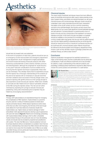
Figure 4: Skin discolouration, hollowing and gaunt appearance following hyaluronidase injections to the tear trough
should also be treated with oral antibiotics. In the event of symptoms of vision loss, patients should be sent as an emergency to their local accident and emergency department or eye department. Acute management is highly specialised and would include decreasing intraocular pressure with either intravenous acetazolamide or direct anterior chamber puncture and decompression, although the prognosis for visual recovery is very poor.13 Vascular complications must be avoided and by ensuring that the injection technique is appropriate, these risks can be minimised. This critically means that it is essential to ensure that the injector has a thorough understanding of the anatomy of the area to be injected with an emphasis on vascular branches and their variations and to be aware of the key danger areas. Low pressure injecting with gentle pressure on the syringe plunger is also essential to avoid intravascular injection. Wider bore needles or cannulas are also helpful and can reduce hydrostatic pressure of the filler during the injection phase. The risk can be further minimised by aspirating the syringe to exclude intravascular positioning. Using cold compresses prior to injection may be helpful by encouraging vasoconstriction.14 Management Therapy with hyaluronidase is an essential technique for managing the complications of filler injections. Hyaluronidase is a powerful enzyme used for a variety of medical purposes to break down natural connective tissues. Whilst deeper injections to other areas of the face are often more forgiving, the thinness of the tear trough areas allows for much less tolerance of adverse results from hyaluronidase use. Higher doses can lead to damage of native extracellular matrix glycosaminoclycans, leaving a gaunt appearance with poor skin texture (Figure 4). Patients are increasingly presenting with hyaluronidase-related damage, which is often untreatable. Hyaluronidase therapy to the periocular area can have devastating results and should only be provided by experienced practitioners with caution. The view that fillers are a ‘natural substance’ and can be simply dissolved without complications is outdated. Given that we are now understanding more about how fillers interact with the body we need to move away from the simplistic view of fillers as an easily reversible therapy and any periocular hyaluronidase therapy must be delivered with extreme caution.
The sheer range of aesthetic techniques means that many different types of chemicals and drugs are often used in close proximity to the eyes. Eyelash tinting, acid peels and retinoid therapies can all cause chemical injuries and corneal damage. Although caution should be undertaken when using chemicals around the eyes, inadvertent inoculation of the ocular surface can be a common presenting scenario to the ophthalmologist. Acid injuries are less severe than alkali injuries, but can cause quite significant corneal epithelial damage and denudement. Corneal protection is predominantly a form of barrier immunity and any compromise of the epithelium can lead to infections and pain. In the event of ocular inoculation, an eyebath should be available on the premises for immediate washing, or running a sterile saline bag gently onto the eye with the patient leaned over the sink. Rapid washing is essential and the pH of the tear film should be measured to ensure a neutral pH is achieved and this can be confirmed with universal indicator paper. Patients should then be referred to the local accident and emergency department if they remain in pain as they will often require a bandage contact lens and antibiotic therapy, as well as close review.15
Conclusion Optimal complication management for aesthetic treatments is a major unmet training need, and this is particularly true for periocular complications. If safe and effective treatments are to be provided for patients, a basic understanding of the relevant anatomy and physiology underlying these treatments is required to allow them to choose the correct product and administer it in a safe fashion. Mr Daniel Ezra is a consultant oculoplastic surgeon at Moorfields Eye Hospital in central London, where he is also the research lead and training director for oculoplastic surgery. He runs a busy private practice based at Harley Street and at Moorfields focusing on periocular and facial aesthetics. Mr Ezra has a special interest in revision surgery after blepharoplasty and managing filler complications, which account for a large proportion of his practice. REFERENCES 1. Ezra DG, Beaconsfield M, Collin R., ‘Surgical anatomy of the upper eyelid: old controversies, new concepts’, Ex Rev Op, 4(1) (2009), pp.47-57. 2. Scheinfeld N., ‘The use of apraclonidine eyedrops to treat ptosis after the administration of botulinum toxin to the upper face’, Dermatol Online J,11(1) (2005), pp.9. 3. Yuksel, NE; Karabas, L; Altintas, O; et al., ‘A comparison of the short-term hypotensive effects and side effects of unilateral brimonidine and apraclonidine in patients with elevated intraocular pressure’, Opthalmologica, 216 1 (2002), pp.45-49. 4. Wollina, U; Konrad, H., ‘Managing adverse events associated with botulinum toxin type A - A focus on cosmetic procedures’, American Journal of Clincial Dermatology, 6 3 (2005), pp.141-150. 5. Sundaram H, Cassuto D., ‘Biophysical characteristics of hyaluronic acid soft-tissue fillers and their relevance to aesthetic applications’, Plastic and Reconstructive Surgery, 132 (4 Suppl 2) (2013) 5S–21S. 6. Beer K, Avelar R., ‘Relationship between delayed reactions to dermal fillers and biofilms: facts and considerations’, Dermatol Surg,40 (11) (2014), pp.1175-1179. 7. Wagner, Ryan D.; Fakhro, Abdulla; Cox, Joshua A.; et al., ‘Etiology, Prevention, and Management of Infectious Complications of Dermal Fillers’, Seminars in Plastic Surgery, 30 2 (2016), pp.83-85. 8. Rzany B, DeLorenzi C., ‘Understanding, avoiding, and managing severe filler complications’, Plast Reconstr Surg, 136 (5, Suppl) (2015), pp.196S-203S. 9. Pimentel de Miranda A, Nassiri N, Goldberg RA., ‘Engorgement of the Angular and Temporal Veins Following Periorbital Hyaluronic Acid Gel Injection’, Ophthalmic Plastic and Reconstructive Surgery, 32(2) (2016), pp.123-6. 10. Beleznay, Katie; Carruthers, Jean D. A.; Humphrey, Shannon; et al., ‘Avoiding and Treating Blindness From Fillers: A Review of the World Literature’, Dermatologic Surgery, 41 10 (2015), pp. 1097-1117. 11. Hwang CJ., ‘Periorbital Injectables: Understanding and Avoiding Complications’, J Cutan Aesthet Surg, Medknow Publications, 9(2) (2016), pp.73-9. 12. Signorini, Massimo; Liew, Steven; Sundaram, Hema; et al., ‘Global Aesthetics Consensus: Avoidance and Management of Complications from Hyaluronic Acid Fillers-Evidence- and Opinion-Based Review and Consensus Recommendations’, Plastic and Reconstructive Surgery, 137 6. 13. Beatty S, Au Eong KG., ‘Acute occlusion of the retinal arteries: current concepts and recent advances in diagnosis and management’, J Accid Emerg Med, 2000 17 (5), pp.324-9. 14. Signorini M, Liew S, Sundaram H, De Boulle KL, Goodman GJ, Monheit G, et al., ‘Global Aesthetics Consensus: Avoidance and Management of Complications from Hyaluronic Acid Fillers-Evidenceand Opinion-Based Review and Consensus Recommendations’, Plastic and Reconstructive Surgery, 137(6) (2016), 961e–71e. 15. Duffy B., ‘Managing chemical eye injuries’, Emerg Nurse, 16(1) (2008), pp.25-9.
Reproduced from Aesthetics | Volume 3/Issue 12 - November 2016