@aestheticsgroup
@aestheticsjournaluk
Aesthetics
aestheticsjournal.com
Understanding Skin Layers and Injectables Miss Priyanka Chadha, Miss Lara Watson and Dr Catrin Wigley provides an overview of the skin’s function and anatomy and its relevance for successful aesthetic treatments The skin is the body’s largest organ, comprising around 16% of a person’s total body weight.1 Its complex structure and range of functions reflect its embryological origin from both ectoderm and mesoderm germ cell layers.2 Its functions are broad, performing a vital role in thermoregulation and water homeostasis, and providing a barrier from harmful agents such as UV rays, pathogens and mechanical trauma, as well as being a key endocrinological organ in vitamin D production.2 Skin also plays an integral role in our everyday psychosocial wellbeing and how we interact with one another, offering information on heritage, age and general health.2 As the skin provides the canvas for almost all aesthetic medicine, a working knowledge of its function and anatomy is essential for clinicians to provide safe treatments, as well as knowing how to optimise results. This article will therefore cover the functional anatomy of the skin in relation to injectable practice, as well as look at the emerging trends that exploit the properties of the integumentary system.
The epidermis
characteristic facial hyperpigmentation seen in melasma.7 Merkel cells are tactile cells that interact with sensory nerves within the basal cell layer of the epidermis. These cells are found in higher concentrations in skin regions that require more touch feedback, such as in the fingertips.4 Langerhan cells, on the other hand, are involved in a variety of T-cell responses and immunity.4 The interface between the epidermis and dermis contains a porous basement membrane that allows the exchange of nutrients such as peptides between the layers, as well as providing structural adherence.8 With age, the epidermis has been observed to become atrophied and so many mild chemical peeling agents, such as glycolic acid, target the epidermis to accelerate the process of desquamation, improving the skin texture and reducing the appearance of fine lines or uneven pigmentation.9
The dermis The dermis is arguably the most clinically relevant skin layer to aesthetic medicine and is where most treatments target their technologies.10 The dermis is the intermediate layer of skin sitting between the epidermis and subcutaneous layer. The role of the dermis is to sustain and support the epidermis, as well as providing elasticity and tensile strength.8 Being connective tissue, the dermis contains ground substance, fibres and cells such as fibroblasts, mast cells and histiocytes.4 The dermis also accommodates vascular and lymphatic channels, nerves and appendages such as hair follicles, sweat glands and sebaceous glands.4,11
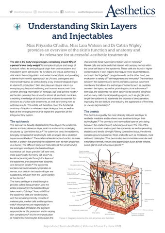
The skin can be broadly classified into three layers: the epidermis, dermis and subcutaneous layer, which is anchored to underlying structures by connective tissue.3 The outermost layer, the epidermis, is largely composed of keratinocyte cells arranged into a stratified squamous epithelium.3 The epidermal keratinocytes function to make keratin, a protein that provides the epidermis with its main properties as a barrier. The different stages of maturation of the keratinocytes are arranged into layers; the basal cell layer, Touch receptor suprabasal cell layer, granular cell layer and, (Meissner’s corpuscle) most superficially, the horny cell layer.4 As Pain receptor keratinocytes migrate through the layers of Capillary network the epidermis, they become less lipophilic Sebaceous gland and denser in keratin.3 The epidermis contains no blood vessels, lymphatics or Erector pili muscle nerves, thus cells in the basal cell layer are Heat receptor (Ruffini endings) supplied by diffusion from the upper portion of the dermis.5 Hair follicle The horny cell layer is shed through a Cold receptor (Krause muscle) process called desquamation, and the entire process from the basal cell layer Motor nerve takes around 28 days.3 Around 95% of Hair bulb the epidermal cells are keratinocytes, Vein and the remaining minority consists of Artery melanocytes, merkel cells and langerhans 6 cells. Melanocytes are responsible for Subcutaneous fat the production of melanin, the pigment Subdermal muscle layer responsible for UV protection and in varying Sweat Deep fascia 6 (eccrine gland) skin complexions. It is the overproduction of melanin by melanocytes that causes the Figure 1: Diagram showing the skin structure and function
Hair
Reproduced from Aesthetics | Volume 8/Issue 7 - June 2021
Epidermis
Dermis
Subcutaneous layer
Nerve endings Pacinian corpuscle (pressure receptor)


